


Abstract
Bovine tuberculosis (bTB) remains endemic in Morocco, where a national control strategy is implemented through screening and culling of infected animals. In this study, seven Mycobacterium bovis (M. bovis) strains were analyzed. Three strains were isolated in 1987, two in 2015 and two between 2022 and 2023. Bacterial culture and isolation were performed according to the protocol recommended by the World Organization for Animal Health (WOAH). PCR confirmed isolates as M. bovis within the Mycobacterium tuberculosis complex (MTBC). Whole genome sequencing (WGS) was performed (short-read sequencing, Illumina platform), followed by phylogenetic tree construction, and determination of clonal complexes and sub-lineages. Six of the seven strains clustered within the Eu2 clonal complex (CC) and sub-lineage La1.7.1, whereas one strain belonged to the CC unknown 2 and sub-lineage La1.2. Single nucleotide polymorphism (SNP)-based analysis revealed genetic distances ranging from 0 to 204. Low SNP distances were observed between the pairs B2–EL2562 and B3–EL2562. High-impact coding SNPs showed variation, although lineage-specific patterns could not be reliably assessed due to the limited sample size. Comparison of M. bovis strains isolated in different years described the distribution of genetic lineages across time in Morocco, based on the available isolates. Within this limited set of isolates, the Eu2 CC (La1.7.1 sublineage) and the unknown2 CC (La1.2 sublineage) were identified across multiple time points; however, given the limited sample size, these findings should be considered descriptive and do not allow conclusions regarding persistence, evolutionary trends, or transmission dynamics. These findings may offer useful information to strengthen the national bTB control strategy and support One Health surveillance of this zoonotic pathogen in Morocco.
1. Introduction
Tuberculosis is a major human disease, causing more than 10 million new cases and over one million deaths in 2024, according to the World Health Organization (WHO) [1].
Bovine tuberculosis (bTB) is a major zoonotic disease affecting multiple animal species and is caused by the Mycobacterium tuberculosis complex (MTBC), predominantly Mycobacterium bovis (M. bovis). Given its zoonotic potential and circulation at the animal–human–environment interface, bTB surveillance and control are relevant within a One Health framework. A recent global assessment estimated the prevalence of zoonotic animal tuberculosis at approximately 10%. Prevalence rates varied across continents and host species, with values below 10% in Africa and Asia, 33.6% in North America, 51% in Europe, and 85.7% in South America [2]. Other studies have reported substantial variation in bTB prevalence across different regions of the world. Prevalence reached up to 50% in parts of the African continent, whereas in Europe the overall proportion of infected cattle herds remained low (0.9%). In North America, prevalence was generally very low and largely confined to specific regions where the disease persists at the livestock–wildlife interface. In Central and South America, bTB is considered endemic, with higher prevalence particularly observed in dairy cattle populations [2,3,4,5,6,7,8].
Despite ongoing control efforts, bTB remains endemic in Morocco [9,10]. According to a survey carried out in 2003 by the Moroccan animal health authority, the average prevalence of bTB in Morocco was 33% at the bovine herd level and 18% at the individual animal level. The prevalence of bTB cases detected during veterinary inspections in the slaughterhouses was 1.7% [11,12].
At the national level, a bTB control strategy has been implemented for several years, aiming to reduce disease prevalence through live animal screening, culling of infected cattle, and systematic monitoring at slaughterhouses. However, bTB cases continue to be reported in numerous herds, for which the source of infection often remains unidentified. Effective traceability of infection sources is therefore essential to strengthen and accelerate the control strategy in Morocco.
Laboratory confirmation of bTB in animals detected through screening or at slaughterhouses is carried out only sporadically. In Morocco, laboratory diagnosis of bTB is performed at the Institut Agronomique et Vétérinaire Hassan II (Rabat, Morocco) (IAV), with existing capabilities for histopathology, bacteriology, PCR and whole genome sequencing (WGS) for detection and confirmation of M. bovis.
Molecular characterization of M. bovis strains in Morocco began in 2018 using spoligotyping [10]. While useful, this method has low discriminatory power and limited resolution when applied to clonal groups or closely related strains [13,14,15].
In recent years, the decreasing cost of WGS has allowed researchers worldwide to apply this high-resolution technique to study M. bovis epidemiology [5,6]. WGS enables precise tracing of infection sources and detailed investigation of transmission dynamics within specific geographic regions. Recent WGS studies have characterized the genomic diversity of Moroccan M. bovis strains and reported potential phylogenetic relationships between Moroccan isolates and M. bovis strains circulating in Europe [9,16].
In Morocco, the diagnostic workflow for bTB is structured within the framework of the national control program. The primary screening method is the single intradermal tuberculin skin test (SIT), which is routinely applied for herd-level surveillance. Bacteriological culture and isolation were performed using Herrold and Löwenstein–Jensen media. Colonies obtained after culture were examined by Ziehl–Neelsen staining to confirm the presence of acid-fast bacilli, while identification and differentiation of M. bovis within the Mycobacterium tuberculosis complex (MTBC) were performed using the previously described multiplex real-time PCR assay [17]. In addition, PCR-based assays are increasingly employed for rapid detection in selected cases. Although WGS is not used for routine diagnosis, it is applied in epidemiological investigations and high-resolution strain characterization.
Despite the availability of diagnostic tools and recent advances in WGS, genomic data on M. bovis circulating in Morocco remain limited, with few isolates characterized at the whole-genome level and limited representation across sampling periods and geographic areas. Consequently, the genomic diversity and phylogenetic relationships of Moroccan M. bovis isolates remain insufficiently documented. Expanding the available genomic data is therefore important to improve the molecular characterization of M. bovis in Morocco and provide baseline information for future genomic and epidemiological surveillance.
This study aimed to (i) characterize M. bovis strains isolated over time from different geographic regions in Morocco and (ii) contribute to enriching the database of M. bovis sequences in Morocco. This study provides new insights into the genomic diversity of M. bovis isolates from Morocco. The findings support evidence-based adjustments to the national control strategy, ultimately contributing to more effective eradication efforts.
2. Materials and Methods
2.1. Regulatory Authorization and Study Compliance
Sampling was conducted during routine veterinary inspections by authorized personnel, without experimental intervention on live animals. Sample collection for this study was authorized by the Moroccan Animal Health Authority (Office National de Sécurité Sanitaire des Produits Alimentaires, ONSSA) (N° 1572 ONSSA/DPPAV/DSA/SAP). Laboratory procedures were carried out following international standards in accordance with the WOAH Manual of Diagnostic Tests and Vaccines for Terrestrial Animals [18].
2.2. Tissue and Bacterial Samples
In 2022, bTB’s suspected tuberculous lesions were collected from four slaughterhouses serving large geographical areas of Morocco. A total of 12 tissue samples presenting lesions suspected of bTB were collected from the selected slaughterhouses. The slaughterhouses were selected based on official bTB surveillance data published by ONSSA in 2021 [19]. Samples were collected from the slaughterhouse of Taroudant, Rabat, El Jadida and Marrakech cities.
Additionally, we included three M. bovis isolates collected by the same laboratory during previous studies performed in 1987. These isolates originated from reactor animals to tuberculin skin test which were subsequently slaughtered.
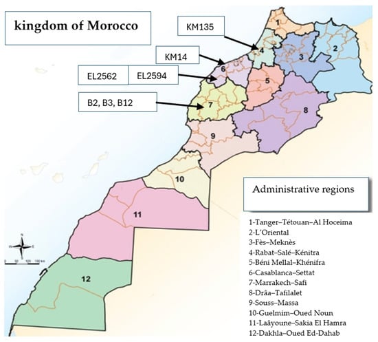
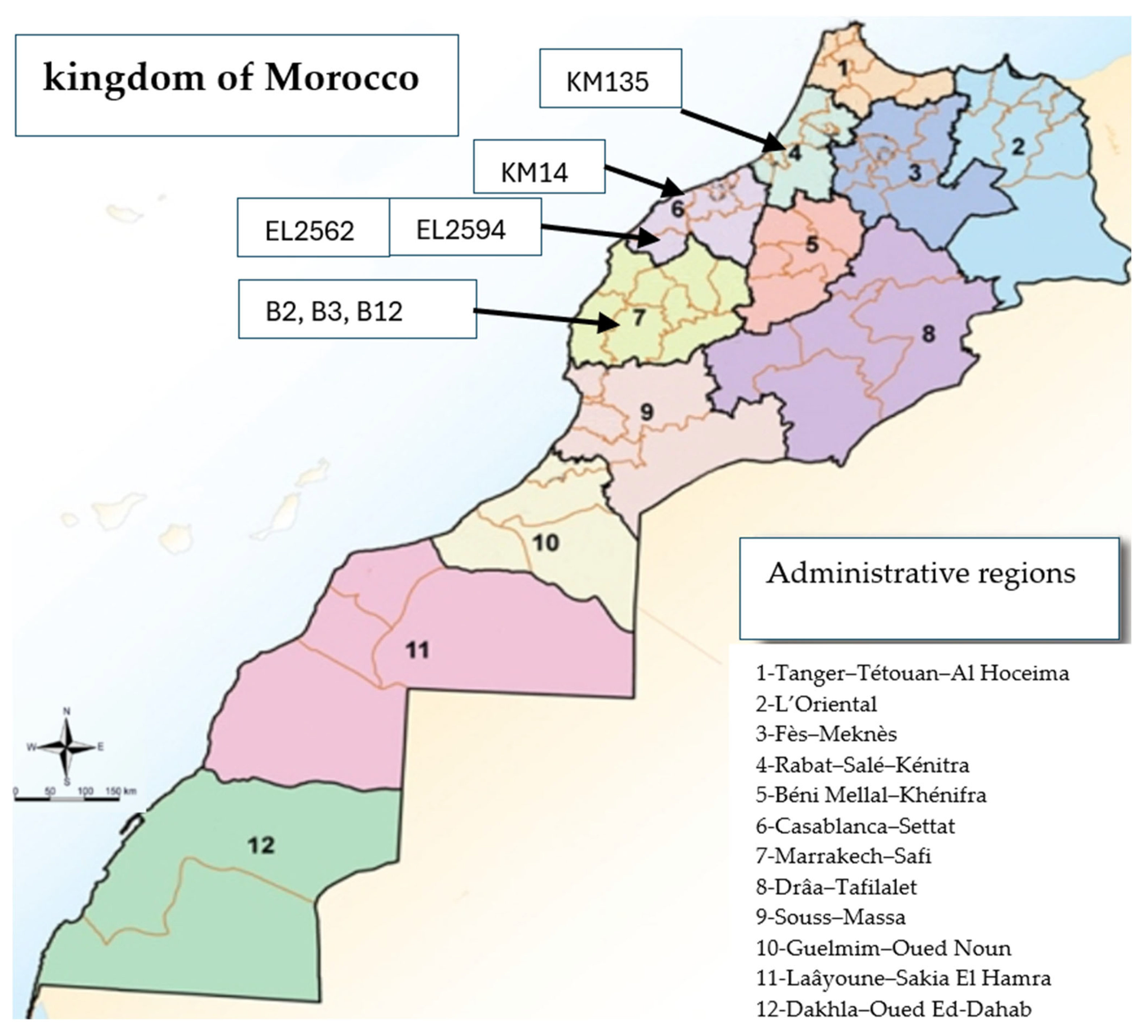
The three animals belonged to the same herd in Marrakech and had been imported several years earlier from France, Germany and the USA [20]. Details of the studied samples are provided in Table 1 along with their geographic distribution as indicated in Figure 1. B2, B3, and B12 correspond to distinct animals; however, all originated from the same herd.
Table 1.
Epidemiological data for bTB-suspect lesions analyzed in this study.
Figure 1.
Geographic distribution of the seven animals sampled in this study.
Sample collection was conducted in accordance with national regulations and with prior authorization from ONSSA. Seven samples were collected aseptically by official veterinarians at postmortem examination. The samples were transported to the Mycobacteriology laboratory at 4 °C and stored at (−20°) until further analysis. For each animal, all available information was recorded, including official animal identification code (SNIT = national system of identification and traceability), sex, age, location of lesions and geographic origin of the animal (retrieved from the SNIT database). The sampled cattle ranged in age from 2 to 8 years. In the present study, bacteriological culture, isolation and molecular characterization were conducted at the Laboratories of the Department of Veterinary Pathology and Public Health, IAV.
The study also included two Mycobacterial isolates obtained from cattle with bTB-suspect lesions during routine investigations performed in 2015 at the Department of Veterinary Pathology and Public Health of the IAV [21]. These isolates had been stored at −80 °C. The animals of origin were both male, aged 2 and 4 years, and came from El Jadida region [10,21].
2.3. Mycobacterial Culture
Mycobacterial isolation was performed according to the technique recommended by the WOAH for the cultivation of the MTBC, as described in the manual of diagnostic tests and vaccines for terrestrial animals [18], and the protocol recommended by National Veterinary Service Laboratories (Ames, IA, USA) [22]. Briefly, collected lymph nodes were trimmed to remove excess fat. The samples were decontaminated for a few seconds using a freshly prepared 3% (v/v) dilution of commercial bleach (12° chlorometric), prepared by adding 3 mL of commercial bleach to 97 mL of distilled water. The lymph nodes were then ground in a sterile mortar containing sterile phenol red broth. The homogenized tissue was disinfected with a 0.5 N NaOH solution for 10 min and subsequently neutralized with 6 N HCl. The suspension was centrifuged at 3000× g for 20 min and the resulting sediment was inoculated onto two culture media. Cultures were incubated at 37 °C for a minimum of 6 weeks and inspected twice weekly [23,24]. Two solid culture media were used: Löwenstein–Jensen (LJ) and Herrold’s egg yolk medium, both of which have been shown in previous studies to be reliable and effective for the isolation of Mycobacterium species [10,21,23].
Smears from suspected bacterial colonies were stained using the Ziehl–Neelsen method. Suspect colonies were then subcultured on both Herrold and LJ media to obtain pure isolates with sufficient biomass for DNA extraction and downstream molecular characterization.
2.4. Real Time PCR Identification Assay
To determine the subtype of the bacterial isolates, real-time PCR testing was carried out. All isolates were first screened for the presence of MTBC DNA, and positive samples were subsequently tested to differentiate M. bovis from M. tuberculosis.
Real time PCR detection of MTBC was performed using VetMAX M. tuberculosis kit (Applied Biosystem, Thermo fisher Scientific, Waltham, MA, USA). The PCR cycling conditions were applied according to the manufacturer’s instructions and previously published protocols [25,26,27,28].
For species differentiation within the MTBC, a real-time PCR assay targeting M. bovis-specific genomic regions was performed using the primers and probe described by Halse et al. (2011) targeting the Regions of Difference (RD) RD1, RD4, RD9, RD12 and ext-RD9 [17]. This assay allowed discrimination of M. bovis from other MTBC members with high specificity. A limitation of this study is that no in silico RD analysis was performed on the WGS data, despite its relevance for classical M. bovis characterization.
For the real-time PCR identification assay, the M. bovis BCG reference strain (Bacillus Calmette–Guérin, Pasteur 1173P2) was used as the positive control, while nuclease-free water served as the negative control.
2.5. DNA Extraction
For the operator and laboratory biosafety, bacterial cultures grown on solid media were heat-inactivated prior to DNA extraction. Briefly, well-isolated colonies were collected from agar plates and resuspended in 2 mL of sterile phosphate-buffered saline (PBS) in screw-cap tubes, resulting in a liquid suspension. Heat inactivation was performed at 100 °C for 30 min in a water bath [29,30]. Mycobacterial DNA was subsequently extracted from the heat-inactivated liquid suspension following the protocol used by the National Veterinary Services Laboratories (USDA/APHIS/VS), with minor adaptations.
A 1× TE buffer was prepared by diluting 100× Tris–EDTA buffer in ultra-distilled water. For mechanical lysis, sealed screw-cap bead-beater tubes were prepared containing 200 µL of 0.1 mm glass beads, 400 µL of 1× TE buffer, and 400 µL of phenol/chloroform/isoamyl alcohol (PCI; 25:24:1, v/v/v). An aliquot of the heat-inactivated bacterial suspension was added to the prepared tubes and homogenized at full speed for 2 min using a bead-beater to ensure effective cell disruption.
Mechanical disruption was performed using a FastPrep-24TM 5G Instrument (MP Biomedicals, Santa Ana, CA, USA). Samples were processed at 6.0 m/s for 2 cycles of 60 s each, with a 5-min cooling interval on ice between cycles to prevent overheating and ensure efficient lysis of the mycobacterial cell wall. These parameters were selected to optimize DNA yield and integrity while ensuring complete mechanical disruption. Following homogenization, phase separation was achieved by centrifugation, and the aqueous phase containing DNA was transferred to a fresh tube containing 30 μL of 3 M sodium acetate. DNA was precipitated by adding 700 µL of ice-cold ethanol and incubating at −20 °C. After centrifugation, the DNA pellet was washed with ice-cold ethanol, air-dried, and resuspended in 1× TE buffer. Extracted DNA was stored at −20 °C until further analysis [31,32,33,34].
DNA quantity and purity was initially evaluated by spectrophotometry (A260/280 ratio) using a Nanodrop ND 8000 (Thermo Fisher Scientific, Wilmington, DE, USA) and quantified fluorometrically with a Qubit 3.0 (Thermofisher, Waltham, MA, USA). DNA integrity was assessed by 1% agarose gel electrophoresis [35]. For downstream applications requiring higher purity, DNA samples were further processed using the MagMAX CORE Mechanical Lysis Module Bundle, which includes the MagMAX CORE Mechanical Lysis Module and MagMAX CORE Nucleic Acid Purification kit (Applied Biosystem, Thermofisher Scientific, Waltham, MA, USA) according to the manufacturer’s instructions. This additional purification step was performed to eliminate residual contaminants and potential PCR or sequencing inhibitors, thereby ensuring high-quality DNA suitable for WGS and library preparation. DNA concentration and purity were assessed spectrophotometrically prior to storage at −20 °C.
All bacteriologically confirmed M. bovis isolates yielding DNA of sufficient quality and quantity for WGS were retained for sequencing, resulting in a final set of seven isolates.
2.6. Whole Genome Sequencing
Genomic DNAs extracted from the seven M. bovis isolates were sent to Eurofins Genomics laboratory (Ebersberg, Germany) for whole WGS. Sequencing libraries were prepared using the TruSeq DNA Library Preparation kit (Illumina, Inc., San Diego, CA, USA) according to the manufacturer’s protocol. Paired-end sequencing (2 × 150 bp) was conducted on an Illumina NovaSeq 6000 platform (Illumina Inc.). The raw data sequencing reads generated for the seven M. bovis isolates in this study have been deposited in the NCBI Sequence Read Archive (SRA) under BioProject accession number PRJNA1337463 with individual accession numbers (SRR35725697, SRR35725698, SRR35725699, SRR35725700, SRR35725701, SRR35725702, SRR35725703).
For comparative genomic analyses, publicly available M. bovis whole-genome sequences were retrieved from GenBank database https://www.ncbi.nlm.nih.gov/datasets/genome/?taxon=1765, accessed on 2 February 2026. These reference genomes included 61 publicly available M. bovis genomes originating from diverse geographical regions worldwide, giving a total of 93 publicly available reference genomes (Supplementary Materials). Together with the 7 isolates newly sequenced in this study, the full analytical dataset comprised 100 strains. Mycobacterium caprae was included as an outgroup for phylogenetic rooting. A detailed breakdown of the dataset composition at each analytical stage is provided in Table 2. Individual accession numbers for all publicly available genomes included in the study are listed in the Supplementary Materials [9,16].
Table 2.
Summary of the genomic dataset used at each analytical stage in this study.
In the present study, bioinformatic processing and downstream genomic analyses were carried out at the Biotechnology Research Unit, Regional Center of Agricultural Research of Rabat, National Institute of Agricultural Research (INRA), Rabat, Morocco.
Accession numbers for all publicly available genomes are listed individually in the Supplementary Materials. Genomes retained after quality filtering are listed explicitly; the SRA accession range SRR31781513–SRR31781567 cited in Section 3.5 does not represent a contiguous block of retained genomes, as not all accessions within that range passed quality filtering criteria.
2.7. Genome Assembly, Variants Calling and Profiling of M. bovis Isolates
Raw sequencing reads generated for each strain were first screened for potential contamination using Kraken2 (version 2.1.2; database: Standard), retaining only reads assigned to the Mycobacteriaceae taxonomic group (taxID = 1762) using the ‘extract_kraken_reads.py’ Python (v2.17.1) script (KrakenTools; https://github.com/jenniferlu717/KrakenTools) (accessed on 15 February 2026).
Prior to genome assembly, raw reads were subjected to quality control and adapter trimming using Trimmomatic v0.39 with the following parameters: paired-end mode; adapters: TruSeq3-PE.fa (Illumina); minimum read length: 36 bp; sliding-window trimming: 4:20; leading and trailing quality threshold: 3. Quality assessment of raw and trimmed reads was performed using FastQC (v0.11.9).
Genome assembly of the trimmed reads was performed using Shovill v0.9.0 (https://github.com/tseemann/shovill, accessed on 15 February 2026), which incorporates the SPAdes assembler with additional read trimming via its internal Trimmomatic module (default Shovill trimming parameters applied). Assembly quality was assessed using QUAST v5.2.0, with M. bovis AF2122/97 (GenBank accession NC_002945.4) as the reference genome. Mean sequencing depth per isolate was calculated following read mapping to AF2122/97 and ranged from 156.7× (KM14) to 385.1× (EL2562) across the seven isolates, as reported in Table 3. Mycobacterium caprae was included as an outgroup for downstream phylogenetic analyses.
Genome annotation of the seven isolates was performed using Prokka v1.14.6 (parameters: --kingdom Bacteria --proteins NC_002945v4). Variant calling for all 100 strains included in the study was performed against the M. bovis AF2122/97 reference genome using Snippy v4.6.0 (https://github.com/tseemann/snippy, accessed on 15 February 2026), with read alignment performed by BWA-MEM, followed by sorting and indexing with SAMtools, and variant detection using FreeBayes. Default Snippy parameters were applied, with a minimum base quality of 20 and a minimum read depth of 10.
Core genome SNPs were extracted using the Snippy-core module, generating a multiple sequence alignment of SNP positions present across all isolates. To improve the reliability of SNP calls and minimize spurious variants arising from mapping ambiguity, repetitive genomic regions and PE/PPE-family genes were excluded from the core SNP alignment using a BED file of masked regions.
Functional annotation of SNP effects was performed using SnpEff v4.3t [36], with the M. bovis AF2122/97 genome annotation (NC_002945.4) as the reference database. SNP effects were classified into four impact categories—high, moderate, low, and modifier—according to their predicted functional consequence, as described in Section 3.5.
The cleaned core SNP alignment was used for maximum likelihood phylogenetic inference using IQ-TREE v2.2.2, with the best-fit substitution model selected automatically by ModelFinder prior to tree construction. The selected model was GTR+F+I+G4, as reported in the IQ-TREE log file. Branch support was assessed using 1000 ultrafast bootstrap replicates (-bb 1000). The resulting tree was visualized and annotated using MEGA 12v.
Variant calling was performed for all 100 strains included in the study—comprising the 7 Moroccan isolates newly sequenced in this study and the 93 publicly available reference genomes (61 Moroccan and 32 international) against the M. bovis AF2122/97 reference genome using snippy-tool (https://github.com/tseemann/snippy, accessed on 15 February 2026).
2.8. Lineage Prediction
To assign lineages, TB-Profiler version 6.6.6 was applied to all 100 strains included in the study (7 newly sequenced Moroccan isolates, 61 publicly available Moroccan reference genomes, and 32 international reference genomes (Table 2)), using raw Illumina sequencing reads as input. TB-Profiler was run using default parameters, which include read mapping with minimap2 or bwa (depending on the input), variant calling, and annotation. The output included lineage classification along with information on key genetic mutations. The resulting lineage assignments were compiled and analyzed to assess the distribution of M. bovis lineages among the Moroccan isolates and the global reference strains included in this study.
3. Results
3.1. Real Time PCR Assay
All strains initially tested positive for the MTBC and were subsequently confirmed as M. bovis by subtype-specific PCR. PCR test results were considered positive when the cycle threshold (Ct) value was below 38 [26]. In the present study, samples with a Ct value ≤ 36 were considered suitable for WGS. Among the seven selected isolates, MTBC-specific Ct values ranged from 14 to 36.
The Ct threshold used to select isolates for WGS was chosen to maximize DNA quality for WGS while recognizing that other studies have applied different thresholds (e.g., Ct < 14 [29] or Ct ≤ 25 [37]). We acknowledge that the selection criterion may bias the dataset toward samples with higher bacterial loads. Additionally, the isolates analyzed in this study originated from heterogeneous sources, including abattoir surveillance and historical import-related cases, with uneven geographic and temporal coverage. These factors may limit the representativeness and generalizability of the dataset and should be considered when interpreting comparative and phylogenetic analyses.
3.2. M. bovis Whole Genome Assembly
WGS was performed on seven M. bovis strains, generating between 1.91 Gb and 3.06 Gb of raw sequencing data per strain. The raw reads were screened and decontaminated using kraken then assembled using the Shovill pipeline, a de novo assembler optimized for bacterial genomes. The resulting assemblies exhibited variability in contiguity and genome size, likely reflecting differences in sequencing quality, coverage depth, and genomic complexity among strains.
Key assembly statistics, including the number of contigs, total genome length, largest contig size, GC content, N50 values, genome coverage and mean sequencing depth are summarized in Table 3. These assemblies provided a robust basis for downstream comparative genomic analyses, structural genome investigations, and the identification of strain-specific genomic features within M. bovis.
Table 3.
Assembly statistics of seven M. bovis strains studied in this work.
3.3. Phylogenetic Analysis
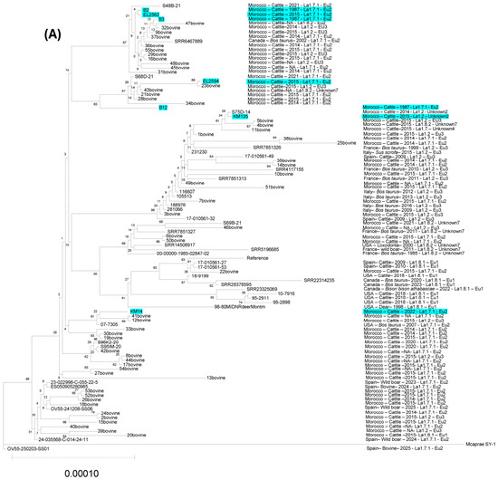
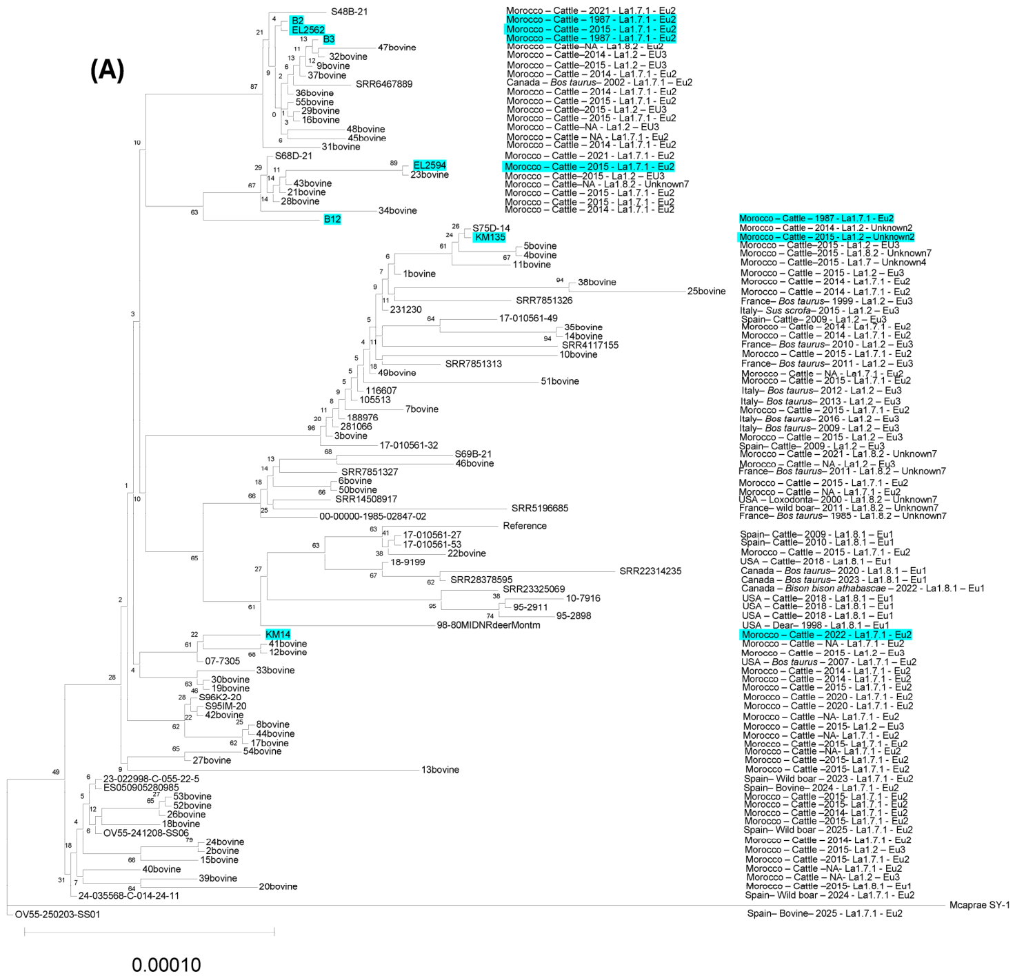
The phylogenetic tree was constructed using M. bovis AF2122/97 as the reference genome, with Mycobacterium caprae included as an outgroup to root the tree and provide an evolutionary reference framework (Figure 2). The seven sequenced Moroccan M. bovis strains were distributed across multiple major branches relative to this reference, alongside 93 publicly available global M. bovis genomes used for comparative context.
Figure 2.
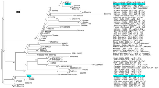
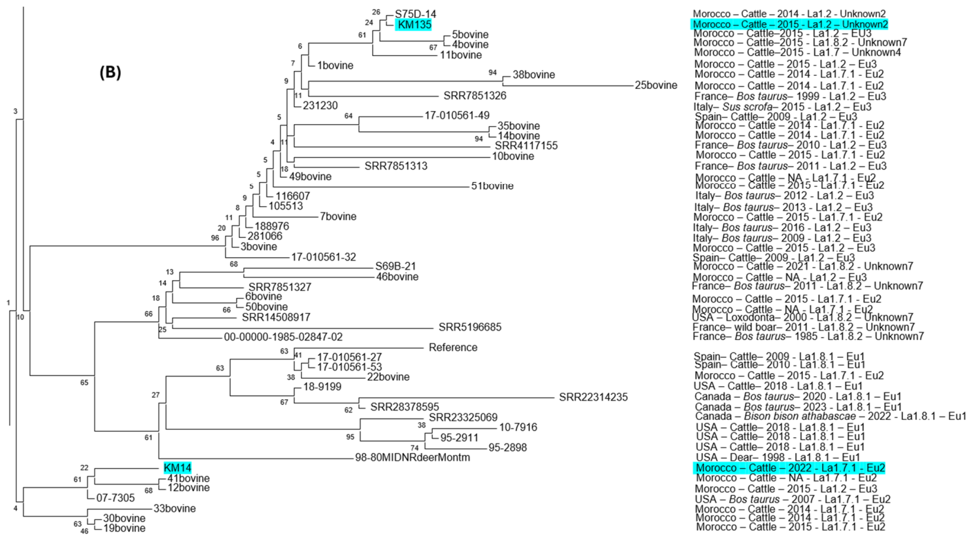
Maximum likelihood phylogenetic tree of 100 M. bovis strains constructed using IQ-TREE v2.2.2 (1000 ultrafast bootstrap replicates). Mycobacterium caprae was used as the outgroup to root the tree. (A) Full phylogenetic tree showing all 100 strains included in the analysis. The seven Moroccan isolates newly sequenced in this study are highlighted in blue. For each strain, the following metadata are displayed at the tip labels: country of origin, host species, year of isolation, sublineage (as assigned by TB-Profiler v6.6.6), and clonal complex. Branch support values ≥ 70% are shown at nodes. (B) Zoomed sub-tree focusing on the clade containing the seven newly sequenced Moroccan isolates and their closest phylogenetic neighbors among the international reference genomes, allowing direct visualization of the relationships between KM14, KM135, and phylogenetically proximate European and North American M. bovis strains. Tip label color coding: blue, Moroccan isolates newly sequenced in this study; black, publicly available Moroccan reference genomes.
The maximum likelihood phylogenetic tree constructed in this study is presented in two complementary panels (Figure 2). Figure 2A shows the full tree comprising all 100 strains, providing the global phylogenetic context within which the seven newly sequenced Moroccan isolates are positioned. Figure 2B presents a zoomed view of the clade containing the seven Moroccan isolates and their closest international neighbors, allowing detailed visualization of the relationships described below.
The three strains isolated in 1987 (B2, B3, and B12) were positioned on two distinct branches of the tree relative to AF2122/97. Strains B2 and B3 clustered closely together, consistent with their shared herd of origin, suggesting a recent common ancestor or a close evolutionary relationship. Strain B12, while originating from the same herd, was positioned on a separate branch, indicating a degree of within-herd genomic divergence. The strains isolated in 2015 (EL2562, EL2594) and 2022–2023 (KM14, KM135) showed high genetic similarity to previously published Moroccan M. bovis strains [9,16] and clustered within the same major clade relative to AF2122/97, indicating phylogenetic stability over time, indicating phylogenetic stability over time. The 2022–2023 isolates also exhibited phylogenetic proximity to several European and North American M. bovis strains, consistent with previously reported trans-continental phylogenetic relationships [9,16].
3.4. Sublineages and Clonal Complex
The genomic characterization of the seven Moroccan M. bovis isolates using TB-Profiler (v6.6.6) is summarized in Table 4 [38,39]. Based on TB-Profiler classification, most isolates (6/7; 86%) were assigned to sublineage La1.7.1, whereas one isolate (KM135; 1/7; 14%) was assigned to sublineage La1.2. In terms of clonal complex classification, six isolates (86%) were assigned to the Eu2 clonal complex, while KM135 was classified as unknown2 (Table 4).
Table 4.
The sublineage of the seven Moroccan M. bovis provided by TBProfiler software [38].
A limitation of this study is the use of TBProfiler (v6.6.6) for lineage assignment, a tool primarily validated for Mycobacterium tuberculosis. Although TBProfiler has been previously applied to M. bovis in the literature [38,40], dedicated pipelines such as KvarQ, which incorporate M. bovis-specific SNP markers, have been shown to provide more appropriate lineage resolution for this species. Accordingly, the lineage and clonal complex assignments reported here, particularly the unknown2/La1.2 classification of isolate KM135, should be interpreted with caution and would benefit from independent validation using M. bovis-specific tools.
3.5. SNP Analysis
SNP analysis was performed against the Mycobacterium bovis AF2122/97 reference genome to evaluate the genomic diversity of Moroccan isolates in a global context (Supplementary Materials).
Among the Moroccan isolates generated in this study (B12, B2, B3, EL2562, EL2594, KM135, and KM14), the number of variants ranged from 674 to 819 SNPs relative to AF2122/97. Most isolates (B12: 674; B2: 711; B3: 710; EL2562: 716; EL2594: 730; KM14: 734) clustered within a relatively narrow divergence range (~670–735 SNPs), suggesting a shared evolutionary background. In contrast, KM135 exhibited a higher divergence level (819 SNPs), indicating possible affiliation with a distinct lineage.
The pairwise comparison among the seven Moroccan isolates revealed genetic distances ranging from 0 to 204 SNPs (Supplementary Materials, Tables S1 and S2). Isolates B2 and B3 were identical at the SNP level (0 SNPs), consistent with their shared herd of origin in Marrakech in 1987. Notably, the lowest inter-isolate distances across different sampling periods were observed between the pairs B2–EL2562 and B3–EL2562 (15 SNPs), representing the closest genomic relationships identified between isolates separated by approximately 28 years (1987 and 2015, respectively). These findings suggest a degree of genomic continuity between historical and more recent Moroccan M. bovis isolates, though the limited sample size and absence of detailed epidemiological metadata preclude any inference on transmission or persistence.
The 61 publicly available Moroccan M. bovis genomes retrieved from public repositories (individual accession numbers listed in Supplementary Materials) showed SNP counts largely ranging between 598 and 791 SNPs relative to AF2122/97, overlapping with the divergence range observed for the newly sequenced Moroccan isolates. It should be noted that the SRA accession range SRR31781513–SRR31781567 previously cited does not represent a contiguous block of retained genomes; the precise list of accessions retained after quality filtering is provided in the Supplementary Materials to ensure full reproducibility of the analyses. This supports the presence of a predominant Moroccan clade characterized by ~650–780 SNPs relative to AF2122/97.
Several international M. bovis genomes included in the dataset displayed markedly different divergence levels. Some strains showed intermediate SNP counts (e.g., SRR12554458: 364 SNPs; SRR4117155: 254 SNPs; SRR5196685: 391 SNPs), whereas others were substantially more distant (e.g., ERR12976152: 811 SNPs; SRR11418337: 788 SNPs). A small subset of strains exhibited lower divergence (e.g., SRR7851378: 63 SNPs), suggesting closer phylogenetic proximity to the AF2122/97 reference lineage.
SNP effects were categorized according to their predicted impact on gene function as: high (affecting splice regions, start or stop codons, or causing frameshift mutations), moderate (non-synonymous substitutions), low (synonymous coding variants, start/stop retained variants, or start gained), and modifier (upstream, downstream, or intergenic variants). High-impact SNPs are predicted to directly alter the functional sequence of genes and are therefore of particular interest for comparative genomic analysis [36,41,42].
Analysis of the Snippy-core output revealed variation in the number and distribution of predicted high-impact SNPs among the seven M. bovis isolates (Table 5). Two distinct counting levels are reported in Table 5 and should be carefully distinguished when interpreting the figures below. First, isolate-level occurrences reflect the number of high-impact SNPs detected per isolate within each functional category; an SNP shared by multiple isolates contributes one count per isolate carrying it. Second, distinct SNP positions reflect the number of unique genomic variant positions identified across all seven isolates within each category, regardless of how many isolates share that position.
Table 5.
Distribution of predicted high-impact SNPs among the seven studied M. bovis.
At the SNP level, stop codon gain variants ranged from 2 to 5 occurrences per isolate, stop codon loss variants ranged from 5 to 6, frameshift mutations ranged from 32 to 48, and splice-region variants ranged from 7 to 8 (Table 5, per-isolate rows). Summed across all seven isolates, these corresponded to 23 isolate-level occurrences of stop codon gain, 36 of stop codon loss, 255 of frameshift mutations, and 50 of splice-region variants (Table 5, row a).
In terms of unique variant positions, 15 distinct stop codon gain SNP positions, 6 distinct stop codon loss SNP positions, 107 distinct frameshift SNP positions, and 8 distinct splice-region SNP positions were identified across the dataset (Table 5, row b). The difference between isolate-level occurrence totals and distinct position counts reflects the degree of SNP sharing among isolates: for example, the 23 isolate-level stop codon gain occurrences map to only 15 distinct genomic positions, indicating that several of these variants are shared across multiple isolates. Similarly, the 255 frameshift occurrences correspond to 107 distinct positions, confirming substantial sharing of frameshift-associated variants within this closely related isolate set.
It should be noted that SNPs with multiple predicted functional effects may be included in more than one annotation category; therefore, the categories presented in Table 5 are not mutually exclusive. Detailed annotations of all identified high-impact SNPs, including predicted effects and the corresponding affected genes, are provided in Supplementary Table S3.
The complete pairwise SNP-distance matrix for all seven Moroccan isolates is provided in Supplementary Table S1, and the SNP distances relative to the M. bovis AF2122/97 reference genome are presented in Supplementary Table S2, to allow full transparency and independent verification of the genomic comparisons reported.
4. Discussion
This study represents the first comprehensive genomic characterization of M. bovis strains isolated from cattle in Morocco over a period spanning more than three decades (1987–2022). Using WGS of seven isolates, we characterized lineage assignments, clonal complexes, and SNP diversity. The obtained results showed that the analyzed Moroccan M. bovis isolates clustered phylogenetically with previously sequenced strains, indicating genetic relatedness among the isolates included in the analysis [9,16]. Mycobacterium caprae was included as an outgroup to root the phylogenetic tree and provide a reliable framework for interpreting phylogenetic relationships between the Moroccan isolates and M. bovis strains included for comparison.
Pairwise SNP-distance analysis showed that isolates B2 and B3 were genomically identical (0 SNPs), consistent with their common herd origin. Among isolates collected at different time points, the lowest SNP distances were observed between B2–EL2562 and B3–EL2562, showing close genomic relatedness despite their different sampling periods. However, the limited number of isolates, the imported origin of the 1987 strains, and the absence of detailed herd contact and animal movement data preclude epidemiological interpretation of these genomic relationships.
The inclusion of three isolates from 1987 was particularly significant, as these represented the earliest M. bovis genomes sequenced from cattle sampled in Morocco; however, these isolates originated from imported animals and did not reflect indigenous Moroccan strains. Comparison with isolates from 2015 and 2022 provided a descriptive overview of lineage distribution across time points, without supporting inferences regarding persistence or genomic evolution. Notably, six of the seven isolates were classified within the Eu2 clonal complex based on TB-Profiler assignment, indicating the frequent representation of this lineage among the isolates analyzed. Given the tool’s primary validation for M. tuberculosis, this finding should be interpreted as a TB-Profiler-based classification that was not independently confirmed using an M. bovis-specific lineage assignment approach. The identification of Eu2-associated isolates in the present study is consistent with previous reports of this clonal complex in M. bovis isolates from Europe and parts of Africa, providing a broader geographical context for the current findings [9,16].
Based on individual animal records retrieved from the SNIT, isolates were associated with animals originating from Casablanca and Rabat. However, these geographic data alone do not allow inference of epidemiological links or transmission routes among the analyzed isolates.
Clonal complex analysis further showed the frequent representation of Eu2, and the La1.7.1 sub-lineage. These results are consistent with previous studies conducted in Morocco and elsewhere that have reported the Eu2 clonal complex and La1.7.1 sublineage among Moroccan M. bovis isolates [9,16]. Within the present dataset, six of the seven isolates were assigned to Eu2/La1.7.1 across three discrete sampling periods (1987, 2015, and 2022–2023). However, given the limited number of isolates, the non-random sampling strategy, and the import origin of the 1987 strains, no inference can be drawn regarding the long-term dominance, persistence, or establishment of this lineage within Moroccan cattle herds. The epidemiological factors underlying the relative frequency of Eu2/La1.7.1 among the analyzed isolates remain to be determined through future studies incorporating larger, representative, and systematically collected datasets.
Interestingly, one isolate was assigned to the unknown2 clonal complex, and phylogenetic analysis placed it within the La1.2 sub-lineage [38]. This CC has also been reported in Morocco in previous studies [9,16]. The detection of this genetically distinct isolate further illustrates the genomic diversity observed among the analyzed M. bovis isolates. Although TBProfiler assigned this isolate to the unknown2 CC and La1.2 sublineage, this classification was not independently validated using a dedicated M. bovis SNP-based lineage assignment approach. Therefore, the present finding should be considered descriptive, and additional comparison with a curated global M. bovis genomic dataset would be required to confirm its phylogenetic placement. The available data are insufficient to determine the origin or epidemiological significance of this isolate. Further surveillance incorporating broader geographic sampling and detailed epidemiological metadata, including information on animal origin and movement, is needed to better characterize the distribution and epidemiological relevance of this lineage.
The SNP-based comparative analysis revealed intra-lineage genetic variation in Moroccan M. bovis isolates despite the shared clonal complex among most isolates. This diversity indicated the presence of genetically distinct M. bovis lineages among the analyzed isolates; however, the available genomic and epidemiological data do not allow conclusions regarding their sources or routes of introduction. Furthermore, future studies should prioritize the use of M. bovis-specific lineage assignment tools, such as KvarQ or custom SNP-based pipelines validated against curated global M. bovis genomic databases, to independently confirm the lineage classifications reported in the present study and to provide a more robust genomic characterization of Moroccan M. bovis isolates.
The identified differences in high-impact SNP profiles illustrate genomic variation among the analyzed isolates. However, given the limited number of isolates, the present dataset was insufficient to reliably assess lineage-specific SNP patterns. Moreover, the analysis was descriptive, and no functional assessment was performed to determine the potential involvement of the affected genes in virulence, metabolism, antigenic variation, or other biological processes. Further studies using larger datasets and functional analyses are therefore required to determine the biological significance of these SNPs.
The observed genomic relationships highlight the potential value of WGS for investigating M. bovis population structure when combined with appropriate epidemiological data. However, genomic similarity alone cannot establish transmission relationships among the analyzed isolates. Integration of WGS with detailed epidemiological metadata, including herd contacts, livestock trade records, and animal movement data, would improve the interpretation of genomic relationships. Future studies should incorporate larger and more geographically representative sampling together with detailed epidemiological metadata to better characterize M. bovis population structure and investigate potential transmission networks in Morocco.
Study Limitations: This study has several limitations. First, the small number of M. bovis isolates and the sampling strategy limit the representativeness of the observed genomic diversity. Second, the 1987 isolates originated from imported cattle and therefore cannot be used to infer the long-term evolution or persistence of indigenous Moroccan lineages. Third, lineage and clonal complex assignment was performed using a single tool, TB-Profiler (v6.6.6), which was primarily developed and validated for M. tuberculosis. Therefore, the lineage and clonal complex assignments reported in this study should be considered TB-Profiler-based classifications that were not independently validated using an M. bovis-specific approach. Cross-validation using dedicated M. bovis lineage assignment tools, such as KvarQ, or a custom SNP-based pipeline benchmarked against a curated global M. bovis genomic dataset was not performed in the present study. Independent validation using M. bovis-specific approaches should therefore be considered a priority for future work, particularly for the unknown2/La1.2 classification of isolate KM135. Finally, although animal origin was retrieved from the SNIT database, detailed epidemiological metadata on herd contacts, livestock trade, and animal movements were limited, preventing robust reconstruction of transmission pathways. Future studies should combine larger, representative genomic datasets with comprehensive epidemiological metadata.
5. Conclusions
In conclusion, this study provided genomic data on Moroccan M. bovis strains and highlighted the utility of WGS in tracing evolutionary relationships and informing bTB control strategies. The identification of Eu2/La1.7.1 and a genetically distinct lineage among the analyzed isolates highlighted the value of continued molecular surveillance to expand knowledge of M. bovis genomic diversity in Morocco and support national bTB control programs. Comparison of M. bovis sequences isolated in 1987 with those obtained over the past fifteen years provided insights into the genomic diversity and temporal patterns of bTB strains across the studied geographic regions of Morocco.
Considering the documented history of livestock imports from Europe and the phylogenetic proximity of certain Moroccan isolates to European M. bovis strains observed in this and previous studies [9,16], future genomic studies integrating Moroccan and European M. bovis isolates with animal importation and movement records would help to investigate potential epidemiological relationships between strains circulating in these regions. Extension of this approach to include isolates from neighboring North African countries would further be required before any inference regarding transboundary bTB dynamics at the regional level could be drawn, a question that the present dataset, containing isolates exclusively from Morocco, is not designed to address. WGS proved instrumental in elucidating the phylogenetic relationships of M. bovis in Morocco. Ongoing WGS-based surveillance will be critical to track the evolution and spread of dominant lineages, including Eu2, and to guide targeted control strategies.
Supplementary Materials
The following supporting information can be downloaded at: https://www.mdpi.com/article/10.3390/amh71040038/s1, Table S1: Number of SNPs between seven studied M. bovis and the reference M. bovis AF2122/97t; Table S2: SNP distances between the studied M. bovis isolates and their closest phylogenetically related strains; Table S3: SNP of M. bovis studied in this work compared to the strains downloaded from NCBI database; Table S4: SNP of seven Moroccan M. bovis studied in this work with type of SNP.
Author Contributions
Conceptualization, M.K., S.F., S.K., M.B., H.L. and J.B.; methodology, M.K., S.F., S.K., M.B., H.L. and J.B.; software, M.K., S.F. and S.K.; validation, M.K., S.F., S.K. and J.B.; formal analysis, M.K., S.F., S.K. and J.B.; investigation, M.K.; writing—original draft preparation, M.K. and S.K.; writing—review and editing, M.K., S.F., S.K., M.B., H.L., M.-L.B. and J.B. All authors have read and agreed to the published version of the manuscript.
Funding
This research received no external funding.
Institutional Review Board Statement
No ethical approval was required for this study as all sampling and handling procedures were conducted by authorized personnel according to Moroccan legislation. Sampling was conducted exclusively under routine veterinary inspection without experimental intervention.
Informed Consent Statement
Not applicable.
Data Availability Statement
The original contributions presented in this study are included in the article/Supplementary Materials. Further inquiries can be directed to the corresponding author.
Acknowledgments
Acknowledgment for the teams from the Contagious Diseases and Avian Pathology Departments for their contributions to the analysis. We thank the Moroccan National Office of Food Safety (ONSSA) for the authorization to realize this work, and the veterinary inspector for their help for performing the samples.
Conflicts of Interest
The authors declare no conflicts of interest.
References
- World Health Organization. Global Tuberculosis Report 2025; World Health Organization: Geneva, Switzerland, 2025; ISBN 978-92-4-011692-4. [Google Scholar]
- Ramos, B.; Pereira, A.C.; Reis, A.C.; Cunha, M.V. Estimates of the Global and Continental Burden of Animal Tuberculosis in Key Livestock Species Worldwide: A Meta-Analysis Study. One Health 2020, 10, 100169. [Google Scholar] [CrossRef] [Scilit]
- Milián-Suazo, F.; González-Ruiz, S.; Contreras-Magallanes, Y.G.; Sosa-Gallegos, S.L.; Bárcenas-Reyes, I.; Cantó-Alarcón, G.J.; Rodríguez-Hernández, E. Vaccination Strategies in a Potential Use of the Vaccine against Bovine Tuberculosis in Infected Herds. Animals 2022, 12, 3377. [Google Scholar] [CrossRef] [Scilit]
- Sorensen, A.; van Beest, F.M.; Brook, R.K. Impacts of Wildlife Baiting and Supplemental Feeding on Infectious Disease Transmission Risk: A Synthesis of Knowledge. Prev. Vet. Med. 2014, 113, 356–363. [Google Scholar] [CrossRef] [Scilit]
- Humphrey, H.M.; Orloski, K.A.; Olea-Popelka, F.J. Bovine Tuberculosis Slaughter Surveillance in the United States 2001–2010: Assessment of Its Traceback Investigation Function. BMC Vet. Res. 2014, 10, 182. [Google Scholar] [CrossRef] [Scilit]
- Dejene, S.W.; Heitkönig, I.M.A.; Prins, H.H.T.; Lemma, F.A.; Mekonnen, D.A.; Alemu, Z.E.; Kelkay, T.Z.; Boer, W.F. de Risk Factors for Bovine Tuberculosis (bTB) in Cattle in Ethiopia. PLoS ONE 2016, 11, e0159083, Correction in PLoS ONE 2017, 12, e0176654. [Google Scholar] [CrossRef] [Scilit]
- Arnot, L.F.; Michel, A. Challenges for Controlling Bovine Tuberculosis in South Africa. Onderstepoort J. Vet. Res. 2020, 87, 8. [Google Scholar] [CrossRef] [Scilit]
- Schiller, I.; Waters, W.R.; Vordermeier, H.M.; Jemmi, T.; Welsh, M.; Keck, N.; Whelan, A.; Gormley, E.; Boschiroli, M.L.; Moyen, J.L.; et al. Bovine Tuberculosis in Europe from the Perspective of an Officially Tuberculosis Free Country: Trade, Surveillance and Diagnostics. Vet. Microbiol. 2011, 151, 153–159, Correction in Vet. Microbiol. 2012, 155, 448. [Google Scholar] [CrossRef] [Scilit]
- Azami, H.Y.; Perea, C.; Stuber, T.; Bouslikhane, M.; Berrada, J.; Aboukhassib, H.; Palau, A.O.A.; Reis, A.C.; Cunha, M.V.; Thacker, T.C.; et al. Phylogenetic Analysis of Mycobacterium bovis Reveals Animal and Zoonotic Tuberculosis Spread between Morocco and European Countries. PLoS Neglected Trop. Dis. 2025, 19, e0011982. [Google Scholar] [CrossRef] [Scilit]
- Azami, H.Y. Prevalence, Molecular Characterization, Transmission Dynamics and Cost Analysis of Bovine Tuberculosis in Morocco. Doctoral Dissertation, Philosophisch-Naturwissenschaftliche Fakultät der Universität Basel, Basel, Switzerland, 2018. [Google Scholar]
- Rapport Annuel au Titre de L’année 2018. Available online: https://www.courdescomptes.ma/publication/rapport-annuel-au-titre-de-l-annee-2018/ (accessed on 28 April 2024).
- Prophylaxis Program (Bovine Tuberculosis). Available online: https://www.onssa.gov.ma/sante-animale-dsa/programme-de-prophylaxie/tuberculose-bovine/2026 (accessed on 28 April 2026).
- Orloski, K.; Robbe-Austerman, S.; Stuber, T.; Hench, B.; Schoenbaum, M. Whole Genome Sequencing of Mycobacterium bovis Isolated from Livestock in the United States, 1989–2018. Front. Vet. Sci. 2018, 5, 253. [Google Scholar] [CrossRef] [Scilit]
- Guimarães, A. Mycobactérie bovis: Du génotypage au séquençage du génome. Microorganisms 2020, 8, 667. [Google Scholar]
- Perea, C.; Ciaravino, G.; Stuber, T.; Thacker, T.C.; Robbe-Austerman, S.; Allepuz, A.; Val, B.P. de Whole-Genome SNP Analysis Identifies Putative Mycobacterium bovis Transmission Clusters in Livestock and Wildlife in Catalonia, Spain. Microorganisms 2021, 9, 1629. [Google Scholar] [CrossRef] [Scilit]
- Khoulane, M.; Fellahi, S.; Khayi, S.; Bouslikhane, M.; Lakhdissi, H.; Berrada, J. First Insight into the Whole Genome Sequencing Whole Variations in Mycobacterium bovis from Cattle in Morocco. Microorganisms 2024, 12, 1316. [Google Scholar] [CrossRef] [Scilit]
- Halse, T.A.; Escuyer, V.E.; Musser, K.A. Evaluation of a Single-Tube Multiplex Real-Time PCR for Differentiation of Members of the Mycobacterium tuberculosis Complex in Clinical Specimens. J. Clin. Microbiol. 2011, 49, 2562–2567. [Google Scholar] [CrossRef] [Scilit]
- WOAH. Manual of Diagnostic Tests and Vaccines for Terrestrial Animals, 12th ed.; CHAPTER 3.1.13; WOAH: Paris, France, 2023. [Google Scholar]
- Office National de Sécurité Sanitaire des Produits Alimentaires (ONSSA). Informations Zoosanitaires Nationales; Office National de Sécurité Sanitaire des Produits Alimentaires (ONSSA): Rabat, Morocco, 2021. Available online: https://www.onssa.gov.ma/sante-animale-dsa/situation-epidemiologique-et-veille-sanitaire/au-niveau-national (accessed on 18 June 2024).
- Bouslikhane, M. Tuberculose Bovine: Étude Épidemiologique et Essai de Détermination de La Cause Des Reactions Faussement Positives à l’IDT; Institut Agronomique et Vétérinaire Hassan II: Rabat, Morocco, 1987. [Google Scholar]
- Rami, S. La Tuberculose Bovine: Contribution à l’étude de La Prévalence Abattoir et Aux Investigations Microbiologiques Des Lésions Tuberculeuses Dans Les Régions de Rabat et d’El Jadida; Institut Agronomique et Vétérinaire Hassan II: Rabat, Morocco, 2016. [Google Scholar]
- U.S. Department of Agriculture (USDA). Laboratory Methods in Veterinary Mycobacteriology; National Veterinary Services Laboratories, Animal and Plant Health Inspection Service: Ames, IA, USA, 1985. [Google Scholar]
- Corner, L.A.; Nicolacopoulos, C. Comparison of Media Used for the Primary Isolation of Mycobacterium bovis by Veterinary and Medical Diagnostic Laboratories. Aust. Vet. J. 1988, 65, 202–205. [Google Scholar] [CrossRef] [Scilit]
- Corner, L.a.L.; Gormley, E.; Pfeiffer, D.U. Primary Isolation of Mycobacterium bovis from Bovine Tissues: Conditions for Maximising the Number of Positive Cultures. Vet. Microbiol. 2012, 156, 162–171. [Google Scholar] [CrossRef] [Scilit]
- Araújo, C.P.; Osório, A.L.A.R.; Jorge, K.S.G.; Ramos, C.A.N.; Souza Filho, A.F.; Vidal, C.E.S.; Vargas, A.P.C.; Roxo, E.; Rocha, A.S.; Suffys, P.N.; et al. Direct Detection of Mycobacterium Tuberculosis Complex in Bovine and Bubaline Tissues through Nested-PCR. Braz. J. Microbiol. 2014, 45, 633–640. [Google Scholar] [CrossRef] [Scilit][Green Version]
- Lorente-Leal, V.; Liandris, E.; Pacciarini, M.; Botelho, A.; Kenny, K.; Loyo, B.; Fernández, R.; Bezos, J.; Domínguez, L.; de Juan, L.; et al. Direct PCR on Tissue Samples To Detect Mycobacterium Tuberculosis Complex: An Alternative to the Bacteriological Culture. J. Clin. Microbiol. 2021, 59, e01404-20. [Google Scholar] [CrossRef] [Scilit]
- Lorente-Leal, V.; Liandris, E.; Castellanos, E.; Bezos, J.; Domínguez, L.; de Juan, L.; Romero, B. Validation of a Real-Time PCR for the Detection of Mycobacterium Tuberculosis Complex Members in Bovine Tissue Samples. Front. Vet. Sci. 2019, 6, 61. [Google Scholar] [CrossRef] [Scilit]
- Liébana, E.; Aranaz, A.; Mateos, A.; Vilafranca, M.; Gomez-Mampaso, E.; Tercero, J.C.; Alemany, J.; Suarez, G.; Domingo, M.; Dominguez, L. Simple and Rapid Detection of Mycobacterium Tuberculosis Complex Organisms in Bovine Tissue Samples by PCR. J. Clin. Microbiol. 1995, 33, 33–36. [Google Scholar] [CrossRef] [Scilit]
- Ortiz, A.P.; Perea, C.; Davalos, E.; Velázquez, E.F.; González, K.S.; Camacho, E.R.; García Latorre, E.A.; Lara, C.S.; Salazar, R.M.; Bravo, D.M.; et al. Whole Genome Sequencing Links Mycobacterium bovis from Cattle, Cheese and Humans in Baja California, Mexico. Front. Vet. Sci. 2021, 8, 674307. [Google Scholar] [CrossRef] [Scilit]
- George, S.; Xu, Y.; Rodger, G.; Morgan, M.; Sanderson, N.D.; Hoosdally, S.J.; Thulborn, S.; Robinson, E.; Rathod, P.; Walker, A.S.; et al. DNA Thermo-Protection Facilitates Whole-Genome Sequencing of Mycobacteria Direct from Clinical Samples. J. Clin. Microbiol. 2020, 58, e00670-20. [Google Scholar] [CrossRef] [Scilit]
- Bouso, J.M.; Planet, P.J. Complete Nontuberculous Mycobacteria Whole Genomes Using an Optimized DNA Extraction Protocol for Long-Read Sequencing. BMC Genom. 2019, 20, 793. [Google Scholar] [CrossRef] [Scilit]
- Bainomugisa, A. Application of Whole Genome Sequencing to Characterize Mycobacterium Tuberculosis Strains Circulating in Far North Queensland, Australia and Western Province, Papua New Guinea. Ph.D. Thesis, The University of Queensland, Brisbane, Australia, 2018. [Google Scholar]
- de Almeida, I.N.; da Silva Carvalho, W.; Rossetti, M.L.; Costa, E.R.D.; de Miranda, S.S. Evaluation of Six Different DNA Extraction Methods for Detection of Mycobacterium Tuberculosis by Means of PCR-IS6110: Preliminary Study. BMC Res. Notes 2013, 6, 561. [Google Scholar] [CrossRef] [Scilit]
- Perea Razo, C.A.; Rodríguez Hernández, E.; Ponce, S.I.R.; Milián Suazo, F.; Robbe-Austerman, S.; Stuber, T.; Cantó Alarcón, G.J. Molecular Epidemiology of Cattle Tuberculosis in Mexico through Whole-Genome Sequencing and Spoligotyping. PLoS ONE 2018, 13, e0201981. [Google Scholar] [CrossRef] [Scilit]
- Koentjoro, M.P.; Donastin, A.; Prasetyo, E.N. A Simple Method of Dna Extraction of Mycobacterium Tuberculosis from Sputum Cultures for Sequencing Analysis. Afr. J. Infect. Dis. 2021, 15, 19–22. [Google Scholar] [CrossRef] [Scilit] [PubMed Central]
- Cingolani, P.; Platts, A.; Wang, L.L.; Coon, M.; Nguyen, T.; Wang, L.; Land, S.J.; Lu, X.; Ruden, D.M. A Program for Annotating and Predicting the Effects of Single Nucleotide Polymorphisms, SnpEff. Fly 2012, 6, 80–92. [Google Scholar] [CrossRef] [Scilit]
- Zeineldin, M.; Camp, P.; Farrell, D.; Lehman, K.; Thacker, T. Whole Genome Sequencing of Mycobacterium bovis Directly from Clinical Tissue Samples without Culture. Front. Microbiol. 2023, 14, 1141651. [Google Scholar] [CrossRef] [Scilit]
- Zwyer, M.; Çavusoglu, C.; Ghielmetti, G.; Pacciarini, M.L.; Scaltriti, E.; Van Soolingen, D.; Dötsch, A.; Reinhard, M.; Gagneux, S.; Brites, D. A New Nomenclature for the Livestock-Associated Mycobacterium Tuberculosis Complex Based on Phylogenomics. Open Res. Eur. 2021, 1, 100. [Google Scholar] [CrossRef] [Scilit]
- Verboven, L.; Phelan, J.; Heupink, T.H.; Van Rie, A. TBProfiler for Automated Calling of the Association with Drug Resistance of Variants in Mycobacterium Tuberculosis. PLoS ONE 2022, 17, e0279644, Correction in PLoS ONE 2023, 18, e0293254. [Google Scholar] [CrossRef] [Scilit]
- Dos Anjos, T.R.; Castro, V.S.; Machado Filho, E.S.; Suffys, P.N.; Gomes, H.M.; Duarte, R.S.; Figueiredo, E.E.d.S.; Carvalho, R.C.T. Genomic Analysis of Mycobacterium Tuberculosis Variant Bovis Strains Isolated from Bovine in the State of Mato Grosso, Brazil. Front. Vet. Sci. 2022, 9, 1006090. [Google Scholar] [CrossRef] [Scilit]
- Healey, A.J.E.; Williams, C.L.; Dimonaco, N.J.; Galloway, T.; Ellis, R.J.; Palkopoulou, E.; Hewinson, R.G.; Friedersdorff, J.C.A. Uncovering the Genomic Landscape of Mycobacterium bovis in Wales. Sci. Rep. 2026, 16, 28546. [Google Scholar] [CrossRef] [Scilit]
- Gibson, A.J.; Passmore, I.J.; Faulkner, V.; Xia, D.; Nobeli, I.; Stiens, J.; Willcocks, S.; Clark, T.G.; Sobkowiak, B.; Werling, D.; et al. Probing Differences in Gene Essentiality Between the Human and Animal Adapted Lineages of the Mycobacterium Tuberculosis Complex Using TnSeq. Front. Vet. Sci. 2021, 8, 760717. [Google Scholar] [CrossRef] [Scilit]

Disclaimer/Publisher’s Note: The statements, opinions and data contained in all publications are solely those of the individual author(s) and contributor(s) and not of MDPI and/or the editor(s). MDPI and/or the editor(s) disclaim responsibility for any injury to people or property resulting from any ideas, methods, instructions or products referred to in the content. |
© 2026 by the authors. Published by MDPI on behalf of the Hellenic Society for Microbiology. Licensee MDPI, Basel, Switzerland. This article is an open access article distributed under the terms and conditions of the Creative Commons Attribution (CC BY) license.