


Abstract
Neurosurgical devices provide clinical benefits that are undermined by the incidence of device-related infections. This study examined the microbiological and clinical characteristics of central nervous system (CNS) infections associated with cerebrospinal fluid (CSF) shunts and drains in neurosurgery patients during August 2022–July 2024. Upon suspicion of CNS infection, CSF samples were submitted for cytology, biochemical analysis, and culture. Identification and susceptibility testing were performed by VITEK-2 and VITEK-MS PRIME (bioMérieux); susceptibility to colistin was determined by broth microdilution (Bruker). The results were interpreted according to EUCAST guidelines. An infection definition was based on IDSA’s guidelines, and the outcome was considered positive upon successful therapeutic intervention. Out of 825 CSFs, 178 showed microbial growth in culture; they corresponded to 102 patients. Neurosurgical devices were implanted in 68 patients and, among them, 29 presented with a CNS infection. The infections attributed to drains and shunts were 72% versus 28%, respectively. Polymicrobial or consecutive infections occurred in 6/29 patients. Overall, 40 pathogens were isolated. The predominant causative agents were Acinetobacter baumannii (40%), Klebsiella pneumoniae (12.5%), coagulase-negative-Staphylococci (12.5%), and Pseudomonas aeruginosa (10%). Neurosurgical devices are easily contaminated and may cause CNS infections, regardless of the device type. The infections’ outcome is multifactorial; timely diagnosis and intervention are of utmost importance.
1. Introduction
Cerebrospinal fluid (CSF) shunts and drains are critical neurosurgical interventions employed in the management of neurological disorders such as hydrocephalus, enabling intracranial pressure monitoring, drug delivery, and prevention of cerebral damage [1]. However, their invasive placement predisposes them to significant risks of microbial colonization and infection, influenced by factors such as surgical proficiency, duration of device implantation, and patient comorbidities [2]. As a result, the widespread application of these devices is associated with an elevated incidence of iatrogenic central nervous system (CNS) infections, which pose substantial clinical challenges [3]. The reported infection rates for neurosurgical devices vary considerably (2–23% for EVD; 2.2–41% for shunts) [3,4], and these infections are linked to adverse outcomes, including increased morbidity and mortality [4].
Τhe present study aims to determine the incidence of CNS infections associated with CSF shunts and drains in a 710-bed tertiary Hellenic hospital over a two-year period (from August 2022 to July 2024). Conducted within a 30-bed Neurosurgery Department that performs approximately 1300 surgeries annually, this investigation elucidates the microbiological profile of the CSF samples and the clinical features of neurosurgery patients, including the complications and CNS infection outcomes. This retrospective study emphasizes vigilance for infection, reflecting the local epidemiology, and also provides insights into the efficacy of the therapeutic interventions implemented.
2. Materials and Methods
This study concerned patients who were hospitalized in the Neurosurgery Department of “G. Gennimatas” General Hospital of Athens, from August 2022 to July 2024. Upon suspicion of CNS infection, CSF was collected from the patients and sent to the hospital’s Clinical Microbiology Laboratory.
The number of white blood cells (WBCs) and red blood cells (RBCs) in the CSF was estimated by a Neubauer counting chamber. A total count of ≤5 WBCs/μL was considered normocytosis (a normal cell count). An elevated leukocyte count, defined as >5 WBCs/μL, was considered pleocytosis. The CSF biochemical parameters, such as glucose and total protein, were analyzed by Alinity (Abbott, Abbott Park, IL, USA). CSF glucose values of 45–80 mg/dL were considered within the normal range, defining normoglycorrhachia. Values <45 mg/dL indicated hypoglycorrhachia, whereas values >85 mg/dL indicated hyperglycorrhachia. CSF total protein cut-offs were set at 10–45 mg/dL, reflecting normoproteinorrhachia. Total protein >50 mg/dL was considered hyperproteinorrhachia.
The clinical samples were initially assessed by Gram and Giemsa staining, followed by culture on selective and differential media to facilitate their identification. The plates were incubated under optimal temperature and atmospheric conditions, depending on the suspected pathogen. More specifically, MacConkey agar, blood agar, and Sabouraud dextrose agar with chloramphenicol and gentamicin were incubated at 37 °C in an aerobic atmosphere; chocolate agar plates were incubated at 37 °C in microaerophilic atmospheric conditions, containing 8–10% CO2 and 70% humidity.
Identification of isolates was conducted using standard microbiological methods, based on colony morphology and biochemical assays, along with automated systems such as VITEK-2 (bioMérieux, Craponne, France) and Matrix-Assisted Laser Desorption/Ionization–Time-of-Flight Mass Spectrometry (MALDI-TOF MS), according to the manufacturer’s instructions (bioMérieux, Craponne, France).
Antimicrobial susceptibility testing (AST) of bacteria was performed using the VITEK 2 automated system; gradient strip tests (LIOFILCHEM S.R.L., Roseto degli Abruzzi, Italy) were applied to determine the Minimum Inhibitory Concentration (MIC) of vancomycin, and in cases where the isolated pathogen could not be tested with automated methods; colistin susceptibility testing was undertaken using broth microdilution (Bruker, Billerica, MA, USA). A lateral flow immunoassay [(NG-TEST® CARBA-5) NG-BIOTECH, Guipry, France] was performed to assess carbapenemase production and differentiate among the KPC, OXA-48, VIM, IMP, and NDM enzymes in carbapenem-resistant pathogens. Antifungal susceptibility testing of yeasts was conducted using the broth microdilution method (MICRONAUT-AM, Bruker, Billerica, MA, USA).
Interpretation of the MIC values was carried out according to the EUCAST Clinical Breakpoint Guidelines [5]. The CDC Tentative MIC breakpoints were considered for the evaluation of antifungal susceptibility testing for Candida auris [6].
When requested, a multiplex molecular syndromic panel was performed on CSF samples using, off label, the Biofire Blood Culture Identification 2 (BCID2) Panel with the Biofire Filmarray Torch system, according to the manufacturer’s instructions (bioMérieux, Craponne, France).
Duplicate results from the same patient were assessed only once, to avoid redundancy in the analysis.
Neurosurgical device-related infection was defined after taking into consideration the 2017 Infectious Diseases Society of America (IDSA) Clinical Practice Guidelines for healthcare-associated ventriculitis and meningitis [7]. Inclusion criteria for this study were as follows: (i) one or more positive CSF cultures; (ii) clinical manifestations consistent with ventriculitis or meningitis (namely, acute cephalalgia, emesis, somnolence, and altered mental status), that might be accompanied by inflammation at the site of insertion or at the distal end; (iii) request for infectious diseases consultation. Cases meeting all criteria were included, regardless of the CSF white blood cell count and glucose levels. Accordingly, this study focused on culture-positive cases and recorded the occurrence of polymicrobial, consecutive, or recurrent infections observed during the two-year study period, examining, in detail, the microbiological profiles and causative pathogens recovered from the CSF cultures. Temporal changes in pathogen diversity were also investigated by subdividing the results into four semesters to assess potential temporal patterns.
The clinical outcome was deemed positive following the successful implementation of therapeutic intervention, as evidenced by clinical improvement and objective assessment criteria, such as resolution of associated symptoms, return to baseline cognitive function, and pathogen eradication, confirmed by follow-up CSF culture.
GNU PSPP Statistical Analysis Software, version 2.0.0-g5b54d1 (Free Software Foundation, Inc.) was implemented for data interpretation. Relative risk with 95% confidence intervals (CI) was calculated using crosstabs in the descriptive statistics options. Qualitative variables were also examined by univariate analysis using the χ2 test. A p-value <0.05 (2-sided test) was considered statistically significant.
3. Results
Overall, 825 CSF specimens were submitted for microbiological investigation. Among them, 178 (21.58%) yielded positive microbial cultures and corresponded to 102 unique neurosurgery inpatients.
3.1. Neurosurgical Device Implantation and Central Nervous System (CNS) Infections
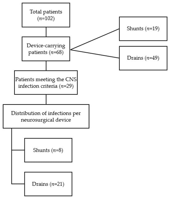
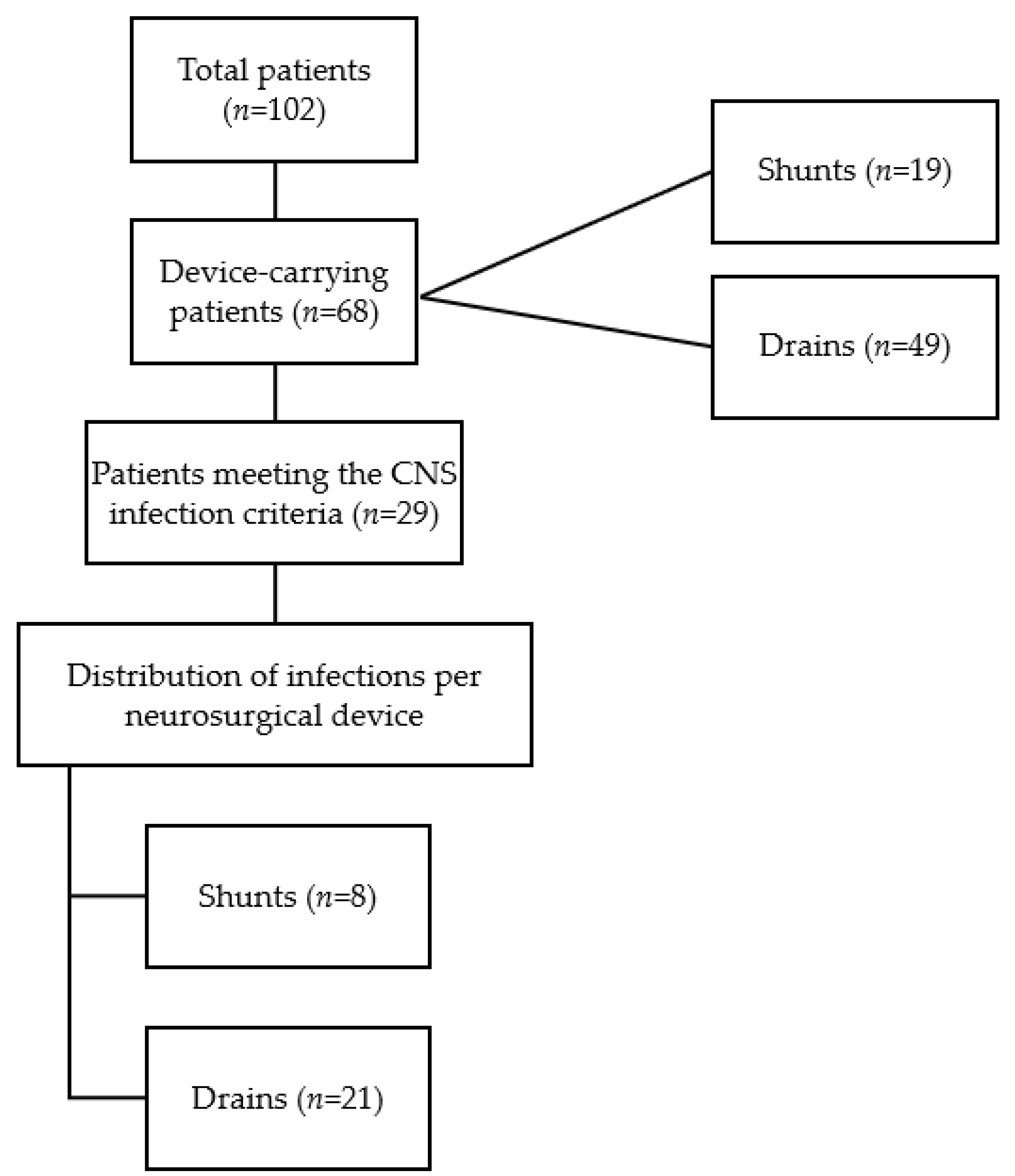
Of the 102 patients with positive CSF cultures, neurosurgical devices were implanted in 68 (67%) individuals. CSF drains were placed in the majority (n = 49; 70%) of the patients, while CSF shunts were used in the remaining ones (n = 19; 28%).
During the study period, 29 out of 68 patients with implanted neurosurgical devices met the study criteria for CNS infection, indicating a 43% incidence of neurosurgical device-related CNS infection in our setting. Among the 29 infected patients, 21 (72%) carried external drains and eight carried (28%) CSF shunts, showing the device proportions among infected patients. The risk of infection in relation to the specific type of neurosurgical device implanted was 42% (8/19) for CSF shunts and 43% (21/49) for external drains (Figure 1).
Figure 1.
Flowchart depicting patient population, neurosurgical device carriage, incidence of infection, and infection distribution by device type.
Patient ages ranged from 24 to 82 years (median 59 years; interquartile range [IQR] 16.5 years). Notably, 76% of infected patients were female. Among the study patients, the predominant surgical indication was intracranial mass resection, accounting for 55% of cases (n = 16), followed by surgical intervention for ruptured cerebral aneurysms (n = 4; 14%) and acoustic neuroma excision (n = 3; 10%) (Table 1).
Table 1.
Demographic indication for hospitalization, complications, and mortality in patients with neurosurgical device-related CNS infections.
3.2. CSF Laboratory Profiles of Patients with Neurosurgical Device-Related Infection
A total of 38 CSF samples with positive, non-repetitive cultures were retrieved from the 29 infected patients—one sample presented with normal laboratory findings (normocytosis, normoglycorrhachia, normoproteinorrhachia). The remaining 37 samples demonstrated pleocytosis, accompanied by heterogeneous biochemical patterns. Among these, the distribution of glucose and protein levels was as follows: 23 samples (62.2%) exhibited hypoglycorrhachia concurrent with hyperproteinorrhachia; eight samples (27%) showed normoglycorrhachia and normoproteinorrhachia; four samples (10.8%) presented with normoglycorrhachia alongside hyperproteinorrhachia; and two samples (5.4%) displayed hyperglycorrhachia with normoproteinorrhachia (Table 2).
Table 2.
Laboratory profile of 38 cerebrospinal fluids analyzed from neurosurgical patients with device-related infection.
3.3. Pathogen Prevalence and Susceptibility Profiles
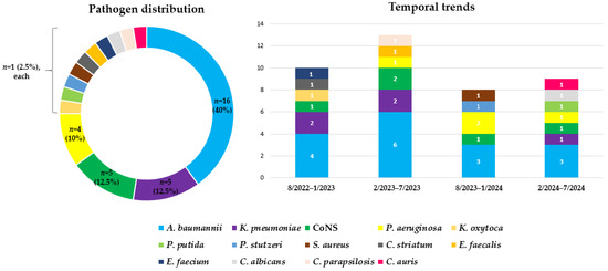
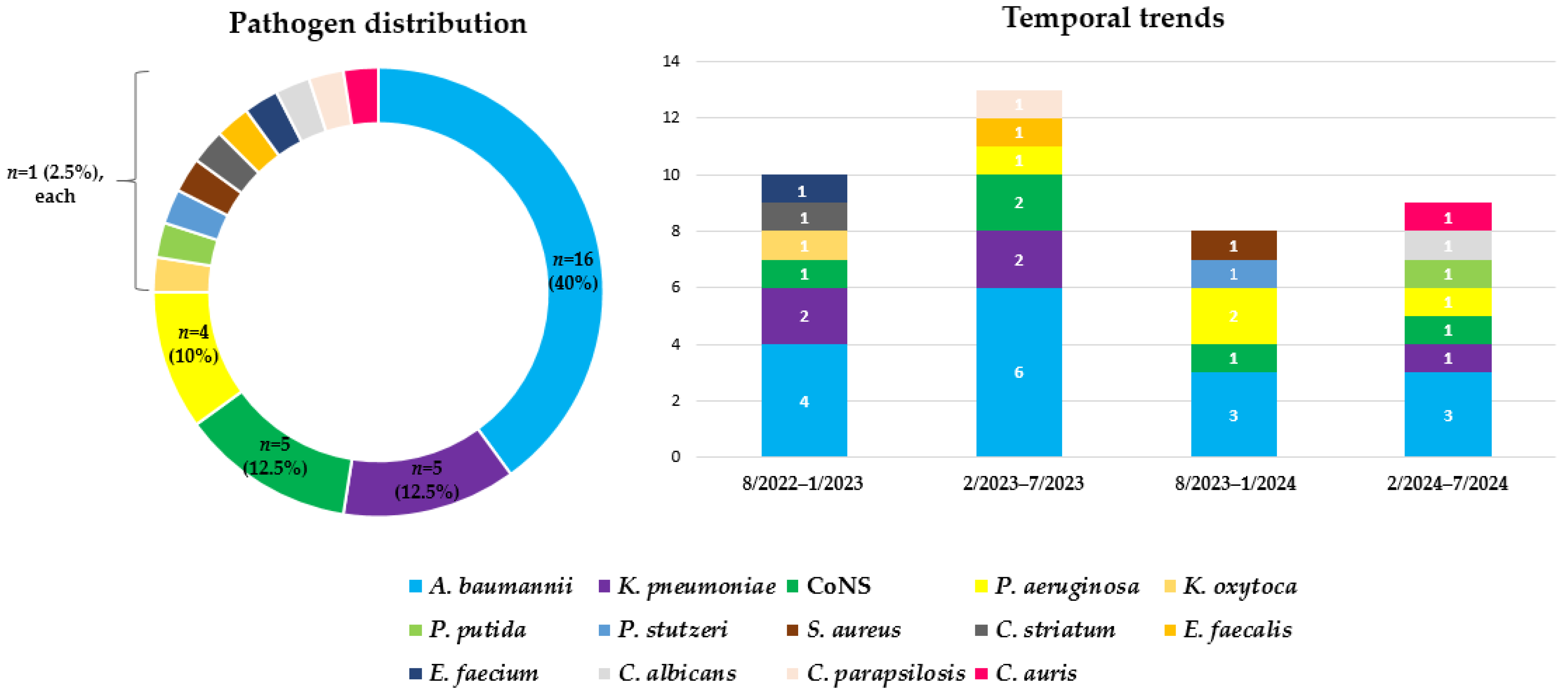
Overall, a total of 40 pathogens were isolated. Acinetobacter baumannii was the most prevalent, accounting for 40% (n = 16) of the total. Klebsiella pneumoniae and coagulase-negative Staphylococci (CoNS) were the second most common, each comprising 12.5% (n = 5) of isolates, while the isolated strains of Pseudomonas aeruginosa accounted for 10% (n = 4) of all pathogens. Klebsiella oxytoca, Pseudomonas putida, and Pseudomonas stutzeri were each isolated in 2.5% (n = 1) of cases. Staphylococcus aureus and Corynebacterium striatum were each identified in 2.5% (n = 1) of the samples, along with Enterococcus faecalis, Enterococcus faecium, and three distinct Candida species (Candida albicans, Candida parapsilosis, and Candida tropicalis) that also contributed 2.5% (n = 1) each. The pathogen profile showed minor changes throughout time. A. baumannii prevailed and was consistently isolated during all four semesters of the study, along with CoNS, whereas only C. auris was first recorded during the last semester of this cohort, in 2024. (Figure 2).
Figure 2.
Overall pathogen distribution and temporal trends in neurosurgical device-related CNS infections. Numbers in the temporal trends graph represent the isolated pathogens.
A. baumannii and K. pneumoniae co-infection was observed in a single female patient who carried a CSF shunt. Consecutive infections were observed in five patients (four with external drains and one with a CSF shunt). A. baumannii was the initial pathogen in two cases, while K. pneumoniae, P. aeruginosa, and P. putida were responsible for the remaining three cases, as elaborated in Table 3.
Table 3.
Characteristics of infections associated with CSF shunts and drains in neurosurgical patients of General Hospital of Athens “G. Gennimatas”.
The antimicrobial susceptibility testing revealed diverse resistance patterns among the bacterial isolates, highlighting the prevalence of multidrug-resistant (MDR) and extensively drug-resistant (XDR) phenotypes. In particular, A. baumannii isolates demonstrated universal resistance to carbapenems; additionally, 62.5% exhibited resistance to colistin. All K. pneumoniae isolates produced carbapenemases, namely KPC (n = 3), NDM (n = 1), and KPC + NDM (n = 1), whereas 40% of the strains were resistant to colistin. Similarly, all P. aeruginosa isolates were resistant to carbapenems, mainly attributable to VIM carbapenemase production (n = 3) and secondary to other mechanisms such as porin downregulation or efflux pump overexpression (n = 1); however, all strains were susceptible to colistin. The one isolate of K. oxytoca displayed XDR characteristics, with resistance to carbapenems (via KPC carbapenemase production) and colistin. The only S. aureus strain was susceptible to all tested antibiotics, indicating a methicillin-susceptible (MSSA) profile. Methicillin resistance was prevalent in 80% of CoNS isolates, while linezolid resistance was encountered in one isolate. Regarding E. faecium, the isolate exhibited a glycopeptide-resistant (GRE) phenotype, which is characteristic of the vanA genotype (Table 4).
Table 4.
Resistance phenotype of the most prevalent pathogens.
As for the fungal pathogens, C. albicans demonstrated susceptibility to all antifungal agents tested; C. auris exhibited resistance to fluconazole, while C. parapsilosis was found to be resistant to both fluconazole and voriconazole. All yeasts were susceptible to echinocandins, polyenes, and pyrimidine analogs.
PCR was performed in five cases, detecting A. baumannii (n = 3), C. albicans (n = 1), and C. tropicalis (n = 1).
3.4. Therapeutic Interventions and Clinical Outcomes
Among the 29 neurosurgery patients, 22 (76%) achieved clinical recovery and/or resolution of infection. All Gram-negative bacteria underwent intrathecal infusion of colistin. Klebsiella strains were treated according to carbapenemase production. Intravenous ceftazidime/avibactam was administered for KPC producers and ceftazidime/avibactam-fosfomycin-colistin for NDM producers. The three VIM-producing P. aeruginosa isolates were managed with meropenem-colistin intravenously. Cases with colistin-resistant A. baumannii infections received intrathecal vancomycin in conjunction with colistin, along with intravenous infusion of different combinations, such as vancomycin-colistin and colistin-ampicillin/sulbactam. These treatment combinations were selected based on the results of in vitro synergistic activity testing. Linezolid and vancomycin were implemented for the treatment of Gram-positive cocci, with the exception of S. aureus, for which cloxacillin was administered. Liposomal amphotericin, both intravenously and intrathecally, was administered for yeast infections.
Mortality was reported in four patients (13.8%), three of whom (75%) were carriers of external drains. In three cases, the infection was monomicrobial and was attributed to carbapenem-resistant K. pneumoniae (NDM), P. aeruginosa (VIM), and A. baumannii. The fourth patient had multiple device-related infections (P. putida, P. aeruginosa, A. baumannii, C. albicans, CoNS, and C. auris).
3.5. Statistical Analysis
Patients with implanted shunts had 0.98 times the risk and 0.97 times the odds of infection compared to patients with drains, showing that the infection risk of shunts and drains was essentially the same, with no statistically significant difference. This was further proved by Chi-square analysis [χ2 (1, N = 68) = 0.00, p = 0.955].
A. baumannii, the predominant pathogen of the study, was not associated with device-specific infections [χ2 (1, N = 39) = 1.02, p = 0.312] (Table 5).
Table 5.
Risk estimate and results of χ2 test of association between device type (shunts versus drains) and infection.
4. Discussion
This retrospective survey was designed to investigate the occurrence and the prevalent characteristics of infections linked to CSF shunts and drains in one of the biggest neurosurgery departments of Greece. To the best of our knowledge, this study is the first published report on neurosurgical device-related infections in Greece, providing data regarding the CSF profile, the epidemiology, the causative agents, the clinical outcome, and the therapeutic interventions, which may prospectively influence neurosurgical therapeutic decisions and infection prevention and control strategies.
In the cohort, the incidence of device-associated CNS infections was 43%, and the device-specific infection risk was similar, accounting for 42% in shunts versus 43% in external drains [8]. These findings exceed those previously documented, where external ventricular drain (EVD) infection rates ranged from 2% to 23%, and cumulative CSF shunt infection rates spanned from 2.2% to 41% [4]. The increased rates in our study may be partially explained by pragmatic factors, such as the high patient turnover in association with the workforce capacity constraints, potentially impacting optimal compliance with infection prevention and control protocols.
To our knowledge, the published studies that directly compare the device-specific infection risks are limited, and further observational studies are required to assess whether the infection risk is associated with the device type. Indeed, contrary to previous findings that report increased burden of infection in external drains [9,10], in our cohort, there was no statistically significant association between device type and occurrence of CNS infection, suggesting comparable insertion and management practices across devices. Along with the continuous monitoring of infection rates and investigation of contributing factors, these findings highlight the need for standardized operating procedures, such as appropriate preoperative antisepsis, sterile precautions, and minimal device manipulation, to reduce infection burden, as previously documented [11,12].
Pleocytosis and hyperproteinorracchia were observed in 27/38 CSFs corresponding to 22 patients, while pleocytosis and elevated protein in association with lower glucose levels were recorded in 23/38 CSFs (18 patients), indicating that normal or borderline CSF profiles do not rule out the possibility of device-related CNS infection in neurosurgical patients, as described in the literature [13]. Early identification is rather based on signs and clinical symptoms consistent with CNS infection (headache, nausea, seizures, neurological deterioration) along with positive CSF cultures or abnormal imaging, after exclusion of systemic infection or alternative causes of the clinical presentation [13].
Traditionally, CoNS and S. aureus predominate in shunt infections, often comprising 70% of cases [14,15]. In the present study, S. aureus (n = 1; 2.5%) was isolated in a shunt infection, whereas CoNS (n = 5; 12.5%) were observed solely in drains. The prevalent pathogens in our cohort were carbapenem-resistant A. baumannii, K. pneumoniae, and P. aeruginosa, as well as CoNS, aligning with a recent report from northern Greece, where these pathogens were responsible for the majority of post-neurosurgical CNS infections, reflecting the endemicity of MDROs in Greece [16,17]. Notably, our study identified a KPC-producing K. oxytoca isolate, a finding rarely documented [18]. Similarly to neurosurgery settings globally [14,19,20,21,22], the shift toward nosocomial MDROs in our institution may be driven by prolonged exposure to broad-spectrum antibiotics during prophylaxis and treatment. The implementation of a more stringent antibiotic stewardship program is warranted, as optimization of empiric and targeted therapy, including timely de-escalation, has been associated with reduced MDRO infections, reduced carbapenem resistance rates, and increased susceptibility rates [23]. In accordance with previous reports of opportunistic fungal invasions in neurosurgical patients, fungal pathogens like Candida species were less common (totaling 7.5%) yet clinically relevant given the challenges associated with their treatment [24,25]. Overall, the infections had no temporal pattern, as opposed to mild-climate countries with high levels of humidity [10].
Positive outcomes were noted in 76% of patients. Currently, there are no specific guidelines for the treatment of CNS infections caused by carbapenem-resistant bacteria, except for A. baumannii. In particular, IDSA recommends intravenous colistin or polymyxin B, with intrathecal or intraventricular administration, if necessary [7]. Several studies, including ours, have reported successful antimicrobial combination therapies [26,27]. As for MDR/XDR K. pneumoniae and P. aeruginosa, intrathecal colistin may have contributed to favorable outcomes, as previously reported [28,29]. Additionally, although our study included limited molecular testing, rapid diagnostic tools such as MALDI-TOF and PCR, combined with close collaboration between microbiologists and infectious diseases specialists, could have facilitated timely and targeted therapy. In our study, we applied the off-label BCID2 panel on CSF samples, focusing on nosocomial pathogens and resistance mechanisms. The results were promising, as they were rapid and accurate (consistent with culture), in accordance with the existing literature [30]. Timely device removal might have also influenced the observed cure rates; however, limited data prevent definitive conclusions.
Shunt-related and drain-related mortality in our setting was similar, 12.5% versus 14.2%, and it was associated with MDR or C. auris infections, whereas a prior study reported hospital mortality of 20% and 27%, respectively, in a setting with a different epidemiology, where the infections were caused predominantly by Gram-positive bacteria [31]. Comparative analysis between shunts and drains in terms of prognosis was not feasible due to the relatively low number of deaths observed during the study period. To address this question, prospective cohort studies, preferably from multiple centers, are essential, keeping in mind the complexity of the factors that may influence the outcome of device-related CNS infections: pathogen virulence, host immune response, duration of device placement, and timeliness of therapeutic intervention.
This study, however, has various limitations as follows: (i) The single-center focus reflects the local epidemiology and resistance patterns. (ii) The retrospective design may have influenced the incidence. Patients with suspected infection may have been excluded if they did not have positive cultures or if cultures were not obtained; additionally, some minor infections that may have resolved spontaneously without clinical intervention or involvement of an infectious diseases specialist may have been inadvertently excluded, resulting in a relatively low number of patients included. (iii) The relatively low sample size and the limited number of shunt-associated infections does not allow for the extraction of statistically powerful results. (iv) Genomic sequencing was not incorporated, and pathogens potentially evading the conventional microbiological methods have been excluded.
5. Conclusions
Neurosurgical devices are associated with a high risk of contamination, often serving as vectors for CNS infections. These infections may be associated with nosocomial pathogens such as A. baumannii, K. pneumoniae, P. aeruginosa, and skin flora (CoNS). Clinical infection may occur even when CSF standard laboratory parameters remain within normal reference ranges, complicating early detection.
In the present cohort, the likelihood of infection does not appear to be influenced by the type of neurosurgical device implanted (CSF shunts versus external drains). However, more research is needed to corroborate this result and further estimate the device-related prognosis.
Prompt clinical recognition and management are critical. Early diagnostic vigilance, including microbiological testing, in combination with targeted antimicrobial therapy and potential device removal, are key to optimizing patient outcomes.
In MDRO endemic settings, antibiotic stewardship is essential to tackle antibiotic resistance, while continuous monitoring along with the implementation and evaluation of rigorous infection prevention and control strategies is of utmost importance to mitigate the infection risk.
Author Contributions
Conceptualization, M.O.; methodology, K.A., A.S., E.K., A.Y., A.A., A.T., M.K., and M.O.; software, K.A.; validation, K.A. and M.O.; formal analysis, K.A.; investigation, K.A., A.S., E.K., A.Y., A.A., A.T., M.K., and M.O.; resources, K.A.; data curation, K.A., A.S., and M.O.; writing—original draft preparation, K.A.; writing—review and editing, K.A., A.S., and M.O.; visualization, K.A. and M.O.; supervision, K.A. and M.O.; project administration, K.A. and M.O.; All authors have read and agreed to the published version of the manuscript.
Funding
This research received no external funding.
Institutional Review Board Statement
This study was approved by the Scientific Council of the General Hospital of Athens ‘G. Gennimatas’ (approval code: 9826).
Informed Consent Statement
Patient consent was waived due to the retrospective nature of the study and the focus on the epidemiology of the infections rather than the patients themselves; moreover, the patients cannot be identified.
Data Availability Statement
The data presented in this study are not publicly available due to institutional and patient privacy regulations. De-identified datasets may be provided by the corresponding author upon reasonable request after institutional approval.
Conflicts of Interest
The authors declare no conflicts of interest.
Abbreviations
The following abbreviations are used in this manuscript:
| CNS | Central Nervous System |
| CSF | Cerebrospinal fluid |
| EUCAST | European Committee on Antimicrobial Susceptibility Testing |
| CDC | Centers for Disease Control and Prevention |
| IDSA | Infectious Diseases Society of America |
| WBCs | White Blood Cells |
| RBCs | Red Blood Cells |
| MALDI-TOF MS | Matrix-Assisted Laser Desorption/Ionization–Time-of-Flight Mass Spectrometry |
| AST | Antimicrobial Susceptibility Testing |
| MIC | Minimum Inhibitory Concentration |
| CoNS | Coagulase-negative Staphylococci |
| MDR | Multidrug-resistant |
| XDR | Extensively drug-resistant |
| PDR | Pandrug-resistant |
| MSSA | Methicillin-resistant Staphylococcus aureus |
| MDRO | Multidrug-resistant organisms |
| EVD | External ventricular drain |
| VP | Ventriculo-peritoneal |
| VRE | Vancomycin-resistant Enterococcus |
References
- Seidelman, J.; Lewis, S.S. Neurosurgical Device-Related Infections. Infect. Dis. Clin. N. Am. 2018, 32, 861–876. [Google Scholar] [CrossRef] [Scilit] [PubMed]
- Allos, H.; Hasbun, R. Current understanding of infection of the ventricles and its complications. Expert Rev. Anti-Infect. Ther. 2024, 22, 1023–1042. [Google Scholar] [CrossRef] [Scilit] [PubMed]
- Sader, E.; Moore, J.; Cervantes-Arslanian, A.M. Neurosurgical Infections. Semin. Neurol. 2019, 39, 507–514. [Google Scholar] [CrossRef] [Scilit] [PubMed]
- Bloch, K.; Hasbun, R. Central nervous system infections associated with neurologic devices. Curr. Opin. Infect. Dis. 2021, 34, 238–244. [Google Scholar] [CrossRef] [Scilit] [PubMed]
- EUCAST. European Committee on Antimicrobial Susceptibility Testing. 2025. Available online: https://www.eucast.org/clinical_breakpoints (accessed on 14 October 2025).
- CDC. Centers for Disease Control and Prevention. Antifungal Susceptibility Testing for C. auris. 2025. Available online: https://www.cdc.gov/candida-auris/hcp/laboratories/antifungal-susceptibility-testing.html (accessed on 14 October 2025).
- Tunkel, A.R.; Hasbun, R.; Bhimraj, A.; Byers, K.; Kaplan, S.L.; Scheld, W.M.; van de Beek, D.; Bleck, T.P.; Garton, H.J.L.; Zunt, J.R. 2017 Infectious Diseases Society of America’s Clinical Practice Guidelines for Healthcare-Associated Ventriculitis and Meningitis. Clin. Infect. Dis. 2017, 64, e34–e65. [Google Scholar] [CrossRef] [Scilit] [PubMed]
- Avgoulea, K.; Spyrou, A.; Kalogianni, E.; Yiaskouri, A.; Athanasopoulou, A.; Tsakalos, A.; Kamperogianni, M.; Orfanidou, M. Two-year retrospective study of neurosurgical infections associated with foreign bodies in a tertiary hospital. In Proceedings of the 12th Panhellenic Congress of Clinical Microbiology and Medical Biopathology, Athens, Greece, 13–15 March 2025. [Google Scholar]
- Faslaperla, R.; Lo Bianco, M.; Palmeri, A.; Betta, P.; Altieri, R.; Barbagallo, G.M.; Ruggieri, M. Short efficacy evaluation of external ventricular drains versus ventriculosubgalelal shunt in the management of neonatal posthemorrhagic hydrocephalus: A retrospective single-center cohort study. Neurosurgery 2023, 93, 622–627. [Google Scholar] [CrossRef] [Scilit] [PubMed]
- Agarwal, S.; Gupta, P.; Venkatesh, V.; Srivastava, C. Microbial Diversity and Infection Burden in Central Nervous System (CNS) Shunt and Drain Devices: A Prospective Observational Study. Cureus 2025, 17, e95294. [Google Scholar] [CrossRef] [Scilit] [PubMed]
- Muram, S.; Isaacs, A.M.; Sader, N.; Holubkov, R.; Fong, A.; Conly, J.; Hamilton, M.G. A standardized infection prevention bundle for reduction of CSF shunt infections in adult ventriculoperitoneal shunt surgery performed without antibiotic-impregnated catheters. J. Neurosurg. 2023, 138, 494–502. [Google Scholar] [CrossRef] [Scilit] [PubMed]
- Pongkaew, C.; Noiphithak, R.; Rukskul, P.; Yodwisithsak, P.; Tantongtip, D.; Nimmannitya, P.; Punyarat, P.; Duangprasert, G.; Kaewwichai, W.; Songphul, S.; et al. Effectiveness of a multimodal strategy to reduce external ventricular drain-associated infection: A quasi-experimental study. Am. J. Infect. Control 2024, 52, 1307–1313. [Google Scholar] [CrossRef] [Scilit] [PubMed]
- Martin, R.M.; Zimmermann, L.L.; Huynh, M.; Polage, C.R. Diagnostic Approach to Health Care- and Device-Associated Central Nervous System Infections. J. Clin. Microbiol. 2018, 56, e00861-18. [Google Scholar] [CrossRef] [Scilit] [PubMed]
- Alqasmi, M.; Kariri, Y.A.; Alenazy, R.; Thabet, M.; Fallata, G.; Alqurainy, N. Ventriculoperitoneal Shunt Infections: Causative Pathogens and Associated Outcomes from Multiple Hospitals in Saudi Arabia. J. Clin. Med. 2025, 14, 2006. [Google Scholar] [CrossRef] [Scilit] [PubMed]
- Enrico, G.; Fusco, E.; Palmetti, M.; Mussa, F.; Sardi, I.; Chiappini, E.; Galli, L.; Venturini, E. Neurosurgical site infections: A retrospective monocentric study of pediatric brain tumor patients. Child’s Nerv. Syst. 2025, 41, 120. [Google Scholar] [CrossRef] [Scilit] [PubMed]
- Markakis, K.; Kapiki, K.; Edric, A.A.A.; Pappas, A.A.; Feretos, G.; Nanoudis, S.; Pilalas, D.; Michailidis, T.; Protonotariou, E.; Skoura, L.; et al. Post-Surgical Central Nervous System Infections in the Era of Multidrug Antibiotic Resistance in Greece-A Single-Center Experience of a Decade. Pathogens 2025, 14, 390. [Google Scholar] [CrossRef] [Scilit] [PubMed]
- European Centre for Disease Prevention and Control (ECDC). Antimicrobial Resistance in the EU/EEA (EARS-Net)—Annual Epidemiological Report for 2024; ECDC: Stockholm, Sweden, 2025. [Google Scholar]
- Jiang, Y.L.; Lyu, Y.Y.; Liu, L.L.; Li, Z.P.; Liu, D.; Tai, J.H.; Hu, X.Q.; Zhang, W.H.; Chu, W.W.; Zhao, X.; et al. Carbapenem-resistant Klebsiella oxytoca transmission linked to preoperative shaving in emergency neurosurgery, tracked by rapid detection via chromogenic medium and whole genome sequencing. Front. Cell. Infect. Microbiol. 2024, 14, 1464411. [Google Scholar] [CrossRef] [Scilit] [PubMed]
- Salmanov, A.G.; Shchehlov, D.V.; Svyrydiuk, O.; Bortnik, I.M.; Mamonova, M.; Kudelskyi, Y.; Rzayeva, F.H. Healthcare-associated infection in neurosurgical patients in Ukraine: Results of a multicenter study (2017–2019). Wiad. Lek. 2021, 74, 1945–1951. [Google Scholar] [CrossRef] [Scilit] [PubMed]
- Corona-Nakamura, A.L.; Arias-Merino, M.J.; Ávila-Esparza, E.I.; Tolentino-Corona, M.L.; Cañedo-Castañeda, C.C.; Flores-Salinas, H.E.; Corona-Macías, J.F.; Vázquez-Arias, M.E. Ventriculitis due to multidrug-resistant gram-negative bacilli associated with external ventricular drain: Evolution, treatment, and outcomes. Front. Neurol. 2024, 15, 1384206. [Google Scholar] [CrossRef] [Scilit] [PubMed]
- Fang, F.; Zhai, X.; Bao, S.; Fan, X.; Bai, R.; Ma, Y.; Dong, X. The Impact of Clinical Pharmacist Consultation on the Treatment of Multi-Drug Resistant Organisms (MDRO) Infections: A Retrospective Observational Study. J. Multidiscip. Healthc. 2025, 18, 2983–2995. [Google Scholar] [CrossRef] [Scilit] [PubMed]
- Al-Mufargi, Y.; Al Busaidi, F.; Al Balushi, Y.; Al Malki, T.; Al Hamdani, M.; AlSaidi, S.; AlYazidi, T.; Alkhatri, R.; Al-Saadi, T. Understanding the burden of multidrug-resistant organisms in neurosurgical care: Resistance trends and risk profiles. J. Neurosci. Rural Pract. 2025, 16, 501–508. [Google Scholar] [CrossRef] [Scilit]
- Yu, J.; Liu, Y.; Qu, R.; Wang, Z.; Zhao, Y.; Zhao, Y.; Zhou, C. Evaluation of a clinical pharmacist-led antimicrobial stewardship program in a neurosurgical intensive care unit: A pre-and post-intervention cohort study. Front. Pharmacol. 2023, 14, 1263618. [Google Scholar] [CrossRef] [Scilit] [PubMed]
- Chen, M.; Chen, C.; Yang, Q.; Zhan, R. Candida meningitis in neurosurgical patients: A single-institute study of nine cases over 7 years. Epidemiol. Infect. 2020, 148, e148. [Google Scholar] [CrossRef] [Scilit] [PubMed]
- Kelly, L.; Walsh, J.; Skally, M.; Dinesh, B.; Burns, K.; O’Connell, K.; MacNally, S.; Humphreys, H.; Fitzpatrick, F. Candida meningitis/ventriculitis over a decade. Increased morbidity and length of stay a concern. Br. J. Neurosurg. 2023, 37, 227–230. [Google Scholar] [CrossRef] [Scilit] [PubMed]
- Theofanopoulos, A.; Fermeli, D.; Vekios, D.; Bizos, A.; Marangos, M.; Constantoyannis, C.; Panagiotopoulos, V.; Assimakopoulos, S.F. Successful treatment of pan-drug resistant Acinetobacter baumannii nosocomial meningitis/ventriculitis by combined intravenous and intrathecal colistin-tigecycline administration: A case series. Infez. Med. 2023, 31, 103–107. [Google Scholar] [CrossRef] [Scilit] [PubMed]
- Gavrilovska, A.D.; Veljanovski, H.; Jovchevski, R. Successful treatment of pan-drug resistant Acinetobacter baumannii meningitis/ventriculitis following craniotomy and external ventricular drainage: A case report. J. Surg. Case Rep. 2024, 2024, rjae603. [Google Scholar] [CrossRef] [Scilit] [PubMed]
- Liu, D.; Niu, J.; Chen, G.; Xu, L. Treatment of Carbapenem-Resistant Multidrug-Resistant Gram-Negative Bacilli with Intracerebroventricular Injection of Polymyxin B: A Retrospective Study. Infect. Drug Resist. 2022, 15, 7653–7666. [Google Scholar] [CrossRef] [Scilit] [PubMed]
- Georgakopoulou, V.E.; Spandidos, D.A.; Papalexis, P.; Gkoufa, A.; Aravantinou-Fatorou, A.; Angelopoulou, E.; Trakas, I.; Trakas, N.; Fotakopoulos, G. Outcomes in meningitis ventriculitis treated with intravenous or intrathecal plus intravenous colistin: A meta analysis. Exp. Ther. Med. 2023, 25, 293. [Google Scholar] [CrossRef] [Scilit] [PubMed]
- Menchinelli, G.; Cacaci, M.; Squitieri, D.; De Angelis, G.; Sanguinetti, M.; Posteraro, B. Improving the diagnosis of central nervous system infections: Experience with the off-label use of FA-BCID2. Microbiol. Spectr. 2026, 14, e03189-25. [Google Scholar] [CrossRef] [Scilit] [PubMed]
- Zeggay, A.; Patry, I.; Chirouze, C.; Bouiller, K. Characteristics and outcomes of cerebrospinal fluid shunt and drain-associated infections. Infect. Dis. Now. 2023, 53, 104665. [Google Scholar] [CrossRef] [Scilit] [PubMed]

Disclaimer/Publisher’s Note: The statements, opinions and data contained in all publications are solely those of the individual author(s) and contributor(s) and not of MDPI and/or the editor(s). MDPI and/or the editor(s) disclaim responsibility for any injury to people or property resulting from any ideas, methods, instructions or products referred to in the content. |
© 2026 by the authors. Published by MDPI on behalf of the Hellenic Society for Microbiology. Licensee MDPI, Basel, Switzerland. This article is an open access article distributed under the terms and conditions of the Creative Commons Attribution (CC BY) license.