


Abstract
Platelet-rich plasma (PRP) is widely used in orthopedics and sports medicine as an autologous product; however, substantial heterogeneity in manufacture and incomplete reporting of preparation parameters limits reproducibility and inter-study comparability.: We performed a PRISMA-guided methodological review of studies describing PRP preparation, subsequently focusing on orthopedic applications. MEDLINE/PubMed, Embase, Scopus, the Cochrane Library, Web of Science, and Google Scholar were searched. 7330 records were retrieved; following merging and de-duplication, more than 2500 unique records were screened. The inclusion criteria for our study required studies on PRP that focused on orthopedic use of this preparation and studies that reported a defined methodology for PRP, reported in the abstract or in the manuscript. Extracted variables covered collection and anticoagulation, centrifugation strategy, cellular composition, activation/lysis, processing environment, storage, and time-to-use. Twenty-three orthopedic studies met the inclusion criteria. Whole blood draw volume and anticoagulant were reported in 15/23 studies each; centrifugation parameters (relative centrifugal force/RPM and duration) in 12/23; and PRP phenotype (e.g., leukocyte-poor vs. leukocyte-rich) in 15/23. Platelet metrics (baseline and/or final platelet count/concentration) were reported in 6/23. Sterility/environmental controls were mentioned in 17/23, whereas storage conditions and time-to-use were described in only 3/23. An explicit exogenous activation agent was reported on 1/23. Orthopedic PRP studies frequently omit critical manufacturing and handling descriptors—particularly platelet dose, leukocyte/lymphocyte handling, temperature control, storage/freezing conditions, and time-to-administration—impairing reproducibility and dose comparability. We propose a pragmatic, standardized protocol for preparation of leucocyte/lymphocyte-depleted PRP for orthopedic use (PRP only, without gelification), together with a minimum set of data and parameters to be evaluated. In our opinion these parameters should be included in future studies in order to standardize the production process. The quality of PRP itself could be impacted by such standardization, and the ability to objectively evaluate the results of studies could be enhanced by facilitating the comparison of data emerging from the literature.
1. Introduction
Platelet-rich plasma (PRP) is an autologous, blood-derived product in which platelets are concentrated above baseline levels within a small volume of plasma. Platelets act as a reservoir of bioactive mediators—stored largely in α-granules—that are released upon activation or lysis, including platelet-derived growth factor, transforming growth factor-β, vascular endothelial growth factor, insulin-like growth factor and a range of chemokines and cytokines. Beyond hemostasis, these mediators are implicated in the modulation of inflammation, angiogenesis, matrix remodeling and cellular proliferation, providing the biological rationale for PRP as an ‘orthobiologic’ intervention in musculoskeletal medicine [1].
PRP is not a unique product but a spectrum of preparations that differ in platelet dose, leukocyte and erythrocyte contamination, activation/lysis status, and delivered volume. This variability arises from pre-analytical factors (patient baseline platelet count, medications affecting platelet function, time from phlebotomy to processing), manufacturing factors (anticoagulant type, centrifugation strategy and device geometry, open versus closed handling), and post-manufacture handling (temperature control, storage, freeze–thaw exposure). Accordingly, preparation methodology is inseparable from product identity: two preparations labelled ‘PRP’ may differ substantially in cellular composition and growth factor release kinetics [1,2,3].
In orthopedics and sports medicine, PRP has become one of the most widely used injectable orthobiologics, principally because it is autologous, can be produced at point of care, and has a favorable safety profile due to the fact that its production process is carried out under aseptic conditions. Current clinical use spans both non-operative and adjunctive surgical settings. Non-operatively, intra-articular PRP is most commonly utilized in knee osteoarthritis, where multiple European consensus initiatives have considered PRP an appropriate option for mild-to-moderate disease (Kellgren–Lawrence grades 0–III/≤3), typically after failure of conservative measures and/or other injectables, while cautioning against routine first-line use or use for advanced radiographic disease. Beyond osteoarthritis, PRP is frequently used for chronic tendinopathies (e.g., lateral epicondylitis and patellar and Achilles tendinopathy), muscle injuries, and as an adjunct in ligament, meniscal and cartilage procedures, although the strength of evidence and recommended use scenarios vary across indications [4,5].
High-quality orthopedic trials are increasingly reporting granular manufacturing variables that directly affect reproducibility and biological interpretation. For example, in a randomized controlled trial of transforaminal epidural PRP for lumbar disc herniation, PRP was prepared from 24 mL of venous blood using a standardized double-spin protocol and the platelet ratio was explicitly reported [6]. In knee osteoarthritis, leukocyte depletion has been applied to generate LP-PRP and both platelet and leukocyte concentrations have been quantified, facilitating comparison between formulations [7]. Similarly, a rotator cuff repair trial detailed double-spin centrifugation forces and a calcium-only activation strategy (without thrombin), as well as intraoperative versus postoperative (2-week) timing of administration [8]. Complementary laboratory work has prepared paired LR-PRP and LP-PRP from the same donors via a double-spin protocol and reported white blood cell subtypes, highlighting the clinical relevance of leukocyte phenotypes [9]. Finally, protocol standardization efforts with volunteers have proposed optimized g-force settings for high platelet recovery, offering a methodological benchmark for reporting [10].
The rapid clinical adoption of PRP has outpaced standardization. Inconsistency in the reporting of fundamental manufacturing parameters continues to limit reproducibility, dose comparability, and ultimately the interpretability of clinical outcomes. To address this, orthopedic-specific reporting guidance has been published (e.g., the Minimum Information for Studies Evaluating Biologics in Orthopedics (MIBO) checklist), alongside broader classification frameworks (e.g., PAW and DEPA) intended to define PRP phenotype and platelet dose. Nevertheless, adherence to these frameworks in orthopedic publications remains variable, and key descriptors—particularly baseline platelet metrics, leukocyte/lymphocyte handling, temperature control, storage/freezing, and time-to-administration—are often omitted [1].
Against this backdrop, our work is deliberately methodological: rather than asking whether PRP ‘works’ for a given orthopedic diagnosis, we asked what PRP actually is in the orthopedic literature and what must be reported to make studies reproducible and comparable. We conducted a systematic methodological review (systematic survey) of PRP preparation reporting in orthopedic studies, mapping the completeness of reporting for variables essential to product identity and reproducibility. We used PRISMA 2020 items to report these aspects, ensuring transparency in identification, screening, eligibility assessment and descriptive synthesis. No quantitative pooling of clinical outcomes was planned [11].
We therefore performed a PRISMA-guided review of studies that described PRP preparation methods and then focused on orthopedic applications. On the basis of the deficiencies identified, we propose, on the basis of our experience, a step-by-step, standardized protocol for preparation of leucocyte/lymphocyte-depleted PRP for orthopedic use (PRP only, without gelification), including guidance on collection, anticoagulation, processing temperature, centrifugation reporting, platelet quantification, controlled platelet rupture/lysis and the role of freezing, and time-to-use.
Study objectives were: (1) to synthesize and compare reported PRP preparation workflows in orthopedic studies and (2) to quantify the completeness of reporting for key manufacturing and handling variables that determine PRP identity. We also present our experience, in the form of a standardized comprehensive protocol proposal with minimum reporting dataset to support reproducible PRP production and more meaningful inter-study comparison in orthopedics. As an initial assessment of the presented protocol, we also wanted to report some preliminary data from the analysis of around 200 cases treated in the last year.
2. Materials and Methods
2.1. Review Design and Reporting Standard
This study is a systematic methodological review (systematic survey) assessing the completeness of reporting of PRP preparation and handling variables in studies in which PRP was applied for orthopedic purposes. The review is reported in accordance with the PRISMA 2020 statement, which provides guidance for transparent reporting of systematic reviews with or without statistical synthesis. Because the outcomes of interest were methodological descriptors (e.g., anticoagulants, centrifugation parameters, cellular composition, storage/lysis) rather than pooled treatment effects, no meta-analysis was planned and findings were synthesized descriptively [6,7,8,9,10,11]. Although the review maps heterogeneity and reporting gaps (features shared with scoping reviews), we retained a systematic review reporting framework because we applied predefined eligibility criteria, duplicate screening, and structured extraction with quantitative descriptions of reporting completeness [12].
2.2. Data Sources and Search Strategy
A comprehensive search was performed in MEDLINE/PubMed, Embase, Scopus, the Cochrane Library, Web of Science, and Google Scholar. The last search date was 24 December 2025. Keyword searches were conducted using the following strings: “prp orthopedic” (2682 results), “platelet rich plasma orthopedic” (3558 results), “prp protocol preparation” (424 results), “platelet rich plasma protocol preparation” (527 results), and “platelet rich plasma protocol preparation orthopedic” (139 results). These raw hit counts represent database interface outputs and include substantial overlap. Results from all searches were merged and de-duplicated prior to screening. To maximize retrieval of studies describing PRP manufacture, we also performed backward citation searching from comprehensive reviews and methodological papers focused on PRP quality control and reporting, and screened any additional records identified. Searches were not restricted by study design. No language limits were imposed at the search stage; however, non-English records were included only if the PRP preparation methodology was clearly extractable.
2.3. Eligibility Criteria
Studies were eligible if they (i) were clinical studies evaluated autologous blood components for non-transfusional use (i.e., PRP) in an orthopedic or sports medicine field, and (ii) were available as full text. Due to the fact that the primary objective was to assess completeness of reporting of PRP manufacture and handling, no minimum threshold of methodological detail was required for inclusion. Instead, all predefined preparation variables (e.g., blood volume, anticoagulants, centrifugation parameters, fractionation approach, cellular composition, activation/lysis, storage, and time-to-administration) were extracted and coded as reported or not reported. Studies were excluded if they did not report an application in an orthopedic field, did not involve PRP (e.g., used other orthobiologics), were not primary studies (e.g., narrative reviews or editorials), were duplicates, or lacked accessible full text. Comprehensive reviews were retained only for backward citation searching and contextual mapping but were not included in the final analysis. Three independent reviewers screened records on the basis of title and abstract and subsequently assessed full texts for eligibility. Disagreements were resolved by consensus. Duplicate records were removed prior to screening.
2.4. Data Extraction and Definitions
A standardized data extraction form was applied to all eligible orthopedic studies. For each predefined PRP manufacturing process variable, reporting was coded as reported (sufficient detail to identify the parameter) or not reported (absent or not extractable). Extracted items included: study design and indication; blood draw volume; anticoagulant type; preparation system (manual vs. commercial kit) and processing environment (open vs. closed/aseptic system workflow where stated); number of centrifugation steps and, for each spin, centrifugal force and duration (reported preferentially as relative centrifugal force (RCF, xg) and duration; where only revolutions per minute (RPM) were provided, the relationship RCF = 1.118 × 10⁻⁵ × r × RPM2 was used conceptually to interpret comparability, with r representing rotor radius); fractionation approach (plasma-based vs. buffy coat-based where described); final PRP volume; platelet metrics (baseline and/or final platelet count and/or dose); leukocyte and erythrocyte content information (including designation as leukocyte-poor or leukocyte-rich where specified); activation/lysis method; storage/freezing (temperature, duration, and cycles where stated); and time from preparation to administration.
Variables were considered ‘reported’ only when the manuscript provided explicit values or unambiguous descriptors. Vague statements such as ‘PRP were prepared according to a standard protocol’ without parameters were coded as not reported.
2.5. Outcomes and Synthesis
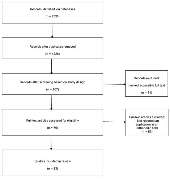
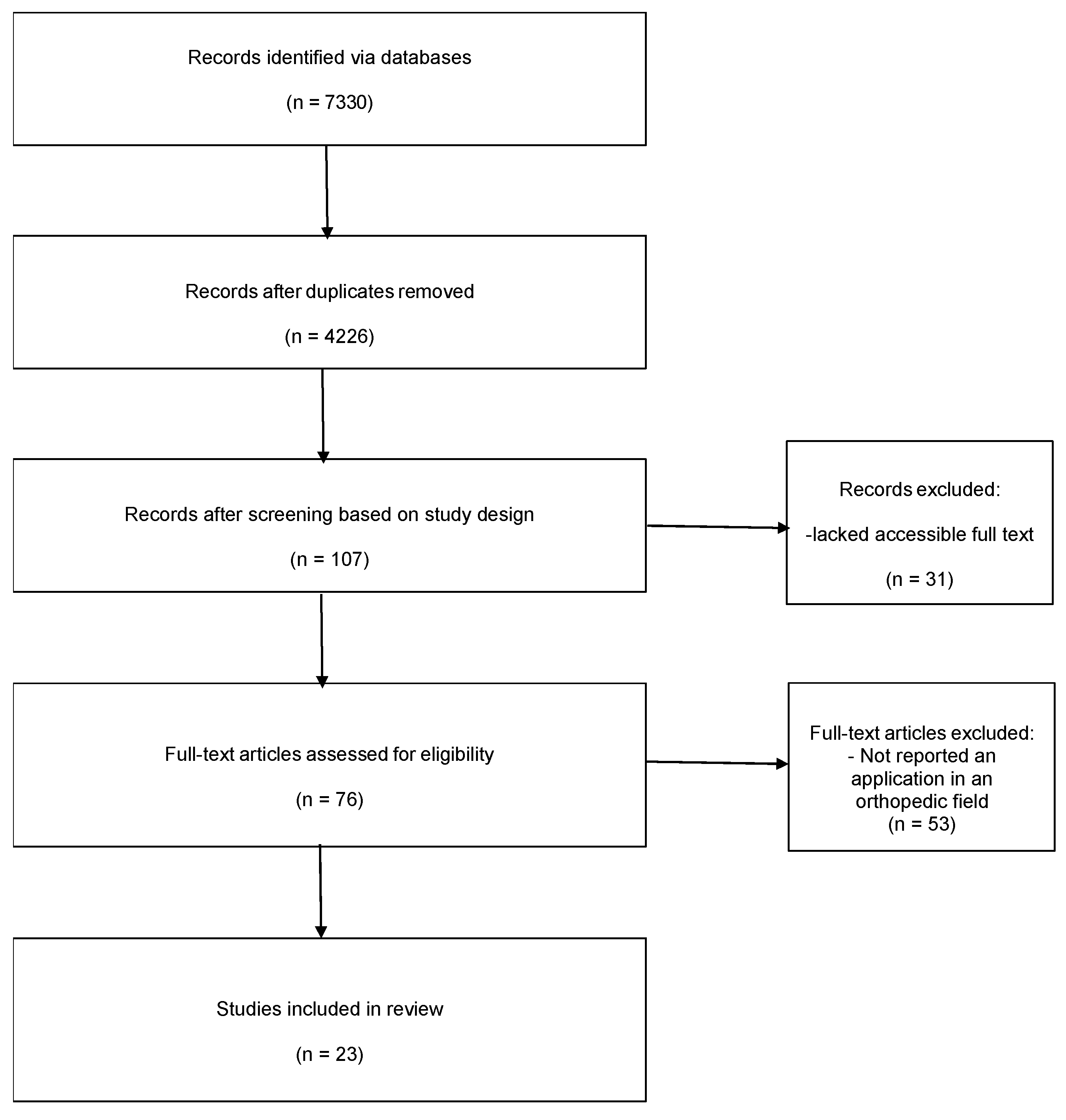
The primary outcome was completeness of reporting for each predefined PRP preparation and handling variable required for reproducibility and dose comparability. For every included study, each variable was coded as reported or not reported and the frequency of reporting was summarized across studies. Secondary outcomes were (i) the distribution of preparation workflows (e.g., single- vs. double-spin; manual vs. kit-based; plasma-based vs. buffy coat-based approaches where identifiable), and (ii) identification of recurrent reporting gaps that informed the proposed standardized protocol and minimum reporting dataset. Formal risk-of-bias assessment was not performed because the review objective was reporting completeness rather than synthesis of clinical effectiveness outcomes. The completed PRISMA 2020 checklist can be found in Supplementary Materials. The PRISMA flow in Figure 1 diagram illustrates the selection of studies for the systematic review.
Figure 1.
PRISMA 2020 flow diagram illustrates the selection of studies for the systematic review. n = number of articles.
2.6. Proposed Standardised Protocol for PRP Preparation in Orthopaedics
The following protocol is intended as a proposal for a reproducible procedure to produce leucocyte-poor PRP intended for orthopedic use (i.e., PRP only, without exogenous activation or gelification).
Our protocol is drafted respecting the latest regulations regarding blood components intended for non-transfusion use within Italy, published on “Gazzetta Ufficiale” of the Italian Republic the 26 September 2019 and its related regulations in force (Ministerial Decree (Ministero della Salute) of 2 November 2015, titled “Disposizioni relative ai requisiti di qualità e sicurezza del sangue e degli emocomponenti” and Ministerial Decree (Ministero della Salute) of 1 August 2019) [13].
Where local regulation and institutional requirements differ, centers could adapt by following the advice below on preserving the minimum reporting dataset.
In our experience, all stages of sample collection, processing, sampling and storage are carried out at the premises of the Unità Operativa Complessa di Medicina Trasfusionale, Azienda Sanitaria Territoriale Macerata, Ospedale Generale Provinciale di Macerata by technicians trained and specialized in this procedure. The sample is administered by an orthopedic specialist, with or without ultrasound guidance, depending on the type of pathology being treated, in dedicated rooms in the Orthopedic Department of the same hospital.
2.6.1. Setting, Governance and Equipment
- PRP should be manufactured within an accredited transfusion service or equivalent controlled laboratory environment using CE-marked, single-use closed systems where feasible.
- All open steps (e.g., sampling for quality control and removal of platelet-poor plasma) should be performed under a class II laminar airflow cabinet with aseptic technique.
- Ambient temperature during processing should be monitored and documented (recommended range 18–25 °C), using calibrated data loggers with scheduled sampling (e.g., every 30 min).
2.6.2. Patient Eligibility and Pre-Collection Requirements
- A recent—within 30–45 days of the collection date—full blood count is required to document baseline platelet concentration.
- Exclusion criteria should include platelet count ≤ 120 × 103/µL, known coagulopathy, therapeutic oral anticoagulation, acetylsalicylic acid use within 10 days, NSAID use within 5 days, suspected or confirmed active infection (bacteremia risk, fever), and absence on written informed consent.
2.6.3. Collection and Anticoagulation
- Collect autologous venous whole blood (e.g., 50 mL) from a peripheral vein using an appropriate sterile kit. In our experience we used Disposable CPunT 60 mL (cod. 10713905) Eltek S.p.A.—Casale Monferrato (Italy).
- Use acid-citrate-dextrose solution A (ACD-A) as anticoagulant; document manufacturer, lot number, and blood-to-anticoagulant ratio [14]. Anticoagulante ACD S.A.L.F. Soluzione anticoagulante e conservante per il sangue—S.A.L.F. S.p.A. Laboratorio Farmacologico, Cenate Sotto (Italy).
2.6.4. Centrifugation-Based Separation (Double-Spin Approach)
- First centrifugation (“soft spin”): 1200× g for 10 min to separate erythrocytes from platelet-containing plasma. Centrifuga Ssitema CPunT (Mod. 10753405) Eltek S.p.A.—Casale Monferrato (Italy).
- After the first spin, under aseptic conditions withdraw a small aliquot (e.g., 0.5 mL) for platelet count as a quality-control checkpoint.
- Leucocyte/lymphocyte exclusion: select the kit setting designed to exclude white cells (where applicable) and manually avoid aspiration of the buffy coat during plasma transfer.
- Second centrifugation (“hard spin”): 1900× g for 10 min to sediment platelets.
- Remove platelet-poor plasma (PPP) while maintaining the platelet pellet at the bottom of the container, then gently resuspend the pellet.
2.6.5. Target Platelet Concentration and Volume Standardisation
- The final product should aim for a platelet concentration of 1.0 × 10⁶/µL (±20%).
- The formula for calculating the final PRP volume (V2) required for each patient iswhere C1 is the post-first-spin platelet concentration, V1 is the total plasma volume after the first spin, and C2 is the target platelet concentration.C1 × V1 = C2 × V2
- Record baseline platelet count, post-first-spin platelet count, estimated platelet recovery, final volume, and expected final platelet concentration. The platelet count was performed with Auto Hematology Analyzer BC-760, Shenzhen Mindray Bio-Medical Electronics Co., Ltd., Shenzhen, China.
2.6.6. Quality Control and Sterility
- Perform sterility testing on the removed PPP (or an equivalent validated surrogate) according to local microbiological protocols.
- Document the hematology analyzer methodology used for platelet counting and quality-control thresholds.
2.6.7. Storage, Platelet Lysis Strategy and Use
- Fresh PRP should be administered as soon as practicable and within 6 h of manufacture if held at room temperature.
- For deferred use, aliquot PRP into sterile, capped syringes or containers, label with patient identifiers and traceability codes, and cryopreserve at −40 °C in a dedicated, monitored freezer [15,16]. In our setting, storage is validated for up to 12 months in a monitored freezer located in the transfusion department.
- Platelet rupture (growth-factor release) should be explicitly reported. If freeze–thaw lysis is intended, document the number of freeze–thaw cycles, thawing conditions, and any post-thaw clarification steps [15,16].
- At administration, document time from thaw to injection, injection volume, anatomical site, and whether any filtration was performed [15,16].
2.6.8. Statistical Analysis of Preliminary Outcomes in Processing PRP with Our Protocol
Additionally, we performed a retrospective descriptive analysis of laboratory and processing variables emerging during PRP manufacture over the past year in consenting patients treated at Unità Operativa Complessa di Medicina Trasfusionale, Azienda Sanitaria Territoriale Macerata, Ospedale Generale Provinciale di Macerata, using de-identified data extracted from routine transfusion service records. The analysis was defined a priori as non-parametric for continuous variables due to the possibility of non-normal distributions and extreme values. Therefore, continuous data were summarized primarily as median and interquartile range (IQR; 25th–75th percentile), with minimum–maximum values also reported. For transparency and comparability, mean and standard deviation (SD) were additionally calculated. A 95% confidence interval for the mean was computed. Data completeness was assessed by reporting the number and percentage of missing values and analyses were performed on available observations for each variable (complete case for that variable). Distributions were visually inspected using histograms and boxplots. Potential outliers were identified using Tukey’s rule (values beyond 1.5 × IQR from the quartiles). Microsoft 365 app for enterprise suite Excel (ver 2601, build 19628.20150) software was used for data analysis and table design. Chat GPT version 5.2 was used to modify the layout of the graph.
3. Results
3.1. Study Selection
Across the five primary search strings, database interfaces returned 7330 records (2682 + 3558 + 424 + 527 + 139). These results were confirmed by the latest research carried out on 24 December 2025. After merging results and removing duplicates, more than 2500 unique records remained for screening. All records were screened by title and then by abstract. Full texts were retrieved for records considered potentially eligible and assessed against the inclusion criteria. Backward citation searching from comprehensive reviews and methodological papers was performed and did not identify additional orthopedic studies meeting the inclusion criteria. Studies were excluded at the full-text stage primarily because the PRP preparation methodology was absent or insufficiently described. The final orthopedic synthesis included 23 studies.
3.2. Characteristics of Included Studies
Across the 23 included orthopedic studies, the designs were predominantly randomized controlled trials. Indications included knee osteoarthritis and other joint or soft tissue conditions [6,17,18] (Table 1). The included studies are summarized in Table 1.
Table 1.
Summary table of the characteristics of the studies that met the inclusion criteria and PRP characteristics (R—reported; NR—not reported; PRP—plasma-rich platelets; LP—leukocyte-poor; ACD—acid citrate dextrose; ACD-A—acid citrate dextrose solution A; EDTA—ethylenediaminetetraacetic acid).
3.3. Completeness of PRP Preparation Reporting
Whole blood draw volume was explicitly reported in 15 of 23 studies reported, and the anticoagulant type in 15 of 23 studies reported. Centrifugation parameters (RCF/RPM and duration) were reported in 12 of 23 studies reported, and leukocyte class or leukocyte content was identifiable in 15 of 23 studies reported. Explicit platelet quantification (number of platelets) acknowledging dose or platelet count was reported in six of 23 studies reported. Aseptic processing conditions or sterility measures were mentioned in 17 of 23 studies reported. By contrast, storage or freezing conditions were described in only three of 23 studies reported, and the time between preparation and administration in three of 23 studies reported. An explicit exogenous activation agent (e.g., calcium chloride) was reported in one of 23 studies reported. Collectively, these findings demonstrate substantial heterogeneity and frequent under-reporting of the variables necessary to reproduce PRP composition and compare doses between studies.
3.4. Preliminary Outcomes of PRP Processed Using the Proposed Protocol
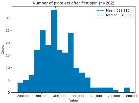
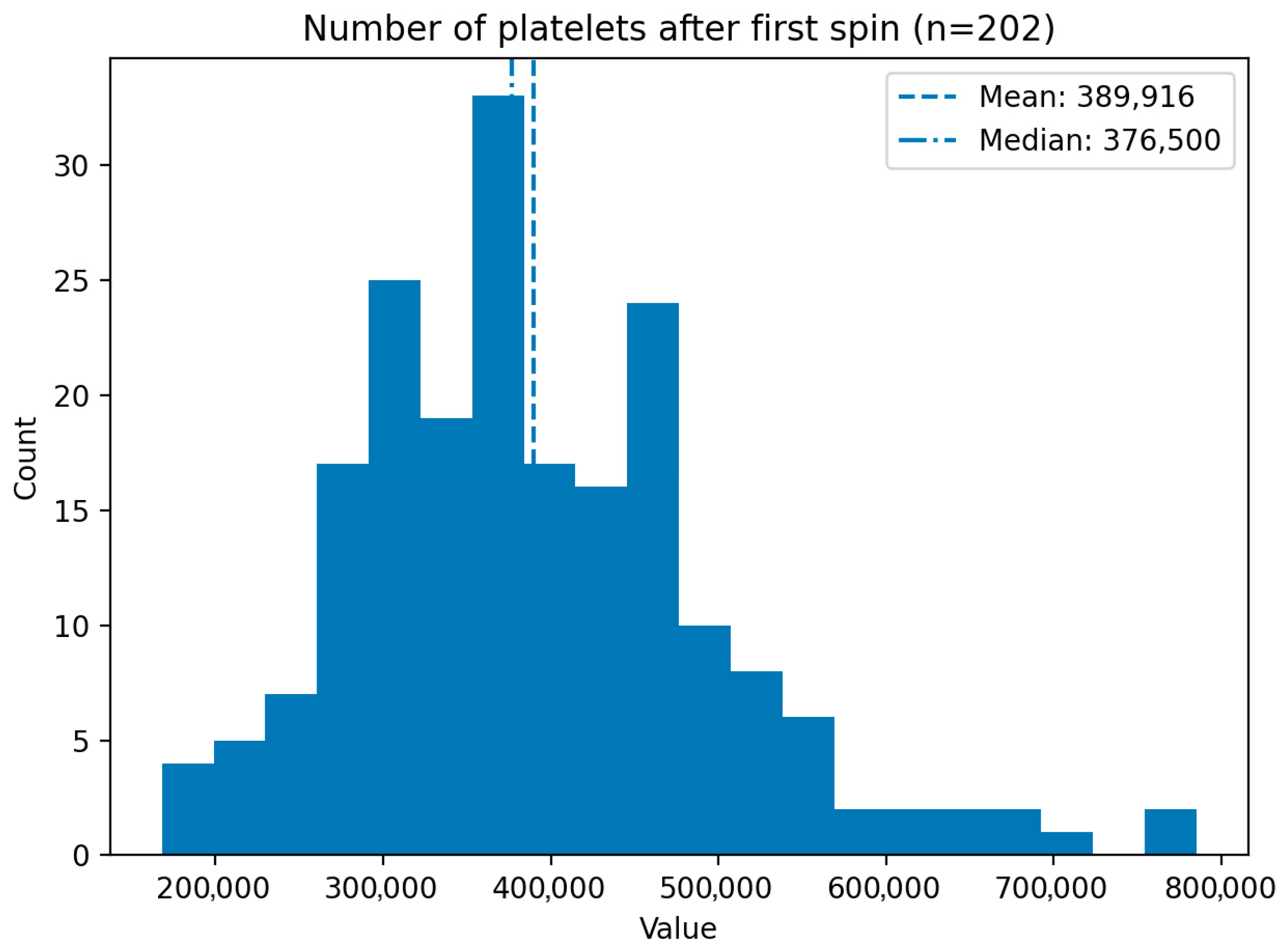
Over the past year, samples of 202 patients were processed and the number of platelets after the first spins averaged 389,916 (SD 108,729) and had a median of 376,500. The interquartile range was 314,500–456,750, with values spanning 168,000–785,000. Mean 95% CI was 374,831–405,001 and the distribution was mildly right-skewed. Four high-end outliers (>670,125) occurred (Figure 2).
Figure 2.
Histogram showing the number of platelets after the first centrifuge spins. Sample of 202 patients. Count—number of observations; Value—number of platelets.
4. Discussion
This PRISMA-guided methodological review shows that the principal limitation to reproducibility in orthopedic PRP studies is not only variation in preparation workflows, but also incomplete reporting of the determinants of PRP identity (platelet dose, cellular composition, activation/lysis status, and handling conditions). In our synthesis, information on whole blood draw volume and anticoagulant type were omitted in a sizeable proportion of studies, and downstream variables—particularly storage conditions and time-to-use—were among the least frequently described. These omissions substantially constrain the interpretability of the orthopedic PRP evidence base because biologically distinct products may be grouped under the same term ‘PRP’ [1,2,3,40].
Although the included literature comprises predominantly comparative clinical designs (including randomized controlled trials), we did not undertake a formal risk-of-bias appraisal (e.g., RoB 2/ROBINS-I) because our objective was not to estimate treatment effects but to evaluate reporting completeness for PRP manufacture and handling. Nevertheless, for complex blood-derived interventions, under-reporting of product-defining variables represents a distinct methodological threat to validity. When platelet dose, leucocyte phenotype (LP vs. LR), activation/lysis status and handling conditions are not defined, participants may receive biologically non-equivalent interventions despite identical labelling as ‘PRP’. This introduces intervention misclassification, weakening causal inference and reducing both the internal interpretability and external reproducibility of clinical conclusions [1,2,3,40].
4.1. Platelet Dose and Upstream Determinants
From a manufacturing perspective, baseline hematological status and capture efficiency are primary determinants of the delivered platelet dose. Yet orthopedic studies frequently do not report baseline platelet count or do not specify whether patients with thrombocytopenia, platelet dysfunction or relevant drug exposure were excluded. This is problematic because platelet yield—and therefore growth factor payload—is bounded by the patient’s starting platelet concentration, and pre-analytical factors such as acetylsalicylic acid and NSAID exposure can modulate platelet responsiveness and mediator release, introducing avoidable biological noise. Accordingly, our proposed protocol mandates a recent pre-collection full blood count and defined medication washout windows (e.g., acetylsalicylic acid within 10 days; NSAIDs within 5 days), together with exclusion of active infection and other high-risk conditions. These requirements align with orthobiologic reporting guidance that emphasizes baseline blood parameters as the minimum information necessary to interpret biologic interventions [1,2,3]. A recurring limitation across trials is that platelet enrichment is reported using heterogeneous metrics (fold increase, platelet ratio, absolute concentration, or not at all), and the concept of delivered platelet dose (concentration × injected volume) is rarely addressed. Examples of good practice show why these factors matters. In an orthopedic randomized trial of transforaminal epidural PRP for lumbar disc herniation, Wongjarupong et al. prepared PRP from 24 mL venous blood using a standardized double-spin protocol and reported a mean platelet ratio of 2.86 ± 0.85, enabling at least partial inference of enrichment [6]. By contrast, in knee osteoarthritis, Di Martino et al. reported absolute platelet and leukocyte concentrations for both LR-PRP and leukodepleted LP-PRP, facilitating clearer interpretation of dose and phenotype [7]. Similarly, methodological validation work can provide benchmarks for capture efficiency: Muthu et al. reported 86–99% platelet recovery and a 6.4 ± 0.8-fold increase using a first centrifugation at 100× g for 15 min followed by 1600× g for 20 min, reinforcing the importance of reporting centrifugation parameters in ×g rather than rpm [10].
Because centrifugation parameters are central determinants of PRP phenotype, they should be reported in a comparable form. When rpm alone is reported, translation to RCF requires rotor radius and device geometry; without this, apparently similar “double-spin” procedures may produce different platelet and leucocyte yields. These considerations directly support a minimum dataset requiring, for each spin, RCF (×g), time, rotor information, and temperature/holding conditions [1,2,3,40].
4.2. Collection of Samples and Traceability
The collection step is often treated as a trivial precursor to centrifugation; however, it is a major source of variability. Whole blood volume, venepuncture technique, tube or syringe type, and mixing quality influence platelet activation and recovery, while the collection system determines traceability and contamination risk. In our review, whole blood draw volume was explicitly reported in only a subset of articles focused on orthopedic field, limiting comparison of platelet dose between trials. We recommend that future orthopedic PRP work reports (as mandatory items) the volume collected, the collection system (kit/manufacturer and whether it is a closed circuit), lot numbers for disposables, and the blood-to-anticoagulant ratio. These parameters are not only essential for reproducibility, but also for traceability and hemovigilance within regulated clinical environments [1,2,3,40,41]. Articles in the literature show the consequences of inconsistent reporting even for basic upstream variables. PRP was obtained from 24 mL of venous blood in a lumbar disc herniation RCT, whereas in a rotator cuff RCT at least 30 mL of peripheral blood was collected into a pre-loaded syringe with acid citrate dextrose (ACD) solution before double-spin processing [6,8]. Without consistent reporting of volume processed, anticoagulant ratio and enrichment/dose metrics, it becomes difficult to interpret whether apparent outcome differences reflect biological dose, phenotype, indication, or procedural context.
4.3. Anticoagulant and Device Effects
Commercial kits may improve standardization through fixed geometry, controlled transfer steps, and partially closed handling, whereas manual methods can be flexible but are more operator-dependent and may increase the risk of cellular contamination or inadvertent buffy coat aspiration. The choice of anticoagulants is similarly consequential; citrate-based anticoagulants (including ACD-A) are widely used in PRP manufacture because they chelate calcium and reduce premature platelet activation during processing. Experimental and transfusion-derived data suggest ACD-A better preserves platelet function compared with some alternative anticoagulants, and regenerative medicine studies have reported anticoagulant-dependent differences in PRP potency. Consequently, our protocol specifies ACD-A with explicit reporting of manufacturers and ratio [14,15,16,17,18,19,20,21,22,23,24,25,26,27,28,29,30,31,32,33,34,35,36,37,38,39,40,41,42]. Device- and consumable-specific steps can directly determine the final cellular phenotype. Di Martino et al. generated LP-PRP using a leukodepletion filter and compared it with LR-PRP in knee osteoarthritis; in this context, the filter is not merely an accessory detail but a defining element of the intervention [7]. Such examples highlight why future reports should describe not only that a “kit” was used, but which kit, how it separates fractions, and whether it includes leukodepletion or other phenotype-defining mechanisms [40,41].
4.4. PRP Phenotype: Leucocyte/Lymphocyte Handling
A persistent source of heterogeneity in orthopedic PRP is the leukocyte fraction. Leucocyte-rich (LR) preparations typically contain higher concentrations of pro-inflammatory cytokines, which may be advantageous in selected indications but may also increase post-injection pain or swelling and potentially promote catabolic signaling within cartilage. Conversely, leucocyte-poor (LP) PRP is commonly advocated for intra-articular indications such as knee osteoarthritis. In our dataset, leukocyte information was more frequently identifiable than many other manufacturing variables; however, explicit reporting of how leucocytes were included or excluded (buffy coat capture vs. avoidance, kit setting, and manual aspiration strategy) was inconsistent. Because lymphocytes are a major cellular component of the buffy coat, failure to state whether the buffy coat was intentionally included, partially included, or excluded impairs biological interpretation. Our proposed protocol is explicitly leucocyte/lymphocyte-depleted; in addition to selecting a kit configuration designed to minimize white cell carryover, lymphocytes are excluded manually by avoiding buffy coat aspiration during plasma transfer. We recommend that future studies report quantitative leukocyte differentials where possible, or at minimum state whether a buffy coat fraction was included and how LP/LR status was achieved [1,2,4,5,7,40,43]. Recent orthopedic studies illustrate both good practice and persisting gaps. Di Martino et al. quantified leukocyte concentrations (including near-complete leukodepletion in the LP-PRP arm), and Jayaram et al. reported white blood cell subtype counts when preparing paired LR-PRP and LP-PRP from the same donors. Conversely, other studies describe PRP formulations qualitatively without reporting absolute leukocyte counts or centrifugation parameters in the abstract, limiting reproducibility [7,9,44,45].
4.5. Activation/Lysis Status and Platelet Rupture Strategy
PRP can be administered as an intact platelet suspension (with in vivo activation) or deliberately activated/lysed ex vivo to release platelet α-granule contents before administration. Exogenous activation using calcium salts or thrombin can induce immediate degranulation but may also promote gelification and fibrin matrix formation, which is not desired in some orthopedic applications. An alternative is controlled platelet rupture via freeze–thaw cycling, producing a PRP lysate with high immediate growth factor availability without gasification. Across orthopedic studies, explicit reporting of an activation agent or lysis strategy was limited, making it unclear whether trials compared intact PRP or functionally lysed products. In other medical applications, systematic syntheses of PRP quality control and laboratory optimization studies have emphasized that activation/lysis status should be considered a defining characteristic of the product rather than an optional procedural detail. A recent cross-specialty systematic review by Rahman and colleagues [40] highlighted pervasive heterogeneity in PRP characterization and reporting, reinforcing the need for minimum manufacturing and quality-control descriptors even when clinical outcomes are not the primary endpoint [1,2]. Regarding the rotator cuff, an RCT by Liu et al., provides a notable example of reproducible reporting. PRP was activated with calcium (without thrombin), and centrifugation forces (1889× g for 4 min and 2009× g for 3 min) and calcium gluconate dose (0.3 mL of 10% solution) were documented in a figure caption [8]. This level of detail is uncommon and should be encouraged, particularly where activation is used.
4.6. Handling Conditions After Preparation: Temperature, Storage/Freezing and Time-to-Use
Temperature is a critical, but rarely reported, pre-analytical variable. Platelet activation and mediator release are sensitive to processing, storage temperature and excursions during centrifugation, transfers and interim holding could influence the activation status of the final product. Despite plausibility as a major confounder, few studies focused on use of PRP in the orthopedic field report environmental temperature or centrifuge temperature control. We therefore propose routine monitoring and documentation of ambient processing temperature (recommended as 18–25 °C) using calibrated data loggers, and reporting of any controlled-temperature steps [3,40,41,46].
Freezing may be used for two distinct purposes: (i) logistical biobanking (delayed administration) and (ii) intentional platelet lysis to increase growth factor release. When freezing is undertaken, reporting is often incomplete with respect to temperature, duration, freeze–thaw cycles, and post-thaw handling. “Freezing: yes/no” is therefore insufficient; studies should specify temperature, duration, container type, number of freeze–thaw cycles, thawing conditions and whether platelet remnants were removed [15,16]. In our proposed workflow, freezing is integrated both as a storage strategy and, where intended, as a controlled lysis step. Storage is validated in our setting for up to 12 months at −40 °C, with traceability and temperature monitoring and with explicit documentation of freeze–thaw exposure [15,16].
Time-to-use is similarly central to PRP identity, because it determines the extent of spontaneous platelet activation, mediator release, and potential microbial risk. Yet the time between blood draw, PRP manufacture and administration is often not reported in orthopedic trials. For fresh products, centers should define and report a maximum hold time at room temperature and holding conditions; for cryopreserved products, reports should include the interval from thaw to administration and any post-thaw processing (e.g., filtration). In our protocol, cryopreserved material may be used within the validated one-year storage window, with documentation of thaw timing and administration details [15,16]. The rotator cuff trial also illustrates clinically relevant temporal heterogeneity—PRP was applied intraoperatively and a booster injection administered at 2 weeks post-operation, implying a need for staged preparation or reproducible batch manufacture [8].
4.7. Prevention of Bacterial Contamination and Sterility Assurance
Although PRP is autologous, bacterial contamination remains a clinically relevant risk because it may occur at multiple points in the workflow—during venepuncture, transfer/fractionation, centrifugation, and any post-processing handling or storage. Recent analyses of PRP workflows emphasise that contamination risks are identifiable both at blood collection and during PRP production, and that risk mitigation requires facility-specific measures, particularly optimised skin disinfection and controlled handling during preparation [47]. Prevention of bacterial growth relies on strict adherence to aseptic technique, minimising opportunities for environmental exposure, and robust quality systems [48,49].
In practice, infection prevention should be treated as a core manufacturing and reporting domain: defer patients with suspected infection; apply validated skin antisepsis with adequate contact/drying time; preferentially use closed, single-use systems; perform any unavoidable open steps under a Class II laminar airflow cabinet with trained personnel and documented aseptic technique; and minimise manipulation while defining and reporting a maximum room-temperature hold time [47]. Where cryopreservation is used, freezing may limit bacterial proliferation but does not sterilise the product; therefore, centres should ensure traceability and specify their sterility/quality-control approach (environmental controls and any sterility testing strategy where required) as part of the minimum reporting dataset [50].
4.8. Our Experience: A Standardised Protocol and Minimum Reporting Dataset
Taken together, these findings support the hypothesis that methodological standardization in the preparation of PRP in orthopedic applications should prioritize: (i) definition and reporting of whole blood collected and processed and the delivered platelet dose (or at minimum baseline and final platelet metrics with injected volume); (ii) explicit leucocyte/lymphocyte handling and phenotype definition (LP vs. LR and how achieved); (iii) clear specification of activation/lysis status; and (iv) controlled handling conditions, including temperature, storage/freezing and time-to-use. Adoption of a minimum manufacturing and reporting dataset consistent with orthopedic biologic reporting guidance would improve reproducibility, facilitate meaningful inter-study comparison and strengthen translational interpretation of PRP in orthopedics [3,15,40,46].
Other variables, in our opinion, which could be considered and reported in order to standardize the production process, include venepuncture approach; anticoagulant type and ratio; kit/centrifuge model and rotor; and g-force, time and temperature for each spin.
4.9. General and Particular Restrictions
Our protocol complies with the most recent Italian regulations governing blood components for non-transfusion use, as published in the “Gazzetta ufficiale” of the Italian Republic on 26 September 2019 [13]. These updates respond to the expanding clinical use of blood-derived products in procedures such as joint injections and soft tissue therapies, and aim to mitigate the risk of administering PRP and other autologous products processed outside hospital transfusion laboratories, thereby ensuring quality, traceability, and safety.
In Italy, the primary framework is the Ministerial Decree (Ministero della Salute) of 2 November 2015, “Disposizioni relative ai requisiti di qualità e sicurezza del sangue e degli emocomponenti”, which harmonised national requirements for donor selection, collection, processing, testing, storage, and distribution. The Decree of 1 August 2019 amends the 2015 text (notably Article 20) to provide a more specific definition of blood components for non-transfusion use—including allogeneic and autologous products—and to delineate permitted applications (e.g., topical application, intra-tissue or intra-articular infiltration, use in surgical sites, and regulated in vitro procedures), while explicitly excluding advanced cellular therapies such as hematopoietic stem cells used under experimental or advanced therapy protocols [13].
This approach is aligned with international trends toward stricter oversight of non-transfusion blood-derived products: EU hemovigilance legislation requires donor-to-recipient traceability, World Health Organization guidance links traceability to effective quality systems, and the European Directorate for the Quality of Medicines and HealthCare Blood Guide has introduced standards for components “for topical use or injection”. In addition, the European Commission has indicated that the Substances of Human Origin (SoHO) Regulation will apply from 7 August 2027, further reinforcing the need for hospital-grade processing and documentation [50,51,52,53].
This review has limitations inherent to its PRISMA-reported, methodological design; intermediate screening counts were not fully logged, and no formal risk-of-bias tool was applied. Reporting completeness may be underestimated when methods were described only in supplements or figures. Furthermore, our search strategy prioritized preparation terminology and orthopedic indications, so relevant studies using atypical nomenclature may have been missed, and heterogeneity in PRP classification limits cross-study comparability.
Furthermore, our protocol proposal cannot provide definitive evidence for the in vitro or clinical effectiveness of the PRP produced with the indicated methodology. In addition, implementation within a national regulatory framework—albeit broadly consistent with European principles—may affect reproducibility across settings.
Clinical outcome data are not reported because they are still being collected and analyzed; they will be addressed in future studies. On the basis of an annual caseload of approximately 200 treated patients, preliminary observations suggest satisfactory platelet yields—reported as encouraging relative to the literature—together with improvement in symptom regression and quality of life, allowing many patients to resume activities after a single treatment course.
Our proposal of a definite, common, straightforward, and replicable protocol is, in our opinion, an essential step toward improving the quality and comparability of future studies in this area.
5. Conclusions
Orthopedic PRP studies frequently lack sufficient methodological detail regarding PRP manufacture to support reproducibility and inter-study comparability. In a field where the preparation itself is intrinsically variable, failure to report the delivered platelet dose, cellular composition (including leucocyte/lymphocyte content), processing conditions, and activation/lysis and storage strategies undermine the interpretability and generalizability of clinical outcomes. We therefore propose a pragmatic, standardized protocol for preparation of leucocyte-poor PRP for orthopedic indications, alongside a minimum reporting dataset encompassing collection, anticoagulation, kit/centrifugation parameters, temperature control, baseline and final platelet metrics, lymphocyte exclusion strategy, platelet rupture/lysis, storage/freezing conditions, and time-to-use. Adoption of these standards would enable higher-quality evidence generation and safer, more consistent translation of PRP into orthobiologic practice.
Future prospective, clinical studies should implement these reporting standards to permit robust dose–response analyses and reliable comparison of PRP phenotypes across indications.
Supplementary Materials
The following supporting information can be downloaded at: https://www.mdpi.com/article/10.3390/labmed3010008/s1, PRISMA 2020 checklist [11].
Author Contributions
Conceptualization, M.B., G.C. and M.T.; methodology, M.B., G.C. and M.T.; validation, M.B., G.C., I.C., D.D.B. and R.P.; formal analysis, M.B., G.C., M.T. and D.D.B.; investigation, G.C., M.T., D.D.B. and M.B.; resources, M.T., I.C. and D.D.B.; data curation, M.B. and G.C.; writing—original draft preparation, G.C. and M.B.; writing—review and editing, M.B., G.C. and M.T.; visualization, M.B., G.C., A.G. and R.P.; supervision, I.C., R.P. and A.G.; project administration, I.C., R.P. and A.G. All authors have read and agreed to the published version of the manuscript.
Funding
This research received no external funding.
Institutional Review Board Statement
Ethical review and approval were waived for this study, according to the policy of our institution, because the original data reported in this manuscript derives from a retrospective analysis of routinely collected laboratory quality-control data generated during standard PRP production and administration. The dataset was fully de-identified prior to analysis, no additional procedures were performed for research purposes, and the study posed no risk to participants. We did not require formal ethical committee approval because informed consent was not obtained. The study was conducted in accordance with the Declaration of Helsinki.
Informed Consent Statement
Informed consent was obtained from all subjects involved in the study for the use of data relating to the procedure performed for research purposes or publication in an anonymous manner. For the review of the literature, no innovative data was collected, no new databases were produced, no data analysis was carried out that was not already present and published in the literature. Informed consent was not required.
Data Availability Statement
The raw data supporting the conclusions of this article will be made available by the authors on request.
Acknowledgments
During the preparation of this manuscript/study, the authors used OpenAI, ChatGPT ver. 5.2 for the purposes of modifying the layout of Figure 2. The authors would like to thank Agnese Pruni for her interest and external participation. The authors have reviewed and edited the output and take full responsibility for the content of this publication.
Conflicts of Interest
The authors declare no conflicts of interest.
References
- DeLong, J.M.; Russell, R.P.; Mazzocca, A.D. Platelet-rich plasma: The PAW classification system. Arthroscopy 2012, 28, 998–1009. [Google Scholar] [CrossRef] [Scilit]
- Magalon, J.; Chateau, A.L.; Bertrand, B.; Louis, M.L.; Silvestre, A.; Giraudo, L.; Veran, J.; Sabatier, F. DEPA classification: A proposal for standardising PRP use and a retrospective application of available devices. BMJ Open Sport Exerc. Med. 2016, 2, e000060. [Google Scholar] [CrossRef] [Scilit]
- Murray, I.R.; Geeslin, A.G.; Goudie, E.B.; Petrigliano, F.A.; LaPrade, R.F. Minimum Information for Studies Evaluating Biologics in Orthopaedics (MIBO): Platelet-Rich Plasma and Mesenchymal Stem Cells. J. Bone Jt. Surg. Am. 2017, 99, 809–819. [Google Scholar] [CrossRef] [Scilit]
- Laver, L.; Filardo, G.; Sanchez, M.; Magalon, J.; Tischer, T.; Abat, F.; Bastos, R.; Cugat, R.; Iosifidis, M.; Kocaoglu, B.; et al. The use of injectable orthobiologics for knee osteoarthritis: A European ESSKA-ORBIT consensus. Part 1: Blood-derived products (platelet-rich plasma). Knee Surg. Sports Traumatol. Arthrosc. 2024, 32, 783–797. [Google Scholar] [CrossRef] [Scilit]
- Kon, E.; de Girolamo, L.; Laver, L.; Andriolo, L.; Andia, I.; Bastos, R.; Beaufils, P.; Biant, L.; Bøe, B.; Boffa, A.; et al. Platelet-rich plasma injections for the management of knee osteoarthritis: The ESSKA-ICRS consensus. Knee Surg. Sports Traumatol. Arthrosc. 2024, 32, 2938–2949. [Google Scholar] [CrossRef] [Scilit]
- Wongjarupong, A.; Pairuchvej, S.; Laohapornsvan, P.; Kotheeranurak, V.; Jitpakdee, K.; Yeekian, C.; Chanplakorn, P. “Platelet-Rich Plasma” Epidural Injection an Emerging Strategy in Lumbar Disc Herniation: A Randomized Controlled Trial. BMC Musculoskelet. Disord. 2023, 24, 335. [Google Scholar] [CrossRef] [Scilit]
- Di Martino, A.; Boffa, A.; Andriolo, L.; Romandini, I.; Altamura, S.A.; Cenacchi, A.; Roverini, V.; Zaffagnini, S.; Filardo, G. Leukocyte-Rich versus Leukocyte-Poor Platelet-Rich Plasma for the Treatment of Knee Osteoarthritis: A Double-Blind Randomized Trial. Am. J. Sports Med. 2022, 50, 609–617. [Google Scholar] [CrossRef] [Scilit] [PubMed]
- Liu, B.; Jeong, H.J.; Yeo, J.H.; Oh, J.H. Efficacy of Intraoperative Platelet-Rich Plasma Augmentation and Postoperative Platelet-Rich Plasma Booster Injection for Rotator Cuff Healing: A Randomized Controlled Clinical Trial. Orthop. J. Sports Med. 2021, 9, 23259671211006100. [Google Scholar] [CrossRef] [Scilit]
- Jayaram, P.; Mitchell, P.J.T.; Shybut, T.B.; Moseley, B.J.; Lee, B. Leukocyte-Rich Platelet-Rich Plasma Is Predominantly Anti-inflammatory Compared with Leukocyte-Poor Platelet-Rich Plasma in Patients with Mild-Moderate Knee Osteoarthritis: A Prospective, Descriptive Laboratory Study. Am. J. Sports Med. 2023, 51, 2133–2140. [Google Scholar] [CrossRef] [Scilit] [PubMed]
- Muthu, S.; Krishnan, A.; Ramanathan, K.R. Standardization and Validation of a Conventional High Yield Platelet-Rich Plasma Preparation Protocol. Ann. Med. Surg. 2022, 82, 104593. [Google Scholar] [CrossRef] [Scilit] [PubMed]
- Page, M.J.; McKenzie, J.E.; Bossuyt, P.M.; Boutron, I.; Hoffmann, T.C.; Mulrow, C.D.; Shamseer, L.; Tetzlaff, J.M.; Akl, E.A.; Brennan, S.E.; et al. The PRISMA 2020 statement: An updated guideline for reporting systematic reviews. BMJ 2021, 372, n71. [Google Scholar] [CrossRef] [Scilit] [PubMed]
- Tricco, A.C.; Lillie, E.; Zarin, W.; O’Brien, K.K.; Colquhoun, H.; Levac, D.; Moher, D.; Peters, M.D.J.; Horsley, T.; Weeks, L.; et al. PRISMA Extension for Scoping Reviews (PRISMA-ScR): Checklist and Explanation. Ann. Intern. Med. 2018, 169, 467–473. [Google Scholar] [CrossRef] [Scilit] [PubMed]
- Latest Regolations About Blood Components Intended for Non-Transfusion Use Within Italy. GU-Serie-Generale-n.-226-del-26-09-2019-Ministero-della-Salute-Decreto-1-Agosto-2019. 2019. Available online: https://www.gazzettaufficiale.it/eli/id/2019/09/26/19A05957/SG (accessed on 4 March 2026).
- Oneto, P.; Zubiry, P.R.; Schattner, M.; Etulain, J. Anticoagulants Interfere with the Angiogenic and Regenerative Responses Mediated by Platelets. Front. Bioeng. Biotechnol. 2020, 8, 223. [Google Scholar] [CrossRef] [Scilit]
- Strandberg, G.; Sellberg, F.; Sommar, P.; Ronaghi, M.; Lubenow, N.; Knutson, F.; Berglund, D. Standardizing the freeze-thaw preparation of growth factors from platelet lysate. Transfusion 2017, 57, 1058–1065. [Google Scholar] [CrossRef] [Scilit]
- Etulain, J.; Mena, H.A.; Meiss, R.P.; Frechtel, G.; Gorzalczany, S.; Schattner, M. An optimised protocol for platelet-rich plasma preparation to improve its angiogenic and regenerative properties. Sci. Rep. 2018, 8, 1513. [Google Scholar] [CrossRef] [Scilit] [PubMed]
- Dulic, O.; Rasovic, P.; Lalic, I.; Kecojevic, V.; Gavrilovic, G.; Abazovic, D.; Maric, D.; Miskulin, M.; Bumbasirevic, M. Bone Marrow Aspirate Concentrate versus Platelet Rich Plasma Hyaluronic Acid for the Treatment of Knee Osteoarthritis. Medicina 2021, 57, 1193. [Google Scholar] [CrossRef] [Scilit]
- Dubin, J.; Leucht, P.; Murray, M.; Pezold, R.; Staff of the American Academy of Orthopaedic Surgeons on Behalf of the Platelet-Rich Plasma (PRP) for Knee Osteoarthritis Technology Overview Workgroup and Contributors. American Academy of Orthopaedic Surgeons Technology Overview Summary: Platelet-Rich Plasma (PRP) for Knee Osteoarthritis. J. Am. Acad. Orthop. Surg. 2024, 32, 296–301. [Google Scholar] [CrossRef] [Scilit]
- Breton, A.; Leplat, C.; Picot, M.; Aouinti, S.; Taourel, P.; Laffont, I.; Julia, M.; Cyteval, C. Prediction of clinical response to corticosteroid or platelet-rich plasma injection in plantar fasciitis with MRI: A prospective, randomized, double-blinded study. Diagn. Interv. Imaging 2022, 103, 217–224. [Google Scholar] [CrossRef] [Scilit] [PubMed]
- Nouri, F.; Babaee, M.; Peydayesh, P.; Esmaily, H.; Raeissadat, S.A. Comparison between the effects of ultrasound guided intra-articular injections of platelet-rich plasma (PRP), high molecular weight hyaluronic acid, and their combination in hip osteoarthritis: A randomized clinical trial. BMC Musculoskelet. Disord. 2022, 23, 856. [Google Scholar] [CrossRef] [Scilit]
- Huang, H.Y.; Hsu, C.W.; Lin, G.C.; Lin, H.S.; Chou, Y.J.; Liou, I.H.; Sun, S.F. Comparing efficacy of a single intraarticular injection of platelet-rich plasma (PRP) combined with different hyaluronans for knee osteoarthritis: A randomized-controlled clinical trial. BMC Musculoskelet. Disord. 2022, 23, 954. [Google Scholar] [CrossRef] [Scilit]
- Dong, C.; Zhao, C.; Wang, F. Clinical benefit of high tibial osteotomy combined with the intervention of platelet-rich plasma for severe knee osteoarthritis. J. Orthop. Surg. Res. 2022, 17, 405. [Google Scholar] [CrossRef] [Scilit]
- Zielinski, M.A.; Evans, N.E.; Bae, H.; Kamrava, E.; Calodney, A.; Remley, K.; Benyamin, R.; Franc, D.; Peterson, M.R.; Lovine, J.; et al. Safety and Efficacy of Platelet Rich Plasma for Treatment of Lumbar Discogenic Pain: A Prospective, Multicenter, Randomized, Double-blind Study. Pain Physician 2022, 25, 29–34. [Google Scholar]
- Keene, D.J.; Alsousou, J.; Harrison, P.; O’Connor, H.M.; Wagland, S.; Dutton, S.J.; Hulley, P.; Lamb, S.E.; Willett, K.; PATH-2 Trial group. Platelet-rich plasma injection for acute Achilles tendon rupture. Two-year follow-up of the PATH-2 randomized, placebo-controlled, superiority trial. Bone Jt. J. 2022, 104-B, 1256–1265. [Google Scholar] [CrossRef] [Scilit]
- Duan, W.; Su, X.; Yu, Z.; Jiang, M.; Zhao, L.; Giannoudis, P.V.; Guo, J.J. No Benefit to Platelet-rich Plasma Over Placebo Injections in Terms of Pain or Function in Patients with Hemophilic Knee Arthritis: A Randomized Trial. Clin. Orthop. Relat. Res. 2022, 480, 2361–2370. [Google Scholar] [CrossRef] [Scilit]
- Won, S.J.; Kim, D.Y.; Kim, J.M. Effect of platelet-rich plasma injections for chronic nonspecific low back pain. Medicine 2022, 101, e28935. [Google Scholar] [CrossRef] [Scilit] [PubMed]
- Wang, Y.C.; Lee, C.L.; Chen, Y.J.; Tien, Y.C.; Lin, S.Y.; Chen, C.H.; Chou, P.P.; Huang, H.T. Comparing the Efficacy of Intra-Articular Single Platelet-Rich Plasma(PRP) versus Novel Crosslinked Hyaluronic Acid for Early-Stage Knee Osteoarthritis: A Prospective, Double-Blind, Randomized Controlled Trial. Medicina 2022, 58, 1028. [Google Scholar] [CrossRef] [Scilit] [PubMed]
- Agarwal, V.; Gupta, A.; Singh, H.; Kamboj, M.; Popli, H.; Saroha, S. Comparative Efficacy of Platelet-Rich Plasma and Dry Needling for Management of Trigger Points in Masseter Muscle in Myofascial Pain Syndrome Patients: A Randomized Controlled Trial. J. Oral Facial Pain Headache 2022, 36, 253–262. [Google Scholar] [CrossRef] [Scilit]
- Kotb, S.Y.; Sherif, N.M.; Saleh, H.A.; Ahmed, S.F.; Sakr, H.M.; Taeimah, M.O. The role of intra-articular injection of autologous platelet-rich plasma versus corticosteroids in the treatment of synovitis in lumbar facet joint disease. Saudi Med. J. 2022, 43, 1200–1208. [Google Scholar] [CrossRef] [Scilit] [PubMed]
- Paget, L.D.A.; Reurink, G.; de Vos, R.J.; Weir, A.; Moen, M.H.; Bierma-Zeinstra, S.M.A.; Stufkens, S.A.S.; Goedegebuure, S.; Krips, R.; Maas, M.; et al. Platelet-Rich Plasma Injections for the Treatment of Ankle Osteoarthritis. Am. J. Sports Med. 2023, 51, 2625–2634. [Google Scholar] [CrossRef] [Scilit]
- Gobbi, A.; Dallo, I.; D’ambrosi, R. Autologous microfragmented adipose tissue and leukocyte-poor platelet-rich plasma combined with hyaluronic acid show comparable clinical outcomes for symptomatic early knee osteoarthritis over a two-year follow-up period: A prospective randomized clinical trial. Eur. J. Orthop. Surg. Traumatol. 2023, 33, 1895. [Google Scholar] [CrossRef] [Scilit]
- Zhou, Y.; Li, H.; Cao, S.; Han, Y.; Shao, J.; Fu, Q.; Wang, B.; Wu, J.; Xiang, D.; Liu, Z.; et al. Clinical efficacy of intra-articular injection with P-PRP and L-PRP in treating knee cartilage lesion: A randomized controlled trial. Orthop. Surg. 2023, 15, 740–749. [Google Scholar] [CrossRef] [Scilit]
- Tschopp, M.; Pfirrmann, C.W.A.; Fucentese, S.F.; Brunner, F.; Catanzaro, S.; Kühne, N.; Zwyssig, I.; Sutter, R.; Götschi, T.; Tanadini, M.; et al. A Randomized Trial of Intra-articular Injection Therapy for Knee Osteoarthritis. Investig. Radiol. 2023, 58, 355–362. [Google Scholar] [CrossRef] [Scilit]
- Kamble, P.; Prabhu, R.M.; Jogani, A.; Mohanty, S.S.; Panchal, S.; Dakhode, S. Is Ultrasound (US)-Guided Platelet-Rich Plasma Injection More Efficacious as a Treatment Modality for Lateral Elbow Tendinopathy Than US-Guided Steroid Injection?: A Prospective Triple-Blinded Study with Midterm Follow-up. Clin. Orthop. Surg. 2023, 15, 454–462. [Google Scholar] [CrossRef] [Scilit]
- Kirschner, J.S.; Cheng, J.; Creighton, A.; Santiago, K.; Hurwitz, N.; Dundas, M.; Beatty, N.; Kingsbury, D.; Konin, G.; Abutalib, Z.; et al. Efficacy of ultrasound-guided glenohumeral joint injections of leukocyte-poor platelet-rich plasma versus hyaluronic acid in the treatment of glenohumeral osteoarthritis: A randomized, double-blind controlled trial. Clin. J. Sport Med. 2022, 32, 558–566. [Google Scholar] [CrossRef] [Scilit]
- Sharma, R.; Chaudhary, N.K.; Karki, M.; Sunuwar, D.R.; Singh, D.R.; Pradhan, P.M.S.; Gyawali, P.; Duwal Shrestha, S.K.; Bhandari, K.K. Effect of platelet-rich plasma versus steroid injection in plantar fasciitis: A randomized clinical trial. BMC Musculoskelet. Disord. 2023, 24, 172. [Google Scholar] [CrossRef] [Scilit]
- Zhuang, W.; Li, T.; Li, Y.; Zhang, Y.; Gao, J.; Wang, X.; Ding, Q.; Li, W. The varying clinical effectiveness of single, three and five intraarticular injections of platelet-rich plasma in knee osteoarthritis. J. Orthop. Surg. Res. 2024, 19, 284. [Google Scholar] [CrossRef] [Scilit] [PubMed]
- Navani, A.; Ambach, M.; Calodney, A.; Rosenthal, R.; Li, G.; Mahoney, C.B.; Everts, P.A. The Safety and Effectiveness of Orthobiologic Injections for Discogenic Chronic Low Back Pain: A Multicenter Prospective, Crossover, Randomized Controlled Trial with 12 Months Follow-up. Pain Physician 2024, 27, E65–E77. [Google Scholar] [CrossRef] [Scilit] [PubMed]
- Ye, Z.; Chen, H.; Qiao, Y.; Wu, C.; Cho, E.; Wu, X.; Li, Z.; Wu, J.; Lu, S.; Xie, G.; et al. Intra-Articular Platelet-Rich Plasma Injection After Anterior Cruciate Ligament Reconstruction: A Randomized Clinical Trial. JAMA Netw. Open 2024, 7, e2410134. [Google Scholar] [CrossRef] [Scilit] [PubMed] [PubMed Central]
- Rahman, E.; Rao, P.; Abu-Farsakh, H.N.; Thonse, C.; Ali, I.; Upton, A.E.; Baratikkae, S.Y.; Carruthers, J.D.A.; Mosahebi, A.; Heidari, N.; et al. Systematic Review of Platelet-Rich Plasma in Medical and Surgical Specialties: Quality, Evaluation, Evidence, and Enforcement. J. Clin. Med. 2024, 13, 4571. [Google Scholar] [CrossRef] [Scilit]
- Tey, R.V.; Haldankar, P.; Joshi, V.R.; Raj, R.; Maradi, R. Variability in Platelet-Rich Plasma Preparations Used in Regenerative Medicine: A Comparative Analysis. Stem Cells Int. 2022, 2022, 3852898. [Google Scholar] [CrossRef] [Scilit] [PubMed]
- Aizawa, H.; Kawabata, H.; Sato, A.; Masuki, H.; Watanabe, T.; Tsujino, T.; Isobe, K.; Nakamura, M.; Nakata, K.; Kawase, T. A Comparative Study of the Effects of Anticoagulants on Pure Platelet-Rich Plasma Quality and Potency. Biomedicines 2020, 8, 42. [Google Scholar] [CrossRef] [Scilit]
- Dohan Ehrenfest, D.M.; Rasmusson, L.; Albrektsson, T. Classification of Platelet Concentrates: From Pure Platelet-Rich Plasma (P-PRP) to Leucocyte-and Platelet-Rich Fibrin (L-PRF). Trends Biotechnol. 2009, 27, 158–167. [Google Scholar] [CrossRef] [Scilit] [PubMed]
- Banerjee, S.; Munde, K.; Kunal, K. Mucoid Degeneration of the Anterior Cruciate Ligament: Can Platelet-Rich Plasma Combined with Reductionplasty Provide Better Results? Regen. Med. 2022, 17, 617–626. [Google Scholar] [CrossRef] [Scilit] [PubMed]
- Mishra, A.K.; Skrepnik, N.V.; Edwards, S.G.; Jones, G.L.; Sampson, S.; Vermillion, D.A.; Ramsey, M.L.; Karli, D.C.; Rettig, A.C. Efficacy of Platelet-Rich Plasma for Chronic Tennis Elbow: A Double-Blind, Prospective, Multicenter, Randomized Controlled Trial of 230 Patients. Am. J. Sports Med. 2014, 42, 463–471. [Google Scholar] [CrossRef] [Scilit] [PubMed]
- Denessen, E.J.S.; Van Den Kerkhof, D.L.; Jeurissen, M.L.J.; Wetzels, R.J.H.; Verhezen, P.W.M.; Henskens, Y.M.C. Determining the Optimal Storage Time and Temperature for Performing Platelet Function Assays and Global Hemostasis Assays. Platelets 2022, 33, 416–424. [Google Scholar] [CrossRef] [Scilit]
- Arita, A.; Saita, Y.; Fujiwara, N.; Homma, Y.; Tobita, M. Microbial contamination risks and clinical safety in platelet-rich plasma therapy and evaluation of rapid microbial detection methods. Regen. Ther. 2025, 31, 101043. [Google Scholar] [CrossRef] [Scilit]
- Szczepiorkowski, Z.M.; Pagano, M.B. Platelet components and bacterial contamination: Hospital perspective. Hematol. Am. Soc. Hematol. Educ. Program. 2022, 2022, 430–436. [Google Scholar] [CrossRef] [Scilit]
- White, S.K.; Schmidt, R.L.; Walker, B.S.; Metcalf, R.A. Bacterial contamination rate of platelet components by primary culture: A systematic review and meta-analysis. Transfusion 2020, 60, 986–996. [Google Scholar] [CrossRef] [Scilit]
- EDQM-European Directorate for the Quality of Medicines & HealthCare. Guide to the Preparation, Use and Quality Assurance of Blood Components. Available online: https://www.edqm.eu/en/blood-guide (accessed on 29 December 2025).
- European Union Guide Directive 2002/98/EC of the European Parliament of the Council of 27 January 2003. Setting Standards of Quality Safety for the Collection Testing Processing Storage Distribution of Human Blood Blood Components Amending Directive, 2001/83/EC. Available online: https://eur-lex.europa.eu/legal-content/EN/TXT/PDF/?uri=CELEX%3A32002L0098 (accessed on 29 December 2025).
- World Health Organization. Health Product Policy and Standards (HPS), External Quality Assessment of Transfusion Laboratory Practice: Guidelines on Establishing an EQA Scheme in Blood Group Serology. 2024. Available online: https://www.who.int/teams/health-product-policy-and-standards/standards-and-specifications/blood-and-products-of-human-origin/quality-and-safety/quality-systems (accessed on 29 December 2025).
- European Union. Regulation (EU) 2024/1938 of the European Parliament and of the Council of 13 June 2024 on Standards of Quality and Safety for Substances of Human Origin Intended for Human Application and Repealing Directives 2002/98/EC and 2004/23/EC (Text with EEA Relevance). Available online: https://eur-lex.europa.eu/legal-content/EN/TXT/?uri=OJ:L_202401938 (accessed on 29 December 2025).

Disclaimer/Publisher’s Note: The statements, opinions and data contained in all publications are solely those of the individual author(s) and contributor(s) and not of MDPI and/or the editor(s). MDPI and/or the editor(s) disclaim responsibility for any injury to people or property resulting from any ideas, methods, instructions or products referred to in the content. |
© 2026 by the authors. Licensee MDPI, Basel, Switzerland. This article is an open access article distributed under the terms and conditions of the Creative Commons Attribution (CC BY) license.