


Abstract
Oral cavity and maxillofacial aspergillosis can take one of four clinically different forms: primary invasive aspergillosis of the oral cavity, invasive aspergillosis of the paranasal sinuses with secondary involvement of the oral cavity, chronic granulomatous invasive fungal rhinosinusitis, and non-invasive maxillary sinus fungus ball. The basis of this narrative review was a structured search of PubMed, Scopus, Cochrane CENTRAL and ClinicalTrials.gov databases. Each of the statements will be identified according to whether it is supported by direct evidence on oral or maxillofacial conditions, or whether it is an extrapolation from invasive fungal rhinosinusitis of mixed fungal aetiology or from invasive aspergillosis occurring at other sites. Direct evidence on primary invasive oral aspergillosis is restricted to small case series from a single centre involving 12–16 patients and individual case reports. Violaceous gingival margin and spontaneous pain progressing to a necrotic ulcer should be suspicious in patients with leukaemia and neutropenia; diagnosis relies on histopathology and culture. Local control in the case series was achieved mostly via haematological remission. Pooled mortality rate of about 31% is derived from acute invasive fungal rhinosinusitis of mixed aetiology and does not apply to the disease of the mouth. Antifungal treatment strategy is based on guidelines and clinical trials of invasive aspergillosis and mucormycosis. Liposomal amphotericin B is used until the exclusion of Mucorales, and either voriconazole or isavuconazole after confirmation of Aspergillus infection, in combination with debridement whenever possible. The fungus ball is related to endodontic and other dental procedures in case–control studies and is removed endoscopically without antifungals. There are no registered clinical trials for oral or sinonasal aspergillosis.
1. Introduction
There are four types of the Aspergillus infection in the mouth and the maxillofacial region, and their respective host, natural history, prognosis and management differ (Table 1). Form 1 is the primary invasive oral aspergillosis that involves the invasion of blood vessels of the gingiva, palate, tongue or jaw. Most descriptions of this condition have been found in patients with haematological malignancy undergoing chemotherapy-induced neutropenia [1,2]. Form 2 is the invasive sinonasal aspergillosis with secondary involvement of the mouth by contiguous spread. The most common primary sites of orofacial involvement in early series included the paranasal sinuses and the nasal cavity rather than the mouth [3]. Form 3 is the chronic granulomatous invasive fungal rhinosinusitis which occurs in immunocompetent, mostly middle-aged adults as a slowly growing orbital, cheek or palatal mass. The disease is most commonly reported in Sudan, India and Saudi Arabia [4]. Form 4, maxillary sinus fungus ball, represents a non-invasive extramucosal accumulation of fungal hyphae. It is usually seen in immunocompetent subjects who are associated with dental procedures [5,6,7].
Invasive oral aspergillosis is uncommon. A 2003 review reported 23 cases of primary invasive oral aspergillosis in the English-language literature [8]. An updated literature review has been recently published [9]. The largest dedicated series included 12–16 cases each [1,2,10,11]. These four series are all from a Japanese centre and may have overlapping patients. Thus, the evidence base is limited to small case series and case-level reviews. Prognosis and management statements in the field frequently depend on cohorts of acute invasive fungal rhinosinusitis (AIFRS) of mixed fungal aetiology, where in many series Mucorales dominates [12,13,14,15], and from clinical trials of invasive aspergillosis of all sites, including lung disease [16,17,18]. These data are difficult to generalise to oral disease.
An oral practitioner has an interest in all four of these conditions. In patients with leukaemia, the marginal gingiva is the primary site for invasive oral aspergillosis [1,2,10]. Children with invasive fungal sinusitis have fever and/or oral or nasal lesions more often than adults [19]. The non-invasive form, fungus ball has been repeatedly associated with root-canal treatment and other dental procedures [5,20,21,22]. This review aims to summarise the features of diagnosis, prognosis and treatment of each type separately. A further aim is to differentiate between the statements based on direct oral/maxillofacial evidence and those based on indirect evidence.
Table 1.
The four clinical forms of oral and maxillofacial aspergillosis. Entries give the design and size of the source; figures from invasive fungal rhinosinusitis of mixed fungal aetiology are labelled as indirect.
2. Methods
2.1. Design
This is a narrative review based on structured, reproducible literature search. The manuscript was prepared with reference to the Scale for the Assessment of Narrative Review Articles (SANRA) [45].
2.2. Information Sources and Search
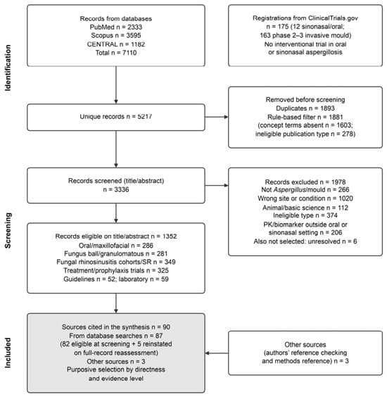
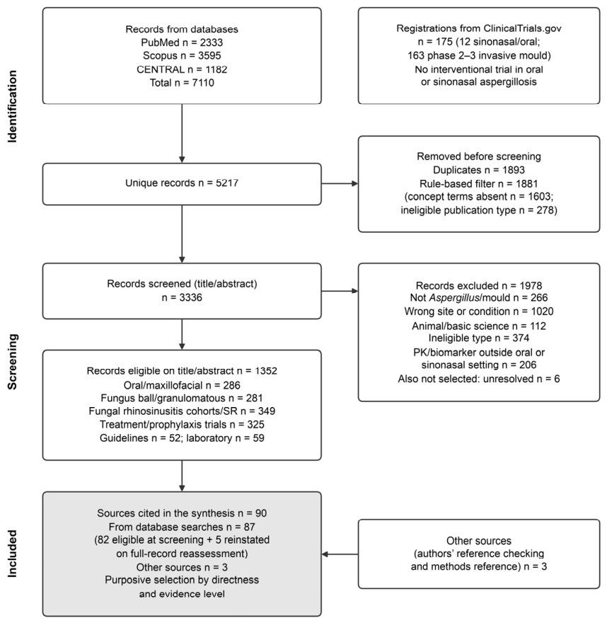
PubMed/MEDLINE and ClinicalTrials.gov were searched until July 30 2026, and Scopus and the Cochrane Central Register of Controlled Trials (CENTRAL) were searched until the same date, from inception. The PubMed search strategy consisted of three blocks. Block A combined Aspergillus, aspergillosis or fungus ball terms with oral, maxillofacial, dental and maxillary-sinus terms (direct evidence). Block B combined invasive fungal rhinosinusitis terms with prognosis and diagnosis terms. Block C combined invasive aspergillosis, invasive mould disease or mucormycosis with trial and guideline terms. The Scopus and CENTRAL strategies were translations of these blocks, and the full strings are shown in Supplementary Table S1. ClinicalTrials.gov was searched for registrations on oral, sinonasal or maxillary-sinus aspergillosis and for phase 2–3 treatment trials for invasive aspergillosis or mould disease. Searches were limited to English language articles. Additional sources identified by the authors by hand-searching references from the original submission are also included in Figure 1.
Figure 1.
Flow of records through the structured literature search (searches run on July 30, 2026). Screening was assisted by a large-language-model classifier; sources were selected purposively from the eligible records for the narrative synthesis.
2.3. Eligibility Criteria
The sources of direct evidence were human reports of each of the four forms. Preferred sources of direct evidence were systematic reviews, cohorts and case series of at least three patients, but individual case reports were used to illustrate characteristics not mentioned in series. The sources of indirect evidence were cohorts of at least 10 patients or systematic reviews of invasive fungal rhinosinusitis that reported mortality, prognostic factors or diagnostic accuracy; randomised or phase 2–3 treatment trials for invasive aspergillosis, invasive mould disease or mucormycosis; and current international guidelines and consensus definitions. Laboratory studies were considered eligible if they addressed issues relevant to the oral or sinus disease (oral isolates, dental materials). We did not consider non-English reports, conference abstracts, letters and editorials with no original data, exclusively animal studies, allergic fungal rhinosinusitis, works on mixed Aspergillus-Mucorales infections and non-Aspergillus infections.
2.4. Selection Process
Following the de-duplication stage, an eligibility filter based on a set of predetermined rules excluded any publications that did not meet the inclusion criteria concerning publication type, and any records with a title and abstract that did not contain at least one of the concept terms. Titles and abstracts were reviewed by a single reviewer assisted by a large-language-model classifier. A second classification phase was conducted on uncertain records on a quality assurance sample of 600 records; an agreement rate of 59.5% (Cohen’s κ = 0.50) was achieved regarding the exact codes and 70.0% (κ = 0.43) on include/exclude decisions, with disagreements being settled through re-adjudication. Sources were purposefully selected from the eligible records for the synthesis with regard to directness and evidence level. The record selection process is presented in Figure 1.
2.5. Appraisal and Labelling of Evidence
There was no use of risk-of-bias criteria since the direct evidence comes mostly from case series and case reports. Rather, the sources mentioned in the narrative have been classified according to the level of evidence hierarchy as per the Oxford Centre for Evidence-Based Medicine 2011 (OCEBM). Levels are denoted using letters where D-O is for direct evidence from oral or maxillofacial aspergillosis, D-S is for direct evidence from maxillary sinus fungus ball/chronic granulomatous disease, I-S is for indirect evidence from invasive fungal rhinosinusitis of mixed fungal aetiology, I-IA is for indirect evidence from invasive aspergillosis or mould disease elsewhere, principally in the lung. G is for guidelines and consensus and L is for laboratory studies. The design, sample size and population for each numerical estimate are provided in the text and tables. Supplementary Table S2 displays the design, population, directness category, evidence level and verification source of each cited reference.
3. Clinical Forms and Their Recognition
3.1. Primary Invasive Oral Aspergillosis (Form 1)
The most informative descriptions of primary invasive oral aspergillosis are those from the series of patients with acute leukaemia from a single centre, and thus they refer to a single highly selected population (direct evidence, OCEBM level 4). The earliest sign is a purple discolouration of the marginal gingiva, accompanied by intense spontaneous pain [1,10]. It progresses to a solitary ulcer covered by a grey necrotic pseudomembrane [2,10], which involves the contiguous mucosa, muscle and bone [1]. Myoken et al. proposed three clinical-pathological stages in 12 patients: an early stage (n = 3) of purple marginal gingiva with superficial fungal invasion; an advanced stage (n = 4) of grey necrotic ulcer with pseudomembrane and vascular invasion at its base; and a late stage (n = 5) with destruction of the alveolar bone and adjacent facial tissues [10]. A 12-patient series showed that in neutropenic patients, the suspicion of infection was raised by persistent fever without a source, gingival pain, facial swelling and solitary grey necrotic ulcer [2]. The diagnosis of Aspergillus stomatitis was made a median of 23 days after the admission to the hospital, while the patient was neutropenic [2].
Other locations are known only from single-case reports. In the 1985 series of 13 patients with leukaemia, black, ulcerative, escharotic lesions were due to direct tissue destruction by the fungus and vascular thrombosis [3]. Hard-palate necrosis may perforate the palate and produce an oronasal fistula. In one patient with acute lymphoblastic leukaemia, the fistula lasted for six months after the control of the infection and required surgical closure [27]. The tongue involvement has been described as an ulcerated grey lesion [8]. It has also been described as a hard and elevated lesion which enlarged to 2 × 4 cm in two days and became grey-black [46]. Alveolar involvement results in sequestration of the premolar and molar alveolar processes [28]. In two publications from the same Japanese centre, A. flavus accounted for 10 out of 12 and 10 out of 13 culture-positive Aspergillus infections [2,11]. These populations overlap, and oral infection with A. fumigatus and A. niger has also been described [47,48]. Therefore, empirical reasoning should not necessarily include A. fumigatus because of A. flavus predominance which relies on data from a single centre.
3.2. Sinonasal Aspergillosis with Oral Extension (Form 2) and Its Differential Diagnosis
Sinonasal infection that develops into invasive disease and proceeds to the mouth may appear as palatal or alveolar necrosis. Three overlapping conditions alter the management. First, mucormycosis presents as a black necrotic palatal or maxillary lesion which cannot be differentiated from aspergillosis based on its clinical features. Misdiagnosis of Mucorales as Aspergillus is the most frequent mistake in morphological identification of fungi [38]. Voriconazole has no therapeutic effect on Mucorales, so histopathological examination and culturing, not the clinical picture, determine the management [40]. Co-infection with Aspergillus and Mucorales has been described in leukaemia [47], glioma treatment, maxillofacial trauma, and post-COVID-19 with diabetes [47,49,50,51].
Second, oral aspergillosis may occur with pulmonary infection. Concomitant pulmonary aspergillosis was present in seven out of 13 patients in the 1985 orofacial series [3]. In one mechanically ventilated patient with COVID-19, A. niger from a gingival pocket swab preceded a positive bronchoalveolar lavage culture and computed tomography (CT) scan evidence of invasive pulmonary aspergillosis by day 14; strain identity was not confirmed [48]. Based on the case-series data, it is reasonable to consider a chest CT after confirmation of invasive oral aspergillosis in an immunocompromised patient. It is an expert opinion, not an evidence-based recommendation.
Third, dental infection and procedures may precede invasive sinonasal infection. In 95 consecutive biopsy-proven AIFRS cases from a single US centre, 11 (11.6%) had a dental infection within the preceding two weeks [12]. These patients were more frequently diabetic and more frequently had orbital and nasal floor invasion, but their mortality rate was not different [12]. Ten out of 11 dental infection cases were Mucorales, and only one was Aspergillus, and this observation indirectly suggests a relation with aspergillosis.
3.3. Chronic Granulomatous Invasive Fungal Rhinosinusitis (Form 3)
In a systematic review of chronic granulomatous invasive fungal sinusitis in immunocompetent patients, 88.2% had proptosis, 35.1% had intracranial involvement, and A. flavus accounted for 64% of described isolates (direct evidence, case-level review, OCEBM level 4) [4]. In an Indian cohort of 18 patients, A. flavus was isolated in 88.9% [30]. A Chinese cohort compared 11 granulomatous and 44 non-granulomatous cases of chronic invasive infection (Aspergillus in 92.7% of the total); there were no differences in treatment and prognosis [52].
3.4. Maxillary Sinus Fungus Ball (Form 4)
The fungus ball is usually asymptomatic or manifests itself as unilateral chronic sinusitis, and is incidentally discovered in imaging investigations [24,25]. In a series of 1717 paranasal sinus fungus balls, 76.6% were maxillary and 94.7% were unilateral; Aspergillus spp. accounted for 72.8% of identified cases [31]. The association with dental treatment is based on old series and case–control studies; causality cannot be established in these designs. In a 1989 French series of 85 patients, a radiopaque intrasinus foreign body was present in 94%. In 85% of them, the authors regarded the foreign body as root canal material of maxillary premolars or molars [5]. In a second series of 34 otherwise healthy patients, 33 had undergone endodontic treatment, and the authors considered the radiologic appearance to be pathognomonic in 26 (76%) [20].
In an Italian case–control study, endodontic treatment of maxillary teeth was observed in 91 out of 102 patients with a fungus ball, and in 113 out of 306 controls (crude odds ratio 14.13, 95% confidence interval [CI] 7.25–27.54); controls were drawn from a dental practice, which may have biassed the estimate [21]. A Korean case–control study with chronic rhinosinusitis controls reported a lower odds ratio of 2.15 (95% CI 1.26–3.67) [22]. In a 13-centre Korean cohort of 203 fungus balls, root canal material extending into the sinus was present in only 4.4% and dental implants in 3.9% [6]. The same study compared 141 odontogenic fungus balls with odontogenic sinusitis: purulence was less common (52.5% vs. 100%, because purulence was part of the definition of odontogenic sinusitis), and so was apical periodontitis (19.9% vs. 44.2%) [6]. Dental implants were more common in maxillary teeth of patients with fungus balls than in age-matched bacterial sinusitis controls (9/120 vs. 2/120 teeth, p = 0.031). The proportion of patients with implants did not differ significantly (5/30 vs. 1/30, p = 0.085) [53]. Case reports described fungus balls after the extrusion of amalgam [54], and in conjunction with zygomatic [55] and conventional [56] implants. In a case–control study of 13 fungus balls after sinus grafting, all had received deproteinized bovine bone (p = 0.0001) [57]. In vitro, A. fumigatus forms biofilm on all five tested sinus augmentation bone substitutes [58].
Two laboratory observations suggest, but do not prove, a mechanism. Soluble zinc oxide, a component of many root canal sealers, promotes in vitro growth of all six tested Aspergillus species, stimulating A. niger to a minor extent; root canal sealers were not tested [59]. CT densitometry classifies sinus concretions as inorganic (>1500 Hounsfield units [HU], attributed to dental materials) or organic (<1500 HU). The density of concretions in aspergillosis was 2567 ± 714 HU, versus 1160 ± 1008 HU in sinuses without aspergillosis [32]. In a second series, the density of concretions was 2868 HU, similar to root filling material [60]. Thus, dense intrasinus opacity suggests but does not confirm a fungus ball of dental origin; histology of the removed material confirms the diagnosis.
4. Diagnosis
The gold standard remains tissue biopsy with histopathology and culture. In EORTC/MSGERC definitions, proven mould infection implies the presence of hyphae causing pathological changes in the biopsy or needle aspiration, and this category is valid for any patient irrespective of immunostatus [34]. Culture from the cavity of a paranasal sinus cannot be counted as sterile site culture [34]. In the lesions of the oral cavity, hyphae of Aspergillus with transverse septa and dichotomous branching in association with thrombotic vascular infarction are described [1]. These characteristics are specific but not diagnostic because, in oral tissues, the hyphae were indistinguishable from those of other hyaline moulds [11]. Hyphae of Mucorales measure 6–16 µm and have a ribbon-like form and a paucity of septa with irregular branching; thus, their width and form provide more reliable means than septation and branching angles [38]. The cultivation of oral tissue samples usually fails [11]. The in situ hybridization with the DNA probe of Aspergillus demonstrated a strong reaction in all 13 cases of culture-confirmed aspergillosis in the oral tissue samples of 16 patients [11]. The ESCMID–ECMM–ERS guideline recommends identification up to the species-complex level [39]. The IDSA guideline suggests antifungal susceptibility testing in the cases of suspected resistance and/or treatment failure [40].
The serum galactomannan is not the substitute for biopsy. Prospective study conducted at the single centre included 17 neutropenic onco-haematological patients receiving prophylaxis of voriconazole with the suspicion of invasive aspergillosis without pulmonary involvement. The sensitivity was 0.69 (9/13) and specificity 1.0 (4/4) [33]. The galactomannan was positive in only two of the six cases of proven disease confined to the mouth (the sensitivity was stated in the text as 0.50) and in seven of the seven cases with the involvement of the nose or sinuses [33]. The meta-analysis of acute invasive Aspergillus sinusitis (five studies, 118 patients, 62 with the disease) demonstrated the pooled sensitivity of 0.63 (95% CI 0.50–0.74) and specificity of 0.65 (95% CI 0.51–0.76) [61]. Neither a positive nor a negative result must hinder biopsy. Surveillance swabs are also not informative: of 657 oral and nasal swabs from 102 allogeneic transplant recipients, seven nasal swabs and one oral swab yielded Aspergillus [62].
Indirect evidence for suspected sinonasal disease comes from AIFRS of mixed aetiology. In the one US centre, 40 patients were biopsied on suspicion of AIFRS, 26 of whom had the biopsy-confirmed disease. Facial pain, the platelet count of less than 50,000 cells/mm3 and an abnormal CT (mostly bilateral sinus opacification) were associated with a positive biopsy on bivariate analysis [26]. In the meta-analysis of nine studies, the intraoperative frozen section had a pooled sensitivity of 0.83 (95% CI 0.80–0.87) and specificity of 0.99 (95% CI 0.93–1.00) for AIFRS [35]. In the multidisciplinary consensus statement evidence was graded across 12 topics. The recommendation is to diagnose AIFRS through a combination of history and examination, laboratory and histopathological methods, and endoscopy [63]. The MRI identifies the involvement of orbital apex and cerebral arteries which predicts visual loss in AIFRS (56% Mucorales) [13].
5. Outcomes and Prognosis by Clinical Form
Outcome data differ in population, study design and selection of patients in the four forms, and they are presented separately below. There is no pooled estimate of mortality for the primary invasive oral aspergillosis.
5.1. Localised Within Mouth (Form 1)
Among 12 neutropenic patients with acute leukaemia and Aspergillus stomatitis, 10 survived following treatment with amphotericin B and surgery with neutrophil recovery [2]. In an earlier series of 16 patients with leukaemia, oral infections were controlled in 15 of those, who experience improvement in haematological status [1]. In the 1985 orofacial series, infections were not controlled in the absence of leukaemia remission with amphotericin B and were controlled in the two patients who achieved remission [3]. These optimistic figures are derived from overlapping single-centre series where treatment response and bone marrow recovery are not separately assessable and are subject to selection and publication bias (direct evidence, OCEBM level 4).
5.2. Invasive Sinonasal Disease, Including Oral Extension (Form 2; Indirect Evidence)
Most outcome data are derived from AIFRS of mixed fungal aetiology. A systematic review and meta-analysis encompassed 205 studies and 48,437 immunocompromised patients of which 10,311 (21.3%) had AIFRS. The pooled mortality was 31.2% (95% CI 28.3–34.3%) [42]. The mortality was 41.9% for studies published before 2013 and 28.2% thereafter. The pooled prevalence of AIFRS among immunocompromised patients was 11.8% (95% CI 7.9–17.2%) overall and 16.6% (95% CI 8.7–29.2%) in studies published from 2013 to 2025. The pooled morbidity was 37.0% overall (39.3% before and 36.4% after 2013) and consisted primarily of vision loss, proptosis and orbital exenteration [42]. In an earlier systematic review of 52 studies and 807 patients (all OCEBM level 4), overall survival was 49.7%. Advanced age and intracranial involvement predicted mortality on multivariable analysis, while diabetes, surgery and liposomal amphotericin B were predictive of survival on univariable analysis [64].
Worse outcomes have been noted in single-centre series. In 71 patients with pathology-confirmed acute invasive fungal sinusitis in one US centre during 1992 to 2022, the six-month survival was 39%. A symptom-based staging system, taking into account comorbidity burden and the duration of severe neutropenia predicted six-month survival of 75%, 41% and 18% in stages A, B and C, respectively, with a C statistic of 0.63. The system has not been externally validated [43]. In 26 biopsy-confirmed cases in another US centre, 30-day all-cause mortality was 50% (13/26), and overall mortality was 88.5% (23/26) [26]. In a cohort of 109 patients with acute invasive fungal sinusitis, 72.6% of the infections were due to zygomycetes. Palatal involvement was not an adverse prognostic feature while intracranial involvement was an independent predictor of mortality [15]. None of these estimates is relevant for primary oral aspergillosis.
5.3. Orbital and Intracranial Extension (Indirect Evidence)
Orbital and intracranial extension is the most consistently reported adverse feature. In an MRI study of 78 patients with AIFRS (93 affected orbits; 56% Mucorales), orbital apex involvement (relative risk [RR] 2.0) and cerebral artery involvement (RR 1.8) predicted poor visual outcome [13]. Facial soft tissue (hazard ratio [HR] 4.9), nasolacrimal (HR 5.0) and intracranial involvement (HR 3.5) predicted increased mortality, while orbital soft tissue involvement predicted lower mortality (HR 0.3) [13]. In 258 pooled patients with diabetes with invasive fungal rhinosinusitis (62.9% mucormycosis, 24.2% aspergillosis), cavernous sinus extension independently predicted mortality (HR 2.6, 95% CI 1.2–5.4) with twelve-month survival being 43.9% vs. 73.9% [14]. In a separate cohort of 65 patients with diabetes from four hospitals in Thailand, Malaysia and Myanmar, both cavernous sinus extension (HR 5.1, 95% CI 1.4–18.2) and intracranial extension (HR 3.4, 95% CI 1.0–11.3) independently predicted mortality. Six-month survival was 51.4% with and 83.6% without cavernous involvement. Current use of diabetic medications also predicted lower mortality (HR 0.2, 95% CI 0.1–0.9) [29].
5.4. Chronic Granulomatous Invasive Disease (Form 3)
In a systematic review of immunocompetent patients, complete resolution or improvement was observed in 91.3%, with mortality varying from 5.9% to 22.2% in different series [4]. A second systematic review of granulomatous invasive fungal sinusitis (41 studies, 89 patients, 92.1% immunocompetent) found that 51 (57.3%) were cured, 24 (27.0%) had residual disease and 14 (15.7%) died [65]. In that review, 74.2% of the cases received combined medical and surgical treatment, of whom 60.6% recovered. Skull base or intracranial extension predicted mortality (26.5% vs. 2.5%; odds ratio [OR] 14.1, 95% CI 1.8–113.2) [65]. Recurrence after a short initial antifungal course and poor adherence has been reported in a fourteen-year course that resulted in death following surgery [23].
5.5. Maxillary Sinus Fungus Ball (Form 4)
In a systematic review and meta-analysis of 14 studies, eleven of them retrospective case series, a pooled success rate of 98.4% (95% CI 97.4–99.3%) following endoscopic sinus surgery has been reported [44]. In a series of 13 immunocompetent patients followed for six months to four years (mean 31 months), the antrostomy remained patent and no recurrence was found [37]. Untreated fungus balls tend to increase slowly in size. In a screening series, 14 out of 29 low-grade sinuses progressed by two or more grades during a mean period of eight years and no invasive growth was seen [24]. In 83 incidental cases (mean age 78 years; median follow-up 59 months), 4.8% became symptomatic and 6.0% required surgery; of the 49 patients with repeat imaging, 34 (69.4%) did not show any radiological progression [25].
5.6. Host and Treatment Factors
The recurrent finding across the four forms of invasion is the dependency of outcome on the reversal of the immune defect, especially neutrophil recovery [1,2,3]. Timing of surgery has been evaluated only in the context of the sinonasal disease. In the 26-patient series, the longer interval between biopsy and surgery predicted 30-day mortality [26]. In 60 patients with post-COVID-19 acute invasive fungal sinusitis, early and aggressive surgical debridement was associated with better prognosis, while renal impairment, previous intensive care unit admission, skin involvement and intracranial spread were associated with worse outcomes [66]. In the same 26-patient series, early initiation of antifungal therapy prior to surgery was associated with 30-day mortality [26]. That might represent confounding by indication, whereby sicker patients were empirically treated earlier, and a series of that size cannot distinguish the two effects. In a series of 47 patients with post-COVID-19 aspergillosis osteomyelitis of the jaw, all of them received corticosteroids and 44.7% were diabetic; there were no deaths, but the patients with rhino-orbito-cerebral extension were excluded [67].
6. Management
There are no randomised trials that have been performed to evaluate aspergillosis of the mouth and/or sinuses. Thus, the following antifungal treatment protocols are suggested based on studies done in cases of invasive aspergillosis and mucormycosis. This disease is managed in two stages. In the first stage, empirical therapy is administered, and in the second, the organism is identified (Table 2).
Table 2.
Evidence from randomised trials, a network meta-analysis, a post hoc analysis and an in vitro study on antifungal agents for invasive aspergillosis and other invasive mould infections. Except for the last two rows, none of the populations had oral or sinonasal disease.
6.1. Empirical Phase: Organism Not Yet Identified
A necrotic palatal or maxillary lesion in an immunocompromised patient may be due to Aspergillus, Mucorales or both, which cannot be differentiated clinically. The global mucormycosis guideline strongly recommends immediate initiation of treatment for suspected mucormycosis in immunocompromised patients. Its first-line recommendation is liposomal amphotericin B 5–10 mg/kg/day, given from the start of treatment at full dose (strong recommendation). Isavuconazole and posaconazole (extended-release tablet or intravenous) are first-line alternatives of moderate strength [38]. IDSA recommends liposomal amphotericin B as an alternative primary treatment when Mucorales infection is a possibility in addition to Aspergillus resistance to voriconazole [40]. There are no guidelines on the management of an oral lesion in which Mucorales has not been excluded. The initiation of treatment with liposomal amphotericin B is an extrapolation based on these recommendations. Treatment should be reviewed as soon as histology, cultures or molecular diagnosis is obtained.
6.2. Targeted Phase: Invasive Aspergillosis Confirmed and Mucorales Excluded
Once Aspergillus infection is confirmed and Mucorales infection is excluded, azoles should be chosen. The ESCMID–ECMM–ERS guideline considers isavuconazole and voriconazole as the preferred first-line treatment for invasive pulmonary aspergillosis. It supports liposomal amphotericin B moderately and does not recommend the use of combined therapy as primary treatment [39]. IDSA recommends voriconazole as a primary treatment (strong recommendation, high-quality evidence) and liposomal amphotericin B or isavuconazole as alternatives (strong, moderate). The addition of voriconazole with an echinocandin may be considered in selected patients (weak, moderate) [40]. An Australasian guideline recommends voriconazole as the preferred first-line treatment, including for extrapulmonary disease [73].
The evidence from the randomised clinical trials is summarised in Table 2. Voriconazole was superior to amphotericin B deoxycholate in 277 patients with invasive aspergillosis. Response at 12 weeks was 52.8% and 31.6%, and 12-week survival was 70.8% and 57.9%, respectively [17]. In 527 patients with suspected invasive mould infection, isavuconazole was non-inferior to voriconazole in terms of all-cause mortality at day 42 (19% vs. 20%; adjusted difference −1.0%, 95% CI −7.8 to 5.7) and had lower frequency of drug-related adverse events (42% vs. 60%) [16]. Posaconazole was non-inferior to voriconazole in 575 patients with invasive aspergillosis (day-42 mortality 15% vs. 21%). It had lower frequency of treatment-related adverse events (30% vs. 40%) [18]. In a 2026 meta-analysis, there was no significant mortality difference between voriconazole and isavuconazole, posaconazole or voriconazole plus anidulafungin. Five randomised clinical trials and 1293 patients were analysed in this meta-analysis; the quality of evidence was moderate [68]. Voriconazole plus anidulafungin resulted in a 6-week mortality of 19.3% vs. 27.5% with voriconazole alone, the difference was not statistically significant (p = 0.087) [70]. Monitoring of therapeutic drug levels is strongly recommended by ESCMID–ECMM–ERS for voriconazole regardless of its formulation and posaconazole suspension [39]. IDSA recommends the monitoring of triazole concentrations at steady state (strong, moderate) [40].
The high-dose liposomal amphotericin B clinical trial should be interpreted in its own population. Among 201 patients with proven or probable invasive mould infection, for 97% of them with aspergillosis, the 10 mg/kg/day dose did not provide increased favourable response compared with 3 mg/kg/day (46% vs. 50%) and was more toxic, causing more nephrotoxicity and hypokalaemia [69]. This evidence relates to invasive aspergillosis. It cannot be extrapolated to the mucormycosis, for which the guideline recommendation is 5–10 mg/kg/day [38].
Three caveats relate to oral infection. Firstly, none of the randomised clinical trials have provided outcomes in patients with oral or sinus fungal infection or A. flavus specifically. The results from randomised clinical trials should be considered as indirect evidence and cannot be interpreted as comparative efficacy of the agents in the management of oral aspergillosis. Secondly, the only site-specific evidence comes from a post hoc analysis of 50 patients with invasive fungal sinusitis treated with isavuconazole in two randomised clinical trials. Day-42 and day-84 survival was 82% and 70%, respectively. Mucorales or Aspergillus caused 88% of the infections in this group, and there were no results stratified by genus [71]. Thirdly, the only information regarding susceptibility of oral isolates is from nine isolates. In those, amphotericin B (minimum inhibitory concentration [MIC] 0.861 µg/mL) and itraconazole (MIC 0.194 µg/mL) were the most effective agents, and fluconazole was ineffective (MIC > 64 µg/mL) [72]. Voriconazole and isavuconazole were not tested. Fluconazole has no role, and species-level identification is recommended so that atypical or resistant isolates are identified [39,40].
6.3. Surgery and Reconstruction in Invasive Aspergillosis
For invasive Aspergillus sinusitis, IDSA recommends a combination of surgical treatment and systemic voriconazole or a lipid formulation of amphotericin B (strong recommendation, moderate-quality evidence). In the narrative, it is added that extensive debridement is preferable to perform once thrombocytopenia is corrected [40]. The mucormycosis guideline supports early complete surgical debridement whenever possible (strong recommendation) [38]. The AIFRS consensus supports resection whenever possible in combination with antifungal therapy and reversal of immunosuppression [63]. Evidence on the impact of surgical treatment on survival is based on retrospective and confounding data. Surgery was positively associated with survival on univariate analysis in the systematic review of 807 patients [64]. Early debridement was positively associated with prognosis in 60 post-COVID-19 patients [66]. In oral aspergillosis, the evidence is based on case series and individual cases. In the leukaemia series, antifungal treatment was combined with surgery or debridement, and the outcome depended on haematological recovery [1,2]. Case reports describe early surgical intervention with antifungals and G-CSF [74], alveolar sequestrectomy [28], and surgical removal of A. flavus infection in the palate with high doses of antifungals [75]. Thus, debridement of the devitalised palatal, alveolar and sinonasal tissue is recommended if it is possible, in coordination with haematology, but not studied prospectively.
More radical surgery is not always necessary. In the orbital cranium Aspergillus infection, 125 azole-treated patients were compared to 153 controls. No patient in the azole group had undergone meningeal or brain resection (11% in the control group), but disease-specific mortality was lower (21% vs. 52%) [76]. This is a retrospective comparison of mixed fungal aetiology. Hyperbaric oxygen as a supplement to surgical debridement and amphotericin B has been reported in a single case of temporal-mandibular joint aspergillosis [77]. Once the infection is controlled, the palatal fistula can be closed surgically [27]. Bilateral maxillectomy can be prosthetically reconstructed [78], and extensive buccal/midfacial defect after stem-cell transplantation can be reconstructed using delayed free flap [79] (individual cases).
6.4. Chronic Granulomatous Invasive Aspergillosis
The management of published case series includes surgical excision or debulking with long-term azole treatment, with tendency for less radical surgery with introduction of azoles [4]. Among 18 patients with invasive aspergillosis, excision with itraconazole or voriconazole therapy was more favourable than first-line amphotericin B, which had caused residual disease [30]. In the systematic review of 89 patients, the apparent advantage of endoscopic debulking and voriconazole therapy (recovery 76.9% vs. 56%) was not statistically significant (p = 0.548) [65]. The optimal duration of treatment remains unknown.
6.5. Maxillary Sinus Fungus Ball
In cases of non-invasive fungus balls, IDSA recommends surgical removal with sinus ostium enlargement when necessary. Surgical removal of an uncomplicated fungus ball by endoscopic method is usually curative, and local or systemic antifungals are not indicated for the maxillary sinus fungus ball. Systemic antifungals may be advisable for sphenoid sinusitis or suspected mucosal invasion [40]. A review of two decades of surgical experience has shown preference of endoscopic sinus surgery with middle-meatal antrostomy; it does not support antifungal drugs and advises against the Caldwell-Luc approach because of its effect on the sinus physiology [7]. An endoscopic canine-fossa approach, alone or in combination with middle-meatal antrostomy, resulted in the complete removal of fungus balls with no recurrence in 31 patients. Such an approach facilitates removal of foreign material from the sinus [80]. In 1717 cases, all of them treated by endoscopic surgery, antimycotics were not recommended either before or after the surgery [31]. Where implant rehabilitation is planned, staged clearance followed by delayed sinus-floor elevation has been reported in 11 patients with 3–10 years follow-up [81].
Asymptomatic incidentally detected fungus balls do not necessarily require operation. An international consensus statement (19 specialists, modified Delphi) concludes that watchful waiting may be considered for asymptomatic patients with proper advice and assessment. It recommends treatment for symptomatic patient or if they become or are already immunosuppressed, and before sinus augmentation [41]. Low risk of symptomatic progression in elderly patients with incidental lesions supports this strategy [25]. Radiopaque intra-antral foreign body should, thus, prompt dental and otorhinolaryngological consultation, but not automatic removal. Removal is necessary if the patient is symptomatic, there is a fungus ball, the patient needs immunosuppression or sinus surgery, or the foreign material is accessible. Authors of case reports suggest removal of extruded root canal filling material from the sinus [82] (case reports and consensus, OCEBM level 4–5).
6.6. Prevention in the Dental Setting
For the iatrogenic form, the preventive measures include avoiding extrusion of the root canal filling material beyond the apical foramen [82,83] and careful work with implant placement and sinus augmentation in the proximity of the sinus floor [41,53] (case reports, case–control and expert opinion). For invasive forms, the prevention is mainly dependent on the haematology department. In a single-centre retrospective study of 536 neutropenic patients with acute leukaemia, invasive gingival aspergillosis developed in 15 out of 192 high-risk patients (7.8%) in 39 months without prophylaxis. The disease developed in 3 out of the 198 (1.5%) patients after the introduction of itraconazole at 100 mg/day, and in none of the 146 patients at 200 mg/day [84]. The European Conference on Infections in Leukaemia suggests posaconazole as the drug of choice when the incidence of invasive mould infection exceeds 8% [85]. The assessment and elimination of odontogenic infection before myelosuppression are recommended widely. However, the evidence is limited to expert opinion and case reports, one of which describes a non-healing socket and oroantral fistula after extraction during chemotherapy [86].
7. Practical Considerations for the Oral Clinician
A table listing practical considerations arising from the above evidence is provided in Table 3. The source of evidence supporting each consideration is indicated for each, and the indirect ones are labelled accordingly.
Table 3.
Practical considerations for the oral clinician and their evidence basis.
8. Gaps in the Evidence
First, there is no outcome-validated staging system for oral or orofacial invasive aspergillosis. The three-stage oral–histological scheme is descriptive [10]. The only clinical severity staging was developed in 71 patients with sinonasal disease at one centre and has not been externally validated [43]. Second, debridement extent and timing have never been compared prospectively. The rationale for early debridement is based on retrospective sinonasal data [26,66] and on oral case series, in which surgery has been confounded with haematological recovery [1,3]. Third, the optimal duration of antifungal therapy is unknown for each form. An inadequate initial treatment duration in one fatal case of chronic granulomatous disease may have contributed to recurrence [23]. Discontinuation guided by positron emission tomography–computed tomography (PET-CT) has been reported only in case studies, e.g., Aspergillus necrotising otitis externa with temporomandibular involvement [87]. Fourth, morbidity is under-reported. Pooled morbidity in AIFRS stayed at around 37% when mortality decreased; palatal fistula, maxillectomy defects, and speech and swallowing complications are only found in case reports [42].
Fifth, the trials supporting antifungal choice do not provide sub-group analysis by mouth, nasal cavities or A. flavus. A search on ClinicalTrials.gov dated 30 July 2026 revealed no interventional studies in oral, maxillofacial or invasive sinonasal aspergillosis. Two new antifungals are currently at phase 3 stage of development for invasive mould disease. Olorofim, an orotomide, achieved day-42 global response rate of 28.7% and day-42 all-cause mortality of 11.9% in a monotherapy phase 2b study involving 202 patients with limited treatment options, 101 of whom had Aspergillus infection, including 22 with azole-resistant strains [88]. A comparison with liposomal amphotericin B is already complete (NCT05101187). Fosmanogepix, a Gwt1 inhibitor administered parenterally and orally, provided 40% global response rate and day-42 mortality of 25% in a phase 2 study involving 20 patients [89]. Phase 3 trial of invasive mould disease is underway (NCT06925321). Olorofim has no activity against Mucorales [90], thus limiting its utility in cases of mixed infection.
9. Limitations of This Review
This is a narrative review, and the selection of literature for analysis was not comprehensive. The screening has been done by one reviewer with machine help, and classification agreement was moderate. Embase and Web of Science have not been searched, and English reports only were included, so relevant articles might have been overlooked. The direct evidence regarding invasive oral aspergillosis includes small single-centre series, most likely overlapping, and case reports. This type of evidence is subject to selection and publication bias, favouring successful and unusual cases. Prognostic and therapeutic conclusions rely on indirect evidence from invasive fungal rhinosinusitis of mixed aetiology and invasive aspergillosis in other anatomical sites. Risk-of-bias assessment has not been performed formally. The moderate agreement (κ = 0.43) mainly reflects disagreement on how eligible records were classified, not on whether they were relevant. All discordant and uncertain records were re-assessed independently by the reviewers. Screening also captured 92% (71/77) of a reference set of known relevant articles. Because this narrative review selects the most direct and highest-level sources for each clinical form, missing occasional low-level reports is unlikely to change its conclusions.
10. Conclusions
Oral and maxillofacial forms of aspergillosis differ in their hosts, clinical features, and prognoses, and results from one form should not be extrapolated to another. In a neutropenic patient, violaceous gingival margin with severe spontaneous pain or necrotic oral ulcer of grey colour should alert one to an invasive fungal infection. It calls for immediate biopsy with histology and culture, and a serum galactomannan test is not a substitute. Direct evidence comes from small case series. In those series, the outcome has been mostly dependent on haematological recovery. There is no mortality estimate specific to primary oral aspergillosis, and pooled mortality of about 31% of AIFRS concerns a different, mixed-pathogen population. Treatment recommendations are made based on guidelines and trials in invasive aspergillosis and mucormycosis. Liposomal amphotericin B is the drug of choice until a Mucorales infection is excluded. It should be followed by voriconazole or isavuconazole once Aspergillus infection has been confirmed, and the debridement of devitalized tissue if possible. In an immunocompetent patient, a fungus ball in the maxilla could potentially occur after dental interventions and may be removed with an endoscopic procedure without the administration of antifungal drugs. Watchful waiting might be considered in asymptomatic incidental lesions. Prevention of extrusion of root canal filling material into the sinus cavity looks reasonable, despite lack of validation of its effectiveness.
Supplementary Materials
The following supporting information can be downloaded at: https://www.mdpi.com/article/10.3390/joma3020026/s1, Table S1: Full search strategies for PubMed, Scopus, CENTRAL and ClinicalTrials.gov; Table S2: Design, population, directness category, evidence level and verification source for each cited reference.
Author Contributions
Conceptualization, F.F., A.S., S.Z., V.Z., K.P., C.C., M.T., P.D., N.S. and A.P.; methodology, F.F., A.S., S.Z., V.Z., K.P., C.C., M.T., P.D., N.S. and A.P.; validation, F.F., A.S., S.Z., V.Z., K.P., C.C., M.T., P.D., N.S. and A.P.; formal analysis, F.F., A.S., S.Z., V.Z., K.P., C.C., M.T., P.D., N.S. and A.P.; investigation, F.F., A.S., S.Z., V.Z., K.P., C.C., M.T., P.D., N.S. and A.P.; writing—original draft preparation, F.F., A.S., S.Z., V.Z., K.P., C.C., M.T., P.D., N.S. and A.P.; writing—review and editing, F.F., A.S., S.Z., V.Z., K.P., C.C., M.T., P.D., N.S. and A.P.; supervision, A.P. All authors have read and agreed to the published version of the manuscript.
Funding
This research received no external funding.
Data Availability Statement
The original contributions presented in this study are included in the article/Supplementary Materials. Further inquiries can be directed to the corresponding author.
Conflicts of Interest
The authors declare no conflict of interest.
References
- Sugata, T.; Myoken, Y.; Kyo, T.; Fujihara, M. Invasive oral aspergillosis in immunocompromised patients with leukemia. J. Oral Maxillofac. Surg. 1994, 52, 382–386. [Google Scholar] [CrossRef] [Scilit] [PubMed]
- Myoken, Y.; Sugata, T.; Kyo, T.; Fujihara, M.; Kohara, T.; Katsu, M.; Tamura, M.; Mikami, Y. Invasive Aspergillus stomatitis in patients with acute leukemia: Report of 12 cases. Clin. Infect. Dis. 2001, 33, 1975–1980. [Google Scholar] [CrossRef] [Scilit] [PubMed]
- Dreizen, S.; Bodey, G.P.; McCredie, K.B.; Keating, M.J. Orofacial aspergillosis in acute leukemia. Oral Surg. Oral Med. Oral Pathol. 1985, 59, 499–504. [Google Scholar] [CrossRef] [Scilit] [PubMed]
- Rupa, V.; Peter, J.; Michael, J.S.; Thomas, M.; Irodi, A.; Rajshekhar, V. Chronic Granulomatous Invasive Fungal Sinusitis in Patients With Immunocompetence: A Review. Otolaryngol. Head Neck Surg. 2023, 168, 669–680, Correction in Otolaryngol. Head Neck Surg. 2023, 168, 560. https://doi.org/10.1177/01945998221133597. [Google Scholar] [CrossRef] [Scilit] [PubMed]
- Legent, F.; Billet, J.; Beauvillain, C.; Bonnet, J.; Miegeville, M. The role of dental canal fillings in the development of Aspergillus sinusitis. A report of 85 cases. Arch. Otorhinolaryngol. 1989, 246, 318–320. [Google Scholar] [CrossRef] [Scilit] [PubMed]
- Im, E.; Donaldson, L.; Adelman, A.; Adappa, N.D.; Chen, Y.W.; Chapurin, N.; Douglas, J.E.; D’Souza, G.E.; Eide, J.; Espinosa, M.; et al. Clinical Features and Dental Pathologies in Maxillary Sinus Fungal Balls and Odontogenic Sinusitis. Laryngoscope 2026, 136, 2913–2924. [Google Scholar] [CrossRef] [Scilit] [PubMed]
- Costa, F.; Polini, F.; Zerman, N.; Robiony, M.; Toro, C.; Politi, M. Surgical treatment of Aspergillus mycetomas of the maxillary sinus: Review of the literature. Oral Surg. Oral Med. Oral Pathol. Oral Radiol. Endod. 2007, 103, e23–e29. [Google Scholar] [CrossRef] [Scilit] [PubMed]
- Correa, M.E.; Soares, A.B.; de Souza, C.A.; Cintra, M.L.; Jorge, J.; Almeida, O.P.; Vargas, P.A. Primary aspergillosis affecting the tongue of a leukemic patient. Oral Dis. 2003, 9, 49–53. [Google Scholar] [CrossRef] [Scilit] [PubMed]
- Fuqua, T.H.; Sittitavornwong, S.; Knoll, M.; Said-Al-Naief, N. Primary invasive oral aspergillosis: An updated literature review. J. Oral Maxillofac. Surg. 2010, 68, 2557–2563. [Google Scholar] [CrossRef] [Scilit] [PubMed]
- Myoken, Y.; Sugata, T.; Kyo, T.I.; Fujihara, M. Pathological features of invasive oral aspergillosis in patients with hematologic malignancies. J. Oral Maxillofac. Surg. 1996, 54, 263–270. [Google Scholar] [CrossRef] [Scilit] [PubMed]
- Myoken, Y.; Sugata, T.; Mikami, Y.; Murayama, S.Y.; Fujita, Y. Identification of Aspergillus species in oral tissue samples of patients with hematologic malignancies by in situ hybridization: A preliminary report. J. Oral Maxillofac. Surg. 2008, 66, 1905–1912. [Google Scholar] [CrossRef] [Scilit] [PubMed]
- Fischer, J.L.; Kosaraju, N.; Lucarelli, K.M.; Sears, C.M.; Lee, J.T.; Beswick, D.M.; Rootman, D.B.; Wang, M.B.; Suh, J.D. Influence of Dental Intervention and Disease on Acute Invasive Fungal Rhinosinusitis Severity and Outcomes. Am. J. Rhinol. Allergy 2026, 40, 46–53. [Google Scholar] [CrossRef] [Scilit] [PubMed]
- Idowu, O.O.; Soderlund, K.A.; Laguna, B.; Ashraf, D.C.; Arnold, B.F.; Grob, S.R.; Winn, B.J.; Russell, M.S.; Kersten, R.C.; Dillon, W.P.; et al. Magnetic Resonance Imaging Prognostic Findings for Visual and Mortality Outcomes in Acute Invasive Fungal Rhinosinusitis. Ophthalmology 2022, 129, 1313–1322. [Google Scholar] [CrossRef] [Scilit] [PubMed]
- Nyunt, T.P.K.; Mullol, J.; Snidvongs, K. Predictive factors for invasive fungal rhinosinusitis in diabetic patients: Systematic review and data re-analysis. Asian Pac. J. Allergy Immunol. 2021, 39, 1–8. [Google Scholar] [CrossRef] [Scilit] [PubMed]
- Varghese, L.; Malleshappa, V.; Yadav, B.K.; Kurien, R.; Rupa, V. Risk factors and predictors of mortality in acute invasive fungal sinusitis—A single-institution experience. J. Laryngol. Otol. 2022, 136, 1320–1327. [Google Scholar] [CrossRef] [Scilit] [PubMed]
- Maertens, J.A.; Raad, I.I.; Marr, K.A.; Patterson, T.F.; Kontoyiannis, D.P.; Cornely, O.A.; Bow, E.J.; Rahav, G.; Neofytos, D.; Aoun, M.; et al. Isavuconazole versus voriconazole for primary treatment of invasive mould disease caused by Aspergillus and other filamentous fungi (SECURE): A phase 3, randomised-controlled, non-inferiority trial. Lancet 2016, 387, 760–769. [Google Scholar] [CrossRef] [Scilit] [PubMed]
- Herbrecht, R.; Denning, D.W.; Patterson, T.F.; Bennett, J.E.; Greene, R.E.; Oestmann, J.W.; Kern, W.V.; Marr, K.A.; Ribaud, P.; Lortholary, O.; et al. Voriconazole versus amphotericin B for primary therapy of invasive aspergillosis. N. Engl. J. Med. 2002, 347, 408–415. [Google Scholar] [CrossRef] [Scilit] [PubMed]
- Maertens, J.A.; Rahav, G.; Lee, D.G.; Ponce-de-León, A.; Ramírez Sánchez, I.C.; Klimko, N.; Sonet, A.; Haider, S.; Diego Vélez, J.; Raad, I.; et al. Posaconazole versus voriconazole for primary treatment of invasive aspergillosis: A phase 3, randomised, controlled, non-inferiority trial. Lancet 2021, 397, 499–509, Correction in Lancet 2021, 398, 490. https://doi.org/10.1016/S0140-6736(21)01745-1. [Google Scholar] [CrossRef] [Scilit] [PubMed]
- Dacey, S.; Velu, P.S.; Wilson, N.; Levi, J.R.; Paz-Lansberg, M. Invasive fungal sinusitis: A comparison of pediatric versus adult cases. Am. J. Otolaryngol. 2024, 45, 104143. [Google Scholar] [CrossRef] [Scilit] [PubMed]
- Beck-Mannagetta, J.; Necek, D. Radiologic findings in aspergillosis of the maxillary sinus. Oral Surg. Oral Med. Oral Pathol. 1986, 62, 345–349. [Google Scholar] [CrossRef] [Scilit] [PubMed]
- Mensi, M.; Piccioni, M.; Marsili, F.; Nicolai, P.; Sapelli, P.L.; Latronico, N. Risk of maxillary fungus ball in patients with endodontic treatment on maxillary teeth: A case-control study. Oral Surg. Oral Med. Oral Pathol. Oral Radiol. Endod. 2007, 103, 433–436. [Google Scholar] [CrossRef] [Scilit] [PubMed]
- Park, G.Y.; Kim, H.Y.; Min, J.Y.; Dhong, H.J.; Chung, S.K. Endodontic treatment: A significant risk factor for the development of maxillary fungal ball. Clin. Exp. Otorhinolaryngol. 2010, 3, 136–140. [Google Scholar] [CrossRef] [Scilit] [PubMed]
- Badri, R.; Fisher, M.C.; Fahal, A.H. Paresthesia and Facial Bone Destruction due to Chronic Granulomatous Invasive Aspergillosis in the Left Maxillary Sinus. Clin. Case Rep. 2026, 14, e72477. [Google Scholar] [CrossRef] [Scilit] [PubMed]
- Han, S.C.; Bae, J.; Park, J.A.; Lee, J.Y.; Anatriera Sumarsono, R.A.; Rhee, C.S.; Shim, Y.J.; Han, D.H. Natural Course of the Maxillary Sinus Fungus Ball: Results From Seoul National University Hospital Healthcare Center. Laryngoscope 2025, 135, 1315–1320. [Google Scholar] [CrossRef] [Scilit] [PubMed]
- Margulis, I.; Shopen, Y.; Zaseeva, T.; Eid, S.; Yakubovich, I.; Shochat, T.; Soudry, E. Natural history of incidental paranasal sinus fungal balls: Evidence from a decade-long imaging-based cohort. Rhinology 2025, 63, 716–724. [Google Scholar] [PubMed]
- Mahomva, C.R.; Hoying, D.; Sbeih, F.; Chaaban, M.R. A diagnostic algorithm for early diagnosis and management of acute invasive fungal sinusitis. Am. J. Otolaryngol. 2022, 43, 103611. [Google Scholar] [CrossRef] [Scilit] [PubMed]
- Karabulut, A.B.; Kabakas, F.; Berköz, O.; Karakas, Z.; Kesim, S.N. Hard palate perforation due to invasive aspergillosis in a patient with acute lymphoblastic leukemia. Int. J. Pediatr. Otorhinolaryngol. 2005, 69, 1395–1398. [Google Scholar] [CrossRef] [Scilit] [PubMed]
- Filippi, A.; Dreyer, T.; Bohle, R.M.; Pohl, Y.; Rosseau, S. Sequestration of the alveolar bone by invasive aspergillosis in acute myeloid leukemia. J. Oral Pathol. Med. 1997, 26, 437–440. [Google Scholar] [CrossRef] [Scilit] [PubMed]
- Nyunt, T.P.K.; Abdullah, B.; Khaing, M.M.; Seresirikachorn, K.; Shukri, N.M.; Aeumjaturapat, S.; Chusakul, S.; Kanjanaumporn, J.; Harvey, R.J.; Snidvongs, K. Overall survival and prognostic factors in diabetic patients with invasive fungal rhinosinusitis. Asian Pac. J. Allergy Immunol. 2023, 41, 347–352. [Google Scholar] [CrossRef] [Scilit] [PubMed]
- Rupa, V.; Maheswaran, S.; Ebenezer, J.; Mathews, S.S. Current therapeutic protocols for chronic granulomatous fungal sinusitis. Rhinology 2015, 53, 181–186. [Google Scholar] [CrossRef] [Scilit] [PubMed]
- Liu, X.; Liu, C.; Wei, H.; He, S.; Dong, S.; Zhou, B.; Zhang, L.; Li, Y. A retrospective analysis of 1717 paranasal sinus fungus ball cases from 2008 to 2017. Laryngoscope 2020, 130, 75–79. [Google Scholar] [CrossRef] [Scilit] [PubMed]
- Krennmair, G.; Lenglinger, F. Maxillary sinus aspergillosis: Diagnosis and differentiation of the pathogenesis based on computed tomography densitometry of sinus concretions. J. Oral Maxillofac. Surg. 1995, 53, 657–663. [Google Scholar] [CrossRef] [Scilit] [PubMed]
- Pagliarone, M.J.; Innocentini, L.M.A.R.; Bortolotto, F.; Galves, V.T.M.; Ricz, H.M.A.; Ferrari, T.C.; Cunha, R.L.G.; Simões, B.P.; de Macedo, L.D. Is galactomannan a useful tool for triage and diagnosis of oral invasive aspergillosis? Hematol. Transfus. Cell Ther. 2025, 47, 103687. [Google Scholar] [CrossRef] [Scilit] [PubMed]
- Donnelly, J.P.; Chen, S.C.; Kauffman, C.A.; Steinbach, W.J.; Baddley, J.W.; Verweij, P.E.; Clancy, C.J.; Wingard, J.R.; Lockhart, S.R.; Groll, A.H.; et al. Revision and Update of the Consensus Definitions of Invasive Fungal Disease From the European Organization for Research and Treatment of Cancer and the Mycoses Study Group Education and Research Consortium. Clin. Infect. Dis. 2020, 71, 1367–1376. [Google Scholar] [CrossRef] [Scilit] [PubMed]
- Kim, D.H.; Kim, S.W.; Hwang, S.H. Usefulness of intraoperative frozen section for diagnosing acute invasive fungal rhinosinusitis: A systematic review and meta-analysis. Int. Forum Allergy Rhinol. 2021, 11, 1347–1354. [Google Scholar] [CrossRef] [Scilit] [PubMed]
- Bahethi, R.; Talmor, G.; Choudhry, H.; Lemdani, M.; Singh, P.; Patel, R.; Hsueh, W. Chronic invasive fungal rhinosinusitis and granulomatous invasive fungal sinusitis: A systematic review of symptomatology and outcomes. Am. J. Otolaryngol. 2024, 45, 104064. [Google Scholar] [CrossRef] [Scilit] [PubMed]
- Costa, F.; Polini, F.; Zerman, N.; Sembronio, S.; Toro, C.; Politi, M. Functional endoscopic sinus surgery for the treatment of Aspergillus mycetomas of the maxillary sinus. Minerva Stomatol. 2008, 57, 117–125. [Google Scholar] [PubMed]
- Cornely, O.A.; Alastruey-Izquierdo, A.; Arenz, D.; Chen, S.C.A.; Dannaoui, E.; Hochhegger, B.; Hoenigl, M.; Jensen, H.E.; Lagrou, K.; Lewis, R.E.; et al. Global guideline for the diagnosis and management of mucormycosis: An initiative of the European Confederation of Medical Mycology in cooperation with the Mycoses Study Group Education and Research Consortium. Lancet Infect. Dis. 2019, 19, e405–e421. [Google Scholar] [CrossRef] [Scilit] [PubMed]
- Ullmann, A.J.; Aguado, J.M.; Arikan-Akdagli, S.; Denning, D.W.; Groll, A.H.; Lagrou, K.; Lass-Flörl, C.; Lewis, R.E.; Munoz, P.; Verweij, P.E.; et al. Diagnosis and management of Aspergillus diseases: Executive summary of the 2017 ESCMID-ECMM-ERS guideline. Clin. Microbiol. Infect. 2018, 24, e1–e38. [Google Scholar] [CrossRef] [Scilit] [PubMed]
- Patterson, T.F.; Thompson, G.R.; Denning, D.W.; Fishman, J.A.; Hadley, S.; Herbrecht, R.; Kontoyiannis, D.P.; Marr, K.A.; Morrison, V.A.; Nguyen, M.H.; et al. Practice Guidelines for the Diagnosis and Management of Aspergillosis: 2016 Update by the Infectious Diseases Society of America. Clin. Infect. Dis. 2016, 63, e1–e60. [Google Scholar] [CrossRef] [Scilit] [PubMed]
- Saibene, A.M.; Allevi, F.; Calvo-Henriquez, C.; Dauby, N.; Dondossola, D.; Hervochon, R.; Lechien, J.R.; Lobo-Duro, D.; Locatello, L.G.; Maniaci, A.; et al. Comprehensive management of paranasal sinus fungus balls: A Young-IFOS consensus statement. Int. Forum Allergy Rhinol. 2023, 13, 877–885. [Google Scholar] [CrossRef] [Scilit] [PubMed]
- Candelo, E.; Vasudevan, S.S.; Osuoha, G.C.; Elijah, I.; Alhalabi, A.; Ward, B.A.; Arias-Valderrama, O.; Chelf, C.; Abello-Vaamonde, J.A.; Verez, A.; et al. Proportion, Morbidity, and Mortality of Acute Invasive Fungal Rhinosinusitis in Immunocompromised Populations: A Systematic Review and Meta-analysis. JAMA Otolaryngol. Head Neck Surg. 2026, 152, 301–312. [Google Scholar] [CrossRef] [Scilit] [PubMed]
- Munyemana, M.A.; Kallogjeri, D.; Chernock, R.; Farrell, N.F.; Schneider, J.S.; Piccirillo, J.F.; Roland, L.T. Prognostic Factors for Survival Using a Clinical Severity Staging System Among Patients With Acute Invasive Fungal Sinusitis. JAMA Otolaryngol. Head Neck Surg. 2024, 150, 328–334. [Google Scholar] [CrossRef] [Scilit] [PubMed]
- Fadda, G.L.; Allevi, F.; Rosso, C.; Martino, F.; Pipolo, C.; Cavallo, G.; Felisati, G.; Saibene, A.M. Treatment of Paranasal Sinus Fungus Ball: A Systematic Review and Meta-Analysis. Ann. Otol. Rhinol. Laryngol. 2021, 130, 1302–1310. [Google Scholar] [CrossRef] [Scilit] [PubMed]
- Baethge, C.; Goldbeck-Wood, S.; Mertens, S. SANRA-a scale for the quality assessment of narrative review articles. Res. Integr. Peer Rev. 2019, 4, 5. [Google Scholar] [CrossRef] [Scilit] [PubMed]
- Bor, O.; Cagri Dinleyici, E.; Kiraz, N.; Dundar, E.; Akdeniz Akgun, N. Successful treatment of tongue aspergillosis caused by Aspergillus flavus with liposomal amphotericin B in a child with acute lymphoblastic leukemia. Med. Mycol. 2006, 44, 767–770. [Google Scholar] [CrossRef] [Scilit] [PubMed]
- Vučićević Boras, V.; Jurlina, M.; Brailo, V.; Đurić Vuković, K.; Rončević, P.; Bašić Kinda, S.; Vidović Juras, D.; Gabrić, D. Oral Mucormycosis and Aspergillosis in the Patient with Acute Leukemia. Acta Stomatol. Croat. 2019, 53, 274–277. [Google Scholar] [CrossRef] [Scilit] [PubMed]
- Fiema, M.; Wlodarczyk, A.; Wojkowska-Mach, J.; Garlicki, J.; Gregorczyk-Maga, I. Atypical Presentation of Aspergillus niger Infection in the Oral Cavity as a Prediction of Invasive Pulmonary Aspergillosis in a Patient with COVID-19: Case Report and Literature Review. Microorganisms 2022, 10, 1630. [Google Scholar] [CrossRef] [Scilit] [PubMed]
- Chermetz, M.; Gobbo, M.; Rupel, K.; Ottaviani, G.; Tirelli, G.; Bussani, R.; Luzzati, R.; Di Lenarda, R.; Biasotto, M. Combined Orofacial Aspergillosis and Mucormycosis: Fatal Complication of a Recurrent Paediatric Glioma-Case Report and Review of Literature. Mycopathologia 2016, 181, 723–733. [Google Scholar] [CrossRef] [Scilit] [PubMed]
- Sardeshmukh, A.A.; Kshirsagar, R.A.; Prasannakumar, P.; Nair, V.S.; Jha, G.K.; Warade, P. Mucormycosis and Aspergillosis Infection Following Maxillofacial Trauma: Case Report. Indian J. Otolaryngol. Head Neck Surg. 2023, 75, 1245–1249. [Google Scholar] [CrossRef] [Scilit] [PubMed]
- Jawanda, M.K.; Narula, R.; Gupta, S.; Sharma, V.; Gupta, P.; Kaur, M. Dual Fungal Infections (Aspergillosis and Mucormycosis) in a Diabetic Mellitus Patient Leading to Maxillary Sinusitis as a Post-COVID Manifestation: First Case Report. Acta Medica 2021, 64, 227–231. [Google Scholar] [CrossRef] [Scilit] [PubMed]
- Zhou, L.H.; Wang, X.; Wang, R.Y.; Zhao, H.Z.; Jiang, Y.K.; Cheng, J.H.; Huang, L.P.; Chen, Z.Q.; Wang, D.H.; Zhu, L.P. Entities of Chronic and Granulomatous Invasive Fungal Rhinosinusitis: Separate or Not? Open Forum Infect. Dis. 2018, 5, ofy228. [Google Scholar] [CrossRef] [Scilit] [PubMed]
- Han, S.A.; Kim, S.; Seo, Y.; Yang, S.K.; Rhee, C.S.; Han, D.H. Dental implant as a potential risk factor for maxillary sinus fungus ball. Sci. Rep. 2024, 14, 2483. [Google Scholar] [CrossRef] [Scilit] [PubMed]
- Burnham, R.; Bridle, C. Aspergillosis of the maxillary sinus secondary to a foreign body (amalgam) in the maxillary antrum. Br. J. Oral Maxillofac. Surg. 2009, 47, 313–315. [Google Scholar] [CrossRef] [Scilit] [PubMed]
- Sato, F.R.; Sawazaki, R.; Berretta, D.; Moreira, R.W.; Vargas, P.A.; de Almeida, O.P. Aspergillosis of the maxillary sinus associated with a zygomatic implant. J. Am. Dent. Assoc. 2010, 141, 1231–1235. [Google Scholar] [CrossRef] [Scilit] [PubMed]
- Jang, S.B.; Lee, S.T.; Choi, S.Y.; Kwon, T.G.; Kim, J.W. Aspergillosis of the Maxillary Sinus Associated With Dental Implant. J. Oral Implantol. 2022, 48, 210–214. [Google Scholar] [CrossRef] [Scilit] [PubMed]
- Scolozzi, P.; Perez, A.; Verdeja, R.; Courvoisier, D.S.; Lombardi, T. Association between maxillary sinus fungus ball and sinus bone grafting with deproteinized bovine bone substitutes: A case-control study. Oral Surg. Oral Med. Oral Pathol. Oral Radiol. 2016, 121, e143–e147. [Google Scholar] [CrossRef] [Scilit] [PubMed]
- Stacchi, C.; Del Lupo, V.; Berton, F.; Lombardi, T.; Bressan, R.; Di Lenarda, R.; Lagatolla, C. Aspergillus fumigatus biofilm formation on different bone substitutes used in maxillary sinus augmentation: An in vitro analysis. Int. J. Implant Dent. 2019, 5, 22. [Google Scholar] [CrossRef] [Scilit] [PubMed]
- Willinger, B.; Beck-Mannagetta, J.; Hirschl, A.M.; Makristathis, A.; Rotter, M.L. Influence of zinc oxide on Aspergillus species: A possible cause of local, non-invasive aspergillosis of the maxillary sinus. Mycoses 1996, 39, 361–366. [Google Scholar] [CrossRef] [Scilit] [PubMed]
- Lenglinger, F.X.; Krennmair, G.; Müller-Schelken, H.; Artmann, W. Radiodense concretions in maxillary sinus aspergillosis: Pathogenesis and the role of CT densitometry. Eur. Radiol. 1996, 6, 375–379. [Google Scholar] [CrossRef] [Scilit] [PubMed]
- Chang, S.W.; Nam, J.S.; Ha, J.G.; Kim, N.W.; Almarzouq, W.F.; Kim, C.H.; Yoon, J.H.; Cho, H.J. Detecting serum galactomannan to diagnose acute invasive Aspergillus sinusitis: A meta-analysis. Eur. Arch. Otorhinolaryngol. 2022, 279, 793–800. [Google Scholar] [CrossRef] [Scilit] [PubMed]
- Nihtinen, A.; Anttila, V.J.; Richardson, M.; Ruutu, T.; Juvonen, E.; Meri, T.; Volin, L. Invasive Aspergillus infections in allo-SCT recipients: Environmental sampling, nasal and oral colonization and galactomannan testing. Bone Marrow Transplant. 2010, 45, 333–338. [Google Scholar] [CrossRef] [Scilit] [PubMed]
- Roland, L.T.; Humphreys, I.M.; Le, C.H.; Babik, J.M.; Bailey, C.E.; Ediriwickrema, L.S.; Fung, M.; Lieberman, J.A.; Magliocca, K.R.; Nam, H.H.; et al. Diagnosis, Prognosticators, and Management of Acute Invasive Fungal Rhinosinusitis: Multidisciplinary Consensus Statement and Evidence-Based Review with Recommendations. Int. Forum Allergy Rhinol. 2023, 13, 1615–1714. [Google Scholar] [CrossRef] [Scilit] [PubMed]
- Turner, J.H.; Soudry, E.; Nayak, J.V.; Hwang, P.H. Survival outcomes in acute invasive fungal sinusitis: A systematic review and quantitative synthesis of published evidence. Laryngoscope 2013, 123, 1112–1118. [Google Scholar] [CrossRef] [Scilit] [PubMed]
- Baqays, A.; Almutawa, S.; Alsabti, R.; Alsughayer, L.; Campbell, S.; Almaflehi, N.; Albaharana, H.; Alsaleh, S. Systematic Review of Granulomatous Invasive Fungal Sinusitis Management. Laryngoscope Investig. Otolaryngol. 2025, 10, e70086. [Google Scholar] [CrossRef] [Scilit] [PubMed]
- Abdelmeguid, A.S.; El-Okda, M.M.A.; Abozaid, M.A.M.; Ebada, H.A. Prognostic factors in post-Covid-19 acute invasive fungal sinusitis. J. Laryngol. Otol. 2024, 138, 1176–1180. [Google Scholar] [CrossRef] [Scilit] [PubMed]
- Datarkar, A.; Gadve, V.; Dhoble, A.; Palve, D.; Daware, S.; Anukula, H.; Walkey, D. Osteomyelitis of Jaw Bone due to Aspergillosis in Post-COVID-19 Patients: An Observational Study. J. Maxillofac. Oral Surg. 2024, 23, 308–315. [Google Scholar] [CrossRef] [Scilit] [PubMed]
- Gu, Q.; Chen, Y.; Ding, P.; He, J.; Zhang, H.; Chen, D. Antifungal Treatment Regimens as Primary Therapy for Invasive Aspergillosis: A Systematic Review and Network Meta-analysis. Drugs 2026, 86, 927–941. [Google Scholar] [CrossRef] [Scilit] [PubMed]
- Cornely, O.A.; Maertens, J.; Bresnik, M.; Ebrahimi, R.; Ullmann, A.J.; Bouza, E.; Heussel, C.P.; Lortholary, O.; Rieger, C.; Boehme, A.; et al. Liposomal amphotericin B as initial therapy for invasive mold infection: A randomized trial comparing a high-loading dose regimen with standard dosing (AmBiLoad trial). Clin. Infect. Dis. 2007, 44, 1289–1297. [Google Scholar] [CrossRef] [Scilit] [PubMed]
- Marr, K.A.; Schlamm, H.T.; Herbrecht, R.; Rottinghaus, S.T.; Bow, E.J.; Cornely, O.A.; Heinz, W.J.; Jagannatha, S.; Koh, L.P.; Kontoyiannis, D.P.; et al. Combination antifungal therapy for invasive aspergillosis: A randomized trial. Ann. Intern. Med. 2015, 162, 81–89, Correction in Ann. Intern. Med. 2019, 170, 220. https://doi.org/10.7326/L18-0712. [Google Scholar] [CrossRef] [Scilit] [PubMed]
- Durand, M.L.; Kitt, T.M.; Song, Y.; Marty, F.M. Isavuconazole Treatment of Invasive Fungal Sinusitis: A Post Hoc Analysis of the SECURE and VITAL Trials. Clin. Infect. Dis. 2021, 73, e1380–e1383. [Google Scholar] [CrossRef] [Scilit] [PubMed]
- Myoken, Y.; Sugata, T.; Myoken, Y.; Kyo, T.; Fujihara, M.; Mikami, Y. Antifungal susceptibility of Aspergillus species isolated from invasive oral infection in neutropenic patients with hematologic malignancies. Oral Surg. Oral Med. Oral Pathol. Oral Radiol. Endod. 1999, 87, 174–179. [Google Scholar] [CrossRef] [Scilit] [PubMed]
- Douglas, A.P.; Smibert, O.C.; Bajel, A.; Halliday, C.L.; Lavee, O.; McMullan, B.; Yong, M.K.; van Hal, S.J.; Chen, S.C. Australasian Antifungal Guidelines Steering Committee Consensus guidelines for the diagnosis and management of invasive aspergillosis, 2021. Intern. Med. J. 2021, 51, 143–176. [Google Scholar] [CrossRef] [Scilit] [PubMed]
- Myoken, Y.; Sugata, T.; Kyo, T.; Fujihara, M.; Mikami, Y. Early surgical management of invasive gingival aspergillosis in a neutropenic patient with leukemia: A case report. Int. J. Oral Maxillofac. Surg. 1997, 26, 51–53. [Google Scholar] [CrossRef] [Scilit] [PubMed]
- Cho, H.; Lee, K.H.; Colquhoun, A.N.; Evans, S.A. Invasive oral aspergillosis in a patient with acute myeloid leukaemia. Aust. Dent. J. 2010, 55, 214–218. [Google Scholar] [CrossRef] [Scilit] [PubMed]
- Tessler, I.; Shemesh, R.; Sherman, G.; Soudry, E.; Chen, S.C.A.; Ziv, O.; Kordeluk, S.; Bar-On, D.; Novikov, I.; Yakirevitch, A. Impact of azole antifungal treatment on outcome in acute invasive fungal rhinosinusitis with orbitocranial involvement: A surgical perspective. Rhinology 2023, 61, 561–567. [Google Scholar] [CrossRef] [Scilit] [PubMed]
- Lo, W.L.; Chang, R.C.; Yang, A.H.; Kao, S.Y. Aspergillosis of the temporomandibular joint following irradiation of the parotid region: A case report. Int. J. Oral Maxillofac. Surg. 2003, 32, 560–562. [Google Scholar] [CrossRef] [Scilit] [PubMed]
- Mustakim, K.R.; Eo, M.Y.; Kim, M.J.; Yoon, H.J.; Kim, S.M. Appropriate Total Rehabilitation in the Bilateral Maxillectomy Defected Diabetic Patient Secondary to Aggressive Aspergillosis. J. Craniofac. Surg. 2023, 34, e517–e520. [Google Scholar] [CrossRef] [Scilit] [PubMed]
- Lim, H.; Kim, M.W.; Lee, J.W. Delayed Anterolateral Thigh Free Flap Reconstruction for Extensive Buccal-Midfacial Necrosis Caused by Invasive Aspergillosis After Pediatric PBSCT: A 7-Year Follow-Up. J. Craniofac. Surg. 2026. [Google Scholar] [CrossRef] [Scilit] [PubMed]
- Chobillon, M.A.; Jankowski, R. What are the advantages of the endoscopic canine fossa approach in treating maxillary sinus aspergillomas? Rhinology 2004, 42, 230–235. [Google Scholar] [PubMed]
- Khoury, F.; Khoury, E.J.; Hampe, T. Management of maxillary sinus aspergillosis followed by implant-prosthetic rehabilitation: A retrospective study of 11 consecutively treated patients with a follow-up period of 3 to 10 years. Int. J. Oral Implantol. 2025, 18, 333–346. [Google Scholar]
- Khongkhunthian, P.; Reichart, P.A. Aspergillosis of the maxillary sinus as a complication of overfilling root canal material into the sinus: Report of two cases. J. Endod. 2001, 27, 476–478. [Google Scholar] [CrossRef] [Scilit] [PubMed]
- Giardino, L.; Pontieri, F.; Savoldi, E.; Tallarigo, F. Aspergillus mycetoma of the maxillary sinus secondary to overfilling of a root canal. J. Endod. 2006, 32, 692–694. [Google Scholar] [CrossRef] [Scilit] [PubMed]
- Myoken, Y.; Sugata, T.; Kyo, T.; Fujihara, M.; Mikami, Y. Itraconazole prophylaxis for invasive gingival aspergillosis in neutropenic patients with acute leukemia. J. Periodontol. 2002, 73, 33–38. [Google Scholar] [CrossRef] [Scilit] [PubMed]
- Maertens, J.A.; Girmenia, C.; Brüggemann, R.J.; Duarte, R.F.; Kibbler, C.C.; Ljungman, P.; Racil, Z.; Ribaud, P.; Slavin, M.A.; Cornely, O.A.; et al. European guidelines for primary antifungal prophylaxis in adult haematology patients: Summary of the updated recommendations from the European Conference on Infections in Leukaemia. J. Antimicrob. Chemother. 2018, 73, 3221–3230. [Google Scholar] [CrossRef] [Scilit] [PubMed]
- Patrick, A.; Saeed, K.; Kumar, N. Multiple myeloma and pulmonary aspergillosis: Dental treatment prior to chemotherapy and management of post extraction complications. BMJ Case Rep. 2020, 13, e235038. [Google Scholar] [CrossRef] [Scilit] [PubMed]
- Noroy, L.; Pujo, K.; Leghzali-Moise, H.; Saison, J. Aspergillus necrotizing otitis externa with temporomandibulozygomatic involvement. Eur. Ann. Otorhinolaryngol. Head Neck Dis. 2020, 137, 127–129. [Google Scholar] [CrossRef] [Scilit] [PubMed]
- Maertens, J.A.; Thompson, G.R.; Spec, A.; Donovan, F.M.; Hammond, S.P.; Bruns, A.H.W.; Rahav, G.; Shoham, S.; Johnson, R.; Rijnders, B.; et al. Olorofim for the treatment of invasive fungal diseases in patients with few or no therapeutic options: A single-arm, open-label, phase 2b study. Lancet Infect. Dis. 2025, 25, 1177–1188. [Google Scholar] [CrossRef] [Scilit] [PubMed]
- Hodges, M.R.; Tawadrous, M.; Cornely, O.A.; Thompson, G.R.; Slavin, M.A.; Maertens, J.A.; Dadwal, S.S.; Rahav, G.; Hazel, S.; Almas, M.; et al. Fosmanogepix for the Treatment of Invasive Mold Diseases Caused by Aspergillus Species and Rare Molds: A Phase 2, Open-Label Study (AEGIS). Clin. Infect. Dis. 2025, 81, e302–e309. [Google Scholar] [CrossRef] [Scilit] [PubMed]
- Hoenigl, M.; Sprute, R.; Egger, M.; Arastehfar, A.; Cornely, O.A.; Krause, R.; Lass-Flörl, C.; Prattes, J.; Spec, A.; Thompson, G.R.; et al. The Antifungal Pipeline: Fosmanogepix, Ibrexafungerp, Olorofim, Opelconazole, and Rezafungin. Drugs 2021, 81, 1703–1729. [Google Scholar] [CrossRef] [Scilit] [PubMed]

Disclaimer/Publisher’s Note: The statements, opinions and data contained in all publications are solely those of the individual author(s) and contributor(s) and not of MDPI and/or the editor(s). MDPI and/or the editor(s) disclaim responsibility for any injury to people or property resulting from any ideas, methods, instructions or products referred to in the content. |
© 2026 by the authors. Published by MDPI on behalf of the Oman Medical Association. Licensee MDPI, Basel, Switzerland. This article is an open access article distributed under the terms and conditions of the Creative Commons Attribution (CC BY) license.