


Abstract
Unexpected postoperative impairment of consciousness may reflect residual anesthesia, encephalopathy, brain injury or nonconvulsive seizures. To appraise bilateral bispectral index (BIS) monitoring and its potential role in postoperative neurological surveillance. This critical narrative review used a focused search of four bibliographic databases and the manual selection of contextual literature. Direct bilateral BIS evidence was distinguished from sedation studies and other EEG technologies. A retrospective series of 15 patients with established nonconvulsive status epilepticus (NCSE) described potentially useful follow-up patterns. A comparison in 48 patients undergoing presurgical epilepsy assessment did not demonstrate reliable seizure detection against 40-channel EEG. Neither direct cohort studied postoperative ICU seizure surveillance. Evidence from sedation, ischemia and prognosis addresses different clinical questions. When already available, bilateral BIS may prompt signal checks and neurological reassessment. Suspected seizures require diagnostic EEG irrespective of the BIS display. The proposed postoperative application requires prospective validation.
1. Introduction
Postoperative patients may fail to recover consciousness as expected after anesthesia because of residual hypnotic or opioid effects, metabolic and toxic encephalopathies, seizures, cerebral ischemia or hemorrhage, hypoperfusion, hypothermia, and other overlapping disorders. Neuromuscular blockade further masks clinically observable motor activity. Nonconvulsive seizures and nonconvulsive status epilepticus (NCSE) are particularly difficult to distinguish from drug-induced unconsciousness without electroencephalography (EEG). Continuous EEG (cEEG) is the diagnostic and monitoring reference standard in appropriately selected critically ill patients, yet immediate access to equipment, neurophysiologists, and interpretation may be restricted outside specialist neurocritical units [1,2].
Processed frontal EEG monitors were designed mainly to estimate the hypnotic component of general anesthesia rather than to diagnose epileptic seizures. The bispectral index (BIS) reduces features derived from EEG into a numerical indicator; newer bilateral systems also display frontal raw EEG, a color density spectral array (CDSA; also termed a density spectral array or spectrogram), spectral edge frequency (SEF), suppression ratio (SR), signal quality index (SQI), electromyographic (EMG) activity, and left–right trends [3,4,5,6]. These complementary displays invite clinical interpretation, but they must not be considered interchangeable with the spatially extensive, multi-channel EEG montage used to diagnose seizures.
Postoperative Presentations Addressed in This Review
The review addresses three overlapping presentations: delayed awakening after anesthesia, unexplained impairment during ongoing sedation or neuromuscular blockade, and new neurological deterioration after an initial recovery or stable postoperative course. Residual drugs, physiological disturbances, diffuse brain dysfunction and structural injury are competing or concurrent explanations. The evidence is interpreted in relation to the clinical presentation and the timing of drug exposure, rather than assuming that any one BIS pattern identifies its cause.
Diagnostic EEG is generally obtained for a clinical indication rather than assumed to be routine after complex surgery. Following abdominal, vascular, trauma, neurosurgical or cardiac operations, a hypnotic monitor may already be present when a patient develops an unexplained neurological change. This creates a practical question about which limited frontal findings warrant further assessment. Conventional EEG or cEEG remains necessary when nonconvulsive seizures are suspected [1,2,7,8].
After cardiac surgery with cardiopulmonary bypass, NCSE was detected in 11 of 1457 patients (0.8% overall and 11 of 89 who underwent clinically indicated EEG), while NCSE was found in 3 of 92 adults selected for EEG during extracorporeal membrane oxygenation (ECMO) [9,10]. After surgery for suspected glioma, continuous EEG identified nonconvulsive seizures in 5 of 100 patients, including 1 with exclusively nonconvulsive events, illustrating a distinct endpoint and monitoring strategy [11].
Our primary objective is to appraise direct evidence for neurological warning signals from bilateral BIS and consider its applicability to postoperative critical care. Gimson and Smith [8] mapped seizure-related evidence across processed EEG devices, Taccone et al. [12] discussed bedside EEG from an intensivist perspective, and Chamorro-Jambrina and Chamorro-Falero [13] addressed processed EEG in the context of neuroprotection. We compare two distinct uses of bilateral BIS: following EEG-confirmed NCSE and detecting previously unrecognized seizures. We then translate that distinction into an expert opinion workflow that links new frontal waveform, spectral, asymmetry or suppression changes to bedside checks and escalation to diagnostic EEG.
2. Materials and Methods: Narrative Review Approach
2.1. Literature Search, Study Selection and Eligibility Assessment
A clinically focused narrative search was conducted from 20 June to 20 October 2026 in PubMed/MEDLINE, Scopus, Web of Science and the Cochrane Library. Table 1 shows the database-specific query frameworks. Their indexing and syntax differ, and the combinations were intended to target the clinical question rather than retrieve all cited material. Publication age was not an automatic exclusion criterion; earlier foundational literature was eligible. Inclusion and exclusion criteria were agreed before screening (Table 2). SANRA informed reporting [14].
Table 1.
Sources, documented query frameworks, selection priorities and interpretation of the narrative search. Abbreviations: BIS, bispectral index; CDSA, color density spectral array; DSA, density spectral array; EEG, electroencephalography; ICU, intensive care unit; NCSE, nonconvulsive status epilepticus.
Table 2.
Inclusion and exclusion criteria, selection stages and permissible evidentiary roles. Additional abbreviations: pEEG, processed electroencephalography; SEF, spectral edge frequency; SR, suppression ratio.
Manual selection used known titles and authors, publisher records, identifiers and cited-reference trails to identify or verify literature on foundational signal processing, sedation, artifacts, ischemia, prognosis and implementation. Contextual sources selected manually were included in the 41 retained items. Each retained item was counted once in the final total, regardless of the route by which it was identified. The database-specific yields in Table 1 describe the database searches. The final total does not give a separate count for manual selection.
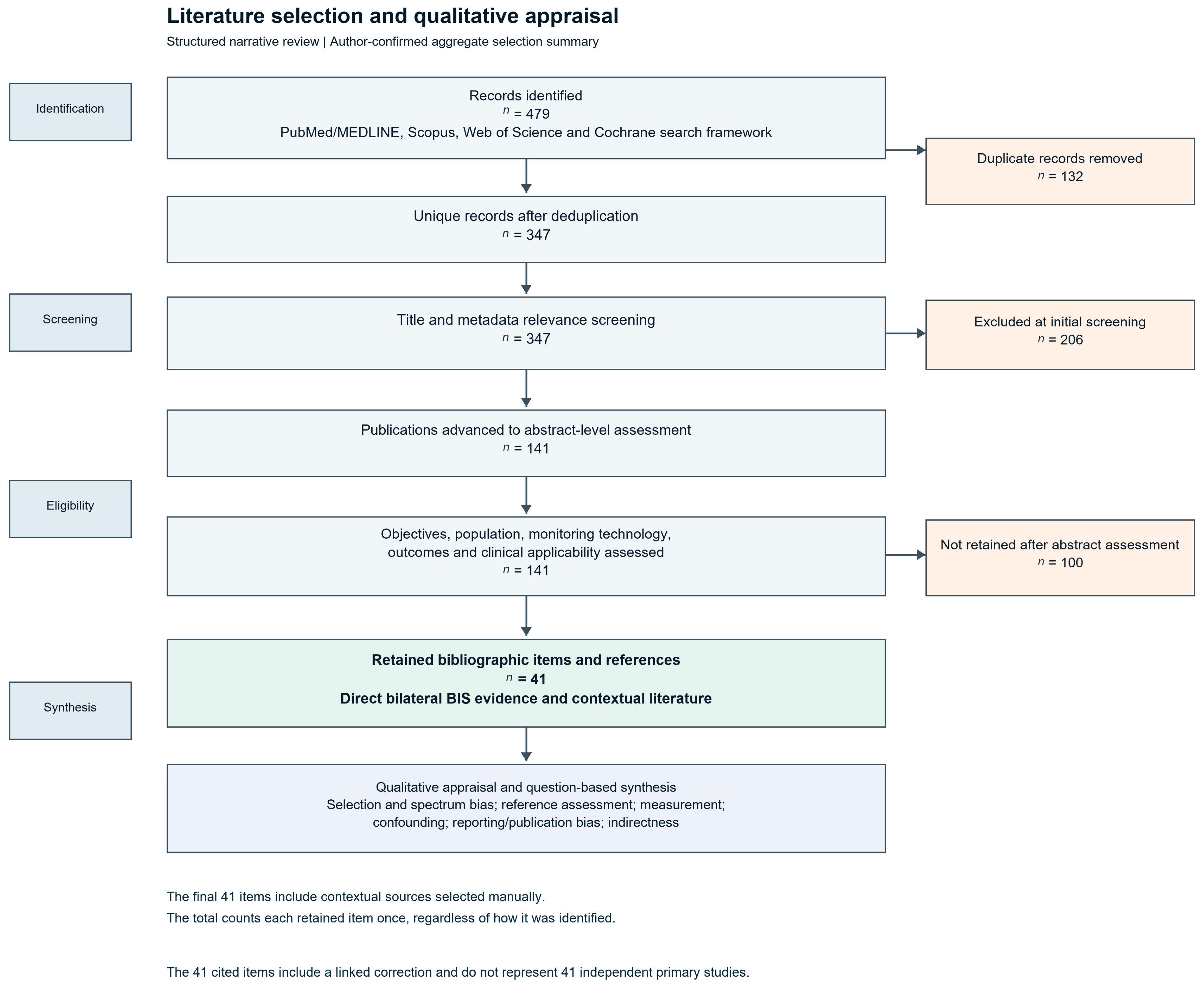
The four databases returned 479 bibliographic records; 172 from PubMed/MEDLINE, 146 from Scopus, 119 from Web of Science and 42 from the Cochrane Library. Removal of 132 duplicates left 347 unique records. Title and metadata screening excluded 206 records: unrelated clinical topic (86), non-EEG technology (49), population or setting without reasonable applicability (32), absence of relevant outcomes (21), and ineligible publication type (18). The remaining 141 publications underwent abstract-level assessment; 100 were excluded for insufficient contribution to the clinical questions (36), insufficient methodological information (24), limited applicability (19), redundancy without an independent contribution (11), or findings not interpretable for the review objectives (10). The recorded selection summary ended with 41 unique bibliographic items (Figure 1), including the manually selected contextual sources. The retained items comprised primary investigations, reviews, guidance and supporting publications, including a correction linked to one case report. They are not 41 independent clinical studies [15].
Figure 1.
Aggregate literature selection for the critical narrative review. The recorded counts were 479 database records, 132 duplicates removed, 347 unique records, 206 title or metadata exclusions, 141 abstracts assessed, 100 further exclusions and 41 retained bibliographic items. The final count includes manually selected contextual sources, counted once per item. The flow does not allocate the retained items by retrieval route. The 41 items include a linked correction and do not represent 41 independent primary studies.
Eligibility covered adult perioperative, postoperative, intensive care and neurocritical care populations, together with foundational physiology and other populations when a defensible connection to the clinical questions existed. Eligible technologies included bilateral BIS and relevant raw or processed EEG measures (CDSA/DSA, SEF and SR); other platforms and reduced montages were used as indirect evidence. Original observational studies, randomized trials, systematic reviews, meta-analyses, consensus documents and selectively informative case reports were considered according to their contribution. Table 2 consolidates the inclusion and exclusion criteria and their application at each selection stage.
Title and metadata screening assessed clinical scope, EEG relevance, population and publication type. Abstract assessment examined whether the population, device and outcome were sufficiently described to inform at least one prespecified clinical question (Table 2). Studies and cases were assigned different evidentiary roles. Documented bilateral acquisition and EEG confirmation were required to support direct seizure-related inferences. Full-text consultation informed the interpretation of retained publications after selection. There was no separate full-text eligibility stage.
Two reviewers independently assessed the 141 records advanced to abstract evaluation. Their initial decisions agreed for 126 records. Of the 15 discrepancies, 11 were resolved by consensus and 4 through additional review. These counts describe the selection process.
2.2. Critical Appraisal and Potential Sources of Bias
The qualitative appraisal examined patient selection, the index test, the reference standard, and flow and timing for the direct seizure studies. It also considered applicability to postoperative critical care, confounding, publication and reporting bias, and overlap between primary studies and reviews. Study-level observations are presented in Section 3.2. The four diagnostic domains organize the narrative judgments; no formal QUADAS-2 ratings or certainty grades were assigned.
Selection and spectrum bias. The direct studies enrolled selected patients with established NCSE or patients undergoing presurgical epilepsy evaluation. The first design lacks a seizure-negative denominator. The second permits a contemporaneous comparison but does not represent postoperative patients with mixed causes of impaired consciousness [16,17].
Measurement and diagnostic reference. Frontal coverage and preprocessing limit the index test. EEG simultaneity, coverage, event adjudication and reader independence affect how closely an apparent change can be linked to electrographic events. Intermittent reference EEG cannot establish continuous agreement across a BIS recording [8,15,16,17,18,19].
Pharmacological physiological and technical confounding. Propofol, volatile agents, dexmedetomidine, ketamine and benzodiazepines have agent-dependent EEG effects that may resemble, obscure or modify abnormalities. Hypothermia, cerebral hypoperfusion, metabolic disturbances, EMG contamination, electrical interference and electrode displacement introduce additional uncertainty. Paralysis can remove EMG without establishing deeper hypnosis or seizure cessation. These issues particularly affect uncontrolled observations and cases. Numerical indices and spectra must therefore be reconciled with raw EEG, signal quality, medication timing and clinical physiology [5,6,7,20,21,22,23,24].
Selective reporting and publication bias. Positive case reports lack a denominator for missed events and false alarms. The appraisal also distinguished surrogate endpoints, such as BIS trends and sedative dose, from independently assessed diagnostic or clinical outcomes [8,25,26,27].
Indirectness and transferability. Reduced-electrode montages, cEEG-derived qEEG panels and other pEEG platforms differ in spatial coverage and processing. Their accuracy cannot be assigned to bilateral BIS [28,29]. Correlation between BIS and clinical sedation scales is distinct from agreement at an individual treatment threshold and from seizure-detection validity; the Heavner synthesis excluded assessments during neuromuscular blockade [26]. Randomized sedation studies, expert recommendations and pooled objective-monitoring evidence address selected aspects of sedation management, not autonomous NCSE diagnosis [7,25,27,30]. Exploratory ischemia and prognostic associations likewise require population-specific validation [31,32].
2.3. Evidence Integration and Qualitative Synthesis
Evidence was organized into six groups: direct seizure and NCSE observations; ischemia and postoperative prognosis; sedation; EEG terminology and management guidance; physiology, artifacts and other EEG technologies; and implementation or transport. Reviews and the correction linked to a case report were treated as contextual publications [15].
Synthesis was qualitative because populations, settings, hardware, electrode positions, reference standards, thresholds and outcome definitions were heterogeneous. The detection of electrographic seizures was considered separately from sedative titration, pharmacologically induced burst suppression, spectral description, prognosis and implementation feasibility. Particular attention was paid to the difference between observing changes during an established condition and identifying or excluding that condition in an undifferentiated population. No new effect-size pooling or diagnostic meta-analysis was performed; a harmonized set of comparable diagnostic thresholds, reference assessments and contingency data was not assembled. This should not be read as proof that a meta-analysis of a differently defined question would be impossible.
The 41-item count refers to the original selection. This revision cites additional clinical and technical sources to contextualize seizure frequency, rapid-response EEG and the specifications of BIS VISTA.
Full texts were consulted for 38 of the 41 retained publications. Three sources were used at abstract level: Chamorro-Jambrina and Chamorro-Falero [13], Brophy et al. [33], and Ruijter et al. [34] (TELSTAR). The synthesis limits its use of those sources to the information available in the abstracts. Bibliographic details and persistent identifiers were checked through 20 October 2026.
Generative AI assistance. AI-assisted work comprised manuscript revision, bibliographic checking and the preparation of synthetic educational graphics, as described in the Acknowledgments. Study results are derived from the cited publications. The study selection counts reflect the review team’s reported selection process.
3. Results: Evidence Relevant to Bilateral BIS and Its Postoperative Application
3.1. Evidence Directly Addressing Seizures and NCSE
Hernández-Hernández and Fernández-Torre retrospectively studied 15 comatose neurocritical-care patients with established NCSE monitored with bilateral BIS for at least 24 h [16]. The interval from NCSE diagnosis to BIS initiation was 8 h (range 0.5–31 h), and monitoring lasted 7.8 ± 6.5 days on average. Reported CDSA findings included increased spectral power, SEF in the delta–theta range, variable asymmetry and fluctuating BIS values. The selected design supports descriptive follow-up during known NCSE and cannot estimate screening performance.
Fàbregas et al. recorded bilateral four-channel BIS and simultaneous conventional 40-channel EEG in 48 nonanesthetized patients undergoing presurgical epilepsy assessment. Twenty-one had at least one electrographic seizure. The evaluated BIS parameters did not reliably identify seizures; nominal SQI differences did not survive adjustment for multiple comparisons [17]. Limited frontal sampling is one plausible explanation. The study does not isolate that mechanism or establish how the device performs in a postoperative ICU.
A brief correspondence on nonconvulsive status and BIS also illustrates the longstanding clinical interest in this question [35]. As a letter without a diagnostic denominator, it cannot establish detection accuracy.
The American Clinical Neurophysiology Society (ACNS) standardized terminology defines electrographic seizure based on sufficient frequency/duration or definite evolution; electrographic status epilepticus corresponds to continuous electrographic seizure activity for at least 10 min or cumulative seizure burden of at least 20% of an hour [36]. These definitions require interpretable EEG data of sufficient technical quality and spatial coverage. A compressed BIS trend cannot be used to classify the ictal–interictal continuum, brief potentially ictal rhythmic discharges, or NCSE to these standards. Salzburg criteria likewise provide clinical EEG diagnostic framing, not a shortcut for interpreting a depth-of-anesthesia monitor [37].
Case reports illustrate changes that may prompt clinical reassessment. Fernández-Torre and Hernández-Hernández described recurring CDSA changes in focal NCSE [18], and Iturri Clavero et al. reported intraoperative seizures with bilateral BIS VISTA [19]. In the case reported by Ntahe, the bilateral sensor was used earlier in the ICU course. The illustrated recurrence used a unilateral two-channel BIS Quatro sensor, with spikes, spike–wave complexes or polyspikes visible in the time-domain displays. A subsequent conventional EEG confirmed recurrence [15]. These cases cannot quantify missed events or false alarms or benefit from acting on the display. For a published clinical BIS trace, see Figure 1 in Ntahe’s case report [15].Published bilateral CDSA observations are described in the 15-patient series [16].
The published correction to the Ntahe case adds references concerning anesthetic and barbiturate suppression monitoring [15]. It does not provide an additional patient or a diagnostic denominator, and is therefore linked to the case rather than counted as independent clinical evidence.
Reduced-montage diagnostic EEG addresses a different question. A meta-analysis of eight studies with 3458 EEG samples using 7–10 electrodes reported pooled sensitivity of 75% (95% CI 68–80%) and specificity of 97% (95% CI 95–98%) [28]. Those estimates describe the studied montages. Kaleem et al. evaluated trained ICU nurses reading quantitative trends derived from cEEG; their results concern that workflow rather than the interpretation of bilateral BIS [29].
Evidence beyond seizures deserves separate consideration. In a prospective single-center aSAH cohort, 64 patients underwent two short BIS recording sessions and 16 developed delayed cerebral ischemia. A median BIS below 85 on either side during days 4–6 was associated with ischemia (adjusted OR 6.2; 95% CI 1.28–30) [31]. This exploratory association, with wide uncertainty and intermittent sampling, is not a validated screening threshold for mixed postoperative populations or a basis for claiming superiority to transcranial Doppler. In 24 postoperative neurosurgical patients, Chan et al. related 728 BIS readings and propofol exposure to 3-month functional outcomes [32]. Repeated measurements do not create 728 independent patients; the association does not establish a bilateral BIS prognostic rule or justify individual outcome predictions.
3.2. Methodological Strength, Bias and Transferability of the Included Literature
Table 3 summarizes study designs and results. Table 4 applies the four diagnostic domains to the main seizure evidence and highlights the principal limitations of the remaining publications.
Table 3.
Principal studies and evidence syntheses: design, population, reported findings and applicability to bilateral BIS. Abbreviations: aSAH, aneurysmal subarachnoid hemorrhage; DCI, delayed cerebral ischemia; DFCF, delirium- and coma-free; OR, odds ratio; RCT, randomized controlled trial; RASS, Richmond Agitation–Sedation Scale; RSS, Ramsay Sedation Scale; SAS, Sedation–Agitation Scale; SQI, signal quality index.
Table 4.
Qualitative methodological appraisal, potential sources of bias and consequences for interpretation. No formal tool-based risk-of-bias scores or GRADE ratings are assigned.
3.3. Bilateral BIS Architecture, Raw Signal and Spectral Variables
The BIS number is a proprietary composite derived from processed frontal EEG. Its numerical scale was validated predominantly to help guide hypnotic anesthesia, not to identify the spatial, morphological, or evolving features required for electrographic seizure diagnosis [3,4]. A reassuring number can coexist with a pathologic EEG pattern; conversely, a sudden increase may reflect EMG or altered drug effects rather than an ictal event. The numerical trend should therefore be considered one element of a multimodal signal rather than the primary neurological outcome.
With a compatible BISx4 and bilateral sensor, BIS VISTA can display four frontal EEG waveforms, two from each side [43,44]. The adhesive array is centered on the forehead and extends laterally toward both temples. The scrolling time-domain traces permit inspection of the sampled waveform, and CDSA shows power by time and frequency. For left and right sampled EEG total powers P_L and P_R, BIS VISTA expresses ASYM as the relative contribution of one hemisphere to their sum: 100 × P_L/(P_L + P_R) on a 0–100 scale, with 50 indicating equal power and the display identifying the side with greater power [45]. ASYM cannot localize a lesion or identify seizure onset. The VISTA operating manual describes selectable waveform display filters (2–70 Hz with mains notch enabled and 0.25–100 Hz with filtering off) [44]. These are display settings. Available outputs depend on model and software. The perioperative report explicitly identifies VISTA [19]. The comparative study describes four-channel bilateral monitoring [17]. The published reports do not establish the firmware or algorithm versions.
Spectrogram interpretation improves on a purely index-driven approach but introduces its own limitations. Bright bands, shifts in alpha power, rhythmic fluctuations, or suppression stripes are patterns for correlation with the raw EEG, sedative exposure, and clinical findings—not seizure diagnoses. The standardized spectrogram nomenclature evaluated by Zafar and colleagues used full EEG-derived spectrograms and cannot be transferred directly as a validated classifier to four-channel frontal BIS [46]. Recent anesthetic-focused reviews underscore the physiological utility of spectrograms while recognizing the limitations of frontal sampling and signal-processing algorithms [6,13].
The frontal raw EEG is valuable in that it allows the user to distinguish some gross waveforms from an index that may lag behind changes. However, not all epileptic discharges have a frontal field, and a few electrodes cannot reliably establish the full spatial distribution, evolution or morphology needed for ACNS seizure classification. CDSA converts a time-frequency series into a color map; its apparent visual simplicity is a trade-off. Color-scale normalization, temporal resolution, filtering and artifacts must be checked before a new bright band or interhemispheric difference is interpreted as brain activity [6,20,36,46].
The technical quality of a particular recording matters at least as much as any inferred pattern. SQI helps identify poor data quality, but a high SQI describes fidelity of the recorded frontal signal rather than coverage of distant cortical regions. EMG from frontalis or temporal muscles can raise apparent power at higher frequencies and distort indices; conversely, paralysis can remove EMG without guaranteeing adequate hypnosis or eliminating electrographic seizures. Temperature, cranial perfusion, sensor placement, electrical interference and electrode displacement must be addressed in parallel. Table A1 in Appendix A defines the EEG and BIS terminology used in this review.
3.4. Pharmacological Signatures: Expected Sedation Versus Neurological Alarm
Sedation studies are summarized here because drug effects are a major confounder in postoperative surveillance. A Cochrane review found insufficient evidence that BIS-guided sedation improved clinical outcomes in ventilated adults [25]. Heavner et al. found a pooled BIS–sedation scale correlation of 0.68 (95% CI 0.61–0.74) in 24 studies (1235 patients), with heterogeneity. Assessments during neuromuscular blockade were excluded [26]. Neither synthesis validates a BIS threshold for seizure recognition.
A single-center randomized trial involving 99 critically ill patients compared BIS-targeted deep sedation with clinical assessment. BIS guidance reduced propofol use (approximately 1.44 versus 1.77 mg/kg/h) and the proportion of monitored time with BIS values below 40, but the primary outcome of delirium- and coma-free days was not significantly improved overall (p = 0.1) [27]. A favorable subgroup analysis among patients sedated for more than 24 h should be interpreted as exploratory rather than conclusive. Thus, lighter dosing under BIS monitoring is not evidence that BIS prevents delirium, diagnoses seizures, or improves neurological outcomes in an unselected postoperative ICU population.
A Delphi consensus of 21 experts considered frontal processed EEG with a continuous spectrogram suitable for assessing sedation depth in selected critically ill adults whose clinical evaluation is limited, particularly during profound sedation or paralysis; adequate staff training and the recognition of monitor-specific limitations were emphasized [7]. This is a consensus about sedation monitoring, not a recommendation to replace cEEG for suspected NCSE. Familiarity with anesthetic EEG physiology is essential because propofol, volatile agents, dexmedetomidine, ketamine, opioids, and high-dose GABAergic agents have different spectral signatures [5,22,23,24]. Table 5 summarizes clinically relevant patterns without assigning drug-independent BIS thresholds.
Table 5.
Pharmacological and physiological EEG signatures and practical bilateral BIS interpretation.
Distinguishing expected drug effects from neurological pathology is essential before invoking the concept of an alarm. During propofol-induced unconsciousness, frontal coherent alpha oscillations can occur alongside slow and delta power, and the balance of these signatures changes with depth and patient vulnerability. Sevoflurane shares several slow and alpha features but is not identical across the theta and higher-frequency spectrum. Dexmedetomidine yields a sleep-like pattern with slow rhythms and spindles, whereas ketamine and nitrous oxide can change high-frequency power in ways that break simple numerical hypnotic assumptions. None of these signatures is pathognomonic of normal cerebral function [5,6,22,23,24].
A clinically meaningful deviation is therefore relational rather than defined by a single threshold. One should ask whether the observed change matches a recent bolus, infusion adjustment, emergence phase, hypothermia, hypotension, hypoglycemia, technical disturbance or muscle activity. A stable BIS value of 40–60 during propofol anesthesia is not evidence that the patient cannot be having a focal or generalized electrographic seizure; and a higher BIS value during ketamine, movement or frontalis contraction is not proof of seizure or light hypnosis. A trend becomes more concerning when there is compatible clinical change, persistent raw EEG rhythm or suppression, and no convincing pharmacological or artifact explanation.
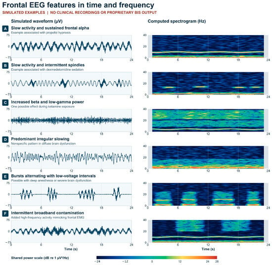
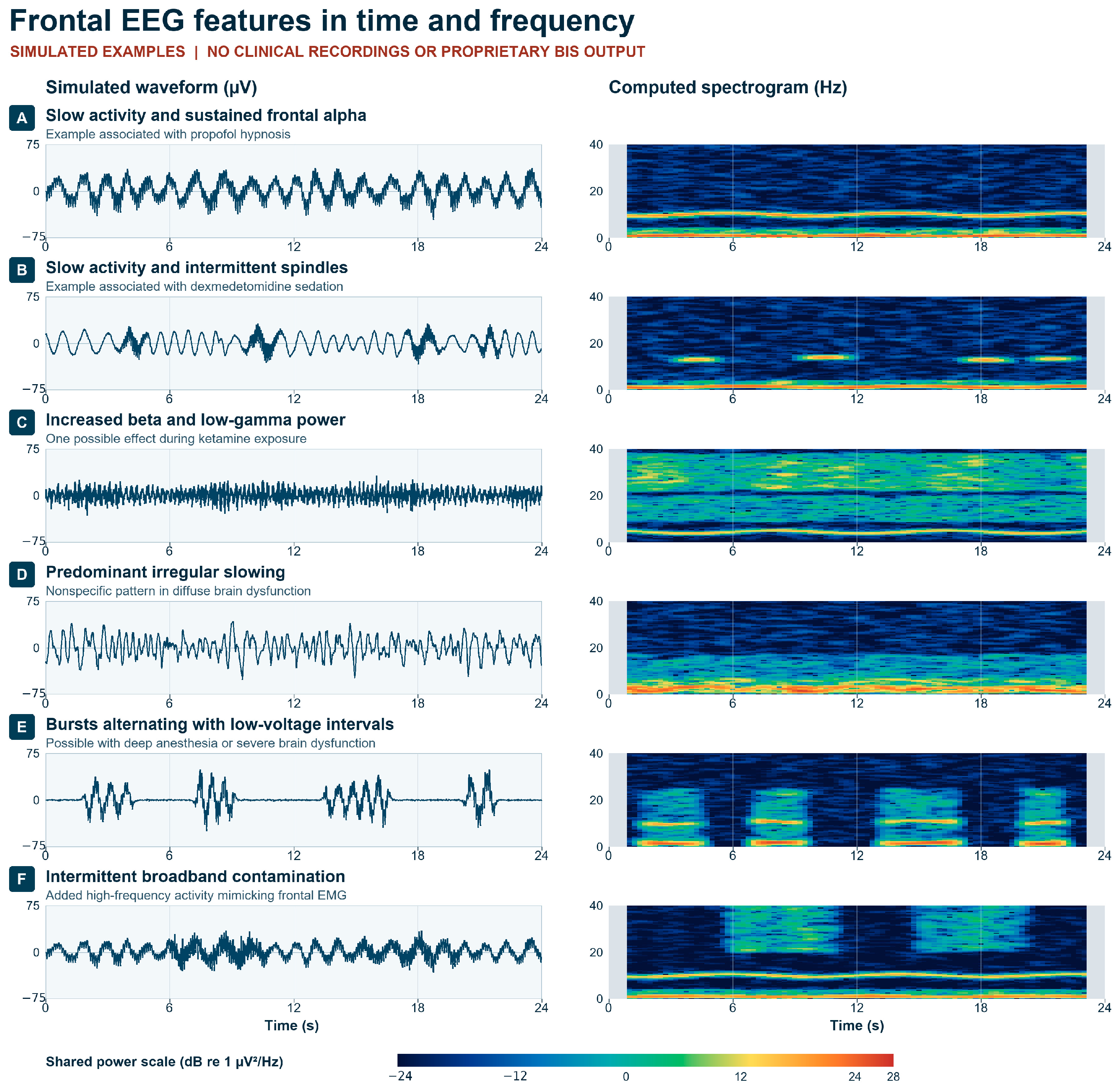
A 2025 synthesis of objective sedation monitoring found no reduction in ventilation duration; sedative-sparing results were heterogeneous [30]. A separate ICU narrative review emphasized the limited validation of BIS in this setting [40]. Figure 2 pairs simulated traces with spectrograms to illustrate how alpha activity, spindles, fast activity, attenuation and contamination change time–frequency displays. Similar appearances may occur under different drugs and clinical conditions. The power palette is illustrative and device settings may differ.
Figure 2.
Simulated frontal EEG waveforms and their computed spectrograms. (A) Slow activity and sustained alpha, a pattern associated with propofol hypnosis. (B) Slow activity with intermittent spindle-range oscillations, as can occur with dexmedetomidine. (C) Increased beta and low-gamma power, one possible effect during ketamine exposure. (D) Predominant irregular slowing, a nonspecific pattern in diffuse brain dysfunction. (E) Bursts alternating with low-voltage intervals. (F) Broadband contamination added to an underlying signal to illustrate an EMG-like effect. Each panel shows a 24 s simulated trace sampled at 128 Hz and its own one-sided power spectral density (2 s Hann windows, 0.25 s step, 0–40 Hz; shared scale −24 to 28 dB re 1 µV2/Hz). Gray margins indicate the absence of centered windows. The examples illustrate individual features described in the existing literature [5,6,20,21,22]. They do not reproduce patient recordings, BIS processing, drug-specific responses or diagnostic thresholds. See Section 3.1 for published clinical examples.
3.5. Encephalopathy, Interictal Activity, IIC, Electrographic Seizures and Burst Suppression
ACNS 2021 terminology differentiates electrographic seizures, electrographic status epilepticus, periodic discharges, rhythmic delta activity and the ictal–interictal continuum (IIC). Definite EEG seizure classification depends on features such as discharges above specified frequencies, unequivocal evolution, duration and contextual modifiers interpreted in a multichannel recording; electrographic status may be defined by continuous seizure lasting at least 10 min or by a substantial seizure burden during an hour. A frontal proprietary processed display is not equipped to apply those criteria reliably, particularly for distant or deep generators [36].
A diffuse encephalopathic pattern is not simply an earlier stage of NCSE. A patient can show metabolic slowing, periodic or triphasic-appearing discharges or burst suppression from multiple causes. The IIC describes uncertain ictal significance that requires integration with brain lesion, level of consciousness, clinical course and EEG reactivity; it is not a universally progressive ladder from interictal abnormality to seizure. Importantly, an increasing ratio of suppression or an evolving visual band does not itself establish treatment response or injury [33,36,37]. Table A2 in Appendix A summarizes these distinctions and their interpretation safeguards.
In refractory status epilepticus, continuous anesthetic treatment may intentionally produce burst suppression. An available frontal SR can supplement trend observation, while cEEG guides electrographic assessment and the detection of breakthroughs [33]. The TELSTAR abstract [34] concerns the suppression of selected rhythmic and periodic EEG patterns after cardiac arrest. It provides no basis for BIS-guided treatment or for generalizing its results to postoperative NCSE.
3.6. Artifacts, Sensor Position and Physiological Confounders
Noncerebral interference is a major limitation across processed EEG platforms. EMG, cardiac contamination, electrical interference, movement, sensor displacement, poor contact and hardware-specific processing can distort indices or spectral displays. Removing frontal EMG with neuromuscular blockade may lower an index without increasing hypnotic drug effect [20].
Physiological confounding is equally important. Hypothermia, cerebral hypoperfusion, low voltage associated with severe brain injury, encephalopathy, drug accumulation, and burst suppression can all alter BIS-related parameters; the same signal appearance may have different meanings depending on mechanism. A newly elevated suppression ratio may be an intended therapeutic target during formally EEG-guided treatment of refractory status, excessive anesthetic effect, a temperature effect, or severe global cerebral dysfunction. Likewise, asymmetric spectra can arise from structural pathology, electrode problems, or normal differences rather than an ictal focus.
The 2026 discussion of processed EEG and neuroprotection offers an appealing broader framework for integrating raw EEG, spectrograms, and indices in anesthetic and critical care practice [13]. Its proposals should be presented as interpretive and potentially preventive rather than as proof that bilateral BIS identifies cerebral ischemia or reduces postoperative brain injury. Table 6 emphasizes actionable differential diagnosis and signal confirmation rather than unsupported sensitivity estimates.
Table 6.
Expected sedation, possible neurological alarm patterns, and a staged clinical response.
Beg, Kim and Tung’s 2026 scoping review included 48 publications describing 31 types of non-pharmacological artifacts across BIS, Entropy and SedLine; 37 publications reported falsely increased hypnotic indices [20]. These publication counts are not routine-practice artifact incidence estimates. Shivering, craniofacial activity, handling, electrical equipment and cable or sensor movement warrant attention in postoperative care. An abrupt index change accompanying EMG elevation raises concern for contamination but cannot by itself exclude concurrent cerebral pathology.
Artifacts can also affect the apparent left-to-right asymmetry. A unilateral contact problem, displaced sensor, cranial wound or unexpected electrode position may simulate lateralized cortical power. Conversely, correct contact on both sides cannot rescue the fundamental absence of central, parietal, posterior temporal or deep sources from frontal acquisition. For that reason, a normal frontal display should never lower the priority of urgent EEG when clinical concern for seizures is substantial.
3.7. From Bedside Concern to EEG Confirmation: A Staged Pathway
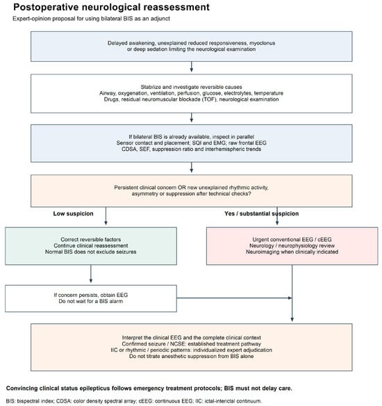
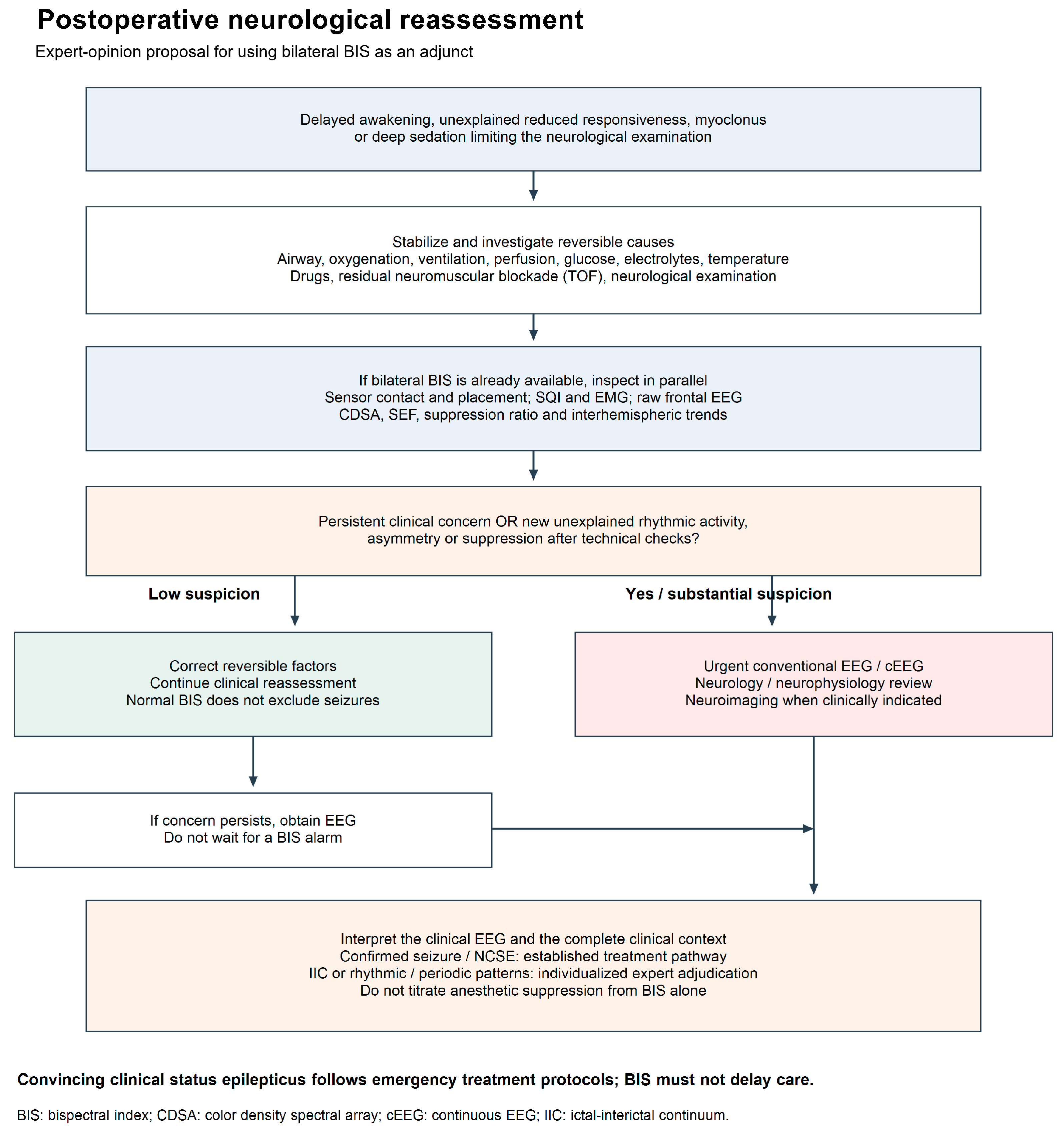
Figure 3 presents an expert opinion escalation proposal for delayed awakening, unexplained reduced responsiveness, myoclonus or concern for electrographic seizures. Initial assessment prioritizes airway, oxygenation, ventilation, hemodynamics, temperature, glucose, electrolytes, drug exposure, residual neuromuscular blockade and neurological examination. The review of sensor placement, SQI/EMG, raw EEG, CDSA, SEF, SR and asymmetry occurs in parallel when a BIS monitor is already available.
Figure 3.
Postoperative neurological reassessment pathway based on expert opinion. Clinical stabilization and investigation proceed alongside inspection of the limited frontal signals. Persistent concern warrants urgent diagnostic EEG regardless of a reassuring BIS display. The pathway awaits prospective validation.
Initiate diagnostic EEG urgently when NCSE is suspected, in parallel with stabilization and the investigation of reversible causes. Unexplained delayed awakening, new focal signs, persistent myoclonus or repeated or evolving frontal rhythmicity, new side-to-side power imbalance, or unexpectedly sustained suppression should prompt reassessment. If the concern arises from BIS alone, review the raw trace, SQI/EMG, electrodes, medication timing and physiology promptly; persistence or recurrence after correctable causes are addressed strengthens the indication for EEG. The bedside clinician initiates escalation, and EEG interpretation requires a clinician trained in critical care EEG. Do not wait for an arbitrary BIS duration or threshold, or allow a stable display to defer EEG when suspicion is substantial. Use cEEG where ongoing or intermittent events are suspected and arrange urgent imaging for appropriate focal signs or other red flags [1,2,29,33,36,37].
3.8. Practical Use in Deployed, Transport and Resource-Constrained Environments
When conventional EEG setup is delayed, commercial rapid-response reduced-montage EEG can provide an earlier diagnostic recording. In the prospective observational DECIDE study of 181 ICU patients with suspected nonconvulsive seizures, median time to acquisition was 5 min with Ceribell Rapid-EEG versus 239 min to conventional EEG in that workflow [47]. The study measured access and bedside decision-making. In a retrospective evaluation of 353 Rapid-EEG recordings, the Claritγ high-burden alert flagged all nine recordings classified as status epilepticus by expert review and flagged 24 additional recordings, 21 of which contained highly epileptiform patterns [48]. Automated alerts can prioritize qualified review of the waveform. A negative alert cannot exclude seizures. Conventional EEG or cEEG remains appropriate when the limited recording is nondiagnostic or suspicion persists [1,2,12,28,29].
Transport and deployed settings introduce vibration, electrical interference, sensor and consumable constraints, variable temperature and staffing limitations. Reports concerning alternative sensor placement, hypnotic monitoring and aeromedical use address implementation [38,39,41,42]. They do not establish the recognition of neurological events or benefit during evacuation. A standard or modified BIS montage cannot substitute for a plan to obtain diagnostic EEG, including remote interpretation or transfer when appropriate.
3.9. Complementary Neurophysiological Methods
Conventional EEG and cEEG allow assessment of spatial distribution, morphology, evolution and reactivity. The bilateral frontal SedLine platform also offers time-domain and spectral displays, but its Patient State Index, montage and processing differ from BIS. Reports of artifacts across platforms do not establish equivalent diagnostic performance [6,20].
Auditory or tactile stimulation during diagnostic EEG can help document background reactivity when clinically appropriate. Sedation and stimulus-related artifacts complicate interpretation. Intermittent photic stimulation addresses specific routine EEG indications, such as suspected photosensitivity, and is not proposed here as routine postoperative seizure surveillance. Evoked potentials address stimulus–response pathway function and may complement selected assessments of impaired consciousness. They serve a different purpose from identifying spontaneous seizures [1,2,12,36].
Machine learning tools may help flag segments in diagnostic EEG for review. Their performance depends on the recording montage, training data, event definitions and artifacts. EEG- or evoked-response-based brain–computer interfaces are an emerging approach to questions such as covert command-following or communication. These are outside the device-specific evidence examined here, and neither automated EEG analysis nor brain–computer interfaces are established by the reviewed studies as postoperative BIS applications.
4. Discussion
The proposed postoperative role is to document available frontal trends and expedite reassessment when a change remains unexplained after technical and physiological checks. Its value depends on timely access to formal EEG and should be judged against false alarms, false reassurance and unnecessary treatment [2,7,12].
Our work deliberately distinguishes bilateral BIS from general critical care quantitative EEG. The latter encompasses many montage configurations and algorithms, and the published accuracy estimates vary by electrode number, location, reader training and the EEG abnormality of interest. The reduced-montage diagnostic meta-analysis and trained-nurse study provide a strong rationale for prospective investigation, but cannot be mathematically transplanted to a proprietary, spatially restricted BIS monitor [28,29].
In Role 2 facilities and aeromedical care, the proposal is limited to signal and workflow feasibility. Published hypnosis and sensor-placement reports cannot establish that a neurological change will be recognized in transit [38,39,41,42].
Weighted Answers to the Major Clinical Questions
Table 7 links the evidence to practical decisions. The key distinction is between clinical surveillance and diagnostic confirmation. A visible frontal change can prompt assessment; EEG remains necessary when the patient’s presentation suggests seizures.
Table 7.
Clinical questions, evidence limitations and defensible practice conclusions. This qualitative synthesis does not assign GRADE certainty ratings.
5. Limitations
This narrative review used clinically focused retrieval and qualitative synthesis and is therefore susceptible to selection and publication bias. It was not designed as an exhaustive systematic review or a pooled diagnostic-accuracy assessment. Heterogeneity in populations, endpoints, devices and reference standards limits comparisons across studies. Qualitative methodological appraisal identifies important concerns but does not constitute a formally scored risk-of-bias or certainty assessment.
Most favorable seizure reports concern selected patients already diagnosed with NCSE or monitored during anesthetic treatment. They do not establish early recognition in undifferentiated postoperative patients. Existing evidence does not quantify a clinically reliable bilateral BIS lead time, false-alarm burden, net outcome benefit or cost effectiveness. Neither the selection workflow (Figure 1) nor the clinical pathway (Figure 3) constitutes a validated intervention. Exploratory ischemia and prognostic associations also require independent validation.
Three sources were examined through abstracts [13,33,34]. They provide limited contextual support and were not used to appraise diagnostic performance. Heterogeneity in device and software reporting also limits comparisons across studies.
Figure 2 illustrates selected signal features based on published descriptions. It contains no validated clinical pattern templates. A single frontal trace and its spectrum cannot establish spatial distribution, identify a drug or confirm a seizure. The clinical BIS observations in Section 3.1 should be interpreted within their original recording configurations and populations.
6. Directions for Future Research
Future investigations should prospectively enroll consecutive high-risk postoperative or mixed ICU patients, record bilateral BIS and adequately sampled cEEG simultaneously, and use blinded adjudication of electrographic seizures and clinically important IIC patterns. Analyses should report per-patient and per-event sensitivity, specificity, positive predictive value in realistic event prevalence, time to recognition, false alarm burden, technical failure rate, and clinically actionable outcomes. Device generations and electrode positions should be reported explicitly, with prespecified performance of the numerical BIS value versus raw EEG, CDSA, asymmetry, SEF, and SR. External validation across sedative classes, brain injuries, neuromuscular blockade, and non-neurocritical populations will be essential.
Equally important are pragmatic implementation trials evaluating staff education, whether an alarm leads to timely cEEG, unintended overtreatment, patient-centered outcomes, and whether costs and resource use justify deployment. The priority is not to redefine a diagnostic role for BIS based on compelling screen images, but to determine whether the device offers measurable additional value without displacing accepted neurophysiological standards.
Before military or emergency medical deployment, each device–sensor configuration needs formal characterization under vibration, transport, power variability, changes in patient temperature, cranial injuries, electromagnetic interference, prolonged monitoring and reduced staffing. Staff training must distinguish patterns due to anesthetic dose, artifacts and epilepsy; operational benefit should be evaluated against easier access to rapid-response EEG rather than assumed from routine BIS availability.
7. Conclusions
- Bilateral BIS may add contextual information to neurological surveillance in deeply sedated or difficult-to-assess postoperative critical care patients; the most important display is the combination of the limited raw EEG, spectra, trends and clinical context, not the number alone.
- Evidence directly assessing seizure recognition by bilateral BIS remains sparse and conflicting. In a simultaneous comparative study, seizure detection was not reliable; a small retrospective series suggests only possible utility for following known NCSE.
- A new evolving spectral pattern, asymmetry, unexplained suppression or a discrepancy between the displayed signal and the clinical/drug context should motivate technical validation and urgent neurological reassessment, potentially expediting conventional EEG/cEEG.
- The frontal montage cannot rule out focal or nonconvulsive seizures, classify the IIC definitively, or replace full EEG in diagnosis or treatment titration. No sensitivity or specificity estimate from full EEG or other reduced montage studies can be transferred to BIS.
- Interpretation should systematically account for sedatives, neuromuscular blockade, temperature, perfusion, electrode placement and EMG/electrical artifacts; unsafe false reassurance and alarm-driven overtreatment are competing risks.
- Military, Role 2 and aeromedical use remains a feasibility question. Available hypnosis and sensor-placement reports support examining the practical conditions of use. The validation of neurological surveillance would require prospective comparison against diagnostic EEG.
- Exploratory associations with delayed cerebral ischemia and postoperative functional outcome broaden the research agenda but do not establish validated BIS thresholds for diagnosis, treatment or individual prognosis.
Author Contributions
Investigation (literature screening), A.S.-R.; investigation (abstract assessment for literature selection), M.C.D.M.; validation (independent literature review), A.J.G.-S. and A.I.R.-M.; formal analysis (qualitative synthesis and integration), J.M.-M.; data curation and writing—original draft, I.A.-V.; supervision and project administration, E.G.-M., A.S.-R. and E.G.-M. contributed to additional assessment of unresolved eligibility decisions. All authors have read and agreed to the published version of the manuscript.
Funding
This research received no external funding.
Institutional Review Board Statement
Not applicable; this is a review of published literature without new human participant recruitment.
Informed Consent Statement
Not applicable; no identifiable patient information or original clinical recordings are included.
Data Availability Statement
The numerical study results derive from the cited publications. The Figure 2 waveforms are simulated and the analysis parameters are provided in the caption. No new patient-level data were generated.
Acknowledgments
During preparation of this manuscript, ChatGPT (GPT-6, OpenAI; used during manuscript preparation and revision in October 2026)assisted with substantive manuscript revision, bibliographic checking and preparation of synthetic educational graphics. The authors are responsible for source verification, interpretation and the final submitted content. The tool was not a source of patient data, clinical adjudication or study-selection counts.
Conflicts of Interest
The authors declare no conflicts of interest.
Appendix A. EEG and BIS Terminology and Pattern Interpretation
The following tables provide additional definitions and interpretation safeguards supporting the main review.
Table A1.
EEG and BIS terminology and interpretation safeguards.
Table A2.
Encephalopathy, interictal, ictal–interictal continuum, ictal and suppression patterns: distinctions without an implied progression.
References
- Herman, S.T.; Abend, N.S.; Bleck, T.P.; Chapman, K.E.; Drislane, F.W.; Emerson, R.G.; Gerard, E.E.; Hahn, C.D.; Husain, A.M.; Kaplan, P.W.; et al. Consensus statement on continuous EEG in critically ill adults and children, part I: Indications. J. Clin. Neurophysiol. 2015, 32, 87–95. [Google Scholar] [CrossRef] [Scilit] [PubMed]
- Hirsch, L.J.; Gopaul, M.T. EEG in the critical care setting. Clin. Neurophysiol. 2026, 182, 2111431. [Google Scholar] [CrossRef] [Scilit] [PubMed]
- Sigl, J.C.; Chamoun, N.G. An introduction to bispectral analysis for the electroencephalogram. J. Clin. Monit. 1994, 10, 392–404. [Google Scholar] [CrossRef] [Scilit] [PubMed]
- Rampil, I.J. A primer for EEG signal processing in anesthesia. Anesthesiology 1998, 89, 980–1002. [Google Scholar] [CrossRef] [Scilit] [PubMed]
- Purdon, P.L.; Sampson, A.; Pavone, K.J.; Brown, E.N. Clinical electroencephalography for anesthesiologists: Part I: Background and basic signatures. Anesthesiology 2015, 123, 937–960. [Google Scholar] [CrossRef] [Scilit] [PubMed]
- Mukai, A.; Yang, J.T.; Wu, S.C.; Wang, T.C.; Lin, F.S.; Wu, C.Y. From index to insight: Clinical perspectives on electroencephalographic spectrogram-guided anesthesia—A narrative review. Korean J. Anesthesiol. 2026, 79, 139–151. [Google Scholar] [CrossRef] [Scilit] [PubMed]
- Rasulo, F.A.; Hopkins, P.; Lobo, F.A.; Pandin, P.; Matta, B.; Carozzi, C.; Romagnoli, S.; Absalom, A.; Badenes, R.; Bleck, T.; et al. Processed electroencephalogram-based monitoring to guide sedation in critically ill adult patients: Recommendations from an international expert panel-based consensus. Neurocrit. Care 2023, 38, 296–311. [Google Scholar] [CrossRef] [Scilit] [PubMed]
- Gimson, A.; Smith, M. Processed EEG from depth of anaesthesia monitors and seizures: A scoping review. Seizure 2021, 91, 198–206. [Google Scholar] [CrossRef] [Scilit] [PubMed]
- Skhirtladze-Dworschak, K.; Felli, A.; Aull-Watschinger, S.; Jung, R.; Mouhieddine, M.; Zuckermann, A.; Tscherkno, E.; Dworschak, M.; Pataraia, E. The Impact of Nonconvulsive Status Epilepticus after Cardiac Surgery on Outcome. J. Clin. Med. 2022, 11, 5668. [Google Scholar] [CrossRef] [Scilit] [PubMed]
- Amorim, E.; Firme, M.S.; Zheng, W.L.; Shelton, K.T.; Akeju, O.; Cudemus, G.; Yuval, R.; Westover, M.B. High incidence of epileptiform activity in adults undergoing extracorporeal membrane oxygenation. Clin. Neurophysiol. 2022, 140, 4–11. [Google Scholar] [CrossRef] [Scilit] [PubMed]
- Elf, K.; Ronne-Engström, E.; Semnic, R.; Rostami-Berglund, E.; Sundblom, J.; Zetterling, M. Continuous EEG monitoring after brain tumor surgery. Acta Neurochir. 2019, 161, 1835–1843. [Google Scholar] [CrossRef] [Scilit] [PubMed]
- Taccone, F.S.; Ozkaya, T.; Baggiani, M.; Rasulo, F.A. EEG for bedside monitoring: The intensivist’s point of view. Crit. Care 2026, 30, 195. [Google Scholar] [CrossRef] [Scilit] [PubMed]
- Chamorro-Jambrina, C.; Chamorro-Falero, C. Neuroprotection. Usefulness of processed electroencephalogram monitors in anesthesia and critical care. Med. Intensiv. (Engl. Ed.) 2026, 50, 502539. [Google Scholar] [CrossRef] [Scilit] [PubMed]
- Baethge, C.; Goldbeck-Wood, S.; Mertens, S. SANRA—A scale for the quality assessment of narrative review articles. Res. Integr. Peer Rev. 2019, 4, 5. [Google Scholar] [CrossRef] [Scilit] [PubMed]
- Ntahe, A. Early diagnosis of nonconvulsive status epilepticus recurrence with raw EEG of a bispectral index monitor. Case Rep. Crit. Care 2018, 2018, 1208401, Erratum in Case Rep. Crit. Care 2018, 2018, 7147626. https://doi.org/10.1155/2018/7147626. [Google Scholar] [CrossRef] [Scilit] [PubMed]
- Hernández-Hernández, M.A.; Fernández-Torre, J.L. Color density spectral array of bilateral bispectral index system: Electroencephalographic correlate in comatose patients with nonconvulsive status epilepticus. Seizure 2016, 34, 18–25. [Google Scholar] [CrossRef] [Scilit] [PubMed]
- Fàbregas, N.; Valencia, J.F.; Belda, I.; Tercero, A.; Hervias, A.; Villafuerte, S.; Gambús, P.L.; Carreño, M.; Valero, R. Bilateral bispectral index monitoring performance in the detection of seizures in nonanesthetized epileptic patients: An observational study. J. Neurosurg. Anesthesiol. 2022, 34, 419–423. [Google Scholar] [CrossRef] [Scilit] [PubMed]
- Fernández-Torre, J.L.; Hernández-Hernández, M.A. Utility of bilateral Bispectral index (BIS) monitoring in a comatose patient with focal nonconvulsive status epilepticus. Seizure 2012, 21, 61–64. [Google Scholar] [CrossRef] [Scilit] [PubMed]
- Iturri Clavero, F.; Tamayo Medel, G.; de Orte Sancho, K.; González Uriarte, A.; Iglesias Martínez, A.; Martínez Ruíz, A. Use of BIS VISTA bilateral monitor for diagnosis of intraoperative seizures, a case report. Rev. Esp. Anestesiol. Reanim. 2015, 62, 590–595. [Google Scholar] [CrossRef] [Scilit] [PubMed]
- Beg, A.; Kim, C.N.; Tung, A. Sources, impacts, and mechanisms of non-pharmacological artifacts in processed electroencephalography monitoring during anaesthesia and intensive care: A scoping review. BJA Open 2026, 19, 100573. [Google Scholar] [CrossRef] [Scilit] [PubMed]
- Guay, C.S.; Agrawal, U.; Tseng, B.; Gallo, S.; Schreier, D.R.; Brown, E.N. Clinical Electroencephalography for Anesthesiologists and Intensivists: Part 2—Physiologic Signatures and Active Management. Anesthesiology 2025, 143, 1595–1618. [Google Scholar] [CrossRef] [Scilit] [PubMed]
- Purdon, P.L.; Pierce, E.T.; Mukamel, E.A.; Prerau, M.J.; Walsh, J.L.; Wong, K.F.K.; Salazar-Gomez, A.F.; Harrell, P.G.; Sampson, A.L.; Cimenser, A.; et al. Electroencephalogram signatures of loss and recovery of consciousness from propofol. Proc. Natl. Acad. Sci. USA 2013, 110, E1142–E1151. [Google Scholar] [CrossRef] [Scilit] [PubMed]
- Akeju, O.; Westover, M.B.; Pavone, K.J.; Sampson, A.L.; Hartnack, K.E.; Brown, E.N.; Purdon, P.L. Effects of sevoflurane and propofol on frontal electroencephalogram power and coherence. Anesthesiology 2014, 121, 990–998. [Google Scholar] [CrossRef] [Scilit] [PubMed]
- Brown, E.N.; Lydic, R.; Schiff, N.D. General anesthesia, sleep, and coma. N Engl. J. Med. 2010, 363, 2638–2650. [Google Scholar] [CrossRef] [Scilit] [PubMed]
- Shetty, R.M.; Bellini, A.; Wijayatilake, D.S.; Hamilton, M.A.; Jain, R.; Karanth, S.; Namachivayam, A. BIS monitoring versus clinical assessment for sedation in mechanically ventilated adults in the intensive care unit and its impact on clinical outcomes and resource utilization. Cochrane Database Syst. Rev. 2018, 2, CD011240. [Google Scholar] [CrossRef] [Scilit] [PubMed]
- Heavner, M.S.; Gorman, E.F.; Linn, D.D.; Yeung, S.Y.A.; Miano, T.A. Systematic review and meta-analysis of the correlation between bispectral index (BIS) and clinical sedation scales: Toward defining the role of BIS in critically ill patients. Pharmacotherapy 2022, 42, 667–676. [Google Scholar] [CrossRef] [Scilit] [PubMed]
- Huespe, I.; Giunta, D.; Acosta, K.; Avila, D.; Prado, E.; Sanghavi, D.; Carboni Bisso, I.; Giannasi, S.; Carini, F.C. Comparing Bispectral Index monitoring vs clinical assessment for deep sedation in the ICU: Effects on delirium reduction and sedative drug doses—A randomized trial. Chest 2024, 166, 733–742. [Google Scholar] [CrossRef] [Scilit] [PubMed]
- Lin, Y.C.; Lin, H.A.; Chang, M.L.; Lin, S.F. Diagnostic accuracy of reduced electroencephalography montages for seizure detection: A frequentist and Bayesian meta-analysis. Neurophysiol. Clin. 2025, 55, 103044. [Google Scholar] [CrossRef] [Scilit] [PubMed]
- Kaleem, S.; Kang, J.H.; Sahgal, A.; Hernandez, C.E.; Sinha, S.R.; Swisher, C.B. Electrographic seizure detection by neuroscience intensive care unit nurses via bedside real-time quantitative EEG. Neurol. Clin. Pract. 2021, 11, 420–428. [Google Scholar] [CrossRef] [Scilit] [PubMed]
- Jaworska, N.; Hezam, A.; Poulin, T.; Kromm, J.A.; Burry, L.D.; Niven, D.J.; Fiest, K.M. Evaluation of Objective Sedation Monitoring Practices in Critically Ill Adult Patients: A Systematic Review and Meta-Analysis. Crit. Care Explor 2025, 7, e1297. [Google Scholar] [CrossRef] [Scilit] [PubMed]
- Hernández-Hernández, M.A.; Cherchi, M.S.; Torres-Díez, E.; Orizaola, P.; Martín-Láez, R.; Fernández-Torre, J.L. Bispectral index monitoring to detect delayed cerebral ischemia after aneurysmal subarachnoid hemorrhage. J. Crit. Care 2022, 72, 154154. [Google Scholar] [CrossRef] [Scilit] [PubMed]
- Chan, D.Y.C.; Li, L.F.; Lui, W.M.; Poon, C.C.M.; Tsang, A.C.O.; Leung, G.K.K. Predictive value of Bispectral Index (BIS) in emergency neurosurgical patients: Loss of BIS reactivity to propofol predicts poor functional outcomes. Clin. Neurol. Neurosurg. 2022, 221, 107382. [Google Scholar] [CrossRef] [Scilit] [PubMed]
- Brophy, G.M.; Bell, R.; Claassen, J.; Alldredge, B.; Bleck, T.P.; Glauser, T.; LaRoche, S.M.; Riviello, J.J.; Shutter, L.; Sperling, M.R.; et al. Guidelines for the evaluation and management of status epilepticus. Neurocrit. Care 2012, 17, 3–23. [Google Scholar] [CrossRef] [Scilit] [PubMed]
- Ruijter, B.J.; Keijzer, H.M.; Tjepkema-Cloostermans, M.C.; Blans, M.J.; Beishuizen, A.; Tromp, S.C.; Scholten, E.; Horn, J.; van Rootselaar, A.F.; Admiraal, M.M.; et al. Treating rhythmic and periodic EEG patterns in comatose survivors of cardiac arrest. N. Engl. J. Med. 2022, 386, 724–734. [Google Scholar] [CrossRef] [Scilit] [PubMed]
- Chamorro, C.; Romera, M.A.; Balandín, B.; Valdivia, M. Nonconvulsive status and bispectral index. Crit. Care Med. 2008, 36, 2218–2219. [Google Scholar] [CrossRef] [Scilit] [PubMed]
- Hirsch, L.J.; Fong, M.W.K.; Leitinger, M.; LaRoche, S.M.; Beniczky, S.; Abend, N.S.; Lee, J.W.; Wusthoff, C.J.; Hahn, C.D.; Westover, M.B.; et al. American Clinical Neurophysiology Society’s standardized critical care EEG terminology: 2021 version. J. Clin. Neurophysiol. 2021, 38, 1–29. [Google Scholar] [CrossRef] [Scilit] [PubMed]
- Leitinger, M.; Beniczky, S.; Rohracher, A.; Gardella, E.; Kalss, G.; Qerama, E.; Höfler, J.; Hess Lindberg-Larsen, A.; Kuchukhidze, G.; Dobesberger, J.; et al. Salzburg consensus criteria for non-convulsive status epilepticus: Approach to clinical application. Epilepsy Behav. 2015, 49, 158–163. [Google Scholar] [CrossRef] [Scilit] [PubMed]
- Fernández Grande, V.; Navarro Suay, R. Estudio comparativo de la concordancia entre los registros BIS y CONOX para la monitorización de la hipnosis cerebral en anestesia general. Sanid. Mil. 2026, 82, 71–75. [Google Scholar] [CrossRef] [Scilit]
- Rodríguez-Mejías, A.; Ramos-Rubio, D.; Sánchez-Roldán, A.; Gallego-Colón, E. Monitorización de las bajas durante las aeroevacuaciones mediante el uso del índice biespectral: Revisión sistemática. Sanid. Mil. 2025, 81, 173–177. [Google Scholar]
- Manning, J.; Chiasson, M. Does Bispectral Index Monitoring Belong in the Intensive Care Unit Setting?: A Narrative Review of Evidence. Dimens Crit. Care Nurs. 2025, 44, 231–238. [Google Scholar] [CrossRef] [Scilit] [PubMed]
- Puente Barbas, J.A.; Navarro Suay, R.; Gutiérrez Ortega, C.; Gilsanz Rodríguez, F. Diferentes alternativas para ubicar el sensor del monitor BIS facilitando su empleo en el ámbito militar. Sanid. Mil. 2018, 74, 90–96. [Google Scholar] [CrossRef]
- Puente Barbas, J.A.; Navarro Suay, R.; Gutiérrez Ortega, C.; Gilsanz Rodríguez, F. Empleo del índice biespectral para monitorización de la hipnosis en sedación durante anestesia regional, experiencia en tres pacientes militares. Sanid. Mil. 2016, 72, 190–193. [Google Scholar]
- U.S. Food and Drug Administration. 510(k) Summary K072286: BIS EEG VISTA Monitor System and BISX. Available online: https://www.accessdata.fda.gov/cdrh_docs/pdf7/K072286.pdf (accessed on 15 November 2026).
- Aspect Medical Systems, Inc. BIS VISTA Monitoring System Operating Manual. Document 070-0069, rev. 3.00. 2008. Available online: https://studylib.net/doc/25768529/nsn000036-eeg-monitors-bis-vista-operator-manual (accessed on 15 November 2026).
- Aspect Medical Systems, Inc. BIS Monitor Serial Port Technical Specification. Appendix B, ASYM (VISTA Bilateral Only), Reproduced in González Cava, J.M., Monitorización de la Información del Electroencefalograma en el Proceso Anestésico. Available online: https://riull.ull.es/xmlui/bitstream/handle/915/298/Monitorizaciondelainformaciondelelectroencefalogramaenelprocesoanestesico.pdf?isAllowed=y&sequence=1 (accessed on 15 November 2026).
- Zafar, S.F.; Amorim, E.; Williamsom, C.A.; Jing, J.; Gilmore, E.J.; Haider, H.A.; Swisher, C.; Struck, A.; Rosenthal, E.S.; Ng, M.; et al. A standardized nomenclature for spectrogram EEG patterns: Inter-rater agreement and correspondence with common intensive care unit EEG patterns. Clin. Neurophysiol. 2020, 131, 2298–2306. [Google Scholar] [CrossRef] [Scilit] [PubMed]
- Vespa, P.M.; Olson, D.M.; John, S.; Hobbs, K.S.; Gururangan, K.; Nie, K.; Desai, M.J.; Markert, M.; Parvizi, J.; Bleck, T.P.; et al. Evaluating the Clinical Impact of Rapid Response Electroencephalography: The DECIDE Multicenter Prospective Observational Clinical Study. Crit. Care Med. 2020, 48, 1249–1257. [Google Scholar] [CrossRef] [Scilit] [PubMed]
- Kamousi, B.; Karunakaran, S.; Gururangan, K.; Markert, M.; Decker, B.; Khankhanian, P.; Mainardi, L.; Quinn, J.; Woo, R.; Parvizi, J. Monitoring the Burden of Seizures and Highly Epileptiform Patterns in Critical Care with a Novel Machine Learning Method. Neurocrit. Care 2021, 34, 908–917. [Google Scholar] [CrossRef] [Scilit] [PubMed]

Disclaimer/Publisher’s Note: The statements, opinions and data contained in all publications are solely those of the individual author(s) and contributor(s) and not of MDPI and/or the editor(s). MDPI and/or the editor(s) disclaim responsibility for any injury to people or property resulting from any ideas, methods, instructions or products referred to in the content. |
© 2026 by the authors. Licensee MDPI, Basel, Switzerland. This article is an open access article distributed under the terms and conditions of the Creative Commons Attribution (CC BY) license.