


Abstract
Clinical Rationale and Objectives: Refractory out-of-hospital cardiac arrest (OHCA) is associated with very poor outcomes when treated with conventional cardiopulmonary resuscitation alone, particularly when low-flow time is prolonged. Prehospital extracorporeal cardiopulmonary resuscitation (ECPR) may shorten the interval to extracorporeal perfusion, but its implementation requires a highly standardized operational framework to ensure appropriate patient selection, procedural safety, and efficient use of specialized resources. This manuscript describes the PrehospECPR-GOKVI operational framework, a standardized prehospital ECPR protocol developed by the Gottsegen National Cardiovascular Center in cooperation with the Hungarian National Ambulance Service and the Hungarian Air Ambulance. ECPR Pathway: The programme is based on a dedicated two-person ECPR team, consisting of an experienced physician and a paramedic, deployed with mobile ECPR-specific equipment, including point-of-care ultrasound, a pre-primed VA-ECMO circuit, an ECMO console, and a mobile oxygenator/gas blender. The protocol defines strict inclusion and exclusion criteria, emphasizing witnessed OHCA, age below or apparently below 50 years, initial shockable rhythm or selected pulseless electrical activity with suspected pulmonary embolism, refractory cardiac arrest lasting at least 15 min, no-flow time below 5 min or signs of life, and the feasibility of establishing ECMO flow within 60 min from collapse or emergency call. Two mandatory sonographic STOP criteria are incorporated before cannulation: relevant pericardial effusion or suspected aortic dissection, and inability to safely visualize the femoral artery, femoral vein, and bifurcation. The operational workflow further specifies dispatch activation, ALS continuation, equipment layout, ultrasound-guided femoro-femoral cannulation, failed-access management, air-free circuit connection, post-flow stabilization, receiving-centre notification, and transport to GOKVI. A target interval of no more than 15 min from initiation of ECPR-specific steps to ECMO flow is mandated. Conclusions: The PrehospECPR-GOKVI framework describes a locally developed, standardized operational pathway intended to support early identification, safe procedural preparation, and structured delivery of prehospital ECPR for selected patients with refractory OHCA. The protocol is designed to reduce avoidable delays, standardize ALS–ECPR coordination, and incorporate predefined sonographic safety checkpoints before cannulation. As no patient-level data are reported in this manuscript, feasibility, safety, complication rates, survival, neurological outcomes, resource utilization, and transferability remain to be evaluated prospectively after programme launch.
1. Clinical Rationale and Objectives
Out-of-hospital cardiac arrest (OHCA) remains a primary challenge in contemporary emergency medicine, characterized by persistently high mortality rates and profound risks of devastating neurological injury. For the subset of patients presenting with refractory cardiac arrest—defined as the failure to achieve the return of spontaneous circulation (ROSC) despite standard advanced life support (ALS) measures—the prognosis under conventional cardiopulmonary resuscitation (CPR) declines precipitously. Epidemiological data indicate that survival with favourable neurological function is exceedingly low after 15 min of conventional efforts, and virtually non-existent beyond a 30 min resuscitation window [1,2,3,4]. While conventional chest compressions provide a vital bridge, they deliver only a fraction of normal cardiac output, falling short of satisfying systemic and cerebral metabolic demands during prolonged low-flow states.
In response to these limitations, extracorporeal cardiopulmonary resuscitation (ECPR), utilizing veno-arterial extracorporeal membrane oxygenation (VA-ECMO), has emerged as a transformative therapeutic modality. By rapidly restoring systemic perfusion and gas exchange at resting physiologic levels, ECPR offers a temporary circulatory bridge, preserving end-organ and cerebral viability while definitive corrective interventions are pursued. Although in-hospital ECPR programmes have demonstrated survival advantage over conventional CPR, this advantage diminishes rapidly as the time from initial collapse to the initiation of extracorporeal flow exceeds the critical 60 min mark [5,6,7]. This strict temporal window underpins the clinical rationale for shifting the initiation of extracorporeal life support from the emergency department directly to the prehospital scene.
Implementing a prehospital ECPR programme introduces formidable logistical, technical, and environmental challenges that diverge significantly from controlled, in-hospital intensive care conditions. To support resource-aware deployment and incorporate predefined safety measures, it is essential to establish highly structured, standardized operational frameworks. This paper presents the comprehensive prehospital operational framework developed by the Gottsegen National Cardiovascular Center (GOKVI) in close cooperation with the Hungarian National Ambulance Service (OMSZ) and Hungarian Air Ambulance Non-Profit Ltd. (HEMS). By detailing strict inclusion and exclusion criteria, deploying specialized point-of-care technology, and enforcing precise sonographic safety checkpoints, this protocol seeks to minimize prehospital low-flow duration while preventing procedural futility [8].
The PrehospECPR-GOKVI operational framework is structured as a standardized operating procedure regulating the deployment and on-scene implementation of prehospital ECPR for OHCA. A foundational pillar of this framework is formalized inter-institutional cooperation between the highly specialized, tertiary hospital-based providers of the Gottsegen National Cardiovascular Center (GOKVI), the centralized Hungarian National Ambulance Service (OMSZ), and the Hungarian Air Ambulance Non-Profit Ltd. (HEMS).
At present, the manuscript describes an operational framework, and no patients have yet been enrolled or analysed. Following programme launch, prospective evaluation of clinical outcomes, procedural feasibility, and complication rates is planned.
To avoid overinterpretation, the present manuscript should be interpreted in three distinct domains. First, the rationale for the programme is based on the time-sensitive pathophysiology of refractory OHCA and the hypothesis that earlier extracorporeal flow may reduce low-flow duration in carefully selected patients. Second, the operational content of the protocol consists of dispatch criteria, eligibility verification, ALS–ECPR role separation, sonographic STOP criteria, cannulation workflow, failed-access management, post-flow stabilization, and transport coordination. Third, clinical and system-level outcomes—including feasibility, safety, cannulation success, complications, survival, neurological recovery, dispatch accuracy, cost-effectiveness, resource utilization, and reproducibility—remain unproven within the present manuscript and require prospective evaluation.
2. Programme Setting and Governance
Prehospital ECPR differs fundamentally from in-hospital extracorporeal life support with respect to clinical environment, available personnel, diagnostic resources, sterility, equipment access, communication pathways, and exposure to logistical uncertainty. Whereas in-hospital ECPR is performed within a controlled critical-care or catheterization-laboratory setting with immediate multidisciplinary support, prehospital ECPR must be delivered in dynamic and often unpredictable environments, including private homes, public spaces, workplaces, and transport settings. These circumstances require a highly protocolized operational structure in which candidate selection, dispatch activation, scene organization, ALS–ECPR role separation, ultrasound-based safety checkpoints, cannulation workflow, circuit connection, and transport preparation are standardized before deployment (Table 1).
Table 1.
Summary of the PrehospECPR-GOKVI operational framework. Key personnel, equipment, dispatch criteria, activation thresholds, safety checkpoints, target times, transport procedures, and quality-assurance elements of the prehospital ECPR protocol.
Within the PrehospECPR-GOKVI programme, overall clinical governance is provided by GOKVI as the designated tertiary ECMO centre. This governance role includes protocol ownership, medical oversight, definition of inclusion and exclusion criteria, specification of equipment and training standards, supervision of team competence, coordination with OMSZ and HEMS, post-mission debriefing, quality assurance, and prospective evaluation of programme performance. Thus, although the intervention is performed in the prehospital field in close cooperation with ambulance providers, the programme remains embedded in a tertiary cardiovascular and ECMO infrastructure to ensure procedural consistency, patient safety, and continuity of care from scene activation through transport and definitive in-hospital management.
Emergency calls in Hungary are received through the national 112 emergency call system. Calls are initially processed at one of two national call centres located in Miskolc and Szombathely, which form part of the Central Emergency Call System. From there, calls are routed according to territorial responsibility to the appropriate regional ambulance dispatch group. In the case of overflow, calls may be transferred to another regional dispatch group. The PrehospECPR-GOKVI programme primarily operates within the Central Hungarian Region, where most activations are expected to originate from the Pest County Dispatch Group. However, because overflow dispatching may involve other regional dispatch groups, education and protocol familiarization are required across all relevant dispatch units.
ECPR team activation is performed by the regional ambulance dispatch group. In the primary dispatch phase, a parallel ECPR alert is issued automatically for every patient with suspected cardiac arrest who is potentially younger than 50 years, together with the dispatch of the nearest ALS-capable ambulance unit. At this stage, no additional ECPR-specific criteria are required beyond potential age eligibility and suspected circulatory arrest. The nearest available BLS or ALS unit, including a helicopter emergency medical service unit when indicated, is dispatched according to the decision of the dispatch group, while the ECPR team is activated in parallel. Thus, the dispatch hierarchy consists of the regional dispatch group, the nearest appropriate ambulance or HEMS unit, and the GOKVI ECPR team.
Communication between dispatch, ambulance units, and the ECPR team is conducted through the OMSZ-integrated TETRA radio system and tablet-based communication platform. The ECPR team may be activated not only during the primary emergency-call processing phase but also at the request of an on-scene OMSZ unit. For example, activation may occur if a patient initially treated for ST-elevation myocardial infarction develops cardiac arrest during prehospital care. However, dispatching the ECPR unit remains the responsibility of the regional dispatch group and is performed via TETRA radio.
The senior supervisor consultant, corresponding to the duty senior medical officer, is incorporated into the dispatch pathway. This physician may support, advise on, or, when necessary, override the ECPR activation decision and can provide real-time professional guidance to the dispatch centre, the on-scene OMSZ unit, and the ECPR team.
Legal and operational responsibility is divided according to the phase and type of care. All non-ECPR-specific patient care remains the responsibility of OMSZ. The decision to perform ECPR can only be made by the ECPR team. Once the ECPR decision is made and ECPR-specific procedural steps are initiated, responsibility for the ECPR intervention lies with the ECPR team, while conventional ALS-related tasks remain coordinated with the OMSZ unit.
At programme launch, the PrehospECPR-GOKVI service is organized around one dedicated ECPR unit, consisting of the ECPR emergency physician vehicle and the designated physician–paramedic ECPR team. Initial availability is planned for a maximum of 12 h per day, with the long-term objective of achieving continuous 24/7 coverage.
The ECPR team is based at the Gottsegen National Cardiovascular Center in Budapest and remains there while awaiting deployment. The primary service area is the approximately 30 km operational radius around GOKVI, provided that the ECPR-specific procedure can realistically be started within 45 min from cardiac arrest or emergency call. This catchment area is predominantly urban, reflecting the population density and road network of Budapest and its metropolitan surroundings. However, selected peri-urban or rural missions may also be eligible depending on geographic conditions, traffic, accessibility, and estimated time to arrival.
Return transport from the scene to GOKVI is expected to require approximately 30–45 min on average. Therefore, dispatch decisions must consider not only distance from GOKVI but also scene accessibility, traffic conditions, warning-signal driving feasibility, ALS availability, and the probability of achieving ECMO flow within the predefined 60 min time window.
The expected activation volume is estimated at approximately two to three alerts per 24 h period, corresponding to approximately one alert during a 12 h shift. Because of the restrictive eligibility criteria and time constraints, the rate of aborted missions is expected to be high, consistent with international experience in comparable prehospital ECPR systems. Approximately one cannulation per week is anticipated during the initial implementation phase. Actual activation frequency, dispatch accuracy, eligibility yield, aborted missions, cannulation rate, response intervals, transport times, complications, and outcomes will be prospectively recorded after programme launch.
3. Protocol-Development Methodology
The development of the PrehospECPR-GOKVI protocol was initiated through a structured review of the available scientific literature, international ECPR recommendations, published prehospital ECPR experiences [8,9], and established principles of extracorporeal life support in refractory cardiac arrest. This evidence overview was used to define the clinical rationale, candidate-selection criteria, procedural safety requirements, time targets, team roles, equipment needs, and post-cannulation transport priorities. In the next phase, these principles were adapted to the Hungarian healthcare environment, with particular attention to the centralized organization of the Hungarian National Ambulance Service, the operational role of the Hungarian Air Ambulance, the geographic and traffic characteristics of the Budapest region, and the receiving capacity of the Gottsegen National Cardiovascular Center as a tertiary ECMO centre.
Subsequently, the draft protocol underwent iterative multidisciplinary discussion with representatives of OMSZ and HEMS. These consultations focused on aligning hospital-based ECPR requirements with prehospital operational realities, including dispatch activation, ALS availability, scene management, division of responsibility between the ambulance and ECPR teams, communication channels, equipment placement, transport logistics, and abort criteria. Feedback from these discussions was incorporated into successive protocol versions until a harmonized operational framework was achieved that was clinically consistent, logistically feasible, and compatible with Hungarian emergency medical service structures.
4. Team Composition and Training
The GOKVI ECPR team consists of one physician, experienced in the care of critically ill patients and, ideally, with prehospital experience, and one paramedic, for whom advanced prehospital experience, scene-management competence, and authorization to drive an emergency vehicle using warning signals are essential requirements. The team may also be supplemented by a trainee or assistant. To support efficient deployment of specialized personnel, all team members provide in-hospital care between prehospital assignments, allocated by the Chief of Department of Cardiovascular Critical Care, while maintaining dispatch readiness. At the beginning of each ECPR shift, the ECPR emergency physician vehicle must be checked against the equipment checklist, even if no previous mission has occurred; the paramedic takes over the vehicle and documents this on the inspection sheet.
Problem-oriented scenario simulation training is performed with paramedics as a core component of team preparation. The training uses a puncturable silicone phantom model incorporating a silicone vascular system connected to a fluid reservoir, thereby enabling repeated practice of ultrasound-guided femoral vessel identification, needle puncture, guidewire handling, sheath insertion, dilation, cannula handover, and air-free circuit connection under controlled but procedurally realistic conditions. This low-cost, reusable simulation platform allows the physician–paramedic team to rehearse the exact spatial arrangement, equipment sequence, communication structure, and role allocation required during prehospital ECPR, while minimizing risk to patients and preserving procedural fidelity. Scenarios are deliberately problem-oriented and include technical and environmental challenges such as poor vessel visualization, difficult ergonomics, guidewire instability, cannula-handling errors, incomplete de-airing, equipment misplacement, and communication breakdowns. This simulation-based preparation is consistent with the broader ECMO training literature [10]. Each session is followed by structured debriefing, with emphasis on closed-loop communication, anticipation of safety-critical steps, adherence to STOP criteria, and reduction in scene-to-flow time without compromising cannulation safety.
5. Dispatch and Eligibility
Based on the emergency call or an on-scene report, dispatch and mobilization for cardiac arrest meeting ECPR criteria are initiated by the OMSZ dispatch group or the duty medical officer via TETRA handheld radio. During primary dispatch, ECPR activation is triggered by suspected cardiac arrest in a patient who is potentially younger than 50 years. Additional inclusion and exclusion criteria are subsequently verified by the on-scene OMSZ unit and the ECPR team before ECPR-specific procedural steps are initiated.
In accordance with OMSZ standards, the activation target is 1 min during daytime and 2 min at night after receipt of the mission. A fundamental prerequisite for effective on-scene ECPR is the presence of an ALS-level unit. If such a unit has not been dispatched, the ECPR team must notify dispatch assertively while en route. If feedback from the on-scene ambulance unit, after rigorous verification of inclusion and exclusion criteria, indicates that ECPR is contraindicated, or if the scene cannot be reached, even using warning signals, within 45 min from the emergency call or cardiac arrest, OMSZ aborts the mission after consultation with the ECPR team so that the unit can remain available for dispatch. The ECPR team then immediately returns to the GOKVI base.
5.1. Inclusion Criteria
- Refractory cardiac arrest persisting for at least 15 min from collapse or call time (if the duration is shorter than this and all other criteria are fulfilled, sheath insertion may be performed).
- An initial shockable rhythm, including ventricular fibrillation or pulseless ventricular tachycardia.
- Initial PEA if pulmonary embolism is the most likely cause, subject to prior telephone consultation and consensus decision with the current GOKVI duty physician.
- Age below 50 years or estimated age below 50 years.
- Witnessed cardiac arrest or signs of life during resuscitation.
- No-flow time of less than 5 min, or signs of life during resuscitation. If lay or ambulance chest compressions are judged, based on the handover or direct observation, not to be at least satisfactory in quality, this period must be considered no-flow time.
- Signs of life during resuscitation include movement of the limbs, eye opening, coughing, or regular breathing. Gasping and constricting pupils are not considered signs of life.
5.2. Exclusion Criteria
- ECMO flow would be initiated more than 60 min after collapse, defined as the time of the emergency call. Considering the target time for the intervention, no more than 45 min may elapse before the procedure is started.
- ETCO2 below 10 mmHg, assessed during adequate-quality chest compressions and evaluated by the ECPR team.
- Traumatic cardiac arrest, or cardiac arrest secondary to haemorrhagic shock.
- A history of severe cardiac, pulmonary, or neurological disease; active advanced malignancy; or a frailty score greater than 4, indicating that survival beyond one year is unlikely or that complex intensive care treatment is contraindicated.
For operational and registry reporting, the time of the emergency call is used as the standardized reference point when exact witnessed collapse time is unavailable or uncertain; when exact witnessed collapse time is known, both collapse-to-flow and call-to-flow intervals should be recorded separately.
If all inclusion criteria are met and all exclusion criteria are absent, the ECPR intervention must be initiated without delay. In all other cases, ECPR is absolutely contraindicated and cannulation is prohibited.
6. Operational ECPR Pathway
ECPR is an extremely time-critical procedure, and its performance in the prehospital phase entails numerous challenges, including environmental constraints and limited personnel. Consequently, the process must be highly regulated, standardized, and protocolized; deviation is permissible only in cases of force majeure or when clearly justified. Any protocol deviation must be documented and discussed during the mandatory debriefing to identify opportunities for further improvement. The fundamental objective is that no more than 15 min may elapse between the initiation of ECPR-specific steps and the start of ECMO flow (Figure 1 and Figure 2).
Figure 1.
Prehospital ECPR operational workflow from dispatch activation to transport. The figure illustrates the complete PrehospECPR-GOKVI decision pathway for refractory out-of-hospital cardiac arrest, beginning with dispatch screening and activation of the ECPR team, followed by confirmation of ALS availability, on-scene verification of inclusion and exclusion criteria, and determination of whether ECPR is indicated. The workflow shows the mandatory continuation of high-quality ALS during all decision and procedural steps, the two sonographic STOP criteria, the transition to ultrasound-guided femoro-femoral VA-ECMO cannulation, the failed-access and abort pathways, air-free circuit connection, initiation of ECMO flow, post-flow stabilization, receiving-centre notification, and transport to GOKVI for definitive post-ECPR care. The figure also highlights the key protocol time targets: dispatch activation within 1 min during daytime or 2 min at night, initiation of ECPR-specific steps within 45 min from collapse or emergency call, ECMO flow within 60 min, and no more than 15 min from the start of ECPR-specific tasks to extracorporeal flow. Abbreviations: ALS, advanced life support; ECPR, extracorporeal cardiopulmonary resuscitation; ECMO, extracorporeal membrane oxygenation; GOKVI, Gottsegen National Cardiovascular Center; OHCA, out-of-hospital cardiac arrest; OMSZ, Hungarian National Ambulance Service; STOP, protocol-defined safety termination criterion; VA-ECMO, veno-arterial extracorporeal membrane oxygenation.
Figure 2.
Team roles, equipment layout, and procedural sequence during prehospital ECPR. The figure illustrates the coordinated physician–paramedic workflow during on-scene ECPR, including scene preparation, equipment positioning, ultrasound-guided femoral vessel assessment, sterile field establishment, cannulation preparation, division of ECPR-specific tasks, air-free circuit connection, initiation of ECMO flow, cannula fixation, and transition to transport readiness. The schematic emphasizes role separation, closed-loop communication, sonographic safety checkpoints, and the 15 min target from initiation of ECPR-specific steps to extracorporeal flow. Abbreviations: ALS, advanced life support; CPR, cardiopulmonary resuscitation; ECPR, extracorporeal cardiopulmonary resuscitation; ECMO, extracorporeal membrane oxygenation; GOKVI, Gottsegen National Cardiovascular Center; MOC, Mobile Oxygenator/Gas Blender; OMSZ, Hungarian National Ambulance Service; STOP, protocol-defined safety termination criterion; VA-ECMO, veno-arterial extracorporeal membrane oxygenation.
The protocol is designed primarily for the coordinated interaction of two providers, a physician and a paramedic. They initially perform partly separate tasks, after which the care pathways converge, and the team members complete the intervention together.
The basic premise is that if all inclusion criteria are met, all exclusion criteria can be safely ruled out, and the patient is indeed in refractory cardiac arrest, the indication for ECPR can be established. Accordingly, the first task is to obtain the relevant clinical information, confirming it through closed-loop communication to avoid misunderstandings. Simultaneously, the paramedic assesses whether the ergonomics of CPR-ECPR can be further optimized at the scene. If ECPR is indicated, this decision must be stated loudly and unequivocally within the ECPR team and, importantly, also to the ALS team. The ambulance unit performing conventional CPR must be clearly informed that the on-scene OMSZ ALS unit retains full responsibility for standard advanced life support and systemic resuscitation, whereas the GOKVI team assumes definitive responsibility for the patient and the procedural field once ECMO cannulation is initiated.
When ECPR is initiated, the paramedic begins preparing their own ECPR-specific steps and, if this has not already occurred, delegates to the OMSZ unit the complete removal of the patient’s clothing, including all underwear. In parallel, the physician switches on the pocket ultrasound device, then performs a focused examination of the heart and of the major vessels in the inguinal and femoral regions. If the cardiac examination reveals a significant pericardial effusion and/or clear signs of aortic dissection, the ECPR intervention must not be performed, and the process must be stopped: this is the first STOP criterion.
The primary objective is right-sided ipsilateral cannulation. However, if the femoral great vessels—the common femoral vein, common femoral artery, and bifurcation—cannot be safely identified on either side by ultrasound, the process must also be stopped despite the indication: this is the second STOP criterion. Published reports and registry data indicate that vascular and cannulation-related complications remain important safety concerns during femoro-femoral ECPR [11,12]. However, the precise contribution of inadequate ultrasound visualization varies across systems and is not uniformly reported. For this reason, the present protocol treats inadequate visualization as a practical safety threshold rather than as a quantitatively validated predictor of cannulation failure.
For this reason, cannulation is not continued when ultrasound visualization of the great vessels is inadequate for safe vessel identification and guidewire verification.
If the physician member of the ECPR team identifies either STOP criterion, this must be communicated immediately to the paramedic before the ECMO cannulas are opened. Further preparation of ECPR-specific processes is then discontinued, and patient care continues according to the currently applicable ALS protocol. Responsibility for care returns to the on-scene OMSZ ALS unit, and in legal terms the patient remains under OMSZ care.
6.1. ECPR-Specific Tasks of the Paramedic
The paramedic begins ECPR-specific tasks only if it has been confirmed that conventional ALS can be safely delegated to the OMSZ units and the ECPR team physician has instructed them to proceed.
As the first step, the paramedic places the equipment required for the intervention in its dedicated position: ALS bag, if the team’s own bag is present; ECMO console; ultrasound system; Mobile Oxygenator/Gas Blender (MOC); oxygen; and cannula bag. Equipment and bag ergonomics are of critical importance. The paramedic generally works on the patient’s left side, below the plane of the mechanical chest compression device, optimally at the level of the patient’s pelvis.
The paramedic switches on the ECMO console, turns it toward the physician, places it near the physician’s right hand at the lower level of the patient’s rib cage, and completes the final priming steps of the pre-primed device according to the attached priming protocol.
During priming, the paramedic receives or prepares the preactivated ultrasound system, supports the tablet next to the ECMO on its left side, turns it directly toward the physician to facilitate later puncture, and places the ultrasound probe visibly next to the tablet, ensuring that it will not later be covered by the sterile drape.
The paramedic switches on the MOC system, places it toward their left hand at the level of the patient’s mid-thigh, turns it toward the physician, connects the MOC system to the ECMO oxygenator using the dedicated oxygen connector, and connects the MOC to the oxygen source after confirming that the source is open.
During these tasks, the ECPR physician will ask the paramedic to assist with sterile draping. The physician, already in sterile attire, covers the patient with a sterile drape in a cranio-caudal direction; on the patient’s left side, the paramedic helps pull the caudal corner of the sterile drape well beyond the legs.
Only after the preceding steps, and with the approval of the ECPR physician, does the paramedic open the ECMO cannulas of the pre-agreed size, both venous and arterial, and drop them without touching into the sterile field, preferably in the midline at the level of the patient’s mid-thigh.
The paramedic then opens four metal clamps and, if available, the arterial pressure monitoring line next to the ECMO cannulas, again without touching them.
The paramedic puts on the sterile gown and gloves placed by the physician in the lower left corner of the sterile field and assumes the role of second operator on the patient’s left side at pelvic level to assist with cannulation.
6.2. ECPR-Specific Tasks of the Physician
The physician’s intervention-specific steps begin once both STOP criteria have been excluded. At this point, the physician confirms to the paramedic that they may continue their own steps.
The physician is positioned on the patient’s right side, opposite the paramedic, at the level of the patient’s pelvis. As the first step, the physician places the ECMO bag behind them and removes the sterile unit pack and disinfectant. The disinfectant is then applied generously to both inguinal and femoral regions.
The physician then opens the sterile unit pack for their own use. The upper half of the first sterile drape may be handled non-sterilely; when unfolded, it forms a rectangular drape module, with the previously non-sterile upper portion positioned underneath. The drape should be drawn in approximately 30–50 cm lateral to the patient, extending downward from the pelvic plane.
The sterile unit pack contains two sets of sterile gowns and gloves. The physician dons the sterile gown, with lay assistance if necessary; tying at the back is important, whereas full wrapping may be omitted. The physician then dons sterile gloves.
Using the large sterile drape included in the unit pack, the physician covers the patient in a cranio-caudal direction starting from the chest compression device, taking care to identify and leave accessible the future puncture sites. The physician must also ensure that the equipment, ultrasound system, and the drape module already prepared by the paramedic on the opposite side are not covered. Assistance from the paramedic should be requested for sterile draping, as the drape may otherwise catch around the patient’s left leg.
After establishing the sterile field, the physician places the second set of sterile gown and gloves for the paramedic in the lower left corner of the sterile field.
Ideally, on the drape module to the physician’s right hand, the physician prepares the equipment required for puncture and cannulation as follows:
- Assemble both 6F sheaths;
- Fill the sterile container with normal saline;
- Fill the two 50 mL and two 5 mL syringes with fluid; these may be left in the container;
- Flush the 6F sheaths;
- Attach a 5 mL syringe to the echogenic Seldinger needle, fill it with 2 mL of fluid, and flush the needle;
- Prepare the super-stiff guidewires with the J-tip forward;
- Place the dilators in a clearly visible location, optimally in the midline;
- Place gauze pads, sterile scissors, scalpel, needle holder, and sutures in a clearly visible location;
- Position the ECMO cannulas previously opened by the paramedic within easy reach and place them side by side, taking care to preserve laterality.
After the above steps have been completed, the physician applies sterile ultrasound gel to the designated access site and then reaches for the ultrasound probe using the sterile ultrasound cover, into which the paramedic carefully drops the probe.
Before ultrasound-guided puncture, the physician confirms that image settings are appropriate, including depth and gain. This is the final opportunity to request assistance from the paramedic to adjust image settings, as by this point the paramedic will have completed their own tasks and will subsequently also be sterile.
6.3. Joint ECPR-Specific Tasks of the Physician and the Paramedic
After all preparations have been completed, the physician performs ultrasound guidance with the left hand and punctures the anterior wall of the right common femoral vein with the right hand. After setting down the ultrasound probe, the physician advances the Seldinger guidewire of the 6F sheath into the vessel, implants the sheath, removes its dilator and guidewire, aspirates blood from the sheath using the prefilled 5 mL syringe, and then flushes it. The ipsilateral common femoral artery is then punctured and cannulated in the same manner, with flushing performed at the end. The paramedic may assist with the process if requested. If ipsilateral cannulation is unsuccessful, contralateral cannulation may be attempted, but only if ultrasound visualization is adequate.
The physician then advances the super-stiff guidewires into both sheaths in mediolateral, vein-artery sequence. Immediate sequential advancement of both guidewires is particularly important because on-scene opportunities to verify wire position are limited; one of the best predictors of correct VA cannulation is visualization of the two wires side by side in two distinct vessels on ultrasound. This confirmation is an obligatory prerequisite for predilation before cannula insertion. The paramedic may assist in advancing the guidewires. If guidewire positions are appropriate, administration of 5000 IU unfractionated heparin is strongly recommended; the decision is made by the ECMO team physician. If administered, intravenous delivery is delegated to the OMSZ unit, and completion is confirmed using closed-loop communication. Some international providers do not administer heparin in the prehospital setting; however, this increases the risk of clot formation within the cannulas.
The intervention continues in a mediolateral, veno-arterial sequence. The physician carefully withdraws the sheath from the venous guidewire while continuously stabilizing the guidewire with the left hand and may compress any bleeding with gauze. The paramedic removes the sheath from the end of the wire and threads on the requested dilator. Two types of dilators are used in this protocol: gradually tapered vascular dilators, which progressively widen from the tip, and sequential dilators. To save time, it may be preferable to make a skin incision alongside the wire with a scalpel and use the gradually tapered vascular dilators; however, the operator decides whether to use this alone or to supplement dilation with sequential dilators. The aim is safe and successful cannulation, while recognizing throughout the ECPR process that each minute of delay is associated with an approximately 2% reduction in survival [2]. During dilation, extreme care must be taken to prevent guidewire dislodgement; accordingly, either the physician or the paramedic must continuously secure the guidewire, with ongoing communication between them. Holding the wire at the end of the dilator farthest from the patient may help prevent dislodgement. Communication is therefore of particular importance throughout the process.
Once the physician considers predilation adequate, they ask the paramedic to thread the venous ECMO cannula onto the guidewire and simultaneously hand the metal clamp to the physician. The physician implants the venous cannula and, while holding it firmly with the left hand, asks the paramedic to remove the cannula dilator and guidewire together with a careful but rapid and decisive movement. Before withdrawal of the dilator and guidewire, the physician parks the clamp on the designated soft segment of the cannula and clamps the cannula immediately at the end of withdrawal.
For arterial cannulation, the steps described for venous cannulation are repeated.
Once both cannulas have been implanted, the paramedic reaches for or requests the circuit located at the back of the ECMO system and hands it sterilely to the physician. Care must be taken to ensure that the arterial pressure monitoring introducer system within the circuit is not accidentally discarded. At this point, the paramedic’s sterile role is considered complete.
The physician receives the circuit sterilely, places the remaining clamps on both the venous and arterial limbs, cuts the tubing at the designated site or removes the Y-connector joining the tubes, and then connects the venous cannula to the venous limb, followed by the arterial cannula to the arterial limb, ensuring an air-free connection. If the fluid depth is sufficient, this air-free connection may be performed in the fluid container; alternatively, the paramedic may provide normal saline for de-airing from the prefilled 50 mL syringe, without requiring sterility. Before any connection, both cannulas must be briefly back-bled immediately beforehand to remove any thrombi that may have formed during the process.
After successful air-free connection, the physician removes the clamps from both cannulas and tubing in the usual veno-arterial sequence, then asks the paramedic to start ECMO and increase the pump speed to the desired value. At the same time, either team member verifies that the MOC module is delivering the desired FsO2 and the appropriate sweep-gas flow.
If available, the physician then introduces the fiber-optic invasive arterial blood pressure monitoring wire through the preformed side port of the arterial limb, advances it until resistance is met, and secures it at the designated site.
The physician carefully secures both cannulas and limbs with one suture each.
6.4. No ALS Unit Present at the Scene
Regardless of whether the ECPR team is the first unit to arrive at the scene or a BLS-level ambulance unit is performing extended BLS, the primary task is the rapid establishment of ALS and the collection of information relevant to inclusion and exclusion criteria, preferably directly from the witness. Resuscitation must be performed in accordance with the European Resuscitation Council ALS guidelines [13,14]; however, from the perspective of ECPR, early endotracheal intubation and ventilation are critical. A supraglottic airway used by a BLS unit should be replaced with an endotracheal tube, intravenous or intraosseous access above the diaphragm must be established, and the mechanical chest compression device must be applied. If the ECPR team is the only unit present, application of the mechanical chest compression device is the first step, as two-person ALS is otherwise not realistic. A BLS unit at the scene may temporarily provide chest compressions, but the quality of these compressions is critical for subsequent care. If a BLS unit is present at the scene, after the above steps have been completed and ALS has been established, it should be considered whether ventilation and medication administration can be delegated. With appropriate scene management, the ECPR team may be expected to focus specifically on the intervention. Otherwise, high-quality ALS remains the priority, and the ECPR intervention must be postponed until the ALS unit arrives. If the ECPR team is the first responding unit, performing the ECPR intervention while simultaneously providing high-quality ALS is unrealistic until additional units arrive. Therefore, ECPR must not be prioritized over ALS.
6.5. ECPR Not Indicated
The inclusion and exclusion criteria, together with the requirement for good ultrasound visualization of the femoral vessels, substantially limit the frequency of ECPR interventions. In the Netherlands, which is considered a pioneer of on-scene ECPR and has nationwide coverage, approximately 8% of all dispatches resulted in cannulation [15]. In such cases, contraindicating ECPR at the scene despite expectations may present a communication challenge; team members should therefore reinforce one another in making decisions consistent with the operational framework. If the patient is not an ECPR candidate and no ALS unit is present at the scene, the objective is to establish and provide high-quality ALS until the ALS unit arrives. If an ALS unit is already present at the scene, the goal is to restore dispatch readiness as soon as possible. The ECPR team should provide technical assistance to the ALS unit as needed, including intubation or venous cannulation. If return of spontaneous circulation (ROSC) occurs in the presence of the ECPR team, the team supports further care, including ECG, screening echocardiography, and disposition decisions such as catheterization laboratory transfer or regional care, particularly when GOKVI is the destination institution.
6.6. No Cardiac Arrest
During prehospital patient care, the situation encountered on arrival may differ substantially from that presumed based on the emergency call. Consequently, particularly when the ECPR team is the first arriving unit, there may be cases in which acute illness or injury is present at the scene, but cardiac arrest is no longer present or may not have occurred at all. In such cases, the ECMO team is obliged to initiate patient care, although knowledge of OMSZ standard operating procedures is explicitly not required. The patient may be left in the care of a BLS unit only if the patient is stable and ALS-level support is not expected to be required either for ongoing care or for hospital transport. Otherwise, the ECPR team remains at the scene until an ALS unit arrives. In uncertain cases, the paramedic, the OMSZ dispatch group, or the duty medical officer may assist in decision-making.
7. Safety and Contingency Procedures
Safety in the PrehospECPR-GOKVI pathway is ensured through predefined termination criteria, closed-loop communication, role separation, and immediate fallback to conventional advanced life support whenever the procedural risk exceeds the expected benefit. The two ultrasound-based STOP criteria represent the principal safety gates before opening the ECMO cannulas: first, relevant pericardial effusion or signs suggestive of aortic dissection; second, inability to identify the femoral artery, femoral vein, and bifurcation with sufficient confidence for ultrasound-guided cannulation. If either criterion is present, the ECPR-specific procedure is discontinued immediately, responsibility for standard resuscitation remains with or returns to the OMSZ ALS unit, and patient care continues according to the applicable ALS protocol (Table 2).
Table 2.
Safety-critical events and predefined contingency actions during prehospital ECPR. The table summarizes key procedural, vascular, circuit, oxygenation, anticoagulation, infection-prevention, and termination-related risks together with the corresponding preventive and immediate contingency actions.
The two sonographic STOP criteria should be interpreted as locally defined procedural safety gates rather than as validated diagnostic algorithms or outcome-proven innovations. The first STOP criterion combines two distinct sonographic concerns that require different interpretation. Relevant pericardial effusion is defined as circumferential or large effusion, effusion associated with right-sided chamber compression when interpretable, or effusion judged by the ECPR physician to be compatible with tamponade physiology in the clinical context. Suspected aortic pathology is based on direct visualization of an intimal flap, marked aortic root dilatation, intramural haematoma when visible, or a combination of suggestive sonographic findings and clinical context. During mechanical chest compressions, these findings may be difficult to interpret because image acquisition is intermittent and artefact-prone; therefore, the STOP criterion is intentionally conservative and based on the operator’s ability to identify a high-risk mechanical cause with sufficient confidence.
Equipment and oxygen contingency plans should include pre-mission checklist verification, backup clamps, spare sterile access equipment, adequate oxygen cylinder reserve, confirmation of MOC function before cannulation, and the ability to stop the procedure before cannulas are opened if a critical device or oxygen supply is unavailable. If oxygen or ECMO console failure occurs after cannulation, the circuit is clamped as needed, ALS is optimized, troubleshooting is performed according to the device checklist, and transport decisions are coordinated with GOKVI.
Contingency procedures begin at dispatch and continue throughout the mission. If updated information obtained en route or on scene confirms that inclusion criteria are not met, exclusion criteria are present, the scene cannot be reached within the time window required to achieve extracorporeal flow within 60 min from collapse or emergency call, or no ALS-level support can be established, the ECPR mission is aborted or postponed. In these circumstances, the ECPR team supports the on-scene unit only as needed, including airway management, vascular access, ultrasound assessment, or disposition decisions after ROSC, while restoring ECPR dispatch readiness as soon as feasible.
Cannula selection should be predefined before deployment and confirmed on scene according to patient habitus, sonographic vessel diameter, and expected flow requirements. If adequate arterial vessel diameter cannot be confirmed, arterial cannulation should not proceed. Distal limb ischaemia is recognized as a major complication of femoro-femoral VA-ECMO. Prehospital insertion of a distal perfusion catheter is not included in the present protocol; rather, distal limb perfusion is planned and managed after arrival at GOKVI, where fluoroscopy, surgical backup, and vascular imaging are available [16].
To reduce vascular injury, the number of puncture attempts should be limited. A failed ipsilateral arterial or venous puncture may be followed by one contralateral attempt only if ultrasound visualization is adequate and the operator judges that the 15 min ECPR-specific time target remains achievable. Repeated blind or uncertain puncture attempts are prohibited. Failure to obtain safe venous or arterial access, inability to maintain guidewire control, or inability to verify two guidewires in two separate vessels mandates termination of the ECPR cannulation pathway and continuation of conventional ALS.
During cannulation, prevention of avoidable vascular injury and air entrainment is prioritized over speed whenever these aims conflict. If ipsilateral venous or arterial access cannot be obtained safely, contralateral access may be attempted only when sonographic visualization is adequate. Guidewire position must be verified by ultrasound before predilation; if the two wires cannot be confirmed in two separate vessels, dilation and cannula insertion must not proceed. This approach has not been validated in this programme as equivalent to fluoroscopy, transoesophageal echocardiography, or radiographic confirmation. Throughout dilation and cannula advancement, either the physician or the paramedic must continuously secure the guidewire, and each handover of wires, dilators, cannulas, clamps, and circuit limbs must be explicitly confirmed.
In the absence of fluoroscopy, transoesophageal echocardiography, or radiographic confirmation, the protocol relies on continuous ultrasound guidance and confirmation of two guidewires in two anatomically distinct femoral vessels before predilation. This approach is intended to reduce the risk of arterial–venous misplacement and inadvertent dilation of the wrong vessel. However, ultrasound confirmation is operator-dependent and may be limited by obesity, poor acoustic windows, vessel overlap, ongoing chest-compression-related movement, environmental constraints, and difficult ergonomics. For this reason, ultrasound visualization is treated as a safety gate rather than a definitive imaging substitute; if wire position cannot be confirmed with confidence, dilation and cannula insertion are not permitted. After cannulation, venous and arterial position are reassessed by ultrasound when possible, together with circuit behaviour, drainage, return pressure/flow, colour and direction of blood flow, haemodynamic response, oxygenation, and the absence of progressive groin swelling or bleeding. Definitive confirmation and correction remain hospital-based responsibilities after arrival at GOKVI [11,12].
Anticoagulation is not automatic. The default prehospital dose is 5000 IU unfractionated heparin after guidewire position has been confirmed and before or during cannulation, but administration remains at the discretion of the ECPR physician. Heparin may be withheld or reduced when major bleeding, traumatic arrest, suspected intracranial haemorrhage, known severe coagulopathy, recent major surgery, or another clinically relevant contraindication is present. When heparin is withheld, the team should minimize circuit stasis, avoid unnecessary delays before flow initiation, and communicate the decision explicitly to the receiving ECMO team [17].
Circuit-related contingencies focus on avoiding clot formation, air embolism, uncontrolled bleeding, and accidental disconnection. Before connecting the circuit, both cannulas must be briefly back-bled to remove potential thrombi, and the venous and arterial limbs must be connected in an air-free manner, preferably under fluid or with saline de-airing. Clamps are removed only after secure connections have been confirmed. If major bleeding, cannula malposition, suspected air entry, circuit disconnection, pump malfunction, or inability to establish adequate ECMO flow occurs, the team must immediately halt further procedural progression, maintain manual control of the cannulas and circuit, restore high-quality ALS as required, and contact the receiving GOKVI team for real-time support.
After ECMO flow has been initiated, the patient must not be moved until the cannulas, circuit limbs, oxygen supply, MOC system, ECMO console, monitoring lines, and stretcher-mounted components have been checked and secured. The physician verifies flow, pump function, alarm limits, oxygenation, sweep-gas delivery, pulsatility, mean arterial pressure, and the absence of visible circuit air or progressive bleeding. The cannulas are secured with sutures and external fixation before transfer; during each movement, one team member maintains direct control of the circuit and cannula exit sites.
Infection-prevention procedures are adapted to the prehospital environment. The protocol requires maximal feasible sterility, including wide exposure of the groins, generous skin disinfection, sterile gowns and gloves, sterile draping, sterile ultrasound cover and gel, and minimization of unnecessary movement around the sterile field. If environmental contamination, restricted space, rain, crowding, lighting, or scene safety prevents maintenance of a minimum sterile field, cannulation should be delayed, relocated if feasible, or abandoned if procedural sterility and safety cannot be achieved.
If vascular injury, suspected retroperitoneal bleeding, uncontrolled groin haemorrhage, guidewire loss, suspected air entrainment, cannula malposition, circuit disconnection, pump malfunction, oxygen supply failure, or inability to establish adequate ECMO flow occurs, further procedural progression is stopped immediately. Manual pressure is applied for external bleeding, cannulas and guidewires are secured, air entry is prevented by clamping and maintaining fluid-filled connections, high-quality ALS is resumed or intensified as required, and the receiving GOKVI team is contacted for real-time support. In the presence of uncontrolled haemorrhage, loss of vascular access, uncorrectable circuit failure, or inability to establish stable ECMO flow, the ECPR attempt is terminated and care reverts to conventional ALS or post-ROSC management as clinically appropriate.
After cannulation, ECPR may be terminated if stable extracorporeal flow cannot be achieved despite troubleshooting; if catastrophic vascular injury or uncontrolled haemorrhage occurs; if circuit or oxygen failure cannot be corrected; if cannula position is unsafe or cannot be stabilized for transport; if newly recognized exclusion criteria indicate futility; or if the receiving ECMO physician and field ECPR physician agree that continuation would expose the patient and team to disproportionate risk without realistic benefit. All such decisions must be documented and reviewed during mandatory debriefing.
Transport contingencies are based on continuous monitoring and early communication with the receiving centre. Before departure, GOKVI is notified using the ATMISTER format, and a video call may be used when rapid cross-checking of ECMO settings is required. If deterioration occurs during loading or transport, the ambulance must stop when necessary to allow safe troubleshooting of cannula position, circuit integrity, gas supply, pump flow, bleeding, or ALS priorities. Any deviation from the predefined pathway, any aborted ECPR attempt, and any technical or communication problem must be documented and reviewed during the mandatory debriefing, so that recurrent hazards can be incorporated into training, equipment checks, and future protocol revisions.
8. Post-ECPR Management and Transport
Further fixation of the cannulas requires considerable effort because cannula displacement in the prehospital environment is a real risk and may constitute a fatal complication. A pressure bag is placed over the cannulas and inflated. A pelvic binder is then applied over this, and additional fixation is achieved at mid-thigh level using a tourniquet or Peha bandage. The arterial pressure monitor must be secured to the arterial limb using its specific fasteners.
After ECMO initiation, inadequate flow is managed by reassessing venous drainage, cannula position, circuit integrity, pump speed, volume status, and mechanical obstruction. Differential hypoxaemia is suspected when upper-body oxygenation remains inadequate despite established femoral VA-ECMO flow and should prompt reassessment of native cardiac output, lung function, ventilation, sweep gas and oxygen delivery, and transport urgency. Left ventricular distension cannot be definitively treated in the field; it is monitored indirectly through pulsatility, arterial pressure, pulmonary oedema, oxygenation, and echocardiographic findings when feasible, and triggers early receiving-centre notification for in-hospital decompression strategies if required.
If blood gas analysis is available, the paramedic inserts the appropriate ABG module into the ECMO system and initiates blood gas analysis; this is an optional step. The physician verifies appropriate ECMO function, including the desired flow, alarm limits, pump operation, presence of pulsatility, target oxygenation, mean arterial pressure, and related parameters, as detailed later. Standard post-resuscitation measures are then performed according to the ABCDE approach; obtaining a 12-lead ECG is particularly important, as described in the ECPR post-resuscitation protocol. The patient is carefully transferred onto a scoop stretcher and then onto the ambulance stretcher. The paramedic attaches the ECMO mount to the frame of the mobile patient stretcher, after which the ECMO and MOC systems are secured separately. Throughout this process, the physician continuously holds the ECMO circuit. Telephone notification to GOKVI is then performed using the ATMISTER format. Optionally, a video call may be arranged for rapid cross-checking, primarily focused on ECMO settings, between the ECMO team physician, the GOKVI duty physician, and the perfusionist. The patient is placed into the OMSZ vehicle. The ECPR team physician also boards the vehicle and accompanies the patient to GOKVI, while the paramedic organizes the remaining ECMO team equipment at the scene and drives the ECPR vehicle back to GOKVI, escorting the ambulance.
According to the ELSO adult ECPR interim guideline [18], post-ECPR care should be treated as the immediate continuation of resuscitation rather than as a separate transport phase. After extracorporeal flow is established, the team should rapidly confirm adequate VA-ECMO support, secure cannulas and circuit connections, avoid air entrainment and circuit interruption, maintain continuous monitoring, and communicate early with the receiving ECMO centre. Initial management should focus on restoring systemic perfusion, identifying and treating the reversible cause of cardiac arrest, protecting the brain after global ischaemia, correcting severe metabolic derangement, and screening for ECMO-related complications, including bleeding, vascular injury, limb ischaemia, haemolysis, oxygenator or pump dysfunction, and inadequate drainage or return flow [19].
A future randomized study is planned to investigate the role of therapeutic hypothermia, or targeted temperature management, in this specific patient population after prehospital ECPR. The study will aim to determine whether controlled post-resuscitation temperature modulation can improve neurological recovery and survival while maintaining safety in patients treated with prehospital VA-ECMO for refractory OHCA.
Compared with previously reported ECPR programmes, the PrehospECPR-GOKVI operational framework should be interpreted primarily as an operational implementation model rather than as an outcomes study. The ARREST trial [6] and the randomized Prague OHCA trial [20] evaluated hospital-centred or intra-arrest transport strategies in which ECPR was initiated after transfer to an experienced cardiac centre, whereas the present operational framework shifts cannulation to the prehospital scene to minimize low-flow time. The secondary analysis of the Prague OHCA trial [3] further supports the importance of careful candidate selection when interpreting ECPR outcomes in refractory OHCA. The INCEPTION trial [5] tested early ECPR within a multicentre Dutch EMS–hospital network, while ON-SCENE [15] and CHEER3 [9] are more closely aligned with the present concept because they evaluate prehospital initiation of VA-ECMO. However, the GOKVI model differs by combining tertiary ECMO-centre governance, a centralized national ambulance structure, HEMS cooperation, a compact physician–paramedic ECPR team, restrictive eligibility criteria, explicit dispatch and collapse-to-flow time thresholds, mandatory sonographic STOP criteria, a standardized failed-access pathway, and detailed post-flow stabilization and transport procedures. Unlike ARREST, Prague OHCA, INCEPTION, and CHEER3, the current manuscript does not yet report survival, neurological outcome, or complication data; these endpoints will require prospective evaluation after programme launch.
9. Quality Assurance and Planned Evaluation
Following programme launch, prospective evaluation should focus on predefined clinical, procedural, and system-level endpoints. These should include dispatch accuracy, eligibility yield, time from emergency call to ECPR-team activation, time from collapse or call to ECPR-specific task initiation, time from ECPR-specific task initiation to ECMO flow, cannulation success, failed-access frequency, protocol deviations, vascular and bleeding complications, circuit-related complications, transport safety, survival to hospital discharge, neurological outcome, resource utilization, and potential organ-donation pathways. These endpoints will be necessary to evaluate feasibility, safety, clinical effectiveness, resource use and transferable beyond the local implementation setting.
The implementation of the standardized PrehospECPR-GOKVI operational framework represents a structured approach to refractory out-of-hospital cardiac arrest. Transitioning a complex, highly invasive intervention such as extracorporeal membrane oxygenation to the prehospital field requires a precise balance between clinical speed and procedural control. As outlined in this framework, implementation is expected to depend on close inter-institutional collaboration between a specialized hospital-based ECPR team and the primary prehospital emergency medical infrastructure. By restricting eligibility to tightly defined clinical cohorts—characterized by young age, witnessed arrest, and shockable initial rhythms—and maintaining a strict 15 min target from initiation of ECPR-specific tasks to ECMO flow, the protocol is designed to target the patient populations most likely to achieve neurological recovery.
A key contribution of this operational framework is the integration of objective sonographic STOP criteria. Enforcing mandatory checkpoints for mechanical contraindications and requiring high-quality ultrasound visualization of the femoral vessels directly addresses the technical risks associated with prehospital cannulation. These safety gates are intended to reduce the likelihood of high-risk vascular injury when field conditions are unfavourable for successful cannulation. Additionally, by formally incorporating transport procedures, the operational framework provides a pragmatic assessment of the programme’s broader clinical value. This protocolized approach may provide an adaptable operational framework for regional healthcare systems seeking to implement prehospital extracorporeal life support for selected patients with otherwise refractory cardiac arrest.
10. Limitations and Generalizability
The PrehospECPR-GOKVI operational framework should be interpreted as a locally developed operational protocol for selected patients with refractory OHCA rather than as evidence of clinical benefit. Its design links prehospital emergency care with tertiary cardiovascular and ECMO expertise, with the aim of standardizing dispatch, eligibility assessment, cannulation preparation, safety checkpoints, and transport coordination. However, whether this structure results in improved feasibility, safety, survival, neurological outcome, resource utilization, or reproducibility remains unknown and requires prospective evaluation.
Several limitations should be considered when interpreting and applying this operational framework. First, this manuscript describes a standardized protocol rather than reporting outcome data from a completed clinical cohort; therefore, its impact on survival, neurological outcome, complication rates, dispatch efficiency, and cost-effectiveness will require prospective evaluation. Second, the protocol is intentionally restrictive, focusing on highly selected patients with witnessed refractory OHCA, short no-flow time, predominantly shockable rhythms, younger age, and the realistic possibility of establishing ECMO flow within approximately 60 min. These criteria are designed to preserve benefit and safety, but they substantially limit the number of eligible patients and may reduce generalizability to broader cardiac arrest populations. The age criterion of below, or apparently below, 50 years is unusually restrictive compared with several established ECPR programmes. However, it is clearly presented as a deliberate local programme decision rather than as a general recommendation. Third, implementation depends on system-level prerequisites that may not be available in all regions, including a centralized dispatch structure, rapid ALS response, a dedicated and continuously available ECPR team, specialized mobile ECMO equipment, prehospital ultrasound, mechanical chest compression, and immediate access to a tertiary ECMO centre. Fourth, the protocol is highly time-sensitive and may be affected by traffic, distance, scene accessibility, weather, bystander information, quality of preceding CPR, and the availability of sufficient personnel at the scene. Fifth, safe cannulation is dependent on adequate ultrasound visualization of femoral vessels and on operator experience; patients with difficult anatomy, obesity, poor acoustic windows, severe peripheral vascular disease, or challenging scene ergonomics may therefore be excluded despite otherwise favourable clinical characteristics.
Finally, because prehospital ECPR is performed in an uncontrolled environment, residual risks remain despite protocolization, including vascular injury, bleeding, air entrainment, cannula displacement, circuit malfunction, delayed transport, and competing priorities between ALS and ECPR-specific tasks. The strict STOP criteria and mandatory debriefing process are intended to mitigate these risks, but they cannot eliminate them. Consequently, the operational framework should be interpreted as a context-specific operational model that requires local adaptation, structured training, continuous quality assurance, and prospective data collection before broader conclusions can be drawn regarding safety, effectiveness, and transferability.
Generalizability is therefore conditional rather than universal. The transferable elements of the PrehospECPR-GOKVI operational framework include the use of strict eligibility criteria, centralized dispatch activation, explicit ALS-ECPR role separation, sonographic STOP criteria, standardized equipment layout, closed-loop communication, structured transport notification, and mandatory post-mission debriefing. However, successful replication in other regions would require adaptation to local ambulance organization, geography, traffic patterns, hospital catchment areas, ECMO centre density, legal responsibility structures, staffing models, equipment availability, training pathways, and funding mechanisms. Systems with fragmented EMS governance, long transport distances, limited prehospital physician availability, restricted ultrasound competence, or delayed access to a high-volume ECMO centre may not be able to reproduce the same time targets or procedural workflow without substantial modification.
Accordingly, the present protocol should be viewed as a model for regional implementation rather than as a directly exportable universal standard. Its main value lies in demonstrating how a tertiary cardiovascular centre and a national ambulance system can translate guideline-based ECPR principles into a field-operational pathway with predefined safety gates. Before adoption elsewhere, each component should be tested against local dispatch data, expected eligible case volume, achievable response intervals, cannulation expertise, transport logistics, and receiving-centre capacity. Prospective registry-based monitoring will be essential to determine whether the operational framework remains safe, efficient, and clinically meaningful when applied outside the original Budapest-centred network.
11. Conclusions
The PrehospECPR-GOKVI operational framework presents a standardized regional protocol for the prehospital initiation of ECPR in highly selected patients with refractory OHCA. The protocol integrates centralized dispatch activation, strict eligibility criteria, ALS–ECPR role separation, sonographic STOP criteria, ultrasound-guided femoro-femoral cannulation, failed-access pathways, air-free circuit connection, post-flow stabilization, and transport coordination with a tertiary ECMO centre. Its purpose is to provide a structured operational pathway intended to reduce avoidable delays and improve procedural consistency in the prehospital environment. As no patient-level outcome data are reported, feasibility, safety, complications, survival, neurological outcome, resource utilization, cost-effectiveness, and transferability cannot be inferred from the present manuscript and should be evaluated prospectively after programme launch.
Author Contributions
Conceptualization, C.S., P.Ó., A.A., Z.B., A.B., L.S.E., M.M., G.C., R.G., M.C. and P.A.; methodology, C.S., P.Ó., A.A., Z.B., A.B., L.S.E., M.M., G.C., R.G., M.C. and P.A.; formal analysis, C.S., P.Ó., A.A., Z.B., A.B., L.S.E., M.M., G.C., R.G., M.C. and P.A.; investigation, C.S., P.Ó., A.A., Z.B., A.B., L.S.E., M.M., G.C., R.G., M.C. and P.A.; resources, C.S., P.Ó., A.A., Z.B., A.B., L.S.E., M.M., G.C., R.G., M.C. and P.A.; writing—original draft preparation, C.S., P.Ó., A.A., Z.B., A.B., L.S.E., M.M., G.C., R.G., M.C. and P.A.; writing—review and editing, C.S., P.Ó., A.A., Z.B., A.B., L.S.E., M.M., G.C., R.G., M.C. and P.A.; visualization, C.S. and P.Ó.; supervision, C.S. and P.A.; project administration, C.S., P.Ó., A.A., Z.B., A.B., L.S.E., M.M., G.C., R.G., M.C. and P.A. All authors have read and agreed to the published version of the manuscript.
Funding
This research received no external funding.
Institutional Review Board Statement
This operational protocol has been designed, implemented, and conducted in strict accordance with the ethical principles enunciated in the Declaration of Helsinki governing medical research involving human subjects, as well as all applicable national regulatory frameworks and institutional guidelines. As this manuscript describes an operational protocol and does not involve the analysis of patient-level data, formal ethics committee approval was not required.
Informed Consent Statement
Not applicable.
Data Availability Statement
The original contributions presented in this study are included in the article. Further inquiries can be directed to the corresponding author.
Acknowledgments
During the preparation of this manuscript, the authors used Microsoft Copilot (Version 2605, Microsoft Corporation, Redmond, WA, USA) for grammatical review and stylistic refinement. The authors critically reviewed, verified, and edited all AI-assisted output and take full responsibility for the accuracy, integrity, and final content of the manuscript.
Conflicts of Interest
The authors declare no conflicts of interest.
Abbreviations
The following abbreviations are used in this manuscript:
| ABG | arterial blood gas |
| ABCDE | Airway, Breathing, Circulation, Disability, Exposure |
| ALS | advanced life support |
| BLS | basic life support |
| CPR | cardiopulmonary resuscitation |
| ECG | electrocardiogram |
| ECPR | extracorporeal cardiopulmonary resuscitation |
| ECMO | extracorporeal membrane oxygenation |
| ETCO2 | end-tidal carbon dioxide |
| FsO2 | fraction of oxygen in sweep gas |
| GOKVI | Gottsegen National Cardiovascular Center |
| HEMS | Hungarian Air Ambulance Non-Profit Ltd. |
| IU | international units |
| MOC | Mobile Oxygenator/Gas Blender |
| OHCA | out-of-hospital cardiac arrest |
| OMSZ | Hungarian National Ambulance Service |
| PEA | pulseless electrical activity |
| ROSC | return of spontaneous circulation |
| STOP | protocol-defined safety termination criterion |
| VA-ECMO | veno-arterial extracorporeal membrane oxygenation |
References
- Low, C.J.W.; Ling, R.R.; Ramanathan, K.; Chen, Y.; Rochwerg, B.; Kitamura, T.; Iwami, T.; Ong, M.E.H.; Okada, Y. Extracorporeal cardiopulmonary resuscitation versus conventional CPR in cardiac arrest: An updated meta-analysis and trial sequential analysis. Crit. Care 2024, 28, 57. [Google Scholar] [CrossRef] [Scilit]
- Inoue, A.; Hifumi, T.; Sakamoto, T.; Kuroda, Y. Extracorporeal cardiopulmonary resuscitation for out-of-hospital cardiac arrest in adult patients. J. Am. Heart Assoc. 2020, 9, e015291. [Google Scholar] [CrossRef] [Scilit] [PubMed]
- Rob, D.; Smalcova, J.; Smid, O.; Kral, A.; Kovarnik, T.; Zemanek, D.; Kavalkova, P.; Huptych, M.; Komarek, A.; Franek, O.; et al. Extracorporeal versus conventional cardiopulmonary resuscitation for refractory out-of-hospital cardiac arrest: A secondary analysis of the Prague OHCA trial. Crit. Care 2022, 26, 330. [Google Scholar] [CrossRef] [Scilit] [PubMed]
- Okada, Y.; Komukai, S.; Irisawa, T.; Yamada, T.; Yoshiya, K.; Park, C.; Nishimura, T.; Ishibe, T.; Kobata, H.; Kiguchi, T.; et al. In-hospital extracorporeal cardiopulmonary resuscitation for patients with out-of-hospital cardiac arrest: An analysis by time-dependent propensity score matching using a nationwide database in Japan. Crit. Care 2023, 27, 442. [Google Scholar] [CrossRef] [Scilit] [PubMed]
- Suverein, M.M.; Delnoij, T.S.R.; Lorusso, R.; Brandon Bravo Bruinsma, G.J.; Otterspoor, L.; Elzo Kraemer, C.V.; Vlaar, A.P.J.; van der Heijden, J.J.; Scholten, E.; den Uil, C.; et al. Early Extracorporeal CPR for Refractory Out-of-Hospital Cardiac Arrest. N. Engl. J. Med. 2023, 388, 299–309. [Google Scholar] [CrossRef] [Scilit] [PubMed]
- Yannopoulos, D.; Bartos, J.; Raveendran, G.; Walser, E.; Connett, J.; Murray, T.A.; Collins, G.; Zhang, L.; Kalra, R.; Kosmopoulos, M.; et al. Advanced reperfusion strategies for patients with out-of-hospital cardiac arrest and refractory ventricular fibrillation (ARREST): A phase 2, single centre, open-label, randomised controlled trial. Lancet 2020, 396, 1807–1816. [Google Scholar] [CrossRef] [Scilit] [PubMed]
- Matsuyama, T.; Irisawa, T.; Yamada, T.; Hayakawa, K.; Yoshiya, K.; Noguchi, K.; Nishimura, T.; Ishibe, T.; Yagi, Y.; Kiguchi, T.; et al. Impact of low-flow duration on favorable neurological outcomes of extracorporeal cardiopulmonary resuscitation after out-of-hospital cardiac arrest: A multicenter prospective study. Circulation 2020, 141, 1031–1033. [Google Scholar] [CrossRef] [Scilit] [PubMed]
- Lamhaut, L.; Hutin, A.; Puymirat, E.; Jouan, J.; Raphalen, J.H.; Jouffroy, R.; Jaffry, M.; Dagron, C.; An, K.; Dumas, F.; et al. A pre-hospital extracorporeal cardio pulmonary resuscitation (ECPR) strategy for treatment of refractory out-of-hospital cardiac arrest: An observational study and propensity analysis. Resuscitation 2017, 117, 109–117. [Google Scholar] [CrossRef] [Scilit] [PubMed]
- Richardson, S.A.C.; Anderson, D.; Burrell, A.J.C.; Byrne, T.; Coull, J.; Diehl, A.; Gantner, D.; Hoffman, K.; Hooper, A.; Hopkins, S.; et al. Pre-hospital ECPR in an Australian metropolitan setting: A single-arm feasibility assessment-The CPR, pre-hospital ECPR and early reperfusion (CHEER3) study. Scand. J. Trauma Resusc. Emerg. Med. 2023, 31, 100. [Google Scholar] [CrossRef] [Scilit] [PubMed]
- Duinmeijer, W.C.; Fresiello, L.; Swol, J.; Torrella, P.; Riera, J.; Obreja, V.; Puślecki, M.; Dąbrowski, M.; Arens, J.; Halfwerk, F.R. Simulators and simulations for extracorporeal membrane oxygenation: An ECMO scoping review. J. Clin. Med. 2023, 12, 1765. [Google Scholar] [CrossRef] [Scilit] [PubMed]
- Richardson, A.S.; Schmidt, M.; Bailey, M.; Pellegrino, V.A.; Rycus, P.T.; Pilcher, D.V. ECMO cardio-pulmonary resuscitation (ECPR), trends in survival from an international multicentre cohort study over 12 years. Resuscitation 2017, 112, 34–40. [Google Scholar] [CrossRef] [Scilit] [PubMed]
- Wang, L.; Li, C.; Hao, X.; Rycus, P.; Tonna, J.E.; Alexander, P.; Fan, E.; Wang, H.; Yang, F.; Hou, X. Percutaneous cannulation is associated with lower rate of severe neurological complication in femoro-femoral ECPR: Results from the Extracorporeal Life Support Organization Registry. Ann. Intensive Care 2023, 13, 77. [Google Scholar] [CrossRef] [Scilit] [PubMed]
- Soar, J.; Böttiger, B.W.; Carli, P.; Jiménez, F.C.; Cimpoesu, D.; Cole, G.; Couper, K.; D’Arrigo, S.; Deakin, C.D.; Ek, J.E.; et al. European Resuscitation Council Guidelines 2025 Adult Advanced Life Support. Resuscitation 2025, 215, 110769. [Google Scholar] [CrossRef] [Scilit] [PubMed]
- Wigginton, J.G.; Agarwal, S.; Bartos, J.A.; Coute, R.A.; Drennan, I.R.; Haamid, A.; Kudenchuk, P.J.; Link, M.S.; Panchal, A.R.; Pelter, M.M.; et al. Part 9: Adult Advanced Life Support: 2025 American Heart Association Guidelines for Cardiopulmonary Resuscitation and Emergency Cardiovascular Care. Circulation 2025, 152, S538–S577. [Google Scholar] [CrossRef] [Scilit] [PubMed]
- Ali, S.; Moors, X.; van Schuppen, H.; Mommers, L.; Weelink, E.; Meuwese, C.L.; Kant, M.; van den Brule, J.; Kraemer, C.E.; Vlaar, A.P.J.; et al. A national multi centre pre-hospital ECPR stepped wedge study; design and rationale of the ON-SCENE study. Scand. J. Trauma Resusc. Emerg. Med. 2024, 32, 31. [Google Scholar] [CrossRef] [Scilit] [PubMed]
- Lamb, K.M.; DiMuzio, P.J.; Johnson, A.; Batista, P.; Moudgill, N.; McCullough, M.; Eisenberg, J.A.; Hirose, H.; Cavarocchi, N.C. Arterial protocol including prophylactic distal perfusion catheter decreases limb ischemia complications in patients undergoing extracorporeal membrane oxygenation. J. Vasc. Surg. 2017, 65, 1074–1079. [Google Scholar] [CrossRef] [Scilit] [PubMed]
- McMichael, A.B.V.; Ryerson, L.M.; Ratano, D.; Fan, E.; Faraoni, D.; Annich, G.M. 2021 ELSO Adult and Pediatric Anticoagulation Guidelines. ASAIO J. 2022, 68, 303–310. [Google Scholar] [CrossRef] [Scilit] [PubMed]
- Richardson, A.S.C.; Tonna, J.E.; Nanjayya, V.; Nixon, P.; Abrams, D.C.; Raman, L.; Bernard, S.; Finney, S.J.; Grunau, B.; Youngquist, S.T.; et al. Extracorporeal cardiopulmonary resuscitation in adults. Interim guideline consensus statement from the Extracorporeal Life Support Organization. ASAIO J. 2021, 67, 221–228. [Google Scholar] [CrossRef] [Scilit] [PubMed]
- Nolan, J.P.; Sandroni, C.; Böttiger, B.W.; Cariou, A.; Cronberg, T.; Friberg, H.; Genbrugge, C.; Haywood, K.; Lilja, G.; Moulaert, V.R.M.; et al. European Resuscitation Council and European Society of Intensive Care Medicine guidelines 2021: Post-resuscitation care. Resuscitation 2021, 161, 220–269. [Google Scholar] [CrossRef] [Scilit] [PubMed]
- Belohlavek, J.; Smalcova, J.; Rob, D.; Franek, O.; Smid, O.; Pokorna, M.; Horak, J.; Mrazek, V.; Kovarnik, T.; Kral, A.; et al. Effect of intra-arrest transport, extracorporeal cardiopulmonary resuscitation, and immediate invasive assessment and treatment on functional neurologic outcome in refractory out-of-hospital cardiac arrest: A randomized clinical trial. JAMA 2022, 327, 737–747. [Google Scholar] [CrossRef] [Scilit] [PubMed]
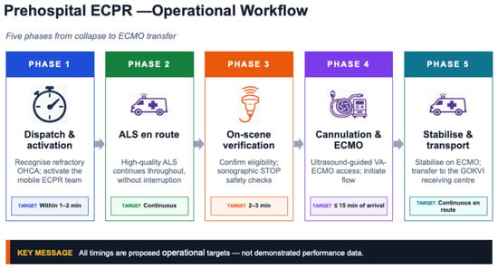
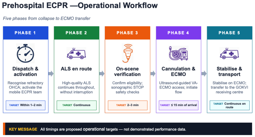
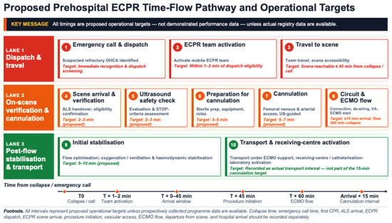
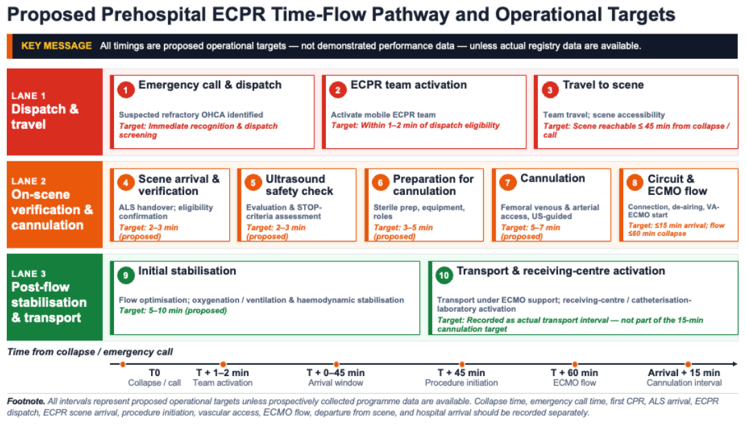

Disclaimer/Publisher’s Note: The statements, opinions and data contained in all publications are solely those of the individual author(s) and contributor(s) and not of MDPI and/or the editor(s). MDPI and/or the editor(s) disclaim responsibility for any injury to people or property resulting from any ideas, methods, instructions or products referred to in the content. |
© 2026 by the authors. Licensee MDPI, Basel, Switzerland. This article is an open access article distributed under the terms and conditions of the Creative Commons Attribution (CC BY) license.