


Abstract
Accurate molecular typing of amyloid deposits is essential because morphologically similar deposits arise from clinically distinct precursor proteins that require different investigations and treatment. Mass spectrometry (MS)-based proteomics has become a reference approach for amyloid typing, but reliable diagnosis depends on the entire tissue-to-report workflow rather than on instrumentation alone. This review examines specimen selection and fixation, Congo red-guided enrichment and laser microdissection, protein extraction and digestion, LC–MS/MS acquisition, database searching, protein inference, precursor assignment, diagnostic ambiguity, quality control, reporting and clinical implementation. Established evidence supports bottom-up LC–MS/MS, particularly on enriched formalin-fixed paraffin-embedded tissue, for identifying common, hereditary, rare and unexpected amyloid precursors. However, inadequate sampling, low peptide recovery, incomplete sequence representation, competing amyloidogenic proteins and inappropriate interpretation can produce inconclusive or misleading results despite technically successful MS acquisition. Emerging strategies, including data-independent acquisition, targeted proteomics, variant-aware and patient-specific sequence analysis, spatial proteomics and machine-learning-supported interpretation, are promising but remain variably validated. The principal challenge is therefore shifting from demonstrating that MS can identify amyloid precursors to ensuring reproducible, transparent and clinically integrated typing across specimens, laboratories and health-care systems.
1. Introduction
Amyloidoses are molecularly heterogeneous disorders in which structurally similar fibrils arise from many unrelated precursor proteins. The 2024 International Society of Amyloidosis (ISA) nomenclature recognizes 42 human fibril proteins, and precursor identity directly affects investigation, prognosis and therapy [1,2]. Diagnosis therefore separates two questions: whether amyloid is present and which protein forms the fibrils. Congo red staining remains the reference histological method for confirming amyloid deposition, while microscopy establishes its spatial distribution. Mass spectrometry (MS) enables molecular typing by identifying the deposited fibril precursor [3,4].
Antibody-based methods preserve spatial information, but their performance depends on reagent repertoire, tissue processing, and expertise. Proteomic typing complements antibody-based methods by analysing Congo red-positive tissue without requiring a predefined antibody panel, and has been validated in routine biopsies and large reference-laboratory series [2,5,6,7]. Genetic testing addresses a distinct question: the presence of a hereditary molecular basis for disease, rather than the identity of the protein forming the tissue deposit.
This review addresses MS-based amyloid typing as an end-to-end diagnostic process: specimen selection and preservation, amyloid localization and enrichment, sample preparation, MS acquisition, database searching and protein inference, interpretation of competing evidence, and clinicopathological integration. Access to MS and its position in the diagnostic pathway vary among clinical centres [8,9,10], but the central analytical principle is constant: a defensible result is generated by the complete chain, not by the mass spectrometer in isolation.
While classic biochemical and antibody-based strategies asked narrower questions about suspected precursors, pathology-guided proteomics enables broad molecular interrogation of the deposit itself. This broader search space is especially important when phenotype, tissue site or monoclonal-protein studies can create misleading diagnostic assumptions. Nevertheless, this broader analytical coverage does not eliminate the need for histological localization or subsequent clinical interpretation [4,11]. Building on the established evidence for MS-based amyloid typing, the present review seeks to integrate three aspects that are often addressed separately: the relationship between analytical failure modes and quality control across the diagnostic workflow (Section 7 and Section 8); the hierarchical interpretation of proteomic evidence, distinguishing confident protein identification from defensible precursor assignment (Section 6); and the translation of analytical findings and diagnostic uncertainty into structured clinical reporting (Section 9). Rather than proposing universally applicable thresholds or replacing existing clinical recommendations, this integrated perspective aims to provide a practical framework for understanding how analytical evidence is converted into a reliable and clinically actionable diagnosis.
This review was developed through iterative, targeted literature searches in PubMed [12], Scopus [13], and Web of Science [14], supplemented by the authors’ previously compiled literature collections and backward and forward citation tracking. Searches were last updated in August 2026, and were further complemented by AI-assisted literature-discovery tools, namely Consensus [15] and ChatGPT (model GPT-5.6 Sol) [16], including its Deep Research functionality [17]. Publications were selected primarily for their relevance to human amyloidosis diagnosis and typing, with emphasis on original methodological studies, analytical and clinical validation studies, large reference-laboratory series, direct methodological comparisons, and relevant guidelines and consensus documents. Foundational studies were considered alongside more recent publications through 2026. When comparing analytical approaches, particular attention was given to specimen characteristics, diagnostic performance, validation design, reproducibility, quality-control criteria and reported limitations. Important findings and bibliographic information were checked against the original publications wherever accessible. General proteomics literature was included when necessary to explain technologies or analytical principles for which amyloid-specific evidence remains limited, with this distinction maintained in the interpretation. Given the heterogeneity of study designs, specimens, analytical workflows and diagnostic endpoints, the literature was synthesized as a narrative review rather than subjected to a formal systematic review or quantitative meta-analysis.
2. The Diagnostic Problem: From Amyloid Detection to Amyloid Typing
2.1. Confirming Amyloid Deposition
Histological confirmation and molecular typing are distinct diagnostic steps. Congo red remains the reference tissue method, although diagnostic birefringence is not restricted to the classic description of “apple-green,” because the observed colour varies with section characteristics and optical configuration [9,18,19]. Fluorescence can improve visualization when deposits are scant, but false-positive staining remains possible and weak or focal amyloid may still be overlooked. These limitations are evident both from fluorescence-based studies and from reports of reduced Congo red affinity in some deposits containing truncated transthyretin (TTR) [20,21,22].
Histomorphology can establish the presence and distribution of fibrillar deposits, but it does not identify the precursor protein. MS is therefore used principally to type histologically defined amyloid. In difficult cases, an amyloid-associated proteomic pattern may provide supporting evidence, but it should lead to pathological reassessment rather than be treated automatically as a substitute for Congo red confirmation [4,23]. In practice, pathology defines the lesion and its location; proteomics then identifies the protein responsible for the deposit.
2.2. Why Amyloid Typing Is Difficult
Amyloid typing is difficult because structurally unrelated fibril proteins can produce similar histological appearances and overlapping clinical phenotypes. While ISA recognizes 42 human amyloid fibril proteins, large clinical proteomics series have demonstrated substantial precursor diversity across anatomical sites. Morphology can inform the differential diagnosis but cannot identify the deposited precursor protein [1,2,24]. The main clinically relevant amyloid types and their precursor proteins are summarized in Table 1.
Table 1.
Representative clinically relevant amyloid types and precursor proteins, distribution and genetic considerations.
The analytical target may itself be heterogeneous. Deposits can contain protein fragments, sequence variants and other proteoforms, while processing-related modifications may further complicate interpretation; formalin-induced modifications can even mimic genuine sequence variation [25]. Amyloid light-chain (AL) amyloidosis poses an additional challenge because each clonal light chain has a patient-specific variable-region sequence that may be incompletely represented in standard protein databases [26].
Antibody-based typing can perform very well in experienced laboratories, but its accuracy remains dependent on antibody panels, epitope preservation and interpretative expertise [11,27]. Clinical information is not definitive either. A monoclonal gammopathy may coexist with TTR amyloidosis (ATTR), while hereditary forms can be misclassified as AL when a circulating monoclonal protein is also present [28,29,30].
True dual amyloidosis creates a different interpretative problem. In a reference-laboratory series comprising 51,309 specimens, Dalland et al. [31] identified 111 patients with two amyloid types. When more than one plausible amyloidogenic protein is detected, spatial distribution, relative abundance and precursor-specific evidence must be assessed before distinguishing background signal from genuine co-deposition.
Across all these situations, the key question is not simply whether a plausible amyloidogenic protein is detected, but whether the analytical and pathological evidence links that protein to the deposit.
2.3. Where MS Fits in the Diagnostic Algorithm
In tissue-based diagnosis, MS provides the molecular typing step between histological confirmation of amyloid and clinicopathological classification of the disease. In the conventional tissue pathway, pathology establishes the presence and location of amyloid, proteomics identifies the fibril precursor, and haematological, genetic and organ-specific investigations then determine the underlying disease process [5,6].
MS is particularly valuable when antibody-based typing is inconclusive, technically limited or discordant, although some centres deploy broad proteomic typing earlier in the diagnostic pathway. These differences largely reflect local expertise, access to specialist laboratories and referral practice rather than a universally accepted testing hierarchy [3,4,9,10].
Cardiac ATTR is an important exception. In appropriately selected patients, validated bone-avid tracer scintigraphy combined with exclusion of a monoclonal gammopathy can establish ATTR cardiomyopathy without tissue biopsy [32,33]. Tissue typing remains important when these criteria are not fulfilled or when clinical, imaging and laboratory findings are discordant. Moreover, identification of TTR in a deposit does not distinguish wild type from variant ATTR; that distinction requires genetic evidence [34]. Tissue MS therefore defines the molecular composition of the deposit, but not, by itself, disease distribution, clonal origin or hereditary status. Those conclusions depend on clinicopathological integration.
The complete diagnostic pathway, from clinical suspicion and tissue selection to molecular typing, clinicopathological integration and reporting, is summarized in Figure 1.
Figure 1.
End-to-end workflow for mass spectrometry-based amyloid typing. Histopathology establishes and localizes the amyloid deposit, after which pathology-guided sampling and proteomic analysis generate evidence for identification of the fibril-forming protein. Computational interpretation integrates precursor-specific evidence with the broader amyloid-associated proteome and competing candidates. The molecular assignment is subsequently reconciled with pathological, haematological, genetic and clinical findings to produce the final diagnostic interpretation and report.
3. Specimens and the Pre-Analytical Phase
3.1. Tissue Types
Specimen selection should prioritize sufficient, representative and spatially identifiable amyloid deposits, rather than relying solely on the least invasive sampling site. Formalin-fixed paraffin-embedded (FFPE) material remains the dominant substrate for clinical proteomic typing, and large series have demonstrated successful analysis across major involved organs as well as surrogate biopsy sites [2,35,36].
For surrogate tissues, the ability to detect amyloid should be distinguished from the ability to type it once present. Proteomic typing of fat aspirates can be effective once amyloid is present; however, the sensitivity of amyloid detection in this tissue varies with amyloid type and disease burden and can be lower in ATTR [37,38]. Kidney and endomyocardial biopsies are particularly useful when clinically indicated because deposits can be localized and selectively sampled. Whole-biopsy cardiac proteomics without prior spatial enrichment is feasible, but the published evidence is more limited than for laser microdissection (LMD)-based workflows applied across multiple tissue types [39,40].
When several specimens are available, the most informative analytical sample is usually the one with the clearest residual deposit and the least confounding surrounding tissue. A negative or low-burden surrogate biopsy should therefore not preclude analysis of an involved-organ specimen that contains demonstrable amyloid.
Fresh and fresh-frozen tissues can also be used for MS-based amyloid typing, but bulk unfixed tissue should not be regarded as a general replacement for pathology-guided enrichment. Noborn et al. [40] initially considered 78 fresh-frozen endomyocardial biopsies without LMD, of which three were excluded because of insufficient material or information. Of the remaining 75, 65 fulfilled the study’s predefined four-precursor classification criterion, and 61 of these 65 assignments agreed with the reference diagnosis. Unfixed fat aspirates can likewise support clinical typing when amyloid is present: Vrana et al. reported 88% sensitivity and 96% specificity during assay validation, with a reported typing yield of 90% among 366 amyloid-positive clinical aspirates [38]. Frozen-tissue LMD has also been demonstrated in kidney, although the available comparative evidence is insufficient to show consistent diagnostic superiority over FFPE material [41].
By contrast, Congo red-stained FFPE sections are directly compatible with validated LMD–LC–MS/MS workflows, and successful typing has also been reported from unstained FFPE sections selected with reference to an adjacent stained section [7,42]. Unfixed material may offer advantages in selected settings, but those advantages do not remove the value of spatial selection. Enrichment is especially important when deposits are sparse, focal, heterogeneous or potentially mixed. At present, FFPE-based, deposit-guided workflows retain the broadest clinical validation.
3.2. Tissue Fixation and Processing
FFPE material supports both routine and retrospective amyloid proteomics, but fixation should be treated as part of the analytical process rather than as a neutral pre-analytical step. Formaldehyde crosslinking and chemical modification can affect protein extraction and peptide representation, and general FFPE proteomics studies have shown that these effects can introduce fixation-dependent bias despite overall analytical robustness [43,44].
Amyloid-specific experience confirms that FFPE tissue is suitable for clinical typing but also illustrates artefacts with direct interpretative consequences. In particular, formalin-associated lysine methylation can mimic genuine sequence variants in TTR and immunoglobulins, creating a potential source of false variant assignment if sample-processing effects are not considered [25].
Congo red staining is compatible with subsequent dissection and MS analysis in established workflows [5,6]. Section thickness, however, is better regarded as a validated assay parameter than as a universal requirement, because laboratories differ in section preparation, tissue area collected and downstream processing [7,45]. Fixation conditions, additional staining or processing steps and archival history should therefore be documented and incorporated into local validation, particularly when peptide recovery is poor or sequence-variant interpretation is clinically important.
3.3. Identification of Amyloid-Containing Regions
Beyond confirming the presence of amyloid, Congo red also helps define the tissue region whose proteome will ultimately be interpreted. Pathology-guided localization is therefore part of the analytical workflow because it links the molecular measurement to the lesion and reduces contribution from unrelated surrounding tissue [5,7,45].
Polarization remains important for confirming Congo red-positive deposits, while fluorescence can facilitate practical localization when deposits are small or difficult to visualize. Because fluorescence is not completely specific, however, the region selected for proteomic analysis should still be supported by morphology and, where appropriate, pathologist review [22,46].
Sampling should also reflect deposit architecture. Large, homogeneous deposits can be collected contiguously, whereas focal vascular, glomerular or interstitial deposits may require selective or spatially separate sampling, particularly when heterogeneity or dual amyloidosis is suspected [39]. Retaining annotated images and compartment information provides traceability and can be important when proteomic findings later prove discordant with the original pathological interpretation.
3.4. Amyloid Enrichment
Enrichment determines how closely the measured proteome reflects the amyloid deposit itself. Its main purpose is to increase the contribution of deposit-associated proteins relative to plasma, extracellular matrix and other background signals, which can otherwise mask the signal of the fibril precursor.
LMD is the most broadly validated enrichment strategy, particularly for small, focal or heterogeneous deposits, and has enabled clinical typing from very limited amyloid-containing regions [5,7]. Minimum tissue input, however, is not universal. Published requirements for dissected area, replicate number and protein yield reflect differences in tissue type, preparation, instrumentation and diagnostic calling criteria. Inadequate amyloid content should therefore be treated as a specimen-adequacy problem rather than compensated for by over-interpreting weak or sparse spectra [6,7,41].
Manual microdissection can provide an alternative when deposits are well demarcated. Phipps et al. [42] developed and validated a workflow in which amyloid-containing regions were recovered from deparaffinized 10-µm FFPE sections with a 29-gauge needle under a dissecting microscope. In a test series, 22 of 23 previously characterized amyloid cases yielded the expected assignment, whereas the remaining specimen was classified as sampling-inadequate rather than incorrectly typed. The workflow incorporated multiple separate scrapes and an explicit proteomic adequacy metric, suggesting that replicate collection and objective assessment of amyloid content can partly mitigate the lower spatial precision of manual sampling [42]. However, the evidence base remains substantially narrower than for LMD. The distinction between development and independent testing should be retained: the initial dataset comprised 121 FFPE blocks and 468 individual scrapes, and the 23-case known-type test subset included five repeated development cases. The expected assignment was obtained in 18 of the 19 non-repeated cases (95% as reported), while the remaining non-repeated case was deemed sampling inadequate [42].
Importantly, Congo red-guided manual dissection should not be conflated with direct scraping of an already Congo red-stained diagnostic slide. In the best-validated clinical workflow, the analytical section itself was unstained and was sampled according to an adjacent Congo red section [42]. Other studies have shown that manually selected Congo red-defined amyloid-rich regions can yield high-quality proteomic data, supporting the general feasibility of microscope-guided manual enrichment [47,48]. These studies were primarily designed for compositional proteomics rather than routine clinical typing. Moreover, coarse needle-based dissection can collect substantial adjacent non-amyloid tissue, increasing background complexity and reducing the spatial specificity of the measured proteome [7]. Manual dissection is therefore most defensible for larger, conspicuous deposits and as a laboratory-validated fallback when LMD is unavailable; equivalence or non-inferiority to LMD for small, vascular, glomerular or heterogeneous deposits has not been established.
Whole-biopsy or minimally enriched approaches can also yield successful amyloid typing in selected organ-specific workflows, particularly when amyloid burden is substantial, but they sacrifice the spatial specificity of deposit-directed sampling and remain less broadly validated across heterogeneous tissues and focal deposits [40].
The choice between LMD, manual dissection and whole-tissue analysis depends primarily on whether amyloid can be histologically localized, on deposit size and heterogeneity, and on the extent to which the laboratory has validated each sampling strategy. In general, LMD remains the preferred approach for small, focal or heterogeneous deposits; manual dissection is most defensible for larger, conspicuous deposits when deposit boundaries can be recognized reliably; and whole-tissue workflows are best regarded as organ-specific alternatives rather than substitutes for deposit-directed enrichment. When clinical suspicion remains high despite negative or equivocal histology, the specimen should first be reassessed rather than automatically proceeding to whole-tissue proteomics. Figure 2 summarizes this evidence-informed sampling framework.
Figure 2.
Proposed evidence-informed framework for selecting amyloid enrichment strategy before MS-based typing. This figure represents a practical interpretive framework derived from the published literature rather than a universally validated clinical algorithm.
Because enrichment directly changes the biological material entering the MS workflow, the sampling strategy and downstream interpretation model should be validated as a single analytical system. The principal specimen types and pre-analytical workflows used for MS-based amyloid typing, together with their main strengths and limitations, are summarized in Table 2.
Table 2.
Major specimen types and pre-analytical workflows for MS-based amyloid typing.
4. Proteomic Sample Preparation
4.1. Deparaffinization and Rehydration
FFPE workflows require effective paraffin removal. Deparaffinization must remove wax without disrupting the spatial workflow or downstream extraction. Conventional xylene/alcohol approaches are widely used, but no amyloid-specific solvent sequence is demonstrably superior, and the complete sectioning–staining–dissection–extraction workflow needs to be considered for proper validation [49,50].
4.2. Protein Extraction from FFPE Material
Extraction should recover a representative, protease-accessible complement of deposit-associated proteins from crosslinked FFPE tissue while minimizing losses of low-abundance analytes and maintaining compatibility with downstream MS. Heat-assisted crosslink mitigation with sonication is common, and both mild-detergent and stronger sodium dodecyl sulphate (SDS)/trapping workflows can succeed [49,50,51]. No extraction chemistry is clearly superior across amyloid types. More aggressive solubilization may improve protein recovery but necessitate additional cleanup steps that risk analyte loss when only limited microdissected material is available. Extraction performance should therefore be judged by recovery and sequence coverage of diagnostically relevant precursors, rather than by total protein yield alone.
4.3. Reduction and Alkylation
Reduction and alkylation are workflow dependent. They may improve overall protein sequence coverage, which may be particularly important for Ig light chains, but benefit varies with precursor and extraction chemistry; laboratories should validate these two steps against diagnostic sequence recovery and artefact formation [7,41,49].
4.4. Proteolytic Digestion
Trypsin remains the dominant protease because its cleavage pattern is compatible with established LC–MS/MS and database-search workflows. Enzyme amount, digestion time and temperature vary with workflow and recovered substrate; the important performance variable is reproducible generation of informative peptides rather than a universal enzyme-to-tissue ratio [41,50,52]. Most established amyloid-typing workflows use overnight tryptic digestion, commonly for approximately 16–18 h, but prolonged digestion is not necessarily advantageous. General proteomics studies show that extended trypsin exposure can alter the recoverable peptide population through continued specific and nonspecific proteolysis, including generation of short peptides with poor LC–MS detectability, while prolonged incubation at mildly alkaline pH and 37 °C can increase artefactual modifications such as deamidation [53,54,55]. Whether amyloid precursor-derived peptides are especially susceptible to such losses has not been systematically established. Digestion time should therefore be validated together with enzyme amount and reaction conditions against precursor sequence coverage, missed cleavage and reproducibility. Excess missed cleavage can reduce effective sequence coverage and should therefore be monitored as an assay/QC characteristic [7]. A candidate represented by few peptides in a digest with globally poor cleavage quality should carry less confidence than the same peptide pattern in an otherwise well-performing sample. Alternative strategies, including sequential LysC/trypsin digestion and accelerated microwave-assisted preparation, are feasible but have less amyloid-specific validation and have not shown broad superiority to conventional tryptic workflows [49,52].
Beyond cleavage efficiency, the recovery and identification of precursor-derived peptides depend on several sequence-dependent physicochemical and analytical factors. The distribution of protease cleavage sites determines the length and composition of the resulting peptides, which may be too short for confident identification, too long for efficient LC–MS/MS analysis, or poorly soluble under the conditions employed. Moreover, peptide recovery is not uniform: hydrophobicity, solubility and interactions with sample-handling surfaces can lead to selective losses during transfer, storage and chromatographic preparation, particularly when only small amounts of microdissected material are available [56,57]. Consequently, even complete enzymatic digestion does not guarantee representative recovery of the precursor sequence.
Peptides that are successfully recovered also differ substantially in their ability to be detected and identified by MS. Peptide length, amino-acid composition, hydrophobicity, basicity and charge-state distribution influence chromatographic retention, electrospray ionization efficiency and fragmentation behaviour [58,59,60]. Poorly retained peptides may be lost during reversed-phase trapping or elute in chromatographic regions with substantial background, whereas strongly retained or poorly soluble peptides may exhibit reduced recovery or unfavourable elution profiles. Coelution with more abundant peptides can further produce ion suppression, and in data-dependent acquisition, low-intensity precursors are less likely to be selected for fragmentation [61]. Even when precursor ions are detected, sequence- and charge-dependent fragmentation may yield spectra of insufficient quality for confident database identification. Recent peptide-detectability studies have further demonstrated that peptides present at comparable abundance can produce markedly different MS responses, reinforcing the distinction between peptide abundance and analytical observability [62]. For amyloid typing, these effects are particularly relevant when the diagnostic evidence depends on a limited number of precursor-specific, clonotypic or variant-containing peptides. Their absence from an LC–MS/MS dataset may reflect incomplete recovery, poor ionization, insufficient fragmentation or unsuccessful identification rather than genuine absence from the deposited protein. Therefore, digestion efficiency, peptide recovery and MS detectability should be recognized as complementary determinants of sequence coverage and diagnostic confidence, rather than treating missed-cleavage rates as a comprehensive measure of peptide-level analytical performance.
Rapid digestion formats based on immobilized or thermostable trypsin, including commercial systems such as SMART Digest and experimental on-line immobilized-enzyme reactors, can substantially shorten proteolysis and facilitate automation; however, their performance is preparation- and analyte-dependent, and amyloid-specific clinical validation is currently lacking [63,64].
4.5. Peptide Cleanup/Desalting
Peptide cleanup removes salts, detergents and other LC- or electrospray-incompatible compounds and may concentrate the digest. Clinical workflows use several C18- or solid-phase formats without evidence that one sorbent is intrinsically superior [49,52,65]. Because every adsorption, wash and transfer can lose peptide, cleanup should be scaled to input and validated for recovery, residual background and LC compatibility, particularly in microdissected specimens [66].
More integrated solid-phase preparation strategies can reduce the number of transfers by combining contaminant removal with protein capture, digestion and peptide recovery. Suspension trapping (S-Trap), which permits detergent-rich extraction followed by on-trap digestion, has now been applied directly to amyloid tissue. In a recent study of 47 laser-microdissected heart and kidney specimens, S-Trap preparation combined with DIA and high-flow LC–MS/MS enabled amyloidogenic-protein assignment in approximately 96% of both training and test sets while reducing sample preparation to less than 8 h [67]. Other low-input or streamlined platforms, such as SP3, offer similar opportunities to minimize transfers and facilitate automation in general proteomics, but amyloid-specific clinical validation remains limited [68,69].
4.6. Low-Input Proteomics Considerations
Low input is intrinsic to selective amyloid enrichment. When protein yield cannot be measured reliably, standardized tissue input, adjusted enzyme amounts and reduced reaction volumes can help maintain reproducible digestion [7,41,49]. At trace input, surface adsorption and environmental contamination become proportionally important; general low-input untargeted methods such as nanoPOTS and SP3 show how reduced volumes and consolidated handling can improve recovery, but these data remain extrapolations unless validated specifically for amyloid typing [68,70].
Recent low-input FFPE protocols can analyse much smaller captured areas than most validated amyloid assays, but those values should not be treated as clinical amyloid limits of detection [71,72]. The diagnostic consequence is central: poor recovery can mimic biological absence. Analytical sensitivity therefore belongs to the complete tissue-to-spectrum workflow, not the instrument alone.
Low-input handling also amplifies stochasticity between replicate dissections. Replicates are therefore useful not only as technical precision measurements but as evidence that a precursor signal survives independent sampling of the deposit. Conversely, discordance between replicates can indicate heterogeneity, contamination or proximity to the analytical limit and should trigger review before a weak candidate is promoted to a diagnostic call.
Key sample-preparation variables and their potential diagnostic consequences are summarized in Table 3.
Table 3.
Major sample-preparation variables and their potential propagation into diagnostic uncertainty in MS-based amyloid typing.
5. Mass Spectrometry Strategies for Amyloid Typing
5.1. Bottom-Up Shotgun LC–MS/MS
Bottom-up LC–MS/MS analysis, also known as shotgun proteomics, remains the core of tissue amyloid proteomics. Proteins from an enriched deposit are digested to peptides that are separated by reversed-phase HPLC, and tandem MS spectra are then matched to protein sequence databases. The approach is untargeted in the diagnostic sense: common, hereditary and unexpected precursors can be interrogated within the same run, although extraction, digestion, ionization, acquisition and database representation still bias what is observed [2,73].
Its clinical maturity is supported by the foundational LMD workflow, independent implementations and large reference-laboratory series [2,5,73,74,75]. Nanoflow LC has predominated because of its sensitivity at low sample input, while high-resolution tandem MS enables accurate measurement of peptide precursor ions and sequence-informative fragmentation. In data-dependent acquisition (DDA), only selected precursor ions undergo fragmentation; dynamic exclusion reduces repeated selection of abundant ions and can increase proteome coverage [50].
Clinical workflows generally use label-free evidence such as spectral counts or intensity-based metrics. These measurements can strengthen precursor assignment but are not direct measures of fibril mass and must not be equated with the diagnosis itself [49]. The strength of shotgun LC–MS/MS is diagnostic breadth; its corresponding limitations are stochastic, intensity-biased sampling and the need for structured interpretation.
5.2. DDA Versus DIA
In general proteomics, data-dependent acquisition (DDA) methods sample selected precursors and can miss low-abundance peptides, whereas data-independent acquisition (DIA) samples predefined m/z windows more systematically and generally improve reproducibility at the cost of greater computational complexity [76]. Amyloid-specific evidence is promising but still limited. Aguilan et al. [50] showed fewer missing observations with short-gradient DIA, and Mann et al. [67] reported approximately 96% amyloid-precursor assignment in a 47-patient high-flow LC-DIA series; multicentre same-specimen validation across rare and mixed amyloidoses is still lacking. DIA should therefore be judged by final precursor-classification performance, not by peptide yield or missingness alone.
5.3. Targeted MS
Unlike untargeted shotgun proteomics, targeted approaches such as SRM/MRM and PRM quantify predefined peptides with high sensitivity and can streamline analysis when the diagnostic differential is restricted to a limited number of candidate precursors. Amyloid-specific studies support their use in focused cardiac and renal applications [65,77,78,79]. For example, Park et al. evaluated an MRM panel targeting common precursors in 16 Congo red-positive specimens and ten Congo red-negative controls, identifying the relevant targets in all 16 selected positive specimens [79]. Li et al. developed a renal PRM approach using ten discovery and 26 validation cases, together with an endogenous reference-protein panel; the restricted amyloid-type range and modest cohort sizes limit extrapolation to broad routine typing [78].
The limitation is inherent to the targeted approach: rare precursors, private sequence variants and clonotypic immunoglobulin sequences that are not represented in the predefined target panel may remain undetected. Targeted MS is therefore complementary to, rather than a general replacement for, broad proteomic typing, and a negative result has exclusionary value only for analytes included within the validated panel.
5.4. MALDI and Mass Spectrometry Imaging
Matrix-assisted laser desorption/ionization mass spectrometry imaging (MALDI-MSI) acquires spectra directly across tissue sections while preserving spatial coordinates, allowing amyloid-associated or precursor-derived signals to be mapped back to histology. Cardiac, gastrointestinal/pulmonary and renal studies have demonstrated feasibility for spatial classification and tissue segmentation [80,81,82,83]. Its main potential advantage is the ability to resolve spatial heterogeneity and potentially distinguish mixed deposits. Current limitations include small study cohorts, incomplete precursor coverage and technically demanding on-tissue preparation and data analysis. MALDI-MSI therefore remains investigational for routine broad amyloid typing, with clinical implementation still requiring multicentre validation across tissue types, rare precursors and analytical platforms.
5.5. Emerging Technologies
Field asymmetric waveform ion mobility spectrometry (FAIMS) and trapped ion mobility spectrometry (TIMS) introduce an additional gas-phase separation step before mass analysis and may improve selectivity or reduce spectral complexity, although amyloid-specific validation remains limited [50,71]. In parallel, shorter gradients, higher-flow LC and greater automation may improve throughput and analytical robustness; Mann et al. [67] provide early clinical evidence for a high-flow DIA workflow.
Top-down MS examines intact proteins or proteoforms without prior proteolytic digestion, whereas native MS aims to preserve non-covalent interactions and aspects of higher-order structure during analysis. Both may provide information that is inaccessible to conventional bottom-up workflows, particularly at the proteoform or structural level, but neither is established for routine tissue typing [84]. Their value will ultimately depend on whether they improve reproducible precursor assignment and clinically relevant interpretation rather than simply increasing analytical depth.
The principal analytical characteristics, limitations, evidence base and current clinical position of these MS strategies are summarized in Table 4.
Table 4.
Comparison of MS strategies for amyloid typing.
6. From Spectra to Diagnosis: Bioinformatics and Interpretation
6.1. Protein Identification
The primary computational output of MS data analysis is a hierarchy of peptide-spectrum matches (PSMs), peptide identifications and inferred proteins, not a diagnosis. Search-engine settings, database composition—including contaminant, immunoglobulin and variant sequences—and software versions must therefore be treated as defined components of the assay [7]. False-discovery-rate control estimates identification error within the search model, commonly using target-decoy strategies, but does not account for incorrect tissue sampling, database omission or biological context [85]. Identification confidence and diagnostic confidence are distinct.
Protein inference is particularly relevant when peptides are shared among homologous proteins or immunoglobulin chains. Unique, high-quality precursor-specific peptides can be more discriminatory than numerous shared peptides, and sequence coverage is itself shaped by protein length, cleavage sites, fixation and peptide detectability [86]. Laboratories therefore use different combinations of FDR, score, peptide, replicate and protein-level criteria [7,87]. No universal minimum peptide count or sequence-coverage threshold establishes an amyloid precursor assignment. High-quality spectra can correctly identify a plasma-derived protein without establishing that it constitutes the amyloid fibrils. Conversely, a diagnostically decisive variant or clonotypic peptide may be represented by relatively few spectra.
6.2. What Constitutes an Amyloid Proteomic Signature?
Amyloid deposits contain recurrent non-fibrillar proteins, most consistently serum amyloid P component (SAP), apolipoprotein E (ApoE) and apolipoprotein A-IV (ApoA-IV). Their recurrent detection across amyloid types underlies the concept of an amyloid-associated proteomic signature. This signature can support the assessment of specimen adequacy but cannot, by itself, distinguish AL, ATTR, amyloid A amyloidosis (AA) or other amyloid types [6,38,88].
The classical three-protein signature is supportive rather than obligatory. In the Brisbane series, at least two of the three proteins were detected in 76% of successfully typed cases, while renal studies suggest that individual signature components vary with amyloid subtype [6,89]. Broader multivariate amyloid-associated profiles may improve classification [90], but many extracellular-matrix and plasma proteins lack subtype specificity. Signature proteins must therefore be interpreted in the context of tissue morphology and enrichment and should neither substitute for Congo red confirmation nor be confused with the fibril precursor.
Detection of the signature can strengthen confidence that a small dissection is genuinely amyloid-rich when precursor evidence is otherwise coherent. Conversely, absence of the signature should prompt review of specimen adequacy but does not, by itself, invalidate a strong precursor assignment. If an amyloid-associated profile is present but no convincing precursor is identified, the appropriate result is indeterminate rather than a forced classification. This logic is incorporated into the decision framework in Figure 3.
Figure 3.
Interpretation decision tree for mass spectrometry-based amyloid typing. The decision pathway begins with assessment of specimen and analytical adequacy, followed by evaluation of credible fibril precursor evidence based on peptide specificity, sequence coverage, relative abundance or enrichment, and competing amyloidogenic proteins. The amyloid-associated proteomic signature, including serum amyloid P component (SAP), apolipoprotein E (ApoE) and apolipoprotein A-IV (ApoA-IV), provides supporting evidence but is not an obligatory prerequisite for precursor assignment. Its absence alone does not invalidate an otherwise well-supported molecular identification. Inadequate specimens or analytical results should be reported as insufficient for typing, whereas analytically adequate specimens without a defensible precursor assignment should be classified as indeterminate, with appropriate investigation of database limitations, immunoglobulin variable sequences, sequence variants and rare precursors. Detection of multiple plausible precursors requires assessment of their independent molecular evidence and spatial distribution before concluding dual amyloidosis. Subtype-specific interpretation should include haematological correlation for suspected immunoglobulin-derived amyloid, germline testing when hereditary disease is possible, and assessment of competing proteins and clinicopathological concordance. The framework is conceptual; evidence thresholds and diagnostic-confidence categories require assay-specific validation. Ig, immunoglobulin; LC–MS/MS, liquid chromatography–mass spectrometry; MS, mass spectrometry; QC, quality control.
Microdissected Congo red-positive material should therefore be considered an amyloid-deposit proteome rather than a preparation of purified fibrils. In cardiac AL amyloidosis, direct analysis of extracted fibrils has shown that the deposited light chain may comprise a heterogeneous population of fragments spanning variable and constant domains, demonstrating that precursor identity and mature fibril proteoform are not necessarily equivalent [91]. At the other end of the evidentiary spectrum, spatial proteomics has shown that vitronectin can reproducibly co-localize with several unrelated amyloid types without defining the fibril precursor [92]. Larger same-organ studies likewise show that the accessory proteome is partly shared and partly context dependent: cardiac AL and ATTR deposits contain overlapping associated proteins but differ quantitatively in complement, contractile and keratin-associated components, whereas renal amyloidosis shows distinct complement-dominated patterns across several precursor classes [93,94]. Mere detection within the dissected region does not demonstrate incorporation into the fibril.
6.3. How Is the Amyloid-Forming Protein Assigned?
The central interpretative task is to determine which identified protein forms the amyloid fibrils, or whether two distinct precursors are present in cases of dual amyloidosis. A universal “most abundant protein wins” rule is not defensible because amyloid-rich tissue also contains plasma proteins, extracellular-matrix components and recurrent amyloid-associated proteins, while peptide detectability and MS response vary substantially between proteins [88]. Precursor assignment should instead integrate the quality and specificity of precursor-derived peptides, sequence coverage, relative abundance or enrichment, reproducibility across replicates or dissections, expected tissue background, amyloid-associated context and the presence of competing amyloidogenic candidates.
Laboratories formalize these dimensions in different ways. Ke et al. [95] combined precursor, light-chain and heavy-chain scores in renal amyloidosis; Noborn et al. [40] used relative dominance among four common cardiac precursors; Taylor et al. [45] applied an AFib-specific pattern to distinguish deposited fibrinogen alpha chain (FGA) from plasma background; and Hollfoth et al. [49] normalized candidate intensities to SAP and explored machine-learning classification. These approaches can reduce interpretative subjectivity, but their numerical thresholds depend on tissue type, the candidate search space, the abundance metric and the complete analytical workflow. Inter-laboratory concordance despite different software pipelines is reassuring, but it does not establish a universal calling threshold [87]. The different endpoints illustrate the limits of direct comparison: Ke et al. correctly classified 44 of 50 renal validation specimens (88% as reported) using laboratory-derived spectral-count criteria [95], whereas Canetti et al. observed 34/40 initial agreement and 37/40 concordance after review when two centres reprocessed identical MS raw data (85% and 92.5%, respectively, as reported) [87]. The latter measures computational agreement, not full-process clinical accuracy.
The workflow should first establish specimen and analytical adequacy, and then evaluate credible precursor-specific evidence against expected tissue background and competing amyloidogenic candidates, with amyloid-associated proteins providing supporting evidence where present. Subtype-specific or quantitative criteria should be applied when required. The resulting molecular assignment should then be reconciled with pathology and clinical data. Clinical context is most useful for exposing contradictions and directing the next etiological investigation; it should not override strong molecular evidence simply because the result is unexpected.
Relative abundance must likewise be judged against expected tissue background. Immunoglobulins, fibrinogen, apolipoproteins and other plasma-derived proteins may be present within the analysed region without constituting fibrils. A confident precursor call therefore depends on patterns such as disproportionate enrichment of one chain or isoform, sequence coverage extending into diagnostically informative regions, concordance across separate dissections, and the absence of a stronger competing amyloidogenic protein.
Non-precursor proteins may nevertheless provide useful subtype-supportive information when interpreted within the correct organ and sampling context. In cardiac amyloid deposits, ATTR showed relative enrichment of complement and contractile proteins, whereas AL showed greater representation of keratin-associated proteins; within AL, clusterin was more abundant in κ than in λ deposits [94]. Whole-biopsy cardiac proteomics independently found higher complement abundance in ATTR than AL, supporting complement as a contextual cardiac signal rather than a standalone discriminator [96]. In kidney, a 2,650-case proteomic atlas identified particularly distinct, complement-rich profiles in AFib, AA and AApoCII and substantial within-type heterogeneity in AL and ALECT2 [93]. In a separate renal cohort, APOA4 was absent from ALECT2 deposits while SAP/APCS and APOE remained detectable [89]. Taken together, these data support an organ-stratified proteomic atlas as a probabilistic second layer of interpretation that can strengthen or weaken a precursor-based assignment, rather than the use of any single accessory protein as a universal subtype marker.
Algorithmic classifiers may help formalize multidimensional precursor assignment, but their diagnostic performance depends critically on training-set composition, subtype representation and validation design. Palstrøm et al. [90] developed support vector machine classifiers using proteomic data from 75 amyloid-positive and 78 amyloid-negative biopsies, subsequently achieving correct subtype classification in 102 of 103 prospectively collected blinded cases. However, the validation cohort was dominated by ATTR (68 cases) and ALλ (25 cases), with substantially fewer AA (six) and ALκ (four) specimens, and the model was restricted to these four subtype categories. More recently, Hollfoth et al. [49] reported approximately 94% accuracy using an XGBoost classifier incorporating approximately 160 amyloid-related proteins, although the machine-learning evaluation involved only 30 training and 18 test specimens from a predominantly cardiac cohort. These results demonstrate the feasibility of automated interpretation but do not establish generalizability to rare, mixed or previously unrepresented amyloid types. Moreover, internal or single-centre validation cannot establish transferability across tissue types, enrichment strategies and analytical platforms. Clinical implementation would therefore require broader external validation, adequate representation of uncommon subtypes, calibrated diagnostic confidence and predefined procedures for ambiguous or out-of-distribution results.
Diagnostic confidence is consequently multidimensional. Several concordant features may provide stronger evidence than a single dominant abundance metric, while meaningful discordance should lower confidence even when one candidate ranks clearly first. Although quantitative interpretation frameworks differ in their abundance metrics, normalization strategies and numerical thresholds, they show increasing convergence in their underlying diagnostic principles. Across approaches, reliable precursor assignment depends on analytical and specimen adequacy, credible precursor-specific peptide evidence, evaluation of relative abundance or enrichment against tissue background and competing amyloidogenic proteins, and clinicopathological concordance [40,45,49,95]. This convergence remains conceptual rather than numerical: quantitative thresholds are generally derived within specific tissue types, candidate panels and analytical workflows, and their transferability across laboratories has not been established [40,49,87,95]. Laboratories should therefore define transparent decision rules that distinguish required from supportive evidence and specify which discrepancies trigger review. These rules need not be identical between centres, but they should be explicit enough to support analytical validation, reproducibility testing and meaningful external comparison. Harmonization should prioritize shared evidence requirements, adequacy criteria and consistent handling of diagnostic uncertainty rather than the premature adoption of universal quantitative thresholds.
6.4. Immunoglobulin Light-Chain Amyloidosis
AL presents a distinctive interpretative problem because the disease-defining light chain is patient-specific. Constant-region peptides can establish κ or λ class, but they are shared across many immunoglobulins and may arise from polyclonal blood or tissue background; by themselves, they do not demonstrate clonality. Variable-region and clonotypic peptides provide stronger evidence because they link the deposit to a restricted immunoglobulin sequence [26].
Dedicated sequence-template strategies can recover variable-region information that is missed by conventional reference-database searches. Dasari et al. [26] achieved high recovery of amyloidogenic variable-gene families, and Kourelis et al. [97] assigned a variable-gene family in most cases from a large systemic and localized AL cohort. Failure to recover a clonotypic peptide, however, does not exclude AL. Somatic sequence variation, unfavourable tryptic peptide properties and low amyloid abundance can all limit coverage.
Augmented immunoglobulin databases, de novo sequencing and patient-specific sequence resources can strengthen difficult assignments. Hollfoth et al. [49] resolved otherwise unexplained AL-λ cases after de novo sequence analysis, while Zhu et al. [98] linked reconstructed urinary monoclonal light chains to matched renal deposits in eight patients. AL evidence is therefore best viewed hierarchically: isotype dominance provides the weakest level of support, variable-gene-family assignment provides stronger evidence, and patient-specific clonotypic sequence evidence provides the most direct molecular link to the pathogenic light chain.
This distinction matters because family-level assignment and clonotype identification are not equivalent. A variable-gene-family assignment shows that recovered peptides are compatible with a particular immunoglobulin gene family but does not, by itself, identify the patient-specific clonal rearrangement, whereas a patient-specific clonotypic sequence can connect the deposited protein much more directly to the responsible clone. Neither should be inferred from constant-region dominance alone. Haematological and proteomic investigations are therefore complementary: serum, urine and marrow studies identify the clone, whereas tissue proteomics determines whether its product is represented in the amyloid deposit.
6.5. Heavy-Chain (AH) and Heavy/Light-Chain (AHL) Amyloidosis
Heavy-chain (AH) and heavy/light-chain (AHL) amyloidosis extend the same inference problem to immunoglobulin heavy chains. Proteomics can assess heavy- and light-chain peptides simultaneously and is particularly useful when antibody-based typing is equivocal. Sethi et al. [99] identified heavy-chain material in diagnostically difficult renal cases, and Nasr et al. [100] characterized five AH and eleven AHL patients, concluding that a substantial proportion would not have been classified correctly without proteomic analysis.
As with light chains, detection of heavy-chain constant regions alone does not establish monoclonality or prove incorporation into fibrils. A convincing AH or AHL assignment requires coherent enrichment of the relevant heavy chain, variable-region evidence where recoverable, appropriate light-chain findings and exclusion of a stronger competing precursor. Because these entities are rare, the evidence base remains dominated by specialist renal series.
AHL is particularly susceptible to overcalling because both heavy- and light-chain peptides can originate from circulating immunoglobulins rather than from the amyloid fibrils. Spatial enrichment and coherent relative evidence are therefore especially important before assigning combined heavy/light-chain amyloidosis. When heavy-chain evidence is weak or confined to shared constant regions, AL with incidental heavy-chain signal may be a more defensible interpretation unless additional peptide, pathological or clonal evidence supports genuine, combined fibril composition.
6.6. ATTR
Strong TTR peptide evidence in an amyloid-enriched region supports ATTR, but it does not by itself distinguish ATTRwt from ATTRv. Variant-aware searches can identify pathogenic substitutions when the altered peptide is analytically observable; Dasari et al. [101] reported high performance for curated hereditary variants. Brown et al. [34], however, failed to detect pathogenic TTR variants in a subset of biopsy-proven ATTR cases, showing that absence of a variant peptide cannot establish ATTRwt.
Positive variant-like evidence also requires caution. Formalin-associated lysine methylation can mimic the p.Val142Ile TTR substitution and produce false variant assignments in FFPE tissue [25]. Tissue proteomics therefore establishes the deposited protein and may provide sequence-level information, whereas germline testing establishes constitutional TTR status and remains necessary for hereditary classification and family counselling.
This asymmetry should be reflected in reporting. A well-supported variant peptide may prompt or reinforce targeted genetic investigation, but failure to detect one cannot be used to exclude hereditary disease. Conversely, a variant-like spectrum from FFPE material should remain provisional until the peptide sequence and potential modification chemistry have been reviewed. Proteomics and germline testing answer related but different questions: one characterizes the deposit, the other the inherited genotype.
6.7. Other Hereditary Amyloidoses
Rare hereditary amyloidoses require integration of precursor identity with sequence-level evidence because several relevant proteins, including fibrinogen and apolipoproteins, are also normal circulating constituents. AFib illustrates the problem clearly: detection of FGA alone is not sufficient, whereas disproportionate FGA enrichment, informative C-terminal coverage and a pathogenic variant together provide much stronger evidence for deposit-specific assignment [45]. Variant-aware workflows can identify both known and novel substitutions, although not every pathogenic sequence is analytically recoverable [101].
Genotype and deposited protein must also remain conceptually separate. Rowczenio et al. [102] showed that an APOA1 variant could be incidental when the actual deposit was AL, while reports of AApoAII, AApoCII and AApoCIII demonstrate differing patterns of mutant and wild-type incorporation into fibrils [103,104,105]. A robust strategy therefore combines broad precursor identification, variant-aware or de novo sequence interrogation and germline confirmation when hereditary disease is suspected. Discordance between these layers should be reported explicitly rather than resolved by forcing them into a single conclusion.
These disorders also illustrate why “pathogenic variant present” and “amyloid caused by that variant” are not synonymous. Demonstrating the encoded protein within the deposit materially strengthens causal attribution, whereas identification of a variant without corresponding deposit evidence may represent an incidental inherited finding. Conversely, failure to observe the mutant peptide may reflect unfavourable cleavage, ionization or sequence coverage rather than true absence from the fibril. Genetics is therefore essential, but it should not substitute for molecular identification of the tissue deposit.
6.8. Rare and Novel Amyloid Proteins
Untargeted proteomics is particularly valuable when the precursor is rare or clinically unexpected. The Mayo reference series identified 21 established amyloid types in routine diagnostic practice, while organ-specific studies have revealed entities such as ALECT2 in settings where AL or AA might otherwise dominate the differential diagnosis [2,106]. Broad detection, however, also increases the risk of mistaking an enriched bystander for the fibril precursor.
A proposed novel amyloid protein therefore requires a higher evidentiary threshold than an established type. Convincing evidence should include enrichment and spatial association with bona fide amyloid, exclusion of known precursors, coherent peptide or fragment mapping, and either recurrence or strong orthogonal confirmation; hereditary claims additionally require appropriate genetic evidence. EFEMP1 illustrates this progression from initial identification and biochemical support to repeated clinical detection with pathological localization [107,108]. AApoCII similarly combined phenotype, proteomic identification, mutant peptide detection and genetics [104]. Recognition by the ISA ultimately distinguishes an accepted human amyloid fibril protein from a candidate amyloid-associated protein.
The threshold should be especially high for a single unusual case. Recurrent identification in independent patients is powerful evidence, but a one-off observation can still be persuasive when spatial localization, biochemical characterization, deposited-fragment mapping and genetics converge. What should be avoided is assigning a new amyloid type based on a single abundant protein detected in amyloid-rich tissue without evidence that it forms the fibril. Novelty is therefore primarily an evidentiary problem, not simply a consequence of searching a broader database.
7. Diagnostic Ambiguity and Failure Modes
7.1. Analytical and Interpretative Ambiguity
Diagnostic ambiguity should be traced to the workflow stage that limits confidence, rather than labelled generically as “MS failure.” An analytically successful run may still be non-diagnostic because the sampled deposit is insufficient, processing compromises peptide recovery, background obscures the candidate precursor, the search space does not contain the relevant sequence, or the specimen genuinely contains more than one plausible amyloidogenic protein.
Inadequate tissue or amyloid input typically results in weak, incomplete or irreproducible precursor evidence and should prompt reassessment of the Congo red-positive area, repeat or enlarged sampling, or selection of another specimen. Clinical workflows therefore need to distinguish blocks that cannot provide adequate amyloid from specimens that are successfully processed but remain untyped. Minimum tissue requirements are consequently assay-specific adequacy criteria rather than universal biological thresholds [6,7]. Poor fixation, extraction or digestion can also produce low-information spectra; recovery metrics, missed-cleavage rates, identification counts and replicate QC can help identify these processing-related failures [7,75].
Background creates a different problem by generating an abundant signal that is not specific to the deposit. This is particularly important for proteins that are normally present in plasma or tissue. FGA detection alone, for example, does not establish AFib, while κ- or λ-constant-region peptides may reflect polyclonal immunoglobulin background rather than AL [26,45]. Multiple amyloidogenic candidates create a related ambiguity. Dalland et al. [31] documented genuine dual amyloidosis in 111 patients, most commonly involving AL and ATTR, with some combinations occupying separate tissue compartments. A second candidate should therefore neither be dismissed automatically nor accepted as co-deposition without spatially separate sampling, replicate support and evidence of relative enrichment.
Non-classifiable outputs should also be reported precisely. A specimen or analysis that fails to meet validated adequacy criteria should be classified as “insufficient for typing”, whereas an analytically adequate result without sufficient evidence for a defensible precursor assignment should be classified as “indeterminate”. Neither outcome establishes the absence of amyloid when deposition has already been confirmed histologically. A “negative” result, where applicable, should refer only to the non-detection of a specifically defined analyte within the validated scope of the assay, rather than to the exclusion of amyloidosis [4,109]. Database incompleteness can produce false-negative assignments, whereas permissive variant searching may generate false positives, including formalin-related TTR sequence artefacts [25,101]. LC–MS carryover can likewise produce low-level apparent secondary precursors and should be excluded during QC review before biological significance is assigned [7]. Corrective action should therefore be directed at the stage responsible for the loss of confidence.
Some uncertainties lie outside the analytical scope of tissue proteomics. A clear AL-λ result in salivary gland, skin or another localized site identifies the fibril protein but does not establish whether disease is localized or systemic [6,110]. Likewise, an unexpected precursor should trigger reconsideration of the clinical model rather than automatic rejection of the molecular result, because both prior clinical classification and MS interpretation can occasionally remain discordant or incomplete [111].
When competing candidates remain similarly prominent, spatial enrichment may be more informative than acquiring additional spectra from the same mixed tissue. Di Tommaso et al. [112] showed that comparing candidate abundance between paired amyloid and non-amyloid regions could resolve cases that were ambiguous by routine score or PSM inspection. Although this remains a proof-of-concept rather than a validated universal algorithm, it illustrates an important principle: diagnostic specificity may come from enrichment relative to local background, not simply from the absolute number of spectra assigned to a candidate.
7.2. Discordance Between MS and Other Clinical Evidence
Disagreement between MS findings and antibody-based typing (IHC/IF), results from different specimens, evidence of a circulating clone, genetic findings or the clinical phenotype should be treated as diagnostically informative. The first step is to verify the specimen and molecular evidence: re-examine Congo red localization and the sampled region, inspect precursor-specific peptides and replicate results, assess digestion quality and carryover, and repeat spatially resolved microdissection when heterogeneous deposits are suspected. Because dual amyloidosis may affect different compartments or different organs, sampling can provide a biological as well as a technical explanation for disagreement [31].
Orthogonal methods should then be chosen according to the unresolved question rather than treated as automatic arbiters. Immunoelectron microscopy can provide antibody-based evidence localized directly to fibrils, whereas proteomics offers broad molecular identification; combined use can resolve cases that remain incomplete by either approach alone [4,109]. Immunoglobulin-derived assignments require haematological correlation without assuming that a circulating clone necessarily produced the tissue deposit, while precursors associated with hereditary disease require germline testing where appropriate [34].
Recent discordant cases further show that disagreement may reveal incorrect previous typing, mismatch between clone and deposit, or genuinely coexisting diseases rather than analytical error alone [113,114]. Persistent discordance should therefore trigger structured re-analysis using the most relevant next step (repeat or spatial sampling, an alternative tissue, targeted pathology review, haematological reassessment or sequence analysis) and should remain explicitly reportable if unresolved. When discordance remains unresolved, reporting an indeterminate result is preferable to privileging either method without sufficient evidence [9].
The re-investigation should follow the nature of the discordance. When MS conflicts with histology, localization and specimen adequacy should be revisited. When it conflicts with an antibody result, the key questions are whether antibody staining is fibril-localized and whether the proteomic candidate is supported by specific peptide evidence. When it conflicts with a clone or genotype, the relevant issue is whether that finding establishes causality for the deposit. This directed approach is more informative than repeating the entire diagnostic work-up and helps distinguish technical disagreement from true biological complexity.
The principal sources of diagnostic ambiguity, their characteristic analytical patterns, associated risks and appropriate corrective actions are summarized in Table 5.
Table 5.
Major sources of diagnostic ambiguity and analytical failure in mass spectrometry-based amyloid typing, with their characteristic patterns, potential diagnostic consequences and recommended approaches to resolution.
8. Analytical Validation, Quality Control and Quality Assurance
Clinical amyloid proteomics requires a quality system spanning tissue selection through interpretation and reporting. Validation establishes fitness for intended use; routine quality control monitors continued performance; quality assurance (QA) governs procedures, competence, traceability and corrective action; and external quality assessment (EQA)/proficiency testing provides independent challenge. The objective is reproducible diagnostic classification rather than procedural uniformity between laboratories [7,115,116].
In this review, quality control (QC) refers to the operational procedures, control materials, metrics and acceptance criteria used to monitor whether individual analytical processes and runs are performing within predefined limits. Quality assurance (QA) is used more broadly for the organizational system that ensures sustained reliability of the diagnostic service, including assay validation, standard operating procedures, personnel competency, document and change control, internal QC, external quality assessment/proficiency testing, corrective actions and continuing performance review. QC is therefore considered a component of QA rather than a synonym for it.
8.1. Assay Validation
Validation should assess whether the complete workflow reproducibly provides correct precursor assignments and appropriately identifies specimens that are inadequate or cannot be classified. Accuracy should therefore be assessed using well-characterized specimens spanning relevant precursor classes, tissues and analytical challenges, with composite clinicopathological reference diagnoses where no single comparator is definitive.
Importantly, the reference diagnosis against which MS-based amyloid typing is validated should be established independently of the assay being evaluated, to avoid incorporation bias and potentially inflated estimates of diagnostic accuracy. Where no single definitive comparator is available, a predefined composite reference diagnosis may integrate histopathological findings, validated antibody-based typing, immunoelectron microscopy, genetic information and relevant clinicopathological evidence. However, histological confirmation establishes amyloid deposition rather than its molecular type, while identification of a pathogenic germline variant does not by itself establish the identity of the deposited protein. Reference diagnoses should therefore be independently adjudicated using appropriate orthogonal evidence, without incorporating the result of the index MS assay, and preferably with assessors blinded to that result. Cases lacking a sufficiently supported independent reference diagnosis should be identified explicitly rather than automatically excluded or assigned a definitive type. In addition, agreement with previously MS-established diagnoses should be distinguished from diagnostic accuracy measured against an independent reference standard. These considerations are consistent with the STARD reporting guidelines and the QUADAS-2 framework for assessing bias in diagnostic-accuracy studies [117,118].
For a clinical typing assay, precision should be assessed primarily through repeatability and reproducibility of the final amyloid-type assignment; peptide-intensity precision alone is insufficient. Analytical sensitivity should likewise refer to the minimum amyloid input that can be reliably typed by the complete workflow, as published tissue-area thresholds are method- and laboratory-specific rather than universal [7]. Validation should also include difficult cases near diagnostic decision boundaries, including scant deposits, immunoglobulin background, competing precursors, rare hereditary forms and suboptimal material. The assay should also predefine reportable outcomes, including a supported precursor assignment, indeterminate typing and insufficient material or analysis. Negative findings should be restricted to explicitly validated analyte-specific targets.
The published evidence for analytical and clinical validation spans initial assay development, blinded comparisons, reference-laboratory implementation, restricted-target studies, quantitative interpretation algorithms and formal molecular-pathology validation. Performance estimates should therefore be interpreted in relation to specimen selection, the precursor classes represented, sample-adequacy and calling rules, reference diagnosis and the treatment of unclassifiable results. In particular, the proportion of specimens receiving a molecular assignment (typing yield), agreement with a previous classification (concordance), and accuracy against an independent reference standard are distinct endpoints. Table S1 summarizes representative studies with explicit numerical criteria and study denominators; laboratory-derived cutoffs are presented as study-specific decision criteria rather than universally validated or transferable clinical thresholds.
8.2. Pre-Analytical QC
Pre-analytical QC establishes that the correct specimen and the intended amyloid-containing region were analysed. Patient and specimen identity, Congo red positivity, anatomical compartment, adequacy of residual deposit and the sampling or microdissection strategy should be documented. When the sampled amyloid falls below the assay’s validated input requirement, the result should be qualified as insufficient or the specimen rejected for typing, rather than interpreting weak molecular evidence as absence of the precursor [6,7,111]. These records should permit later reconstruction of what was sampled, particularly when a weak or discordant result requires review.
8.3. Sample-Processing QC
Processing QC should identify failures in deparaffinization, extraction, digestion and cleanup before they are mistaken for absent or weak precursor evidence. Useful indicators include overall peptide/protein recovery, missed-cleavage behaviour, amyloid-associated proteins, precursor representation and replicate concordance. Ideally, process controls that undergo preparation alongside patient material should complement instrument-only reference samples [7]. Importantly, batch performance and specimen adequacy are different questions. A satisfactory process control does not demonstrate that an individual sample contained sufficient amyloid, so both process-level and sample-level QC are required.
Quantitative adequacy criteria have been implemented in individual workflows. Phipps et al. required at least two suitable spatial scrapes and combined PEVAC thresholds to establish case adequacy, whereas Chapman et al. required at least 50 identified protein groups, less than 30% missed cleavage, minimum amyloid-associated and precursor PSM evidence, and concordance across repeated dissections [7,36]. These examples illustrate the value of prespecified sample-level rules, but their numerical values are not universal acceptance limits. Normalization and QC reference strategies in amyloid proteomics are assay-specific rather than universal. Conti et al. used actin for myocardium and FABP4 for adipose tissue as tissue-reference proteins, monitored IgG heavy-chain peptides as an indicator of blood contamination, and included horse apomyoglobin as an exogenous process reference in a targeted MRM workflow [65]. Li et al. used prelamin-A/C, vimentin, vinculin and histone H4 as an endogenous reference panel for renal PRM, whereas Hollfoth et al. normalized amyloid-related iBAQ intensities to APCS/SAP in predominantly cardiac FFPE tissue [49,78]. These approaches have not established a cross-organ reference protein. PARK7/DJ-1 has been proposed as a relatively invariant reference in general quantitative proteomics [119] but to our knowledge has not been validated as an endogenous normalizer or adequacy marker in human amyloid-typing workflows. Clinically validated workflows therefore favour multiple complementary controls covering tissue adequacy, processing performance, blanks, replicate concordance and LC–MS system suitability, rather than reliance on a single endogenous normalization denominator [7].
Whether a universal endogenous reference protein represents a realistic goal depends on the purpose. A tissue-derived reference would need to remain quantitatively stable across organs with markedly different cellular and extracellular compositions, as well as across varying degrees of amyloid infiltration. Such invariance has not been demonstrated, making tissue- or compartment-specific reference strategies more defensible when normalization to the surrounding tissue is required [65,78]. Conversely, normalization to an amyloid-associated protein such as APCS/SAP offers a potentially more transferable approach, but its quantitative stability across organs, amyloid types and deposit burdens remains insufficiently validated [49]. A universal endogenous reference should therefore not be excluded as a research objective, although broader harmonization may be more realistically achieved through validated, context-appropriate normalization strategies combined with standardized process controls and analytical QC, rather than reliance on a single reference protein [7,119].
Importantly, these reference strategies are not interchangeable: endogenous proteins, exogenous process standards, post-digestion peptide standards and LC–MS system-suitability materials interrogate different sources of variation, and none provides a quantitatively traceable control of the complete tissue-to-result pathway.
8.4. LC QC
LC performance should be monitored longitudinally using laboratory-defined metrics such as retention-time stability, pressure, peak shape, signal and carryover. The exact limits are platform-specific; their purpose is to detect drift before it compromises low-abundance diagnostic evidence [7,116]. Carryover is particularly important in amyloid typing because residual precursor peptides from a preceding sample can mimic a low-level secondary amyloidogenic protein and should be excluded using blanks and appropriate reanalysis.
8.5. MS QC
Routine MS QC should confirm stable mass accuracy, sensitivity, fragmentation performance and identification depth using reference materials that interrogate complementary aspects of LC–MS performance. In the clinically validated amyloid workflow of Chapman et al. [7], synthetic peptide standards were used for frequent system-suitability testing: a peptide retention-time calibration mixture (PRTC; with Promega 6 × 5 LC–MS/MS reference material used during validation) monitored retention time, precursor and fragment-ion mass accuracy, peak width, signal intensity and fragmentation reproducibility, with testing performed at least every 24 h and before and after patient batches. A commercial HeLa digest provided a more complex proteome reference for monitoring sensitivity and identification depth and was analysed around batches and following maintenance or instrument intervention. These instrument-level materials were complemented by known amyloid-positive and amyloid-negative FFPE tissues processed alongside patient specimens as process controls, and by blank injections to assess carryover [7]. No universally accepted or certified amyloid-proteomics reference material currently exists; therefore, the material, frequency of analysis and acceptance limits should be established and validated for the individual laboratory workflow [7,115,116]. The relevant QC endpoint is stable instrument performance sufficient to support the assay’s validated typing criteria, not identical protein-identification counts across biologically different specimens.
Stable-isotope-labelled peptides or proteins and non-human protein spike-ins have also been used in targeted or quantitative amyloid workflows, but these control only the analytical steps occurring after their addition and do not replace tissue-based process controls [65,88].
8.6. Bioinformatics QC
The computational workflow is part of the diagnostic assay and requires the same version control and change management as the wet-laboratory method. Search software, sequence and contaminant databases, enzyme and modification settings, mass tolerances, FDR procedures and protein-inference rules should be documented and verified after relevant changes. Expanded sequence databases can improve detection of variants or immunoglobulin sequences but may also increase false matching, so updates should not be adopted without evaluation [120]. Revalidation should be proportional to the potential effect of the change on the final precursor assignment. Raw data, processed identifications, analytical parameters, pathology/microdissection records and the issued interpretation should remain traceable.
8.7. Interpretation QC
Interpretation itself requires QC because the same protein-identification output can lead to different diagnostic conclusions if reporting rules are implicit. Laboratories should therefore define the evidence required for precursor assignment, including precursor-specific peptides, competing amyloidogenic proteins, amyloid-associated context and replicate behaviour. Difficult cases, particularly light-chain background, rare variants, multiple candidates, and clinicopathological discordance, benefit from second review and, where necessary, peptide- or spectrum-level inspection [7,8,121]. Competent interpretation of the data is therefore part of the validated assay, rather than a separate discretionary step. Interpretation requires familiarity not only with proteomic data but also with tissue background, immunoglobulin biology, hereditary variants, common artefacts and the laboratory’s validated decision rules.
8.8. Internal QC and External Quality Assessment/Proficiency Testing
Internal QC should combine system-suitability testing, process controls, carryover blanks, longitudinal monitoring and periodic reanalysis of characterized cases. Predefined acceptance or action criteria should be established for QC measures that determine whether a specimen or analytical run is reportable, including minimum tissue input, process-control performance, digestion efficiency/missed-cleavage rate, minimum peptide or protein identification yield, replicate concordance, carryover in blanks, LC–MS system suitability, mass accuracy and identification performance. Bioinformatic criteria, including false discovery rates (FDR) thresholds, and the evidence required for a reportable amyloid-type assignment should likewise be defined prospectively. Other variables, such as chromatographic pressure, retention-time drift, peak width, signal intensity and spray stability, may be most appropriately evaluated against laboratory-specific longitudinal warning and action limits rather than universal numerical cutoffs. Amyloid-specific external quality assessment remains limited; where formal schemes are unavailable, blinded inter-laboratory exchanges can test the end-to-end workflow. The goal is to obtain comparable diagnostic performance across validated platforms, not identical instruments or software [8,116,122].
The principal QC domains that should be monitored across the amyloid proteomics workflow, together with their practical purpose and potential corrective implications, are summarized in Table 6.
Table 6.
Suggested quality-control parameters across the pre-analytical, analytical, computational and interpretative stages of mass spectrometry-based amyloid typing, with their intended purpose and potential corrective action.
The principal sources of uncertainty across the diagnostic chain and the corresponding stage-specific controls and corrective checkpoints are summarized in Figure 4.
Figure 4.
Sources of uncertainty and matched control points across the analytical chain for mass spectrometry-based amyloid typing. The workflow is divided into pre-analytical, analytical, and computational, interpretative and clinical stages. For each stage, representative sources of uncertainty are shown above the central diagnostic pathway and corresponding controls or corrective checkpoints below it. Pre-analytical controls address specimen representativeness, amyloid localization and enrichment; analytical controls address sample preparation, chromatographic performance and MS/MS acquisition; and downstream controls address database and search configuration, precursor interpretation and clinicopathological integration. The framework emphasizes that uncertainty can be introduced throughout the tissue-to-report pathway and that reliable typing therefore requires stage-specific controls rather than reliance on MS performance alone. FDR, false discovery rate; Ig, immunoglobulin; LC, liquid chromatography; MS/MS, tandem mass spectrometry; RT, retention time.
9. Reporting the Mass Spectrometry Result
The clinical report is the point at which analytical evidence is translated into a diagnostic conclusion. The report should identify the specimen and analysed deposit, state the assigned precursor using accepted nomenclature, and summarize the precursor-specific and amyloid-associated evidence supporting the interpretation. It should communicate diagnostic confidence, material analytical limitations, competing candidates or interpretations, and any required clinicopathological, haematological or genetic follow-up. Detailed peptide- and protein-level results should remain traceable in the laboratory record rather than being reproduced in full in the clinical report. Whenever reportability depends on a quantitative decision rule or specimen-adequacy threshold, the laboratory record should document the rule, observed values, replicate results and any justified deviation, even when the clinical report presents only a concise interpretation. This facilitates audit, reassessment and comparison of indeterminate or discordant cases [7,42]. When MS is performed on a Congo red-defined deposit, the wording should not imply that proteomics independently established the presence of amyloid.
The report must also distinguish what the molecular assignment establishes from what requires further clinical or genetic investigation. AL typing requires haematological correlation to define the underlying clone and disease extent; identification of TTR in tissue does not by itself distinguish ATTRwt from ATTRv; and other hereditary-capable precursors may require germline confirmation [34]. The report should therefore preserve the distinction between molecular typing and aetiological diagnosis. “TTR amyloid identified” is a tissue-level result, whereas assignment as ATTRwt or ATTRv may require genetic evidence. Likewise, “AL-λ amyloid identified” does not by itself establish systemic disease or prove that a particular circulating clone is responsible for the deposit.
Insufficient for typing indicates that specimen or analytical adequacy requirements have not been met, whereas indeterminate describes an adequately analysed specimen for which no defensible precursor assignment can be established. For histologically confirmed deposits, neither outcome should be reported as negative for amyloidosis; any negative finding must be explicitly restricted to a defined analytical target and the validated scope of the assay. Standardized report templates and qualified review can improve consistency [7]. No internationally accepted confidence terminology currently exists for amyloid MS; if categories such as high confidence, supported with limitation, indeterminate or insufficient for typing are used, they should be explicitly defined and locally validated rather than presented as consensus nomenclature. In practice, the accompanying interpretation and recommended next step are often more informative than the confidence label itself.
These principles can be implemented through a structured minimum reporting dataset that ensures the analytical conclusion, its limitations and the required clinical follow-up are communicated consistently (Table 7).
Table 7.
Recommended elements of a clinical report for mass spectrometry-based amyloid typing, including specimen and analytical adequacy, precursor assignment, interpretative limitations and subtype-specific recommendations.
10. Comparison with Alternative Typing Methods
Amyloid-typing methods answer different questions and are therefore best regarded as complementary. IHC and IF preserve spatial antibody-based information and can perform very well in experienced laboratories, although their performance depends on antibody repertoire, tissue processing and interpretation [123]. MS provides broad molecular identification without requiring a predefined antibody panel and can resolve cases that remain unclassified by immunotyping [11]. IEM adds antibody-based localization at the fibril level and can clarify cases that remain unresolved after MS or conventional immunotyping [109].
Untargeted LMD–LC–MS/MS provides the broadest single tissue search space, but requires specialized tissue enrichment, instrumentation, informatics and interpretative expertise. Targeted MS gains sensitivity and analytical simplicity by restricting that search space, with a corresponding loss of breadth. Both MALDI-MSI and histology-guided LMD proteomics retain spatial molecular context, but MALDI-MSI additionally enables direct mapping of molecular distributions across tissue sections. Its clinical validation for routine amyloid typing remains more limited. Genetic testing addresses a different question: it establishes germline sequence and hereditary status but does not determine which protein constitutes a particular tissue deposit. Method selection should therefore be driven by the unresolved diagnostic question rather than by a fixed technological hierarchy [3,8].
The comparison is consequently multidimensional. Antibody methods are strongest in spatial localization and accessibility; proteomics in molecular coverage; IEM in fibril-localized antibody evidence; targeted MS in focused sensitivity; MSI in spatial molecular mapping; and genetics in inherited sequence determination. Their failure modes differ accordingly. The most useful additional test is the one that resolves the outstanding diagnostic uncertainty; no single method should be treated as universally superior. The principal advantages, limitations and most appropriate diagnostic roles of established and emerging approaches to amyloid typing are compared in Table 8.
Table 8.
Comparison of established and emerging methods for amyloid typing, highlighting their principal advantages, limitations and most appropriate diagnostic roles.
11. Clinical Implementation
Clinical implementation is a service-design problem rather than simply an instrument-purchase decision. Access remains concentrated in referral networks because successful amyloid proteomics depends on coordinated pathology-guided sampling, low-input proteomics, validated bioinformatics and specialist interpretation [10]. Centralization offers advantages in case volume, exposure to rare and mixed amyloidoses, development of reference datasets and proficiency testing, as illustrated by large reference-centre series [2]. Local testing may shorten diagnostic pathways, but only where sample volume, infrastructure and expertise support a fully validated workflow; otherwise, referral to a specialist service remains preferable [121].
A viable service requires coordinated pathology, MS and bioinformatics capability, together with access to haematological, genetic and organ-specific expertise for clinical integration. Turnaround time, cost and contingency arrangements should be defined locally. The approximately 14-business-day target reported by Chapman et al. [7] and the approximately USD 300 per-case estimate described in one Brazilian setting are useful implementation examples but should not be treated as universal benchmarks [111].
Distributed implementation depends more on harmonized performance than on identical technical workflows. Intercentre exchange of raw data has shown concordant typing despite differences in computational pipelines, while broader proteomics experience supports the use of shared QC materials across heterogeneous platforms [87,116]. Regulatory and accreditation requirements remain jurisdiction-specific, but instruments, software, databases, staff competence and interpretative rules all require formal document and change control. Traceability should extend from the tissue block and annotated region through analysis to the final report, with appropriate governance of potentially identifying sequence information [120]. Multidisciplinary review should remain available, particularly for AL, hereditary, dual and otherwise discordant cases.
Case volume and interpretative expertise are closely linked. Rare amyloid types, dual deposits and sequence-level ambiguities occur often enough in high-volume centres to sustain specialist competence, whereas they may be encountered only sporadically in smaller laboratories. Regional models can therefore divide responsibilities, with local centres performing tissue recognition and referral preparation while centralized laboratories provide MS acquisition, interpretation and specialist review. Such models can broaden access without requiring every institution to reproduce the full capabilities of a reference laboratory, provided chain of custody, digital pathology exchange and reporting responsibility are clearly defined.
12. Future Perspectives
Future progress should be measured by reproducibility of amyloid typing, not simply by the number of peptides identified. For sample preparation and enrichment, priorities include cross-laboratory validation of interoperable procedures and image-guided region selection that reduces operator variability without removing pathology oversight. For MS acquisition, the key evidence will be multicentre studies analysing matched specimens to establish whether new strategies maintain or improve precursor-classification performance across common, rare and mixed amyloidosis. Current high-flow DIA results are encouraging but remain single-centre [50,67].
Sequence-aware interpretation should also be a priority. Patient-specific immunoglobulin reconstruction, de novo sequencing and variant-aware searches can recover information absent from canonical proteomes, but broader search spaces also increase false-match risk and therefore require explicit orthogonal genetic or clonal confirmation [98]. Standardized spectral/reference resources could support DIA and rare-precursor interpretation if they include variants, immunoglobulin diversity, fixation-related modifications and tissue backgrounds rather than becoming AL/ATTR-centric closed libraries [124]. Machine-learning classifiers likewise show promise as decision support, but external transportability, calibration and failure handling remain prerequisites for autonomous use [49,90].
Spatial proteomics may ultimately integrate molecular typing with tissue architecture. However, it requires larger multi-organ cohorts and validation [80,81]. Equally important are amyloid-specific reference materials, because safe decentralization depends on demonstrable interlaboratory comparability [116,122]. Applications beyond typing, including deposit biology, spatial heterogeneity, biomarker discovery, and treatment-response research, remain substantially less validated and should stay distinct from the established diagnostic indication.
Automation should be judged by whether it removes variability from the steps that are genuinely automatable. Robotic digestion can standardize chemistry, whereas deciding whether a faint vascular focus is amyloid or whether two deposits should be sampled separately remains a pathology problem. Future systems are likely to combine automation with expert checkpoints rather than eliminate human review from the end-to-end pathway.
The same principle applies to decentralization. Faster instruments and simplified software may reduce technical barriers, but safe distribution requires reference materials, transparent algorithms, external testing and defined routes for rare or discordant cases. The field’s most important technological advance may consequently be a reproducible quality and interpretation infrastructure that allows different laboratories to reach the same conclusion, rather than a single new acquisition mode.
13. Conclusions
Mass spectrometry has transformed amyloid typing by enabling broad molecular identification of the deposited precursor protein without a predetermined antibody panel. Its reliability, however, is conditional on the complete analytical chain: representative tissue, accurate localization and enrichment, adequate recovery and acquisition, appropriate sequence representation and protein inference, resolution of competing candidates, and clinicopathological integration. The next priority is not simply deeper or faster MS, but lower failure rates, reproducible interpretation, harmonized quality control, external testing, transparent reporting and clinically integrated implementation. Emerging methods should be adopted only when amyloid-specific validation shows that they preserve or improve the final diagnostic assignment across laboratories and difficult cases.
The principal evidence gap is no longer whether MS can detect amyloid-associated proteins, but whether complete analytical workflows provide accurate and reproducible typing at low sample input, across laboratories and rare or mixed amyloidoses, and after changes in instrumentation or software. That shift from analytical capability to controlled clinical inference is the basis for wider implementation.
Supplementary Materials
The following supporting information can be downloaded at: https://www.mdpi.com/article/10.3390/targets4040038/s1, Table S1: Representative quantitative evidence for clinical MS amyloid-typing workflows.
Author Contributions
Conceptualization, G.C.J.; data curation, G.C.J.; writing—original draft preparation, G.C.J.; writing—review and editing, G.C.J., C.F.M., P.C.R., A.P. and P.F.P.; visualization, G.C.J., C.F.M., P.C.R., A.P. and P.F.P. All authors have read and agreed to the published version of the manuscript.
Funding
This work was performed at Centro de Química Estrutural (CQE)—Instituto Superior Técnico, a research unit funded by Fundação para a Ciência e Tecnologia by national funds through FCT/MECI (PIDDAC) projects UID/00100/2025 (https://doi.org/10.54499/UID/00100/2025), UID/PRR/00100/2025 (https://doi.org/10.54499/UID/PRR/00100/2025) and UID/PRR2/00100/2025 (https://doi.org/10.54499/UID/PRR2/00100/2025). Institute of Molecular Sciences is an Associate Laboratory funded by Fundação para a Ciência e Tecnologia by national funds through FCT/MECI (PIDDAC) project LA/P/0056/2020 (https://doi.org/10.54499/LA/P/0056/2020). C.F.M. was supported by a Postdoctoral Fellowship, PF-24-1291669-01-CDP, from the American Cancer Society (https://doi.org/10.53354/ACS.PF-24-1291669-01-CDP.pc.gr.222075).
Institutional Review Board Statement
Not Applicable.
Informed Consent Statement
Not Applicable.
Data Availability Statement
No new primary experimental data were generated. Independently calculated percentages were derived from numerical results reported in the cited publications, as documented in Supplementary Table S1. Data sharing is not applicable to this article.
Conflicts of Interest
Pedro C. Rosado is employed by Klivon Scientific Lda. The remaining authors declare that the research was conducted in the absence of any commercial or financial relationships that could be construed as a potential conflict of interest.
Abbreviations
The following abbreviations are used in this manuscript:
| AFib | fibrinogen A alpha-chain amyloidosis |
| AH | immunoglobulin heavy chain amyloidosis |
| AHL | immunoglobulin heavy/light-chain amyloidosis |
| AL | immunoglobulin light-chain amyloidosis |
| ApoA-IV | apolipoprotein A-IV |
| ApoE | apolipoprotein E |
| ATTR | transthyretin amyloidosis |
| DDA | data-dependent acquisition |
| DIA | data-independent acquisition |
| FDR | false discovery rate |
| FFPE | formalin-fixed paraffin-embedded |
| FGA | fibrinogen alpha chain |
| IHC | immunohistochemistry |
| IF | immunofluorescence |
| ISA | International Society of Amyloidosis |
| LC–MS/MS | liquid chromatography–tandem mass spectrometry |
| LMD | laser microdissection |
| MS | mass spectrometry |
| PRM | parallel reaction monitoring |
| QA | quality assurance |
| QC | quality control |
| S-Trap | suspension trapping |
| SAP | serum amyloid P component |
| SDS | sodium dodecyl sulphate |
| SRM/MRM | selected/multiple reaction monitoring |
| TTR | transthyretin |
References
- Buxbaum, J.N.; Eisenberg, D.S.; Fändrich, M.; McPhail, E.D.; Merlini, G.; Saraiva, M.J.M.; Sekijima, Y.; Westermark, P. Amyloid nomenclature 2024: Update, novel proteins, and recommendations by the International Society of Amyloidosis (ISA) Nomenclature Committee. Amyloid 2024, 31, 249–256. [Google Scholar] [CrossRef] [Scilit] [PubMed]
- Dasari, S.; Theis, J.D.; Vrana, J.A.; Rech, K.L.; Dao, L.N.; Howard, M.T.; Dispenzieri, A.; Gertz, M.A.; Hasadsri, L.; Highsmith, W.E.; et al. Amyloid Typing by Mass Spectrometry in Clinical Practice: A Comprehensive Review of 16,175 Samples. Mayo Clin. Proc. 2020, 95, 1852–1864. [Google Scholar] [CrossRef] [Scilit] [PubMed]
- Gonzalez-Lopez, E.; McPhail, E.D.; Salas-Anton, C.; Dominguez, F.; Gertz, M.A.; Dispenzieri, A.; Dasari, S.; Milani, P.; Verga, L.; Grogan, M.; et al. Histological Typing in Patients with Cardiac Amyloidosis: JACC Review Topic of the Week. J. Am. Coll. Cardiol. 2024, 83, 1085–1099. [Google Scholar] [CrossRef] [Scilit] [PubMed]
- Rezk, T.; Gilbertson, J.A.; Mangione, P.P.; Rowczenio, D.; Rendell, N.B.; Canetti, D.; Lachmann, H.J.; Wechalekar, A.D.; Bass, P.; Hawkins, P.N.; et al. The complementary role of histology and proteomics for diagnosis and typing of systemic amyloidosis. J. Pathol. Clin. Res. 2019, 5, 145–153. [Google Scholar] [CrossRef] [Scilit] [PubMed]
- Vrana, J.A.; Gamez, J.D.; Madden, B.J.; Theis, J.D.; Bergen, H.R., 3rd; Dogan, A. Classification of amyloidosis by laser microdissection and mass spectrometry-based proteomic analysis in clinical biopsy specimens. Blood 2009, 114, 4957–4959. [Google Scholar] [CrossRef] [Scilit] [PubMed]
- Mollee, P.; Boros, S.; Loo, D.; Ruelcke, J.E.; Lakis, V.A.; Cao, K.L.; Renaut, P.; Hill, M.M. Implementation and evaluation of amyloidosis subtyping by laser-capture microdissection and tandem mass spectrometry. Clin. Proteom. 2016, 13, 30. [Google Scholar] [CrossRef] [Scilit] [PubMed]
- Chapman, J.R.; Baik, J.; Mereuta, O.M.; Yi, S.; Walland, A.C.; Flor, K.; Wooten, A.; Wardrope, J.; Ritorto, M.S.; Dogan, A. Validation of a Mass Spectrometry-Based Proteomics Molecular Pathology Assay. Mol. Cell. Proteom. 2026, 25, 101487. [Google Scholar] [CrossRef] [Scilit] [PubMed]
- Greco, F.; McPhail, E.; Canetti, D.; Masri, A.; Ferrari Chen, Y.F.; Menichetti, L.; Aimo, A.; Franzini, M.; Ciccone, L.; Pucci, A.; et al. Current Standards and Perspectives in Proteomics for Cardiac Amyloidosis. Anal. Chem. 2026, 98, 7149–7158. [Google Scholar] [CrossRef] [Scilit] [PubMed]
- Dendooven, A.; Vandendriessche, A.; Koshy, P.J.; Timmermans, F.; Speeckaert, M.; Kint, N.; Van Aelst, L.; Vekemans, M.C.; Meuleman, N.; Pouleur, A.C.; et al. Belgian recommendations for tissue diagnosis of amyloidosis. Acta Clin. Belg. 2026, 81, 427–440. [Google Scholar] [CrossRef] [Scilit] [PubMed]
- Naiki, H.; Johnson, M.; Walters, K.; Carpinteiro, A.; Cibeira, M.T.; D’Souza, A.; Kastritis, E.; Maurer, M.S.; Mendelson, L.; Merlini, G.; et al. Global patterns of amyloid typing: Results of a survey by the International Society of Amyloidosis (ISA). Amyloid 2025, 32, 139–144. [Google Scholar] [CrossRef] [Scilit] [PubMed]
- Gilbertson, J.A.; Theis, J.D.; Vrana, J.A.; Lachmann, H.; Wechalekar, A.; Whelan, C.; Hawkins, P.N.; Dogan, A.; Gillmore, J.D. A comparison of immunohistochemistry and mass spectrometry for determining the amyloid fibril protein from formalin-fixed biopsy tissue. J. Clin. Pathol. 2015, 68, 314–317. [Google Scholar] [CrossRef] [Scilit] [PubMed]
- PubMed. Available online: https://pubmed.ncbi.nlm.nih.gov/ (accessed on 19 August 2026).
- Scopus: Abstract and Citation Database. Available online: https://www.elsevier.com/products/scopus (accessed on 19 August 2026).
- Web of Science Core Collection. Available online: https://clarivate.com/academia-government/scientific-and-academic-research/research-discovery-and-referencing/web-of-science/web-of-science-core-collection/ (accessed on 19 August 2026).
- Consensus: AI-Powered Academic Search Engine. Available online: https://consensus.app/ (accessed on 3 November 2026).
- Introducing ChatGPT. Available online: https://openai.com/index/chatgpt/ (accessed on 3 November 2026).
- Introducing Deep Research. Available online: https://openai.com/index/introducing-deep-research/ (accessed on 3 November 2026).
- El-Meanawy, A.; Mueller, C.; Iczkowski, K.A. Improving sensitivity of amyloid detection by Congo red stain by using polarizing microscope and avoiding pitfalls. Diagn. Pathol. 2019, 14, 57. [Google Scholar] [CrossRef] [Scilit] [PubMed]
- Howie, A.J. Origins of a pervasive, erroneous idea: The “green birefringence” of Congo red-stained amyloid. Int. J. Exp. Pathol. 2019, 100, 208–221. [Google Scholar] [CrossRef] [Scilit] [PubMed]
- Pinedo Pichilingue, A.; Thayyil, A.; Dai, D.F.; Rastogi, P. Diagnostic performance of fluorescence microscopy with a tetramethylrhodamine isothiocyanate filter in identifying renal amyloidosis. Histopathology 2023, 83, 722–732. [Google Scholar] [CrossRef] [Scilit] [PubMed]
- Ihse, E.; Ybo, A.; Suhr, O.; Lindqvist, P.; Backman, C.; Westermark, P. Amyloid fibril composition is related to the phenotype of hereditary transthyretin V30M amyloidosis. J. Pathol. 2008, 216, 253–261. [Google Scholar] [CrossRef] [Scilit] [PubMed]
- Clement, C.G.; Truong, L.D. An evaluation of Congo red fluorescence for the diagnosis of amyloidosis. Hum. Pathol. 2014, 45, 1766–1772. [Google Scholar] [CrossRef] [Scilit] [PubMed]
- Herrera, G.A. Renal amyloidosis with emphasis on the diagnostic role of electron microscopy. Ultrastruct. Pathol. 2020, 44, 325–341. [Google Scholar] [CrossRef] [Scilit] [PubMed]
- Larsen, B.T.; Mereuta, O.M.; Dasari, S.; Fayyaz, A.U.; Theis, J.D.; Vrana, J.A.; Grogan, M.; Dogan, A.; Dispenzieri, A.; Edwards, W.D.; et al. Correlation of histomorphological pattern of cardiac amyloid deposition with amyloid type: A histological and proteomic analysis of 108 cases. Histopathology 2016, 68, 648–656. [Google Scholar] [CrossRef] [Scilit] [PubMed]
- Canetti, D.; Rendell, N.B.; Di Vagno, L.; Gilbertson, J.A.; Rowczenio, D.; Rezk, T.; Gillmore, J.D.; Hawkins, P.N.; Verona, G.; Mangione, P.P.; et al. Misidentification of transthyretin and immunoglobulin variants by proteomics due to methyl lysine formation in formalin-fixed paraffin-embedded amyloid tissue. Amyloid 2017, 24, 233–241. [Google Scholar] [CrossRef] [Scilit] [PubMed]
- Dasari, S.; Theis, J.D.; Vrana, J.A.; Meureta, O.M.; Quint, P.S.; Muppa, P.; Zenka, R.M.; Tschumper, R.C.; Jelinek, D.F.; Davila, J.I.; et al. Proteomic detection of immunoglobulin light chain variable region peptides from amyloidosis patient biopsies. J. Proteome Res. 2015, 14, 1957–1967. [Google Scholar] [CrossRef] [Scilit] [PubMed]
- Schonland, S.O.; Hegenbart, U.; Bochtler, T.; Mangatter, A.; Hansberg, M.; Ho, A.D.; Lohse, P.; Rocken, C. Immunohistochemistry in the classification of systemic forms of amyloidosis: A systematic investigation of 117 patients. Blood 2012, 119, 488–493. [Google Scholar] [CrossRef] [Scilit] [PubMed]
- Geller, H.I.; Singh, A.; Mirto, T.M.; Padera, R.; Mitchell, R.; Laubach, J.P.; Falk, R.H. Prevalence of Monoclonal Gammopathy in Wild-Type Transthyretin Amyloidosis. Mayo Clin. Proc. 2017, 92, 1800–1805. [Google Scholar] [CrossRef] [Scilit] [PubMed]
- Maleszewski, J.J.; Murray, D.L.; Dispenzieri, A.; Grogan, M.; Pereira, N.L.; Jenkins, S.M.; Judge, D.P.; Caturegli, P.; Vrana, J.A.; Theis, J.D.; et al. Relationship between monoclonal gammopathy and cardiac amyloid type. Cardiovasc. Pathol. 2013, 22, 189–194. [Google Scholar] [CrossRef] [Scilit] [PubMed]
- Lachmann, H.J.; Booth, D.R.; Booth, S.E.; Bybee, A.; Gilbertson, J.A.; Gillmore, J.D.; Pepys, M.B.; Hawkins, P.N. Misdiagnosis of hereditary amyloidosis as AL (primary) amyloidosis. N. Engl. J. Med. 2002, 346, 1786–1791. [Google Scholar] [CrossRef] [Scilit] [PubMed]
- Dalland, J.C.; Dasari, S.; Theis, J.D.; Howard, M.T.; Vrana, J.A.; Dao, L.N.; Chiu, A.; Rech, K.L.; Larson, D.P.; Nasr, S.H.; et al. Dual amyloidosis: A clinicopathologic and proteomic analysis of 111 patients. Hum. Pathol. 2025, 166, 105954. [Google Scholar] [CrossRef] [Scilit] [PubMed]
- Gillmore, J.D.; Maurer, M.S.; Falk, R.H.; Merlini, G.; Damy, T.; Dispenzieri, A.; Wechalekar, A.D.; Berk, J.L.; Quarta, C.C.; Grogan, M.; et al. Nonbiopsy Diagnosis of Cardiac Transthyretin Amyloidosis. Circulation 2016, 133, 2404–2412. [Google Scholar] [CrossRef] [Scilit] [PubMed]
- Rauf, M.U.; Hawkins, P.N.; Cappelli, F.; Perfetto, F.; Zampieri, M.; Argiro, A.; Petrie, A.; Law, S.; Porcari, A.; Razvi, Y.; et al. Tc-99m labelled bone scintigraphy in suspected cardiac amyloidosis. Eur. Heart J. 2023, 44, 2187–2198. [Google Scholar] [CrossRef] [Scilit] [PubMed]
- Brown, E.E.; Lee, Y.Z.J.; Halushka, M.K.; Steenbergen, C.; Johnson, N.M.; Almansa, J.; Tedford, R.J.; Cingolani, O.; Russell, S.D.; Sharma, K.; et al. Genetic testing improves identification of transthyretin amyloid (ATTR) subtype in cardiac amyloidosis. Amyloid 2017, 24, 92–95. [Google Scholar] [CrossRef] [Scilit] [PubMed]
- Holub, D.; Flodrova, P.; Pika, T.; Flodr, P.; Hajduch, M.; Dzubak, P. Mass Spectrometry Amyloid Typing Is Reproducible across Multiple Organ Sites. BioMed Res. Int. 2019, 2019, 3689091. [Google Scholar] [CrossRef] [Scilit] [PubMed]
- Abe, R.; Katoh, N.; Takahashi, Y.; Takasone, K.; Yoshinaga, T.; Yazaki, M.; Kametani, F.; Sekijima, Y. Distribution of amyloidosis subtypes based on tissue biopsy site—Consecutive analysis of 729 patients at a single amyloidosis center in Japan. Pathol. Int. 2021, 71, 70–79. [Google Scholar] [CrossRef] [Scilit] [PubMed]
- Garcia, Y.; Collins, A.B.; Stone, J.R. Abdominal fat pad excisional biopsy for the diagnosis and typing of systemic amyloidosis. Hum. Pathol. 2018, 72, 71–79. [Google Scholar] [CrossRef] [Scilit] [PubMed]
- Vrana, J.A.; Theis, J.D.; Dasari, S.; Mereuta, O.M.; Dispenzieri, A.; Zeldenrust, S.R.; Gertz, M.A.; Kurtin, P.J.; Grogg, K.L.; Dogan, A. Clinical diagnosis and typing of systemic amyloidosis in subcutaneous fat aspirates by mass spectrometry-based proteomics. Haematologica 2014, 99, 1239–1247. [Google Scholar] [CrossRef] [Scilit] [PubMed]
- Sethi, S.; Vrana, J.A.; Theis, J.D.; Leung, N.; Sethi, A.; Nasr, S.H.; Fervenza, F.C.; Cornell, L.D.; Fidler, M.E.; Dogan, A. Laser microdissection and mass spectrometry-based proteomics aids the diagnosis and typing of renal amyloidosis. Kidney Int. 2012, 82, 226–234. [Google Scholar] [CrossRef] [Scilit] [PubMed]
- Noborn, F.; Thomsen, C.; Vorontsov, E.; Bobbio, E.; Sihlbom, C.; Nilsson, J.; Polte, C.L.; Bollano, E.; Vukusic, K.; Sandstedt, J.; et al. Subtyping of cardiac amyloidosis by mass spectrometry-based proteomics of endomyocardial biopsies. Amyloid 2023, 30, 96–108. [Google Scholar] [CrossRef] [Scilit] [PubMed]
- Aoki, M.; Kang, D.; Katayama, A.; Kuwahara, N.; Nagasaka, S.; Endo, Y.; Terasaki, M.; Kunugi, S.; Terasaki, Y.; Shimizu, A. Optimal conditions and the advantages of using laser microdissection and liquid chromatography tandem mass spectrometry for diagnosing renal amyloidosis. Clin. Exp. Nephrol. 2018, 22, 871–880. [Google Scholar] [CrossRef] [Scilit] [PubMed]
- Phipps, W.S.; Smith, K.D.; Yang, H.Y.; Henderson, C.M.; Pflaum, H.; Lerch, M.L.; Fondrie, W.E.; Emrick, M.A.; Wu, C.C.; MacCoss, M.J.; et al. Tandem Mass Spectrometry-Based Amyloid Typing Using Manual Microdissection and Open-Source Data Processing. Am. J. Clin. Pathol. 2022, 157, 748–757. [Google Scholar] [CrossRef] [Scilit] [PubMed]
- Tanca, A.; Pagnozzi, D.; Falchi, G.; Biosa, G.; Rocca, S.; Foddai, G.; Uzzau, S.; Addis, M.F. Impact of fixation time on GeLC-MS/MS proteomic profiling of formalin-fixed, paraffin-embedded tissues. J. Proteom. 2011, 74, 1015–1021. [Google Scholar] [CrossRef] [Scilit] [PubMed]
- Sprung, R.W., Jr.; Brock, J.W.; Tanksley, J.P.; Li, M.; Washington, M.K.; Slebos, R.J.; Liebler, D.C. Equivalence of protein inventories obtained from formalin-fixed paraffin-embedded and frozen tissue in multidimensional liquid chromatography-tandem mass spectrometry shotgun proteomic analysis. Mol. Cell. Proteom. 2009, 8, 1988–1998. [Google Scholar] [CrossRef] [Scilit] [PubMed]
- Taylor, G.W.; Gilbertson, J.A.; Sayed, R.; Blanco, A.; Rendell, N.B.; Rowczenio, D.; Rezk, T.; Mangione, P.P.; Canetti, D.; Bass, P.; et al. Proteomic Analysis for the Diagnosis of Fibrinogen Aalpha-chain Amyloidosis. Kidney Int. Rep. 2019, 4, 977–986. [Google Scholar] [CrossRef] [Scilit] [PubMed]
- Lee, A.Y.S.; Bayly, A.; Lin, M.W. Evaluation of Polarized Light and Fluorescence Microscopy of Congo Red Stain in the Diagnosis of Renal Amyloidosis. Lab. Med. 2021, 52, 574–577. [Google Scholar] [CrossRef] [Scilit] [PubMed]
- Treitz, C.; Muller-Marienburg, N.; Meliss, R.R.; Urban, P.; Axmann, H.D.; Siebert, F.; Becker, K.; Martens, K.; Behrens, H.M.; Gericke, E.; et al. ATTR- and AFib amyloid—Two different types of amyloid in the annular ligament of trigger finger. Amyloid 2023, 30, 394–406. [Google Scholar] [CrossRef] [Scilit] [PubMed]
- Treitz, C.; Gottwald, J.; Gericke, E.; Urban, P.; Meliss, R.R.; Axmann, H.D.; Siebert, F.; Becker, K.; Tholey, A.; Rocken, C. Quantitative proteome profiling provides evidence of an activation of the complement cascade in ATTR amyloidosis. Amyloid 2022, 29, 102–109. [Google Scholar] [CrossRef] [Scilit] [PubMed]
- Hollfoth, V.; Ali, A.; Bag, E.; Riemenschneider, P.; Mattern, S.; Luibrand, J.; Jarboui, M.A.; Singer, K.; Goeppert, B.; Franz-Wachtel, M.; et al. Accurate MS-Based Diagnostic Amyloid Typing Using Endogenously Normalized Protein Intensities in Formalin-Fixed Paraffin-Embedded Tissue. Mol. Cell. Proteom. 2025, 24, 101040. [Google Scholar] [CrossRef] [Scilit] [PubMed]
- Aguilan, J.T.; Lim, J.; Racine-Brzostek, S.; Fischer, J.; Silvescu, C.; Cornett, S.; Nieves, E.; Mendu, D.R.; Aliste, C.M.; Semple, S.; et al. Effect of dynamic exclusion and the use of FAIMS, DIA and MALDI-mass spectrometry imaging with ion mobility on amyloid protein identification. Clin. Proteom. 2024, 21, 47. [Google Scholar] [CrossRef] [Scilit] [PubMed]
- Van Haver, D.; Dendooven, A.; Impens, F. Proteomics-Based Analysis and Diagnosis of Formalin-Fixed Paraffin-Embedded Amyloidosis Samples. Methods Mol. Biol. 2023, 2718, 213–233. [Google Scholar] [CrossRef] [Scilit] [PubMed]
- Sun, W.; Sun, J.; Zou, L.; Shen, K.; Zhong, D.; Zhou, D.; Sun, W.; Li, J. The Successful Diagnosis and Typing of Systemic Amyloidosis Using A Microwave-Assisted Filter-Aided Fast Sample Preparation Method and LC/MS/MS Analysis. PLoS ONE 2015, 10, e0127180. [Google Scholar] [CrossRef] [Scilit] [PubMed]
- Lin, Z.; Ren, Y.; Shi, Z.; Zhang, K.; Yang, H.; Liu, S.; Hao, P. Evaluation and minimization of nonspecific tryptic cleavages in proteomic sample preparation. Rapid Commun. Mass Spectrom. 2020, 34, e8733. [Google Scholar] [CrossRef] [Scilit] [PubMed]
- Hildonen, S.; Halvorsen, T.G.; Reubsaet, L. Why less is more when generating tryptic peptides in bottom-up proteomics. Proteomics 2014, 14, 2031–2041. [Google Scholar] [CrossRef] [Scilit] [PubMed]
- Hao, P.; Ren, Y.; Alpert, A.J.; Sze, S.K. Detection, evaluation and minimization of nonenzymatic deamidation in proteomic sample preparation. Mol. Cell. Proteom. 2011, 10, O111.009381. [Google Scholar] [CrossRef] [Scilit] [PubMed]
- Swaney, D.L.; Wenger, C.D.; Coon, J.J. Value of using multiple proteases for large-scale mass spectrometry-based proteomics. J. Proteome Res. 2010, 9, 1323–1329. [Google Scholar] [CrossRef] [Scilit] [PubMed]
- Stejskal, K.; Potěšil, D.; Zdráhal, Z. Suppression of peptide sample losses in autosampler vials. J. Proteome Res. 2013, 12, 3057–3062. [Google Scholar] [CrossRef] [Scilit] [PubMed]
- Nielsen, M.L.; Savitski, M.M.; Kjeldsen, F.; Zubarev, R.A. Physicochemical properties determining the detection probability of tryptic peptides in Fourier transform mass spectrometry. A correlation study. Anal. Chem. 2004, 76, 5872–5877. [Google Scholar] [CrossRef] [Scilit] [PubMed]
- Mallick, P.; Schirle, M.; Chen, S.S.; Flory, M.R.; Lee, H.; Martin, D.; Ranish, J.; Raught, B.; Schmitt, R.; Werner, T.; et al. Computational prediction of proteotypic peptides for quantitative proteomics. Nat. Biotechnol. 2007, 25, 125–131. [Google Scholar] [CrossRef] [Scilit] [PubMed]
- Fusaro, V.A.; Mani, D.R.; Mesirov, J.P.; Carr, S.A. Prediction of high-responding peptides for targeted protein assays by mass spectrometry. Nat. Biotechnol. 2009, 27, 190–198. [Google Scholar] [CrossRef] [Scilit] [PubMed]
- Budamgunta, H.; Maes, E.; Willems, H.; Menschaert, G.; Schildermans, K.; Kumar, A.A.; Boonen, K.; Baggerman, G. Multiple solvent elution, a method to counter the effects of coelution and ion suppression in LC-MS analysis in bottom up proteomics. J. Chromatogr. B 2019, 1124, 256–264. [Google Scholar] [CrossRef] [Scilit] [PubMed]
- Abdul-Khalek, N.; Picciani, M.; Shouman, O.; Wimmer, R.; Overgaard, M.T.; Wilhelm, M.; Gregersen Echers, S. To Fly, or Not to Fly, That Is the Question: A Deep Learning Model for Peptide Detectability Prediction in Mass Spectrometry. J. Proteome Res. 2025, 24, 2709–2726. [Google Scholar] [CrossRef] [Scilit] [PubMed]
- Twigg, C.A.I.; Perez, J.M.; Ryu, J.; Hanson, B.K.; Barrera Estrada, V.J.; Thomas, S.N. Evaluation of Serum Proteome Sample Preparation Methods to Support Clinical Proteomics Applications. J. Am. Soc. Mass Spectrom. 2024, 35, 2659–2669. [Google Scholar] [CrossRef] [Scilit] [PubMed]
- Moore, S.M.; Hess, S.M.; Jorgenson, J.W. Extraction, Enrichment, Solubilization, and Digestion Techniques for Membrane Proteomics. J. Proteome Res. 2016, 15, 1243–1252. [Google Scholar] [CrossRef] [Scilit] [PubMed]
- Conti, M.; Poppi, I.; Cavedagna, T.M.; Zamagni, E.; Leone, O.; Corti, B.; Milandri, A.; Bacci, F.; Ramazzotti, E.; Mancini, R.; et al. A targeted proteomics approach to amyloidosis typing. Clin. Mass Spectrom. 2018, 7, 18–28. [Google Scholar] [CrossRef] [Scilit] [PubMed]
- Rappsilber, J.; Mann, M.; Ishihama, Y. Protocol for micro-purification, enrichment, pre-fractionation and storage of peptides for proteomics using StageTips. Nat. Protoc. 2007, 2, 1896–1906. [Google Scholar] [CrossRef] [Scilit] [PubMed]
- Mann, M.W.; Chen, F.; Zhu, C.; Lu, C.; Liang, B.; Kambham, N.; Troxell, M.L.; Luo, R.Y. Suspension trapping and data-independent acquisition enable high-flow liquid chromatography-tandem mass spectrometry-based amyloidosis typing in clinical laboratories. Clin. Proteom. 2026. [Google Scholar] [CrossRef] [Scilit]
- Müller, T.; Kalxdorf, M.; Longuespée, R.; Kazdal, D.N.; Stenzinger, A.; Krijgsveld, J. Automated sample preparation with SP3 for low-input clinical proteomics. Mol. Syst. Biol. 2020, 16, e9111. [Google Scholar] [CrossRef] [Scilit] [PubMed]
- Hughes, C.S.; Moggridge, S.; Müller, T.; Sorensen, P.H.; Morin, G.B.; Krijgsveld, J. Single-pot, solid-phase-enhanced sample preparation for proteomics experiments. Nat. Protoc. 2019, 14, 68–85. [Google Scholar] [CrossRef] [Scilit] [PubMed]
- Zhu, Y.; Piehowski, P.D.; Zhao, R.; Chen, J.; Shen, Y.; Moore, R.J.; Shukla, A.K.; Petyuk, V.A.; Campbell-Thompson, M.; Mathews, C.E.; et al. Nanodroplet processing platform for deep and quantitative proteome profiling of 10–100 mammalian cells. Nat. Commun. 2018, 9, 882. [Google Scholar] [CrossRef] [Scilit] [PubMed]
- Aguilan, J.; Horton, M.; Pessin, J.E.; Sidoli, S. A Spatial Proteomics Approach to Isolate and Analyze Amyloid Proteins Using Laser Capture Microdissection and Mass Spectrometry. Methods Mol. Biol. 2026, 3015, 121–134. [Google Scholar] [CrossRef] [Scilit] [PubMed]
- Musiani, D.; Cuomo, A. Low-Input Proteomics Protocols for Laser-Captured FFPE Samples with Orbitrap Astral DIA-MS. Methods Mol. Biol. 2026, 3015, 95–108. [Google Scholar] [CrossRef] [Scilit] [PubMed]
- Theis, J.D.; Dasari, S.; Vrana, J.A.; Kurtin, P.J.; Dogan, A. Shotgun-proteomics-based clinical testing for diagnosis and classification of amyloidosis. J. Mass Spectrom. 2013, 48, 1067–1077. [Google Scholar] [CrossRef] [Scilit] [PubMed]
- Colombat, M.; Gaspard, M.; Camus, M.; Dalloux-Chioccioli, J.; Delas, A.; Poullot, E.; Moktefi, A.; Francois, A.; Moreau, A.; Gibier, J.B.; et al. Mass spectrometry-based proteomics in clinical practice amyloid typing: State-of-the-art from a French nationwide cohort. Haematologica 2022, 107, 2983–2987. [Google Scholar] [CrossRef] [Scilit] [PubMed]
- Canetti, D.; Rendell, N.B.; Gilbertson, J.A.; Botcher, N.; Nocerino, P.; Blanco, A.; Di Vagno, L.; Rowczenio, D.; Verona, G.; Mangione, P.P.; et al. Diagnostic amyloid proteomics: Experience of the UK National Amyloidosis Centre. Clin. Chem. Lab. Med. 2020, 58, 948–957. [Google Scholar] [CrossRef] [Scilit] [PubMed]
- Frohlich, K.; Fahrner, M.; Brombacher, E.; Seredynska, A.; Maldacker, M.; Kreutz, C.; Schmidt, A.; Schilling, O. Data-Independent Acquisition: A Milestone and Prospect in Clinical Mass Spectrometry-Based Proteomics. Mol. Cell. Proteom. 2024, 23, 100800. [Google Scholar] [CrossRef] [Scilit] [PubMed]
- Wang, K.; Liu, R.; Li, L.; Duan, X.; Zhao, X.; Cong, H.; Gao, X.; Yin, K.; Gao, Z.; Zhou, Z. Development of a novel targeted LC-MS/MS methods for the typing of cardiac amyloidosis. Anal. Chim. Acta 2025, 1372, 344453. [Google Scholar] [CrossRef] [Scilit] [PubMed]
- Li, Y.; Zhang, Y.; Zhou, X.; Xue, X.; Wang, M.; Kang, D.; Zhou, Y.; Hu, R.; Quan, S.; Xing, G.; et al. Precise diagnosis and typing of early-stage renal immunoglobulin-derived amyloidosis by label-free quantification of parallel reaction monitoring-based targeted proteomics. BMC Nephrol. 2023, 24, 50. [Google Scholar] [CrossRef] [Scilit] [PubMed]
- Park, J.; Lee, G.Y.; Choi, J.O.; Jeon, E.S.; Kim, K.; Kim, J.S.; Lee, S.Y. Development and Validation of Mass Spectrometry-Based Targeted Analysis for Amyloid Proteins. Proteom. Clin. Appl. 2018, 12, e1700106. [Google Scholar] [CrossRef] [Scilit] [PubMed]
- Bindi, G.; Cazzaniga, G.; Alberici, F.; Mescia, F.; Mannino, M.; Maggioni, A.; Galimberti, S.; Capitoli, G.; Fisogni, S.; Magni, F.; et al. Single-slide detection and typing of AL renal amyloidosis: Combining mass spectrometry imaging and digital pathology. Amyloid 2025, 32, 335–343. [Google Scholar] [CrossRef] [Scilit] [PubMed]
- Bindi, G.; Smith, A.; Oliveira, G.; Eccher, A.; Vatrano, S.; Alberici, F.; Cazzaniga, G.; Galimberti, S.; Capitoli, G.; Magni, F.; et al. Spatial resolution of renal amyloid deposits through MALDI-MSI: A combined digital and molecular approach to monoclonal gammopathies. J. Clin. Pathol. 2024, 77, 402–410. [Google Scholar] [CrossRef] [Scilit] [PubMed]
- Schurmann, J.; Gottwald, J.; Rottenaicher, G.; Tholey, A.; Rocken, C. MALDI mass spectrometry imaging unravels organ and amyloid-type specific peptide signatures in pulmonary and gastrointestinal amyloidosis. Proteom. Clin. Appl. 2021, 15, e2000079. [Google Scholar] [CrossRef] [Scilit] [PubMed]
- Winter, M.; Tholey, A.; Kristen, A.; Rocken, C. MALDI Mass Spectrometry Imaging: A Novel Tool for the Identification and Classification of Amyloidosis. Proteomics 2017, 17, 1700236. [Google Scholar] [CrossRef] [Scilit] [PubMed]
- Theberge, R.; Infusini, G.; Tong, W.; McComb, M.E.; Costello, C.E. Top-Down Analysis of Small Plasma Proteins Using an LTQ-Orbitrap. Potential for Mass Spectrometry-Based Clinical Assays for Transthyretin and Hemoglobin. Int. J. Mass Spectrom. 2011, 300, 130–142. [Google Scholar] [CrossRef] [Scilit] [PubMed]
- Elias, J.E.; Gygi, S.P. Target-decoy search strategy for increased confidence in large-scale protein identifications by mass spectrometry. Nat. Methods 2007, 4, 207–214. [Google Scholar] [CrossRef] [Scilit] [PubMed]
- Huang, T.; Wang, J.; Yu, W.; He, Z. Protein inference: A review. Brief. Bioinform. 2012, 13, 586–614. [Google Scholar] [CrossRef] [Scilit] [PubMed]
- Canetti, D.; Brambilla, F.; Rendell, N.B.; Nocerino, P.; Gilbertson, J.A.; Di Silvestre, D.; Bergamaschi, A.; Lavatelli, F.; Merlini, G.; Gillmore, J.D.; et al. Clinical Amyloid Typing by Proteomics: Performance Evaluation and Data Sharing Between Two Centres. Molecules 2021, 26, 1913. [Google Scholar] [CrossRef] [Scilit] [PubMed]
- Ogawa, M.; Shintani-Domoto, Y.; Nagashima, Y.; Ode, K.L.; Sato, A.; Shimizu, Y.; Ohashi, K.; Roehrl, M.H.A.; Ushiku, T.; Ueda, H.R.; et al. Mass spectrometry-based absolute quantification of amyloid proteins in pathology tissue specimens: Merits and limitations. PLoS ONE 2020, 15, e0235143. [Google Scholar] [CrossRef] [Scilit] [PubMed]
- Wang, S.; Li, D.; Zhang, X.; Zhou, F.; Wang, S. Identifying the quantity profiles of amyloid signature proteins in different types of renal amyloidosis. BMC Nephrol. 2025, 26, 576. [Google Scholar] [CrossRef] [Scilit] [PubMed]
- Palstrom, N.B.; Rojek, A.M.; Moller, H.E.H.; Hansen, C.T.; Matthiesen, R.; Rasmussen, L.M.; Abildgaard, N.; Beck, H.C. Classification of Amyloidosis by Model-Assisted Mass Spectrometry-Based Proteomics. Int. J. Mol. Sci. 2021, 23, 319. [Google Scholar] [CrossRef] [Scilit] [PubMed]
- Lavatelli, F.; Mazzini, G.; Ricagno, S.; Iavarone, F.; Rognoni, P.; Milani, P.; Nuvolone, M.; Swuec, P.; Caminito, S.; Tasaki, M.; et al. Mass spectrometry characterization of light chain fragmentation sites in cardiac AL amyloidosis: Insights into the timing of proteolysis. J. Biol. Chem. 2020, 295, 16572–16584. [Google Scholar] [CrossRef] [Scilit] [PubMed]
- Winter, M.; Tholey, A.; Kruger, S.; Schmidt, H.; Rocken, C. MALDI-mass spectrometry imaging identifies vitronectin as a common constituent of amyloid deposits. J. Histochem. Cytochem. 2015, 63, 772–779. [Google Scholar] [CrossRef] [Scilit] [PubMed]
- Charalampous, C.; Dasari, S.; McPhail, E.; Theis, J.D.; Vrana, J.A.; Dispenzieri, A.; Leung, N.; Muchtar, E.; Gertz, M.; Ramirez-Alvarado, M.; et al. A proteomic atlas of kidney amyloidosis provides insights into disease pathogenesis. Kidney Int. 2024, 105, 484–495. [Google Scholar] [CrossRef] [Scilit] [PubMed]
- Kourelis, T.V.; Dasari, S.S.; Dispenzieri, A.; Maleszewski, J.J.; Redfield, M.M.; Fayyaz, A.U.; Grogan, M.; Ramirez-Alvarado, M.; Abou Ezzeddine, O.F.; McPhail, E.D. A Proteomic Atlas of Cardiac Amyloid Plaques. JACC Cardiooncolohy 2020, 2, 632–643. [Google Scholar] [CrossRef] [Scilit] [PubMed]
- Ke, M.; Li, X.; Wang, L.; Yue, S.; Zhao, B. A stepwise data interpretation process for renal amyloidosis typing by LMD-MS. BMC Nephrol. 2022, 23, 144. [Google Scholar] [CrossRef] [Scilit] [PubMed]
- Netzel, B.C.; Charlesworth, M.C.; Johnson, K.L.; French, A.J.; Dispenzieri, A.; Maleszewski, J.J.; McPhail, E.D.; Grogan, M.; Redfield, M.M.; Weivoda, M.; et al. Whole tissue proteomic analyses of cardiac ATTR and AL unveil mechanisms of tissue damage. Amyloid 2025, 32, 72–80. [Google Scholar] [CrossRef] [Scilit] [PubMed]
- Kourelis, T.V.; Dasari, S.; Theis, J.D.; Ramirez-Alvarado, M.; Kurtin, P.J.; Gertz, M.A.; Zeldenrust, S.R.; Zenka, R.M.; Dogan, A.; Dispenzieri, A. Clarifying immunoglobulin gene usage in systemic and localized immunoglobulin light-chain amyloidosis by mass spectrometry. Blood 2017, 129, 299–306. [Google Scholar] [CrossRef] [Scilit] [PubMed]
- Zhu, Y.; Wang, X.; Zhou, X.; Wang, S.; Ji, P.; Yan, Z.; Chen, K.; Yu, X.; Liu, G.; Yang, L.; et al. Mass Spectrometry-Based De Novo Sequencing of N-Glycosylated Light Chains: Linking Urinary Proteoforms to In Situ Renal Amyloid Deposits. Mol. Cell. Proteom. 2026, 25, 101652. [Google Scholar] [CrossRef] [Scilit] [PubMed]
- Sethi, S.; Theis, J.D.; Leung, N.; Dispenzieri, A.; Nasr, S.H.; Fidler, M.E.; Cornell, L.D.; Gamez, J.D.; Vrana, J.A.; Dogan, A. Mass spectrometry-based proteomic diagnosis of renal immunoglobulin heavy chain amyloidosis. Clin. J. Am. Soc. Nephrol. 2010, 5, 2180–2187. [Google Scholar] [CrossRef] [Scilit] [PubMed]
- Nasr, S.H.; Said, S.M.; Valeri, A.M.; Sethi, S.; Fidler, M.E.; Cornell, L.D.; Gertz, M.A.; Dispenzieri, A.; Buadi, F.K.; Vrana, J.A.; et al. The diagnosis and characteristics of renal heavy-chain and heavy/light-chain amyloidosis and their comparison with renal light-chain amyloidosis. Kidney Int. 2013, 83, 463–470. [Google Scholar] [CrossRef] [Scilit] [PubMed]
- Dasari, S.; Theis, J.D.; Vrana, J.A.; Zenka, R.M.; Zimmermann, M.T.; Kocher, J.P.; Highsmith, W.E., Jr.; Kurtin, P.J.; Dogan, A. Clinical proteome informatics workbench detects pathogenic mutations in hereditary amyloidoses. J. Proteome Res. 2014, 13, 2352–2358. [Google Scholar] [CrossRef] [Scilit] [PubMed]
- Rowczenio, D.; Dogan, A.; Theis, J.D.; Vrana, J.A.; Lachmann, H.J.; Wechalekar, A.D.; Gilbertson, J.A.; Hunt, T.; Gibbs, S.D.; Sattianayagam, P.T.; et al. Amyloidogenicity and clinical phenotype associated with five novel mutations in apolipoprotein A-I. Am. J. Pathol. 2011, 179, 1978–1987. [Google Scholar] [CrossRef] [Scilit] [PubMed]
- Valleix, S.; Verona, G.; Jourde-Chiche, N.; Nedelec, B.; Mangione, P.P.; Bridoux, F.; Mange, A.; Dogan, A.; Goujon, J.M.; Lhomme, M.; et al. D25V apolipoprotein C-III variant causes dominant hereditary systemic amyloidosis and confers cardiovascular protective lipoprotein profile. Nat. Commun. 2016, 7, 10353. [Google Scholar] [CrossRef] [Scilit] [PubMed]
- Sethi, S.; Dasari, S.; Plaisier, E.; Ronco, P.; Nasr, S.H.; Brocheriou, I.; Theis, J.D.; Vrana, J.A.; Zimmermann, M.T.; Quint, P.S.; et al. Apolipoprotein CII Amyloidosis Associated with p.Lys41Thr Mutation. Kidney Int. Rep. 2018, 3, 1193–1201. [Google Scholar] [CrossRef] [Scilit] [PubMed]
- Prokaeva, T.; Akar, H.; Spencer, B.; Havasi, A.; Cui, H.; O’Hara, C.J.; Gursky, O.; Leszyk, J.; Steffen, M.; Browning, S.; et al. Hereditary Renal Amyloidosis Associated with a Novel Apolipoprotein A-II Variant. Kidney Int. Rep. 2017, 2, 1223–1232. [Google Scholar] [CrossRef] [Scilit] [PubMed]
- Mereuta, O.M.; Theis, J.D.; Vrana, J.A.; Law, M.E.; Grogg, K.L.; Dasari, S.; Chandan, V.S.; Wu, T.T.; Jimenez-Zepeda, V.H.; Fonseca, R.; et al. Leukocyte cell-derived chemotaxin 2 (LECT2)-associated amyloidosis is a frequent cause of hepatic amyloidosis in the United States. Blood 2014, 123, 1479–1482. [Google Scholar] [CrossRef] [Scilit] [PubMed]
- Dao, L.N.; Kurtin, P.J.; Smyrk, T.C.; Theis, J.D.; Dasari, S.; Vrana, J.A.; Dispenzieri, A.; Nasr, S.H.; McPhail, E.D. The novel form of amyloidosis derived from EGF-containing fibulin-like extracellular matrix protein 1 (EFEMP1) preferentially affects the lower gastrointestinal tract of elderly females. Histopathology 2021, 78, 459–463. [Google Scholar] [CrossRef] [Scilit] [PubMed]
- Tasaki, M.; Ueda, M.; Hoshii, Y.; Mizukami, M.; Matsumoto, S.; Nakamura, M.; Yamashita, T.; Ueda, A.; Misumi, Y.; Masuda, T.; et al. A novel age-related venous amyloidosis derived from EGF-containing fibulin-like extracellular matrix protein 1. J. Pathol. 2019, 247, 444–455. [Google Scholar] [CrossRef] [Scilit] [PubMed]
- Abildgaard, N.; Rojek, A.M.; Moller, H.E.; Palstrom, N.B.; Nyvold, C.G.; Rasmussen, L.M.; Hansen, C.T.; Beck, H.C.; Marcussen, N. Immunoelectron microscopy and mass spectrometry for classification of amyloid deposits. Amyloid 2020, 27, 59–66. [Google Scholar] [CrossRef] [Scilit] [PubMed]
- Chiu, A.; Dasari, S.; Nasr, S.H.; Dispenzieri, A.; Dao, L.N.; Dalland, J.C.; Howard, M.T.; Larson, D.P.; Rech, K.L.; Theis, J.D.; et al. Salivary gland amyloidosis: Proteomic identification and clinicopathologic characterization of 57 cases. Hum. Pathol. 2024, 151, 105628. [Google Scholar] [CrossRef] [Scilit] [PubMed]
- Szor, R.S.; Castelli, J.B.; Schuch, R.A.; Carvalho, V.M.; Rocha, V. Assessing the consistency of mass spectrometry, a clinical-laboratory model, and immunohistochemistry in amyloid subtyping: A Brazilian experience. Clin. Proteom. 2025, 22, 35. [Google Scholar] [CrossRef] [Scilit] [PubMed]
- Di Tommaso, S.; Chauveau, B.; Dourthe, C.; Dupuy, J.W.; Saltel, F.; Bail, B.L.; Raymond, A.A. Quantitative enrichment of amyloid precursors refines mass spectrometry-based amyloidosis diagnosis. Clin. Proteom. 2026, 23, 25. [Google Scholar] [CrossRef] [Scilit] [PubMed]
- da Silva, M.L.; Pais, T.; Boavida, J.; Branco, C.; Pinheiro, P.; Justino, G.; Lopes, J.A.; Rodrigues, N. Diagnostic discordance in amyloidosis: Lessons from proteomic amyloid typing. Clin. Kidney J. 2026, 19, sfag297. [Google Scholar] [CrossRef] [Scilit] [PubMed]
- Pais, T.; Branco, C.; Pereira, M.; Ruivo, L.B.; López-Presa, D.; Moreno, R.; Góis, M.; Pinheiro, P.; Justino, G.; Jorge, S.; et al. Amyloid Light-Chain Amyloidosis Masquerading as Serum Amyloid Amyloidosis: The diagnostic value of mass spectrometry—A case report. Kidney Int. Case Rep. 2026, 1, 100159. [Google Scholar] [CrossRef] [Scilit]
- Bittremieux, W.; Tabb, D.L.; Impens, F.; Staes, A.; Timmerman, E.; Martens, L.; Laukens, K. Quality control in mass spectrometry-based proteomics. Mass Spectrom. Rev. 2018, 37, 697–711. [Google Scholar] [CrossRef] [Scilit] [PubMed]
- Chiva, C.; Olivella, R.; Staes, A.; Mendes Maia, T.; Panse, C.; Stejskal, K.; Douche, T.; Lombard, B.; Schuhmann, A.; Loew, D.; et al. A Multiyear Longitudinal Harmonization Study of Quality Controls in Mass Spectrometry Proteomics Core Facilities. J. Proteome Res. 2025, 24, 397–409. [Google Scholar] [CrossRef] [Scilit] [PubMed]
- Bossuyt, P.M.; Reitsma, J.B.; Bruns, D.E.; Gatsonis, C.A.; Glasziou, P.P.; Irwig, L.; Lijmer, J.G.; Moher, D.; Rennie, D.; de Vet, H.C.W.; et al. STARD 2015: An updated list of essential items for reporting diagnostic accuracy studies. BMJ 2015, 351, h5527. [Google Scholar] [CrossRef] [Scilit] [PubMed]
- Whiting, P.F.; Rutjes, A.W.; Westwood, M.E.; Mallett, S.; Deeks, J.J.; Reitsma, J.B.; Leeflang, M.M.; Sterne, J.A.; Bossuyt, P.M. QUADAS-2: A revised tool for the quality assessment of diagnostic accuracy studies. Ann. Intern. Med. 2011, 155, 529–536. [Google Scholar] [CrossRef] [Scilit] [PubMed]
- Wisniewski, J.R.; Mann, M. A Proteomics Approach to the Protein Normalization Problem: Selection of Unvarying Proteins for MS-Based Proteomics and Western Blotting. J. Proteome Res. 2016, 15, 2321–2326. [Google Scholar] [CrossRef] [Scilit] [PubMed]
- Bandeira, N.; Deutsch, E.W.; Kohlbacher, O.; Martens, L.; Vizcaino, J.A. Data Management of Sensitive Human Proteomics Data: Current Practices, Recommendations, and Perspectives for the Future. Mol. Cell. Proteom. 2021, 20, 100071. [Google Scholar] [CrossRef] [Scilit] [PubMed]
- Hill, M.M.; Dasari, S.; Mollee, P.; Merlini, G.; Costello, C.E.; Hazenberg, B.P.C.; Grogan, M.; Dispenzieri, A.; Gertz, M.A.; Kourelis, T.; et al. The Clinical Impact of Proteomics in Amyloid Typing. Mayo Clin. Proc. 2021, 96, 1122–1127. [Google Scholar] [CrossRef] [Scilit] [PubMed]
- Horan, M.P.; Hoffmann, P.; Briggs, M.T.; Condina, M.; Herbert, S.; Ito, J.; Rodger, A.; McKay, M.; Maltby, D.; Crossett, B.; et al. An external quality assurance trial to assess mass spectrometry protein testing facilities for identifying multiple human peptides. Anal. Bioanal. Chem. 2019, 411, 6575–6581. [Google Scholar] [CrossRef] [Scilit] [PubMed]
- Barreca, A.; Bottasso, E.; Veneziano, F.; Giarin, M.; Nocifora, A.; Martinetti, N.; Attanasio, A.; Biancone, L.; Benevolo, G.; Roccatello, D.; et al. Immunohistochemical typing of amyloid in fixed paraffin-embedded samples by an automatic procedure: Comparison with immunofluorescence data on fresh-frozen tissue. PLoS ONE 2021, 16, e0256306. [Google Scholar] [CrossRef] [Scilit] [PubMed]
- Klein, J.; Lam, H.; Mak, T.D.; Bittremieux, W.; Perez-Riverol, Y.; Gabriels, R.; Shofstahl, J.; Hecht, H.; Binz, P.A.; Kawano, S.; et al. The Proteomics Standards Initiative Standardized Formats for Spectral Libraries and Fragment Ion Peak Annotations: mzSpecLib and mzPAF. Anal. Chem. 2024, 96, 18491–18501. [Google Scholar] [CrossRef] [Scilit] [PubMed]
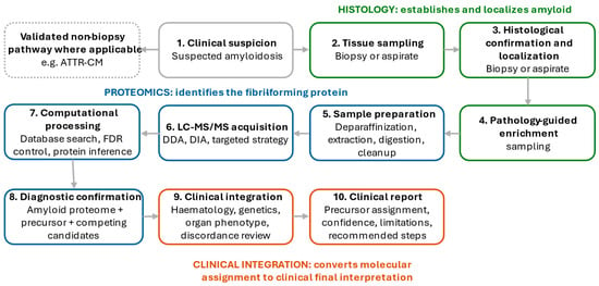
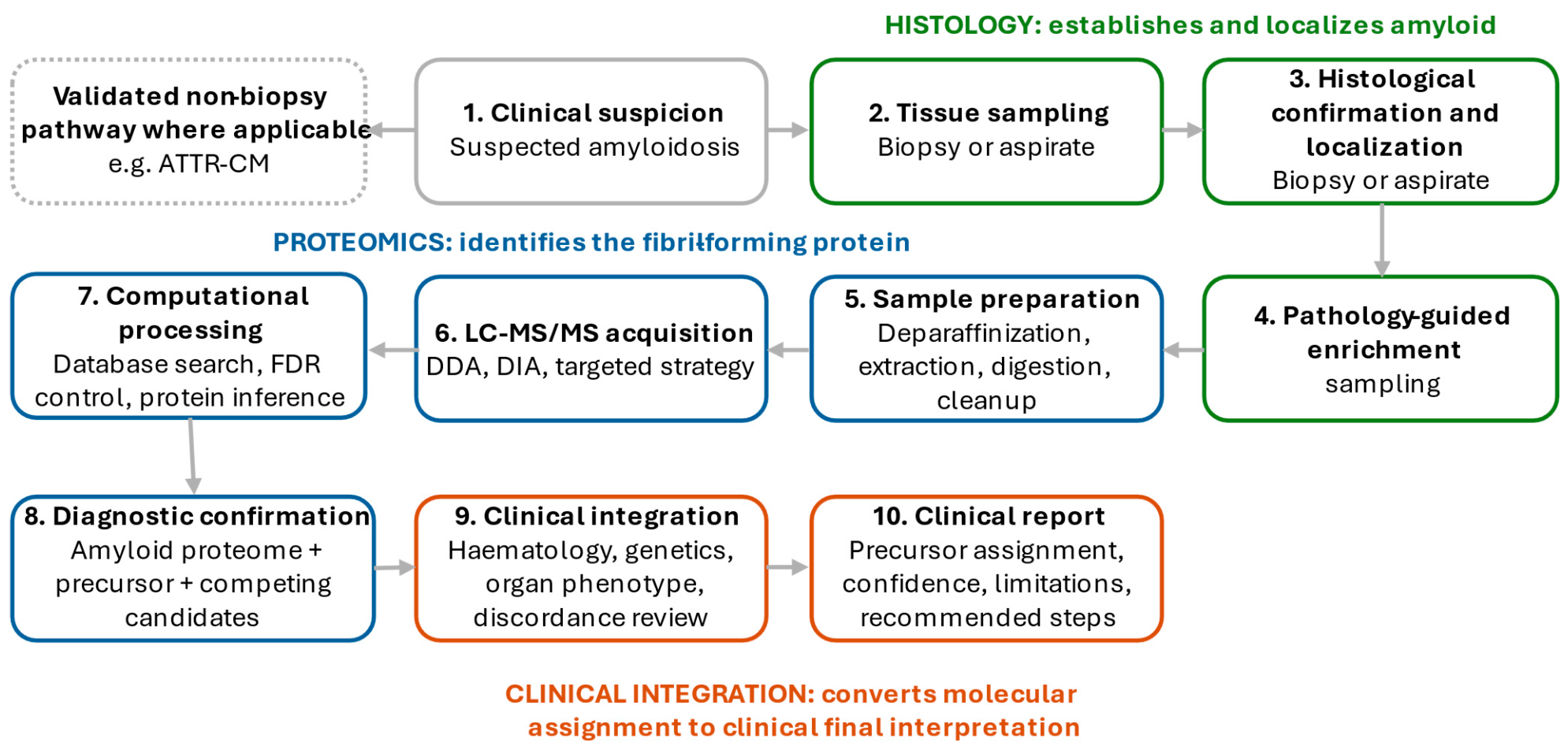
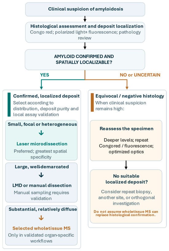
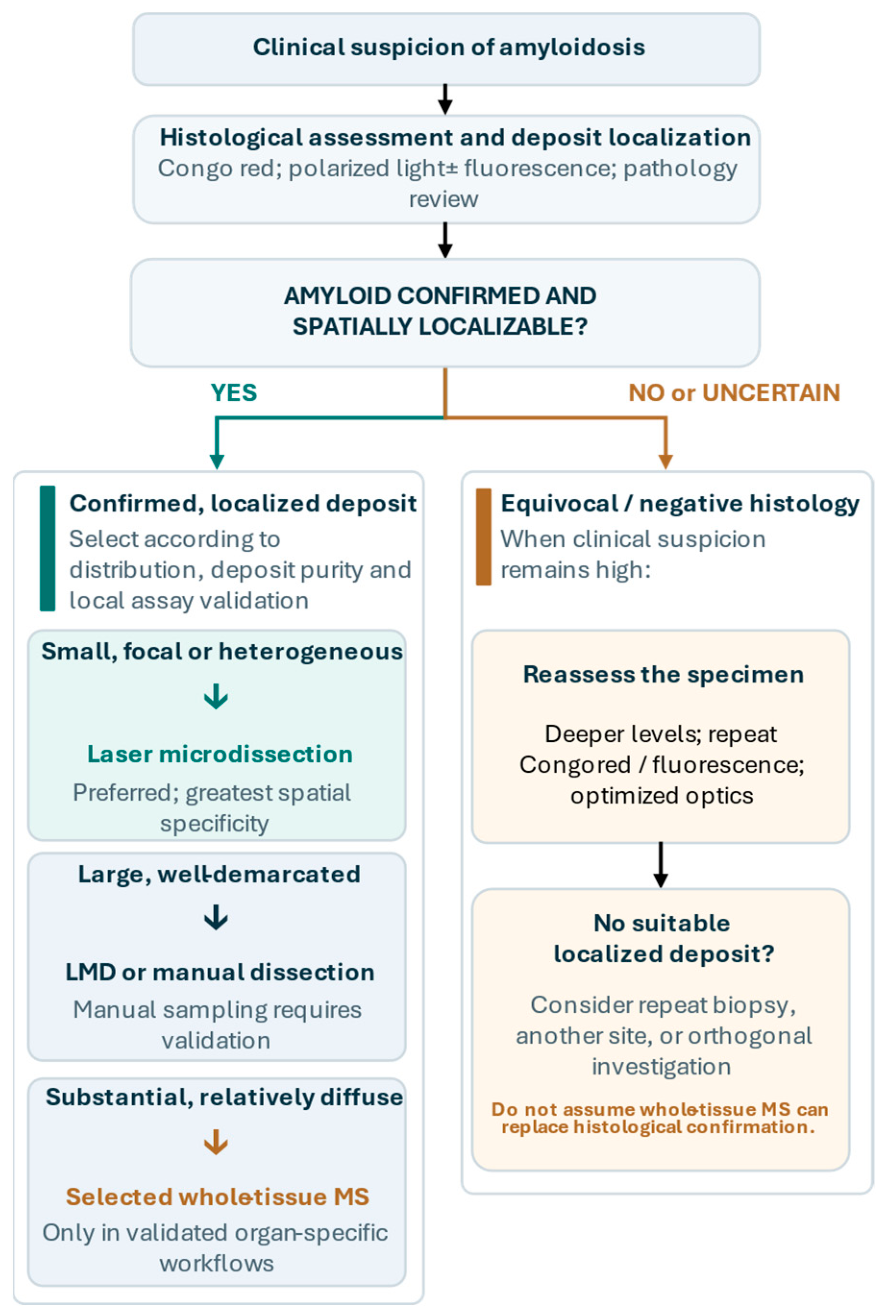
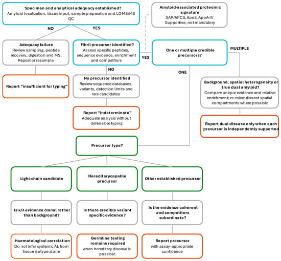
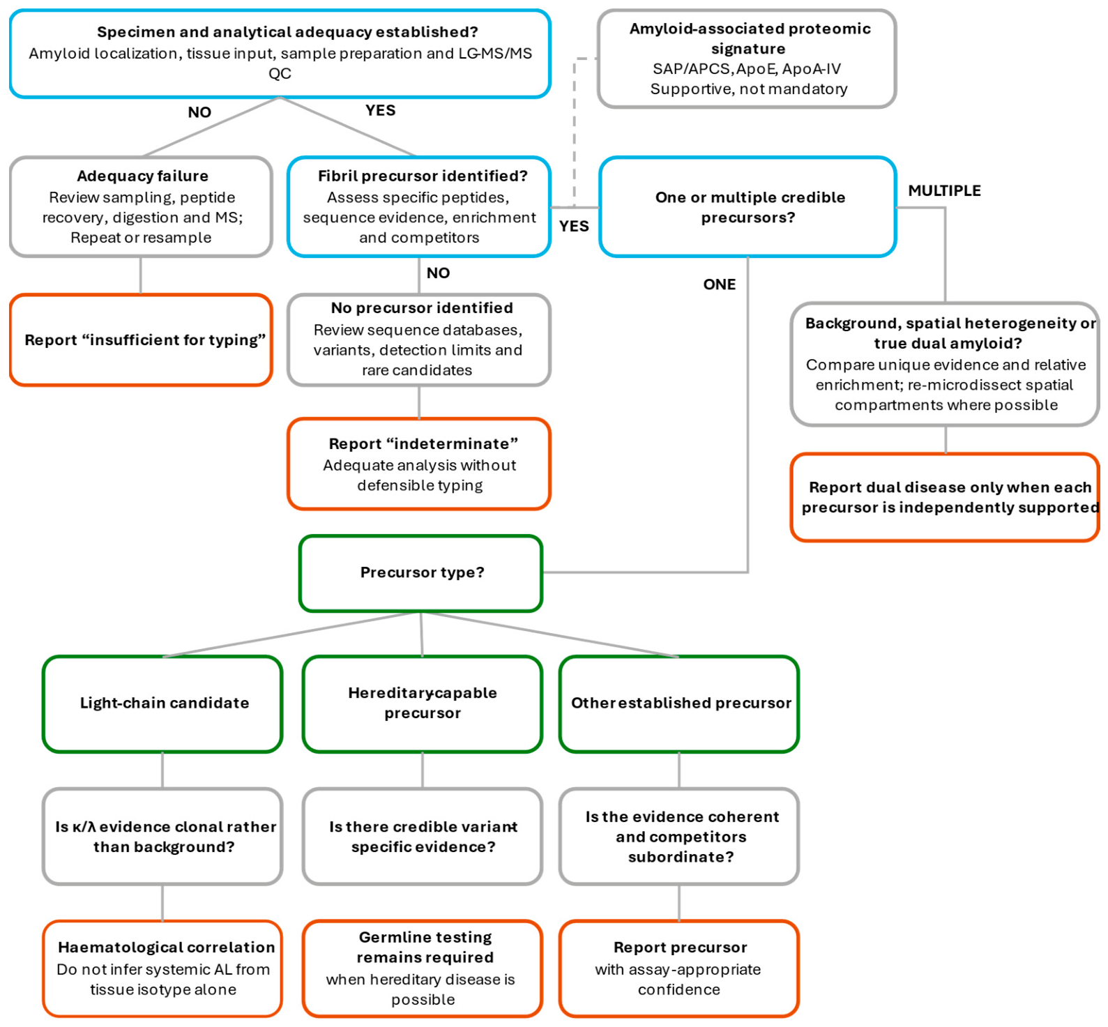
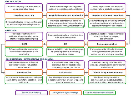
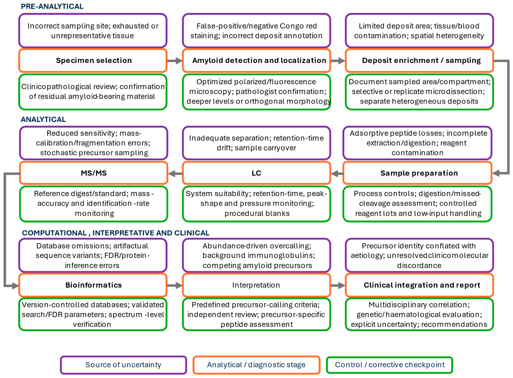

Disclaimer/Publisher’s Note: The statements, opinions and data contained in all publications are solely those of the individual author(s) and contributor(s) and not of MDPI and/or the editor(s). MDPI and/or the editor(s) disclaim responsibility for any injury to people or property resulting from any ideas, methods, instructions or products referred to in the content. |
© 2026 by the authors. Licensee MDPI, Basel, Switzerland. This article is an open access article distributed under the terms and conditions of the Creative Commons Attribution (CC BY) license.