


Abstract
Systemic sclerosis (SSc) is a rare, clinically heterogeneous connective tissue disease defined by the triad of autoimmunity, microvasculopathy, and fibrosis of the skin and internal organs. Among the immune effectors implicated in SSc, B cells have moved from being regarded mainly as the source of disease-defining autoantibodies to being recognized as multifunctional drivers of disease through antibody-independent mechanisms. This review summarizes current understanding of B-cell biology in SSc across three domains. First, we consider the classical role of B cells as antibody-producing cells, including the diagnostic and prognostic value of SSc-specific autoantibodies, the evidence for polyclonal B-cell activation and altered B-cell homeostasis, and the contested question of autoantibody pathogenicity. Second, we examine antibody-independent functions, focusing on cytokine production by effector and regulatory B-cell subsets, the B-cell-activating factor (BAFF) axis, antigen presentation, and T-cell co-stimulation, together with the imbalance between pro-inflammatory effector B cells and interleukin (IL)-10-producing regulatory B cells that characterizes active disease. Third, we review B cells as therapeutic targets, from the rituximab experience—including the randomized DESIRES trial and its open-label extension—through anti-BAFF strategies to the emerging use of CD19-directed chimeric antigen receptor (CAR) T-cell and CAR-natural killer (NK) cell therapies that achieve deep, durable B-cell depletion. We conclude by highlighting unresolved questions, including patient selection, the balance between depleting pathogenic and protective B-cell subsets, biomarker development, and the long-term safety of deep B-cell depletion. A B-cell-centered view of SSc pathogenesis provides a coherent framework that links autoimmunity to fibrosis and is increasingly translatable into the clinic.
1. Introduction
Systemic sclerosis (SSc) is a multisystem autoimmune connective tissue disease characterized by three interrelated pathological processes: immune dysregulation and autoimmunity, a small-vessel vasculopathy, and excessive deposition of extracellular matrix leading to fibrosis of the skin and internal organs [1,2]. The disease is clinically heterogeneous, ranging from limited cutaneous SSc with predominantly distal skin involvement to diffuse cutaneous SSc (dcSSc) with rapidly progressive skin thickening and a high burden of internal organ disease. Interstitial lung disease (ILD) and pulmonary arterial hypertension are now the leading causes of disease-related death, and SSc carries the highest case-specific mortality of any systemic rheumatic disease [1,2]. Despite advances, the events that connect the initial loss of immune tolerance to the eventual fibrotic phenotype remain incompletely understood.
A defining and almost universal feature of SSc is the presence of circulating autoantibodies. More than 90% of patients are positive for antinuclear antibodies, and the majority carry one of several mutually exclusive SSc-specific autoantibodies whose specificities correlate closely with clinical subset, organ involvement, and prognosis [1,3]. The reproducible association between particular autoantibody specificities and distinct disease trajectories has long implicated the humoral immune system, and therefore B cells, in the pathogenesis of SSc. This relationship has motivated sustained interest in B cells as both biomarkers and therapeutic targets [4,5,6].
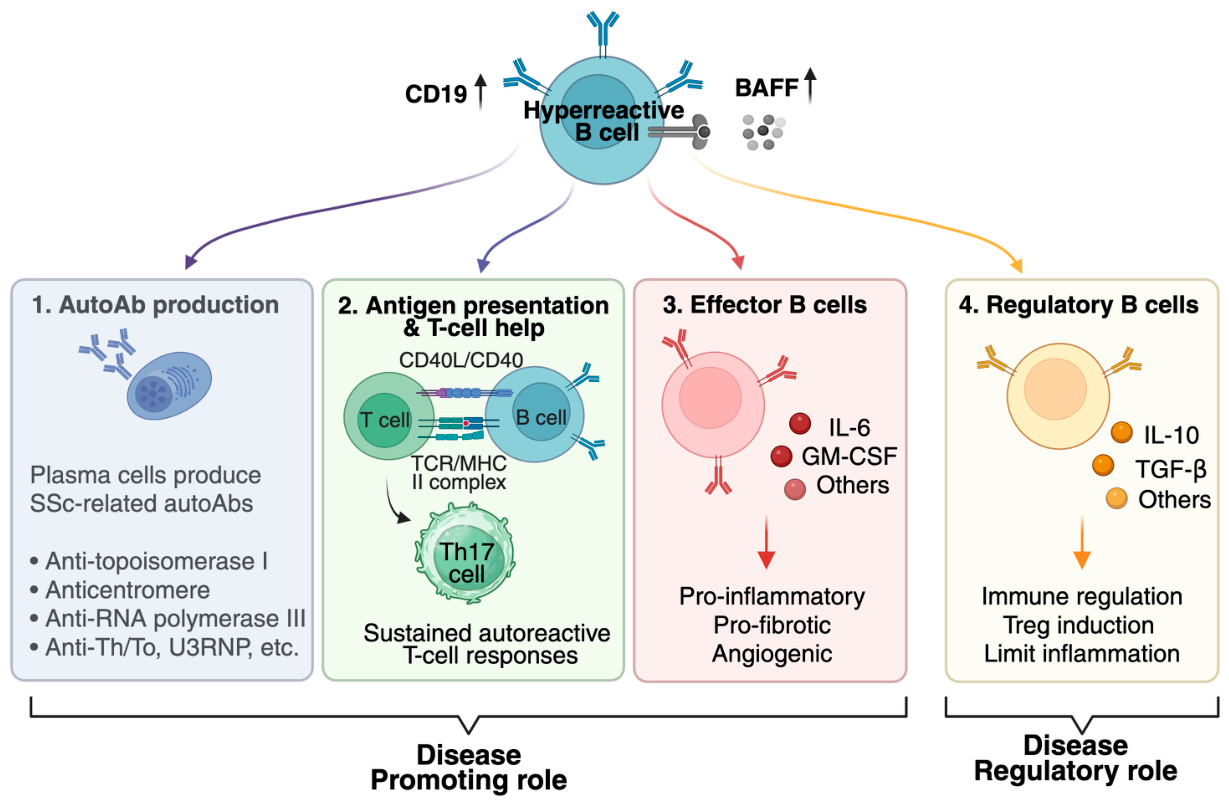
Over the past two decades, the conceptual role of B cells in SSc has broadened considerably. B cells are no longer viewed solely as the precursors of autoantibody-secreting plasma cells; they are now understood to shape the immune response through a range of antibody-independent activities, including the secretion of pro-inflammatory and pro-fibrotic cytokines, the presentation of antigen to T cells, the provision of co-stimulatory signals, and the negative regulation of inflammation by specialized regulatory B-cell (Breg) subsets [4,6,7]. Importantly, not all B cells promote disease: effector B cells (Beff) and Breg cells exert opposing influences, and an imbalance between them appears to be a feature of active SSc [3,5,8] (Figure 1). This more nuanced picture has both refined our understanding of disease mechanisms and complicated the design of B-cell-directed therapies, because indiscriminate depletion removes protective as well as pathogenic populations.
The clinical importance of this question is amplified by the limitations of current treatment. For most of its history, SSc has lacked disease-modifying therapy, and management has relied on organ-based symptomatic treatment and broad immunosuppression with agents such as mycophenolate mofetil and cyclophosphamide [9,10]. These approaches have modest and variable effects on the fibrotic process, and outcomes in progressive skin and lung disease remain unsatisfactory [2,9]. A mechanistically targeted, immunologically rational therapy is therefore a long-standing unmet need, and the B-cell compartment—simultaneously a biomarker, an effector, and a tractable drug target—has emerged as one of the most attractive points of intervention [4,6,7].
In this review, we synthesize current knowledge of B cells in SSc around three themes that mirror the expanding appreciation of their biology: (i) B cells as antibody-producing cells; (ii) the antibody-independent functions of B cells, with emphasis on cytokine production and antigen presentation; and (iii) B cells as therapeutic targets, including conventional depletion with rituximab and the rapidly evolving field of cellular immunotherapy. Throughout, we aim to distinguish well-supported observations from areas of continuing uncertainty, and to indicate where the field is moving.
Figure 1.
The multifaceted roles of B cells in systemic sclerosis (SSc). Hyperreactive B cells (CD19 overexpression, elevated BAFF) act through four functions: (1) plasma-cell autoantibody production (anti-topoisomerase I, anticentromere, anti-RNA polymerase III, anti-Th/To, U3RNP); (2) antigen presentation and CD40L/CD40 T-cell help, sustaining autoreactive (Th17) responses; and (3) effector B cells secreting IL-6, GM-CSF, and other pro-inflammatory, pro-fibrotic, and angiogenic mediators—together disease-promoting—versus (4) regulatory B cells producing IL-10 and TGF-β to limit inflammation and induce Treg (disease-regulatory). The effector–regulatory balance determines the net B-cell contribution to disease. Created in BioRender. Matsushita, T. (2026) https://BioRender.com/eiza87w.
2. B Cells as Antibody-Producing Cells
2.1. SSc-Specific Autoantibodies and Their Clinical Associations
The most clinically entrenched role of B cells in SSc is the generation of autoantibodies. The classical SSc-specific antibodies—anti-topoisomerase I (anti-Scl-70), anti-centromere, and anti-RNA polymerase III—are largely mutually exclusive and define recognizable clinical phenotypes [1,3]. Anti-topoisomerase I is associated with diffuse cutaneous disease and a higher risk of ILD; anti-centromere antibodies typically accompany limited cutaneous disease and an increased risk of pulmonary hypertension; and anti-RNA polymerase III is linked to rapidly progressive skin involvement and scleroderma renal crisis [1,3]. Additional, less common specificities—including antibodies against U3 ribonucleoprotein (fibrillarin), Th/To, and U11/U12 ribonucleoprotein—further refine prognostic subgroups, underscoring the remarkable degree to which the fine specificity of the autoantibody response maps onto distinct disease trajectories [1,11]. Because these specificities are typically present at or before clinical onset, rarely overlap, and remain stable over the course of disease, they are widely used for diagnosis, subclassification, and risk stratification [1,3]. The strength and reproducibility of these antibody–phenotype relationships are among the most compelling indirect arguments for a central role of the B-cell compartment in disease; even if the antibodies themselves are not the principal effectors, their tight clinical correlations indicate that the B-cell program generating them is closely coupled to the mechanisms that determine disease expression [11,12].
2.2. Polyclonal B-Cell Activation and Altered B-Cell Homeostasis
Beyond the production of specific autoantibodies, SSc is marked by more generalized B-cell hyperreactivity. Hypergammaglobulinemia and polyclonal B-cell activation are common, and detailed phenotyping has revealed reproducible disturbances in the circulating B-cell compartment. In a landmark study, Sato and colleagues demonstrated that patients with SSc have expanded numbers of CD27-negative naive B cells together with reduced but chronically activated memory B cells; the memory cells displayed increased expression of the activation markers CD80, CD86, and CD95, underwent augmented spontaneous apoptosis, and retained an enhanced capacity to secrete immunoglobulin G upon stimulation [13]. Critically, expression of CD19—a cell-surface signal-transduction molecule that sets the threshold for B-cell activation and regulates autoantibody production—was significantly increased on both naive and memory B cells in SSc [13]. Even modest CD19 overexpression is sufficient to break tolerance and induce autoantibodies in experimental models, and the finding of CD19 overexpression in human SSc B cells provides a mechanistic link between intrinsic B-cell hyperreactivity and the autoantibody response [12,14].
These homeostatic abnormalities have been complemented by studies of the B-cell receptor (BCR) repertoire. High-throughput sequencing of the immunoglobulin heavy-chain repertoire in SSc has identified abnormalities in clonal composition and complementarity-determining region 3 characteristics consistent with antigen-driven selection and polyclonal activation [15]. Collectively, the homeostatic and repertoire data indicate that the B-cell compartment in SSc is intrinsically dysregulated rather than simply responding to an external stimulus [6,12]. Beyond the naive and memory compartments, additional B-cell subsets are increasingly implicated. Circulating plasmablasts and long-lived plasma cells sustain autoantibody output and, because they down-regulate CD20, largely escape conventional depletion (Section 2.4). Transitional B cells, a physiological source of regulatory IL-10, may be functionally altered and thereby contribute to the regulatory deficit discussed below [13,16]. Attention has also turned to age-associated B cells (ABCs; CD19 + CD21 − CD11c + T-bet+), an IgD−CD27−“double-negative”-enriched population that expands with chronic inflammation and drives autoreactive IgG and extrafollicular responses across autoimmune diseases; although best characterized in lupus and Sjögren’s syndrome, their specific contribution to SSc remains to be formally defined and is an open question [17]. A more granular, subset-resolved map of B-cell heterogeneity is therefore likely to refine both pathogenic understanding and therapeutic targeting. Innate signals further amplify this intrinsic activation: Toll-like receptors—notably TLR7, TLR9, and the TLR homologue CD180—are engaged by endogenous nucleic-acid and damage-associated ligands in SSc, driving B-cell activation, type I interferon responses, and a pathological shift in BAFF-receptor expression, and TLR-pathway variants have been proposed as susceptibility factors, linking innate immunity to the polyclonal B-cell activation characteristic of the disease [18]. Sex is a further modifier: SSc shows a strong female predominance, and X-linked immune-gene dosage, sex hormones (estrogens being generally immunostimulatory and profibrotic), and fetal microchimerism have all been proposed as contributors. Fetal DNA and cells persisting after pregnancy have been identified in SSc skin lesions, suggesting that pregnancy-associated microchimerism could supply an allogeneic stimulus to autoreactive B cells, although a causal role remains unproven [19].
2.3. The Contested Pathogenicity of Autoantibodies
Whether SSc-associated autoantibodies are directly pathogenic or principally serve as biomarkers of an aberrant immune response remains unresolved. The classical anti-nuclear specificities recognize intracellular antigens, and a direct fibrogenic mechanism for them has been difficult to establish [5,11]. Attention has therefore turned to functional autoantibodies directed against cell-surface or matrix targets—such as antibodies against the platelet-derived growth factor receptor, endothelial cells, and fibroblasts—that could, in principle, transmit pro-fibrotic or pro-inflammatory signals; however, the reproducibility and pathogenic relevance of several of these specificities remain debated [5,11]. This controversy is best illustrated by stimulatory autoantibodies to the platelet-derived growth factor receptor (PDGFR): using functional bioassays, these were reported to be specific for SSc and to convert normal fibroblasts to a pro-fibrotic phenotype, arguing for a direct pathogenic role [20]. Studies using immunochemical rather than bioactivity assays, however, detected anti-PDGFR reactivity in healthy individuals and other conditions and questioned its disease specificity, and independent groups were unable to reproduce the original functional findings [21]. On balance, most SSc autoantibodies are best regarded as robust biomarkers of a dysregulated B-cell response, whereas direct pathogenicity has been convincingly demonstrated for few, if any, individual specificities. A further line of evidence underscores the cooperative nature of the autoantibody response: the antibody response to DNA topoisomerase I in SSc requires collaboration between autoreactive T and B cells, indicating that B cells act not in isolation but within an integrated adaptive immune network [22]. Regardless of whether individual autoantibodies are effectors or markers, the autoantibody response reflects an underlying B-cell abnormality that is itself a legitimate therapeutic target [4,14].
2.4. Plasma Cells and the Limits of an Antibody-Centered View
An important corollary of the autoantibody data concerns the cells that ultimately secrete immunoglobulin. Terminally differentiated long-lived plasma cells, which reside in the bone marrow and inflamed tissue, down-regulate the surface markers CD20 and, in many cases, CD19, and are therefore relatively resistant to conventional B-cell-depleting antibodies that target these molecules [4,6]. This biology helps to explain a recurring clinical observation: depletion of CD20-positive B cells with rituximab produces only modest and inconsistent reductions in established SSc-specific autoantibody titers, even when circulating and tissue B cells are effectively eliminated [23]. The implication is twofold. First, the clinical benefit of B-cell depletion in SSc cannot be attributed solely to a fall in autoantibody levels and must derive substantially from the antibody-independent functions discussed below. Second, strategies that reach the plasma-cell compartment or that target antigens retained on plasma cells—such as B-cell maturation antigen—may be required to durably reset the autoantibody response, a consideration that has directly motivated the cellular therapies described later in this review [6,24].
3. Antibody-Independent Functions of B Cells
A major shift in the understanding of SSc has been the recognition that B cells influence disease through mechanisms that do not depend on antibody secretion. These functions—cytokine production, antigen presentation, co-stimulation, and immune regulation—position B cells as nodal cells that can both amplify and restrain the inflammatory and fibrotic processes of SSc [4,6,7].
3.1. Cytokine Production and the Effector/Regulatory Balance
B cells are an important source of immunoregulatory and pro-inflammatory cytokines. In SSc, the functional polarization of B cells appears to be tilted toward a pro-inflammatory, pro-fibrotic profile. Effector B cells that produce interleukin (IL)-6 are of particular interest because IL-6 is a well-established driver of fibroblast activation and collagen synthesis, and IL-6-producing effector B cells promote fibrosis in experimental scleroderma [25]. Horii and colleagues quantified the balance between IL-6-producing effector B cells and IL-10-producing regulatory B cells in patients with SSc and found that the frequency of IL-6-producing effector B cells was significantly elevated, while IL-10-producing regulatory B cells were reduced, relative to healthy controls; the resulting increase in the effector-to-regulatory (Beff/Breg) ratio correlated positively with the modified Rodnan skin score and the extent of ILD [3]. This observation provides a direct clinical link between B-cell cytokine polarization and the two cardinal manifestations of SSc—skin and lung fibrosis—and supports the concept of an effector/regulatory imbalance as a core feature of active disease, a framework that has been articulated as the central organizing principle of B-cell function in autoimmunity [8,26].
The relevance of the IL-6 axis to fibrosis is supported more broadly by the biology of the cytokine: IL-6 promotes the differentiation of fibroblasts into matrix-producing myofibroblasts, supports T-helper-17 responses, and drives plasmablast differentiation, situating B-cell-derived IL-6 within a self-reinforcing inflammatory–fibrotic loop [3,12]. Direct genetic evidence comes from the bleomycin-induced scleroderma model: mice with a B-cell-specific deficiency in IL-6 develop attenuated skin and lung fibrosis, whereas mice with a B-cell-specific deficiency in IL-10 develop more severe fibrosis, demonstrating that the cytokine output of B cells—not their mere presence—determines the fibrotic outcome [25]. Consistent with this, B-cell-deficient and CD19-manipulated mice show altered fibrosis and autoantibody production, indicating that B-cell signaling thresholds shape the fibrotic phenotype rather than merely accompanying it [12].
Beyond IL-6, B cells in SSc have been reported to produce other disease-relevant mediators. A subset of granulocyte–macrophage colony-stimulating factor (GM-CSF)-producing B cells has been described in SSc and proposed to contribute to pathogenesis through innate-like pro-inflammatory activity [27]. The cytokine milieu that sustains B-cell activation is itself altered: serum levels of IL-27, a cytokine that can modulate T-cell, B-cell, and fibroblast activity, are elevated in SSc and associate with disease features [28]. The net effect of these mediators is to couple B-cell activation to the activation of the principal fibrotic effector cell of SSc, the myofibroblast, providing a cellular pathway by which adaptive immune dysregulation is translated into matrix deposition [3,4].
3.2. The BAFF Axis
The B-cell-activating factor (BAFF, also known as BLyS) is a key survival and differentiation signal for B cells, and the BAFF axis is consistently dysregulated in SSc. Serum BAFF levels are elevated in patients with SSc, correlate with skin and overall disease severity, and are accompanied by enhanced BAFF signaling in SSc B lymphocytes, establishing BAFF as a driver of the B-cell hyperreactivity characteristic of the disease [29]. Consistent with these findings, circulating BAFF—together with the chemokine CXCL13, a marker of germinal-center and follicular activity—has been proposed as a biomarker of B-cell activation and lymphoid organization in SSc and other connective tissue diseases [30,31]. Several features of BAFF biology bear on this hyperreactivity. Although BAFF is produced chiefly by myeloid cells, neutrophils, and T cells, activated B cells can themselves secrete BAFF, establishing an autocrine loop that may reinforce their own survival. Host genetics also contribute: a common TNFSF13B (BAFF) variant yields a shorter, microRNA-insensitive transcript that raises circulating soluble BAFF, up-regulates humoral immunity and has been linked to autoimmune-disease susceptibility, providing a genetic mechanism for defective BAFF regulation [32]. Finally, non-immune cells feed the axis: platelets, chronically activated in the SSc vasculopathy, express CD40 ligand and release immunostimulatory and profibrotic mediators (including TGF-β and serotonin), positioning them as a link between vascular injury, B-cell co-stimulation, and fibrosis [33]. Beyond acting as a survival factor, BAFF actively shapes the effector/regulatory balance: in the experimental scleroderma model, BAFF expanded IL-6-producing effector B cells while suppressing IL-10-producing regulatory B cells, directly linking the BAFF axis to the pathogenic cytokine imbalance described above [25]. The dependence of pathogenic B cells on BAFF therefore provides a rationale for therapeutic blockade of this pathway and helps explain why B-cell-activation biomarkers track with disease activity [25,30].
3.3. Antigen Presentation and T-Cell Help
As professional antigen-presenting cells, B cells capture antigen through their BCR, process it, and present peptides to CD4-positive T cells while delivering co-stimulatory signals through CD80 and CD86. The increased expression of CD80 and CD86 on SSc memory B cells indicates a heightened capacity for T-cell co-stimulation [13], and the requirement for T–B collaboration in the anti-topoisomerase I response demonstrates that this interaction is operative in SSc [22]. It should be emphasized, however, that much of the mechanistic evidence for B-cell antigen presentation and co-stimulation derives from animal models, whereas direct demonstration of antigen-specific B cell–T cell interactions within human SSc tissue remains limited. The strongest human evidence is indirect—the cognate T–B dependence of the anti-topoisomerase I response [22], the activated, co-stimulation-competent phenotype of circulating SSc B cells [13], and the presence of B cell–T cell aggregates in affected skin and lung [34]—and this gap should be kept in mind when extrapolating from experimental systems. Through antigen presentation and co-stimulation, B cells can shape effector T-cell differentiation, sustain autoreactive T-cell responses, and contribute to the organization of tertiary lymphoid structures in affected tissues. Consistent with a tissue-level role, B-cell infiltrates have been identified in the lungs of patients with SSc-associated ILD, placing B cells at the site of fibrotic injury rather than confined to the circulation [34]. Such infiltrates, sometimes organized into tertiary lymphoid structures together with T cells and follicular dendritic cells, can support local antigen presentation, affinity maturation, and in situ generation of autoreactive B cells, creating a self-sustaining focus of adaptive immune activity within affected organs [4,34]. The proximity of these cells to fibroblasts and the local production of cytokines and chemokines such as CXCL13, which recruits B cells into lymphoid aggregates, link the antigen-presenting function of B cells to the spatial organization of disease [31,34]. These antigen-presenting and tissue-localized functions provide a mechanistic basis for the efficacy of B-cell depletion that is independent of any effect on autoantibody titers, because removing B cells dismantles both a source of T-cell help and a structural component of the local immune response [4,6].
3.4. Regulatory B Cells
Counterbalancing the pro-inflammatory activities of effector B cells is a population of regulatory B cells that restrain inflammation, principally through the production of IL-10. The existence of such cells was established in autoimmune models, in which regulatory B cells were shown to inhibit disease initiation while other B cells promote disease progression [8]; the corresponding human IL-10-competent subset, termed B10 cells, was subsequently defined and shown to parallel mouse regulatory B cells [16]. In SSc, this protective compartment is deficient. Matsushita and colleagues identified a human Breg subset enriched within CD24-high CD27-positive B cells and showed that the frequency of IL-10-producing Breg cells was significantly reduced in patients with SSc compared with healthy controls [35]. Lower Breg levels were associated with the presence of ILD and correlated inversely with the titers of anti-topoisomerase I and anti-centromere antibodies, suggesting that loss of B-cell-mediated regulation permits both autoantibody production and organ involvement [35]. Notably, Breg frequencies increased after treatment in parallel with reduced disease activity, indicating that the regulatory deficit is at least partly reversible and may track therapeutic response [35]. Mechanistically, Breg cells limit inflammation not only through IL-10 but also by supporting regulatory T-cell populations and restraining effector T-cell and dendritic-cell activation, so their loss in SSc removes a brake on multiple arms of the immune response simultaneously [5,35]. The IL-10-producing regulatory B-cell program defined through this body of work is not confined to scleroderma; the same regulatory mechanism operates broadly across autoimmune disease, indicating that the Breg deficiency observed in SSc reflects the failure of a fundamental, broadly acting immunoregulatory pathway [26]. The coexistence of expanded pro-inflammatory effector B cells and contracted regulatory B cells reinforces the view that it is the balance, rather than the absolute number, of B-cell subsets that determines their net contribution to disease, and it raises a therapeutic dilemma that recurs throughout this review: any non-selective depletion strategy will remove protective Breg cells alongside pathogenic effectors [3,5,8].
3.5. Integrating the Antibody-Independent Functions
Taken together, the cytokine-producing, antigen-presenting, co-stimulatory, and regulatory activities of B cells form an integrated picture in which B cells sit at the interface between adaptive immunity and tissue fibrosis. The same compartment that generates autoantibodies also amplifies inflammation through IL-6 and GM-CSF, sustains autoreactive T cells through antigen presentation, depends on BAFF for survival, and normally restrains itself through IL-10-producing Breg cells [4,6,7]. In SSc, multiple nodes of this network are deranged simultaneously: B-cell homeostasis is altered, effector cytokine output is increased, regulatory function is diminished, and the supporting BAFF environment is expanded. This multiplicity of mechanisms explains why therapeutic removal of B cells can have effects that extend well beyond a reduction in autoantibody levels, and it provides a strong conceptual rationale for B-cell-targeted treatment [5,6].
4. B Cells as Therapeutic Targets
4.1. Rationale and Early Experience with Rituximab
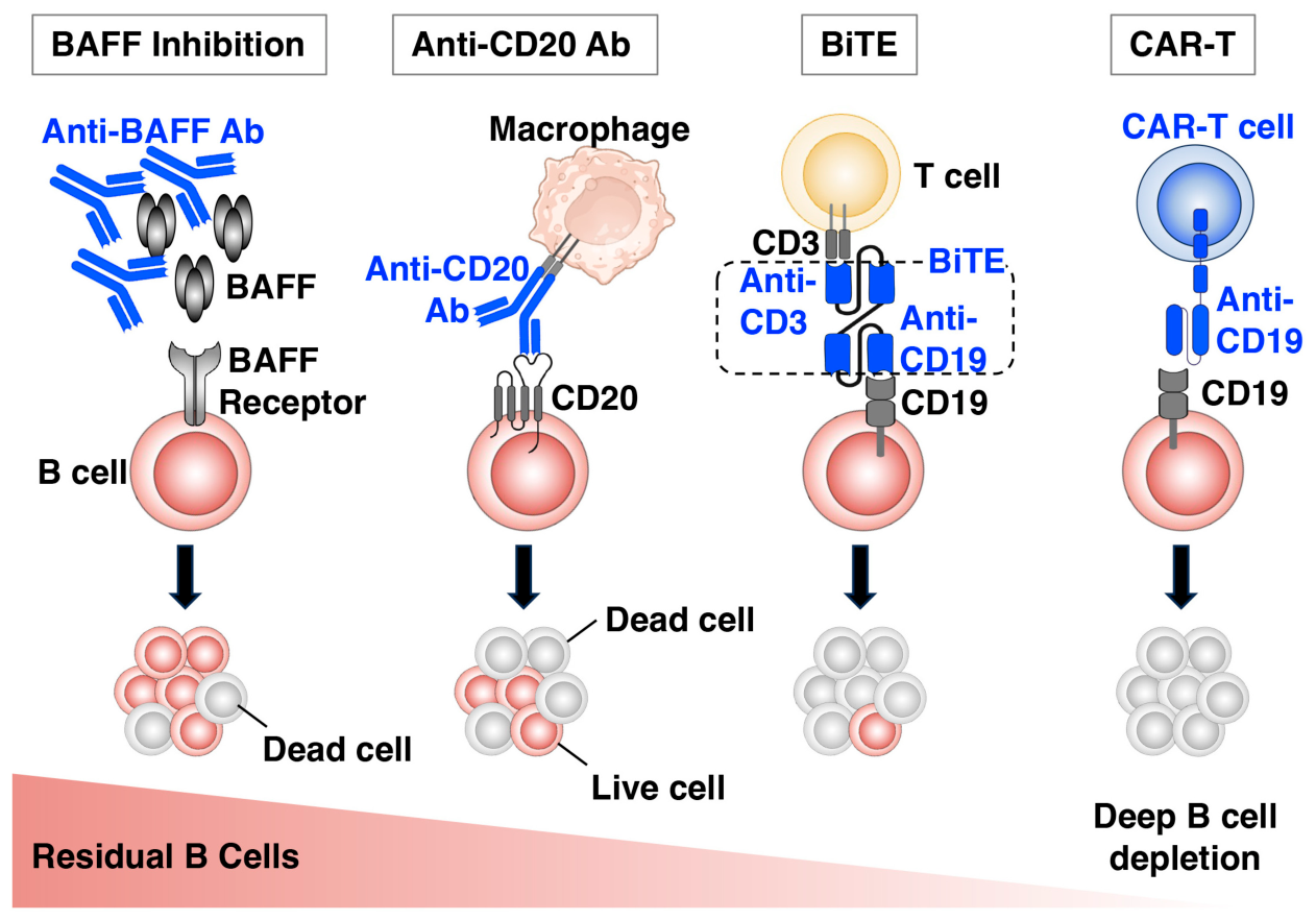
The combination of disease-defining autoantibodies, intrinsic B-cell hyperreactivity, pro-fibrotic cytokine production, and deficient B-cell regulation provides a strong rationale for therapeutically targeting the B-cell compartment in SSc [4,6,36] (Figure 2). Table 1 compares the principal B-cell-targeted strategies discussed below—rituximab, belimumab, CD19-directed CAR-T and CAR-NK cells, and blinatumomab—by molecular target, mechanism of action, current stage of clinical development in SSc, and key efficacy and safety considerations. This rationale is grounded in preclinical evidence: in the tight-skin mouse model of SSc, B-lymphocyte depletion reduced both skin fibrosis and autoimmunity, providing direct proof of concept that eliminating B cells can attenuate the fibrotic process [37]. The first and most extensively studied approach has been depletion of CD20-positive B cells with the monoclonal antibody rituximab. Early open-label studies established proof of principle. In a pilot study of patients with dcSSc, Lafyatis and colleagues showed that two infusions of rituximab safely and effectively depleted circulating and dermal B cells; although autoantibody titers changed only modestly and skin scores did not improve significantly over six months in this small, short trial, the study confirmed that tissue B cells could be eliminated and that the approach was well tolerated [23]. Other early reports and case series, including work by Daoussis and colleagues and a contemporaneous synthesis of the available evidence, suggested potential benefit on skin and lung outcomes and helped frame rituximab as a promising option warranting controlled evaluation [38,39].
Figure 2.
Mechanisms of B-cell-targeted therapies in systemic sclerosis (SSc). Four strategies acting on distinct targets: BAFF inhibition (e.g., belimumab) blocks BAFF–BAFF-receptor survival signaling, leaving residual B cells; an anti-CD20 antibody (e.g., rituximab) drives macrophage-mediated B-cell killing; a CD19 × CD3 bispecific T-cell engager (BiTE; e.g., blinatumomab) redirects endogenous T cells to kill B cells; and an anti-CD19 CAR-T cell achieves deep B-cell depletion. Ab, antibody; BAFF, B-cell-activating factor; CAR, chimeric antigen receptor.
Table 1.
Comparison of B-cell-targeted therapeutic strategies in systemic sclerosis.
4.2. The DESIRES Trial and Controlled Evidence
The pivotal advance in the rituximab experience was the DESIRES trial, a double-blind, investigator-initiated, randomized, placebo-controlled study of rituximab in SSc. The trial demonstrated that rituximab significantly improved skin thickening, as measured by the modified Rodnan skin score, compared with placebo, providing the first high-quality randomized evidence that B-cell depletion modifies the fibrotic phenotype of SSc [40]. The subsequent open-label extension reported that improvements in skin score were maintained over longer-term follow-up and that the treatment was generally well tolerated, supporting durability of effect [41]. Evidence has also accumulated for benefit in SSc-associated ILD: a systematic review and meta-analysis concluded that rituximab is associated with stabilization or improvement of lung function in SSc-ILD, and B-cell depletion has been used as a strategy in patients with disease resistant to conventional immunosuppression [42,43,44]. Broader systematic reviews of rituximab across immune-mediated disorders have reinforced its overall safety profile [45]. These data collectively informed the 2023 update of the European Alliance of Associations for Rheumatology (EULAR) recommendations for the treatment of SSc, in which rituximab is now considered among the options for skin and lung involvement, reflecting a substantial shift in the evidence base [9,46].
The rituximab experience is also instructive mechanistically. Because depletion of CD20-positive B cells has only a limited effect on established autoantibody titers [23], the clinical benefit observed in DESIRES and in SSc-ILD is most plausibly mediated by removal of the antibody-independent functions of B cells—cytokine secretion, antigen presentation, and T-cell co-stimulation—rather than by a reduction in autoantibodies [4,6,40]. This interpretation aligns with the broader reconceptualization of B cells in SSc and reinforces the rationale for targeting the compartment as a whole. At the same time, the responses to rituximab are partial and not uniform: many patients show stabilization rather than reversal of fibrosis, the optimal timing and re-treatment schedule are not established, and incomplete tissue depletion together with sparing of long-lived plasma cells may limit the depth and durability of the response [23,43]. These limitations have driven interest in more complete forms of B-cell depletion.
4.3. Targeting the BAFF Axis and Other B-Cell-Directed Strategies
In parallel with depletion strategies, modulation of the BAFF survival pathway has been explored, given the consistent elevation of BAFF in SSc and its correlation with disease activity [30,31]. The preclinical rationale is unusually direct and long-standing: an early study showed that a BAFF antagonist attenuated the development of skin fibrosis in tight-skin mice [47], and later work in the bleomycin-induced scleroderma model clarified the underlying mechanism, demonstrating that BAFF inhibition attenuates both skin and lung fibrosis specifically by depleting IL-6-producing effector B cells while preserving IL-10-producing regulatory B cells—that is, by correcting the effector/regulatory imbalance rather than by indiscriminate B-cell removal [25]. This mechanism is particularly attractive because it addresses the central limitation of depletion therapy, the simultaneous loss of protective Bregs. Clinical evaluation of BAFF blockade in SSc itself has begun. In a randomized, double-blind, placebo-controlled pilot trial, belimumab added to background mycophenolate mofetil in early diffuse cutaneous SSc did not produce a statistically significant skin benefit over placebo in this small study, yet responders showed marked down-regulation of profibrotic and B-cell-signaling gene programs not seen with placebo [48]. This pattern—clinical equivalence but a distinct molecular signature—suggests that the relevant target population and pharmacodynamic readout may be narrower than unselected SSc and motivates larger, biomarker-enriched trials, including sequential regimens combining B-cell depletion with BAFF blockade. B-cell-activation biomarkers such as BAFF and CXCL13 may help identify patients most likely to respond and to monitor that response [30,31]. As with depletion, a recurring consideration is the need to preserve, or ideally restore, the protective IL-10-producing Breg population whose deficiency characterizes active disease [8,35].
A complementary direction is combination and sequential therapy. Because depletion with rituximab spares long-lived plasma cells while anti-BAFF treatment reduces the survival of newly generated autoreactive B cells, sequential use of the two has a clear biological logic: depletion followed by BAFF blockade could blunt the post-depletion surge in BAFF that promotes the re-emergence of autoreactive clones [30,31]. More broadly, the heterogeneity of SSc argues against a single uniform B-cell-directed strategy and in favor of biomarker-guided selection, in which the choice and intensity of B-cell-targeted therapy are matched to a patient’s B-cell activation profile, autoantibody specificity, and the balance between inflammatory and established fibrotic disease [3,9,30]. The same biomarkers that illuminate pathogenesis—BAFF, CXCL13, the Beff/Breg ratio, and autoantibody dynamics—may thus serve a dual role as tools for treatment stratification [3,30,31,35].
4.4. Chimeric Antigen Receptor Cell Therapy: Deep B-Cell Depletion
The most striking recent development is the application of CD19-directed chimeric antigen receptor (CAR) cell therapy to SSc and other autoimmune diseases. Unlike antibody-mediated depletion, which leaves residual B cells in lymphoid tissue, CAR T cells can achieve deep, tissue-penetrating elimination of the B-cell lineage, a concept that has been described as “B-cell depletion 2.0” [49]. In a now widely cited case series, Müller and colleagues reported that autologous CD19 CAR T-cell therapy induced drug-free remission across several autoimmune diseases, including SSc, with reconstitution of a naive B-cell repertoire and sustained clinical responses on follow-up [50]. The mechanistic appeal of this approach lies in the depth of depletion it achieves and in the nature of B-cell reconstitution that follows: after transient aplasia, B cells return as antigen-inexperienced naive cells, and the autoantibody-secreting and autoreactive populations that characterized the disease are not simply suppressed but appear to be reset, raising the prospect of durable, treatment-free remission rather than chronic disease control [49,50]. This distinguishes the goal of cellular therapy in autoimmunity—a lasting immune reset—from its goal in oncology, and it provides a conceptual bridge from the antibody-independent biology of B cells to a curative-intent intervention. Follow-up data are cautiously encouraging: across the initial autoimmune-disease experience, B cells reconstituted as naive cells within roughly three to four months of infusion, and treated patients—including those with SSc—have maintained drug-free responses without relapse over follow-up now approaching two years, with generally preserved protective serology and predominantly low-grade cytokine release syndrome [50]. Because SSc is a chronic disease, however, these observation periods remain short relative to its natural history, and the durability of remission, completeness of immune reconstitution, and long-term infection and malignancy risks require substantially longer study. Focused experience in SSc has followed: a case series of CD19-targeting CAR T-cell therapy in patients with diffuse SSc reported improvement in skin and stabilization of organ involvement [51], and third-generation CD19 CAR T cells have been used in anti-topoisomerase I-positive SSc patients [52].
To address the cost, complexity, and manufacturing constraints of autologous therapy, allogeneic and off-the-shelf platforms are being developed. Allogeneic CD19-targeted CAR T-cell therapy has been applied to patients with severe myositis and SSc [53], and a first-in-human report described the use of induced pluripotent stem cell-derived CD19/B-cell maturation antigen (BCMA) dual-targeting CAR-natural killer (NK) cells in a patient with SSc, raising the possibility of scalable, repeatable cellular therapy [24]. Detailed translational studies are beginning to define the immunological consequences of these treatments; integrated clinical and proteomic profiling of CD19 CAR T-cell therapy in progressive SSc has started to characterize the changes in circulating mediators that accompany clinical response [54]. T-cell-engaging approaches that do not require cell manufacture, such as the bispecific anti-CD3/CD19 molecule blinatumomab, have also been explored in severe SSc [55]. Several reviews have framed these advances within the wider movement of cellular immunotherapy into rheumatology [56].
4.5. Safety, Patient Selection, and Barriers to Implementation
The enthusiasm generated by deep B-cell depletion must be tempered by considerations of safety, access, and patient selection. CAR cell therapies carry risks including cytokine release syndrome, neurotoxicity, prolonged cytopenias, and infection related to sustained B-cell aplasia and hypogammaglobulinemia, and the long-term consequences in autoimmune disease—where unlike in oncology, the goal is durable immune reset rather than malignancy eradication—are not yet defined [56,57]. Practical barriers, including manufacturing logistics, cost, the need for lymphodepleting conditioning, and limited specialist capacity, currently restrict access and have been explicitly cataloged as obstacles to wider use in rheumatology [57]. Equally important is the unresolved biological question of how to deplete pathogenic effector B cells while preserving or restoring protective regulatory B cells; current depletion strategies remove both [8,35]. Robust biomarkers—potentially built on measures of B-cell activation, the Beff/Breg balance, and autoantibody dynamics—will be needed to select patients, time intervention, and monitor response [3,30,35].
Several specific uncertainties will shape how deep B-cell depletion is positioned in practice. The durability of remission after a single course of CAR cell therapy, the kinetics and quality of B-cell reconstitution, and whether protective humoral immunity (for example, to vaccines and prior infections) recovers adequately are not yet established in SSc, where follow-up remains short relative to the natural history of the disease [24,54,56]. The choice between autologous, allogeneic, and off-the-shelf platforms involves trade-offs between manufacturing time, persistence, immunogenicity, and the risk of graft-versus-host reactions, and the optimal target antigen—CD19 alone or in combination with B-cell maturation antigen to reach the plasma-cell compartment—remains to be defined [24,53]. Patient selection is equally unresolved: the strongest rationale exists for early, rapidly progressive diffuse disease with active immunological features, whereas the value of B-cell-directed therapy in long-standing, predominantly fibrotic disease with little inflammatory activity is likely to be limited [9,40]. Finally, the cost and infrastructure required for cellular therapy raise questions of equity and access that will need to be addressed if these treatments are to move beyond specialist centers [57].
5. Conclusions and Future Perspectives
The understanding of B cells in SSc has evolved from a narrow focus on autoantibody production to a broad appreciation of their multifunctional contribution to disease. B cells in SSc are intrinsically hyperreactive, with altered homeostasis and CD19 overexpression; they generate the autoantibodies that define clinical subsets; and they shape the inflammatory and fibrotic milieu through IL-6 and other cytokines, antigen presentation, T-cell co-stimulation, and a BAFF-dependent survival program. A consistent theme is the imbalance between expanded pro-inflammatory effector B cells and deficient IL-10-producing regulatory B cells, which links B-cell biology directly to the severity of skin and lung fibrosis [3,8,35].
This mechanistic framework has translated into therapeutic progress. Rituximab now rests on randomized evidence from the DESIRES trial and is reflected in updated treatment recommendations, while CD19-directed CAR T-cell and CAR-NK therapies offer the prospect of deep, durable, and potentially reset-inducing B-cell depletion [24,40,41,46,50]. Several questions remain central to the next phase of research: which patients benefit most and at what disease stage; how to selectively target pathogenic B-cell subsets while sparing regulatory populations; how to balance the efficacy of deep depletion against its infectious and immunological risks; and which biomarkers can guide these decisions [8,35,57]. Addressing these questions will require integration of detailed B-cell immunophenotyping with carefully designed clinical trials, ideally incorporating mechanistic read-outs—B-cell subset composition, cytokine output, and autoantibody and BAFF dynamics—alongside validated clinical endpoints such as the modified Rodnan skin score, lung function, and composite response indices [3,30,35,40]. Such trials would help to resolve whether the therapeutic goal should be transient depletion, sustained suppression, or a durable immune reset, and whether interventions can be refined to spare or rebuild the regulatory B-cell compartment whose deficiency contributes to disease [8,35]. BAFF inhibition, which in preclinical scleroderma and early human studies shifts the balance from effector toward regulatory B cells, exemplifies such a rebalancing strategy and offers an alternative to indiscriminate depletion [25], and is now being tested clinically in SSc [48].
A coherent narrative now connects the strands of this review. The same B-cell compartment that produces the autoantibodies used to classify SSc is intrinsically hyperreactive, amplifies fibrosis through cytokines such as IL-6, sustains autoreactive T cells through antigen presentation, depends on BAFF for survival, and fails to restrain itself because of a deficiency of regulatory B cells [3,4,6,12,35]. This integrated dysfunction explains why removing or resetting B cells can modify a disease once regarded as refractory, and why the benefit of depletion extends beyond any change in autoantibody levels. A B-cell-centered view of SSc therefore not only unifies the immunological and fibrotic features of the disease but also provides one of the most promising and rapidly advancing avenues for treatment, with the trajectory from rituximab to engineered cellular therapies illustrating how mechanistic insight into B-cell biology is being translated, in little more than a decade, into tangible clinical progress.
Funding
This research received no external funding.
Institutional Review Board Statement
Not applicable.
Informed Consent Statement
Not applicable.
Data Availability Statement
No new data were created or analyzed in this study. Data sharing is not applicable to this article.
Conflicts of Interest
The author declares no conflicts of interest.
References
- Jerjen, R.; Nikpour, M.; Krieg, T.; Denton, C.P.; Saracino, A.M. Systemic Sclerosis in Adults. Part I: Clinical Features and Pathogenesis. J. Am. Acad. Dermatol. 2022, 87, 937–954. [Google Scholar] [CrossRef] [Scilit]
- Rosendahl, A.-H.; Schönborn, K.; Krieg, T. Pathophysiology of Systemic Sclerosis (Scleroderma). Kaohsiung J. Med. Sci. 2022, 38, 187–195. [Google Scholar] [CrossRef] [Scilit] [PubMed]
- Horii, M.; Fushida, N.; Ikeda, T.; Oishi, K.; Hamaguchi, Y.; Ikawa, Y.; Komuro, A.; Matsushita, T. Cytokine-Producing B-Cell Balance Associates with Skin Fibrosis in Patients with Systemic Sclerosis. J. Dermatol. 2022, 49, 1012–1019. [Google Scholar] [CrossRef] [Scilit] [PubMed]
- Thoreau, B.; Chaigne, B.; Mouthon, L. Role of B-Cell in the Pathogenesis of Systemic Sclerosis. Front. Immunol. 2022, 13, 933468. [Google Scholar] [CrossRef] [Scilit] [PubMed]
- Yoshizaki, A.; Fukasawa, T.; Ebata, S.; Yoshizaki-Ogawa, A.; Sato, S. Involvement of B Cells in the Development of Systemic Sclerosis. Front. Immunol. 2022, 13, 938785. [Google Scholar] [CrossRef] [Scilit] [PubMed]
- Beesley, C.F.; Goldman, N.R.; Taher, T.E.; Denton, C.P.; Abraham, D.J.; Mageed, R.A.; Ong, V.H. Dysregulated B Cell Function and Disease Pathogenesis in Systemic Sclerosis. Front. Immunol. 2022, 13, 999008. [Google Scholar] [CrossRef] [Scilit] [PubMed]
- Scaletti, C.; Pratesi, S.; Bellando Randone, S.; Di Pietro, L.; Campochiaro, C.; Annunziato, F.; Matucci Cerinic, M. The B-Cells Paradigm in Systemic Sclerosis: An Update on Pathophysiology and B-Cell-Targeted Therapies. Clin. Exp. Immunol. 2025, 219, uxae098. [Google Scholar] [CrossRef] [Scilit] [PubMed]
- Liu, M. Effector and Regulatory B-Cell Imbalance in Systemic Sclerosis: Cooperation or Competition? Clin. Rheumatol. 2024, 43, 2783–2789. [Google Scholar] [CrossRef] [Scilit] [PubMed]
- Pope, J.E.; Denton, C.P.; Johnson, S.R.; Fernandez-Codina, A.; Hudson, M.; Nevskaya, T. State-of-the-Art Evidence in the Treatment of Systemic Sclerosis. Nat. Rev. Rheumatol. 2023, 19, 212–226. [Google Scholar] [CrossRef] [Scilit] [PubMed]
- Jerjen, R.; Nikpour, M.; Krieg, T.; Denton, C.P.; Saracino, A.M. Systemic Sclerosis in Adults. Part II: Management and Therapeutics. J. Am. Acad. Dermatol. 2022, 87, 957–978. [Google Scholar] [CrossRef] [Scilit] [PubMed]
- Liem, S.I.E.; Neppelenbroek, S.; Fehres, C.M.; Wortel, C.; Toes, R.E.M.; Huizinga, T.W.J.; Scherer, H.U.; de Vries-Bouwstra, J.K. Autoreactive B Cell Responses Targeting Nuclear Antigens in Systemic Sclerosis: Implications for Disease Pathogenesis. Semin. Arthritis Rheum. 2022, 58, 152136. [Google Scholar] [CrossRef] [Scilit] [PubMed]
- Hasegawa, M.; Fujimoto, M.; Takehara, K.; Sato, S. Pathogenesis of Systemic Sclerosis: Altered B Cell Function Is the Key Linking Systemic Autoimmunity and Tissue Fibrosis. J. Dermatol. Sci. 2005, 39, 1–7. [Google Scholar] [CrossRef] [Scilit] [PubMed]
- Sato, S.; Fujimoto, M.; Hasegawa, M.; Takehara, K. Altered Blood B Lymphocyte Homeostasis in Systemic Sclerosis: Expanded Naive B Cells and Diminished but Activated Memory B Cells. Arthritis Rheum. 2004, 50, 1918–1927. [Google Scholar] [CrossRef] [Scilit] [PubMed]
- Sato, S. [B cell abnormalities and autoantibody production in systemic sclerosis]. Nihon Rinsho Meneki Gakkai Kaishi 2006, 29, 73–84. [Google Scholar] [CrossRef] [Scilit] [PubMed]
- Shi, X.; Shao, T.; Huo, F.; Zheng, C.; Li, W.; Jiang, Z. An Analysis of Abnormalities in the B Cell Receptor Repertoire in Patients with Systemic Sclerosis Using High-Throughput Sequencing. PeerJ 2020, 8, e8370. [Google Scholar] [CrossRef] [Scilit] [PubMed]
- Iwata, Y.; Matsushita, T.; Horikawa, M.; Dilillo, D.J.; Yanaba, K.; Venturi, G.M.; Szabolcs, P.M.; Bernstein, S.H.; Magro, C.M.; Williams, A.D.; et al. Characterization of a Rare IL-10-Competent B-Cell Subset in Humans That Parallels Mouse Regulatory B10 Cells. Blood 2011, 117, 530–541. [Google Scholar] [CrossRef] [Scilit]
- Xie, G. Age-Associated B Cells in Autoimmune Diseases: Pathogenesis and Clinical Implications. Clin. Rev. Allergy Immunol. 2025, 68, 18. [Google Scholar] [CrossRef] [Scilit]
- Frasca, L.; Lande, R. Toll-like Receptors in Mediating Pathogenesis in Systemic Sclerosis. Clin. Exp. Immunol. 2020, 201, 14–24. [Google Scholar] [CrossRef] [Scilit] [PubMed]
- Artlett, C.M.; Smith, J.B.; Jimenez, S.A. Identification of Fetal DNA and Cells in Skin Lesions from Women with Systemic Sclerosis. N. Engl. J. Med. 1998, 338, 1186–1191. [Google Scholar] [CrossRef] [Scilit] [PubMed]
- Baroni, S.S.; Santillo, M.; Bevilacqua, F. Stimulatory Autoantibodies to the PDGF Receptor in Systemic Sclerosis. N. Engl. J. Med. 2006, 354, 2667–2676. [Google Scholar] [CrossRef] [Scilit] [PubMed]
- Balada, E.; Simeón-Aznar, C.P.; Ordi-Ros, J. Anti-PDGFR-Alpha Antibodies Measured by Non-Bioactivity Assays Are Not Specific for Systemic Sclerosis. Ann. Rheum. Dis. 2008, 67, 1027–1029. [Google Scholar] [CrossRef] [Scilit] [PubMed]
- Kuwana, M.; Medsger, T.A.; Wright, T.M. T and B Cell Collaboration Is Essential for the Autoantibody Response to DNA Topoisomerase I in Systemic Sclerosis. J. Immunol. 1997, 155, 2703–2714. [Google Scholar]
- Lafyatis, R.; Kissin, E.; York, M.; Farina, G.; Viger, K.; Fritzler, M.J.; Merkel, P.A.; Simms, R.W. B Cell Depletion with Rituximab in Patients with Diffuse Cutaneous Systemic Sclerosis. Arthritis Rheum. 2009, 60, 578–583. [Google Scholar] [CrossRef] [Scilit] [PubMed]
- Wang, X.; Zhang, Y.; Jin, Y.; Dai, L.; Yue, Y.; Hu, J.; Liu, X.; Pang, K.; Ye, S.; Chen, Y.; et al. An iPSC-Derived CD19/BCMA CAR-NK Therapy in a Patient with Systemic Sclerosis. Cell 2025, 188, 4225–4238.e12. [Google Scholar] [CrossRef] [Scilit] [PubMed]
- Matsushita, T.; Kobayashi, T.; Mizumaki, K.; Kano, M.; Sawada, T.; Tennichi, M.; Okamura, A.; Hamaguchi, Y.; Iwakura, Y.; Hasegawa, M.; et al. BAFF Inhibition Attenuates Fibrosis in Scleroderma by Modulating the Regulatory and Effector B Cell Balance. Sci. Adv. 2018, 4, eaas9944. [Google Scholar] [CrossRef] [Scilit] [PubMed]
- Matsushita, T. Regulatory and Effector B Cells: Friends or Foes? J. Dermatol. Sci. 2019, 93, 2–7. [Google Scholar] [CrossRef] [Scilit] [PubMed]
- Higashioka, K.; Ota, Y.; Nakayama, T.; Mishima, K.; Ayano, M.; Kimoto, Y.; Mitoma, H.; Akahoshi, M.; Arinobu, Y.; Akashi, K.; et al. GM-CSF-Producing B Cells: A Novel B Cell Subset Involved in the Pathogenesis of Systemic Sclerosis [Abstract]. Arthritis Rheumatol. 2017, 69. Available online: https://acrabstracts.org/abstract/gm-csf-producing-b-cells-a-novel-b-cell-subset-involved-in-the-pathogenesis-of-systemic-sclerosis/ (accessed on 23 June 2026).
- Yoshizaki, A.; Yanaba, K.; Iwata, Y.; Komura, K.; Ogawa, A.; Muroi, E.; Ogawa, F.; Takenaka, M.; Shimizu, K.; Hasegawa, M.; et al. Elevated Serum Interleukin-27 Levels in Patients with Systemic Sclerosis: Association with T Cell, B Cell and Fibroblast Activation. Ann. Rheum. Dis. 2011, 70, 194–200. [Google Scholar] [CrossRef] [Scilit] [PubMed]
- Matsushita, T.; Hasegawa, M.; Yanaba, K.; Kodera, M.; Takehara, K.; Sato, S. Elevated Serum BAFF Levels in Patients with Systemic Sclerosis: Enhanced BAFF Signaling in Systemic Sclerosis B Lymphocytes. Arthritis Rheum. 2006, 54, 192–201. [Google Scholar] [CrossRef] [Scilit] [PubMed]
- Du, A.X.; Gniadecki, R.; Osman, M. Biomarkers of B Cell Activation in Autoimmune Connective Tissue Diseases: More than Markers of Disease Activity. Clin. Biochem. 2021, 100, 1–12. [Google Scholar] [CrossRef] [Scilit] [PubMed]
- Wutte, N.; Kovacs, G.; Berghold, A.; Reiter, H.; Aberer, W.; Aberer, E. CXCL13 and B-Cell Activating Factor as Putative Biomarkers in Systemic Sclerosis. Br. J. Dermatol. 2013, 169, 723–725. [Google Scholar] [CrossRef] [Scilit] [PubMed]
- Steri, M.; Orrù, V.; Idda, M.L. Overexpression of the Cytokine BAFF and Autoimmunity Risk. N. Engl. J. Med. 2017, 376, 1615–1626. [Google Scholar] [CrossRef] [Scilit] [PubMed]
- Ntelis, K.; Bogdanos, D.; Dimitroulas, T.; Daoussis, D. Platelets in Systemic Sclerosis: The Missing Link Connecting Vasculopathy, Autoimmunity, and Fibrosis? Curr. Rheumatol. Rep. 2019, 21, 15. [Google Scholar] [CrossRef] [Scilit] [PubMed]
- Lafyatis, R.; O’Hara, C.; Feghali-Bostwick, C.A.; Matteson, E. B Cell Infiltration in Systemic Sclerosis-Associated Interstitial Lung Disease. Arthritis Rheum. 2007, 56, 3167–3168. [Google Scholar] [CrossRef] [Scilit] [PubMed]
- Matsushita, T.; Hamaguchi, Y.; Hasegawa, M.; Takehara, K.; Fujimoto, M. Decreased Levels of Regulatory B Cells in Patients with Systemic Sclerosis: Association with Autoantibody Production and Disease Activity. Rheumatol. Oxf. 2016, 55, 263–267. [Google Scholar] [CrossRef] [Scilit] [PubMed]
- Yoshizaki, A. [B cell abnormalities and therapeutic strategies in systemic sclerosis]. Nihon Rinsho Meneki Gakkai Kaishi 2016, 39, 197–206. [Google Scholar] [CrossRef] [Scilit] [PubMed]
- Hasegawa, M.; Hamaguchi, Y.; Yanaba, K.; Bouaziz, J.D.; Uchida, J.; Fujimoto, M.; Matsushita, T.; Matsushita, Y.; Horikawa, M.; Komura, K.; et al. B-Lymphocyte Depletion Reduces Skin Fibrosis and Autoimmunity in the Tight-Skin Mouse Model for Systemic Sclerosis. Am. J. Pathol. 2006, 169, 954–966. [Google Scholar] [CrossRef] [Scilit] [PubMed]
- Daoussis, D.; Liossis, S.N.; Tsamandas, A.C.; Kalogeropoulou, C.; Kazantzi, A.; Korfiatis, P.; Yiannopoulos, G.; Andonopoulos, A.P. Is There a Role for B-Cell Depletion as Therapy for Scleroderma? A Case Report and Review of the Literature. Semin. Arthritis Rheum. 2010, 40, 127–136. [Google Scholar] [CrossRef] [Scilit] [PubMed]
- van Laar, J.M. B-Cell Depletion with Rituximab: A Promising Treatment for Diffuse Cutaneous Systemic Sclerosis. Arthritis Res. Ther. 2010, 12, 112. [Google Scholar] [CrossRef] [Scilit] [PubMed]
- Ebata, S.; Yoshizaki, A.; Oba, K.; Kashiwabara, K.; Ueda, K.; Uemura, Y.; Watadani, T.; Fukasawa, T.; Miura, S.; Yoshizaki-Ogawa, A.; et al. Safety and Efficacy of Rituximab in Systemic Sclerosis (DESIRES): A Double-Blind, Investigator-Initiated, Randomised, Placebo-Controlled Trial. Lancet Rheumatol. 2021, 3, E489–E497. [Google Scholar] [CrossRef] [Scilit] [PubMed]
- Ebata, S.; Yoshizaki, A.; Oba, K.; Kashiwabara, K.; Ueda, K.; Uemura, Y.; Watadani, T.; Fukasawa, T.; Miura, S.; Yoshizaki-Ogawa, A.; et al. Safety and Efficacy of Rituximab in Systemic Sclerosis (DESIRES): Open-Label Extension of a Double-Blind, Investigators-Initiated, Randomised, Placebo-Controlled Trial. Lancet Rheumatol. 2022, 4, e546–e555. [Google Scholar] [CrossRef] [Scilit] [PubMed]
- Macrea, M.; Ghazipura, M.; Herman, D.; Barnes, H.; Knight, S.L.; Silver, R.M.; Montesi, S.B.; Raghu, G.; Hossain, T. Rituximab in Patients with Systemic Sclerosis-associated Interstitial Lung Disease: A Systematic Review and Meta-Analysis. Ann. Am. Thorac. Soc. 2024, 21, 317–327. [Google Scholar] [CrossRef] [Scilit] [PubMed]
- Bounia, C.A.; Liossis, S.-N.C. B Cell Depletion Treatment in Resistant Systemic Sclerosis Interstitial Lung Disease. Mediterr. J. Rheumatol. 2022, 33, 1–6. [Google Scholar] [CrossRef] [Scilit] [PubMed]
- Liossis, S.-N.C.; Bounia, C.A. Treating Autoimmune-Related Interstitial Lung Disease With B Cell Depletion. Front. Med. 2022, 9, 937561. [Google Scholar] [CrossRef] [Scilit] [PubMed]
- Kaegi, C.; Wuest, B.; Schreiner, J.; Steiner, U.C.; Vultaggio, A.; Matucci, A.; Crowley, C.; Boyman, O. Systematic Review of Safety and Efficacy of Rituximab in Treating Immune-Mediated Disorders. Front. Immunol. 2019, 10, 1990. [Google Scholar] [CrossRef] [Scilit] [PubMed]
- Del Galdo, F.; Lescoat, A.; Conaghan, P.G.; Bertoldo, E.; Čolić, J.; Santiago, T.; Suliman, Y.A.; Matucci-Cerinic, M.; Gabrielli, A.; Distler, O.; et al. EULAR Recommendations for the Treatment of Systemic Sclerosis: 2023 update. Ann. Rheum. Dis. 2024, 84, 29–40. [Google Scholar] [CrossRef] [Scilit] [PubMed]
- Matsushita, T.; Fujimoto, M.; Hasegawa, M.; Matsushita, Y.; Komura, K.; Ogawa, F.; Watanabe, R.; Takehara, K.; Sato, S. BAFF Antagonist Attenuates the Development of Skin Fibrosis in Tight-Skin Mice. J. Investig. Dermatol. 2007, 127, 2772–2780. [Google Scholar] [CrossRef] [Scilit] [PubMed]
- Gordon, J.K.; Martyanov, V.; Franks, J.M.; Bernstein, E.J.; Szymonifka, J.; Magro, C.; Wildman, H.F.; Wood, T.A.; Whitfield, M.L.; Spiera, R.F. Belimumab for the Treatment of Early Diffuse Systemic Sclerosis: Results of a Randomized, Double-Blind, Placebo-Controlled, Pilot Trial. Arthritis Rheumatol. 2018, 70, 308–316. [Google Scholar] [CrossRef] [Scilit] [PubMed]
- Appalaneni, R.; Achanta, N.; Mohan, C. Chimeric Antigen Receptor T-Cell Therapy in Rheumatology: B-Cell Depletion 2.0. Curr. Opin. Rheumatol. 2023, 36, 126–133. [Google Scholar] [CrossRef] [Scilit] [PubMed]
- Muller, F.; Taubmann, J.; Bucci, L.; Wilhelm, A.; Bergmann, C.; Volkl, S.; Aigner, M.; Rothe, T.; Minopoulou, I.; Tur, C.; et al. CD19 CAR T-Cell Therapy in Autoimmune Disease—A Case Series with Follow-up. N. Engl. J. Med. 2024, 390, 687–700. [Google Scholar] [CrossRef] [Scilit] [PubMed]
- Auth, J.; Müller, F.; Völkl, S.; Bayerl, N.; Distler, J.H.W.; Tur, C.; Raimondo, M.G.; Chenguiti Fakhouri, S.; Atzinger, A.; Coppers, B.; et al. CD19-Targeting CAR T-Cell Therapy in Patients with Diffuse Systemic Sclerosis: A Case Series. Lancet Rheumatol. 2024, 7, e83–e93. [Google Scholar] [CrossRef] [Scilit] [PubMed]
- Merkt, W.; Freitag, M.; Claus, M.; Kolb, P.; Falcone, V.; Röhrich, M.; Rodon, L.; Deicher, F.; Andreeva, I.; Tretter, T.; et al. Third-Generation CD19.CAR-T Cell-Containing Combination Therapy in Scl70+ Systemic Sclerosis. Ann. Rheum. Dis. 2023, 83, 543–546. [Google Scholar] [CrossRef] [Scilit] [PubMed]
- Wang, X.; Wu, X.; Tan, B.; Zhu, L.; Zhang, Y.; Lin, L.; Xiao, Y.; Sun, A.; Wan, X.; Liu, S.; et al. Allogeneic CD19-Targeted CAR-T Therapy in Patients with Severe Myositis and Systemic Sclerosis. Cell 2024, 187, 4890–4904.e9. [Google Scholar] [CrossRef] [Scilit] [PubMed]
- Jia, C.; Lin, W.; Zhao, Z.; Wu, F.; Gu, C.; Sun, Y.; Ding, Z.; Wei, C.; Li, X.; Xie, C.; et al. Integrated Clinical and Proteomic Profiling of CD19 CAR-T Cell Therapy in Progressive Systemic Sclerosis. Arthritis Rheumatol. 2026; early view. [CrossRef] [Scilit] [PubMed]
- Subklewe, M.; Magno, G.; Gebhardt, C.; Bucklein, V.; Szelinski, F.; Arevalo, H.J.R.; Hanel, G.; Dorner, T.; Zugmaier, G.; von Bergwelt-Baildon, M.; et al. Application of Blinatumomab, a Bispecific Anti-CD3/CD19 T-Cell Engager, in Treating Severe Systemic Sclerosis: A Case Study. Eur. J. Cancer 2024, 204, 114071. [Google Scholar] [CrossRef] [Scilit] [PubMed]
- Rangel-Peláez, C.; Martínez-Gutiérrez, L.; Tristán-Manzano, M.; Callejas, J.L.; Ortego-Centeno, N.; Martín, F.; Martín, J. CD19 CAR-T Cell Therapy: A New Dawn for Autoimmune Rheumatic Diseases? Front. Immunol. 2024, 15, 1502712. [Google Scholar] [CrossRef] [Scilit] [PubMed]
- Lungova, K.; Putman, M. Barriers to CAR T-Cell Therapy in Rheumatology. Lancet Rheumatol. 2024, 7, e212–e216. [Google Scholar] [CrossRef] [Scilit] [PubMed]

Disclaimer/Publisher’s Note: The statements, opinions and data contained in all publications are solely those of the individual author(s) and contributor(s) and not of MDPI and/or the editor(s). MDPI and/or the editor(s) disclaim responsibility for any injury to people or property resulting from any ideas, methods, instructions or products referred to in the content. |
© 2026 by the author. Licensee MDPI, Basel, Switzerland. This article is an open access article distributed under the terms and conditions of the Creative Commons Attribution (CC BY) license.