


Abstract
The renin–angiotensin–aldosterone system (RAAS) is a fundamental regulator of blood pressure, electrolyte balance, fluid homeostasis, and tissue remodeling. The discovery of the prorenin receptor (PRR), the protein encoded by the ATP6AP2 gene, has substantially expanded the classical RAAS paradigm by demonstrating that prorenin possesses biological activity beyond its proteolytic conversion to renin. Binding of renin or prorenin to PRR enhances local angiotensin II (Ang II) generation while simultaneously initiating Ang II-independent intracellular signaling pathways, including ERK1/2, mitogen-activated protein kinases, PI3K/Akt, transforming growth factor-β, and nuclear factor-κB, thereby promoting inflammation, oxidative stress, fibrosis, cellular proliferation, and extracellular matrix accumulation. Beyond its receptor function, PRR serves as an essential accessory component of the vacuolar H+-ATPase (V-ATPase) complex, regulating vesicular acidification, lysosomal function, autophagy, protein trafficking, cellular metabolism, and Wnt/β-catenin signaling. These diverse functions explain its indispensable role in embryonic development, cell differentiation, and tissue homeostasis, as evidenced by the embryonic lethality associated with ATP6AP2 gene deficiency. PRR is predominantly localized to intracellular organelles, including the endoplasmic reticulum, Golgi apparatus, endosomes, lysosomes, and autophagic vesicles, although membrane-bound and soluble forms also contribute to physiological and pathological processes. Increasing evidence implies dysregulated PRR signaling in the development and progression of hypertension, cardiovascular disease, chronic kidney disease, diabetes, obesity, and other metabolic disorders. This review summarizes current advances in PRR and soluble PRR biology, discusses unresolved mechanistic and translational questions, and evaluates the potential of PRR as a biomarker and therapeutic target for cardiovascular, renal, and metabolic diseases.
1. Introduction
The RAAS is a central hormonal cascade that regulates blood pressure, electrolyte homeostasis, fluid balance, and tissue remodeling [1,2]. Within this system, renin and its inactive precursor prorenin occupy a pivotal position as the rate-limiting enzymes controlling the generation of angiotensin peptides. Renin is an aspartyl protease produced, stored, and secreted primarily by the juxtaglomerular cells of the kidney and is encoded by the Ren gene located on chromosome 13 in rats and 1 in humans and mice [3,4]. The human Ren1 gene translation produces preprorenin, a 406-amino acid precursor containing a N-terminal signal peptide, a 43-amino acid pro-segment, and the mature catalytic enzyme. Following removal of the signal peptide in the endoplasmic reticulum, prorenin is generated and constitutes the predominant circulating form of the enzyme, accounting for approximately 90% of total plasma renin immunoreactivity [5]. Conversion of prorenin to active renin occurs through proteolytic cleavage of the pro-segment exposing the catalytic cleft required for AGT cleavage [6]. In addition to the classical proteolytic activation, prorenin can undergo a non-proteolytic conformational change upon binding to the PRR, resulting in exposure of its catalytic site without removal of the pro-segment [7]. The discovery of PRR fundamentally expanded the conceptual framework of the RAAS to unravel that prorenin possesses biological activity independent of its conversion to mature renin.
The PRR, originally identified as a specific receptor for both renin and prorenin, represents a critical molecular link between classical angiotensin-dependent signaling and a broad spectrum of intracellular pathways independent of Ang II. In the human, PRR is encoded by the Atp6ap2 gene located on chromosome Xp11.4 and spans approximately 20–25 kb of genomic DNA. It contains 9 exons and 8 introns and gives rise to multiple transcript variants, including the full-length membrane-bound receptor and shorter isoforms generated through alternative splicing [8,9]. The canonical PRR protein consists of 350 amino acids with a molecular mass of approximately 37–39 kDa and is classified as a type I single-pass transmembrane protein [7]. Structurally, it contains a large N-terminal extracellular domain responsible for ligand binding, a single transmembrane domain, and a short C-terminal intracellular domain that interacts with V-ATPase subunits and intracellular signaling complexes. Both renin and prorenin bind primarily to the extracellular N-terminal region, although prorenin exhibits substantially higher affinity than renin because of interactions involving the pro-segment “handle region” [9]. Binding of renin or prorenin to PRR increases catalytic efficiency to convert AGT to local Ang II generation, enhancing Ang II-mediated signaling through angiotensin receptors [2]. In addition, PRR functions as a signaling receptor in its own. Ligand binding induces activation of multiple intracellular cascades, including extracellular signal-regulated kinase 1/2 (ERK1/2), mitogen-activated protein kinases (MAPKs), phosphatidylinositol-3 kinase (PI3K)/Akt, transforming growth factor-β (TGF-β), nuclear factor-κB (NF-κB), and profibrotic pathways that promote inflammation, oxidative stress, cellular proliferation, and extracellular matrix accumulation independently of Ang II generation [9,10,11]. Through these diverse mechanisms, PRR contributes to embryonic development, cell fate determination, metabolic regulation, and tissue homeostasis, highlighting its biological functions far beyond those traditionally attributed to the RAAS.
Beyond its role as a receptor, PRR also functions as an accessory component of the V-ATPase complex [12]. The PRR predominantly localized in intracellular membranes is associated with the endoplasmic reticulum, Golgi apparatus, endosomes, lysosomes, and autophagic vesicles. This localization is consistent with its role in V-ATPase assembly and function [12,13,14], while smaller fractions are present at the plasma membrane. Through its association with V-ATPase, PRR regulates vesicular acidification, protein trafficking, lysosomal function, autophagy, cellular metabolism, and Wnt/β-catenin signaling, explaining why ATP6AP2 gene deletion results in profound developmental defects and embryonic lethality [12,13,15]. The PRR has also been detected in nuclear and mitochondrial-associated compartments, although the functional significance of these localizations remains incompletely understood [16,17]. A further layer of complexity arises from the existence of sPRR, a biologically active circulating form generated by proteolytic cleavage of membrane-bound PRR [8,18]. Several proteases, including furin, A Disintegrin and Metalloproteinase 19 (ADAM19), and site-1 protease (S1P) cleave the extracellular domain from the membrane-anchored receptor [9,19,20,21], a glycoprotein (28–32 kDa) that retains most of the N-terminal extracellular ligand-binding region while lacking the transmembrane and intracellular domains [8]. Released sPRR into the extracellular milieu as a free circulating protein can also be found in extracellular vesicles, including exosomes [22,23]. Retention of the extracellular binding domain allows sPRR to interact with both renin and prorenin, although reported binding affinities and functional consequences may differ from those observed for membrane-bound PRR because the intracellular signaling scaffold is absent [22]. Evidence indicates that sPRR remains biologically active and can stimulate pathways associated with oxidative stress, inflammation, sodium transport, fibrosis, and Wnt/β-catenin signaling. Elevated circulating and urinary sPRR concentrations have been reported in hypertension, chronic kidney disease, diabetes mellitus, heart failure, and obesity, suggesting that sPRR may serve not only as a biomarker of disease activity but also as an active mediator of pathophysiological processes [20,24,25].
Despite more than two decades of research since its discovery, PRR remains one of the most intriguing and controversial molecules in cardiovascular and renal biology. Dysregulated PRR signaling has been implicated in cardiovascular disease, vascular remodeling and development, metabolic disorders, and renal inflammation, and continues to be an area of active investigation. In this review, we critically discuss current knowledge of PRR and sPRR biology, highlighting emerging mechanistic insights, unresolved controversies, their potential roles in disease initiation and progression, and whether targeting PRR could provide novel therapeutic opportunities across multiple organ systems.
2. Prorenin Receptor in Cardiovascular, Metabolic and Renal Diseases
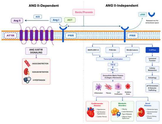
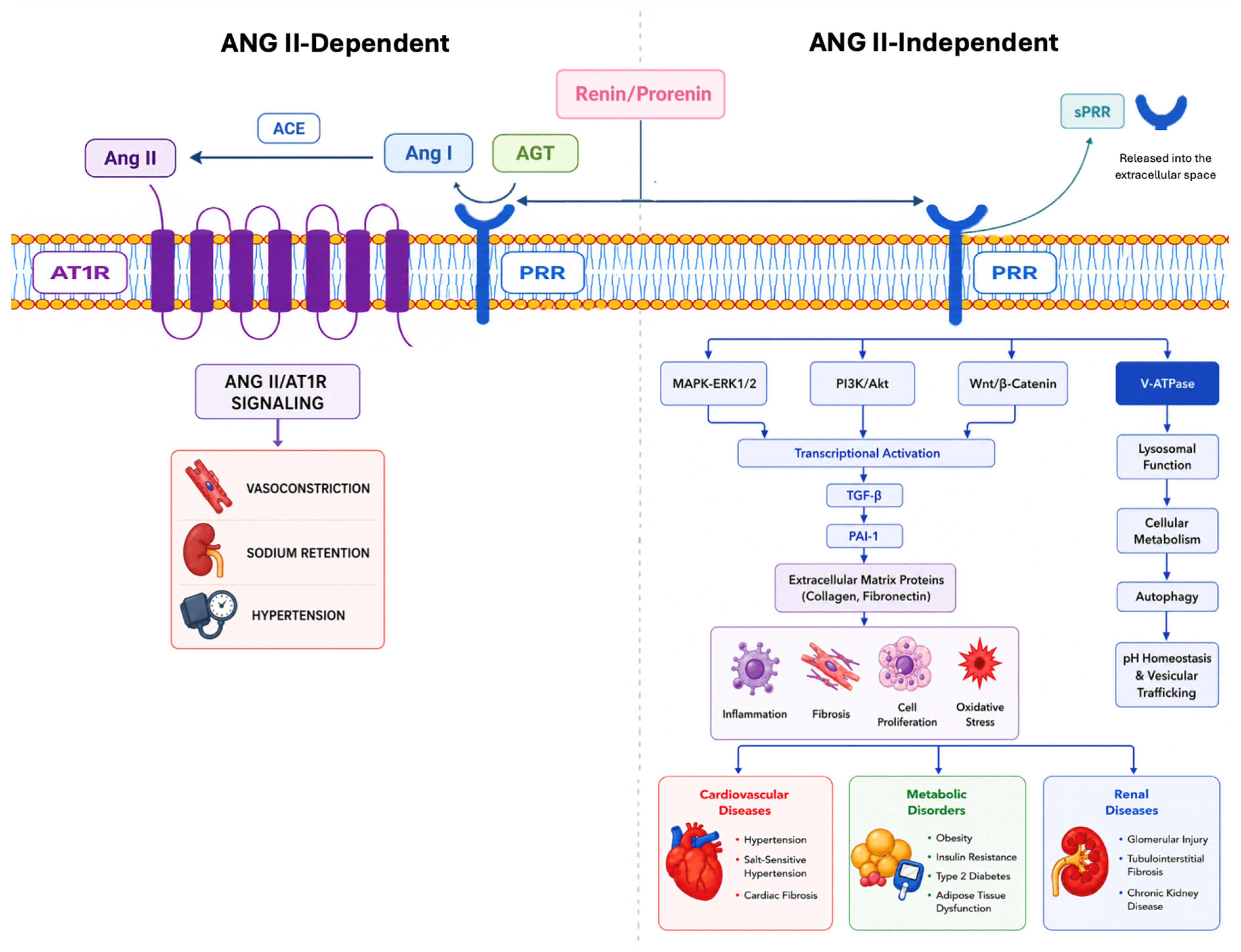
The PRR serves as a central molecular hub regulator linking RAAS activation with intracellular signaling pathways that govern blood pressure, fluid homeostasis, cardiovascular remodeling, and metabolic function (Figure 1). The PRR activation contributes to cardiac hypertrophy and fibrosis, insulin resistance, metabolic syndrome, inflammation, and oxidative stress, thereby promoting the development and progression of cardiovascular and metabolic diseases. These findings identify PRR as a promising therapeutic target for hypertension, heart failure, and related cardiorenal-metabolic disorders [26]. The dysregulation of PRR protein expression and activity have been associated with hypertension, heart failure, chronic kidney disease, diabetic nephropathy, obesity, and metabolic dysfunction, where they contribute to progressive target-organ injury. Consequently, growing experimental and clinical evidence identifies PRR not only as a key mediator of disease progression but also as a promising biomarker and therapeutic target across a broad spectrum of cardio-renal-metabolic disorders.
Figure 1.
A Molecular Hub Integrating RAAS Signaling, Intracellular Metabolism, and Cardiovascular and Renal Remodeling. The PRR functions as a multifunctional signaling platform that links extracellular RAAS activation to intracellular signaling and metabolic regulation. Angiotensin II (Ang II)-dependent: Binding of prorenin or renin to PRR enhances local conversion of angiotensinogen (AGT) to Ang I and subsequent generation of Ang II if angiotensin-converting enzyme (ACE) is present. These actions contribute to amplify tissue RAAS activity independent of circulating renin levels. In parallel, PRR activates Ang II-independent intracellular signaling pathways, including ERK1/2, p38 MAPK, and PI3K/Akt, leading to increased expression of transforming growth factor-β (TGF-β), plasminogen activator inhibitor-1 (PAI-1), and extracellular matrix proteins that promote inflammation, fibrosis, and tissue remodeling. PRR also serves as an essential accessory component of the V-ATPase, regulating lysosomal acidification, vesicular trafficking, autophagy, intracellular pH homeostasis, and cellular metabolism. In the heart, PRR interacts with V-ATPase to regulate calcium handling, contractility, and myocardial integrity, while pathological upregulation promotes fibrosis and cardiac dysfunction. In vascular smooth muscle cells (VSMCs), PRR stimulates ERK1/2-dependent proliferation, inflammation, and vascular remodeling, contributing to hypertension. In the kidney, PRR regulates nephrogenesis through Wnt/β-catenin signaling and, in adulthood, enhances intrarenal RAAS activity, epithelial sodium channel (ENaC) function, oxidative stress, inflammation, and fibrosis, thereby promoting chronic kidney disease progression. Collectively, PRR acts as a central molecular integrator of hormonal, metabolic, and stress-response pathways that drive cardiovascular and renal remodeling.
2.1. Prorenin Receptor in Adipose Tissue Biology and Metabolic Homeostasis
The PRR in adipose tissue modulates biology and obesity-associated metabolic alterations. PRR protein expression is increased in adipose tissue under metabolic stress, including high-fat diet-induced obesity. PRR activation promotes adipocyte hypertrophy, inflammatory cytokine production, and oxidative stress, thereby contributing to adipose tissue dysfunction and systemic insulin resistance [27,28]. In addition, PRR has been implicated in the regulation of adipocyte metabolism through its association with the V-ATPase complex and Wnt/β-catenin signaling-pathways that are critical for adipocyte differentiation and energy homeostasis [27,29].
The metabolic effects of PRR are also closely linked to its function as an accessory protein of the V-ATPase complex. Through this interaction, PRR contributes to the regulation of lysosomal function and cellular metabolism. Disruption of the PRR–V-ATPase pathway impairs intracellular pH and protein degradation—processes increasingly recognized as key contributors to metabolic dysfunction and cellular stress [30]. These findings highlight PRR as a molecular hub integrating extracellular hormonal signals with intracellular metabolic pathways.
2.2. Prorenin Receptor in Cardiac Remodeling and Myocardial Homeostasis
Cardiac PRR protein expression is upregulated in pathological states including diabetes, myocardial infarction, and heart failure, supporting its involvement in maladaptive cardiac remodeling [9,31]. Both low and high sodium diets increase myocardial prorenin/renin levels and PRR protein expression, leading to enhanced fibrotic remodeling under conditions of extreme sodium imbalance [32,33]. Despite these associations, the role of cardiac PRR in systemic BP regulation remains unclear. Constitutive deletion of PRR in cardiomyocytes results in lethal heart failure due to impaired lysosomal acidification, underscoring its essential role in cardiomyocyte homeostasis [14]. In contrast, global or cardiomyocyte-specific PRR overexpression does not significantly alter baseline BP or cardiac morphology. Adenovirus-mediated PRR overexpression in adult rat hearts induces Ang II–independent activation of intracellular signaling pathways and reduces left ventricular ejection fraction, indicating that PRR can directly promote cardiac dysfunction independent of systemic RAAS activation [34]. These findings suggest that cardiac PRR critically regulates myocardial remodeling and function rather than exerting a dominant influence on hemodynamic control.
Beyond its role in RAAS signaling, PRR functions as an accessory protein of the V-ATPase, which is essential for intracellular acidification, vesicular trafficking, and autophagy [13]. Through this interaction, PRR regulates lysosomal function and cellular metabolism. Disruption of the PRR–V-ATPase pathway impairs intracellular pH and protein degradation—processes increasingly recognized as key contributors to metabolic dysfunction and cellular stress [30]. These findings highlight PRR as a molecular hub integrating extracellular hormonal signals with intracellular metabolic pathways. In cardiomyocytes, PRR is closely associated with the V-ATPase and the ryanodine receptor, positioning it at the interface of intracellular calcium handling, lysosomal function, and downstream signaling pathways that influence myocardial contractility and structural integrity [14].
In VSMCs, PRR protein expressed at the cell surface plays a more direct role in BP regulation and vascular remodeling [7,35]. VSMC-specific human PRR overexpression in transgenic rats induces hypertension and tachycardia, whereas PRR deficiency in VSMCs leads to aortic sclerosis without significant changes in BP, highlighting its context-dependent actions within the vasculature. PRR activation in VSMCs stimulates cellular proliferation, ERK1/2 signaling, and the expression of inflammatory and profibrotic mediators, including PAI-1, reinforcing its contribution to vascular injury and remodeling [35]. Collectively, these data indicate that while cardiac PRR predominantly influences myocardial structure and function, vascular PRR plays a more prominent role in the regulation of BP and the progression of vascular pathology. The contribution of vascular PRR extends beyond BP regulation to the structural remodeling of the vessel wall. Independent of Ang II, the activation of PRR by renin or prorenin initiates intracellular signaling pathways—including MAPK-ERK1/2, PI3K/Akt, and Wnt/β-catenin—that regulate VSMC proliferation, migration, and extracellular matrix production [7,11,36]. These processes promote structural changes within the vascular wall, including media thickening, increased collagen deposition, and reduced vascular compliance, which contribute to elevated peripheral resistance and sustained hypertension.
Beyond its effects on VSMCs, PRR also plays a critical role in endothelial cell function and developmental angiogenesis. Endothelial cell-specific deletion of PRR in mice leads to severe defects in retinal vascular development characterized by impaired endothelial cell migration, loss of tip cell polarity, and defective sprouting angiogenesis [36]. These abnormalities reflect dysregulation of genes governing cell migration, polarity, extracellular matrix organization, and angiogenic signaling, and indicate that PRR is critical for proper vascular network formation.
During oxidative stress, the PRR mediates vascular injury by driving maladaptive vascular remodeling. PRR signaling can enhance the production of reactive oxygen species (ROS) through activation of NADPH oxidase pathways, leading to endothelial dysfunction and vascular inflammation [37,38]. These processes facilitate leukocyte recruitment, extracellular matrix reorganization, and vascular wall hypertrophy. PRR-associated signaling through V-ATPase and Wnt/β-catenin pathways may regulate gene programs involved in vascular cell growth and tissue remodeling, linking PRR to both developmental vascular patterning and pathological vascular remodeling in adult tissues [29,36]. These findings position PRR as a central molecular regulator of vascular remodeling, integrating RAAS-dependent and independent signaling that contribute to the structural and functional changes in hypertension and other cardiovascular disease. Collectively, we propose that PRR functions as a molecular hub that integrates metabolic, developmental, and signaling pathways to regulate endothelial cell behavior, angiogenic remodeling, and vascular development.
2.3. Prorenin Receptor in Kidney Development and Renal Homeostasis
The PRR has essential roles in both kidney development and renal homeostasis. During nephrogenesis, deletion of PRR in nephron progenitor cells results in reduced nephron number, podocyte abnormalities, and proteinuria [39]. The PRR is thought to regulate kidney development partly through Wnt/β-catenin signaling, which coordinates ureteric bud branching and nephron formation. In the mature kidney, PRR contributes to the regulation of water and sodium homeostasis. PRR activation increases intrarenal RAAS and ENaC activity, promoting sodium retention and the development of hypertension [24,40,41].
In addition to classical renin production by JG cells, renin and its precursor prorenin are synthesized by the distal nephron segments [42]. Prorenin produced by the collecting duct is regulated by hormones acting on the distal nephron rather than by BP [42,43]. Binding of PRR to renin and prorenin increases local renin enzymatic activity, activates prorenin, and augments intratubular Ang II formation. Increased distal prorenin production and secretion into the lumen has been demonstrated in several experimental hypertensive animal models, including chronic Ang II–infusion, 2-kidney 1-clip (2K1C) Goldblatt hypertension, and transgenic mice with renin deficiency in the collecting duct [40,42,43,44]. Likewise, these findings are present in mice with high fat diet-induced type II diabetes and rats with streptozotocin-induced type 1 diabetes [45,46].
In models of diabetic nephropathy, PRR protein expression is markedly upregulated in glomerular cells, where it stimulates oxidative stress, extracellular matrix accumulation, and inflammatory signaling [9]. These findings, along with evidence that PRR activates Wnt/β-catenin signaling, highlight its critical role in driving renal fibrosis and promoting epithelial–mesenchymal transition during the progression of chronic kidney disease (CKD).
Activation of PRR by its natural agonists, renin and prorenin, promotes kidney fibrosis and inflammation through various pathways independent of the RAAS [9,42]. Its activation directly triggers intracellular signal transduction via ERK1/2 and upregulation of profibrotic genes, including TGF-β1, fibronectin, collagen I, and PAI-1 [45,47,48]. Likewise, PRR exerts a positive feedback action on the Wnt/β-catenin signaling, which exacerbates renal fibrosis and inflammation in hypertensive nephropathy [49]. This crosstalk establishes a feed-forward loop that sustains inflammatory and profibrotic signaling over time and accelerates maladaptive tissue remodeling. Consistent with this pathogenic role, experimental inhibition or knockdown of PRR in ischemia–reperfusion kidney injury models significantly attenuates renal fibrosis and further supports its role as the key amplifier of fibrosis [9].
In addition to promoting fibrosis, PRR amplifies renal inflammatory responses through activation of multiple intracellular signaling pathways. Pathologic upregulation of PRR is strongly associated with enhanced renal and systemic inflammation in disease states including diabetes, hypertension, and renal ischemia. Increased PRR protein expression correlates with activation of ERK1/2 and p38 MAPK, as well as proinflammatory transcription factors such as NF-κB [48,50,51]. Activation of these pathways increases inflammatory mediators including TNF-α, IL-1β, MCP-1, and COX-2 [49,52], and promotes immune cell recruitment and progressive renal injury independent of the RAAS. Although PRR overexpression in healthy animals does not independently induce hypertension or renal injury, it functions as a pathological amplifier to exacerbate inflammation and fibrosis under disease conditions [34]. In the 2K1C model of renovascular hypertension, PRR protein expression is selectively upregulated in the ischemic kidney, which is accompanied by marked increases in TNF-α, IL-1β, MCP-1, COX-2, phosphorylated NF-κB, and collagen type I, despite comparable elevations in intrarenal Ang II levels [9]. Furthermore, targeted intrarenal knockdown of PRR using shRNA significantly attenuates inflammatory cytokine expression, NF-κB activation, fibrosis, and albuminuria without altering BP or renal Ang II levels [48]. In addition, PRR inhibition can reduce inflammation and fibrosis in heart and liver disease models, supporting a broader role for PRR in systemic inflammatory signaling across organs [27,53].
3. The Soluble Prorenin Receptor: Emerging Mechanisms of Disease
The discovery of the sPRR has expanded the conceptual framework of PRR biology from a membrane-bound receptor to a circulating bioactive molecule capable of mediating both local and systemic effects. The sPRR is generated through proteolytic cleavage of the extracellular domain of full-length PRR protein, primarily by site-1 protease (S1P), although furin and ADAM19 also contribute under specific physiological and pathological conditions. Once released into the extracellular space, sPRR is detectable in plasma, urine, and other biological fluids, where its concentration reflects tissue PRR activity and disease severity in several cardio-renal-metabolic disorders [54].
Initially considered merely a cleavage by-product, sPRR is now recognized as a biologically active molecule with endocrine- and paracrine-like properties. Like membrane-bound PRR, sPRR retains the ability to bind both renin and prorenin, thereby enhancing local Ang I generation and amplifying tissue RAAS activation. However, many of its biological effects occur independently of Ang II production. Experimental studies demonstrate that recombinant sPRR directly activates intracellular signaling pathways involved in inflammation, oxidative stress, apoptosis, endothelial dysfunction, and extracellular matrix remodeling, including ERK1/2, MAPK, PI3K/Akt, TGF-β/Smad, and NF-κB signaling. These effects promote vascular remodeling, renal fibrosis, and cardiac hypertrophy even in the presence of conventional RAAS blockade, suggesting that sPRR represents an additional mechanism of tissue injury beyond classical RAAS activation [25].
Experimental studies continue to uncover previously unrecognized physiological actions of sPRR [27,37]. Transgenic mice selectively expressing human sPRR in the renal collecting duct exhibit sex-specific alterations in blood pressure regulation, renal sodium handling, and cardiac remodeling, suggesting that biological responses to sPRR may differ between males and females [55]. These observations introduce an important new dimension to PRR biology and may partially explain sex differences observed in hypertension and cardiorenal disease. One of the most important recent mechanistic advances is the recognition that sPRR directly regulates ENaC activity in collecting duct of the renal medulla [56]. Rather than functioning solely through Ang II generation, sPRR enhances ENaC activity via β-catenin-dependent signaling and proteolytic activation mechanisms, thereby increasing distal sodium reabsorption and contributing to salt-sensitive hypertension. Recent work further demonstrated that furin-mediated post-translational modification is required for the ENaC-activating properties of sPRR, providing new mechanistic insight into how sPRR regulates renal sodium handling independently of membrane-bound PRR. These findings position sPRR as a central mediator linking renal sodium retention, volume expansion, and hypertension [57].
In the vasculature, sPRR promotes endothelial dysfunction and vascular remodeling by enhancing oxidative stress and inflammatory signaling. The sPRR also functions as a signaling ligand capable of interacting with cell-surface receptors beyond PRR itself [58]. In vascular endothelial cells, sPRR has been shown to activate the AT1R in a ligand-independent manner, promoting endothelial dysfunction, oxidative stress, and impaired nitric oxide bioavailability [54]. Additional studies suggest that sPRR influences macrophage activation, inflammatory cytokine production, mitochondrial dysfunction, and metabolic reprogramming, further supporting its role as an active mediator of chronic inflammation rather than simply a biomarker of disease. Although the identity of additional receptors and signaling partners remains under investigation, these observations broaden the biological functions attributed to sPRR considerably.
Clinical investigations have strengthened the association between elevated circulating sPRR concentrations and cardiovascular, renal, and metabolic diseases. In metabolic tissues such as the liver and adipose tissue, sPRR may influence glucose metabolism and insulin sensitivity, potentially linking PRR signaling to broader metabolic dysregulation [27]. Furthermore, increased plasma or serum sPRR levels have consistently been reported in patients with hypertension, chronic kidney disease, diabetic nephropathy, heart failure, obstructive sleep apnea, obesity, and preeclampsia, where higher concentrations correlate with disease severity, renal dysfunction, vascular injury, and adverse clinical outcomes [59,60]. Indeed, circulating sPRR is significantly elevated in patients with heart failure and correlates with indices of cardiac dysfunction, supporting its potential utility as a diagnostic and prognostic biomarker [61].
From a clinical perspective, circulating sPRR has gained attention as a potential biomarker for cardiorenal disease progression. Recent population studies have further extended these observations. The African-PREDICT study [62] demonstrated significant associations between circulating sPRR concentrations and early changes in renal function in apparently healthy young adults, suggesting that sPRR may identify subclinical kidney injury before overt chronic kidney disease develops. Therefore, sPRR is being proposed as a predictor of vascular injury and atherosclerosis, with elevated circulating concentrations correlating with endothelial dysfunction, arterial stiffness, and indices of peripheral arterial disease [62].
4. Soluble Prorenin Receptor as a Biomarker: Clinical Therapeutic Implications and Limitations of Targeting the PRR/sPRR Axis
Interest in sPRR as a metabolic biomarker has also increased substantially. A 2025 meta-analysis integrating seven clinical studies involving more than 1600 participants reported that circulating sPRR demonstrates moderate diagnostic accuracy for gestational diabetes mellitus, supporting its potential as a non-invasive biomarker for identifying women at increased cardiometabolic risk during pregnancy [63]. Although further validation is required, these findings reinforce the concept that sPRR reflects systemic metabolic dysfunction extending beyond classical RAAS activation. Measurement of plasma sPRR levels may provide insights into the activation of tissue RAAS and associated pathological processes, including fibrosis, inflammation, and sodium retention. The sPRR reflects both PRR expression and proteolytic processing; thus, it may serve as an integrative indicator of PRR pathway activation across multiple organs.
Accumulating data supports sPRR value to help identify patients at risk for cardiovascular disease, progressive kidney disease, resistant hypertension, or adverse cardiovascular outcomes; nonetheless several important limitations currently preclude its routine clinical implementation. First, circulating sPRR lacks disease specificity. Elevated serum or plasma concentrations have been reported in chronic kidney disease, hypertension, heart failure, diabetes mellitus, obesity, obstructive sleep apnea, preeclampsia, and several inflammatory disorders [54,59,60]. In consequence, it is difficult to distinguish whether increased sPRR reflects activation of disease-specific pathogenic pathways or merely represents a general marker of tissue injury and chronic inflammation. Furthermore, because PRR is ubiquitously expressed in numerous organs—including the kidney, heart, vasculature, brain, placenta, adipose tissue, liver, pancreas, and immune cells—circulating sPRR likely reflects integrated release from multiple tissues rather than activity within a single organ [26]. Consequently, interpretation of plasma sPRR concentrations in patients with multisystem disease remains challenging.
Another important limitation is the strong influence of renal function on circulating sPRR concentrations. Numerous clinical studies have demonstrated inverse relationships between serum sPRR and estimated glomerular filtration rate (eGFR), suggesting that impaired renal clearance contributes substantially to circulating sPRR accumulation [59,60,64]. Thus, elevated sPRR levels in patients with chronic kidney disease may reflect both increased production and reduced elimination [64]. Similar confounding effects may occur in heart failure and other conditions characterized by declining renal function, limiting the ability of sPRR to serve as an independent biomarker without adjustment for kidney function [61]. Future prospective studies incorporating serial measurements and multivariable analyses will be required to determine whether sPRR provides incremental prognostic value beyond established biomarkers and conventional measures of renal function.
An additional obstacle to the clinical application of sPRR is the lack of assay standardization. Most published studies have employed commercially available enzyme-linked immunosorbent assays (ELISAs), yet these assays differ with respect to antibody specificity, calibration standards, detection ranges, sample handling protocols, and recognition of different PRR fragments [65]. Consequently, absolute circulating sPRR concentrations vary considerably among studies, making direct comparisons difficult and preventing establishment of universally accepted reference intervals or diagnostic cut-off values. Moreover, preanalytical variables—including sample type (serum versus plasma), storage conditions, freeze–thaw cycles, and anticoagulant selection—may further influence measured concentrations. Standardized analytical methods, international reference materials, and multicenter validation studies will therefore be essential before sPRR can be incorporated into routine clinical practice.
Equally important is the unresolved question regarding the origin of tissue circulating sPRR. Although the kidney—particularly collecting duct intercalated cells—appears to be a major source under physiological conditions [24,42], experimental studies indicate that PRR cleavage also occurs in the heart, vascular wall, placenta, adipose tissue, liver, pancreas, and central nervous system [8]. Furthermore, proteolytic processing varies according to disease state, involving S1P, furin, ADAM19, and potentially additional proteases whose relative contributions remain incompletely characterized. Because different tissues may generate sPRR in response to distinct pathological stimuli, circulating concentrations likely represent the integrated output of multiple organs rather than a tissue-specific signal fragment [65]. The development of tissue-specific biomarkers or molecular imaging approaches capable of identifying the principal source of sPRR production would substantially enhance its clinical utility.
The growing recognition that sPRR functions as an active signaling molecule rather than an inert cleavage product has broadened therapeutic interest in the PRR/sPRR axis. Unlike conventional RAAS inhibitors, which primarily suppress systemic Ang II formation or receptor activation, interventions targeting PRR or sPRR could simultaneously inhibit local RAAS activation and Ang II-independent signaling pathways involved in inflammation, oxidative stress, fibrosis, endothelial dysfunction, and sodium retention. Experimental studies have demonstrated that blockade of PRR using decoy peptides such as PRO20 (MCE®, Cat No. HY-P10933; Monmouth Junction, NJ 08852, USA) attenuates hypertension, renal injury, cardiac remodeling, and diabetic nephropathy, whereas inhibition of sPRR generation through modulation of S1P activity or interference with sPRR signaling may represent an emerging therapeutic strategy [20]. More recent mechanistic studies also indicate that inhibition of sPRR-mediated activation of ENaC may reduce sodium retention and salt-sensitive hypertension independently of systemic RAAS blockade [56,64]. These observations suggest that selective targeting of the PRR/sPRR axis may provide additive benefits when combined with ACE inhibitors, angiotensin receptor blockers, direct renin inhibitors, mineralocorticoid receptor antagonists, small-interfering RNA (siRNA) or sodium-glucose cotransporter-2 inhibitors.
Therapeutic targeting of PRR presents important biological challenges. The PRR is indispensable for V-ATPase assembly, lysosomal acidification, autophagy, intracellular trafficking, and Wnt/β-catenin signaling, all of which are essential for normal cellular homeostasis and embryonic development. Global inhibition of PRR therefore carries a substantial risk of disrupting physiological processes unrelated to RAAS activation [54]. Future therapeutic approaches will likely require selective inhibition of pathological ligand-dependent signaling or modulation of sPRR production while preserving the receptor’s constitutive intracellular functions. Tissue-specific delivery systems, monoclonal antibodies, small-molecule inhibitors targeting extracellular PRR interactions, or selective inhibition of disease-associated proteolytic cleavage represent promising avenues currently under investigation. Before translation into clinical practice, however, additional studies are needed to define the long-term safety, tissue specificity, and efficacy of these approaches in human cardiovascular, renal, and metabolic diseases.
Collectively, current evidence establishes sPRR as considerably more than a soluble cleavage product of PRR. It functions as a circulating signaling molecule that integrates RAAS activation with inflammatory, fibrotic, metabolic, and sodium-retaining pathways. Although many mechanistic questions remain unresolved—including the identification of additional receptors, downstream signaling networks, and the relative contributions of tissue-derived versus circulating sPRR—the accumulating experimental and clinical evidence supports its potential utility both as a biomarker of cardio-renal-metabolic disease and as a novel therapeutic target. Future studies should determine whether selective inhibition of sPRR production or signaling can provide additional protection beyond conventional RAAS blockade.
5. Conclusions
Over the past two decades, PRR has emerged as a multifunctional regulator whose roles extend well beyond the RAAS. Although many aspects of its biology remain unresolved, PRR is increasingly recognized as a central signaling hub that integrates RAAS-dependent and RAAS-independent pathways involved in endothelial function, angiogenesis, vascular remodeling, metabolic regulation, and renal homeostasis. Through activation of signaling cascades such as MAPK-ERK1/2, PI3K/Akt, and Wnt/β-catenin, as well as its association with the V-ATPase complex, PRR regulates cellular proliferation, inflammation, oxidative stress, and fibrosis—processes that contribute to cardiovascular, metabolic, and renal disease.
The discovery of sPRR has further expanded our understanding of PRR biology. In addition to its membrane-bound functions, PRR may signal through its soluble form, enabling communication among the kidney, heart, vasculature, and metabolic tissues. Emerging evidence suggests that sPRR acts as a circulating mediator linking tissue RAAS activity with systemic cardiometabolic and cardiorenal dysfunction. These findings highlight the PRR–sPRR axis as a promising therapeutic target. However, further studies are needed to define the tissue-specific functions of PRR, elucidate the mechanisms regulating sPRR generation and signaling, and determine whether modulation of PRR/sPRR pathways can be translated into effective diagnostic and therapeutic strategies for cardiovascular, metabolic, and renal diseases.
Funding
M.C.P., was partially supported by Fundação de Amparo à Pesquisa do Estado de São Paulo (FAPESP Grant No. 2017/17027-0) and the Carol Lavin-Bernick Faculty Grant Award from Tulane University. J.D.P., was a recipient of The Ramadhyani-Bourgeois MD Student Endowed Award.
Institutional Review Board Statement
Not applicable.
Informed Consent Statement
Not applicable.
Data Availability Statement
No new data were created or analyzed in this study. Data sharing is not applicable to this article.
Acknowledgments
The authors thank Tulane University School of Medicine for institutional support.
Conflicts of Interest
The authors declare that there are no potential conflicts of interest relating to this manuscript.
Abbreviations and Acronyms
| ACE | Angiotensin-converting enzyme |
| ADAM19 | A Disintegrin And Metalloproteinase 19 |
| AGT | Angiotensinogen |
| Akt | Protein kinase B |
| Ang I | Angiotensin I |
| Ang II | Angiotensin II |
| ATP6AP2 | ATPase H+-transporting accessory protein 2 (gene encoding PRR) |
| AT1R | Angiotensin II Type 1 Receptor |
| BP | Blood pressure |
| CKD | Chronic kidney disease |
| COX-2 | Cyclooxygenase-2 |
| ELISA | Enzyme-linked immunosorbent assay |
| ENaC | Epithelial sodium channel |
| eGFR | Estimated glomerular filtration rate |
| ERK1/2 | Extracellular signal-regulated kinase 1/2 |
| IL-1β | Interleukin-1 beta |
| IS | Indoxyl sulfate |
| MAPK | Mitogen-activated protein kinase |
| MCP-1 | Monocyte chemoattractant protein-1 |
| MMP9 | Matrix metalloproteinase-9 |
| NF-κB | Nuclear factor κB |
| NOX4 | NADPH oxidase 4 |
| p38 MAPK | p38 mitogen-activated protein kinase |
| PAI-1 | Plasminogen activator inhibitor-1 |
| PI3K | Phosphoinositide 3-kinase |
| PRR | Prorenin receptor |
| PRO20 | 20 amino acid peptide derived from the prorenin pro-segment |
| RAAS | Renin–angiotensin–aldosterone system |
| ROS | Reactive oxygen species |
| sPRR | Soluble prorenin receptor |
| shRNA | Small hairpin RNA |
| siRNA | Small interfering RNA |
| SP-1 | Site-1 protease |
| TGF-β | Transforming growth factor-β |
| TIMP1 | Tissue inhibitor of metalloproteinase-1 |
| TNF-α | Tumor necrosis factor-α |
| V-ATPase | Vacuolar H+-ATPase |
| VSMC | Vascular smooth muscle cell |
| Wnt/β-catenin | Wnt/β-catenin signaling pathway |
| 2K1C | Two-kidney, one-clip (Goldblatt hypertension model) |
References
- Weir, M. The Renin-Angiotensin-Aldosterone System: A Specific Target for Hypertension Management. Am. J. Hypertens. 1999, 12, 205–213. [Google Scholar] [CrossRef] [Scilit] [PubMed]
- Crowley, S.D.; Navar, L.G.; Prieto, M.C.; Gurley, S.B.; Coffman, T.M. Kidney Renin–Angiotensin System: Lost in a RAS Cascade. Hypertension 2024, 81, 682–686. [Google Scholar] [CrossRef] [Scilit] [PubMed]
- Pravenec, M.; Simonet, L.; Křen, V.; Kuneš, J.; Levan, G.; Szpirer, J.; Szpirer, C.; Kurtz, T. The Rat Renin Gene: Assignment to Chromosome 13 and Linkage to the Regulation of Blood Pressure. Genomics 1991, 9, 466–472. [Google Scholar] [CrossRef] [Scilit] [PubMed]
- Kurtz, A. Control of Renin Synthesis and Secretion. Am. J. Hypertens. 2012, 25, 839–847. [Google Scholar] [CrossRef] [Scilit] [PubMed]
- Schalekamp, M.A.D.H.; Deinum, J.; Danser, A.H.J. Short-Lived Active Prorenin: Precursor of So-Called Native Prorenin. Hypertension 2024, 81, 1190–1193. [Google Scholar] [CrossRef] [Scilit] [PubMed]
- Triebel, H.; Castrop, H. The Renin Angiotensin Aldosterone System. Pflüg. Arch.-Eur. J. Physiol. 2024, 476, 705–713. [Google Scholar] [CrossRef] [Scilit] [PubMed]
- Nguyen, G.; Delarue, F.; Burcklé, C.; Bouzhir, L.; Giller, T.; Sraer, J.-D. Pivotal Role of the Renin/Prorenin Receptor in Angiotensin II Production and Cellular Responses to Renin. J. Clin. Investig. 2002, 109, 1417–1427. [Google Scholar] [CrossRef]
- Cousin, C.; Bracquart, D.; Contrepas, A.; Corvol, P.; Muller, L.; Nguyen, G. Soluble Form of the (Pro)Renin Receptor Generated by Intracellular Cleavage by Furin Is Secreted in Plasma. Hypertension 2009, 53, 1077–1082. [Google Scholar] [CrossRef] [Scilit] [PubMed]
- Ichihara, A.; Yatabe, M.S. The (pro)Renin Receptor in Health and Disease. Nat. Rev. Nephrol. 2019, 15, 693–712. [Google Scholar] [CrossRef] [Scilit] [PubMed]
- Jan Danser, A.H.; Batenburg, W.W.; Van Esch, J.H.M. Prorenin and the (pro)Renin Receptor—An Update. Nephrol. Dial. Transplant. 2007, 22, 1288–1292. [Google Scholar] [CrossRef] [Scilit] [PubMed]
- Sakoda, M.; Ichihara, A.; Kaneshiro, Y.; Takemitsu, T.; Nakazato, Y.; Nabi, A.H.M.N.; Nakagawa, T.; Suzuki, F.; Inagami, T.; Itoh, H. (Pro)Renin Receptor-Mediated Activation of Mitogen-Activated Protein Kinases in Human Vascular Smooth Muscle Cells. Hypertens. Res. 2007, 30, 1139–1146. [Google Scholar] [CrossRef] [Scilit] [PubMed]
- Balakumar, P.; Jagadeesh, G. Potential Cross-Talk between (pro)Renin Receptors and Wnt/Frizzled Receptors in Cardiovascular and Renal Disorders. Hypertens. Res. 2011, 34, 1161–1170. [Google Scholar] [CrossRef] [Scilit] [PubMed]
- Advani, A.; Kelly, D.J.; Cox, A.J.; White, K.E.; Advani, S.L.; Thai, K.; Connelly, K.A.; Yuen, D.; Trogadis, J.; Herzenberg, A.M.; et al. The (Pro)Renin Receptor: Site-Specific and Functional Linkage to the Vacuolar H+-ATPase in the Kidney. Hypertension 2009, 54, 261–269. [Google Scholar] [CrossRef] [Scilit] [PubMed]
- Kinouchi, K.; Ichihara, A.; Sano, M.; Sun-Wada, G.-H.; Wada, Y.; Kurauchi-Mito, A.; Bokuda, K.; Narita, T.; Oshima, Y.; Sakoda, M.; et al. The (Pro)Renin Receptor/ATP6AP2 Is Essential for Vacuolar H+-ATPase Assembly in Murine Cardiomyocytes. Circ. Res. 2010, 107, 30–34. [Google Scholar] [CrossRef] [Scilit] [PubMed]
- Oshima, Y.; Kinouchi, K.; Ichihara, A.; Sakoda, M.; Kurauchi-Mito, A.; Bokuda, K.; Narita, T.; Kurosawa, H.; Sun-Wada, G.-H.; Wada, Y.; et al. Prorenin Receptor Is Essential for Normal Podocyte Structure and Function. J. Am. Soc. Nephrol. 2011, 22, 2203–2212. [Google Scholar] [CrossRef] [Scilit] [PubMed]
- Kanda, A.; Noda, K.; Ishida, S. ATP6AP2/(Pro)Renin Receptor Contributes to Glucose Metabolism via Stabilizing the Pyruvate Dehydrogenase E1 β Subunit. J. Biol. Chem. 2015, 290, 9690–9700. [Google Scholar] [CrossRef] [Scilit] [PubMed]
- Shimba, A.; Munakata, S.; Abe, S.; Cui, G.; Kato, R.; Miyazaki, M.; Ichihara, A.; Ikuta, K. PRR Enhances Anti-Tumor Immunity and Suppresses Colitis by Promoting the Development and Survival of Naive T and iNKT Cells. Front. Immunol. 2025, 16, 1566935. [Google Scholar] [CrossRef] [Scilit] [PubMed]
- Cruciat, C.-M.; Ohkawara, B.; Acebron, S.P.; Karaulanov, E.; Reinhard, C.; Ingelfinger, D.; Boutros, M.; Niehrs, C. Requirement of Prorenin Receptor and Vacuolar H+-ATPase–Mediated Acidification for Wnt Signaling. Science 2010, 327, 459–463. [Google Scholar] [CrossRef] [Scilit] [PubMed]
- Nakagawa, T.; Suzuki-Nakagawa, C.; Watanabe, A.; Asami, E.; Matsumoto, M.; Nakano, M.; Ebihara, A.; Uddin, M.N.; Suzuki, F. Site-1 Protease Is Required for the Generation of Soluble (pro)Renin Receptor. J. Biochem. 2017, 161, 369–379. [Google Scholar] [CrossRef] [Scilit] [PubMed]
- Feng, Y.; Peng, K.; Luo, R.; Wang, F.; Yang, T. Site-1 Protease-Derived Soluble (Pro)Renin Receptor Contributes to Angiotensin II–Induced Hypertension in Mice. Hypertension 2021, 77, 405–416. [Google Scholar] [CrossRef] [Scilit] [PubMed]
- Yoshikawa, A.; Aizaki, Y.; Kusano, K.; Kishi, F.; Susumu, T.; Iida, S.; Ishiura, S.; Nishimura, S.; Shichiri, M.; Senbonmatsu, T. The (pro)Renin Receptor Is Cleaved by ADAM19 in the Golgi Leading to Its Secretion into Extracellular Space. Hypertens. Res. 2011, 34, 599–605. [Google Scholar] [CrossRef] [Scilit] [PubMed]
- Suzuki-Nakagawa, C.; Nishimura, M.; Noda, M.; Iwata, H.; Hattori, M.; Ebihara, A.; Suzuki, F.; Nakagawa, T. Intracellular Retention of the Extracellular Domain of the (pro)Renin Receptor in Mammalian Cells. Biosci. Biotechnol. Biochem. 2014, 78, 1187–1190. [Google Scholar] [CrossRef] [Scilit] [PubMed]
- Murray, J.B.; Mikhael, C.; Han, G.; De Faria, L.P.; Rody, W.J.; Holliday, L.S. Activation of (pro)Renin by (pro)Renin Receptor in Extracellular Vesicles from Osteoclasts. Sci. Rep. 2021, 11, 9214. [Google Scholar] [CrossRef] [Scilit] [PubMed]
- Gonzalez, A.A.; Lara, L.S.; Luffman, C.; Seth, D.M.; Prieto, M.C. Soluble Form of the (pro)Renin Receptor Is Augmented in the Collecting Duct and Urine of Chronic Angiotensin II-Dependent Hypertensive Rats. Hypertension 2011, 57, 859–864. [Google Scholar] [CrossRef] [Scilit] [PubMed]
- Wang, B.; Jie, H.; Wang, S.; Dong, B.; Zou, Y. The Role of (pro)Renin Receptor and Its Soluble Form in Cardiovascular Diseases. Front. Cardiovasc. Med. 2023, 10, 1086603. [Google Scholar] [CrossRef] [Scilit] [PubMed]
- Zhu, M.; Yi, X.; Song, S.; Yang, H.; Yu, J.; Xu, C. Principle Role of the (pro)Renin Receptor System in Cardiovascular and Metabolic Diseases: An Update. Cell. Signal. 2024, 124, 111417. [Google Scholar] [CrossRef] [Scilit] [PubMed]
- Gatineau, E.; Arthur, G.; Poupeau, A.; Nichols, K.; Spear, B.T.; Shelman, N.R.; Graf, G.A.; Temel, R.E.; Yiannikouris, F.B. The Prorenin Receptor and Its Soluble Form Contribute to Lipid Homeostasis. Am. J. Physiol.-Endocrinol. Metab. 2021, 320, E609–E618. [Google Scholar] [CrossRef] [Scilit] [PubMed]
- Nichols, K.; Yiannikouris, F. The Role of (Pro)Renin Receptor in the Metabolic Syndrome. Curr. Hypertens. Rev. 2022, 18, 117–124. [Google Scholar] [CrossRef] [Scilit] [PubMed]
- Hoffmann, N.; Peters, J. Functions of the (pro)Renin Receptor (Atp6ap2) at Molecular and System Levels: Pathological Implications in Hypertension, Renal and Brain Development, Inflammation, and Fibrosis. Pharmacol. Res. 2021, 173, 105922. [Google Scholar] [CrossRef] [Scilit] [PubMed]
- Figueiredo, M.; Daryadel, A.; Sihn, G.; Müller, D.N.; Popova, E.; Rouselle, A.; Nguyen, G.; Bader, M.; Wagner, C.A. The (pro)Renin Receptor (ATP6ap2) Facilitates Receptor-Mediated Endocytosis and Lysosomal Function in the Renal Proximal Tubule. Pflüg. Arch.-Eur. J. Physiol. 2021, 473, 1229–1246. [Google Scholar] [CrossRef] [Scilit] [PubMed]
- Li, L.; Cui, Y.; Liu, Y.; Li, H.; Su, Y.; Li, S.; Wang, L.; Zhao, Y.; Wang, S.; Yan, F.; et al. ATP6AP2 Knockdown in Cardiomyocyte Deteriorates Heart Function via Compromising Autophagic Flux and NLRP3 Inflammasome Activation. Cell Death Discov. 2022, 8, 161. [Google Scholar] [CrossRef] [Scilit] [PubMed]
- Hayakawa, Y.; Aoyama, T.; Yokoyama, C.; Okamoto, C.; Komaki, H.; Minatoguchi, S.; Iwasa, M.; Yamada, Y.; Kawamura, I.; Kawasaki, M.; et al. High Salt Intake Damages the Heart through Activation of Cardiac (Pro) Renin Receptors Even at an Early Stage of Hypertension. PLoS ONE 2015, 10, e0120453. [Google Scholar] [CrossRef] [Scilit] [PubMed]
- Okamoto, C.; Hayakawa, Y.; Aoyama, T.; Komaki, H.; Minatoguchi, S.; Iwasa, M.; Yamada, Y.; Kanamori, H.; Kawasaki, M.; Nishigaki, K.; et al. Excessively Low Salt Diet Damages the Heart through Activation of Cardiac (pro) Renin Receptor, Renin-Angiotensin-Aldosterone, and Sympatho-Adrenal Systems in Spontaneously Hypertensive Rats. PLoS ONE 2017, 12, e0189099. [Google Scholar] [CrossRef] [Scilit] [PubMed]
- Moilanen, A.-M.; Rysä, J.; Serpi, R.; Mustonen, E.; Szabò, Z.; Aro, J.; Näpänkangas, J.; Tenhunen, O.; Sutinen, M.; Salo, T.; et al. (Pro)Renin Receptor Triggers Distinct Angiotensin II-Independent Extracellular Matrix Remodeling and Deterioration of Cardiac Function. PLoS ONE 2012, 7, e41404. [Google Scholar] [CrossRef] [Scilit] [PubMed]
- Ma, J.; Li, Y.; Yang, X.; Liu, K.; Zhang, X.; Zuo, X.; Ye, R.; Wang, Z.; Shi, R.; Meng, Q.; et al. Signaling Pathways in Vascular Function and Hypertension: Molecular Mechanisms and Therapeutic Interventions. Signal Transduct. Target. Ther. 2023, 8, 168. [Google Scholar] [CrossRef] [Scilit] [PubMed]
- Patel, N.R.; K C, R.; Blanks, A.; Li, Y.; Prieto, M.C.; Meadows, S.M. Endothelial Cell Polarity and Extracellular Matrix Composition Require Functional ATP6AP2 during Developmental and Pathological Angiogenesis. JCI Insight 2022, 7, e154379. [Google Scholar] [CrossRef] [Scilit] [PubMed]
- Visniauskas, B.; Rajan, K.C.; Abshire, C.M.; Ogola, B.O.; Rosales, C.B.; Galeas-Pena, M.; Sure, V.N.; Sakamuri, S.S.V.P.; Harris, N.R.; Kilanowski-Doroh, I.; et al. High-Plasma Soluble Prorenin Receptor Is Associated with Vascular Damage in Male, but Not Female, Mice Fed a High-Fat Diet. Am. J. Physiol.-Heart Circ. Physiol. 2023, 324, H762–H775. [Google Scholar] [CrossRef] [Scilit] [PubMed]
- Luo, R.; Yang, K.; Wang, F.; Xu, C.; Yang, T. (Pro)Renin Receptor Decoy Peptide PRO20 Protects against Adriamycin-Induced Nephropathy by Targeting the Intrarenal Renin-Angiotensin System. Am. J. Physiol.-Ren. Physiol. 2020, 319, F930–F940. [Google Scholar] [CrossRef] [Scilit]
- Yosypiv, I.V.; Sequeira-Lopez, M.L.S.; Song, R.; De Goes Martini, A. Stromal Prorenin Receptor Is Critical for Normal Kidney Development. Am. J. Physiol.-Regul. Integr. Comp. Physiol. 2019, 316, R640–R650. [Google Scholar] [CrossRef] [Scilit] [PubMed]
- Prieto, M.C.; Reverte, V.; Mamenko, M.; Kuczeriszka, M.; Veiras, L.C.; Rosales, C.B.; McLellan, M.; Gentile, O.; Jensen, V.B.; Ichihara, A.; et al. Collecting Duct Prorenin Receptor Knockout Reduces Renal Function, Increases Sodium Excretion, and Mitigates Renal Responses in ANG II-Induced Hypertensive Mice. Am. J. Physiol.-Ren. Physiol. 2017, 313, F1243–F1253. [Google Scholar] [CrossRef] [Scilit] [PubMed]
- Wang, F.; Lu, X.; Peng, K.; Fang, H.; Zhou, L.; Su, J.; Nau, A.; Yang, K.T.; Ichihara, A.; Lu, A.; et al. Antidiuretic Action of Collecting Duct (Pro)Renin Receptor Downstream of Vasopressin and PGE2 Receptor EP4. J. Am. Soc. Nephrol. JASN 2016, 27, 3022–3034. [Google Scholar] [CrossRef] [Scilit] [PubMed]
- Prieto, M.C.; Gonzalez, A.A.; Visniauskas, B.; Navar, L.G. The Evolving Complexity of the Collecting Duct Renin-Angiotensin System in Hypertension. Nat. Rev. Nephrol. 2021, 17, 481–492. [Google Scholar] [CrossRef] [Scilit] [PubMed]
- Lara, L.S.; Gonzalez, A.A.; Hennrikus, M.T.; Prieto, M.C. Hormone-Dependent Regulation of Renin and Effects on ProreninReceptor Signaling in the Collecting Duct. Curr. Hypertens. Rev. 2022, 18, 91–100. [Google Scholar] [CrossRef] [Scilit] [PubMed]
- Fu, Z.; Kaewsaro, K.; Soodvilai, S.; Kimball, A.; Yang, T. Collecting Duct-Derived (pro)Renin Receptor Contributes to 2-Kidney, 1-Clip-Induced Ischemic Nephropathy and Renovascular Hypertension in Mice. Am. J. Physiol.-Ren. Physiol. 2025, 329, F46–F58. [Google Scholar] [CrossRef] [Scilit] [PubMed]
- Gogulamudi, V.R.; Arita, D.Y.; Bourgeois, C.R.T.; Jorgensen, J.; He, J.; Wimley, W.C.; Satou, R.; Gonzalez, A.A.; Prieto, M.C. High Glucose Induces Trafficking of Prorenin Receptor and Stimulates Profibrotic Factors in the Collecting Duct. Sci. Rep. 2021, 11, 13815. [Google Scholar] [CrossRef] [Scilit] [PubMed]
- Reverte, V.; Gogulamudi, V.R.; Rosales, C.B.; Musial, D.C.; Gonsalez, S.R.; Parra-Vitela, A.J.; Galeas-Pena, M.; Sure, V.N.; Visniauskas, B.; Lindsey, S.H.; et al. Urinary Angiotensinogen Increases in the Absence of Overt Renal Injury in High Fat Diet-Induced Type 2 Diabetic Mice. J. Diabetes Complicat. 2020, 34, 107448. [Google Scholar] [CrossRef] [Scilit] [PubMed]
- Narumi, K.; Sato, E.; Hirose, T.; Yamamoto, T.; Nakamichi, T.; Miyazaki, M.; Sato, H.; Ito, S. (Pro)Renin Receptor Is Involved in Mesangial Fibrosis and Matrix Expansion. Sci. Rep. 2018, 8, 16. [Google Scholar] [CrossRef] [Scilit] [PubMed]
- Gonzalez, A.A.; Zamora, L.; Reyes-Martinez, C.; Salinas-Parra, N.; Roldan, N.; Cuevas, C.A.; Figueroa, S.; Gonzalez-Vergara, A.; Prieto, M.C. (Pro)Renin Receptor Activation Increases Profibrotic Markers and Fibroblast-like Phenotype through MAPK-Dependent ROS Formation in Mouse Renal Collecting Duct Cells. Clin. Exp. Pharmacol. Physiol. 2017, 44, 1134–1144. [Google Scholar] [CrossRef] [Scilit] [PubMed]
- Lin, M.; Wang, D.; Chen, Y.; Chen, G.; Zhou, Y.; Ou, J.; Xiao, L. PRR Promotes Hypertensive Renal Injury by Activating Wnt/β-Catenin Signaling and Inflammation Infiltration in Mice. Biochim. Biophys. Acta Mol. Basis Dis. 2025, 1871, 167517. [Google Scholar] [CrossRef] [Scilit] [PubMed]
- Larenas, P.E.; Cárdenas, P.; Aguirre-Delgadillo, M.; Moris, C.; Casarini, D.E.; Vallotton, Z.; Prieto, M.C.; Gonzalez, A.A. GLUT1 and Prorenin Receptor Mediate Differential Regulation of TGF-β and CTGF in Renal Inner Medullary Collecting Duct Cells during High Glucose Conditions. Biol. Res. 2024, 57, 81. [Google Scholar] [CrossRef] [Scilit] [PubMed]
- Ramkumar, N.; Kohan, D.E. The (pro)Renin Receptor: An Emerging Player in Hypertension and Metabolic Syndrome. Kidney Int. 2019, 95, 1041–1052. [Google Scholar] [CrossRef] [Scilit] [PubMed]
- Gonzalez, A.A.; Luffman, C.; Bourgeois, C.R.T.; Vio, C.P.; Prieto, M.C. Angiotensin II–Independent Upregulation of Cyclooxygenase-2 by Activation of the (Pro)Renin Receptor in Rat Renal Inner Medullary Cells. Hypertension 2013, 61, 443–449. [Google Scholar] [CrossRef] [Scilit] [PubMed]
- Ellmers, L.J.; Rademaker, M.T.; Charles, C.J.; Yandle, T.G.; Richards, A.M. (Pro)Renin Receptor Blockade Ameliorates Cardiac Injury and Remodeling and Improves Function After Myocardial Infarction. J. Card. Fail. 2016, 22, 64–72. [Google Scholar] [CrossRef] [Scilit] [PubMed]
- Yang, T. Soluble (Pro)Renin Receptor in Hypertension. Nephron 2023, 147, 234–243, Erratum in Nephron 2023, 147, 250. [Google Scholar] [CrossRef] [Scilit] [PubMed]
- Arthur, G.; Poupeau, A.; Biel, K.; Osborn, J.L.; Gong, M.; Hinds, T.D.; Lindner, V.; Loria, A.S. Human Soluble Prorenin Receptor Expressed in Mouse Renal Collecting Duct Shows Sex-Specific Effect on Cardiorenal Function. Am. J. Physiol.-Ren. Physiol. 2024, 326, F611–F621. [Google Scholar] [CrossRef] [Scilit] [PubMed]
- Yang, T.; Gao, Z.-X.; Mao, Z.-H.; Wu, P. Soluble (pro)Renin Receptor as a Novel Regulator of Renal Medullary Na+ Reabsorption. Am. J. Physiol.-Ren. Physiol. 2025, 328, F239–F247. [Google Scholar] [CrossRef] [Scilit] [PubMed]
- Zheng, H.; Helms, M.N.; Zou, C.; Zimmerman, E.; Feng, Y.; Yang, T. Furin-Mediated Modification Is Required for Epithelial Sodium Channel-Activating Activity of Soluble (pro)Renin Receptor in Cultured Collecting Duct Cells. Am. J. Physiol.-Ren. Physiol. 2025, 328, F766–F774. [Google Scholar] [CrossRef] [Scilit] [PubMed]
- Visniauskas, B.; Ogola, B.O.; Kilanowski-Doroh, I.; Harris, N.R.; Diaz, Z.T.; Horton, A.C.; Blessinger, S.A.; McNally, A.B.; Zimmerman, M.A.; Arnold, A.C.; et al. Hypertension Disrupts the Vascular Clock in Both Sexes. Am. J. Physiol.-Heart Circ. Physiol. 2024, 327, H765–H777. [Google Scholar] [CrossRef] [Scilit] [PubMed]
- Visniauskas, B.; Arita, D.Y.; Rosales, C.B.; Feroz, M.A.; Luffman, C.; Accavitti, M.J.; Dawkins, G.; Hong, J.; Curnow, A.C.; Thethi, T.K.; et al. Sex Differences in Soluble Prorenin Receptor in Patients with Type 2 Diabetes. Biol. Sex Differ. 2021, 12, 33. [Google Scholar] [CrossRef] [Scilit] [PubMed]
- Watanabe, N.; Bokuda, K.; Fujiwara, T.; Suzuki, T.; Mito, A.; Morimoto, S.; Jwa, S.C.; Egawa, M.; Arai, Y.; Suzuki, F.; et al. Soluble (Pro)Renin Receptor and Blood Pressure During Pregnancy: A Prospective Cohort Study. Hypertension 2012, 60, 1250–1256. [Google Scholar] [CrossRef] [Scilit] [PubMed]
- Xiong, J.; Yang, H.; Yi, X.; Zhou, X.; Tan, W.; Song, S.; Liu, C.; Wang, M.; Zhu, M.; Zheng, L.; et al. Elevated Plasma Soluble (Pro)Renin Receptor as a Potential Indicator for Heart Failure. Peptides 2025, 183, 171337. [Google Scholar] [CrossRef] [Scilit] [PubMed]
- Mokgonyana, P.J.; Mokwatsi, G.G.; Gwini, S.M.; Gafane-Matemane, L.F. The Relationship between Kidney Function and the Soluble (pro)Renin Receptor in Young Adults: The African-PREDICT Study. BMC Nephrol. 2025, 26, 172. [Google Scholar] [CrossRef] [Scilit] [PubMed]
- Aalami, A.H.; Rahimi, M.; Sahebkar, A. Exploring the Soluble (pro)Renin Receptor (sPRR) as a Biomarker in Pathophysiological Disorders: Integrating Machine Learning and Meta-Analysis for Insights into Gestational Diabetes. Expert Syst. Appl. 2025, 275, 126998. [Google Scholar] [CrossRef] [Scilit]
- Yang, T. Potential of Soluble (pro)Renin Receptor in Kidney Disease: Can It Go beyond a Biomarker? Am. J. Physiol.-Ren. Physiol. 2022, 323, F507–F514. [Google Scholar] [CrossRef] [Scilit] [PubMed]
- Zhu, Q.; Yang, T. Enzymatic Sources and Physio-Pathological Functions of Soluble (pro)Renin Receptor. Curr. Opin. Nephrol. Hypertens. 2018, 27, 77–82. [Google Scholar] [CrossRef] [Scilit] [PubMed]
Disclaimer/Publisher’s Note: The statements, opinions and data contained in all publications are solely those of the individual author(s) and contributor(s) and not of MDPI and/or the editor(s). MDPI and/or the editor(s) disclaim responsibility for any injury to people or property resulting from any ideas, methods, instructions or products referred to in the content. |
© 2026 by the authors. Licensee MDPI, Basel, Switzerland. This article is an open access article distributed under the terms and conditions of the Creative Commons Attribution (CC BY) license.