


Abstract
Background/Objectives: Venous thromboembolism (VTE) is a common and potentially preventable complication after hemorrhagic stroke, yet the optimal timing of pharmacologic prophylaxis remains uncertain because of concern for hematoma expansion or rebleeding. Repeat neuroimaging is frequently used to assess hemorrhage stability before initiating deep vein thrombosis (DVT) chemoprophylaxis, although the value of radiographic stability alone remains unclear. This review evaluates the evidence supporting imaging-guided initiation of prophylaxis after intracerebral hemorrhage (ICH) and aneurysmal subarachnoid hemorrhage (aSAH). Methods: A narrative review of the literature was performed focusing on hemorrhage expansion, thrombotic risk, repeat neuroimaging, and timing of pharmacologic prophylaxis in spontaneous ICH and aSAH. Observational studies, limited randomized data, and current guideline recommendations were assessed. Results: In spontaneous ICH, hematoma expansion risk is greatest within the first 24 h, whereas thrombotic risk increases over subsequent days because of immobility and systemic inflammation. Available evidence suggests that early low-dose anticoagulant prophylaxis may be safe in selected patients with stable hemorrhage, although definitions of stability vary and no prospective study has validated repeat imaging as an independent decision trigger. In aSAH, rebleeding risk depends more heavily on aneurysm securement and procedural factors than on imaging appearance alone. Conclusions: Repeat neuroimaging provides important reassurance after hemorrhagic stroke but does not fully capture ongoing biological risks. Current evidence supports a cautious, individualized approach in which radiographic stability informs, but does not independently determine, the timing of pharmacologic VTE prophylaxis.
1. Introduction
Venous thromboembolism (VTE), including deep vein thrombosis (DVT) and pulmonary embolism (PE), remains a major and potentially preventable source of morbidity in patients with acute hemorrhagic stroke. Patients with intracerebral hemorrhage (ICH) and aneurysmal subarachnoid hemorrhage (aSAH) are especially vulnerable because they combine prolonged immobility with systemic inflammation, endothelial activation, reduced mobility, and frequent exposure to invasive devices and prolonged intensive care unit stays [1,2]. Reported rates of DVT after hemorrhagic stroke range from approximately 6–25%, while pulmonary embolism remains a significant contributor to preventable in-hospital mortality in neurocritical care populations [1,3]. At the same time, these same patients are at risk for catastrophic hemorrhagic worsening, including hematoma expansion after ICH and rebleeding after aSAH, which makes clinicians appropriately cautious about introducing anticoagulant prophylaxis too early [2,4]. The practical consequence is a persistent tension between two competing hazards: preventable thrombosis if chemoprophylaxis is delayed too long, and preventable bleeding if it is started too early. This balance is particularly difficult in neurocritical care because even relatively small hemorrhagic changes can produce major neurologic injury.
Current guidelines acknowledge this dilemma but stop short of defining a universal decision rule. The 2022 American Heart Association/American Stroke Association (AHA/ASA) guideline for spontaneous ICH supports intermittent pneumatic compression beginning on the day of diagnosis and states that low-dose unfractionated heparin or low-molecular-weight heparin prophylaxis at 24–48 h from ICH onset may be reasonable once hematoma stability has been demonstrated and the benefit is judged to outweigh hemorrhagic risk [2]. Likewise, the 2023 AHA/ASA guideline for aSAH supports VTE prophylaxis but emphasizes aneurysm securement and procedural context because the biology of hemorrhagic risk differs substantially in this population [4]. However, neither guideline provides a standardized operational definition of stability, nor do they validate repeat imaging as a sufficient stand-alone trigger for chemoprophylaxis initiation. In practice, many clinicians therefore rely on repeat CT imaging at 24 h as reassurance that the period of highest hemorrhagic risk has passed.
Although the primary focus of this review is spontaneous ICH, aSAH is included as a comparator because clinicians frequently encounter the same practical question in both conditions: when is it safe to begin pharmacologic VTE prophylaxis after intracranial bleeding? However, the review emphasizes that the concept of stable imaging should not be applied identically across hemorrhagic stroke subtypes. In ICH, repeat imaging may approximate hematoma quiescence, whereas in aSAH, hemorrhagic risk depends more directly on aneurysm securement, external ventricular drain (EVD) status, and antiplatelet exposure. This distinction highlights the broader hypothesis of this review: that radiographic stability is clinically useful but biologically incomplete and should therefore inform, rather than independently dictate, chemoprophylaxis timing decisions.
Prior work has approached this question from several directions. Observational cohorts and propensity-matched analyses have generally reported that pharmacologic prophylaxis initiated within 24–48 h of ICH is not associated with increased hematoma expansion [5,6], and meta-analyses have reached broadly similar conclusions for prophylaxis begun after the hyperacute period [7,8]. However, the only double-blind randomized comparison of early versus late enoxaparin in ICH was substantially underpowered for thrombotic endpoints [9], and the PREVENTIHS trial was terminated early for slow recruitment [10], leaving the timing question unresolved. Critically, none of these studies was designed to test repeat imaging itself as the decision trigger; imaging stability was applied retrospectively and defined inconsistently. Across the literature, “stability” has been operationalized in incompatible ways—percentage (relative) hematoma growth, absolute volume change, absence of new or expanding hemorrhage, and the specific timing of the repeat scan—so a hemorrhage judged stable under one threshold may be classified as expanding under another [11]. The specific gap this review addresses is therefore not whether early prophylaxis is broadly safe, but whether demonstration of radiographic stability, as currently and variably defined, is itself sufficient to justify chemoprophylaxis initiation, and how this differs across hemorrhagic stroke subtypes.
This question is clinically important because imaging demonstrates the macroscopic appearance of hemorrhage at a specific time point but does not directly measure dynamic vascular fragility, microvascular leakage, clot organization, or systemic thromboinflammatory changes following acute brain injury [12]. The goal of this review is therefore to evaluate whether demonstration of hemorrhage stability on repeat neuroimaging is sufficient to justify initiation of pharmacologic DVT prophylaxis after 24–48 h, or whether imaging should instead be incorporated into a broader individualized and risk-stratified clinical framework.
Review Approach and Literature Selection
This article is a narrative review rather than a systematic review or meta-analysis. This design was chosen because the underlying evidence is heterogeneous in study design, in the definitions used for hemorrhagic stability, and in reported endpoints, and is therefore not readily amenable to quantitative pooling. To assemble the evidence base, we searched PubMed/MEDLINE and Google Scholar through 2025 using combinations of the terms “intracerebral hemorrhage,” “subarachnoid hemorrhage,” “traumatic brain injury,” “venous thromboembolism,” “deep vein thrombosis,” “chemoprophylaxis,” “anticoagulation timing,” “hematoma expansion,” “hematoma stability,” and “repeat neuroimaging.” We prioritized society guidelines, randomized trials, prospective cohorts, propensity-matched analyses, and systematic reviews, and supplemented these with foundational observational studies and additional references identified from the bibliographies of retrieved articles. Because study selection in a narrative review is inherently interpretive, we state our inclusion emphasis explicitly here to improve transparency, to reduce the risk of selection bias, and to clarify that our synthesis reflects the strongest available evidence rather than an exhaustive systematic search.
2. Pathophysiologic Rationale
2.1. Hematoma Expansion Dynamics
The argument for delaying chemoprophylaxis in the hyperacute period rests on the natural history of hemorrhage expansion. In spontaneous ICH, hematoma enlargement is most common early after symptom onset and is strongly associated with neurological deterioration, death, and poor functional outcome [12,13,14]. Foundational observational studies showed that a substantial proportion of patients experience measurable growth in the first hours after presentation, with the greatest risk concentrated in the initial 6–24 h [13,14]. More recent syntheses have reinforced that hematoma expansion remains one of the most important modifiable acute determinants of outcome and is particularly relevant in the first day after hemorrhage [12]. This temporal pattern is the physiologic basis for the common belief that starting anticoagulant prophylaxis before 24 h is more hazardous than waiting until after early expansion risk has begun to decline.
That said, hematoma expansion is not a binary or uniform event. Risk varies with baseline volume, time since symptom onset, blood pressure control, antithrombotic exposure, hemorrhage location, and imaging markers of active bleeding [2,12]. The CT angiography spot sign has been prospectively validated as a predictor of hematoma growth and poor outcome, underscoring that some hemorrhages remain biologically unstable even if the first follow-up scan does not yet show major enlargement [15]. Similarly, debates over what qualifies as clinically meaningful expansion have led to different thresholds in the literature, such as absolute growth, relative growth, or combinations of both, with each definition carrying different prognostic properties [11]. Therefore, a statement that a hematoma is stable is only as good as the timing, measurement method, and risk context in which it was defined.
Direct clinical evidence supports the concern that radiographic stability can be an incomplete surrogate for biological stability. Even when an early follow-up scan shows no interval change, delayed hematoma expansion has been documented in moderate-to-high-risk phenotypes, with expansion reported in approximately 18% of such patients beyond day 3 [16]. The CT-angiography spot sign identifies hemorrhages that remain prone to growth despite an initially unremarkable follow-up appearance [15], and in lobar and cerebral amyloid angiopathy-related hemorrhage the ENRICH-AF trial was halted early in the anticoagulation arm because of unacceptably high rates of recurrent hemorrhagic stroke, confirming that persistent bleeding susceptibility is not abolished by an early stable imaging trajectory [15]. These observations justify considering factors beyond the repeat scan—hemorrhage phenotype, bleeding-risk markers, and coagulation status—when determining the timing of DVT chemoprophylaxis.
In aSAH, the pathophysiologic logic differs. The dominant early hemorrhagic risk is not parenchymal hematoma expansion from diffuse small-vessel rupture but rebleeding from a discrete ruptured aneurysm that remains vulnerable until it is clipped or coiled [4]. A stable head CT in a patient with unsecured aSAH does not establish that the biological source of bleeding has become safe; it only shows that rebleeding has not yet occurred. This distinction is critical because it limits the portability of ICH-based reasoning into aSAH management. In other words, radiographic stability in ICH often approximates quiescence of the hemorrhagic event, whereas in aSAH it may be largely subordinate to aneurysm treatment status [4].
2.2. Thrombotic Risk in Acute Brain Injury
The competing rationale for earlier chemoprophylaxis is the substantial thrombotic risk that develops rapidly after acute brain injury. Neurocritical care patients have multiple components of Virchow’s triad: immobility and paralysis promote venous stasis, endothelial activation follows systemic and local inflammation, and acute illness can produce a hypercoagulable state [1]. In addition, many patients require central lines, mechanical ventilation, or prolonged bed rest, all of which further increase VTE risk [1]. This means that the risk of thrombosis is not delayed until late convalescence; it begins early, often while clinicians are still waiting for the hemorrhage to declare itself.
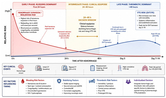
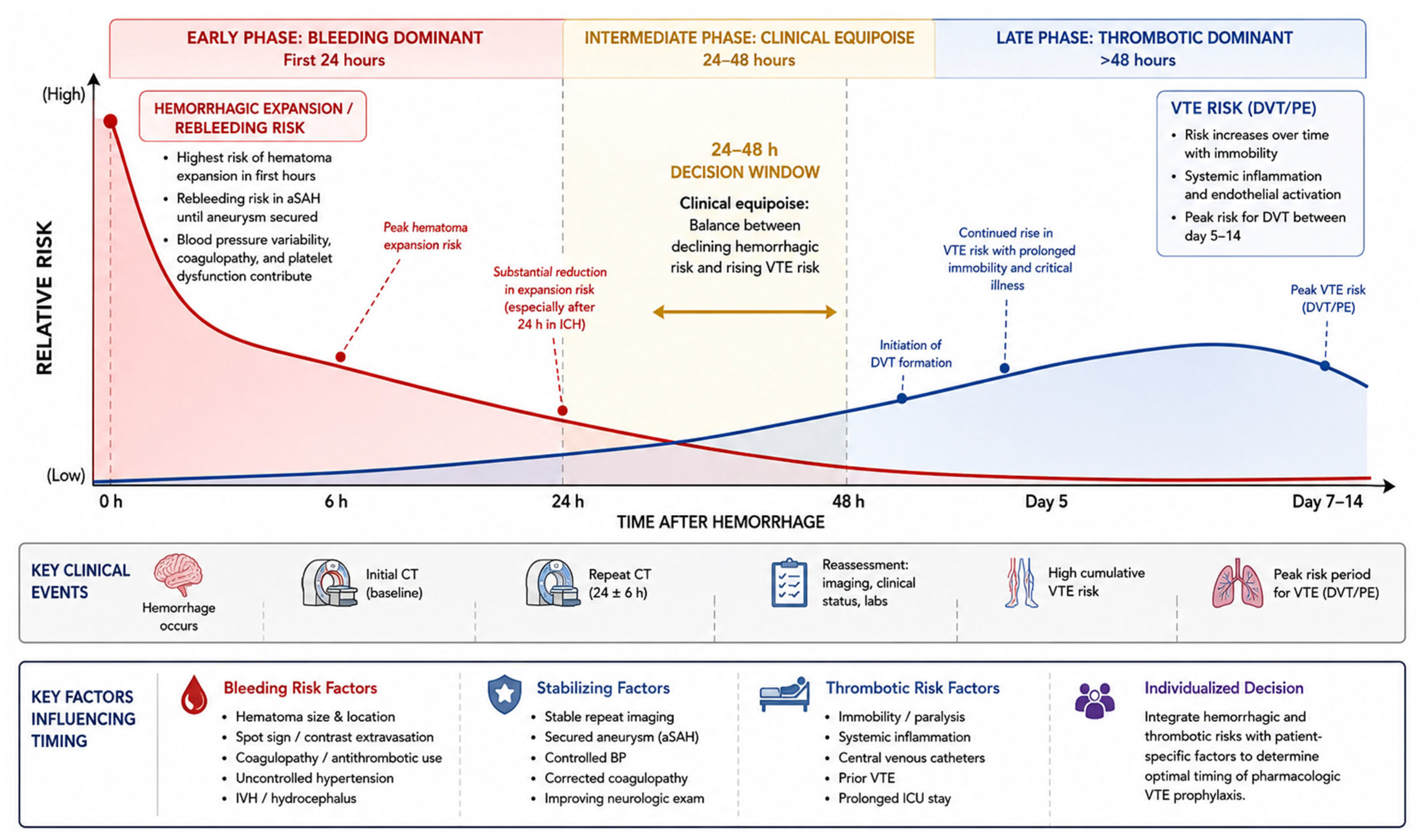
The timing mismatch between bleeding and thrombosis is what creates the clinically relevant 24–48 h decision window. Hemorrhagic instability is most front-loaded, especially in ICH, whereas thrombosis risk accumulates over the following days [1,13,14]. This produces a period in which the net benefit may shift in favor of pharmacologic prophylaxis, particularly in patients whose hemorrhage appears radiographically and clinically stable. However, because the transition is gradual rather than binary, a single repeat scan may not fully capture the point at which bleeding risk has become acceptably low (Figure 1). Imaging can therefore support the timing decision, but it should not be mistaken for a direct measure of the evolving balance between hemorrhagic and thrombotic danger [2,3,4,12].
Figure 1.
Conceptual model illustrating the competing temporal dynamics of hemorrhagic and thrombotic risk following ICH and aSAH. Hemorrhagic expansion and rebleeding risk are greatest during the hyperacute period, particularly within the first 6–24 h after hemorrhage, and gradually decline over time. In contrast, VTE risk progressively increases over subsequent days because of immobility, systemic inflammation, endothelial dysfunction, critical illness, and prolonged intensive care unit stay. The shaded 24–48 h interval represents the clinical equipoise window in which clinicians must balance declining hemorrhagic risk against rising thrombotic risk when considering initiation of pharmacologic prophylaxis.
3. Current Guideline Landscape
3.1. Intracerebral Hemorrhage Guidelines
The 2022 AHA/ASA guideline for spontaneous ICH provides the clearest modern framing of this issue. It recommends intermittent pneumatic compression beginning on the day of diagnosis for VTE prophylaxis and states that low-dose unfractionated heparin or low-molecular-weight heparin can be useful to reduce PE risk; starting pharmacologic prophylaxis at 24 to 48 h from ICH onset may be reasonable to optimize the benefit of thrombosis prevention relative to the risk of hematoma expansion [2]. Notably, this language is cautious and conditional. It does not say that a stable repeat scan is sufficient in all patients, nor does it define exact radiographic criteria for stability. The recommendation is therefore permissive rather than prescriptive.
The Neurocritical Care Society guideline similarly recognizes the importance of prophylaxis while emphasizing the limited evidentiary base and the need for individualized risk assessment in neurocritical care populations [1]. This reflects a broader reality in the field: despite the widespread use of repeated imaging in practice, there is still no universally accepted imaging-based rule that has been prospectively validated. In effect, the guidelines endorse the principle of cautious early prophylaxis in selected patients, without endorsing a strict algorithm for documenting stability. That lack of operational precision is one reason institutional practice remains heterogeneous [1,17].
3.2. Subarachnoid Hemorrhage Guidelines
The 2023 AHA/ASA aSAH guideline also supports VTE prevention, but the framework is more explicitly shaped by aneurysm status and procedural timing [4]. This is appropriate because the bleeding source in aSAH is not considered controlled until aneurysm securement is achieved. Thus, even if a follow-up CT shows no new blood, the patient may still carry a high risk of catastrophic rebleeding if the aneurysm remains untreated. The guideline, therefore, situates chemoprophylaxis within the broader treatment pathway of aneurysm repair, vasospasm surveillance, hydrocephalus management, and device-related bleeding considerations [4].
This condition-specific emphasis highlights an important conceptual gap in broad statements that recommend prophylaxis once stable. Stability means different things in different hemorrhagic stroke syndromes. In ICH, it often refers to cessation of hematoma growth; in aSAH, it is often inseparable from aneurysm occlusion status and procedural context. Treating these as equivalent can oversimplify the decision and risk importing false reassurance from one disease model into another [1,4].
3.3. Comparison and Gaps
Taken together, current guidelines support cautious early chemoprophylaxis in selected patients but do not validate repeat imaging as a stand-alone green light. They endorse a principle of integrated decision-making rather than a rigid, stable scan equals safe anticoagulation rule [1,2,4]. This is an important distinction because the absence of a formal imaging-based threshold is not an accident; it reflects the existing literature’s lack of sufficient prospective evidence to justify a universal imaging trigger.
Beyond restating these recommendations, it is important to appraise the strength of the evidence that underpins them. The 24–48 h initiation window, and the use of repeat neuroimaging as a proxy for hemorrhagic stability, rest largely on observational data and expert consensus rather than on high-quality randomized evidence; in the current guidelines the corresponding statements carry low-to-moderate levels of evidence and are framed with conditional, permissive language such as “may be reasonable” [1,2,4]. No randomized trial has demonstrated that a stable repeat scan, as distinct from elapsed time or favorable clinical selection, is what confers safety. The 24–48 h threshold is therefore best understood as a pragmatic convention derived from the temporal pattern of hematoma expansion rather than as an evidentiary-validated cut-point.
Survey data also suggest that the lack of consensus remains a real-world problem. In a recent survey of clinicians’ opinions on VTE prevention after spontaneous ICH, substantial practice variation persisted in timing and anticoagulant use, and most respondents supported the need for a randomized trial [17]. That finding is telling: even among specialists, the field does not believe the question has been definitively settled. Repeat neuroimaging is clearly useful, but whether it is useful enough on its own remains uncertain.
4. What Does Radiographic Stability Actually Mean?
4.1. Definitions Used in the Literature
One major reason this topic remains unsettled is that radiographic stability lacks a standardized definition. Studies have variably defined stability as no increase in hematoma volume, no clinically meaningful increase according to absolute or relative thresholds, absence of new hemorrhage, or lack of perihematomal extension [11,12]. Dowlatshahi and colleagues demonstrated that different definitions of hematoma expansion vary in their ability to predict poor outcome, emphasizing that the threshold used to define “stability” has important prognostic implications [11]. As a result, a hematoma considered stable under one classification system may be categorized as enlarged under another, complicating comparisons across studies and limiting the development of a universal imaging-based rule.
There is also an important distinction between research definitions and bedside interpretation. Published studies often use volumetric techniques or predefined expansion thresholds, whereas routine clinical practice frequently relies on qualitative radiology terminology such as stable or no significant interval change. Although these descriptions are clinically practical, they are inherently less precise. Consequently, the absence of a standardized operational definition of stability remains a clinically relevant limitation when repeat imaging is used to guide chemoprophylaxis decisions [2,11].
4.2. Timing of Repeat Imaging
The timing of repeat imaging further complicates interpretation. Studies have evaluated follow-up imaging at 6, 12, 24, and 48 h after hemorrhage onset, with different implications at each time point [12,13,14]. Stability demonstrated at 6 h provides different reassurance than stability maintained at 24 or 48 h. Early imaging may fail to capture delayed hematoma enlargement, particularly in higher-risk hemorrhage phenotypes, whereas delayed imaging offers greater reassurance at the expense of prolonged exposure to thrombotic risk if chemoprophylaxis is deferred [2].
Importantly, current evidence does not establish whether a single stable scan at 24 h is universally sufficient or whether additional serial imaging meaningfully improves safety in lower-risk patients. The clinical significance of stability likely depends on hemorrhage phenotype and patient-specific risk factors. For example, small deep hypertensive ICH with controlled blood pressure and stable neurologic examination differs substantially from anticoagulant-associated lobar hemorrhage with intraventricular extension or active contrast extravasation. These distinctions suggest that the meaning of stable imaging is context dependent rather than universally applicable.
4.3. Practical Limitations
Radiographic stability also has practical limitations unrelated to hemorrhage biology itself. Inter-reader variability, differences in slice thickness, qualitative versus volumetric assessment methods, and variability in imaging timing can all influence whether a hemorrhage is classified as unchanged [11]. In some centers, repeat CT imaging is protocolized and rapidly available, whereas in others, logistical constraints may delay imaging and inadvertently postpone prophylaxis decisions. Cost and cumulative radiation exposure are additional considerations, particularly when serial imaging is obtained primarily for reassurance rather than because of clinical deterioration.
Coagulopathic conditions should also be considered independent modifiers of chemoprophylaxis timing rather than being grouped under radiographic stability alone. Patients with anticoagulant-associated ICH, thrombocytopenia, elevated INR, liver dysfunction, disseminated intravascular coagulation, renal dysfunction affecting anticoagulant clearance, or recent antiplatelet exposure may remain biologically unstable despite unchanged repeat imaging. Current ICH guidance emphasizes rapid reversal of anticoagulation-associated hemorrhage and correction of modifiable bleeding risks before antithrombotic decisions are made [2]. Therefore, a stable repeat CT should not be interpreted as sufficient reassurance when systemic hemostatic abnormalities remain uncorrected.
4.4. Does Repeat Imaging Add Independent Predictive Value?
These considerations bear directly on how radiographic stability should influence practice. The pivotal question is whether a stable repeat scan contributes predictive information beyond what is already available from clinical stability and established hemorrhagic risk factors—hemorrhage subtype and location, baseline hematoma volume, blood-pressure control, the CT-angiography spot sign, and coagulation status. Present evidence does not establish such independent, incremental value: imaging stability co-varies with these favorable features and has never been tested prospectively as a stand-alone predictor after adjustment for them. In practice, therefore, repeat imaging is most useful as one confirmatory input within a multivariable risk assessment—capable of down-weighting the estimated bleeding risk in an already-reassuring patient, but insufficient to override adverse clinical or phenotypic signals when they are present. Treating a stable scan as a semi-quantitative “green light,” independent of these modifiers, over-interprets what the current data can support.
5. Evidence Linking Repeat Imaging to Safe Chemoprophylaxis
Repeat neuroimaging is used as the primary trigger for initiating DVT chemoprophylaxis and centers on one key principle: if the hematoma has not expanded on follow-up imaging, the period of highest hemorrhagic risk has likely passed, and the benefit of anticoagulation may outweigh the residual bleeding risk. Significant gaps remain in the use of imaging-guided approaches to assess hematoma stability.
5.1. Observational Studies Supporting Early Prophylaxis
The safety of chemoprophylaxis administered within 24–48 h of ICH onset has been examined in numerous observational studies, most of which show no significant increase in hematoma expansion. A propensity-matched analysis of 235 ICH patients comparing early chemoprophylaxis (initiated on admission) versus conventional timing (>24 h) showed no difference in hematoma expansion (19% vs. 23%, p = 0.5) or VTE incidence (7.5% vs. 8%, p = 0.9) [5]. The study shows that earlier initiation did not confer additional hemorrhagic risk. Similarly, a multicenter analysis from the Consortium of Leaders in Traumatic Thromboembolism [6] evaluated 881 patients with traumatic brain injury and associated ICH, finding that early VTE prophylaxis (≤48 h) was associated with decreased VTE and DVT without increased progression of intracranial hemorrhage or bleeding events.
A meta-analysis by Abdel-Aziz et al. [7] collected data from studies that initiated chemoprophylaxis at or after 24 h and within the first week post-ICH. It was concluded that pharmacologic prophylaxis did not significantly increase the risk of hematoma expansion when initiated after the hyperacute period. Additional single-center retrospective studies have reported that approximately 28% of patients receiving anticoagulants within 48 h showed no association with hematoma growth [13,18]. These findings align with the known temporal pattern of ICH expansion, which is most pronounced during the first 24 h and declines thereafter [11,19]. Collectively, the observational literature suggests that early chemoprophylaxis may be safe in selected patients following stable repeat imaging.
However, several methodological limitations temper these conclusions. Most studies were retrospective and therefore subject to selection bias. For instance, clinicians may have withheld anticoagulation from patients they judged to be at higher bleeding risk based on clinical judgment, imaging findings, or both. Surveillance bias could also complicate interpretation: patients with neurological deterioration were more likely to undergo repeat imaging, creating an asymmetry in hematoma progression detection between early and delayed prophylaxis groups. Additionally, imaging stability was variably defined across studies. Some require no change in volume, and others permit expansion below predetermined thresholds (commonly 6 mL or a 33% relative increase).
5.2. Studies Showing Persistent Risk Despite Apparent Stability
Not all patients with stable early imaging carry an equal risk for subsequent hemorrhagic complications. Emerging evidence has identified high-risk subgroups where radiographic stability may provide false reassurance. Lobar ICH associated with cerebral amyloid angiopathy (CAA) represents the most concerning phenotype. Patients with lobar hemorrhage demonstrate a 2–4 times higher rebleeding risk compared to deep/hypertensive; CAA-related hemorrhages carry recurrence rates of approximately 7.4% per year compared to 1.1% per year for hypertensive arteriopathy [20,21]. This elevated risk continues beyond the acute phase and is not mitigated by early imaging stability.
Real-world evidence can be found in the ENRICH-AF trial, which randomized patients with atrial fibrillation and prior ICH to edoxaban versus antiplatelet therapy. The Data Safety Monitoring Board terminated the lobar ICH and convexity SAH cohorts early due to unacceptably high rates of recurrent hemorrhagic stroke in the anticoagulation arm [15]. Although ENRICH-AF evaluated therapeutic anticoagulation rather than prophylactic, its findings show that, even with an initial imaging trajectory, the location of lobar hemorrhage identifies a population with persistently elevated bleeding susceptibility.
Additional risk factors for delayed hematoma expansion despite early stability include anticoagulant-associated ICH, large baseline hematoma volume (>30 mL), presence of the CT angiography spot sign indicating active extravasation, and intraventricular hemorrhage extension [14,22]. In moderate-to-high-risk ICH phenotypes, studies have documented expansion rates of approximately 18% even after day 3. This suggests that the 24–48 h imaging window may not adequately capture ongoing hemorrhagic evolution in selected patients [16].
In aneurysmal SAH, which originates from a discrete vascular lesion, imaging stability has different implications than in ICH. Rebleeding risk remains approximately 5% in the first 24 h (concentrated in the initial 6 h) and approximately 1% per day for the subsequent month, but this risk persists until the aneurysm is definitively secured by surgical clipping or endovascular coiling [1,23]. A stable early CT that demonstrates no new hemorrhage does not address the fundamental question of aneurysm status. Furthermore, patients who require EVDs are met with catheter-related hemorrhagic complications that interact with anticoagulation timing. Dual antiplatelet therapy for stent-assisted coiling increases EVD-related hematoma rates from 7.7% to 32.3% [9].
5.3. What the Evidence Does Not Show
Despite the accumulated observational data, critical gaps persist in the field’s understanding of hematoma stability and chemoprophylaxis. Most fundamentally, no randomized controlled trial has prospectively validated imaging stability as a trigger for initiating chemoprophylaxis. Even the sole double-blind RCT comparing early versus late prophylaxis timing in ICH [10], in which 139 patients were randomized to receive enoxaparin at 24 h versus 72 h, found no difference in hematoma enlargement (p = 0.927) or clinical outcomes (p = 0.904). It must be noted, though, that this trial was substantially underpowered; only three VTE events occurred across both arms (two early, one late), reflecting stringent adherence to IPC prophylaxis in all patients. The trial cannot isolate whether early prophylaxis is safe because of imaging-confirmed stability or simply because the baseline VTE rate was too low to demonstrate benefit.
The PREVENTIHS trial [10], designed to evaluate enoxaparin initiated beyond 72 h, was terminated early (n = 73) due to recruitment difficulties, which left the question of optimal timing unresolved. A network meta-analysis [8] comparing prophylaxis strategies found IPC superior to no prophylaxis but, due to imprecision, could not establish superiority of pharmacologic over mechanical prophylaxis. This shows the small sample sizes and heterogeneous protocols across existing studies.
Existing studies used variable timing (6, 12, 24, or 48 h post-ictus), different volume thresholds for stability, and inconsistent imaging modalities. Whether a single stable scan is sufficient or whether serial imaging provides incremental safety information remains an unanswered question with potentially positive clinical implications. The imaging criteria used in observational studies have typically been determined retrospectively for research purposes rather than prospectively applied as clinical decision rules.
Finally, the observational literature cannot account for unmeasured confounding. Clinicians who chose to initiate early chemoprophylaxis may have selected patients with more favorable clinical profiles, such as smaller hemorrhages, better neurological status, or absence of coagulopathy, independent of imaging findings. Without randomization, it remains unclear whether imaging stability itself drives safety or whether it correlates with other favorable prognostic features.
6. ICH vs. aSAH: Should the Same Logic Apply?
Current guidelines for both ICH and aSAH support chemoprophylaxis following hemorrhage stabilization, typically within 24–48 h in selected patients. However, the pathophysiology, bleeding trajectory, and procedural considerations differ substantially between these conditions, raising the question of whether condition-specific approaches are more appropriate than a unified imaging-based strategy.
To consolidate these distinctions, Table 1 provides a structured visual summary of how ICH, aSAH, and traumatic intracranial hemorrhage differ in bleeding mechanism, hemorrhagic risk trajectory, and the determinants of chemoprophylaxis timing.
Table 1.
Comparative summary of spontaneous intracerebral hemorrhage (ICH), aneurysmal subarachnoid hemorrhage (aSAH), and traumatic intracranial hemorrhage, contrasting bleeding mechanism, hemorrhagic risk trajectory, the primary determinant of chemoprophylaxis timing, and the reliability of repeat neuroimaging. The three entities correspond to imaging-dependent (ICH), procedure-dependent (aSAH), and imaging-reliable (traumatic ICH) decision frameworks, underscoring that a single “stable imaging at 24–48 h” paradigm is not universally applicable. BP, blood pressure; CAA, cerebral amyloid angiopathy; EVD, external ventricular drain; IVH, intraventricular hemorrhage.
6.1. Fundamental Differences in Bleeding Mechanism
ICH and aSAH arise from distinct vascular mechanisms. Primary ICH results from rupture of small penetrating arteries damaged by chronic hypertension or cerebral amyloid angiopathy, producing parenchymal hemorrhage with a predominantly early expansion phase [2,24]. Approximately 70% of hematoma expansion occurs within the first 24 h, with the highest risk concentrated in the initial 6 h [13,14]. In many lower-risk patients, hemorrhagic activity declines substantially after 24–48 h.
In contrast, aSAH results from rupture of a discrete macrovascular lesion. Rebleeding risk persists until the aneurysm is definitively secured by clipping or coiling [23,25]. Consequently, a stable CT in ICH may suggest hemorrhagic quiescence, whereas a stable CT in aSAH only indicates that rebleeding has not yet occurred. This distinction fundamentally limits the applicability of a uniform “stable imaging” paradigm across hemorrhagic stroke subtypes.
6.2. Aneurysm Securement and Procedural Considerations
In aSAH, chemoprophylaxis decisions are closely linked to aneurysm securement and procedural status. Most centers delay pharmacologic prophylaxis until after aneurysm treatment because unsecured aneurysms carry substantial rebleeding risk [26,27]. Additional complexity arises from procedure-related bleeding considerations, including EVD placement and the use of dual antiplatelet therapy (DAPT) for stent-assisted coiling [28,29].
EVD-associated hemorrhage risk increases significantly in anticoagulated patients and may be further amplified in those receiving DAPT [30,31]. These considerations have no direct analog in most spontaneous ICH cases and support the need for aSAH-specific prophylaxis strategies that account for aneurysm treatment status, device placement, and antiplatelet exposure.
6.3. Temporal Patterns and Subgroup Heterogeneity
The temporal relationship between hemorrhagic and thrombotic risk also differs between conditions. In ICH, hemorrhagic risk is largely front-loaded, creating a potential window for earlier chemoprophylaxis in selected low-risk patients after stability has been demonstrated [13,14]. In aSAH, DVT formation more commonly occurs between days 5–14 and overlaps with the vasospasm window, during which patients are often immobilized and critically ill [30,31].
Subgroup heterogeneity further complicates decision-making. Deep hypertensive ICH generally carries lower long-term recurrence risk and may be more amenable to early imaging-guided prophylaxis [32]. In contrast, lobar ICH associated with cerebral amyloid angiopathy carries substantially greater hemorrhagic vulnerability, and imaging stability may provide false reassurance in this population [32,33]. Similarly, in aSAH, aneurysm securement status remains the dominant determinant of ongoing hemorrhagic risk.
6.4. SAH-Specific Considerations and Potential Therapeutic Implications
Beyond hemorrhagic risk alone, aSAH introduces additional considerations related to delayed cerebral ischemia (DCI), which occurs in approximately 20–30% of patients between days 4–14 [3]. Because vasospasm, inflammation, and microthrombosis contribute to DCI pathophysiology, some investigators have hypothesized that anticoagulation may provide secondary neuroprotective benefit beyond standard VTE prevention.
A systematic review found that therapeutic-dose intravenous heparin administered after aneurysm securement was associated with lower rates of cerebral infarction compared with prophylactic dosing alone [19]. Although these findings require cautious interpretation, they suggest that prophylaxis decisions in aSAH may involve considerations distinct from those in spontaneous ICH.
6.5. Traumatic Intracranial Hemorrhage Anticoagulation Protocols
Traumatic intracranial hemorrhage represents a distinct clinical entity with different hemorrhage expansion dynamics and VTE risk profiles than spontaneous ICH and aSAH. Existing trauma literature generally supports early chemoprophylaxis following stable repeat imaging in selected patients, although injury severity, operative intervention, and invasive monitoring devices remain important modifiers of risk [18,19,21,34,35]. Because traumatic hemorrhage reflects mechanical vascular injury rather than spontaneous small-vessel disease or aneurysmal rupture, extrapolation from traumatic brain injury literature to spontaneous hemorrhagic stroke should be performed cautiously [36,37,38,39].
6.6. Implications for Condition-Specific Protocols
Collectively, the available evidence supports condition-specific rather than universal imaging-based algorithms. In spontaneous ICH, early chemoprophylaxis may be reasonable in selected lower-risk patients with stable repeat imaging and favorable clinical characteristics. In aSAH, aneurysm securement and procedural context remain central determinants of safety, often superseding imaging appearance alone.
Accordingly, a one-size-fits-all “stable imaging at 24–48 h” framework does not adequately capture the biologic and procedural differences across hemorrhagic stroke syndromes. Future protocols should incorporate hemorrhage subtype, aneurysm status, coagulopathy, procedural factors, and individualized bleeding risk rather than relying solely on repeat neuroimaging as an isolated decision trigger.
7. Clinical Decision-Making Framework
Radiographic stability should be treated as one component of the decision, not the entire decision. The most defensible interpretation of the literature is that repeated neuroimaging is useful because it reduces uncertainty, but it does not eliminate it. A stable follow-up scan is more reassuring in a small deep ICH with controlled blood pressure, no spot sign, no anticoagulant exposure, and stable neurologic examination than it is in a large lobar hemorrhage with intraventricular extension or suspected cerebral amyloid angiopathy [2,15,19]. Likewise, in aSAH, a stable scan is helpful only after accounting for aneurysm securement, EVD status, and antiplatelet requirements [4].
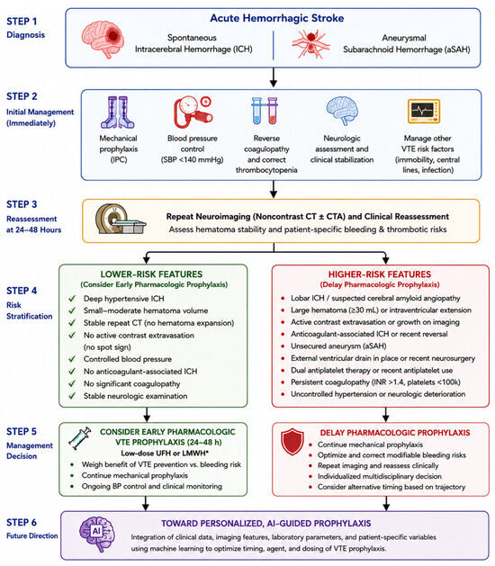
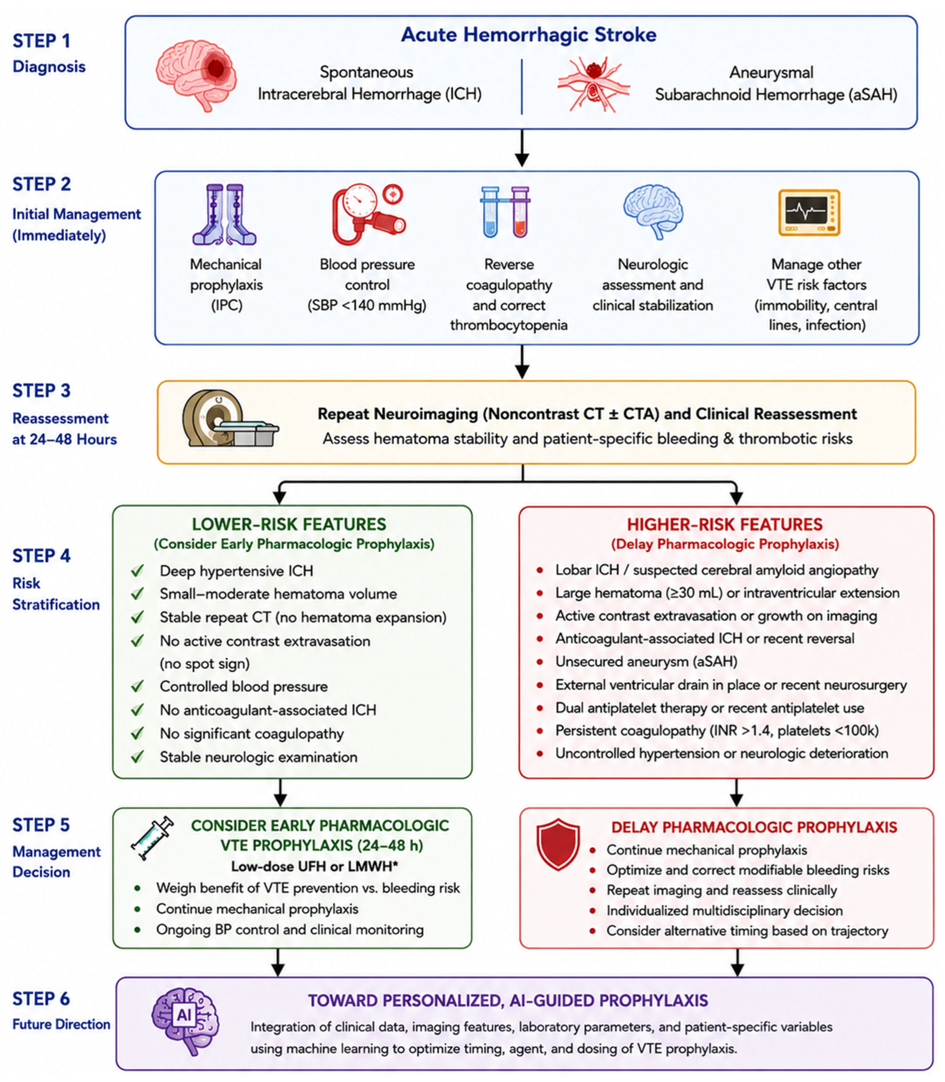
A practical framework, therefore, has to be risk-stratified (Figure 2). In lower-risk spontaneous ICH, especially deep hypertensive hemorrhage with reassuring repeat imaging and no adverse clinical signals, initiating low-dose chemoprophylaxis at 24–48 h is supported by existing guideline language and observational evidence [2,5,6]. In higher-risk phenotypes, repeat imaging still matters, but it should probably not be considered sufficient on its own. These patients may require longer reliance on mechanical prophylaxis, more cautious delay, or repeated reassessment. The literature supports nuance much more strongly than it supports a one-size-fits-all timing rule. This proposed framework is intended to operationalize existing guideline principles into a more individualized and phenotype-specific approach to chemoprophylaxis timing.
Figure 2.
Proposed clinical decision framework for initiation of pharmacologic DVT prophylaxis following spontaneous ICH and aSAH. Initial management includes mechanical prophylaxis, blood pressure control, reversal of coagulopathy, and neurologic stabilization, followed by repeat neuroimaging and clinical reassessment at 24–48 h. Patients with lower-risk ICH features, including deep hypertensive hemorrhage, stable repeat imaging, controlled blood pressure, and absence of coagulopathy or active contrast extravasation, may be considered for early low-dose chemoprophylaxis. In contrast, patients with higher-risk hemorrhagic features, including lobar ICH, cerebral amyloid angiopathy-associated hemorrhage, large hematoma burden, intraventricular extension, unsecured aneurysm, external ventricular drainage, antiplatelet exposure, or persistent coagulopathy, may require delayed pharmacologic prophylaxis and continued reassessment. The figure also highlights future directions involving personalized and artificial intelligence-guided prophylaxis strategies integrating imaging, laboratory, and patient-specific clinical variables.
We emphasize that these hemorrhage subtypes are not interchangeable and are not aggregated here into a single decision rule. The pathways below are applied separately to each entity, and the framework in Figure 2 should be read as three parallel pathways rather than one uniform algorithm.
7.1. Spontaneous Intracerebral Hemorrhage
In spontaneous ICH, the decision is genuinely imaging informed. In lower-risk hemorrhage—small, deep, hypertensive bleeds with controlled blood pressure, no spot sign, no anticoagulant exposure, and a stable neurologic examination—a stable repeat scan at 24–48 h supports initiation of low-dose pharmacologic prophylaxis, consistent with guideline language and observational data [2,5,6]. In higher-risk phenotypes (lobar or CAA-related hemorrhage, large baseline volume, intraventricular extension, active extravasation, or uncorrected coagulopathy), a stable scan is insufficient on its own; these patients warrant continued mechanical prophylaxis, correction of hemostatic abnormalities, and either delayed initiation or repeated reassessment [15,16].
7.2. Aneurysmal Subarachnoid Hemorrhage
In aSAH, the pathway is procedure-dependent rather than imaging-dependent. Pharmacologic prophylaxis is generally deferred until the aneurysm is definitively secured because a stable CT in a patient with an unsecured aneurysm indicates only that rebleeding has not yet occurred [4,23]. Once the aneurysm is secured, timing must additionally account for EVD placement and for antiplatelet exposure related to stent-assisted coiling, both of which raise catheter-associated hemorrhage risk [9,30,31]. Here, repeat imaging is subordinate to aneurysm and device status.
7.3. Traumatic Intracranial Hemorrhage
In traumatic intracranial hemorrhage, the pathway is the most imaging-reliable of the three: stable repeat imaging in selected patients generally supports early prophylaxis, although injury severity, operative intervention, and invasive monitoring devices remain important modifiers, and extrapolation to spontaneous hemorrhagic stroke should be undertaken cautiously [18,36,37,38,39]. Presenting these three pathways separately avoids conflating pathologies that have distinct bleeding sources and risk trajectories.
7.4. Limitations of the Proposed Framework
We acknowledge an inherent limitation of this framework: it is derived from the same observational and consensus-based evidence whose insufficiency this review critiques, and it has not itself been prospectively validated. It is therefore offered as a pragmatic, provisional decision aid that operationalizes existing guideline principles and makes their assumptions explicit, not as a validated algorithm and not as a substitute for the randomized, imaging-triggered trials called for above. Its components should be applied with clinical judgment and revised as higher-quality evidence emerges.
8. Future Directions and Research Priorities
The strongest conclusion from the current literature is not that repeated neuroimaging is unhelpful, but that it has not yet been validated as a sufficient independent trigger. The field needs prospective trials that explicitly test imaging-based initiation strategies rather than merely reporting the safety of clinician-selected early prophylaxis [9,10]. Those studies should standardize how stability is defined, when repeat imaging is obtained, and which clinical features modify the meaning of a stable scan. Without that standardization, stable at 24 h will continue to mean different things across centers and studies.
There is also a growing role for precision medicine and advanced phenotyping in hemorrhagic stroke decision-making. Modern ICH research increasingly recognizes that hemorrhage expansion risk can be stratified using clinical variables, CT features, CTA markers, hemorrhage location, anticoagulant exposure, laboratory coagulation parameters, and disease subtype [12]. Future artificial intelligence and machine-learning models may help integrate these variables into individualized estimates of hemorrhagic and thrombotic risk, allowing clinicians to move beyond binary timing decisions toward personalized prophylaxis strategies. In the future, AI-guided frameworks may assist not only with determining when to initiate prophylaxis, but also which agent, dose, and monitoring strategy are most appropriate for individual patients. Such models remain investigational and will require prospective validation, but they represent a promising step toward precision medicine in hemorrhagic stroke management.
To make these priorities concrete, we propose the following ranked research agenda. (1) Most important, a randomized trial should test an imaging-triggered strategy directly, randomizing radiographically and clinically stable ICH patients at 24 h to immediate versus delayed pharmacologic prophylaxis, rather than reporting the safety of clinician-selected early treatment. (2) Trials should prospectively compare a single stable 24 h scan against serial imaging to determine whether repeated scanning adds incremental safety in lower-risk phenotypes. (3) Studies should stratify by hemorrhage subtype—deep hypertensive versus lobar/CAA-related ICH, secured versus unsecured aSAH, and traumatic ICH—so that subtype-specific thresholds can be derived. (4) Work is needed to define which clinical and coagulation modifiers should override an otherwise stable scan. Underpinning all of these, “stability” should be operationalized with a single prespecified definition—for example, absence of hematoma expansion defined as growth below both an absolute (e.g., <6 mL) and a relative (e.g., <33%) threshold, no new or extending hemorrhage, and no active extravasation (spot sign) on a scan obtained at a fixed interval (e.g., 24 h)—so that “stable at 24 h” carries the same meaning across centers and trials.
9. Conclusions
Demonstration of hemorrhage stability on repeat neuroimaging is clearly useful, but current evidence does not support treating it as a universally sufficient stand-alone justification for starting DVT chemoprophylaxis at 24–48 h. In spontaneous ICH, a stable repeat scan can provide meaningful reassurance, particularly in lower-risk phenotypes, and guideline-supported early prophylaxis appears reasonable in selected patients [2,5,9]. In aSAH, however, the meaning of imaging stability is much more limited unless aneurysm securement and procedural context are also considered [4,28]. Across both diseases, the best reading of the literature is that radiographic stability should inform timing decisions but should not dictate them in isolation.
The lack of consensus should not be interpreted as a failure of the field so much as a sign that clinicians are working at the edge of incomplete evidence. The current literature supports cautious early chemoprophylaxis in selected patients, especially when repeat imaging, neurologic stability, hemorrhage phenotype, and procedural status all align. What it does not yet support is a simplistic rule that one stable scan is enough for everyone. Until higher-quality prospective evidence becomes available, repeat neuroimaging is best viewed as necessary in many cases, helpful in most, and sufficient only when interpreted within a broader clinical context.
Author Contributions
C.R.: Writing—review and editing, Writing—original draft, Software, Resources, Investigation, Formal analysis, Conceptualization, and Data curation. Z.K.: Writing—original draft, Resources, Investigation, and Data curation. B.L.-W.: Writing—review and editing, Visualization, Validation, Supervision, Project administration, Conceptualization, and Methodology. M.K.: Writing—review and editing, Visualization, Validation, Supervision, Project administration, Conceptualization, and Methodology. M.P.: Writing—review and editing, Visualization, Validation, Supervision, Project administration, Methodology, Investigation, Data curation, and Conceptualization. All authors have read and agreed to the published version of the manuscript.
Funding
This study was not supported by any sponsor or funder.
Data Availability Statement
No new data were created or analyzed in this study. Data sharing is not applicable to this article.
Conflicts of Interest
The authors have no conflicts of interest to declare.
References
- Nyquist, P.; Bautista, C.; Jichici, D.; Burns, J.; Chhangani, S.; DeFilippis, M.; Goldenberg, F.D.; Kim, K.; Liu-DeRyke, X.; Mack, W.; et al. Prophylaxis of Venous Thrombosis in Neurocritical Care Patients: An Evidence-Based Guideline: A Statement for Healthcare Professionals from the Neurocritical Care Society. Neurocrit Care 2016, 24, 47–60. [Google Scholar] [CrossRef] [Scilit] [PubMed]
- Greenberg, S.M.; Ziai, W.C.; Cordonnier, C.; Dowlatshahi, D.; Francis, B.; Goldstein, J.N.; Hemphill, J.C., III; Johnson, R.; Keigher, K.M.; Mack, W.J.; et al. 2022 Guideline for the Management of Patients With Spontaneous Intracerebral Hemorrhage. Stroke 2022, 53, e282–e361. [Google Scholar] [CrossRef] [Scilit] [PubMed]
- Kilgore, C.B.; Nair, S.K.; Ran, K.R.; Caplan, J.M.; Jackson, C.M.; Gonzalez, L.F.; Huang, J.; Tamargo, R.J.; Xu, R. Venous thromboembolism in aneurysmal subarachnoid hemorrhage: Risk factors and timing of chemoprophylaxis. Clin. Neurol. Neurosurg. 2023, 231, 107822. [Google Scholar] [CrossRef] [Scilit] [PubMed]
- Hoh, B.L.; Ko, N.U.; Amin-Hanjani, S.; Chou, S.H.-Y.; Cruz-Flores, S.; Dangayach, N.S.; Derdeyn, C.P.; Du, R.; Hänggi, D.; Hetts, S.W.; et al. 2023 Guideline for the Management of Patients With Aneurysmal Subarachnoid Hemorrhage: A Guideline From the American Heart Association/American Stroke Association. Stroke 2023, 54, E314–E370. [Google Scholar] [CrossRef] [Scilit] [PubMed]
- Laurent, D.; Bardhi, O.; Kubilis, P.; Corliss, B.; Adamczak, S.; Geh, N.; Dodd, W.; Vaziri, S.; Busl, K.; Fox, W.C. Early chemoprophylaxis for deep venous thrombosis does not increase the risk of hematoma expansion in patients presenting with spontaneous intracerebral hemorrhage. Surg. Neurol. Int. 2021, 12, 277. [Google Scholar] [CrossRef] [Scilit] [PubMed]
- Kananeh, M.F.; Fonseca-Paricio, M.J.; Liang, J.W.; Sullivan, L.T.; Sharma, K.; Shah, S.O.; Vibbert, M.D. Ultra-early venous thromboembolism (VTE) prophylaxis in spontaneous intracerebral hemorrhage (sICH). J. Stroke Cerebrovasc. Dis. 2021, 30, 105476. [Google Scholar] [CrossRef] [Scilit] [PubMed]
- Cai, Q.; Zhang, X.; Chen, H. Patients with venous thromboembolism after spontaneous intracerebral hemorrhage: A review. Thromb. J. 2021, 19, 93. [Google Scholar] [CrossRef] [Scilit] [PubMed]
- Yogendrakumar, V.; Lun, R.; Khan, F.; Salottolo, K.; Lacut, K.; Graham, C.; Dennis, M.; Hutton, B.; Wells, P.S.; Fergusson, D.; et al. Venous thromboembolism prevention in intracerebral hemorrhage: A systematic review and network meta-analysis. PLoS ONE 2020, 15, e0234957. [Google Scholar] [CrossRef] [Scilit] [PubMed]
- Qian, C.; Huhtakangas, J.; Juvela, S.; Bode, M.K.; Tatlisumak, T.; Savolainen, M.; Numminen, H.; Ollikainen, J.; Luostarinen, L.; Kupila, L.; et al. Early vs. late enoxaparin for the prevention of venous thromboembolism in patients with ICH: A double blind placebo controlled multicenter study. Clin. Neurol. Neurosurg. 2021, 202, 106534. [Google Scholar] [CrossRef] [Scilit] [PubMed]
- Paciaroni, M.; Agnelli, G.; Alberti, A.; Becattini, C.; Guercini, F.; Martini, G.; Tassi, R.; Marotta, G.; Venti, M.; Acciarresi, M.; et al. PREvention of VENous Thromboembolism in Hemorrhagic Stroke Patients—PREVENTIHS Study: A Randomized Controlled Trial and a Systematic Review and Meta-Analysis. Eur. Neurol. 2020, 83, 566–575. [Google Scholar] [CrossRef] [Scilit] [PubMed]
- Dowlatshahi, D.; Demchuk, A.; Flaherty, M.; Ali, M.; Lyden, P.; Smith, E. Defining hematoma expansion in intracerebral hemorrhage. Neurology 2011, 76, 1238–1244. [Google Scholar] [CrossRef] [Scilit] [PubMed]
- Morotti, A.; Boulouis, G.; Dowlatshahi, D.; Li, Q.; Shamy, M.; Salman, R.A.-S.; Rosand, J.; Cordonnier, C.; Goldstein, J.N.; Charidimou, A. Intracerebral haemorrhage expansion: Definitions, predictors, and prevention. Lancet Neurol. 2022, 22, 159–171. [Google Scholar] [CrossRef] [Scilit] [PubMed]
- Brott, T.; Broderick, J.; Kothari, R.; Barsan, W.; Tomsick, T.; Sauerbeck, L.; Spilker, J.; Duldner, J.; Khoury, J. Early hemorrhage growth in patients with intracerebral hemorrhage. Stroke. 1997, 28, 1–5. [Google Scholar] [CrossRef] [Scilit] [PubMed]
- Kazui, S.; Naritomi, H.; Yamamoto, H.; Sawada, T.; Yamaguchi, T. Enlargement of spontaneous intracerebral hemorrhage. Incidence and time course. Stroke 1996, 27, 1783–1787. [Google Scholar] [CrossRef] [Scilit] [PubMed]
- Demchuk, A.M.; Dowlatshahi, D.; Rodriguez-Luna, D.; Molina, C.A.; Blas, Y.S.; Dzialowski, I.; Kobayashi, A.; Boulanger, J.-M.; Lum, C.; Gubitz, G.; et al. Prediction of haematoma growth and outcome in patients with intracerebral haemorrhage using the CT-angiography spot sign (PREDICT): A prospective observational study. Lancet Neurol. 2012, 11, 307–314. [Google Scholar] [CrossRef] [Scilit] [PubMed]
- Hillman, J.; Fridriksson, S.; Nilsson, O.; Yu, Z.; Saveland, H.; Jakobsson, K.E. Immediate administration of tranexamic acid and reduced incidence of early rebleeding after aneurysmal subarachnoid hemorrhage: A prospective randomized study. J. Neurosurg. 2002, 97, 771–778. [Google Scholar] [CrossRef] [Scilit] [PubMed]
- Mendel, R.; Abdelhameed, N.; Salman, R.A.-S.; Cohen, H.; Dowlatshahi, D.; Freemantle, N.; Paciaroni, M.; Parry-Jones, A.; Price, C.; Sprigg, N.; et al. Prevention of venous thromboembolism in acute spontaneous intracerebral haemorrhage: A survey of opinion. J. Neurol. Sci. 2023, 454, 120855. [Google Scholar] [CrossRef] [Scilit] [PubMed]
- Phelan, H.A. Pharmacologic venous thromboembolism prophylaxis after traumatic brain injury: A critical literature review. J. Neurotrauma 2012, 29, 1821–1828. [Google Scholar] [CrossRef] [Scilit] [PubMed]
- Charidimou, A.; Imaizumi, T.; Moulin, S.; Biffi, A.; Samarasekera, N.; Yakushiji, Y.; Peeters, A.; Vandermeeren, Y.; Laloux, P.; Baron, J.-C.; et al. Brain hemorrhage recurrence, small vessel disease type, and cerebral microbleeds. Neurology 2017, 89, 820–829. [Google Scholar] [CrossRef] [Scilit] [PubMed]
- Li, L.; Poon, M.T.C.; Samarasekera, N.E.; Perry, L.A.; Moullaali, T.J.; Rodrigues, M.A.; Loan, J.J.M.; Stephen, J.; Lerpiniere, C.; Tuna, M.A.; et al. Risks of recurrent stroke and all serious vascular events after spontaneous intracerebral haemorrhage: Pooled analyses of two population-based studies. Lancet Neurol. 2021, 20, 437–447. [Google Scholar] [CrossRef] [Scilit] [PubMed]
- Shoamanesh, A.; ENRICH-AF Steering Committee. Anticoagulation in patients with cerebral amyloid angiopathy. Lancet 2023, 402, 1411–1412. [Google Scholar] [CrossRef] [Scilit] [PubMed]
- Davis, S.M.; Broderick, J.; Hennerici, M.; Brun, N.C.; Diringer, M.N.; Mayer, S.A.; Begtrup, K.; Steiner, T. Hematoma growth is a determinant of mortality and poor outcome after intracerebral hemorrhage. Neurology 2006, 66, 1175–1181. [Google Scholar] [CrossRef] [Scilit] [PubMed]
- Connolly, E.S.; Rabinstein, A.A.; Carhuapoma, J.R.; Derdeyn, C.P.; Dion, J.; Higashida, R.T.; Hoh, B.L.; Kirkness, C.J.; Naidech, A.M.; Ogilvy, C.S.; et al. Guidelines for the Management of Aneurysmal Subarachnoid Hemorrhage. Stroke 2012, 43, 1711–1737. [Google Scholar] [CrossRef] [Scilit] [PubMed]
- Magid-Bernstein, J.; Girard, R.; Polster, S.; Srinath, A.; Romanos, S.; Awad, I.A.; Sansing, L.H. Cerebral Hemorrhage: Pathophysiology, Treatment, and Future Directions. Circ. Res. 2022, 130, 1204–1229. [Google Scholar] [CrossRef] [Scilit] [PubMed]
- de Rooij, N.K.; Greving, J.P.; Rinkel, G.J.; Frijns, C.J. Early prediction of delayed cerebral ischemia after subarachnoid hemorrhage. Stroke 2013, 44, 1288–1294. [Google Scholar] [CrossRef] [Scilit] [PubMed]
- van Donkelaar, C.E.; Bakker, N.A.; Veeger, N.J.; Uyttenboogaart, M.; Metzemaekers, J.D.; Luijckx, G.-J.; Groen, R.J.; van Dijk, J.M.C. Predictive Factors for Rebleeding After Aneurysmal Subarachnoid Hemorrhage. Stroke 2015, 46, 2100–2106. [Google Scholar] [CrossRef] [Scilit] [PubMed]
- The Participants in the International Multi-disciplinary Consensus Conference on the Critical Care Management of Subarachnoid Hemorrhage; Vergouwen, M.D.I. Vasospasm Versus Delayed Cerebral Ischemia as an Outcome Event in Clinical Trials and Observational Studies. Neurocrit. Care 2011, 15, 308–311. [CrossRef] [Scilit] [PubMed]
- Manoel, A.L.d.O.; Turkel-Parrella, D.; Germans, M.; Kouzmina, E.; Almendra, P.d.S.; Marotta, T.; Spears, J.; Abrahamson, S. Safety of Early Pharmacological Thromboprophylaxis after Subarachnoid Hemorrhage. Can. J. Neurol. Sci. 2014, 41, 554–561. [Google Scholar] [CrossRef] [Scilit] [PubMed]
- Siironen, J.; Juvela, S.; Varis, J.; Porras, M.; Poussa, K.; Ilveskero, S.; Hernesniemi, J.; Lassila, R. No effect of enoxaparin on outcome of aneurysmal subarachnoid hemorrhage: A randomized, double-blind, placebo-controlled clinical trial. J. Neurosurg. 2003, 99, 953–959. [Google Scholar] [CrossRef] [Scilit] [PubMed]
- Amenta, P.S.; Dalyai, R.T.; Kung, D.; Toporowski, A.; Chandela, S.; Hasan, D.; Gonzalez, L.F.; Dumont, A.S.; Tjoumakaris, S.I.; Rosenwasser, R.H.; et al. Stent-Assisted Coiling of Wide-Necked Aneurysms in the Setting of Acute Subarachnoid Hemorrhage. Neurosurgery 2012, 70, 1415–1429. [Google Scholar] [CrossRef] [Scilit] [PubMed]
- Bruder, M.; Schuss, P.; Konczalla, J.; El-Fiki, A.; Lescher, S.; Vatter, H.; Seifert, V.; Güresir, E. Ventriculostomy-Related Hemorrhage After Treatment of Acutely Ruptured Aneurysms: The Influence of Anticoagulation and Antiplatelet Treatment. World Neurosurg. 2015, 84, 1653–1659. [Google Scholar] [CrossRef] [Scilit] [PubMed]
- Tang, Y.; Zhong, X.; Lin, T.; Zuo, F.; Fu, M.; Wang, L.; Yu, X.; Liu, D.; Zhang, J. Bleeding complications related to external ventricular drainage placement in patients with ruptured intracranial aneurysms: A single-center study. Front. Surg. 2024, 11, 1403668. [Google Scholar] [CrossRef] [Scilit] [PubMed]
- Liang, C.W.; Su, K.; Liu, J.J.; Dogan, A.; Hinson, H.E. Timing of deep vein thrombosis formation after aneurysmal subarachnoid hemorrhage. J. Neurosurg. 2015, 123, 891–896. [Google Scholar] [CrossRef] [Scilit] [PubMed]
- Vergouwen, M.D.; Vermeulen, M.; van Gijn, J.; Rinkel, G.J.; Wijdicks, E.F.; Muizelaar, J.P.; Mendelow, A.D.; Juvela, S.; Yonas, H.; Terbrugge, K.G.; et al. Definition of Delayed Cerebral Ischemia After Aneurysmal Subarachnoid Hemorrhage as an Outcome Event in Clinical Trials and Observational Studies. Stroke 2010, 41, 2391–2395. [Google Scholar] [CrossRef] [Scilit] [PubMed]
- Lukito, P.P.; Lie, H.; Helsa, K.; July, J. Heparin in the treatment of aneurysmal subarachnoid hemorrhage: A systematic review and meta-analysis. Neurosurg. Focus 2022, 52, E9. [Google Scholar] [CrossRef] [Scilit] [PubMed]
- Lin, M.; Davis, J.V.; Wong, D.T. Safety and efficacy of early thromboembolism chemoprophylaxis after intracranial hemorrhage from traumatic brain injury. Am. Surg. 2013, 79, 1050–1053. [Google Scholar] [CrossRef] [Scilit]
- Abdel-Aziz, H.; Dunham, C.M.; Malik, R.J.; Hileman, B.M. Timing for deep vein thrombosis chemoprophylaxis in traumatic brain injury: An evidence-based review. Crit. Care 2015, 19, 96. [Google Scholar] [CrossRef] [Scilit] [PubMed]
- Dengler, B.A.; Haight, T.; Fu, A.; Hafeez, S.J.; Cirivello, M.; Bartanusz, V. Safety of early chemoprophylaxis for venous thromboembolism after traumatic brain injury: A systematic review and meta-analysis. A military traumatic brain injury initiative study. J. Neurosurg. 2024, 142, 1046–1055. [Google Scholar] [CrossRef] [Scilit] [PubMed]
- Rappold, J.F.; Sheppard, F.R.; Ii, S.P.C.; Cuschieri, J.; Ley, E.; Rangel, E.; Seshadri, A.J.; Michetti, C.P. Venous thromboembolism prophylaxis in the trauma intensive care unit: An American Association for the Surgery of Trauma Critical Care Committee Clinical Consensus Document. Trauma Surg. Acute Care Open 2021, 6, e000643. [Google Scholar] [CrossRef] [Scilit] [PubMed]

Disclaimer/Publisher’s Note: The statements, opinions and data contained in all publications are solely those of the individual author(s) and contributor(s) and not of MDPI and/or the editor(s). MDPI and/or the editor(s) disclaim responsibility for any injury to people or property resulting from any ideas, methods, instructions or products referred to in the content. |
© 2026 by the authors. Licensee MDPI, Basel, Switzerland. This article is an open access article distributed under the terms and conditions of the Creative Commons Attribution (CC BY) license.