


Abstract
Objective: Giant thrombosed intracranial aneurysms (GTIAs) are aneurysmal dilatations containing partial or complete intraluminal thrombus. Representing approximately 2–5% of intracranial aneurysms, GTIAs present diagnostic and therapeutic challenges. Optimal management remains controversial and may involve microsurgical, endovascular, or hybrid strategies. Methods: A systematic review was performed in accordance with PRISMA guidelines using PubMed and Embase to identify relevant studies published between January 2000 and August 2025. Eligible studies reported clinical, anatomical, or treatment-related data for patients with GTIAs. Extracted variables included patient demographics, aneurysm characteristics, treatment modality, and outcomes. Data were standardized and analyzed using Python. Complication and recurrence rates were pooled in subsets of studies reporting explicit event counts; comparisons between treatment modalities were assessed using chi-square analysis. Results: In total, 101 studies including 536 patients met our criteria. Mean patient age was 50.2 years. Most aneurysms arose from the anterior circulation (88.2%) involving the internal carotid artery (46.6%) and middle cerebral artery (39.9%). The unweighted mean aneurysm diameter was 39.0 mm, with a patient-weighted mean of 24.3 mm. Hybrid treatment strategies were most frequently reported (62.7%), followed by endovascular (23.3%) and microsurgical (14.0%) approaches. Among studies reporting explicit counts, the pooled complication rate was 17.0% and the recurrence rate was 21.0%. Complication rates did not differ significantly between treatment modalities (p = 0.48), whereas recurrence differed significantly (p = 0.00013). Conclusions: Management of GTIAs remains heterogeneous, with increasing reliance on multimodal and hybrid treatment strategies. However, inconsistent outcome reporting and the predominance of retrospective studies limit definitive comparisons between treatment modalities.
1. Introduction
Giant thrombosed intracranial aneurysms (GTIAs) are typically defined as aneurysmal dilatations measuring ≥25 mm in maximum diameter that contain partial or complete intraluminal thrombus. The complex morphology, diagnostic difficulty, and high risk of neurological complications make these lesions a rare challenging subset of intracranial aneurysms. Unlike smaller or non-thrombosed aneurysms, GTIAs often present with symptoms related to mass effect or thromboembolic phenomena rather than acute hemorrhage [1]. The pathophysiology of GTIAs involves both altered intra-aneurysmal blood flow and progressive remodeling of the aneurysm wall. Stasis and disturbed flow within large aneurysm sacs promote thrombus formation, which in turn can increase wall stress and facilitate aneurysm growth. Histopathological studies and case reports have documented extensive thrombus organization and complex vascular architecture within these aneurysms. Recurrent cycles of thrombosis and partial recanalization contribute to continued expansion and wall instability [2,3]. Although spontaneous complete thrombosis of giant aneurysms has been reported, such events are rare and do not necessarily prevent rupture or other complications [4].
Management of GTIAs remains technically demanding and often controversial. Strategies range from open microsurgical approaches, including thrombectomy with clip reconstruction, aneurysm trapping, and bypass procedures, to endovascular techniques such as flow diversion and stent-assisted occlusion. In complex cases, multimodal or staged approaches combining surgical bypass with subsequent endovascular occlusion have been employed [5]. The presence of intraluminal thrombus and involvement of branching vessels complicates both surgical and endovascular access, emphasizing the need for individualized, multidisciplinary planning.
This systematic review aims to synthesize the available evidence on treatment modalities and clinical outcomes for GTIAs, including analysis of complication and recurrence rates across microsurgical, endovascular, and hybrid management approaches.
2. Materials and Methods
2.1. Protocol and Reporting Standards
The objectives and methodology of this study were prespecified in the International Prospective Register of Systematic Reviews (PROSPERO; ID # CRD420251074714). The systematic review was conducted in accordance with the Preferred Reporting Items for Systematic Reviews and Meta-Analyses (PRISMA) guidelines and the methodological recommendations of the Cochrane Collaboration [6] (Supplementary Table S1).
2.2. Selection Criteria
Eligible studies included randomized controlled trials, nonrandomized interventional studies, cohort studies, case–control studies, case series, and case reports describing patients with giant intracranial aneurysms measuring ≥25 mm in maximum diameter with partial or complete intraluminal thrombosis. Studies were included if they reported at least one of the following: patient demographics, aneurysm characteristics, treatment modality, or clinical outcomes. Articles not published in English or lacking sufficient clinical detail were excluded.
2.3. Literature Search Strategy
A systematic search of PubMed and Embase was conducted to identify relevant studies published between January 2000 and August 2025. The search strategy incorporated combinations of the following keywords: intracranial aneurysm, giant aneurysm, thrombosed, clipping, microsurgical, endovascular, bypass, and hybrid. In addition to the database search, the reference lists of all included articles were manually reviewed to identify additional relevant publications.
2.4. Data Collection Process
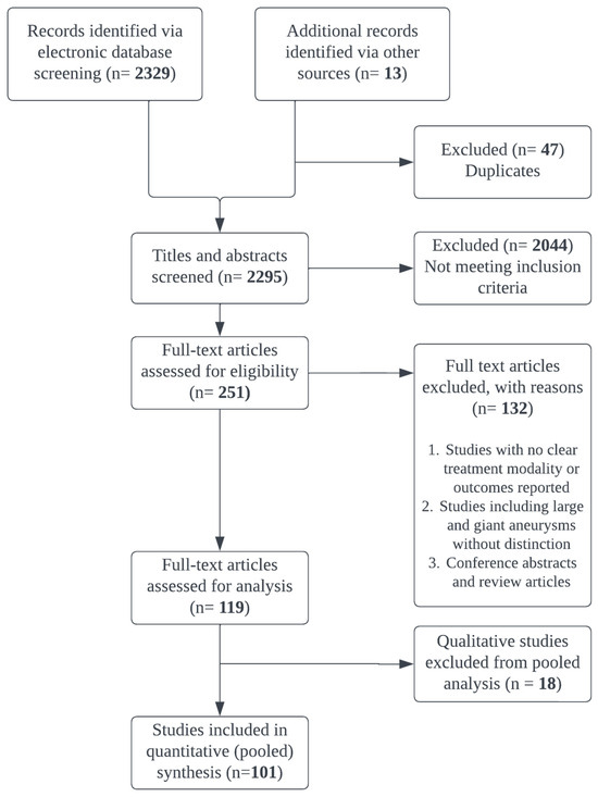
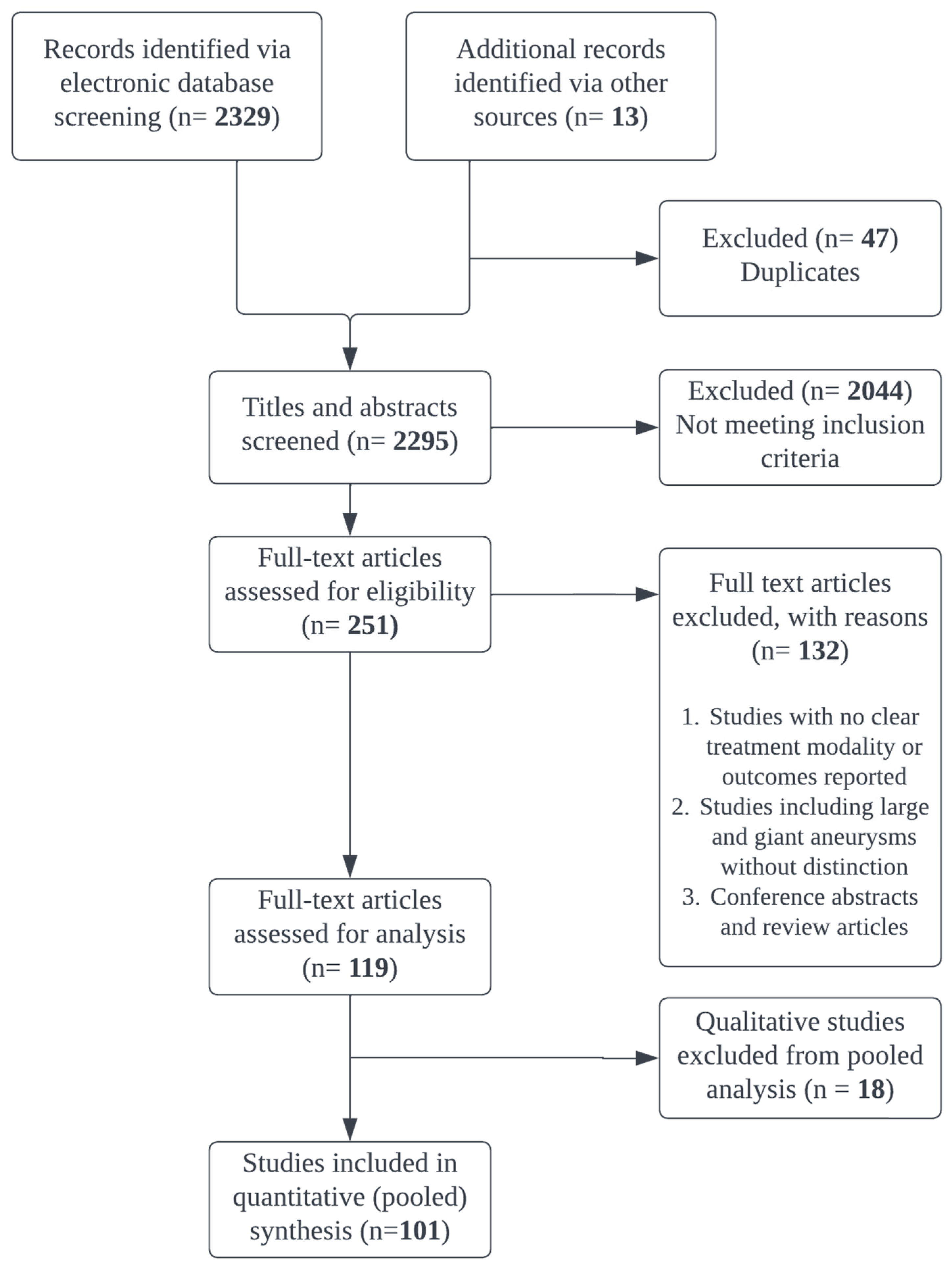
Data were extracted from the 101 included studies into a structured dataset capturing study characteristics, patient demographics, aneurysm morphology, treatment modality, and reported outcomes, as illustrated in Figure 1. Extracted variables included patient age, sex, aneurysm size, location, parent artery involvement, degree of thrombosis when reported, treatment strategy, complications, recurrence or retreatment, and neurological outcomes.
Figure 1.
PRISMA flowchart of the article selection process.
2.5. Data Processing and Statistical Analysis
The extracted dataset was cleaned and analyzed using Python version 3.13.2. Text-based clinical variables were standardized through structured parsing and keyword-based classification. The number of aneurysm cases reported in each study was extracted and aggregated to calculate total patient counts. Age values were converted to numeric form when reported. Sex distribution was calculated only for studies that explicitly reported male and female counts; no imputation was performed for missing values.
Aneurysm size values were extracted from free-text entries, converted to millimeters when necessary, and averaged when multiple values were reported within a study. Both unweighted and patient-weighted summary statistics were calculated to account for heterogeneity in study sample sizes. Parent artery involvement was categorized using keyword-based mapping into standard cerebrovascular territories including the internal carotid artery (ICA), middle cerebral artery (MCA), anterior cerebral artery (ACA), basilar artery, posterior cerebral artery, posterior communicating artery, vertebral artery, and other less frequently reported arteries. Circulation classification (anterior vs. posterior) was inferred from parent vessel anatomy.
Treatment modality was categorized into microsurgical, endovascular, or hybrid approaches based on procedural descriptions. Hybrid approaches included combined or staged microsurgical and endovascular interventions. Outcome data were extracted from structured and semi-structured fields within each publication. For complications and recurrence, explicit event counts were extracted when reported. These counts allowed pooled rate estimation within the subset of studies reporting numeric outcomes. Mortality and functional outcomes were frequently reported qualitatively without standardized denominators and therefore could not be pooled. Comparisons of complication and recurrence rates between treatment modalities were performed using chi-square tests within subsets of studies reporting numeric counts. Statistical significance was defined as p < 0.05.
3. Results
3.1. Study Selection and Patient Characteristics
Out of the 251 full-text articles assessed for eligibility, 119 met the inclusion criteria. However, 18 articles reported measures qualitatively and were, thus, excluded from further analysis. The 101 studies reported extractable quantitative data and were included in the pooled analysis, which consisted of 536 patients with GTIAs. The mean patient age across studies was 50.2 years, with a median age of 54.1 years (interquartile range [IQR] 41.0–64.0; range 9–80 years). Sex distribution was reported for 219 patients, of whom 100 were male (45.7%) and 119 were female (54.3%).
3.2. Aneurysm Characteristics
Most aneurysms arose from the anterior circulation, accounting for 88.2% of cases, whereas 10.8% involved the posterior circulation and 0.9% could not be definitively classified due to incomplete reporting. Parent artery involvement was most frequently reported in the ICA (46.6%) and MCA (39.9%), followed by the basal artery (7.8%). Less common locations included the ACA (1.5%), posterior cerebral artery (1.5%), and vertebral artery (1.3%), while other arterial territories were reported only rarely. Aneurysm size across studies remained large. The unweighted mean aneurysm diameter was 39.0 mm (median 38.0 mm; IQR 29.8–47.3 mm; range 3.5–80 mm). When weighed by study sample size, the patient-weighted mean aneurysm diameter was 24.3 mm. Although the eligibility criteria specified a diameter threshold of ≥25 mm, a small number of included studies reported mixed cohorts of varying sizes and giant aneurysms without stratified reporting; these cases account for the values below this threshold at the lower end of the observed range and for the reduction in the patient-weighted mean relative to the unweighted mean. This is a limitation addressed further in Section 4.7.
3.3. Treatment Approaches
Treatment strategies were heterogeneous across studies. Hybrid treatment approaches, combining microsurgical and endovascular techniques, were the most frequently reported and accounted for 62.7% of cases. Endovascular approaches alone were reported in 23.3% of patients, whereas microsurgical treatment alone accounted for 14.0% of cases. This distribution reflects the increasing adoption of multimodal strategies in the management of complex thrombosed aneurysms. As defined previously in Section 2.5, the hybrid category encompasses any combined or staged use of microsurgical and endovascular techniques within a single treatment course. Due to this relatively broad definition, a high proportion of cases are categorized as hybrid. Hence, this should be considered when interpreting this distribution.
3.4. Outcomes
Complications and recurrence were analyzed within subsets of studies reporting explicit numeric counts. Across studies reporting complication counts, 30 complications were reported among 176 patients, corresponding to a pooled complication rate of 17.0%. When stratified by treatment modality, complication rates were 13.2% for microsurgical treatment, 15.4% for endovascular treatment, and 21.1% for hybrid treatment. No statistically significant difference in complication rates was observed between treatment groups (χ2 p = 0.48).
Recurrence data were available for a larger subset of studies. A total of 91 recurrences were reported among 434 patients, yielding an overall recurrence rate of 21.0%. Recurrence rates differed across treatment modalities, with 2.3% in microsurgical cases, 12.8% in endovascular cases, and 26.3% in hybrid approaches. These differences reached statistical significance (χ2 p = 0.00013).
Mortality and functional neurological outcomes could not be pooled because these outcomes were reported inconsistently across studies. Many publications provided qualitative descriptions such as good recovery or neurological improvement without standardized outcome scales or explicit denominators. Consequently, these endpoints could not be aggregated for quantitative analysis.
4. Discussion
The management of GTIAs continues to represent one of the most formidable challenges in cerebrovascular surgery. These aneurysms, typically defined as measuring greater than 25 mm in diameter, often demonstrate partial thrombosis and complex hemodynamics, posing significant diagnostic and therapeutic difficulties [7]. Despite advancements in endovascular techniques, microsurgical intervention remains an essential treatment modality in selected cases.
The present systematic review synthesizes data from 101 studies encompassing 536 patients, highlighting the considerable heterogeneity in clinical presentation, aneurysm anatomy, treatment strategies, and outcome reporting across the available literature. The following Section 4.1, Section 4.2, Section 4.3, Section 4.4, Section 4.5 and Section 4.6 interpret these findings against the current technical and pathophysiological literature on GTIA management. Moreover, the pooled statistics (treatment distribution, complication and recurrence rates, and anatomical distribution) are linked to their clinical implications.
4.1. Microsurgical Clipping in the Era of Endovascular Therapy
While endovascular treatment has emerged as a first-line approach for many intracranial aneurysms due to its minimally invasive nature, GTIAs often resist complete obliteration via coiling or flow diversion due to intraluminal thrombus, calcified walls, and wide necks [8].These anatomical factors increase the risk of incomplete treatment and aneurysm regrowth or recanalization [9]. Additionally, the presence of a thrombotic mass may prevent the device from achieving adequate wall apposition or lead to migration over time [8,10].
Microsurgical clipping, on the other hand, offers the advantage of direct visualization of the aneurysm sac, neck, and associated perforators. This permits precise manipulation, decompression, and control of the aneurysm, particularly when mass effect is present [11,12]. It also allows for immediate intraoperative adjustments, such as clip repositioning, which are not possible with endovascular techniques. Importantly, microsurgical access may permit the removal of intraluminal thrombus and reduce the risk of delayed rupture.
Within the pooled cohort, microsurgical treatment alone was the least frequently used treatment approach (14.0% of cases), yet was associated with the lowest recurrence rates among the three modalities (2.3% compared to 12.8% for endovascular and 26.3% for hybrid approaches) and an intermediate complication rate (13.2%). This pattern is consistent with the durable and positive outcomes historically attributed to direct clip occlusion. However, as mentioned in Section 4.7, these comparisons were not adjusted for case selection. This means that microsurgical clipping may have been preferentially applied to aneurysms with a more favorable anatomy. Hence, the low recurrence rate should not be interpreted as indicative of superiority.
4.2. Surgical Planning and Anatomical Considerations
Preoperative planning must be meticulous and incorporate multimodal imaging, including Digital Subtraction Angiography, Computed Tomography Angiography, and Magnetic Resonance Imaging [13]. These imaging modalities help determine not only the morphology and patency of the aneurysm but also the relationship with adjacent neural structures and the presence of calcification or thrombus. Moreover, the choice of surgical corridors is critical. In cases involving anterior circulation aneurysms such as those at the anterior communicating artery, MCA, or distal ACA, the surgeon may benefit from utilizing natural or pathological corridors, such as infarcted or atrophic brain tissue, to minimize additional parenchymal injury [14,15]. For posterior circulation aneurysms, skull base approaches or far lateral approaches may be necessary, depending on location and exposure requirements [16,17]. An understanding of collateral circulation is essential when parent artery occlusion or trapping is considered. Balloon test occlusion may assist in assessing collateral adequacy. Inadequate collateral flow necessitates the use of bypass procedures [18,19,20,21,22].
These planning principles are directly relevant to the anatomical distribution observed in this review: 88.2% of the aneurysms arose from the anterior circulation, with the internal carotid artery (46.6%) and middle cerebral artery (39.9%) being the most frequently involved sites, while only 10.8% involved the posterior circulation. Anterior-circulation corridors such as the pterional or minipterional approaches were thus applicable to the vast majority of cases represented in this literature. Contrastingly, skull base or far lateral approaches which pertain more to posterior circulation lesions, are applicable to a comparatively small minority. Given that corridor selection and collateral assessment were not reported consistently across the different studies, this review cannot quantify how anatomical location influences the modality choice or outcome. Thus, this gap represents a target for more granular data extraction in future studies.
4.3. Bypass and Revascularization Techniques
In cases where parent vessel occlusion is required, revascularization may be necessary to preserve distal perfusion. Both extracranial–intracranial and intracranial–intracranial bypasses have been described [23,24,25]. Intracranial–intracranial bypasses are generally preferred when feasible due to their lower risk of kinking and shorter graft length. However, these require careful intraoperative planning and high microsurgical precision.
Bypasses such as superficial temporal artery to MCA or radial artery grafts are employed based on recipient vessel location and caliber [26]. Recent advances in intraoperative tools, such as indocyanine green angiography and Doppler ultrasonography, have enhanced the safety and success of bypass procedures by providing real-time assessment of flow dynamics.
Revascularization techniques (extracranial–intracranial vs. intracranial–intracranial bypass) were not reported consistently in all studies. Therefore, bypass-specific outcomes could not be analyzed separately from the broader hybrid and microsurgical context discussed in Section 3.
4.4. Hybrid Strategies and Intraoperative Considerations
A hybrid approach combining endovascular balloon occlusion or coiling with microsurgical clipping has gained traction in recent years [27,28]. Suction decompression, facilitated by temporary balloon occlusion of the parent artery, can significantly improve visualization and facilitate safe dissection and clip application. This approach is particularly useful in large, high-pressure aneurysms where traditional proximal control is difficult. However, hybrid approaches are not without risks. Anticoagulation during endovascular intervention may increase the risk of intraoperative bleeding during microsurgical exposure. Therefore, careful coordination between surgical and endovascular teams is essential to ensure optimal timing and minimize complications.
The hybrid approach was the most frequently reported strategy, accounting for 62.7% of cases. As noted in Section 3.3, the broad definition applied to this category likely contributed to its high apparent prevalence. Hybrid approaches were also associated with the highest complication (21.1%) and recurrence (26.3%) rates among the three modalities, exceeding both microsurgical (13.2% and 2.3%, respectively) and endovascular (15.4% and 12.8%) treatment alone. Without properly contextualizing the reason behind these findings, this pattern might suggest that hybrid strategies are less safe and effective than single-modality treatment. However, hybrid approaches are, by clinical rationale, typically reserved for the most anatomically challenging aneurysms, including those with extensive intraluminal thrombus, wide necks, branch vessel involvement, or failed attempts at a single-modality approach. The elevated event rates observed here therefore more plausibly reflect the underlying case complexity for which a hybrid strategy was selected, rather than an inherent deficiency of the hybrid approach itself.
4.5. The Role of Dual-Trained Cerebrovascular Neurosurgeons
Given that hybrid strategies represented the majority of reported treatment approaches, the following perspective on dual-trained cerebrovascular neurosurgeons is particularly relevant. It represents the authors’ expert opinion, informed by the patterns observed in the included literature rather than a finding directly derived from the systematic review data. The increasing use of hybrid treatment strategies for giant thrombosed intracranial aneurysms highlights the importance of surgeons trained in both microsurgical and endovascular techniques. As noted in this included literature, management of these lesions often requires individualized approaches that may involve bypass, thrombectomy, clipping, flow diversion, or staged combinations of surgical and endovascular interventions. Surgeons trained exclusively in either microsurgical or endovascular techniques may be limited in their ability to offer the full range of treatment options. In contrast, dual-trained cerebrovascular neurosurgeons are uniquely positioned to evaluate complex aneurysm anatomy and select the most appropriate strategy without bias toward a single modality. This training model, which has been increasingly emphasized in the United States, may facilitate more flexible procedural planning and allow seamless integration of microsurgical and endovascular techniques when managing complex aneurysms [11,20,23,28].
4.6. Pathophysiology and Rupture Risk
The natural history of GTIAs is incompletely understood, but evidence suggests that these aneurysms undergo a cyclical process of mural hemorrhage, thrombus formation, and neovascularization [7]. This may explain their unpredictable clinical course and higher annual rupture rate compared to smaller aneurysms. Intramural thrombus can both protect against rupture by dampening pulsatility and increase risk by creating an inflammatory environment within the aneurysm wall.
Interestingly, several studies suggest that intraoperative rupture rates for GTIAs may actually be lower than smaller, nonthrombotic aneurysms due to the presence of multiple fibrotic thrombus layers [29,30]. However, when rupture does occur, the volume of hemorrhage and risk of neurological deterioration is significantly greater due to the aneurysm’s size.
4.7. Limitations
Several limitations must be acknowledged when interpreting the findings of this systematic review, most of which stem from the structure and reporting quality of the existing literature on GTIAs. A formal methodological quality and risk of bias assessment (e.g., the Newcastle–Ottawa Scale) was not performed for individual studies, and this represents a limitation of the present review. Given that the included literature consists predominantly of retrospective case series and single-case reports, the risk of bias related to selection, reporting, and confounding is expected to be substantial and variable across studies. The current evidence base is composed predominantly of retrospective case series and single-case reports, with no prospective comparative studies identified. Classic microsurgical experiences describing thrombectomy, clip reconstruction, trapping, and bypass, such as those reported by Drake [1], Peerless and Drake [2], Sundt and Piepgras [3], Yasargil et al. [4], and Lawton et al. [5], provided essential technical insights into operative feasibility and natural history but were not designed for comparative outcome analysis and frequently involved highly selected referral populations. As a result, selection bias and incomplete outcome reporting are unavoidable and limit generalizability. Similarly, contemporary endovascular and hybrid series, including early experiences with flow diversion, are observational in nature and lack appropriate control groups [31,32,33]. Consequently, causal inferences regarding the relative effectiveness or safety of different treatment strategies cannot be drawn. These chi-square comparisons were unadjusted for confounders such as aneurysm size, location, morphology, thrombosis degree, treatment era, or follow-up duration, and were drawn from heterogeneous retrospective data; the significant difference in recurrence rates should not be taken as evidence of comparative treatment effectiveness.
Outcome reporting across the included studies was highly heterogeneous and frequently inconsistent. Many publications reported neurological outcomes qualitatively rather than quantitatively or described complications, mortality, or retreatment without providing explicit numerators, denominators, or standardized functional outcome measures. This limitation was present across both microsurgical and endovascular literature, including large institutional series and smaller hybrid cohorts [5,32,33]. For example, several large microsurgical series described outcomes using qualitative terms such as good or poor recovery without standardized functional scales or explicit complication rates [2,3]. As a result, true event rates could not be calculated for many endpoints, and only limited pooled analyses were possible in subsets of studies reporting explicit outcome counts.
Treatment classification represented an additional limitation. Many studies described complex, staged, or multimodal management strategies, such as extracranial–intracranial bypass followed by trapping or delayed parent vessel occlusion, without clearly identifying a dominant treatment modality or attributing outcomes to specific procedural components [5,33]. As a result, outcomes were often reported for the overall treatment course rather than per intervention, complicating interpretation of modality-specific results.
There was also marked heterogeneity in aneurysm definition and characterization. Although a diameter threshold of ≥ 25 mm is defined for giant aneurysms, some studies included aneurysms below this cutoff or combined large and giant aneurysms into a single category or combined thrombosed and non-thrombosed aneurysms without stratified reporting [2,34]. The extent of thrombosis, aneurysm morphology, involvement of branch vessels or perforators, and presence of calcification were inconsistently reported despite their known impact on treatment complexity and outcome [3,33]. The degree of thrombosis, partial versus complete, was variably defined or not quantified despite its known impact on mass effect, surgical complexity, and endovascular feasibility [5,35].
Temporal bias further limits interpretability. The included literature spans several decades, during which significant advances occurred in microsurgical techniques, cerebral revascularization, neuroimaging, endovascular devices, and perioperative care. Outcomes reported in early surgical series are therefore not directly comparable to those reported in more recent endovascular or hybrid studies utilizing flow-diverting stents and modern adjunctive techniques [6,7,10].
Follow-up duration was variable and often limited. While some classic surgical series included long-term follow-up, many endovascular and hybrid studies reported short or unspecified follow-up intervals [5,8]. Given the recognized potential for delayed aneurysm growth, thrombus progression, recanalization, and late neurological deterioration in GTIAs, this limitation restricts the assessment of long-term durability.
Publication bias is also likely substantial. Many reports reflect single-center experiences and small patient cohorts, limiting generalizability and introducing selection bias. Given the rarity and technical complexity of GTIAs, successful or novel treatment strategies are preferentially reported, particularly in single-case reports and small series. Negative outcomes, failed interventions, and conservatively managed cases are likely underrepresented [36].
Collectively, these limitations reflect the intrinsic challenges of studying a rare, heterogeneous, and technically demanding pathology rather than deficiencies of individual studies. They highlight the need for standardized definitions, uniform outcome reporting, and multicenter collaboration to improve the quality of evidence guiding the management of GTIAs.
5. Conclusions
GTIAs represent a rare and uniquely complex subset of cerebrovascular disease characterized by challenging anatomy, evolving pathophysiology, heterogeneous clinical presentation, and substantial treatment-related risk. This systematic review of 101 studies encompassing 536 patients highlights the marked heterogeneity in presentation, management strategies, and outcome reporting across the existing literature. The literature demonstrates considerable variability in treatment approaches. The general trend observed may suggest that multimodal and hybrid approaches are favored, reflecting the limitations of isolated microsurgical or endovascular techniques when faced with extensive intraluminal thrombus, mass effect, and branch vessel involvement [37,38,39,40,41].
However, the predominance of retrospective case series, coupled with inconsistent and incomplete outcome reporting, prevents definitive conclusions from being drawn regarding the superiority of any single treatment modality. This limitation restricts the ability to quantify true risks, assess durability, or perform meaningful comparative analyses between treatment strategies. Future research efforts should prioritize multicenter collaboration, standardized reporting of aneurysm characteristics and outcomes, and the consistent use of validated functional outcome measures with long-term follow-up. Until such data are available, management of giant thrombosed intracranial aneurysms should remain individualized, multidisciplinary, and tailored to patient-specific anatomy, symptomatology, and institutional expertise.
Supplementary Materials
The following supporting information can be downloaded at: https://www.mdpi.com/article/10.3390/jvd5040030/s1. Table S1: PRISMA 2020 checklist.
Author Contributions
Conceptualization, M.P.; methodology, Y.A.-N.; validation, Y.A.-N., D.B., E.D., F.Q.M.S., D.F., D.S., Z.S., B.L.-W., M.K. and M.P.; formal analysis, C.R.; investigation, Y.A.-N., D.B., E.D., F.Q.M.S. and D.F.; resources, M.P.; data curation, D.B., E.D., F.Q.M.S. and D.F.; writing—original draft preparation, Y.A.-N., D.B., E.D., F.Q.M.S. and D.F.; writing—review and editing, Y.A.-N., D.S., Z.S., B.L.-W., M.K. and M.P.; visualization, Y.A.-N.; supervision, B.L.-W., M.K. and M.P.; project administration, M.P. All authors have read and agreed to the published version of the manuscript.
Funding
The authors did not receive support from any organization for the submitted work.
Institutional Review Board Statement
Not applicable. This is a review of existing research and did not involve human subjects.
Data Availability Statement
No new data were created or analyzed in this study. Data sharing is not applicable to this article.
Acknowledgments
The authors wish to thank Mariana Grohowski for editing assistance.
Conflicts of Interest
The authors have no competing interests to declare that are relevant to the content of this article.
References
- Drake, C.G. Giant intracranial aneurysms: Experience with surgical treatment in 174 patients. Clin. Neurosurg. 1979, 26, 12–95. [Google Scholar] [CrossRef] [Scilit]
- Peerless, S.J.; Drake, C.G. Treatment of giant cerebral aneurysms of the anterior circulation. Neurosurg. Rev. 1982, 5, 149–154. [Google Scholar] [CrossRef] [Scilit] [PubMed]
- Sundt, T.M.; Piepgras, D.G., Jr. Surgical approach to giant intracranial aneurysms. Operative experience with 80 cases. J. Neurosurg. 1979, 51, 731–742. [Google Scholar] [CrossRef] [Scilit] [PubMed]
- Yasargil, M.G. Clinical considerations, surgery of the intracranial aneurysms and results. Microneurosurg. Thieme 1984, 2, 33–123. [Google Scholar]
- Lawton, M.T.; Quinones-Hinojosa, A.; Chang, E.F.; Yu, T. Thrombotic intracranial aneurysms: Classification scheme and management strategies in 68 patients. Neurosurgery 2005, 56, 441–454; discussion 441–454. [Google Scholar] [CrossRef] [Scilit] [PubMed]
- Liberati, A.; Altman, D.G.; Tetzlaff, J.; Mulrow, C.; Gøtzsche, P.C.; Ioannidis, J.P.A.; Clarke, M.; Devereaux, P.J.; Kleijnen, J.; Moher, D. The prisma statement for reporting systematic reviews and meta-analyses of studies that evaluate health care interventions: Explanation and elaboration. J. Clin. Epidemiol. 2009, 62, e1–e34. [Google Scholar] [CrossRef] [Scilit] [PubMed]
- Choi, I.S.; David, C. Giant intracranial aneurysms: Development, clinical presentation and treatment. Eur. J. Radiol. 2003, 46, 178–194. [Google Scholar] [CrossRef] [Scilit] [PubMed]
- Sughrue, M.E.; Saloner, D.; Rayz, V.L.; Lawton, M.T. Giant intracranial aneurysms: Evolution of management in a contemporary surgical series. Neurosurgery 2011, 69, 1261–1270; discussion 1270–1271. [Google Scholar] [CrossRef] [Scilit] [PubMed]
- Santoro, A.; Armocida, D.; Paglia, F.; Iacobucci, M.; Berra, L.V.; D’angelo, L.; Cirelli, C.; Guidetti, G.; Biraschi, F.; Cantore, G. Treatment of giant intracranial aneurysms: Long-term outcomes in surgical versus endovascular management. Neurosurg. Rev. 2022, 45, 3759–3770. [Google Scholar] [CrossRef] [Scilit] [PubMed]
- Lylyk, P.; Miranda, C.; Ceratto, R.; Ferrario, A.; Scrivano, E.; Luna, H.R.; Berez, A.L.; Tran, Q.; Nelson, P.K.; Fiorella, D. Curative endovascular reconstruction of cerebral aneurysms with the pipeline embolization device: The buenos aires experience. Neurosurgery 2009, 64, 632–642; discussion 642–643. [Google Scholar] [CrossRef] [Scilit] [PubMed]
- Lee, K.S.; Zhang, J.J.Y.; Nguyen, V.; Han, J.; Johnson, J.N.; Kirollos, R.; Teo, M. The evolution of intracranial aneurysm treatment techniques and future directions. Neurosurg. Rev. 2022, 45, 1–25. [Google Scholar] [CrossRef] [Scilit] [PubMed]
- Chalouhi, N.; Thakkar, V.; Tjoumakaris, S.; Gonzalez, L.F.; Hasan, D.; Rosenwasser, R.; Singhal, S.; Jabbour, P.M. Microsurgical clipping of large and giant cerebral aneurysms: A single-center contemporary experience. J. Clin. Neurosci. 2014, 21, 1424–1427. [Google Scholar] [CrossRef] [Scilit] [PubMed]
- Tollard, E.; Perot, G.; Clavier, E.; Gerardin, E. Imaging of giant cerebral aneurysms. Neurochirurgie 2015, 61, 378–384. [Google Scholar] [CrossRef] [Scilit] [PubMed]
- Li, B.; Zhang, K.; Yu, J. Current state of endovascular treatment of anterior cerebral artery aneurysms. Front. Neurol. 2024, 15, 1396701. [Google Scholar] [CrossRef] [Scilit] [PubMed]
- Nasir, R.; Naqvi, M.e.Z.; Ahmed, S.; Hassan, M.U.; Tariq, R.; Khan, S.A.; Koeskemeier, P.; Khajuria, R.K.; Bajwa, M.H.; Muhammad, S. The morphological, clinical, and prognostic factors in the management of giant anterior communicating artery aneurysms: A systematic review of cases. Brain Spine 2025, 5, 104189. [Google Scholar] [CrossRef] [Scilit] [PubMed]
- Bowles, A.P.; Kinjo, T.; Al-Mefty, O. Skull base approaches for posterior circulation aneurysms. Skull Base Surg. 1995, 5, 251–260. [Google Scholar] [CrossRef] [Scilit] [PubMed]
- Nanda, A.; Konar, S.; Bir, S.C.; Maiti, T.K.; Ambekar, S. Modified far lateral approach for posterior circulation aneurysms: An institutional experience. World Neurosurg. 2016, 94, 398–407. [Google Scholar] [CrossRef] [Scilit] [PubMed]
- Lee, S.H.; Ahn, J.S.; Kwun, B.D.; Park, W.; Park, J.C.; Roh, S.W. Surgical flow alteration for the treatment of intracranial aneurysms that are unclippable, untrappable, and uncoilable. J. Korean Neurosurg. Soc. 2015, 58, 518–527. [Google Scholar] [CrossRef] [Scilit] [PubMed]
- de Lotbiniere-Bassett, M.; Hasen, M.; Couldwell, W.T. Flow reversal bypass surgery for giant intracranial aneurysms: Illustrative cases. J. Neurosurg. Case Lessons 2025, 9, CASE2583. [Google Scholar] [CrossRef] [Scilit] [PubMed]
- Hanel, R.A.; Spetzler, R.F. Surgical treatment of complex intracranial aneurysms. Neurosurgery 2008, 62, 1289–1297; discussion 1297–1299. [Google Scholar] [CrossRef] [Scilit] [PubMed]
- Kalani, M.Y.; Zabramski, J.M.; Nakaji, P.; Spetzler, R.F. Twenty-year follow-up of flow reversal and revascularization for a giant serpentine basilar artery aneurysm. Neurosurgery 2014, 10, E493–E497; discussion E497. [Google Scholar] [CrossRef] [Scilit] [PubMed]
- Miyamoto, S.; Funaki, T.; Iihara, K.; Takahashi, J.C. Successful obliteration and shrinkage of giant partially thrombosed basilar artery aneurysms through a tailored flow reduction strategy with bypass surgery. J. Neurosurg. 2011, 114, 1028–1036. [Google Scholar] [CrossRef] [Scilit] [PubMed]
- Sanai, N.; Zador, Z.; Lawton, M.T. Bypass surgery for complex brain aneurysms: An assessment of intracranial-intracranial bypass. Neurosurgery 2009, 65, 670–683; discussion 683. [Google Scholar] [CrossRef] [Scilit] [PubMed]
- Park, S.J.; Lee, Y.J.; Kim, W.B.; Kim, Y.S.; Kim, T.S.; Joo, S.P. Intracranial-intracranial bypass strategies for the treatment of complex intracranial aneurysms: Anatomical characteristics and surgical intervention. Acta Neurochir. 2024, 166, 42. [Google Scholar] [CrossRef] [Scilit] [PubMed]
- Chen, Y.; Chen, P.; Duan, G.; Li, R.; Li, Z.; Guo, G. Extracranial-intracranial bypass surgery for intracranial aneurysm of the anterior cerebral circulation: A systematic review and meta-analysis. Front. Neurol. 2023, 14, 1174088. [Google Scholar] [CrossRef] [Scilit] [PubMed]
- Newell, D.W. Superficial temporal artery to middle cerebral artery bypass. Skull Base 2005, 15, 133–141. [Google Scholar] [CrossRef] [Scilit] [PubMed]
- Lim, Y.C.; Shin, Y.S.; Chung, J. Combined endovascular and microsurgical procedures as complementary approaches in the treatment of a single intracranial aneurysm. J. Korean Neurosurg. Soc. 2008, 43, 21–25. [Google Scholar] [CrossRef] [Scilit] [PubMed]
- Ulmer, S.; Gruber, P.; Schubert, G.A.; Remonda, L.; Marbacher, S.; Gruter, B.E. Combined microsurgical and endovascular intracranial aneurysm treatment: Interdisciplinary experience using a true hybrid approach and a systematic review of the literature. Brain Sci. 2024, 14, 816. [Google Scholar] [CrossRef] [Scilit] [PubMed]
- Lawton, M.T.; Spetzler, R.F. Surgical management of giant intracranial aneurysms: Experience with 171 patients. Clin. Neurosurg. 1995, 42, 245–266. [Google Scholar] [PubMed]
- International Study of Unruptured Intracranial Aneurysms I. Unruptured intracranial aneurysms--risk of rupture and risks of surgical intervention. N. Engl. J. Med. 1998, 339, 1725–1733. [CrossRef] [Scilit] [PubMed]
- Briganti, F.; Leone, G.; Ugga, L.; Marseglia, M.; Macera, A.; Manto, A.; Delehaye, L.; Resta, M.; Resta, M.; Burdi, N.; et al. Mid-term and long-term follow-up of intracranial aneurysms treated by the p64 flow modulation device: A multicenter experience. J. Neurointerv Surg. 2017, 9, 70–76. [Google Scholar] [CrossRef] [Scilit] [PubMed]
- Fiorella, D.; Woo, H.H.; Albuquerque, F.C.; Nelson, P.K. Definitive reconstruction of circumferential, fusiform intracranial aneurysms with the pipeline embolization device. Neurosurgery 2008, 62, 1115–1120; discussion 1120–1121. [Google Scholar] [CrossRef] [Scilit] [PubMed]
- Kalani, M.Y.; Zabramski, J.M.; Hu, Y.C.; Spetzler, R.F. Extracranial-intracranial bypass and vessel occlusion for the treatment of unclippable giant middle cerebral artery aneurysms. Neurosurgery 2013, 72, 428–435; discussion 435–436. [Google Scholar] [CrossRef] [Scilit] [PubMed]
- Cantore, G.; Santoro, A.; Guidetti, G.; Delfinis, C.P.; Colonnese, C.; Passacantilli, E. Surgical treatment of giant intracranial aneurysms: Current viewpoint. Neurosurgery 2008, 63, 279–289; discussion 289–290. [Google Scholar] [CrossRef] [Scilit] [PubMed]
- van Rooij, W.J.; Sluzewski, M.; Beute, G.N. Endovascular treatment of giant serpentine aneurysms. AJNR Am. J. Neuroradiol. 2008, 29, 1418–1419. [Google Scholar] [CrossRef] [Scilit] [PubMed]
- Nanda, A.; Sonig, A.; Banerjee, A.D.; Javalkar, V.K. Microsurgical management of basilar artery apex aneurysms: A single surgeon’s experience from louisiana state university, shreveport. World Neurosurg. 2014, 82, 118–129. [Google Scholar] [CrossRef] [Scilit] [PubMed]
- Chihi, M.; Jabbarli, R.; Gembruch, O.; Teuber-Hanselmann, S.; Oppong, M.D.; Pierscianek, D.; Radbruch, A.; Glas, M.; Stettner, M.; Sure, U. A rare case of a completely thrombosed bilobed giant intracranial aneurysm of the anterior cerebral artery with spontaneous parent vessel thrombosis: Case report. BMC Neurol. 2019, 19, 297. [Google Scholar] [CrossRef] [Scilit] [PubMed]
- Bastos, L.P.; Perreira, T.; Machado, E. Giant Partially Thrombosed Intracranial Aneurysm. Eurorad 2009, Case 7392. Available online: https://eurorad.org/case/7392 (accessed on 5 March 2026).
- Mura, J.; Alvarez, V.H.; Oberman, D.Z.; Cardenas, A.R.; Rabelo, N.N.; Figueiredo, E.G. Management of giant thrombosed mca aneurysm: Double sta-mca revascularization. World Neurosurg. 2021, 149, 1. [Google Scholar] [CrossRef] [Scilit] [PubMed]
- Borni, M.; Kolsi, F.; Cherif, I.; Boudawara, M.Z. A giant partial thrombosed aneurysm of the internal cavernous carotid artery mimicking a meningioma of the lesser wing of the sphenoid bone. Radiol. Case Rep. 2022, 17, 1325–1329. [Google Scholar] [CrossRef] [Scilit] [PubMed]
- Noh, Y.; Lee, S.H.; Yoon, S.M.; Song, I.H.; Oh, J.S. Staged hybrid treatment for giant thrombosed fusiform aneurysm. J. Cerebrovasc. Endovasc. Neurosurg. 2021, 23, 359–364. [Google Scholar] [CrossRef] [Scilit] [PubMed]
Disclaimer/Publisher’s Note: The statements, opinions and data contained in all publications are solely those of the individual author(s) and contributor(s) and not of MDPI and/or the editor(s). MDPI and/or the editor(s) disclaim responsibility for any injury to people or property resulting from any ideas, methods, instructions or products referred to in the content. |
© 2026 by the authors. Licensee MDPI, Basel, Switzerland. This article is an open access article distributed under the terms and conditions of the Creative Commons Attribution (CC BY) license.