


Abstract
Background: While cardiovascular diseases (CVD) manifest in adulthood, vascular changes may begin in childhood. Early markers of CVD, such as carotid intima-media thickness (CIMT), have not been well studied in children in the African setting. The potential influence of environmental and genetic factors on the CIMT of African children is not well understood. Objective: This study assessed the cardiovascular risk profiles of Ghanaian children and adolescents in rural and urban settings using carotid intima-media thickness (CIMT) and ultrasound-measured adiposity. Methods: A cross-sectional study was conducted involving 343 asymptomatic, healthy school children (10–16 years) from the Ashanti region of Ghana. Participants were recruited from one urban and one rural school. Data collected included height, weight, blood pressure, and ultrasound measurements of CIMT, subcutaneous, preperitoneal, and Visceral fat. Results: The mean CIMT for the cohort was 0.60 ± 0.07 mm. Urban children had significantly higher BMI (p < 0.001), subcutaneous fat (p = 0.005), and preperitoneal fat (p < 0.001) compared to rural children, yet there was no significant difference in CIMT between the two sites (p = 0.497). Multiple linear regression revealed that peritoneal fat thickness (p = 0.029) and male gender (p < 0.001) were significant predictors of CIMT, whereas BMI and blood pressure were not. Conclusions: Ghanaian children exhibit elevated CIMT values compared to Western pediatric cohorts despite having lower BMI and normal blood pressure. The significant correlation between peritoneal (visceral) adiposity and CIMT suggests that fat distribution is a more sensitive indicator of early vascular remodeling than BMI in this population. This highlights the need for population-specific screening strategies that move beyond BMI.
1. Introduction
Cardiovascular diseases (CVDs), including stroke, remain the leading cause of mortality worldwide, accounting for an estimated 20.5 million deaths in 2021, with nearly 80% occurring in low- and middle-income countries [1]. While clinical manifestations of CVD are typically observed in adulthood, current evidence suggests that the pathophysiological processes underlying atherosclerotic disease, including structural and functional vascular alterations, begin during the first decade of life [2,3].
A growing body of studies suggests that stroke in Africans and populations of African ancestry occurs at a younger age and exhibits distinct pathophysiology compared to white populations [1,2]. In addition, the vascular mechanisms underlying these strokes are in stark contrast to those of Caucasians and people of other ethnicities, heavily favoring intracranial vessel disease and small vessel occlusions. Specifically, cardiovascular disease in Africans preferentially involves intracranial small vessel disease occlusions, whereas extracranial carotid artery stenosis is more common in white populations [4].
To understand the origins of these unique adult stroke patterns, it is essential to investigate early-life vascular changes. Computed tomography coronary angiography (CTCA) and coronary artery calcium scoring (CAC) are useful imaging techniques for adults, but their efficacy in children is limited due to challenges such as the need for contrast agents in CTCA and the requirement for stillness in CAC [1]. Risks include radiation exposure and difficulty in detecting calcifications or significant plaque in pediatric populations.
Carotid intima-media thickness (CIMT), measured using B-mode ultrasound, provides an important marker for evaluating the subclinical vascular remodeling that precedes clinical cardiovascular disease without the aforementioned risks to the pediatric population [5,6]. While black populations typically have a lower prevalence of significant extracranial carotid stenosis or focal plaque, they demonstrate greater diffuse arterial wall thickening, which is evident in elevated CIMT [7,8,9]. In addition, since focal atherosclerotic plaques are virtually absent during the first decade of life, utilizing a continuous metric such as CIMT is essential for detecting the subtle, earliest phases of vascular injury. Significantly, research indicates that this extracranial thickening correlates with the severity of intracranial arterial stenosis, suggesting that extracranial and intracranial vascular changes evolve concurrently. As such, if African populations develop vascular risk factors earlier in life, the resulting hemodynamic and endothelial stress may simultaneously accelerate both carotid and intracranial arterial wall thickening. Since overt clinical cardiovascular disease in this demographic primarily manifests in adulthood, investigating the pediatric population is essential to capture the subclinical, pre-plaque phase of vascular remodeling. As such, waiting until adulthood for screening represents a missed opportunity for prevention. Given this predisposition to intracranial small vessel disease and the reported higher CIMT in young black adults, this study sought to characterize early CIMT and adiposity values in Ghanaian children and compare them with published data from other populations.
2. Materials and Methods
2.1. Study Design
This cross-sectional observational study was conducted between September and October 2024. It was reported according to the Strengthening the Reporting of Observational Studies in Epidemiology (STROBE) guidelines. Ethical approval was obtained from the Committee on Human Research, Publications, and Ethics (CHRPE/AP/694/24) of the Kwame Nkrumah University of Science and Technology (KNUST).
2.2. Study Setting
Participants were school children recruited from two schools. One is in an urban area within the heart of the Ashanti region of Ghana, where students are mostly dropped off and picked up after school, and another is in a rural area where students walk a minimum of 5 km on average to school daily. The students in the rural area come from a predominantly farming community with a relatively lower economic outlook, whereas those from the urban area have parents who are mostly in the upper middle class and better off.
2.3. Participants and Recruitment
The inclusion criteria for this study were asymptomatic children between 10 and 16 years with no known chronic illness and who were not being treated for any chronic conditions. These were assessed through self-report by both the parents and the children themselves. The parents of these participants had to give consent for inclusion in the study. Students whose parents did not give consent or who fell outside the stipulated age range were excluded from the study.
2.4. Sample Size
This exploratory study aimed to recruit a representative sample of school-going children from one urban and one rural school in the Ashanti region. Due to the absence of prior CIMT data in Ghanaian pediatric populations, a formal a priori sample size calculation was not feasible, and a pragmatic convenience sampling approach was adopted, enrolling all eligible children from the selected schools during the data collection period (September–October 2024). We purposefully selected one urban and one rural school to observe extremes in lifestyle and socioeconomic factors such as daily physical activity and economic outlook. The achieved sample of 343 participants exceeds the minimum requirement for the planned statistical analysis. Post hoc power analysis using G*Power (Version 3.1.9.7) indicated that this sample size provides >90% power to detect a correlation of r = 0.20 (comparable to the observed correlation between peritoneal fat and CIMT) at α = 0.05 (two-tailed). The sample also allows for stratified analyses by gender and setting while maintaining adequate statistical power. This sample size is comparable to or larger than similar pediatric CIMT studies in other populations [10,11,12,13,14].
2.5. Variables
The main variables of interest to the researchers were blood pressure (systolic and diastolic), body mass index (BMI), measures of adiposity (subcutaneous, preperitoneal, visceral), and carotid intima-media thickness (CIMT).
2.6. Covariates
Taking into consideration potential confounders, the covariates assessed included: Gender was classified as ‘male’ and ‘female,’ and age was categorized as 10 years, 11 years, 12 years, 13 years, 14 years, 15 years, and 16 years.’
2.7. Data Collection
The data collected in this study included height, weight, blood pressure, ultrasound-measured adiposity, and CIMT. The height and weight were taken using an electronic scale with a laser-guided height measurement attachment. Participants were asked to remove their shoes and heavy items of clothing and then stand upright on the scale. After the weight was determined, the machine subsequently measured the height, and this was documented.
The blood pressure was taken using an electronic blood pressure measurement device (ALPHAMED, model: U82RH) with the appropriate cuff size for pediatric populations. Participants rested seated for 5–10 min after their height and weight were taken to allow an accurate measurement of blood pressure. The mean of three [3] measurements taken two minutes apart was recorded as the final blood pressure for each participant.
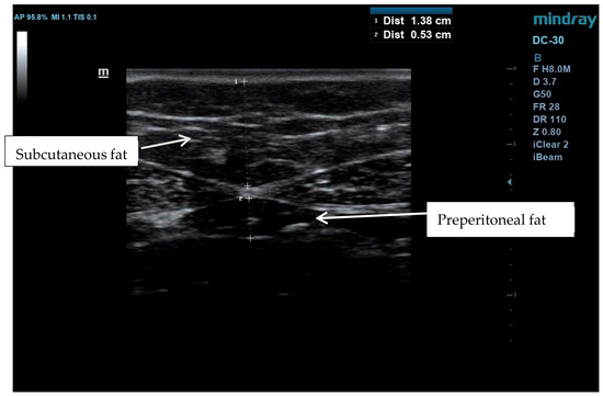
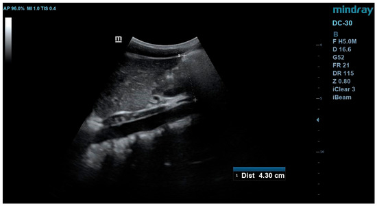
Participants were then led to an ultrasound area where they were asked to lie down supine on a couch. An ultrasound machine (Mindray DC 30 HD, Shenzhen, China) was used to measure adiposity and CIMT. Adiposity measurements were calculated as the arithmetic mean of three measurements taken on the hemisternal line, 1 cm above the umbilical scar. A linear transducer was utilized in a transverse section to measure subcutaneous and preperitoneal fat, while a convex transducer was utilized in a longitudinal section to measure visceral (peritoneal) fat (Figure 1 and Figure 2).
Figure 1.
Measurement of subcutaneous and preperitoneal fat.
Figure 2.
Measurement of visceral fat.
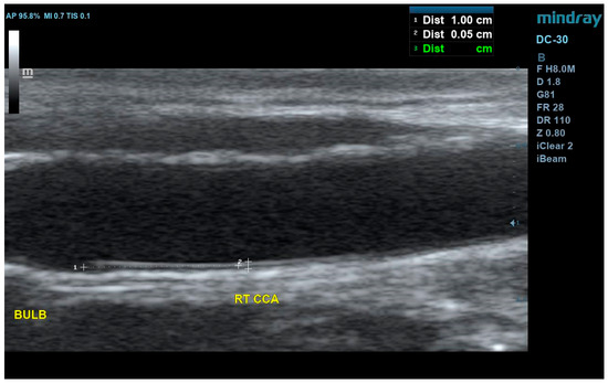
Manual CIMT measurements were taken in the far wall of the distal Common carotid artery about 1 cm from the bulb at a 90-degree angle of insonation for the right and left sides by two independent assessors blinded to the measurements of each other (Figure 3). Measurements were made according to established protocols [15]. There was no significant difference between the right and left sides (p = 0.236). The mean of two measurements was recorded as the value for each side and for each assessor. The mean CIMT for each participant was calculated as the mean of all the measurements taken by the two assessors. A detailed step-by-step protocol for positioning and ultrasound measurements can be found in Appendix A.
Figure 3.
Showing measurements of CIMT in the distal CCA 1 cm from the bulb.
2.8. Interobserver Reliability
Interobserver reliability was assessed using the interclass correlation coefficient (ICC). The ICC yielded a value of 0.699 (95% CI: 0.632–0.771), which indicates a moderate level of reliability.
2.9. Data Analysis
The data was analyzed using Jamovi version 2.7.12. In alignment with the study’s objectives, a descriptive analysis technique was applied. The Shapiro–Wilks test was used to ascertain the normality of the data distribution. This yielded a p < 0.001, indicating a non-normal distribution of data. Visual inspection of the Q-Q plots (Supplementary Materials) confirmed a non-normal distribution. Consequently, Spearman’s rank correlation coefficient (rho) was initially employed. To ensure robustness, logarithmic transformation of CIMT was also attempted, and parametric tests (Pearson’s correlation) were conducted as a sensitivity analysis. Because the parametric and non-parametric results did not differ materially, the non-parametric findings are reported.
3. Results
3.1. Demographic Characteristics of Participants
At the end of the study, a total of 343 participants were recruited. This number comprised 210 (61.2%) females and 133 (38.8%) males. There were 148 participants from the urban area and 195 from the rural area. The age range used for this study was 10 to 16 years. The majority of the respondents were 13 years old. The mean CIMT for the entire study population was 0.60 ± 0.07 mm, with a coefficient of variation (CV) of 13.1%, indicating a moderate degree of dispersion in CIMT values within the cohort. Further details on demographics are provided in Table 1.
Table 1.
Baseline characteristics of the total study cohort.
3.2. Correlation Between CIMT and Variables
Table 2 illustrates the correlation between CIMT and body metrics as well as the covariates. There were significant positive associations between CIMT and peritoneal fat (rho = 0.193, p < 0.001), whereas subcutaneous (rho = −0.116, p = 0.031) and preperitoneal fat (rho = −0.146, p = 0.007) showed weak negative correlations. There was no other significant correlation with any of the other variables.
Table 2.
Correlation matrix of CIMT and factors.
3.3. Comparing Variables from the Two Groups
Table 3 illustrates findings from independent sample t-tests between variables measured from the two sites. There was a significant difference in the height (p < 0.001), weight (p < 0.001), BMI (p < 0.001), subcutaneous fat (p = 0.005), preperitoneal fat (p < 0.001), waist circumference (p < 0.001), and hip circumference (p < 0.001). There was no significant difference in CIMT between the two sites (p = 0.49).
Table 3.
Comparison of anthropometric and vascular variables between rural and urban study sites.
Table 4 illustrates findings from independent sample t-tests between measured variables and genders. Apart from waist circumference, which showed no significant difference between genders (p = 0.598), all other variables showed significant differences.
Table 4.
Comparison of anthropometric and vascular variables between genders.
3.4. Factors Influencing CIMT
A multiple linear regression analysis was employed to investigate the extent to which age, gender, and key cardiovascular risk factors predict carotid intima-media thickness (CIMT). The model specified CIMT as the dependent variable. The model was statistically significant, explaining 11.5% of the variance in CIMT (R = 0.339; R2 =0.115; adjusted R2 = 0.0913; F = 4.82; p < 0.001). While the R2 value indicates that a large proportion of the variance remains unexplained, this suggests other unmeasured genetic, environmental, or lifestyle factors are at play; the model highlights specific significant associations. Notably, traditional risk factors such as BMI and BP were not significantly associated with CIMT in this cohort, whereas male gender (Estimate: 0.03935 [95% CI: 0.020–0.058]; p < 0.001) and visceral fat thickness (Estimate: 9.69 × 10⁻⁴ [95% CI: 1.19 × 10⁻⁴–0.001]; p = 0.026) demonstrated statistically significant positive correlations with CIMT (Table 5). Because Age was evaluated as a continuous variable and found not to significantly influence CIMT within this narrow 10–16-year cohort (p = 0.436), further age-stratified sub-analyses were not performed. Variance Inflation Factor (VIF) values for the updated model were all well within acceptable limits (VIF < 2.14) (Table 6).
Table 5.
Results of multiple linear regression analysis assessing combined predictors of carotid intima-media thickness (CIMT).
Table 6.
Variance inflation factors (VIFs).
4. Discussion
4.1. Summary of Findings
This study was grounded on the observation that adults of African ancestry suffer from a distinct pattern of cerebrovascular disease, notably earlier-onset and intracranial small vessel disease. This study sought to determine if the antecedent of the pattern, namely, elevated CIMT and its risk factors, was detectable in Ghanaian children. This study goes on to present the mean values of BMI, blood pressure, adiposity, and CIMT for a Ghanaian population. Females generally showed higher BMI, blood pressure, and adiposity compared to males, with urban children exhibiting greater BMI and fat thickness than those in rural areas. Importantly, despite these marked differences in superficial adiposity and lifestyle, there was no significant difference in CIMT between the urban and rural cohorts. This study differs from previous research by revealing a significant correlation between CIMT and visceral fat thickness but not with BMI [2]. This adiposity paradox suggests that traditional anthropometric markers such as BMI may not be sensitive indicators of vascular health in African populations [3], consistent with studies from other populations [4,16,17].
4.2. CIMT and Stroke Risk
The mean CIMT values of this population (0.60 ± 0.07 mm) are higher than those reported in numerous international pediatric studies (Table 7) [18,19,20,21,22], including those comparing sex and laterality [23].
Table 7.
CIMT values in the current study compared with those from other studies.
This finding aligns with observations in young Black adults who exhibit higher CIMT than their white counterparts [7] and provides direct evidence that this disparity is present as early as the first decade of life. Given that CIMT is a recognized early surrogate marker of subclinical atherosclerosis, its elevation in childhood suggests a lifelong vascular burden [8]. This early initiation of vascular remodeling may be a key contributor to the high prevalence of intracranial small vessel disease observed in African populations [9,10]. However, the relationship between extracranial CIMT and intracranial pathology is not straightforward. Although Black populations often demonstrate greater CIMT [11], studies have reported lower prevalence of carotid plaque and significant extracranial carotid stenosis compared with White populations [12,13]. This suggests that vascular remodeling in Black populations may manifest more as diffuse arterial wall thickening rather than focal plaque formation in the extracranial carotid arteries. Such a pattern may help explain why stroke in Africans and African-ancestry populations is more frequently associated with intracranial vascular disease rather than extracranial carotid disease.
The relationship between CIMT and intracranial vascular disease provides further support for this interpretation. Several studies have demonstrated that CIMT correlates with intracranial arterial pathology. Lee et al. [14] reported that increased common carotid artery intima-media thickness was associated with greater severity of intracranial arterial stenosis in patients with ischemic stroke (Lee and Yeh., 2007) [14]. Similarly, Kwon et al. demonstrated that reductions in CIMT following medical treatment were accompanied by improvements in intracranial atherosclerosis, suggesting that extracranial and intracranial vascular changes may evolve concurrently [15]. Advanced vessel-wall imaging studies have also demonstrated associations between extracranial atherosclerotic markers and intracranial atherosclerotic burden [26].
4.3. CIMT in Intracranial and Extracranial Vessels
The central hypothesis arising from this study is that the elevated CIMT observed in this population of Ghanaian children is not an isolated finding, but rather a likely peripheral marker of systemic arteriopathy that preferentially affects the intracranial vessels later in life. The Northern Manhattan and Aric studies demonstrated that Blacks have a higher burden of intracranial atherosclerosis [27,28,29]. Our findings suggest that the origin of this disparity may lie in childhood, where vascular remodeling occurs earlier and is driven by factors such as visceral adiposity, even in the absence of general obesity. If Africans and African-ancestry populations develop vascular risk factors such as hypertension earlier and with greater severity, the resulting hemodynamic and endothelial stress may accelerate both carotid and intracranial arterial wall thickening [30]. This may partly explain why stroke occurs at younger ages and why intracranial vascular disease is more prominent in these populations [31,32,33,34,35].
Nevertheless, while the available evidence supports a strong association between CIMT and intracranial vascular disease, the theory that CIMT increases proportionately with intracranial wall thickening, particularly in Africans compared with Whites, remains largely hypothetical. Intracranial arteries differ structurally from extracranial arteries, possessing thinner media and adventitia and lacking a well-developed external elastic lamina [36]. Because of these structural differences, the progression of atherosclerosis may not be strictly proportional between extracranial and intracranial vessels. Consequently, although an early increase in CIMT may serve as a surrogate marker of earlier intracranial vascular involvement, direct imaging studies measuring intracranial vessel wall thickness across racial groups are still needed to confirm this relationship.
4.4. Adiposity and CVD
Comparing our findings to other studies, we found that the mean subcutaneous fat thickness in our population was significantly lower than in studies from other populations [37,38] but higher than values reported in a South African study [39]. In contrast, the mean visceral fat thickness observed in this study was higher than in two international studies [37,38], though still lower than that reported in Germany [40]. Given the recognized association between increased visceral adiposity and elevated cardiovascular risk [41], which is also the case in this population, these findings suggest that Ghanaian children, particularly urban males, may face a higher risk of future CVD when compared to other populations.
Importantly, our data suggest that the elevated CIMT observed in this cohort reflects early pathological atherosclerosis rather than a normal physiological adaptation to growth or hemodynamics. In our regression model, variables representing normal physical maturation, such as age and height, were not significantly associated with CIMT, nor were traditional hemodynamic stressors such as systolic and diastolic blood pressure. Instead, the specific, independent correlation between CIMT and peritoneal fat points toward a localized, pathological mechanism. Visceral adipose tissue is highly metabolically active; its accumulation likely initiates a cascade of obesity-related metabolic changes and systemic inflammation mediated by adipokines and cytokines that drives subclinical endothelial injury and structural vascular remodeling long before traditional clinical markers become abnormal.
4.5. Interpretation of Adiposity Correlations
The weak negative correlations seen between CIMT and both subcutaneous (rho = −0.116) and preperitoneal fat (rho = −0.146) are in contrast with most of the pediatric literature, which typically reports positive associations between all adiposity deposits and vascular remodeling. Several factors likely explain this unexpected finding. Primarily, this inverse relationship appears to be driven by confounding by sex. In our cohort, females showed significantly thicker subcutaneous and preperitoneal fat layers than males, whereas, on the other hand, males presented with significantly higher CIMT. When analyzed as a pooled cohort, this sex-dimorphic pattern of fat distribution and vascular thickening mathematically generates an apparent negative correlation.
Furthermore, pubertal development significantly alters both fat distribution and vascular hemodynamics [42,43]. Because pubertal staging (e.g., Tanner stages) was not assessed, we were unable to adjust for its confounding effects in this 10–16-year-old cohort [44]. Distinct body composition profiles may also play a role; African populations frequently exhibit an ‘adiposity paradox’, characterized by lower superficial fat but higher visceral fat compared to other ethnicities [45], suggesting that Western adiposity-vascular risk paradigms may not directly translate to this population. Finally, the potential for measurement error must be acknowledged. Given that the mean subcutaneous fat thickness in our sample was relatively thin (8.23 mm), manual ultrasound measurements of these superficial layers using a linear transducer are highly susceptible to slight variations in transducer pressure, which may have contributed to the moderate inter-observer reliability (ICC = 0.699) observed in this study.
4.6. Mechanistic Hypotheses for Elevated CIMT in Ghanaian Children
The observation that Ghanaian children exhibit significantly higher CIMT values than Western cohorts, despite having lower BMI and normal blood pressure, suggests a complex interplay of genetic, environmental, and physiological factors.
Genetic vs. Environmental Hypotheses: This was a cross-sectional study, but the “adiposity paradox” we observed points to a unique African phenotype. In pediatric populations, the propensity for diffuse arterial wall thickening over focal plaque formation may be genetically linked to African descent. However, the marked differences in adiposity between urban and rural children in our cohort suggest that environmental changes, particularly urbanization, are likely to accelerate these biological predispositions.
Early-Life Inflammation: Although not measured directly in this study, the close association of visceral fat with CIMT suggests a metabolic and inflammatory mechanism. Visceral adipose is metabolically active and can secrete inflammatory cytokines and adipokines that may promote early endothelial dysfunction and vascular injury in African children, irrespective of total body fat [46,47].
Diet, Physical Activity, and Socioeconomic Factors: Our data suggest a clear difference in lifestyle factors; rural children walk at least 5 km per day and come from farming communities, whereas urban children are driven to school and come from higher socioeconomic backgrounds. Higher BMI and superficial fat measures in urban children suggest that the “nutrition transition” and sedentary behavior associated with higher socioeconomic status in Ghana are already affecting vascular health by age 10.
Mechanistic Summary: We hypothesize that in this population, increased CIMT reflects a peripheral marker of systemic arteriopathy. Early life increased hemodynamic and metabolic stress (mediated by visceral adiposity) in African children may result in accelerated concomitant thickening of the carotid and intracranial arterial walls, which could explain the high burden of intracranial stroke in African adults.
4.7. Implications for Practice and Future Research
4.7.1. Implications for Practice
Currently, pediatric cardiovascular screening in Ghana and much of sub-Saharan Africa mainly depends on BMI and blood pressure measurement [2,48,49]. These traditional markers may provide false reassurance, since children with normal BMI and blood pressure can have elevated CIMT, a direct measure of subclinical atherosclerosis. Clinicians should therefore interpret normal BMI with caution, recognizing that it does not rule out significant vascular remodeling, particularly in children with increased visceral adiposity.
Where ultrasound resources are available, measurements of visceral adiposity offer a more sensitive indicator of early vascular change than BMI, waist circumference, or subcutaneous fat thickness. In areas where ultrasound is not readily available, the waist-to-hip ratio, even though it is less precise, can serve as a pragmatic alternative to capture central adiposity patterns [50]. Locally validated screening algorithms that prioritize fat distribution over simple anthropometric methods should be developed.
The marked adiposity differences between rural and urban children indicate that urbanization is already influencing cardiometabolic health. Clinicians in urban settings should thus maintain a lower threshold for cardiovascular risk assessment, even in children who appear healthy by conventional metrics, especially in male children who demonstrated higher CIMT despite lower adiposity measures than females in our population.
Again, while CIMT is a valuable research and clinical tool, its cost and technical requirements hinder universal pediatric screening in low-resource settings. It may, however, be deployed for targeted screening of children with identifiable risk factors such as significant visceral adiposity, a family history of premature cardiovascular disease, or those in rapidly urbanizing communities to help identify those who may benefit from early intervention.
4.7.2. Implications for Future Research
A critical gap identified by this study is the absence of normative CIMT and visceral adiposity values for West African children. Current comparisons rely on European, North American, and North African cohorts, which may not account for ethnic, genetic, and environmental differences. Large-scale, multi-center studies across Ghana and the West African subregion to establish age- and sex-specific reference percentiles for CIMT and ultrasound-measured adiposity are recommended.
While this study demonstrated elevated CIMT in childhood, the prognostic significance of this finding remains unknown. Do these children develop premature cerebrovascular events? Does childhood CIMT predict adult intracranial atherosclerosis? Longitudinal cohort studies tracking participants from childhood into early adulthood are urgently needed to establish the clinical trajectory of early vascular remodeling in African populations. Such studies should incorporate direct intracranial vessel imaging, such as magnetic resonance angiography, to test the hypothesis that elevated CIMT correlates with later intracranial atherosclerotic burden. Future longitudinal research should prioritize the inclusion of pubertal staging to better delineate the impact of hormonal development on early vascular and adiposity markers in this population.
Our finding that visceral fat thickness, but not BMI, predicts CIMT raises important mechanistic questions. Future research should examine the metabolic and inflammatory pathways that mediate this relationship. Studies incorporating biomarkers (e.g., inflammatory cytokines, adipokines, insulin resistance markers) alongside imaging could clarify whether visceral adipose tissue in African children exhibits unique metabolic activity that drives vascular injury independent of total adiposity.
Suppose visceral adiposity is a modifiable driver of early vascular remodeling, interventions that specifically reduce central fat accumulation warrant investigation. School-based physical activity programs, dietary modifications, and family-centered lifestyle interventions should be tested in randomized controlled trials with CIMT and visceral fat thickness as primary outcomes. Urban–rural differences observed in this study suggest that interventions may need to be context-specific, addressing the distinct environmental and behavioral factors driving adiposity in each setting.
Recognizing that ultrasound and CIMT are not scalable for routine primary care in resource-limited settings, research should focus on developing and validating accessible screening tools. This includes evaluating the predictive performance of simple anthropometric indices (e.g., waist-to-hip ratio, waist-to-height ratio) against ultrasound measures and exploring whether novel approaches such as point-of-care biomarkers or simplified ultrasound protocols can provide acceptable risk stratification at a lower cost.
4.8. Strengths and Limitations
4.8.1. Strength
The major strength of this study was its ability to gain access to a relatively larger sample size for its inclusion and analysis. There was additional information resulting from comparing children from two different areas within the same population to ascertain differences. The use of standardized ultrasound-based measures for both adiposity and carotid imaging is a substantial strength. Again, all measurements were taken according to well-established guidelines, significantly reducing error.
4.8.2. Limitations
The findings of this study should be interpreted considering several limitations. First, the cross-sectional design precludes the assessment of causality or the monitoring of vascular changes over time. Additionally, the study did not include a concurrent age-matched control group from a different ethnic or geographic background, necessitating our reliance on published historical cohorts to contextualize the elevated CIMT values. Secondly, the study relied on self-reported health status without laboratory confirmation (e.g., lipid profiles or fasting glucose), meaning some participants with undiagnosed metabolic conditions may have been included. Information on other potential significant confounders, such as dietary intake, etc., was unavailable. ECG gating was not available for use during the CIMT measurements.
A major limitation of this study is the lack of assessment of pubertal status. Given that our cohort spans 10 to 16 years, participants were at various stages of biological maturation. Puberty is a primary determinant of significant physiological shifts, including the redistribution of adipose tissue, hormonal influences on blood pressure regulation, and the rate of vascular remodeling. Because pubertal staging was not assessed, we were unable to account for these developmental factors in our analysis. Consequently, some of the observed variations in CIMT and its association with peritoneal fat may be confounded by unmeasured pubertal transitions rather than age or environment alone.
Another notable limitation is our sampling strategy, which relied on recruiting participants from only two selected schools. This introduces cluster bias, as students within the same school likely share similar environmental, dietary, and physical activity exposures. Consequently, this clustering effect and the localized nature of the sample mean that the design does not allow for broad generalizability of the findings to the entire Ghanaian pediatric population. Furthermore, while inter-observer reliability was moderate (ICC: 0.699), it suggests some variability between assessors. The use of two independent, blinded assessors and averaging their results is a rigorous attempt to mitigate this. Also, the cause of the significant differences between children in rural and urban areas cannot be established from this study.
5. Conclusions
This study demonstrates that apparently healthy Ghanaian children exhibit higher CIMT values than those reported in pediatric populations from other countries, notwithstanding low body mass index and normal blood pressure profiles. These findings suggest that conventional CVD markers may underestimate early vascular changes in African children. Importantly, visceral adiposity showed a more relevant correlation to CIMT than BMI or blood pressure, reinforcing the importance of fat distribution in early vascular remodeling. While genetic, developmental, and early-life observations play a role, other factors may contribute to these observations, and causal inferences cannot be drawn from this cross-sectional analysis.
The findings of this study ultimately emphasize the need for population-specific reference standards for CIMT and abdominal adiposity in African children and support the integration of non-invasive vascular imaging into pediatric cardiovascular risk assessment frameworks in sub-Saharan Africa. Longitudinal studies are required to clarify whether elevated CIMT in childhood predicts adverse cardiovascular outcomes later in life and to inform targeted prevention strategies, such as promoting physical activity to reduce visceral fat accumulation in urban settings, well before traditional risk factors such as BMI become abnormal.
Supplementary Materials
The following supporting information can be downloaded at: https://www.mdpi.com/article/10.3390/jvd5030024/s1.
Author Contributions
Conceptualization, B.A.A., F.S.S., A.D., I.C.A.-K., and Y.A.W.; methodology, A.D.; software, A.D.; validation, B.A.A., I.C.A.-K., and Y.A.W.; formal analysis, B.A.A.; investigation, B.A.A.; resources, B.A.A.; data curation, B.A.A.; writing—original draft preparation, B.A.A.; writing—review and editing, F.S.S.; visualization, B.A.A.; supervision, A.D., I.C.A.-K., F.S.S., and Y.A.W.; project administration, B.A.A.; funding acquisition, B.A.A. All authors have read and agreed to the published version of the manuscript.
Funding
This research received no external funding.
Institutional Review Board Statement
This study was conducted in accordance with the Declaration of Helsinki and approved by the Institutional Review Board (or Ethics Committee) of Kwame Nkrumah University of Science and Technology (CHRPE/AP/694/24).
Informed Consent Statement
Informed consent was obtained from all subjects involved in the study.
Data Availability Statement
The data presented in this study are available on request from the corresponding author. The data for this study are part of a broader study being evaluated for the award of a PhD in medical imaging. As such, it will not be prudent to share the data before the PhD is completed and or awarded.
Acknowledgments
The authors would like to acknowledge the support of the management, staff, and parents of students in the Ejura Model School and KNUST Basic School during the conduct of this study. We would also like to acknowledge Gabriel Darkwa, Jeremy Ahima Gyasi, Kataru Yahya, Moses Sakyi, and Patience A. Boateng for their support during this study.
Conflicts of Interest
The authors declare no conflicts of interest.
Abbreviations
The following abbreviations are used in this manuscript:
| BMI | Body mass index |
| CIMT | Carotid intima-media thickness |
| SBP | Systolic blood pressure |
| DBP | Diastolic blood pressure |
| CVD | Cardiovascular disease |
| ICC | Interobserver correlation coefficient |
| CV | Coefficient of variation |
| CHRPE | Committee on Human Research Publications and Ethics |
| KNUST | Kwame Nkrumah University of Science and Technology |
Appendix A
- (Ultrasound measurement protocol)
- Adiposity measurements
The measurement of adiposity was taken as the arithmetic mean of three measurements:
- Subcutaneous fat—distance from the skin to the linea alba, measured on the hemisternal line, 1 cm above the umbilical scar, utilizing the linear transducer in a transverse section.
- Preperitoneal fat—distance from the linea alba to the anterior parietal peritoneum, measured on the hemisternal line, 1 cm above the umbilical scar, utilizing the linear transducer in a transverse section.
- Visceral fat—measured with the convex transducer as follows: the distance between the anterior parietal peritoneum and the anterior wall of the aorta measured on the hemisternal line, 1 cm above the umbilical scar, in a longitudinal/sagittal section.
- Carotid Intima media thickness measurements
- The participant is positioned supine on the scan bed with the head resting comfortably.
- Slightly hyperextend and rotate the neck in a direction opposite to the examination.
- Use a 45-degree angle wedge pillow or a suitable pillow to help standardize lateral rotation.
- During the scan, the sonographer may adjust neck position to optimize images, especially in anterior scanning planes.
- The sonographer must be positioned at the head of the participant, with enough space to rest an elbow on the bed.
- Adjust the height and location of the ultrasound system keyboard and monitor, examination bed, and chair to avoid ergonomic injuries [51].
- Intima-media thickness measurements are taken in the far walls of the common carotid arteries using manual B-mode measurement techniques.
References
- Schipper, H.S.; de Ferranti, S. Cardiovascular Risk Assessment and Management for Pediatricians. Pediatrics 2022, 150, e2022057957. [Google Scholar] [CrossRef] [Scilit] [PubMed]
- Agyei, B.A.; Wiafe, Y.A.; Donkor, A.; Anyitey-Kokor, I.C.; Yahya, K.; Sarfo, F.S. The association of blood pressure, body mass index and carotid intima-media thickness as childhood predictors of cardiovascular disease risk: A systematic review and meta-analysis. Health Sci. Rep. 2024, 7, e70033. [Google Scholar] [CrossRef] [Scilit]
- Goyal, A.; Nimmakayala, K.R.; Zonszein, J. Is There a Paradox in Obesity? Cardiol. Rev. 2014, 22, 163–170. [Google Scholar] [CrossRef] [Scilit] [PubMed] [PubMed Central]
- Shetty, S.; Suvarna, R.; Bhattacharya, S.; Seetharaman, K. Visceral Adiposity and Cardiometabolic Risk: Clinical Insights and Assessment. Cardiol. Rev. 2025. [Google Scholar] [CrossRef] [Scilit] [PubMed]
- Diaz, A.; Bia, D.; Zócalo, Y.; Manterola, H.; Larrabide, I.; Lo Vercio, L.; Del Fresno, M.; Cabrera Fischer, E. Carotid Intima Media Thickness Reference Intervals for a Healthy Argentinean Population Aged 11–81 Years. Int. J. Hypertens. 2018, 2018, 8086714. [Google Scholar] [CrossRef] [Scilit] [PubMed]
- Páll, D.; Juhász, M.; Lengyel, S.; Molnár, C.; Paragh, G.; Fülesdi, B.; Katona, E. Assessment of target-organ damage in adolescent white-coat and sustained hypertensives. J. Hypertens. 2010, 28, 2139–2144. [Google Scholar] [CrossRef] [Scilit] [PubMed]
- Hao, G.; Wang, X.; Treiber, F.A.; Davis, H.; Leverett, S.; Su, S.; Kapuku, G. Growth of Carotid Intima-Media Thickness in Black and White Young Adults. J. Am. Heart Assoc. 2016, 5, e004147. [Google Scholar] [CrossRef] [Scilit]
- Fernández-Alvarez, V.; Linares-Sánchez, M.; López, F.; Rinaldo, A.; Ferlito, A. Role of Arterial Stiffness and Carotid Intima-Media Thickness on Subclinical Atherosclerosis and Cardiovascular Risk Assessment. Cardiol. Ther. 2025, 14, 367–383. [Google Scholar] [CrossRef] [Scilit] [PubMed] [PubMed Central]
- Owolabi, M.; Sarfo, F.; Howard, V.J.; Irvin, M.R.; Gebregziabher, M.; Akinyemi, R.; Bennett, A.; Armstrong, K.; Tiwari, H.K.; Akpalu, A.; et al. Stroke in Indigenous Africans, African Americans, and European Americans. Stroke 2017, 48, 1169–1175. [Google Scholar] [CrossRef] [Scilit]
- Gulli, G.; Rutten-Jacobs, L.C.A.; Kalra, L.; Rudd, A.G.; Wolfe, C.D.A.; Markus, H.S. Differences in the distribution of stroke subtypes in a UK black stroke population—Final results from the South London Ethnicity and Stroke Study. BMC Med. 2016, 14, 77. [Google Scholar] [CrossRef] [Scilit] [PubMed] [PubMed Central]
- Whincup, P.H.; Nightingale, C.M.; Owen, C.G.; Rapala, A.; Bhowruth, D.J.; Prescott, M.H.; Ellins, E.A.; Donin, A.S.; Masi, S.; Rudnicka, A.R.; et al. Ethnic differences in carotid intima-media thickness between UK children of black African-Caribbean and white European origin. Stroke 2012, 43, 1747–1754. [Google Scholar] [CrossRef] [Scilit] [PubMed] [PubMed Central]
- Mackinnon, A.D.; Jerrard-Dunne, P.; Porteous, L.; Markus, H.S. Carotid intima-media thickness is greater but carotid plaque prevalence is lower in black compared with white subjects. AJNR Am. J. Neuroradiol. 2010, 31, 1951–1955. [Google Scholar] [CrossRef] [Scilit] [PubMed] [PubMed Central]
- Lal, B.K.; Meschia, J.F.; Brott, T.G.; Jones, M.; Aronow, H.D.; Lackey, A.; Howard, G. Race Differences in High-Grade Carotid Artery Stenosis. Stroke 2021, 52, 2053–2059. [Google Scholar] [CrossRef] [Scilit] [PubMed] [PubMed Central]
- Lee, Y.-H.; Yeh, S.-J. Correlation of common carotid artery intima media thickness, intracranial arterial stenosis and post-stroke cognitive impairment. Acta Neurol. Taiwanica 2007, 16, 207–213. [Google Scholar] [PubMed]
- Kwon, S.U.; Kim, B.J.; Kim, S.R.; Kim, D.-E.; Kim, H.Y.; Lee, J.-H.; Bae, H.-J.; Han, M.-K.; Kang, D.-W.; Kim, J.S.; et al. The response of carotid intima-media thickness to medical treatment is correlated with that of intracranial atherosclerosis. J. Clin. Neurol. 2013, 9, 231–236. [Google Scholar] [CrossRef] [Scilit] [PubMed] [PubMed Central]
- Park, M.H.; Skow, Á.; De Matteis, S.; Kessel, A.S.; Saxena, S.; Viner, R.M.; Kinra, S. Adiposity and carotid-intima media thickness in children and adolescents: A systematic review. BMC Pediatr. 2015, 15, 161. [Google Scholar] [CrossRef] [Scilit] [PubMed]
- Ishfaq, F.; Iqtadar, S.; Lodhi, S.; Kanwal, S.; Amir, H.; Ishfaq, A.; Ishfaq, A. Relationship of visceral adiposity index (VAI) and visceral body fat among metabolically obese normal weight individuals from Pakistan. Obes. Pillars 2024, 12, 100140. [Google Scholar] [CrossRef] [Scilit] [PubMed] [PubMed Central]
- Sorof, J.M.; Alexandrov, A.V.; Garami, Z.; Turner, J.L.; Grafe, R.E.; Lai, D.; Portman, R.J. Carotid ultrasonography for detection of vascular abnormalities in hypertensive children. Pediatr. Nephrol. Berl. Ger. 2003, 18, 1020–1024. [Google Scholar] [CrossRef] [Scilit] [PubMed]
- Kusters, D.M.; Wiegman, A.; Kastelein, J.J.P.; Hutten, B.A. Carotid intima-media thickness in children with familial hypercholesterolemia. Circ. Res. 2014, 114, 307–310. [Google Scholar] [CrossRef] [Scilit] [PubMed]
- Phillips, A.A.; Chirico, D.; Coverdale, N.S.; Fitzgibbon, L.K.; Shoemaker, J.K.; Wade, T.J.; Cairney, J.; O’Leary, D.D. The association between arterial properties and blood pressure in children. Appl. Physiol. Nutr. Metab. Physiol. Appl. Nutr. Metab. 2015, 40, 72–78. [Google Scholar] [CrossRef] [Scilit] [PubMed]
- Baroncini, L.A.V.; de Castro Sylvestre, L.; Pecoits Filho, R. Assessment of intima-media thickness in healthy children aged 1 to 15 years. Arq. Bras. Cardiol. 2016, 106, 327–332. [Google Scholar] [CrossRef] [Scilit]
- Liu, R.S.; Dunn, S.; Grobler, A.C.; Lange, K.; Becker, D.; Goldsmith, G.; Carlin, J.B.; Juonala, M.; Wake, M.; Burgner, D.P. Carotid artery intima–media thickness, distensibility and elasticity: Population epidemiology and concordance in Australian children aged 11–12 years old and their parents. BMJ Open 2019, 9, 23–33. Available online: https://bmjopen.bmj.com/content/9/Suppl_3/23.abstract (accessed on 31 October 2025). [CrossRef] [Scilit]
- Fathi, F.A.; El Ghany, S.M.A.; Taha, A.A.; Hassan, G.F.; Fouad, E. Assessment of carotid intima-media thickness and its relation to cardiovascular risk factors in obese children. J. Med. Sci. Res. 2023, 6, 10. [Google Scholar] [CrossRef] [Scilit]
- Drole Torkar, A.; Plesnik, E.; Groselj, U.; Battelino, T.; Kotnik, P. Carotid Intima-Media Thickness in Healthy Children and Adolescents: Normative Data and Systematic Literature Review. Front. Cardiovasc. Med. 2020, 7, 597768. [Google Scholar] [CrossRef] [Scilit] [PubMed] [PubMed Central]
- Doyon, A.; Kracht, D.; Bayazit, A.K.; Deveci, M.; Duzova, A.; Krmar, R.T.; Litwin, M.; Niemirska, A.; Oguz, B.; Schmidt, B.M.W.; et al. Carotid artery intima-media thickness and distensibility in children and adolescents: Reference values and role of body dimensions. Hypertension 2013, 62, 550–556. [Google Scholar] [CrossRef] [Scilit] [PubMed]
- Zwartbol, M.H.T.; Geerlings, M.I.; Ghaznawi, R.; Hendrikse, J.; van der Kolk, A.G. UCC-SMART Study Group Intracranial Atherosclerotic Burden on 7T MRI Is Associated with Markers of Extracranial Atherosclerosis: The SMART-MR Study. AJNR Am. J. Neuroradiol. 2019, 40, 2016–2022. [Google Scholar] [CrossRef] [Scilit] [PubMed] [PubMed Central]
- Sacco, R.L.; Kargman, D.E.; Gu, Q.; Zamanillo, M.C. Race-ethnicity and determinants of intracranial atherosclerotic cerebral infarction. The Northern Manhattan Stroke Study. Stroke 1995, 26, 14–20. [Google Scholar] [CrossRef] [Scilit] [PubMed]
- Mak, W.; Cheng, T.S.; Chan, K.H.; Cheung, R.T.F.; Ho, S.L. A possible explanation for the racial difference in distribution of large-arterial cerebrovascular disease: Ancestral European settlers evolved genetic resistance to atherosclerosis, but confined to the intracranial arteries. Med. Hypotheses 2005, 65, 637–648. [Google Scholar] [CrossRef] [Scilit]
- Waddy, S.P.; Cotsonis, G.; Lynn, M.J.; Frankel, M.R.; Chaturvedi, S.; Williams, J.E.; Chimowitz, M. Racial Differences in Vascular Risk Factors and Outcomes of Patients With Intracranial Atherosclerotic Arterial Stenosis. Stroke J. Cereb. Circ. 2009, 40, 3. [Google Scholar] [CrossRef] [Scilit] [PubMed] [PubMed Central]
- Matjuda, E.N.; Engwa, G.A.; Anye, S.N.C.; Nkeh-Chungag, B.N.; Goswami, N. Cardiovascular Risk Factors and Their Relationship with Vascular Dysfunction in South African Children of African Ancestry. J. Clin. Med. 2021, 10, 354. [Google Scholar] [CrossRef] [Scilit] [PubMed] [PubMed Central]
- Gorelick, P.B. Cerebrovascular Disease in African Americans. Stroke 1998, 29, 2656–2664. [Google Scholar] [CrossRef] [Scilit]
- Trimble, B.; Morgenstern, L.B. Stroke in Minorities. Neurol. Clin. 2008, 26, 1177–1190. [Google Scholar] [CrossRef] [Scilit] [PubMed] [PubMed Central]
- Howard, V.J. Reasons Underlying Racial Differences in Stroke Incidence and Mortality. Stroke J. Cereb. Circ. 2013, 44, S126–S128. [Google Scholar] [CrossRef] [Scilit] [PubMed] [PubMed Central]
- Martin, C.; Pikel, K.; Wood, S.; Logue, M.; Mascari, R.; Kodumuri, N.; Sen, S. Racial Differences in Small Vessel Disease Measured in MRI of Ischemic Stroke/TIA Patients. J. Neurol. Disord. Stroke 2025, 12, 1234. [Google Scholar] [CrossRef]
- Napoleon, T.; Akinrinde, D.; Ogheneruona, D.O.; Duru, C.N.; Christopher, U.N.; Lawal, S.; Egwu, E.O.; Okoye, C.; Nnagozie, A.; Oluwafunmilayo, E.; et al. A perspective on the burden of stroke among young adults in Africa. Discov. Public Health 2025, 22, 882. [Google Scholar] [CrossRef] [Scilit]
- Ritz, K.; Denswil, N.P.; Stam, O.C.G.; van Lieshout, J.J.; Daemen, M.J.A.P. Cause and mechanisms of intracranial atherosclerosis. Circulation 2014, 130, 1407–1414. [Google Scholar] [CrossRef] [Scilit] [PubMed]
- Jung, J.H.; Jung, M.K.; Kim, K.E.; Kwon, A.R.; Chae, H.W.; Yoon, C.S.; Kim, H.S.; Kim, D.-H. Ultrasound measurement of pediatric visceral fat thickness: Correlations with metabolic and liver profiles. Ann. Pediatr. Endocrinol. Metab. 2016, 21, 75–80. [Google Scholar] [CrossRef] [Scilit] [PubMed] [PubMed Central]
- Stigall, A.N.; Evans, K.D.; Hunter, R.L.; Spees, C.K. The Potential for Using Sonography to Measure Abdominal Adiposity in Children. J. Diagn. Med. Sonogr. 2019, 35, 199–204. [Google Scholar] [CrossRef] [Scilit]
- Schmid-Zalaudek, K.; Brix, B.; Sengeis, M.; Jantscher, A.; Fürhapter-Rieger, A.; Müller, W.; Matjuda, E.N.; Mungamba, M.M.; Nkeh-Chungag, B.; Fredriksen, P.M.; et al. Subcutaneous Adipose Tissue Measured by B-Mode Ultrasound to Assess and Monitor Obesity and Cardio–Metabolic Risk in Children and Adolescents. Biology 2021, 10, 449. [Google Scholar] [CrossRef] [Scilit]
- Reinehr, T.; Wunsch, R. Relationships between cardiovascular risk profile, ultrasonographic measurement of intra-abdominal adipose tissue, and waist circumference in obese children. Clin. Nutr. 2010, 29, 24–30. [Google Scholar] [CrossRef] [Scilit] [PubMed]
- Cesaro, A.; De Michele, G.; Fimiani, F.; Acerbo, V.; Scherillo, G.; Signore, G.; Rotolo, F.P.; Scialla, F.; Raucci, G.; Panico, D.; et al. Visceral adipose tissue and residual cardiovascular risk: A pathological link and new therapeutic options. Front. Cardiovasc. Med. 2023, 10, 1187735. [Google Scholar] [CrossRef] [Scilit] [PubMed] [PubMed Central]
- Adami, F.; Benedet, J.; Takahashi, L.A.R.; da Silva Lopes, A.; da Silva Paiva, L.; de Vasconcelos, F.d.A.G. Association between pubertal development stages and body adiposity in children and adolescents. Health Qual. Life Outcomes 2020, 18, 93. [Google Scholar] [CrossRef] [Scilit] [PubMed] [PubMed Central]
- Kwarteng, E.A.; Shank, L.M.; Faulkner, L.M.; Loch, L.K.; Fatima, S.; Gupta, S.; Haynes, H.E.; Ballenger, K.L.; Parker, M.N.; Brady, S.M.; et al. Influence of Puberty on Relationships Between Body Composition and Blood Pressure: A Cross-Sectional Study. Pediatr. Res. 2023, 94, 781–788. [Google Scholar] [CrossRef] [Scilit] [PubMed] [PubMed Central]
- Luo, J.; Wu, D.; Tian, Y.; Wang, Y.; Zhang, Q.; He, Z.; Wang, H.; Liu, Q. Validity of self-assessment pubertal Tanner stages by realistic color images and Pubertal Development Scale in a longitudinal cohort study. Front. Pediatr. 2024, 12, 1380934. [Google Scholar] [CrossRef] [Scilit] [PubMed] [PubMed Central]
- Reed, R.M.; Nevitt, S.J.; Kemp, G.J.; Cuthbertson, D.J.; Whyte, M.B.; Goff, L.M. Ectopic fat deposition in populations of black African ancestry: A systematic review and meta-analysis. Acta Diabetol. 2022, 59, 171–187. [Google Scholar] [CrossRef] [Scilit] [PubMed] [PubMed Central]
- Alexopoulos, N.; Katritsis, D.; Raggi, P. Visceral Adipose Tissue as a Source of Inflammation and Promoter of Atherosclerosis. Atherosclerosis 2014, 233, 104–112. [Google Scholar] [CrossRef] [Scilit]
- Nakamura, K.; Fuster, J.J.; Walsh, K. Adipokines: A link between obesity and cardiovascular disease. J. Cardiol. 2014, 63, 250–259. [Google Scholar] [CrossRef] [Scilit] [PubMed] [PubMed Central]
- Chung, S.T.; Krenek, A.; Magge, S.N. Childhood Obesity and Cardiovascular Disease Risk. Curr. Atheroscler. Rep. 2023, 25, 405–415. [Google Scholar] [CrossRef] [Scilit] [PubMed] [PubMed Central]
- Meng, Y.; Mynard, J.P.; Smith, K.J.; Juonala, M.; Urbina, E.M.; Niiranen, T.; Daniels, S.R.; Xi, B.; Magnussen, C.G. Pediatric Blood Pressure and Cardiovascular Health in Adulthood. Curr. Hypertens. Rep. 2024, 26, 431–450. [Google Scholar] [CrossRef] [Scilit] [PubMed] [PubMed Central]
- Ribeiro-Filho, F.F.; Faria, A.N.; Kohlmann, O.; Ajzen, S.; Ribeiro, A.B.; Zanella, M.T.; Ferreira, S.R.G. Ultrasonography for the Evaluation of Visceral Fat and Cardiovascular Risk. Hypertension 2001, 38, 713–717. [Google Scholar] [CrossRef] [Scilit] [PubMed]
- Stein, J.H.; Korcarz, C.E.; Hurst, R.T.; Lonn, E.; Kendall, C.B.; Mohler, E.R.; Najjar, S.S.; Rembold, C.M.; Post, W.S. Use of carotid ultrasound to identify subclinical vascular disease and evaluate cardiovascular disease risk: A consensus statement from the American Society of Echocardiography Carotid Intima-Media Thickness Task Force. Endorsed by the Society for Vascular Medicine. J. Am. Soc. Echocardiogr. 2008, 21, 93–111; quiz 189–190. [Google Scholar] [CrossRef] [Scilit] [PubMed]

Disclaimer/Publisher’s Note: The statements, opinions and data contained in all publications are solely those of the individual author(s) and contributor(s) and not of MDPI and/or the editor(s). MDPI and/or the editor(s) disclaim responsibility for any injury to people or property resulting from any ideas, methods, instructions or products referred to in the content. |
© 2026 by the authors. Licensee MDPI, Basel, Switzerland. This article is an open access article distributed under the terms and conditions of the Creative Commons Attribution (CC BY) license.