


Abstract
Background: Vascular malformations are congenital structural abnormalities of the blood vessels that may present at any age. In the vulvovaginal region, these lesions are uncommon and frequently misdiagnosed because their clinical appearance overlaps with common gynecologic conditions, particularly Bartholin’s gland cyst or abscess. Inappropriate surgical intervention without prior vascular evaluation may result in hemorrhage, incomplete treatment, and recurrence. Methods: A structured narrative review of the literature was performed using PubMed/MEDLINE and EMBASE databases (January 2000–April 2024) to summarize the classification, pathophysiology, clinical presentation, imaging characteristics, differential diagnosis, and management of vulvovaginal vascular malformations. Publications addressing vascular anomalies in other anatomical locations were also included when clinically relevant. A representative clinical case confirmed by histopathologic and molecular analysis is presented to illustrate the diagnostic pitfalls. Results: Vulvovaginal vascular malformations are predominantly low-flow venous lesions but may include high-flow arteriovenous malformations. A clinical examination alone is insufficient for diagnosis. Doppler ultrasonography is the recommended initial imaging modality, followed by magnetic resonance imaging to define the lesion extent and flow characteristics. Misdiagnosis most commonly occurs when lesions are treated as Bartholin’s gland pathology without prior imaging. Low-flow lesions are generally managed with sclerotherapy or planned surgical excision, whereas high-flow lesions require embolization and multidisciplinary care. Hormonal and hemodynamic changes, including pregnancy, may precipitate enlargement or thrombosis. Conclusions: Vascular malformations should be considered in the differential diagnosis of atypical vulvar masses. Preoperative imaging is essential in order to avoid inappropriate surgical procedures. A structured diagnostic approach combining clinical assessment and imaging enables correct classification and guides treatment. The presented case demonstrates a typical diagnostic pitfall and emphasizes the importance of recognizing vascular lesions in gynecologic practice.
1. Introduction
Vascular malformations are congenital structural abnormalities of the vascular system that result from errors in the vascular morphogenesis and vessel maturation. Unlike vascular tumors, they have a normal endothelial turnover without cellular proliferation and persist throughout life, often enlarging progressively due to ectasia, thrombosis, and hormonal changes [1,2,3]. They are currently classified according to the International Society for the Study of Vascular Anomalies (ISSVA) into low-flow malformations (capillary, venous, and lymphatic) and high-flow malformations (arteriovenous malformations—AVMs and fistulas). Accurate classification is essential because management and complication risks differ substantially between entities [1,4,5].
Vascular malformations most frequently occur in the head, neck, and extremities. The involvement of the female genital tract is uncommon and remains poorly recognized [6,7]. Vulvar and vaginal lesions are particularly challenging because their clinical appearance overlaps with common benign gynecologic conditions. Patients typically present with a localized swelling, pain, or a rapidly enlarging mass, findings that frequently lead to an initial diagnosis of a Bartholin’s gland cyst or abscess [8]. However, the inappropriate incision or marsupialization of a vascular malformation can result in significant hemorrhage, incomplete treatment, and lesion recurrence [6,9]. Consequently, accurate preoperative identification is crucial for patient safety and appropriate management.
In contrast to the typical Bartholin’s gland pathology, vascular malformations are compressible lesions with variable flow dynamics and their clinical behavior may change during periods of hormonal changes or fluctuations, such as puberty, pregnancy, or perimenopause [2,10]. Venous malformations represent the most common subtype and may remain asymptomatic for years before presenting with thrombosis, pain, or cosmetic deformity [2,11]. High-flow AVMs, although rare, carry substantial risks including ulceration, hemorrhage, and, in extensive cases, high-output cardiac failure [4,12]. Imaging therefore plays a central role in evaluation. Doppler ultrasonography is typically the first diagnostic step, while magnetic resonance imaging (MRI) is used for defining the extent of the lesion and evaluating tissue involvement. Angiography can also be used, but it is reserved primarily for therapeutic planning and endovascular treatment [11,12,13].
Despite the clinical importance of these lesions, the available literature remains limited predominantly to isolated case reports, and practical guidance for gynecologists and general clinicians is scarce [6,7]. Most publications focus on individual presentations rather than providing a structured diagnostic and therapeutic approach. As a result, vascular malformations of the vulva are frequently misdiagnosed and managed surgically without prior imaging, leading to avoidable complications [6,8].
The aim of this study is to provide a comprehensive review of vulvovaginal vascular malformations, including classification, pathophysiology, histology, clinical presentation, imaging characteristics, differential diagnosis, and current management strategies. To illustrate the diagnostic challenges encountered in clinical practice, we additionally present a representative case of a vulvar venous malformation initially presumed to be Bartholin’s gland pathology that was later confirmed by histopathologic and molecular analysis.
2. Materials and Methods
2.1. Study Design and Ethics
This manuscript includes a clinical case report combined with a structured narrative review of the literature. A systematic search strategy was used to identify relevant publications. However, due to the limited number of reported cases and the predominance of case reports, quantitative synthesis was not performed. The case report was exempt from an Institutional Review Board approval under institutional guidelines, as it does not meet the definition of human subject research. Oral and written informed consent for publication was obtained from the patient.
2.2. Literature Search and Study Selection
A structured narrative literature review was conducted to summarize current knowledge regarding vascular malformations of the vulva and female genital tract and to place the presented case into a broader clinical and diagnostic context.
Electronic databases (PubMed/MEDLINE and EMBASE) were searched for studies published between January 2000 and April 2024. Although additional databases such as Web of Science or Scopus may identify further publications, PubMed/MEDLINE and EMBASE were selected because they represent the two largest biomedical literature databases and provide comprehensive coverage of peer-reviewed medical publications. The search combined Medical Subject Headings (MeSH) and free-text keywords related to both anatomical location and disease entity. The primary search terms included the following: “vascular malformation”, “venous malformation”, “arteriovenous malformation”, “vascular anomalies”, “ISSVA classification”, “vulva”, “vaginal”, “perineal”, “female genital tract”, and “Bartholin gland”.
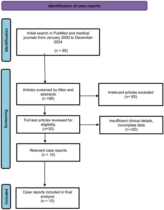
The search strategy identified 107 records across the two databases. After removal of duplicates, 95 titles and abstracts were screened for relevance. Of them, 65 articles were excluded because they did not describe vascular malformations of the female genital tract or did not provide sufficient clinical information. A total of 30 full-text articles were assessed for eligibility, of which only 10 case reports or case series describing vulvar or genital vascular malformations were included in the qualitative synthesis.
Because clinical management principles for vulvar lesions are derived from the broader vascular anomaly literature, additional searches were performed for diagnostic imaging, classification systems, and treatment strategies of peripheral vascular malformations regardless of the anatomical location. Reference lists of relevant articles and review papers were manually screened to identify additional eligible publications.
Because published evidence on vulvovaginal vascular malformations is limited primarily to case reports and small case series, a structured narrative review approach was selected. The aim was to integrate available clinical, imaging, and interventional literature rather than to perform quantitative synthesis. Publications were included if they (1) described vascular malformations involving the vulva, vagina, or perineum; or (2) provided clinically relevant information on classification, imaging characteristics, pathophysiology, or treatment of vascular malformations applicable to genital tract lesions.
Eligible study types included case reports, case series, cohort studies, clinical reviews, imaging studies, and consensus statements. Articles not available in English, conference abstracts without sufficient clinical data, and publications describing vascular tumors (e.g., infantile hemangioma) without relevance to malformations were excluded.
After removal of duplicates, titles and abstracts were screened for relevance. Full-text review was performed for potentially eligible articles. The study selection process is illustrated in Figure 1. Reports specifically describing vulvar vascular malformations or related vascular anomalies were extracted for clinical comparison. Data collected included patient age, clinical presentation, diagnostic approach, imaging modality, treatment strategy, lesion size when available, and reported follow-up or recurrence. The identified cases are summarized in Table 1 to facilitate comparison of diagnostic pathways and therapeutic outcomes reported in the literature.
Figure 1.
Literature review flowchart for vulvar vascular malformations.
Table 1.
Reported cases of vulvar and genital vascular malformations [6,7,14,15,16,17,18,19,20,21].
3. Pathophysiology and Molecular Basis of Vascular Malformations
Vascular malformations arise from errors in embryologic vascular development and are now recognized as disorders of dysregulated angiogenic signaling rather than purely structural anomalies. During normal vasculogenesis and angiogenesis, endothelial cells, pericytes, and smooth-muscle cells undergo coordinated maturation regulated by several signaling pathways, particularly the phosphatidylinositol-3-kinase (PI3K)–AKT–mTOR and TIE2/TEK receptor pathways. Somatic activating mutations affecting these pathways have been identified in a substantial proportion of venous and combined vascular malformations [22,23,24].
Venous malformations are most frequently associated with somatic mutations in the TEK (TIE2) gene, which encodes an endothelial tyrosine kinase receptor essential for vascular maturation and stability. Activating TEK mutations lead to persistent PI3K-AKT signaling, resulting in abnormal venous channel dilation, defective smooth-muscle recruitment, and impaired vessel wall integrity [23,25]. As a consequence, the affected vessels demonstrate ectasia, slow blood flow, and a propensity for localized intravascular thrombosis and phlebolith formation. These lesions are present at birth but may remain clinically inapparent until adolescence or adulthood.
A subset of venous and combined malformations instead harbors somatic mutations in PIK3CA, a gene encoding the catalytic subunit of PI3K. These mutations produce the constitutive activation of the PI3K-AKT-mTOR pathway, leading to endothelial cell survival, abnormal vascular growth, and the progressive enlargement of the lesion [24,26]. PIK3CA-related malformations are also associated with overgrowth syndromes within the PIK3CA-related overgrowth spectrum (PROS), including Klippel–Trénaunay syndrome and CLOVES syndrome [26].
The biological behavior of vascular malformations differs fundamentally from vascular tumors. Infantile hemangiomas demonstrate endothelial proliferation followed by involution, whereas vascular malformations consist of mature but structurally abnormal vessels with a normal endothelial turnover [22,27]. Histologically, venous malformations are composed of dilated vascular channels lined by a single layer of flat endothelium, often with deficient smooth-muscle support and fibrotic stroma, findings consistent with the present case.
Hormonal influences are clinically important. Estrogen receptors (ERs) have been demonstrated in some vascular malformations and lesions may enlarge during puberty, pregnancy, or as a result of hormone therapy use [28,29]. Increased venous pressure, hypervolemia, and hormonal vascular relaxation during pregnancy may exacerbate venous ectasia and thrombosis, explaining the late clinical presentation in previously asymptomatic patients. Thrombotic events within low-flow malformations may produce sudden pain and swelling, commonly prompting clinical suspicion of infection or abscess formation.
Low-flow venous malformations therefore behave as chronic hemodynamic lesions rather than neoplastic processes. A slow flow can lead to intravascular coagulopathy characterized by fibrin deposition and elevated D-dimer levels, which may contribute to pain and progressive enlargement [30]. High-flow AVMs differ substantially, as they contain direct arterial–venous shunts that bypass the capillary bed, producing tissue ischemia, ulceration, and, in extensive lesions, high-output cardiac failure [31].
4. Clinical Classification of Vulvovaginal Vascular Malformations
The correct classification of vascular anomalies is essential because diagnostic evaluation and treatment depend primarily on lesion flow characteristics rather than anatomical location. The ISSVA classification distinguishes vascular tumors from vascular malformations and further categorizes malformations into low-flow and high-flow lesions [1,4,32].
Vascular tumors, such as infantile hemangioma, are characterized by endothelial proliferation and a predictable growth and involution phase. In contrast, vascular malformations are structural abnormalities of mature vessels, present at birth and growing proportionally with the patient without spontaneous regression [1,2,27,33].
4.1. Low-Flow Malformations
Low-flow malformations include capillary, venous, and lymphatic malformations and represent the majority of vascular anomalies encountered in the female genital tract [6,7,30]. Venous malformations are the most frequent subtype affecting the vulva. Clinically, they typically present as soft, compressible bluish lesions that enlarge with dependency or the Valsalva maneuver and may become painful due to localized thrombosis [10,30,33].
Lymphatic malformations may present as clusters of translucent vesicles or diffuse swelling and are prone to recurrent leakage or infection [1,33]. Capillary malformations are uncommon in the vulva but may be associated with combined malformation syndromes [1].
4.2. High-Flow Malformations
High-flow malformations include AVMs and arteriovenous fistulas. These lesions contain direct arterial-to-venous shunts without an intervening capillary bed, resulting in pulsation, warmth, bruit, and a higher risk of hemorrhage [31,32,33,34]. In the vulvar region, AVMs are rare but clinically significant because surgical excision without prior vascular control may cause severe bleeding [6,34].
4.3. Combined and Syndromic Lesions
Some patients present with combined malformations or lesions associated with systemic syndromes such as Klippel–Trénaunay syndrome or other PIK3CA-related overgrowth spectrum disorders [1,26]. The identification of syndromic disease is important because lesions may extend into the pelvis or lower extremities and require multidisciplinary management and imaging beyond the genital tract [30,33].
Because clinical appearance alone is often misleading, classification requires integration of history, physical examination, and imaging findings, particularly Doppler ultrasonography and MRI [11,13,33]. Determining whether a lesion is low-flow or high-flow is the key step guiding treatment selection. Table 2 summarizes these findings.
Table 2.
ISSVA-based classification and clinical features of vulvovaginal vascular malformations.
5. Imaging Evaluation and Diagnostic Approach
Clinical examination alone is insufficient to reliably differentiate vascular malformations from common vulvar lesions. Although a Bartholin’s gland cyst or abscess is typically fluctuant and localized to the posterolateral vestibule, vascular malformations may present with similar swelling, tenderness, and apparent inflammatory change, particularly when thrombosed [8,10]. Misidentification may lead to incision, biopsy, or marsupialization, procedures that can result in significant hemorrhage and incomplete treatment [6,9].
5.1. First-Line Evaluation: Doppler Ultrasonography
An ultrasound with color Doppler is the recommended initial imaging modality for suspected vascular malformations because it is widely available, noninvasive, and capable of assessing flow dynamics [11,13,33,35,36,37,38]. Vascular malformations are broadly classified into low-flow and high-flow lesions according to the ISSVA classification [3]. Low-flow malformations include venous, lymphatic, and capillary malformations, whereas high-flow malformations include arteriovenous malformations and arteriovenous fistulas [3,4]. Low-flow venous malformations typically appear as compressible, hypoechoic, or heterogeneous lesions with a slow venous flow, the absence of arterial waveforms, or even an absent detectable flow. Phleboliths, when present, are highly suggestive and appear as echogenic foci with posterior acoustic shadowing [11,33].
In contrast, high-flow AVMs demonstrate arterialized waveforms with a high peak systolic velocity (PSV), low-resistance waveforms, and, in some cases, direct evidence of arteriovenous shunting on Doppler examination [13,34,39,40,41].
Although there is no universally accepted PSV cutoff, studies suggest that low-flow lesions generally exhibit PSV values < 20–30 cm/s, whereas high-flow lesions demonstrate significantly higher velocities, often exceeding 50–100 cm/s, depending on the degree of arteriovenous shunting [4,13,33,34,40].
These Doppler characteristics are critical for guiding further imaging and treatment planning, particularly in distinguishing lesions that may require endovascular management.
5.2. Second-Line Imaging: MRI
MRI is the imaging modality of choice for defining the lesion extent, tissue infiltration, and involvement of adjacent pelvic structures [11,12,13,39,40,41,42]. Venous malformations are characteristically hyperintense on T2-weighted sequences and enhanced gradually following contrast administration. MRI can also identify intralesional thrombosis and distinguish localized from infiltrative lesions [11,22,33,39,40,41,43].
MRI is particularly important in the vulvar region because lesions may extend into the perineum, pelvic floor, or vagina without clear clinical evidence. Preoperative MRI therefore assists in surgical planning and in determining whether referral for interventional radiology treatment is appropriate.
5.3. Angiography and Interventional Planning
Digital subtraction angiography is not required for the diagnosis of low-flow lesions but is essential for the evaluation of high-flow AVMs when embolization is considered [31,34]. Angiography allows the identification of feeding arteries and venous drainage patterns and is typically performed immediately before therapeutic embolization [44,45,46].
5.4. Suggested Diagnostic Algorithm
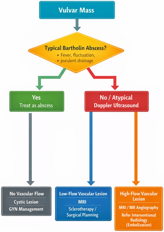
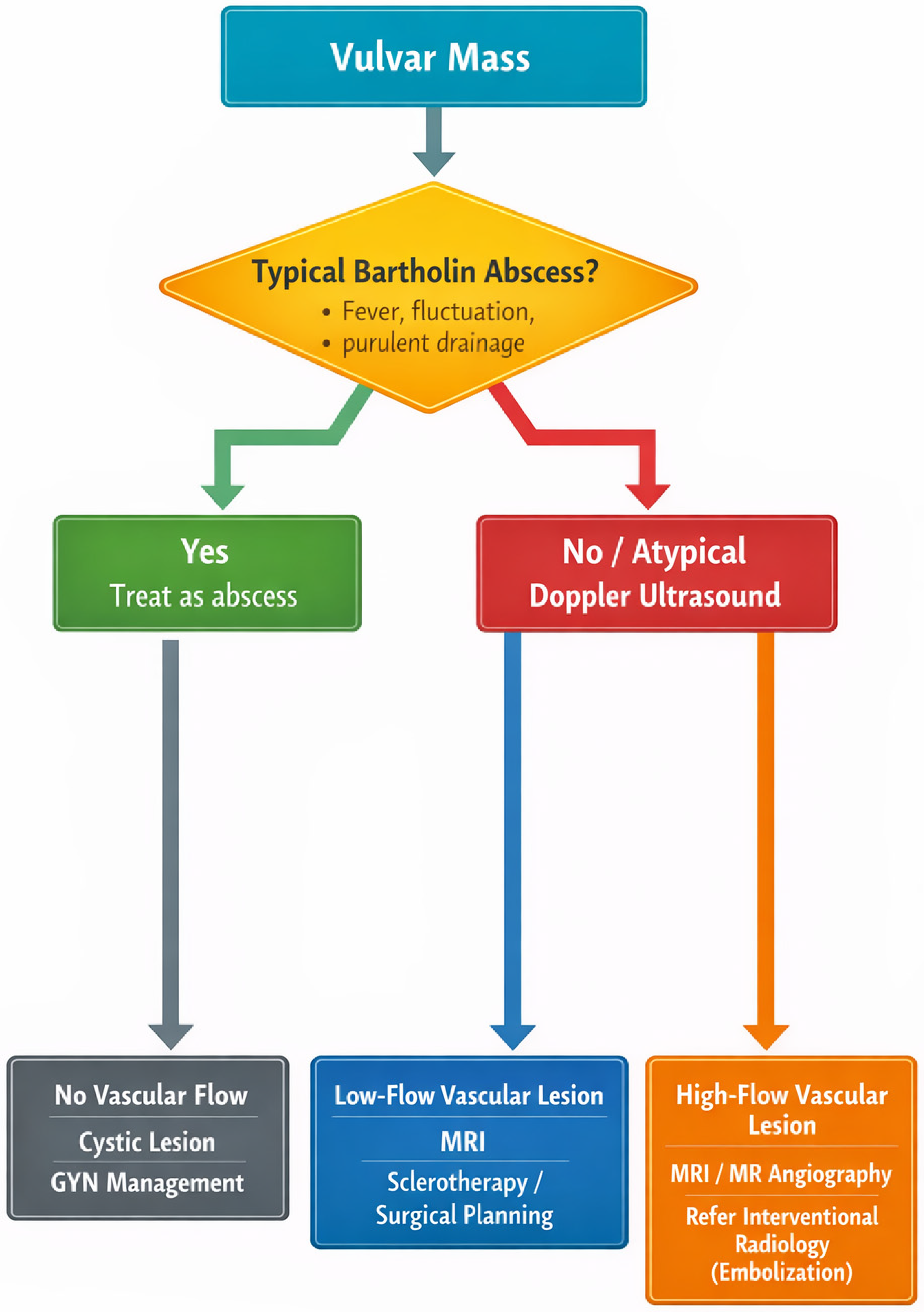
Based on the current evidence, the evaluation of an atypical vulvar mass should follow a structured diagnostic pathway (Figure 2). Clinical features suggesting a vascular lesion include compressibility, bluish discoloration, enlargement with Valsalva maneuver, absence of purulent drainage, and recurrence after prior incision [6,10].
Figure 2.
Diagnostic algorithm for evaluation of an atypical vulvar mass.
The proposed diagnostic algorithm emphasizes the importance of early imaging in atypical vulvar masses. Lesions clinically consistent with a simple Bartholin’s gland cyst or abscess may be managed conventionally. However, the presence of atypical features such as compressibility, a bluish discoloration, recurrent swelling, or the absence of purulent drainage should prompt Doppler ultrasonography prior to surgical treatment. MRI is recommended when a vascular lesion is suspected to define the lesion extent and flow characteristics.
6. Histopathological Evaluation and Differential Diagnosis
Histopathological analysis plays a central role in confirming the diagnosis of vascular malformations, particularly when the clinical and imaging findings are inconclusive. Unlike vascular tumors, vascular malformations consist of mature but structurally abnormal vessels and lack endothelial proliferation [1,2,3,32].
6.1. Venous Malformations
Venous malformations are composed of dilated, irregular vascular channels lined by a single layer of flattened endothelium. Vessel walls are typically thin but may contain variably developed smooth muscle. The surrounding stroma often shows fibrosis and evidence of previous thrombosis. Phleboliths may be present due to chronic slow-flow stasis [30,47,48].
Immunohistochemically, endothelial cells are positive for CD31 and CD34, while smooth muscle markers such as desmin and smooth muscle actin highlight the vessel walls. Importantly, cellular atypia and mitotic activity are absent, distinguishing venous malformations from vascular neoplasms. ER expression has been reported in some lesions and may explain hormonal responsiveness [22,27,49].
6.2. Arteriovenous Malformations
AVMs demonstrate dysplastic arteries and veins connected directly without an intervening capillary bed. Histology reveals thickened arterial walls and irregular vascular channels. Elastic lamina staining may help identify arterial components. These lesions correlate with high-flow findings on imaging and require different treatment strategies [10,33,34].
6.3. Lymphatic Malformations
Lymphatic malformations consist of dilated lymphatic channels lined by a thin endothelium and containing proteinaceous fluid rather than blood. Immunostaining with D2-40 (podoplanin) is helpful in confirming lymphatic origin [34].
6.4. Distinction from Vascular Tumors
The most important histologic differential diagnosis is hemangioma. Infantile hemangiomas show endothelial proliferation and increased mitotic activity and are positive for GLUT-1, a marker absent in vascular malformations [34,49]. This distinction is clinically relevant because hemangiomas may regress spontaneously, whereas malformations persist and often enlarge.
6.5. Mesenchymal Tumors and Other Mimics
Aggressive angiomyxoma and other vulvar mesenchymal tumors may clinically mimic vascular lesions. These tumors demonstrate hypocellular myxoid stroma with spindle cells and characteristic infiltrative margins rather than true vascular channels [34]. Sarcomas show cellular atypia, mitotic figures, and destructive growth patterns.
Histopathology is therefore particularly important when intraoperative findings are unexpected or when a lesion presumed to be cystic proves to be solid, as has occurred in the present case. The correlation of histologic features with imaging findings provides the most accurate diagnosis and guides appropriate management.
7. Differential Diagnosis of Vulvar Vascular Malformations
Vulvar masses are common in gynecologic practice and the majority represent benign conditions such as Bartholin’s gland cysts, abscesses, or vulvar varicosities. Because vascular malformations are rare and frequently painless, they are often not considered initially, leading to misdiagnosis and inappropriate surgical intervention [6,8]. The risk of diagnostic error is the greatest when lesions present acutely with tenderness or enlargement due to thrombosis, which may clinically mimic infection. Clinical differentiation of vulvar masses is presented in Table 3.
7.1. Bartholin’s Gland Cyst and Abscess
Bartholin’s gland pathology is the most frequent cause of a posterolateral vulvar mass. Cysts typically present as fluctuant, well-localized swellings near the vaginal introitus [50]. When infected, abscesses are characterized by severe pain, erythema, fever, and purulent drainage [7,51]. In contrast, vascular malformations are compressible and may enlarge with dependency or the Valsalva maneuver. Pain, if present, is usually mild and related to thrombosis rather than infection [10,30]. The incision of a vascular malformation may result in unexpected bleeding and the absence of purulent material, as observed in the present case.
7.2. Vulvar Varicosities
Vulvar varicosities occur particularly during pregnancy and are associated with venous congestion and pelvic venous insufficiency. Unlike venous malformations, varicosities represent dilated normal veins rather than developmental anomalies. They typically appear as multiple serpiginous superficial vessels and often regress postpartum [10]. Doppler ultrasonography demonstrates venous flow connected to the pelvic venous system, whereas venous malformations form localized venous lakes with a slow or stagnant flow [11,33].
7.3. Vascular Tumors (Hemangioma)
Hemangiomas are vascular tumors characterized by endothelial proliferation, most commonly occurring in infancy. They exhibit a proliferative phase followed by spontaneous involution [27]. In adults, true hemangiomas of the vulva are uncommon and histologically distinct from venous malformations, which lack endothelial hyperplasia and consist of mature ectatic vessels [22,27].
7.4. Aggressive Angiomyxoma and Mesenchymal Tumors
Aggressive angiomyxoma and other mesenchymal tumors should also be considered. These lesions typically present as slow-growing deep perineal masses and may extend into the pelvis. MRI demonstrates infiltrative margins rather than well-defined vascular channels [51]. A histopathologic evaluation is necessary when imaging is inconclusive.
7.5. Malignancy
Although rare, vulvar malignancies such as sarcoma or melanoma may occasionally mimic benign masses. Suspicion should be raised in the presence of rapid growth, ulceration, pigmentation, or lymphadenopathy. Unlike vascular malformations, malignant lesions are generally firm and non-compressible and lack characteristic vascular imaging features [51].
Because clinical examination alone is often insufficient, imaging—particularly Doppler ultrasonography—should be performed when the findings are atypical or when purulent drainage is absent. The recognition of these distinguishing features can prevent unnecessary surgical procedures and reduce morbidity.
Table 3.
Clinical differentiation of vulvar masses.
8. Management of Vulvovaginal Vascular Malformations
The management of vascular malformations depends primarily on lesion flow characteristics, symptom severity, and anatomical extent [52,53]. Not all lesions require treatment; asymptomatic and stable malformations may be managed conservatively with observation and patient education [10,30].
8.1. Conservative Management
Observation is appropriate for small, asymptomatic venous malformations. Patients should be counseled regarding potential enlargement, thrombosis, and bleeding risk. Compression therapy may reduce symptoms in selected lesions, particularly those associated with venous congestion [10,30]. Analgesics and anti-inflammatory medication may be used for pain related to localized thrombosis.
8.2. Sclerotherapy
Sclerotherapy is considered the first-line treatment for most symptomatic low-flow venous malformations [10,33,54,55,56,57]. The procedure involves the percutaneous injection of a sclerosing agent to induce endothelial damage, thrombosis, and fibrosis, resulting in volume reduction of the lesion. Commonly used agents include ethanol, polidocanol, and sodium tetradecyl sulfate [33,54,58,59].
Multiple treatment sessions are often required. Complications may include localized necrosis, nerve injury, and skin ulceration, particularly in superficial lesions. However, outcomes are generally favorable and recurrence rates are lower than with surgery alone [31,43]. Pre-procedural imaging is essential to delineate lesion boundaries and to avoid damage to adjacent structures.
8.3. Laser Therapy
Superficial capillary and some lymphatic malformations may respond to laser therapy, including pulsed dye and Nd:YAG lasers. Laser treatment is particularly useful for cutaneous lesions causing bleeding or cosmetic concern [33]. However, deeper venous malformations are less responsive and typically require sclerotherapy.
8.4. Surgical Excision
Surgical excision may be considered for localized lesions causing significant symptoms or functional impairment. Complete excision is often difficult due to ill-defined margins and infiltration into the surrounding tissue. Surgery without prior imaging or vascular control is associated with a substantial risk of bleeding and recurrence [6,9]. Therefore, excision is typically performed after preoperative imaging and in selected cases, following preoperative sclerotherapy or embolization. In selected cases, particularly in high-flow AVMs or large, highly vascularized lesions, preoperative embolization plays an important role in reducing intraoperative blood loss and facilitating surgical resection. Embolization may be used as a standalone therapy or as an adjunct to surgery, depending on lesion size, location, and flow characteristics.
For low-flow venous malformations, sclerotherapy is often preferred as the first-line treatment, while surgical excision is reserved for well-circumscribed lesions or cases refractory to minimally invasive approaches.
The present case illustrates a typical diagnostic pitfall: the presumed Bartholin’s gland pathology led to incision before a vascular origin was recognized. Planned surgical removal after imaging evaluation would reduce procedural risk.
8.5. Management of High-Flow Arteriovenous Malformations
High-flow AVMs require a fundamentally different approach. Endovascular embolization is the primary therapy and is frequently performed in staged procedures [31,34,54,60,61,62,63]. Surgical excision alone is contraindicated in most cases because uncontrolled bleeding and rapid recurrence may occur if feeding vessels are not occluded [30]. In contrast to low-flow malformations, high-flow lesions typically require a combined approach, with preoperative embolization followed by surgical excision to achieve durable control and minimize recurrence. Therefore, the accurate preoperative classification of the lesion is essential in determining the role of embolization and optimizing the treatment strategy.
8.6. Multidisciplinary Care and Management of Malignancies
The management of vulvar masses requires a structured and multidisciplinary approach, particularly when malignancy is suspected. Although benign entities such as Bartholin’s cysts or vascular malformations are more common, malignant lesions—including vulvar carcinoma, melanoma, sarcoma, and metastatic disease—must always be considered in the differential diagnosis.
Clinical features that should raise suspicion for malignancy include rapid growth, irregular borders, ulceration, bleeding, fixation to underlying structures, and associated lymphadenopathy. In such cases, imaging—preferably MRI—should be performed prior to any surgical intervention to assess the local extent and involvement of adjacent structures [30,33].
A biopsy is essential for a definitive diagnosis and should be performed prior to definitive treatment in all suspicious lesions. Importantly, inappropriate surgical intervention without prior imaging or biopsy may lead to incomplete excision, tumor dissemination, or significant bleeding, particularly in vascular tumors or malignancies with high vascularity [10].
Management should be guided by a histopathological diagnosis and may include wide local excision, sentinel lymph node assessment, or multimodal oncologic treatment. Collaboration with gynecologic oncology, radiology, and vascular specialists is often required to ensure optimal outcomes [10,30,33].
9. Hormonal Influences and Pregnancy Considerations
Hormonal factors play a significant role in the clinical behavior of vascular malformations. Although present at birth, many lesions remain asymptomatic until adolescence or adulthood, when hormonal changes and hemodynamic stress may precipitate enlargement or symptoms [28,29]. Estrogen-mediated vasodilation, increased venous capacitance, and alterations in vascular smooth-muscle tone are believed to contribute to progressive venous ectasia in susceptible vessels [28,64,65].
9.1. Puberty and Hormonal Therapy
Clinical progression is frequently observed during puberty, when increased estrogen levels stimulate vascular relaxation and increased blood flow [28]. The enlargement of venous malformations has also been reported in association with exogenous hormonal therapy, including estrogen-containing contraceptives and hormone replacement therapy. While data are limited, careful monitoring is recommended in patients with known symptomatic lesions.
9.2. Pregnancy
Pregnancy represents the most important physiologic condition affecting vulvovaginal vascular malformations. Physiologic hypervolemia, increased cardiac output, and venous obstruction by the gravid uterus elevate the pelvic venous pressure and may lead to the rapid enlargement of venous malformations [10,30]. Progesterone-mediated venous dilation further contributes to vascular distensibility [31,66,67].
Patients may present with new-onset vulvar swelling, pain, or thrombosis during pregnancy. Lesions previously asymptomatic may become clinically apparent. Thrombosis within low-flow malformations may cause acute tenderness and may mimic infection or abscess formation.
9.3. Risk of Hemorrhage and Delivery Considerations
Large vulvar or vaginal malformations pose a potential risk of hemorrhage during vaginal delivery, particularly if the lesion is traumatized [29,68]. However, cesarean delivery is not routinely required. The mode of delivery should be individualized based on lesion size, location, and bleeding risk. Multidisciplinary planning with obstetrics and interventional radiology may be indicated in extensive lesions.
In selected cases, prepartum embolization or sclerotherapy has been reported to reduce bleeding risk when significant vascular involvement is identified [29,68]. For small, superficial lesions, conservative management and careful intrapartum monitoring are usually sufficient.
9.4. Thrombosis and Coagulopathy
Low-flow venous malformations are associated with localized intravascular coagulopathy due to slow blood flow and fibrin deposition [30]. Pregnancy further increases the risk of thrombosis because of the hypercoagulable state. Patients presenting with acute pain and swelling should be evaluated for thrombosis rather than infection alone [30,69,70,71,72,73].
The recognition of these physiologic effects is essential, as pregnancy-related enlargement or thrombosis may lead to misdiagnosis and inappropriate surgical intervention.
10. Illustrative Case
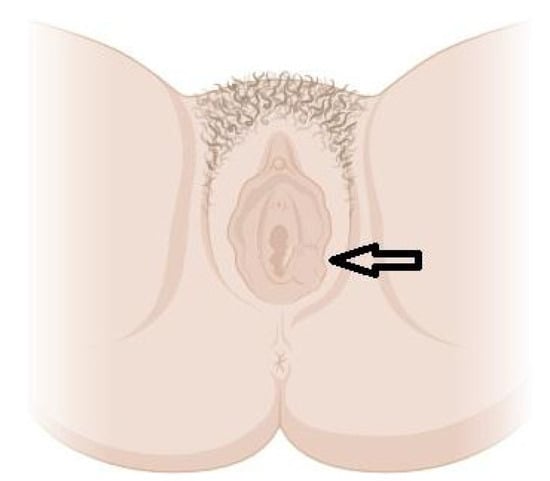
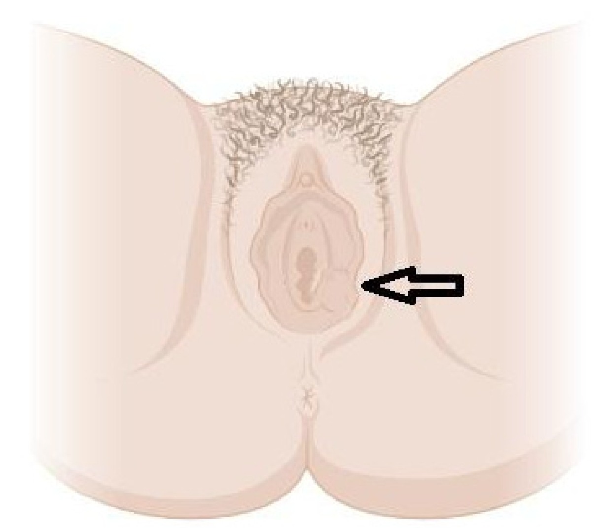
A 48-year-old woman presented to the emergency gynecological clinic with a tender mass near the vaginal introitus that had been progressively enlarging over three days (Figure 3). She reported localized discomfort but no fever, systemic symptoms, urinary complaints, or preceding trauma. Her medical history was unremarkable apart from a prior laparoscopic supracervical hysterectomy for uterine leiomyoma. She had two prior term deliveries.
Figure 3.
An illustration of the lesion localization on the vulva, created based on the clinical case. The arrow shows the location of the lesion.
A physical examination revealed a 4-cm well-circumscribed swelling of the left labium majus. The lesion was mobile and tender with mild overlying erythema, and was clinically interpreted as a Bartholin’s gland abscess.
No preoperative imaging was performed because the lesion was clinically interpreted as a Bartholin’s gland abscess in the emergency setting. Consequently, Doppler ultrasonography or MRI were not obtained prior to surgical intervention. The absence of imaging reflects a common diagnostic pathway described in the literature, where vascular malformations are initially treated as Bartholin gland pathology.
The patient underwent operative incision and attempted marsupialization under short general anesthesia. No purulent material was encountered; instead, a solid encapsulated mass was identified and completely excised. Intraoperative blood loss was minimal and postoperative recovery was uncomplicated. The patient has been followed for 12 months after surgery so far. No evidence of recurrence or new symptoms has been observed to date.
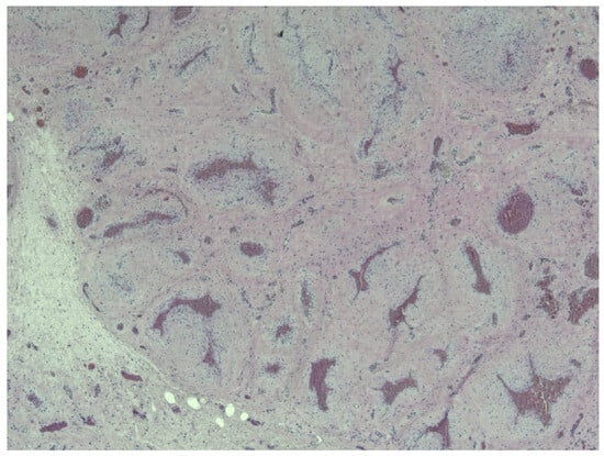
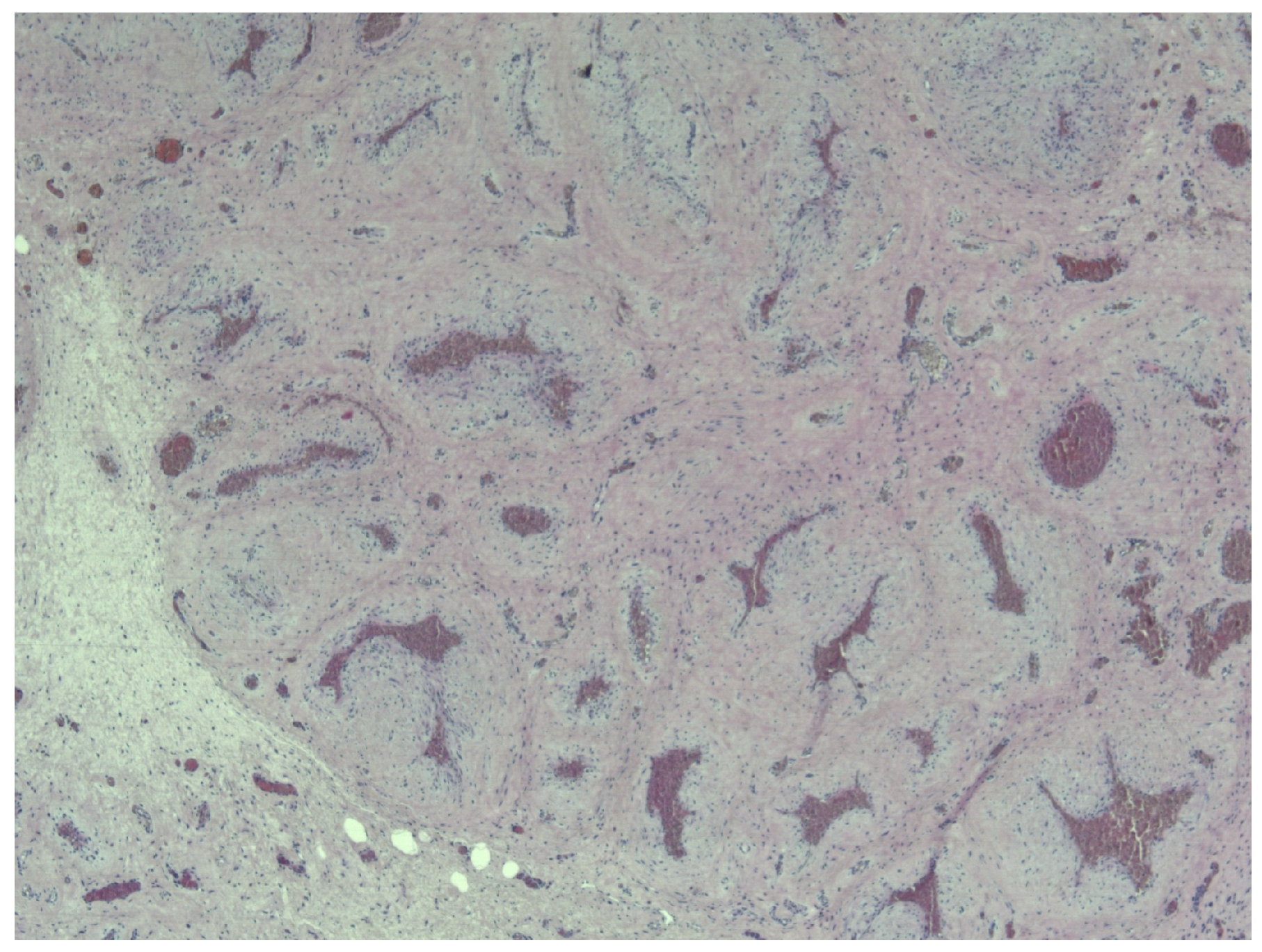
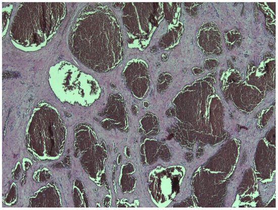
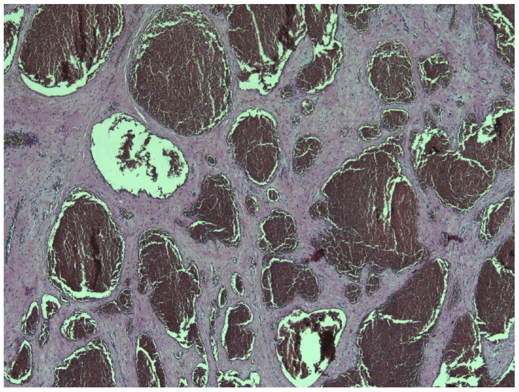
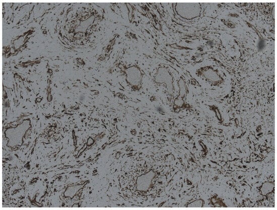
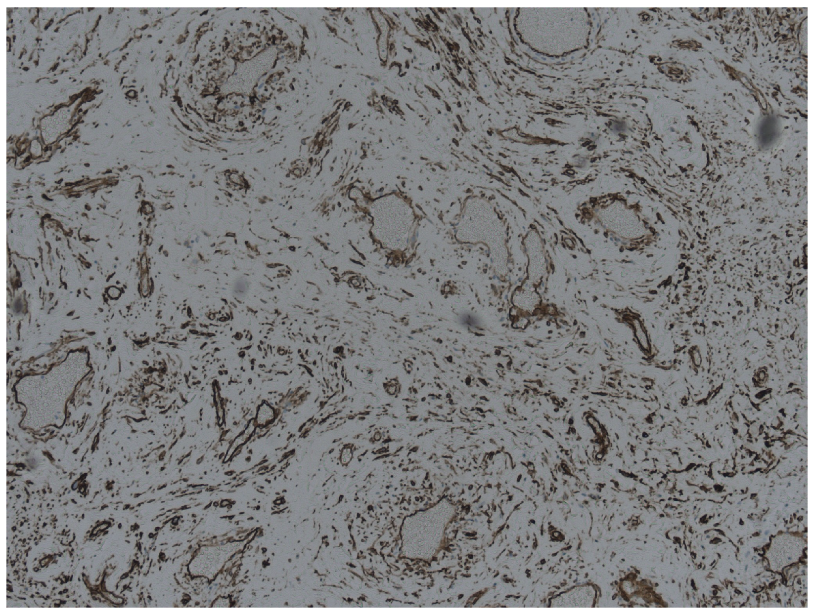
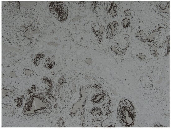
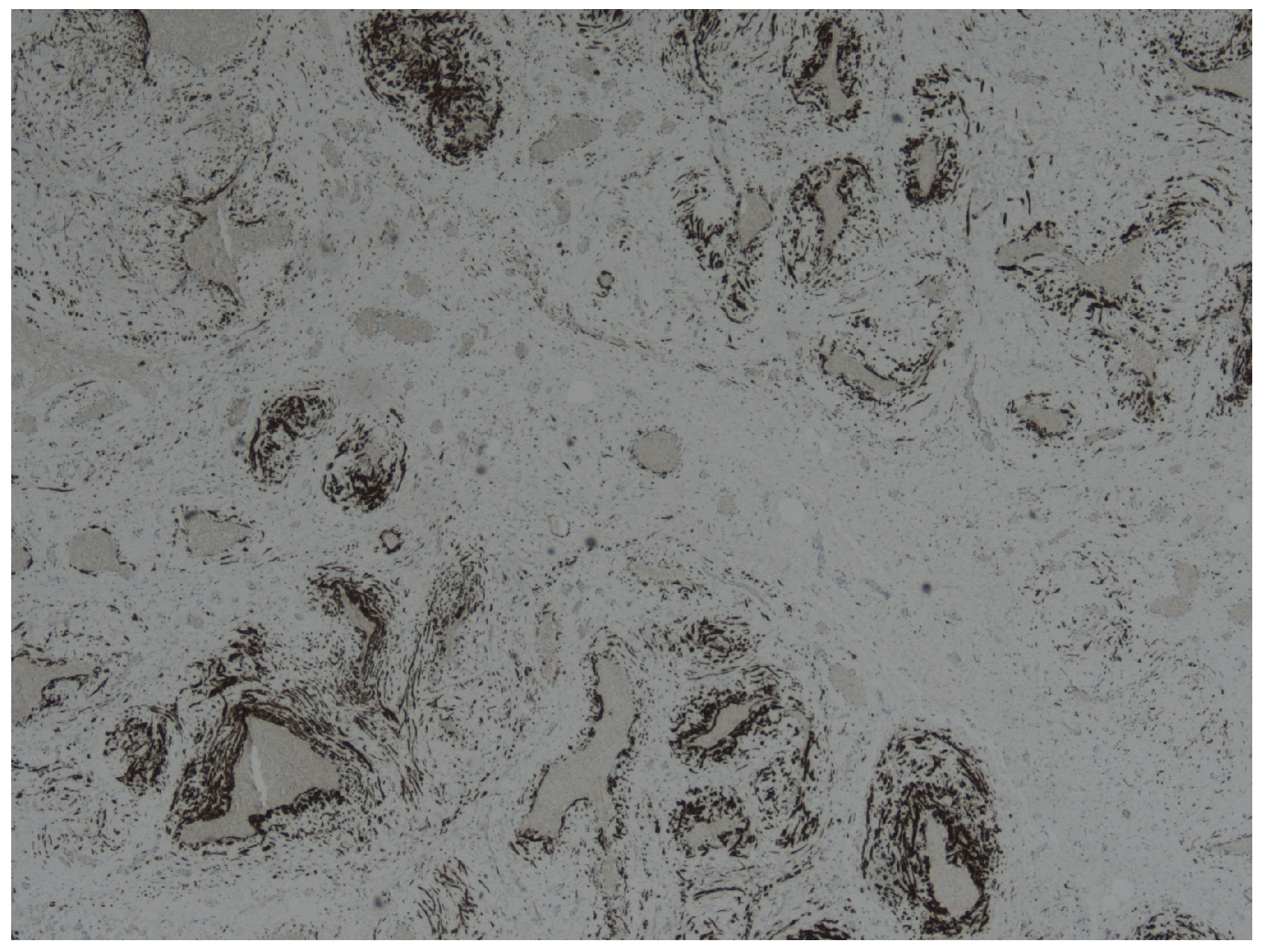
A histopathological examination of the tissue removed from the patient revealed the following: the lesion was a 4 × 2.5 × 2 cm mass of fibrotic tissue with evidence of old hemorrhages. On the cut surface, several blood vessels with focal thromboses were discernible. On a microscopic examination, the lesion was composed of variably cellular fibrotic tissue with unassuming spindled cells, focally with some degenerative atypia. In between, there were numerous vascular spaces filled with blood that were predominantly composed of muscularized blood vessels (Figure 4). In some areas, the vascular spaces were cavernous (Figure 5). Endothelial cells of the vascular spaces were positive for endothelial markers. The spindled cells in the surrounding stroma were positive for immunohistochemical markers CD34 (Figure 6), desmin (Figure 7), and ER, and negative for S100. There was no convincing loss of the expression of Rb. Given the rarity of this finding, the tumor sample was sent for a consultative review to international consultation. Additionally, the tumor sample was subjected to molecular genetic testing.
Figure 4.
A conglomerate of variably sized blood vessels with thick muscular walls and aberrant wall layer structure (HE, original magnification 4×).
Figure 5.
Area of the lesion with more cavernous-like vessel spaces without the muscular walls (HE, original magnification 4×).
Figure 6.
Immunohistochemical reaction for CD34 shows the endothelium as well as positivity in the stromal cells (CD34 IHC, original magnification 4×).
Figure 7.
Immunohistochemical reaction for desmin highlights the smooth muscles in the vessel walls (desmin IHC, original magnification 4×).
It concurred with the diagnosis of a vascular formation and found no evidence of other mesenchymal tumors. Additionally, molecular genetic testing did not reveal any mutations typically associated with other mesenchymal tumors of the lower gynecological tract.
A retrospective review of the case identified several atypical features for Bartholin’s gland pathology, including the absence of purulent drainage, the presence of a solid mass, and relatively mild pain. The case illustrates the diagnostic challenge of vulvar vascular malformations and highlights the importance of preoperative imaging before surgical intervention in atypical vulvar lesions.
11. Discussion
Vulvovaginal vascular malformations are uncommon but clinically significant because they frequently mimic common benign gynecologic conditions. The available literature is dominated by isolated case reports, and a consistent finding across publications is the initial misdiagnosis as Bartholin’s gland cyst or abscess [6,7,8]. This diagnostic error is understandable: both conditions present as localized swelling in the posterolateral vulva and may be associated with tenderness. However, the consequences differ substantially. The incision of a Bartholin abscess is therapeutic, whereas the incision of a vascular malformation may lead to bleeding, incomplete treatment, and recurrence [6,9].
The principal reason for a misdiagnosis is the reliance on clinical examination alone. Vulvar lesions are often evaluated in emergency or outpatient settings where imaging is not routinely performed before surgical treatment. Several reported cases, including the present patient, underwent an operative intervention before the recognition of the vascular nature of the lesion. A review of the published reports shows that preoperative imaging was frequently omitted, particularly when symptoms were acute.
The key clinical distinction is not the anatomical location but the hemodynamic behavior of the lesion. Vascular malformations are disorders of abnormal vessels rather than obstructed glands. Compressibility, bluish discoloration, enlargement with the Valsalva maneuver, and the absence of purulent drainage are important clues suggesting a vascular origin [10,30]. Acute pain may occur due to thrombosis within a low-flow venous malformation and may be misinterpreted as infection. Doppler ultrasonography is therefore the critical first diagnostic step when the findings are atypical.
Our case demonstrates a common scenario: a lesion clinically interpreted as a Bartholin abscess but intraoperatively found to be solid. A histopathologic evaluation confirmed a venous malformation, consistent with the typical structure of ectatic vascular channels lacking endothelial proliferation. Similar diagnostic pathways have been described in previously reported vulvar vascular malformations [6,7]. The case emphasizes that the absence of purulent material at the incision should immediately raise suspicion for an alternative diagnosis.
Management strategies differ markedly according to the lesion type. Low-flow venous malformations are generally best treated with sclerotherapy or carefully planned excision, whereas high-flow AVMs require embolization prior to any surgical intervention [31,34]. Surgery performed without a preoperative vascular assessment carries significant risk and may not be curative due to infiltrative margins. Consequently, imaging evaluation should precede operative management in any atypical vulvar mass. Hormonal and hemodynamic influences further complicate diagnosis. Enlargement during puberty or pregnancy and thrombosis-related pain may prompt acute clinical presentation, particularly in previously unrecognized lesions [28,29,30]. The recognition of these patterns can help clinicians avoid unnecessary surgical procedures.
Misdiagnosis appears to be common in the published literature. Several reported cases describe the initial treatment as a Bartholin’s gland cyst or abscess before the vascular origin of the lesion was recognized. This likely reflects the rarity of vulvar vascular malformations and the frequent reliance on clinical examination alone in emergency settings. Imaging plays a critical role in preventing this error. Doppler ultrasonography has been reported to reliably differentiate low-flow from high-flow lesions and is widely recommended as the initial diagnostic modality, while MRI provides a superior assessment of the lesion extent and tissue infiltration.
Treatment outcomes vary depending on the lesion type and therapeutic approach. Low-flow venous malformations generally respond well to sclerotherapy, often requiring multiple treatment sessions but demonstrating good symptom control and low recurrence rates. Surgical excision may be effective for localized lesions but carries a higher risk of recurrence if the margins are incomplete. High-flow arteriovenous malformations require staged embolization and multidisciplinary management because surgery alone is associated with rapid recurrence and a significant bleeding risk.
Based on the literature and our clinical experience, a practical approach is recommended: lesions typical for a simple Bartholin cyst or abscess may be managed conventionally, but any atypical features—including compressibility, unusual firmness, recurrence, or unexpected intraoperative findings—should prompt Doppler ultrasonography before further intervention.
12. Clinical Practice Recommendations
When to suspect a vascular malformation in a vulvar mass:
- Compressible lesion;
- Bluish discoloration;
- Enlargement with Valsalva maneuver or dependency;
- Mild or intermittent pain;
- Recurrence after previous incision;
- Absence of purulent drainage.
Recommended diagnostic approach:
- Doppler ultrasonography before surgical treatment in atypical lesions;
- MRI when vascular origin is suspected;
- Angiography only for high-flow lesions or pre-embolization planning.
Management principles:
- Avoid incision or biopsy without prior imaging;
- Low-flow venous malformations → sclerotherapy or planned excision;
- High-flow lesions → embolization and multidisciplinary management;
- Consider referral to a vascular anomaly center.
13. Conclusions
Vulvovaginal vascular malformations are rare but important causes of vulvar masses and are frequently misdiagnosed as Bartholin’s gland pathology. The primary clinical risk is inappropriate surgical intervention without a prior vascular evaluation. Doppler ultrasonography followed by MRI allows for accurate classification and guides management.
Low-flow venous malformations are usually managed with sclerotherapy or planned excision, whereas high-flow AVMs require embolization and multidisciplinary care. The awareness of characteristic clinical features and appropriate use of imaging can prevent complications and improve outcomes.
The presented case illustrates a typical diagnostic pitfall and supports the need for a structured diagnostic approach to atypical vulvar lesions.
Author Contributions
Conceptualization, M.J.; methodology, M.B. and M.J.; investigation, M.B.; data curation, M.B.; writing—original draft preparation, M.B. and J.M.; writing—review and editing, M.J., K.D.S., and J.M.; supervision, M.J. All authors have read and agreed to the published version of the manuscript.
Funding
This research received no external funding.
Institutional Review Board Statement
This study was not subject to Institutional Review Board approval in accordance with institutional policy for case reports and reviews of literature.
Informed Consent Statement
Written and oral informed consent has been obtained from the patient to publish this paper, including the case and accompanying images.
Data Availability Statement
The data presented in this study are available from the corresponding author upon reasonable request. Data are not publicly available due to patient privacy and ethical restrictions.
Conflicts of Interest
The authors declare no conflicts of interest.
Abbreviations
The following abbreviations are used in this manuscript:
| AVM | Arteriovenous Malformation |
| CD31 | Cluster of Differentiation 31 (an endothelial cell marker) |
| CD34 | Cluster of Differentiation 34 (an endothelial cell marker) |
| CT | Computed Tomography |
| D2-40 | Podoplanin (immunohistochemical marker of lymphatic endothelium) |
| ER | Estrogen receptor |
| GLUT-1 | Glucose transporter protein-1 (marker characteristic of infantile hemangioma) |
| HE | Hematoxylin and eosin stain |
| IHC | Immunohistochemistry |
| ISSVA | International Society for the Study of Vascular Anomalies |
| MRI | Magnetic Resonance Imaging |
| PI3K | Phosphatidylinositol-3-kinase |
| PSV | Peak systolic velocity |
| PROS | PIK3CA-related overgrowth spectrum |
| Rb | Retinoblastoma protein (a marker used in immunohistochemistry) |
| TEK (TIE2) | Tyrosine kinase receptor expressed on endothelial cells |
| S100 | S 100 protein (a marker used in immunohistochemistry) |
References
- International Society for the Study of Vascular Anomalies (ISSVA). ISSVA Classification for Vascular Anomalies (2018 revision). Available online: https://www.issva.org (accessed on 21 January 2026).
- Mulliken, J.B.; Glowacki, J. Hemangiomas and vascular malformations in infants and children: A classification based on endothelial characteristics. Plast. Reconstr. Surg. 1982, 69, 412–422. [Google Scholar] [PubMed]
- Wassef, M.; Blei, F.; Adams, D.; Alomari, A.; Baselga, E.; Berenstein, A.; Burrows, P.; Frieden, I.J.; Garzon, M.C.; Lopez-Gutierrez, J.-C.; et al. Vascular anomalies classification: Recommendations from the International Society for the Study of Vascular Anomalies. Pediatrics 2015, 136, e203–e214. [Google Scholar] [CrossRef] [Scilit] [PubMed]
- Merrow, A.C.; Gupta, A.; Patel, M.N.; Adams, D.M. 2014 revised classification of vascular lesions from the ISSVA: Radiologic-pathologic update. Radiographics 2016, 36, 1494–1516. [Google Scholar] [CrossRef] [Scilit] [PubMed]
- Chilukuri, S.; Mehta, N.; Monson, L.; Chapman, K. Reclassification and histopathological study of vascular anomalies according to ISSVA (2018 revision) classification. J. Med. Surg. Res. 2021, 9, 204–210. [Google Scholar]
- Pereira, N.; Lin, R.; Zhu, J.; Gusberg, A.; Huang, G. Arteriovenous malformation of the vulva. J. Low. Genit. Tract Dis. 2014, 18, E12–E15. [Google Scholar] [CrossRef] [Scilit]
- Focseneanu, M.A.; Merritt, D.F. Vulvar venous malformations in premenarchal girls: A report of four cases. J. Pediatr. Adolesc. Gynecol. 2012, 25, e53–e54. [Google Scholar] [CrossRef] [Scilit]
- Pundir, J.; Auld, B.J. A review of the management of diseases of the Bartholin’s gland. J. Obstet. Gynaecol. 2008, 28, 161–165. [Google Scholar]
- Nosher, J.L.; Murillo, P.G.; Liszewski, M.; Gendel, V.; Gribbin, C.E. Vascular anomalies: A pictorial review of nomenclature, diagnosis and treatment. World J. Radiol. 2014, 6, 677–692. [Google Scholar] [CrossRef] [Scilit]
- Lee, B.B.; Baumgartner, I.; Berlien, P.; Bianchini, G.; Burrows, P.; Gloviczki, P.; Huang, Y.; Laredo, J.; A Loose, D.; Markovic, J.; et al. Diagnosis and Treatment of Venous Malformations. Consensus Document of the International Union of Phlebology (IUP): Updated 2013. Int. Angiol. A J. Int. Union Angiol. 2015, 34, 97–149. [Google Scholar]
- Bashir, U.; Shah, S.; Jeph, S.; O’Keeffe, M.; Khosa, F. Magnetic resonance imaging of vascular malformations. Pol. J. Radiol. 2017, 82, 731–741. [Google Scholar]
- Lee, B.B.; Lardeo, J.; Neville, R. Arteriovenous malformation: How much do we know? Phlebology 2009, 24, 193–200. [Google Scholar] [CrossRef] [Scilit] [PubMed]
- Flors, L.; Leiva-Salinas, C.; Maged, I.M.; Norton, P.T.; Matsumoto, A.H.; Hurwitz, L.M. MR imaging of soft-tissue vascular malformations: Diagnosis, classification, and therapy follow-up. Radiographics 2011, 31, 1321–1340. [Google Scholar] [CrossRef] [Scilit] [PubMed]
- Brotherton, J.M.; Yazdany, T. Resection of a vulvar arteriovenous malformation in a premenarchal patient. Obstet. Gynecol. 2010, 115, 426–429. [Google Scholar] [CrossRef] [Scilit] [PubMed]
- Herman, A.R.; Morello, F.; Strickland, J.L. Vulvar venous malformations in an 11-year-old girl: A case report. J. Pediatr. Adolesc. Gynecol. 2004, 17, 179–181. [Google Scholar] [CrossRef] [Scilit]
- Shah, R.; Parekh, U. Vulvar cavernous hemangioma. Int. J. Sci. Res. 2018, 7, 226–228. [Google Scholar]
- Revicky, V.; Maina, W.; Cockburn, J.; Stanley, K. Vulval low-flow arteriovenous malformation. Arch. Gynecol. Obstet. 2009, 280, 271–273. [Google Scholar] [CrossRef] [Scilit]
- Van der Woude, D.A.A.; Stegeman, M.; Seelen, J.L. Coiling of a vulvar arterio-venous malformation. BMJ Case Rep. 2011, 2011, bcr0320113972. [Google Scholar] [CrossRef] [Scilit]
- Heller, D.S.; Fitzhugh, V.A.; Cracchiolo, B.; Barrett, T., Jr.; Suidan, R.S. Massive localized lymphedema of the vulva: Report of a case and review of the literature. J. Low. Genit. Tract Dis. 2014, 18, E46–E49. [Google Scholar] [CrossRef] [Scilit]
- Vienet-Legué, L.; Harper, L.; Hamel, A.; Lardy, H.; Aubert, D. Vulvar arteriovenous malformation in a 16-year-old girl. J. Pediatr. Adolesc. Gynecol. 2018, 31, 285–287. [Google Scholar] [CrossRef] [Scilit]
- Sluga, M.C.; Domenech, M.M.; Peremateu, M.S.; Absi, M.L.; Wernicke, A.; Marchitelli, C.E. Report of 4 cases of vulvar lymphangioma: An update. Obstet. Gynecol. Int. J. 2018, 9, 143–146. [Google Scholar] [CrossRef] [Scilit]
- Wassef, M.; Vanwijck, R.; Clapuyt, P.; Boon, L.; Magalon, G. Tumeurs et malformations vasculaires, classification anatomopathologique et imagerie. Ann. Chir. Plast. Esthet. 2006, 51, 263–281. [Google Scholar] [CrossRef] [Scilit] [PubMed]
- Limaye, N.; Wouters, V.; Uebelhoer, M.; Tuominen, M.; Wirkkala, R.; Mulliken, J.B.; Eklund, L.; Boon, L.M.; Vikkula, M. Somatic mutations in angiopoietin receptor gene TEK cause solitary and multiple sporadic venous malformations. Nat. Genet. 2009, 41, 118–124. [Google Scholar] [CrossRef] [Scilit] [PubMed]
- Luks, V.L.; Kamitaki, N.; Vivero, M.P.; Uller, W.; Rab, R.; Bovée, J.V.; Rialon, K.L.; Guevara, C.J.; Alomari, A.I.; Greene, A.K.; et al. Lymphatic and other vascular malformative/overgrowth disorders are caused by somatic mutations in PIK3CA. J. Pediatr. 2015, 166, 1048–1054. [Google Scholar] [CrossRef] [Scilit] [PubMed]
- Soblet, J.; Kangas, J.; Nätynki, M.; Mendola, A.; Helaers, R.; Uebelhoer, M.; Kaakinen, M.; Cordisco, M.; Dompmartin, A.; Enjolras, O.; et al. Blue rubber bleb nevus syndrome is caused by somatic TEK (TIE2) mutations. J. Investig. Dermatol. 2017, 137, 207–216. [Google Scholar] [CrossRef] [Scilit]
- Keppler-Noreuil, K.M.; Rios, J.J.; Parker, V.E.; Semple, R.K.; Lindhurst, M.J.; Sapp, J.C.; Alomari, A.; Ezaki, M.; Dobyns, W.; Biesecker, L.G. PIK3CA-related overgrowth spectrum (PROS): Diagnostic and testing eligibility criteria, differential diagnosis, and evaluation. Am. J. Med. Genet. Part A 2014, 167, 287–295. [Google Scholar] [CrossRef] [Scilit]
- Mulliken, J.B.; Enjolras, O. Congenital hemangiomas and infantile hemangioma: Missing links. J. Am. Acad. Dermatol. 2004, 50, 875–882. [Google Scholar] [CrossRef] [Scilit]
- Uller, W.; Alomari, A.I.; Richter, G.T. Hormonal responsiveness of vascular malformations. Semin. Pediatr. Surg. 2014, 23, 186–191. [Google Scholar] [CrossRef] [Scilit]
- Greene, A.K.; Orbach, D.B. Management of arteriovenous malformations. Clin. Plast. Surg. 2011, 38, 95–106. [Google Scholar] [CrossRef] [Scilit]
- Dompmartin, A.; Vikkula, M.; Boon, L.M. Venous malformation: Update on aetiopathogenesis, diagnosis and management. Phlebology 2010, 25, 224–235. [Google Scholar] [CrossRef] [Scilit]
- Behravesh, S.; Yakes, W.; Gupta, N.; Naidu, S.; Chong, B.W.; Khademhosseini, A.; Oklu, R. Venous malformations: Clinical diagnosis and treatment. Cardiovasc. Diagn. Ther. 2016, 6, 557–569. [Google Scholar] [CrossRef] [Scilit]
- Monroe, E.J. Brief description of ISSVA classification for radiologists. Tech. Vasc. Interv. Radiol. 2019, 22, 100628. [Google Scholar] [CrossRef] [Scilit] [PubMed]
- McCuaïg, C. Update on classification and diagnosis of vascular malformations. Curr. Opin. Pediatr. 2017, 29, 448–454. [Google Scholar] [CrossRef] [Scilit] [PubMed]
- Mulligan, P.R.; Prajapati, H.J.S.; Martin, L.G.; Patel, T.H. Vascular anomalies: Classification, imaging characteristics and implications for interventional radiology treatment approaches. Br. J. Radiol. 2014, 87, 20130392. [Google Scholar] [CrossRef] [Scilit] [PubMed]
- Legiehn, G.M.; Heran, M.K.S. A step-by-step practical approach to imaging diagnosis and interventional radiologic therapy in vascular malformations. Semin. Interv. Radiol. 2010, 27, 209–231. [Google Scholar] [CrossRef] [Scilit]
- Dubois, J.; Soulez, G.; Oliva, V.L.; Berthiaume, M.J.; Lapierre, C.; Therasse, E. Soft-tissue venous malformations in adult patients: Imaging and therapeutic issues. Radiographics 2001, 21, 1519–1531. [Google Scholar] [CrossRef] [Scilit]
- Trop, I.; Dubois, J.; Guibaud, L.; Grignon, A.; Patriquin, H.; McCuaig, C.; Garel, L.A. Soft-tissue venous malformations in pediatric and young adult patients: Diagnosis with Doppler US. Radiology 1999, 212, 841–845. [Google Scholar] [CrossRef] [Scilit]
- Gruber, H.; Peer, S. Ultrasound diagnosis of soft tissue vascular malformations and tumors. Curr. Med. Imaging 2009, 5, 55–61. [Google Scholar] [CrossRef] [Scilit]
- Paltiel, H.J.; Burrows, P.E.; Kozakewich, H.P.; Zurakowski, D.; Mulliken, J.B. Soft-tissue vascular anomalies: Utility of US for diagnosis. Radiology 2000, 214, 747–754. [Google Scholar] [CrossRef] [Scilit]
- Puig, S.; Aref, H.; Chigot, V.; Bonin, B.; Brunelle, F. Classification of venous malformations in children and implications for sclerotherapy. Pediatr. Radiol. 2003, 33, 99–103. [Google Scholar] [CrossRef] [Scilit]
- Vilanova, J.C.; Barceló, J.; Smirniotopoulos, J.G.; Pérez-Andrés, R.; Villalón, M.; Miralles, M. Hemangioma and vascular malformations: MR imaging differentiation. Eur. J. Radiol. 2004, 51, 161–175. [Google Scholar] [CrossRef] [Scilit]
- Majewska, N.K.; Stajgis, P.; Wykrętowicz, M.; Stajgis, M.; Oszkinis, G.; Katulska, K. Peripheral vascular malformations—Modern imaging. Pol. J. Radiol. 2018, 83, 253–259. [Google Scholar] [CrossRef] [Scilit] [PubMed]
- Flors, L.; Leiva-Salinas, C.; Norton, P.T.; Park, A.W.; Ogur, T.; Hagspiel, K.D. Ten Frequently Asked Questions About MRI Evaluation of Soft-Tissue Vascular Anomalies. Am. J. Roentgenol. 2013, 201, W554–W562. [Google Scholar] [CrossRef] [Scilit] [PubMed]
- Legiehn, G.M.; Heran, M.K.S. Venous malformations: Classification, development, diagnosis, and interventional radiologic management. Radiol. Clin. N. Am. 2008, 46, 545–597. [Google Scholar] [CrossRef] [Scilit] [PubMed]
- Yakes, W.F. Endovascular management of high-flow arteriovenous malformations. Semin. Intervent. Radiol. 2004, 21, 49–58. [Google Scholar] [CrossRef] [Scilit]
- Do, Y.S.; Yakes, W.F.; Shin, S.W.; Lee, B.B.; Kim, D.I.; Liu, W.C.; Kim, Y.W.; Choo, S.W.; Yong, H.S.; Kim, S.R.; et al. Ethanol embolization of arteriovenous malformations: Interim results. Radiology 2005, 235, 674–682. [Google Scholar] [CrossRef] [Scilit]
- Chapel, D.B.; Cipriani, N.A.; Bennett, J.A. Mesenchymal lesions of the vulva. Semin. Diagn. Pathol. 2021, 38, 85–98. [Google Scholar] [CrossRef] [Scilit]
- Enjolras, O.; Wassef, M.; Chapot, R. Color Atlas of Vascular Tumors and Vascular Malformations; Cambridge University Press: Cambridge, UK, 2007. [Google Scholar]
- North, P.E.; Waner, M.; Mizeracki, A.; Mihm, M.C., Jr. GLUT1: A newly discovered immunohistochemical marker for juvenile hemangiomas. Hum. Pathol. 2000, 31, 11–22. [Google Scholar] [CrossRef] [Scilit]
- Marzano, D.A.; Haefner, H.K. The Bartholin’s gland cyst: Past, present, and future. J. Low. Genit. Tract Dis. 2004, 8, 195–204. [Google Scholar] [CrossRef] [Scilit]
- Çetin, A.B.; Unsal, M.A.; Basim, G.; Köse, S. Bartholin’s gland excision: An evaluation of 149 cases. Eurasian J. Med. 2018, 34, 179–181. [Google Scholar]
- Lee, B.B.; Baumgartner, I. Contemporary diagnosis of venous malformation. J. Vasc. Diagn. Interv. 2013, 1, 25–34. Available online: https://www.dovepress.com/contemporary-diagnosis-of-venous-malformation-peer-reviewed-fulltext-article-JVD (accessed on 27 January 2026). [CrossRef] [Scilit]
- Judith, N.; Ulrike, E.; Siegmar, R.; Matthias, N.; Jürgen, H. Current concepts in diagnosis and treatment of venous malformations. J. Cranio-Maxillofac. Surg. 2014, 42, 1300–1304. [Google Scholar] [CrossRef] [Scilit]
- Lee, B.B.; Do, Y.S.; Yakes, W.; Kim, D.I.; Mattassi, R.; Hyon, W.S. Management of arteriovenous malformations: A multidisciplinary approach. J. Vasc. Surg. 2004, 39, 590–600. [Google Scholar] [CrossRef] [Scilit] [PubMed]
- Nucci, M.R.; Fletcher, C.D.M. Vulvovaginal soft tissue tumours: Update and review. Histopathology 2000, 36, 97–108. [Google Scholar] [CrossRef] [Scilit] [PubMed]
- Cabrera, J.; García-Olmedo, M.A.; Redondo, P. Treatment of Venous Malformations with Sclerosant in Microfoam Form. Arch. Dermatol. 2003, 139, 1409–1416. [Google Scholar] [CrossRef] [Scilit] [PubMed]
- van der Linden, E.; Pattynama, P.M.T.; Heeres, B.C.; de Jong, S.C.; Hop, W.C.J.; Kroft, L.J.M. Long-term Patient Satisfaction after Percutaneous Treatment of Peripheral Vascular Malformations. Radiology 2009, 251, 926–932. [Google Scholar] [CrossRef] [Scilit]
- Berenguer, B.; Burrows, P.E.; Zurakowski, D.; Mulliken, J.B. Sclerotherapy of craniofacial venous malformations: Complications and results. Plast. Reconstr. Surg. 1999, 104, 1–11. [Google Scholar] [CrossRef] [Scilit]
- Cao, J.; Li, X.; Liu, J.; Wang, Z.; Zhang, Y.; Zhao, Y.; Chen, H.; Xu, L.; Yang, X.; Zhou, Q.; et al. A systematic review and network meta-analysis of the effectiveness of sclerotherapy for venous malformation. J. Vasc. Surg. Venous Lymphat. Disord. 2023, 11, 210–218.e3. [Google Scholar] [CrossRef] [Scilit]
- Puig, S.; Casati, B.; Staudenherz, A.; Paya, K. Vascular low-flow malformations in children: Current concepts for classification, diagnosis and therapy. Eur. J. Radiol. 2005, 53, 35–45. [Google Scholar] [CrossRef] [Scilit]
- Horbach, S.E.R.; Rigter, I.M.; Smitt, J.H.S.; Reekers, J.A.; Spuls, P.I.; van der Horst, C.M.A.M. Intralesional Bleomycin Injections for Vascular Malformations. Plast. Reconstr. Surg. 2016, 137, 244–256. [Google Scholar] [CrossRef] [Scilit]
- Spence, J.; Krings, T.; TerBrugge, K.G.; Agid, R. Percutaneous treatment of peripheral vascular malformations: A systematic review. Can. Assoc. Radiol. J. 2010, 61, 195–201. [Google Scholar] [CrossRef] [Scilit]
- Yakes, W.F.; Rossi, P.; Odink, H. Arteriovenous malformation management. Cardiovasc. Interv. Radiol. 1996, 19, 65–71. [Google Scholar] [CrossRef]
- Choi, D.J.; Alomari, A.I.; Chaudry, G.; Orbach, D.B. Neurointerventional Management of Low-Flow Vascular Malformations of the Head and Neck. Neuroimaging Clin. N. Am. 2009, 19, 199–218. [Google Scholar] [CrossRef] [Scilit]
- Cho, S.K.; Do, Y.S.; Shin, S.W.; Kim, D.I.; Kim, Y.W.; Park, K.B.; Kim, E.J.; Ahn, H.J.; Choo, S.W.; Choo, I.W. Arteriovenous malformations of the body and extremities: Analysis of therapeutic outcomes and approaches according to a modified angiographic classification. J. Endovasc. Ther. 2006, 13, 527–538. [Google Scholar] [CrossRef] [Scilit]
- Alomari, A.; Dubois, J. Interventional management of vascular malformations. Tech. Vasc. Interv. Radiol. 2011, 14, 22–31. [Google Scholar] [CrossRef] [Scilit]
- Greene, A.K. Vascular anomalies: Current overview of the field. Clin. Plast. Surg. 2011, 38, 1–5. [Google Scholar] [CrossRef] [Scilit] [PubMed]
- Kamireddy, A.; Serletti, J.M.; Richter, G.T. Venous malformations: Diagnosis, management, and future directions. Semin. Plast. Surg. 2024, 38, 286–293. [Google Scholar] [CrossRef] [Scilit] [PubMed]
- Fayad, L.M.; Hazirolan, T.; Bluemke, D.A.; Mitchell, S. Vascular malformations in the extremities: Emphasis on MR imaging features that guide treatment options. Skeletal Radiol. 2006, 35, 127–137. [Google Scholar] [CrossRef] [Scilit] [PubMed]
- Greene, A.K.; Alomari, A.I. Management of Venous Malformations. Clin. Plast. Surg. 2011, 38, 83–93. [Google Scholar] [CrossRef] [Scilit]
- Dompmartin, A.; Acher, A.; Thibon, P.; Tourbach, S.; Hermans, C.; Deneys, V.; Pocock, B.; Lequerrec, A.; Labbé, D.; Barrellier, M.-T.; et al. Association of localized intravascular coagulopathy with venous malformations. Arch. Dermatol. 2008, 144, 873–877. [Google Scholar] [CrossRef] [Scilit]
- Mazoyer, E.; Enjolras, O.; Laurian, C.; Houdart, E.; Drouet, L. Coagulation disorders in patients with venous malformation of the limbs and trunk. Arch. Dermatol. 2008, 144, 861–867. [Google Scholar] [CrossRef] [Scilit]
- Boon, L.M.; Mulliken, J.B.; Vikkula, M. RASA1: Variable phenotype with capillary and arteriovenous malformations. Curr. Opin. Genet. Dev. 2005, 15, 265–269. [Google Scholar] [CrossRef] [Scilit]

Disclaimer/Publisher’s Note: The statements, opinions and data contained in all publications are solely those of the individual author(s) and contributor(s) and not of MDPI and/or the editor(s). MDPI and/or the editor(s) disclaim responsibility for any injury to people or property resulting from any ideas, methods, instructions or products referred to in the content. |
© 2026 by the authors. Licensee MDPI, Basel, Switzerland. This article is an open access article distributed under the terms and conditions of the Creative Commons Attribution (CC BY) license.