


Abstract
Background: Fungal infections from Candida species pose a clinical threat due to changing distributions and emerging resistance. Shifts in species, antifungal susceptibility and factors associated with length of hospital stay were studied at a tertiary hospital in Jordan from 2019 to 2024. Methods: A retrospective study analyzed microbiology records at Jordan University Hospital. Isolates were identified with CHROMagar Candida and automated Vitek 2 Compact System. Antifungal susceptibility testing (AST) was CLSI M60-based. Mucosal colonization in non-blood specimens was excluded from clinical criteria. Amphotericin B activity was assessed against Epidemiological Cutoff Values (ECVs) of CLSI M60 and azole Susceptible-Dose-Dependent (S-DD) categories were examined explicitly. Trends before the COVID-19 pandemic and after the pandemic were analyzed using denominator-adjusted species proportions. Results: A total of 182 clinical isolates were analyzed. C. albicans (53.8%) dominated, followed by non-albicans Candida (NAC) species (especially C. glabrata [23.1%]). Voriconazole was the most susceptible (92.9%), while fluconazole showed the lowest susceptibility (63.7%), influenced by resistance in C. krusei (100% resistant) and low susceptibility in C. glabrata (2.4% S-DD). Amphotericin B Wild-Type (WT) profile rate is 91.2% (166/182 isolates; MIC ≤ 2.0 ug/mL per CLSI M60 ECVs), whereas C. glabrata showed an automated resistance signal to caspofungin, with 76.2% having an MIC ≥ 0.5 µg/mL, representing an automated screening signal requiring confirmatory reference broth microdilution testing. Species proportions changed significantly across COVID-19 periods (p < 0.001): C. albicans predominated during the pandemic (70.9%), whereas NAC species extended post-pandemic. Multivariable logistic regression identified patient’s age to be a predictor of hospital length of stay (LOS) ≥ 21 days (OR = 0.982, 95% CI: 0.971–0.993, p = 0.001). Conclusions: C. glabrata showed significant resistance to conventional azoles in Jordan. Automated echinocandin resistance signals need confirmatory reference testing before updating clinical guidelines. Standardized clinical infection criteria, routine AST, and institutional antifungal stewardship programs are robust in tertiary hospitals in Jordan.
Keywords:
Candida spp.; antifungal resistance; Vitek 2; CLSI M60; COVID-19; epidemiology; antifungal stewardship; Jordan 1. Introduction
Candida species causing invasive and local infections are a major healthcare-associated problem worldwide contributing to significant inpatient morbidity, prolonged hospitalization and increased healthcare costs [1,2]. Although Candida albicans has historically been the predominant species responsible for most cases of clinical candidiasis, over the past two decades global epidemiological surveillance has documented a marked shift toward non-albicans Candida (NAC) species, including Candida glabrata, Candida tropicalis, Candida parapsilosis, Candida krusei (updated nomenclature; Pichia kudriavzevii), and the multidrug-resistant (MDR) Candida auris [3,4].
The treatment of candidiasis is increasingly complicated due to intrinsic and acquired resistance to first-line antifungal agents [5]. The primary therapeutic options for invasive candidiasis are echinocandins (caspofungin, micafungin, anidulafungin) and triazoles (fluconazole, voriconazole) [5]. However, widespread use of prophylactic and empirical azole therapy has driven the emergence of resistant strains. C. glabrata, in particular, has intrinsically reduced susceptibility to fluconazole (categorized as Susceptible-Dose-Dependent or Resistant) and secondary mutations in hot-spot regions of FKS1 and FKS2 genes can emerge under echinocandin selective pressure leading to MDR phenotypes [6].
In the Middle East and North Africa (MENA) region, including Jordan, longitudinal surveillance studies examining species-specific Candida distribution and long-term resistance dynamics remain limited [7,8]. Although recent regional reports have evaluated aggregate fungal susceptibility [8,9], few studies have systematically tracked species shifts and susceptibility patterns across consecutive epidemiological phases [10,11,12,13]. Furthermore, the COVID-19 pandemic significantly altered inpatient admission patterns, intensive care unit (ICU) utilization rates, exposure to broad-spectrum antibacterial agents, systemic corticosteroids and immunomodulatory therapies in critically ill patients [12,13]. These changes may have collectively altered fungal colonization dynamics and secondary superinfection risks over time.
To address these critical knowledge gaps, this study evaluated the longitudinal species distribution, species-specific susceptibility profiles for six major antifungal agents (interpreted using updated CLSI M60 performance standards) and clinical predictors of hospital length of stay (LOS) at a major 600-bed tertiary referral hospital in Jordan over a six-year period (2019–2024).
2. Methodology
2.1. Study Design and Setting
This retrospective cross-sectional study was conducted at Jordan University Hospital (JUH), a 600-bed premier tertiary teaching hospital in Amman, Jordan. Microbiological records and clinical electronic medical records from 1 January 2019 to 31 May 2024 were systematically reviewed for species distribution and antifungal susceptibility profiles of clinical Candida isolates.
2.2. Selection Criteria and Colonization vs. Infection Differentiation
Strict inclusion and exclusion criteria were established to ensure high data integrity and clinical relevance. All hospitalized patients with culture-positive clinical specimens for Candida species accompanied by complete species identification and AST records were evaluated.
Because non-blood specimens comprised 95.6% (n = 174) of total isolates—including urine (37.4%), respiratory tract aspirates/sputum (29.7%), wound exudates/abscesses (17.6%), vaginal swabs (6.6%), and catheter tips (4.4%)—rigorous clinical inclusion criteria were enforced to differentiate true clinical infection from mucosal colonization.
Isolates from non-sterile sites were classified as true clinical infections only when all of the following criteria were documented in the medical record: (i) localized or systemic symptoms consistent with fungal infection (e.g., fever > 38.5 °C, leukocytosis, purulent discharge, or new/progressive radiologic pulmonary infiltrates); (ii) heavy fungal growth on repeat culture without bacterial overgrowth; (iii) attending physician-documented clinical diagnosis of candidiasis; and (iv) initiation of targeted systemic antifungal therapy by the care team.
Specimens representing asymptomatic colonization or normal flora were excluded. Exclusion criteria comprised: (i) duplicate isolates recovered from the same patient within a 14-day window (to eliminate persistent case overrepresentation); (ii) polymicrobial fungal/bacterial co-infections; and (iii) specimens with incomplete clinical or laboratory diagnostic data.
2.3. Microbiological Processing and Species Identification
Clinical specimens were collected during routine diagnostic care and processed immediately in the hospital’s accredited central microbiology laboratory. Samples were inoculated onto SDA (SDA + chloramphenicol) at 35 °C for 48–72 h. Presumptive species identification was performed on CHROMagar Candida medium (CHROMagar, Paris, France): C. albicans (light-green colonies), C. tropicalis (metallic blue-gray colonies), C. krusei (pink with pale rough borders) and C. glabrata (smooth dark pink/purple colonies). Species confirmation was done using the automated Vitek 2 Compact System (bioMérieux, Marcy-l’Étoile, France) with YST identification cards according to manufacturer’s instructions.
2.4. Antifungal Susceptibility Testing (AST)
Antifungal susceptibility testing was performed using the automated Vitek 2 Compact System with AST-YS08 card panels (bioMérieux, France) measuring Minimum Inhibitory Concentrations (MICs) for six antifungal agents: amphotericin B, caspofungin, fluconazole, flucytosine, micafungin, and voriconazole. All MIC values were categorized in strict compliance with updated CLSI M60 performance standards (2020/2023). For triazoles (fluconazole and voriconazole), isolates were categorized as Susceptible (S), Susceptible-Dose-Dependent (S-DD), or Resistant (R). In accordance with CLSI M60 guidance, C. glabrata fluconazole response was categorized exclusively as S-DD (MIC ≤ 32 ug/mL) or Resistant (R, MIC ≥ 64 ug/mL); the ‘Intermediate’ category was replaced with ‘S-DD’ for all azoles.
Crucially, because CLSI M60 does not establish S/I/R clinical breakpoints for amphotericin B against Candida species, amphotericin B MIC values were interpreted against CLSI Epidemiological Cutoff Values (ECVs) to categorize isolates as Wild-Type (WT, MIC ≤ 2.0 ug/mL, lacking acquired resistance mechanisms) or Non-Wild-Type (NWT, MIC > 2.0 ug/mL). Quality control was maintained using reference strains C. albicans ATCC 90028 and C. parapsilosis ATCC 22019 [5].
2.5. Epidemiological Periods and Denominator Handling
To assess the impact of the COVID-19 pandemic, data were stratified into three distinct temporal periods: pre-COVID (2019; 1 calendar year), pandemic (2020–2021; 2 calendar years) and post-COVID (2022–May 2024; ~2.5 calendar years). Because the calendar length, hospital admission rates and microbiological sampling volume varied between these periods, raw isolate counts represent diagnostic activity rather than population incidence rates. To avoid admission volume bias, all longitudinal species shifts and susceptibility trends were calculated and normalized using denominator-adjusted species proportions (isolates of a given species/susceptibility class divided by total isolates tested in that period).
2.6. Statistical Analysis
Data were analyzed with SPSS version 27.0 (IBM Corp., Armonk, NY, USA). Descriptive statistics summarized categorical variables as frequencies and percentages, and continuous variables as Standard Deviation (SD) or Interquartile Range (IQR). Distribution of continuous data such as LOS was evaluated prior to selecting the appropriate statistical test. The Kruskal–Wallis test was used to compare non-parametric data, whereas one-way ANOVA was used to compare parametric data. Pearson’s Chi-square test was used to compare categorical data such as hospital stays (short [<21 days] vs. prolonged [≥21 days]).
Predictors of prolonged stay were evaluated using univariable and multivariable logistic regression. Variables with p < 0.25 in univariable analysis were entered into the multivariable model. Odds Ratios (ORs) and 95% Confidence Intervals (CIs) were reported, with p < 0.05 considered statistically significant.
2.7. Ethical Considerations
Ethical approval was granted by the Institutional Review Board (IRB) of Jordan University Hospital (Approval No. 10/2024/7181; March 2024). Informed consent was waived due to the anonymized, retrospective design.
3. Results
3.1. Patient Demographics and Sample Characteristics
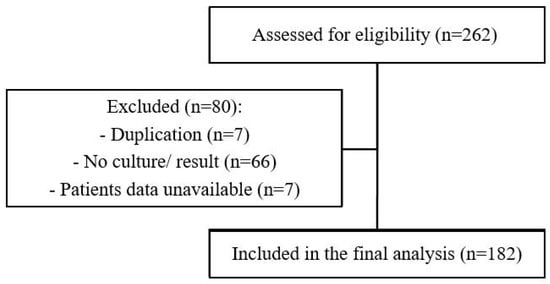
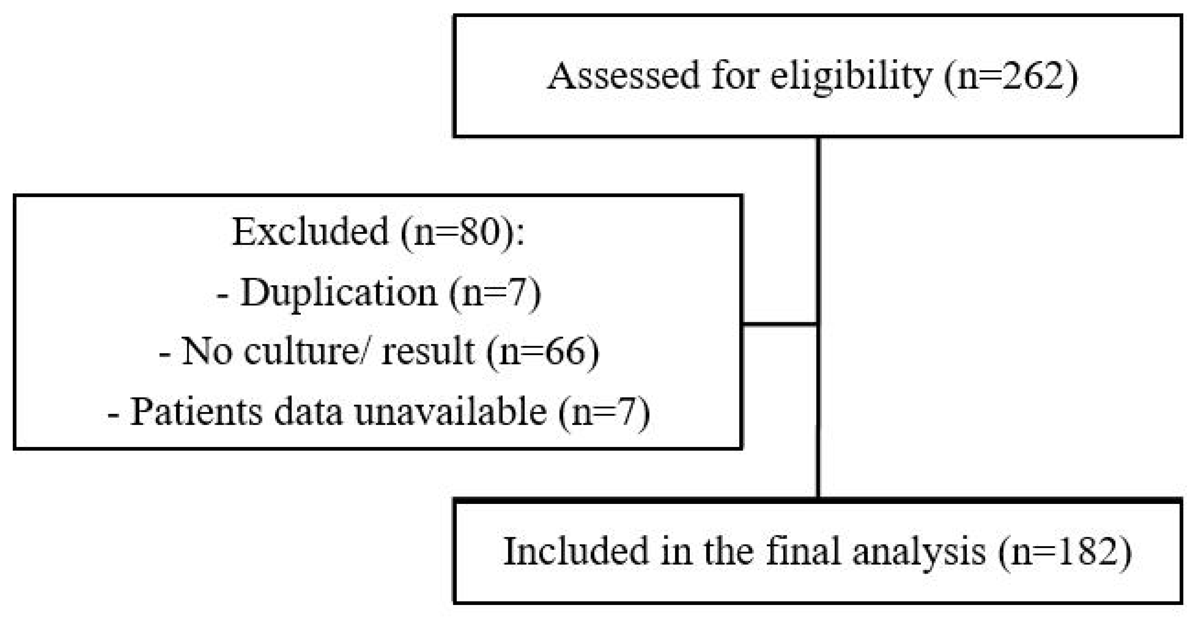
From an initial pool of 262 microbiology records screened between 2019 and 2024 at Jordan University Hospital, 80 specimens were excluded based on pre-specified criteria (e.g., duplicates samples, specimen lacking clinical details, suspected mucosal colonization and polymicrobial/bacterial co-infections). A final cohort of 182 unique patient isolates was included in the analysis (Figure 1).
Figure 1.
Flowchart of study cohort selection, eligibility screening, and clinical specimen filtering.
Demographic parameters and clinical specimen breakdown are detailed in Table 1. Patients aged 60–79 years comprised the largest age group (31.3%, n = 57), followed by pediatric/adolescent patients aged 1–19 years (27.5%, n = 50). Males comprised 63.2% (n = 115) of the cohort. Non-blood specimens comprised 95.6% (n = 174/182) of total isolates—including urine (37.4%, n = 68), respiratory tract aspirates/sputum (29.7%, n = 54), wound exudates/abscesses (17.6%, n = 32), vaginal swabs (6.6%, n = 12), and catheter tips (4.4%, n = 8). Rigorous clinical diagnostic inclusion criteria were enforced to exclude benign mucosal colonization, particularly for non-sterile respiratory specimens (Table 1).
Table 1.
Demographic parameters and clinical specimen breakdown (n = 182).
3.2. Candida Species Distribution and Temporal Shifts
Overall, C. albicans was the most frequent species (53.8%, n = 98), followed by NAC species (46.2%, n = 84) (Table 2).
Table 2.
Candida species distribution among isolates (n = 182).
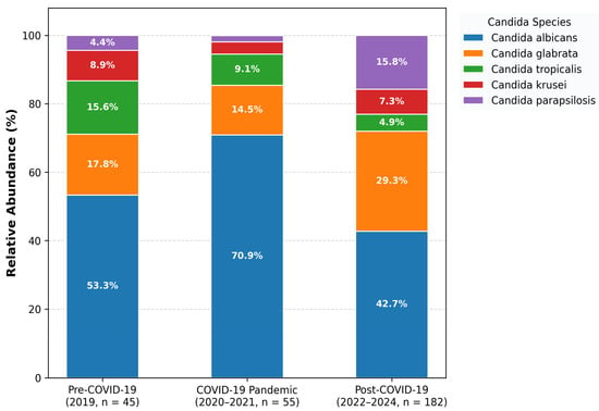
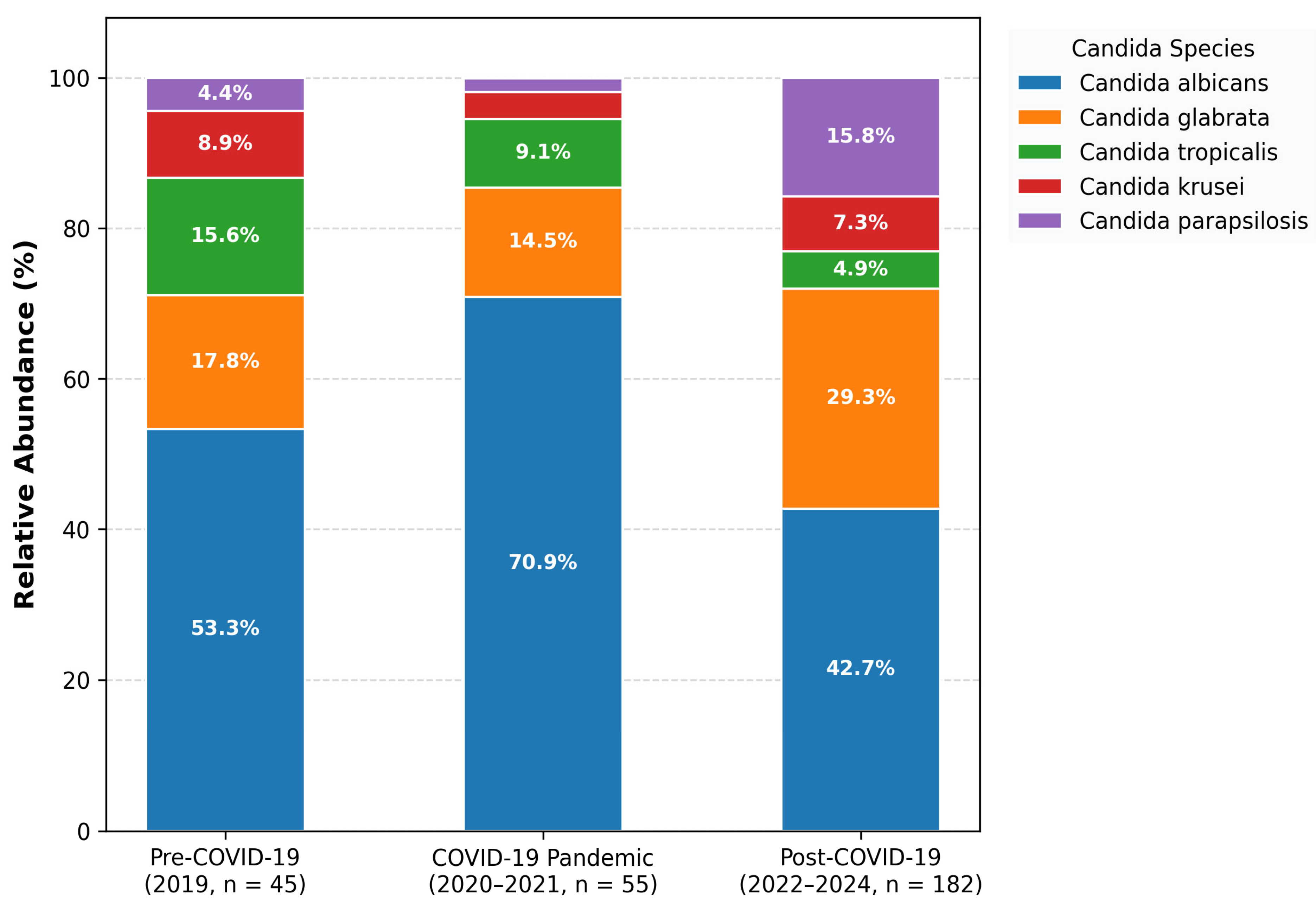
Isolate collection volumes across the study phases were as follows: pre-COVID-19 (2019: n = 45; 45.0 isolates/yr), pandemic (2020–2021: n = 55; 27.5 isolates/yr), and post-COVID-19 (2022–May 2024: n = 82; 33.9 isolates/yr). Denominator-adjusted species proportions shifted significantly across periods (Chi-square = 24.8, p < 0.001; Figure 2). During the pandemic period, C. albicans proportion reached its peak (70.9%), whereas post-pandemic years witnessed a significant expansion in NAC species, particularly C. glabrata and C. tropicalis.
Figure 2.
Relative abundance (%) of Candida species across COVID-19 epidemiological periods. Normalized by period-specific denominators: pre-COVID (2019, n = 45), COVID-19 (2020–2021, n = 55), post-COVID (2022–2024, n = 182). Post-COVID NAC species expansion p < 0.001.
3.3. Length of Hospital Stay Analysis
Length of hospital stay (LOS) ranged from 0 to 402 days, with an overall median of 21.5 days (IQR: 10.75–43.5 days). C. parapsilosis infections were identified in individuals with the longest mean hospitalization periods, 42.06 days (SD, ±39.90 days). Kruskal–Wallis test demonstrated no statistically significant difference in stay duration across different periods: the pre-pandemic, pandemic, and post-pandemic (Kruskal–Wallis p = 0.285). Similarly, categorical evaluation of short (<21 days) versus prolonged (>21 days) hospitalization revealed no significant association with the COVID-19 period (Chi-square = 1.65, p = 0.439).
3.4. Antifungal Susceptibility Patterns
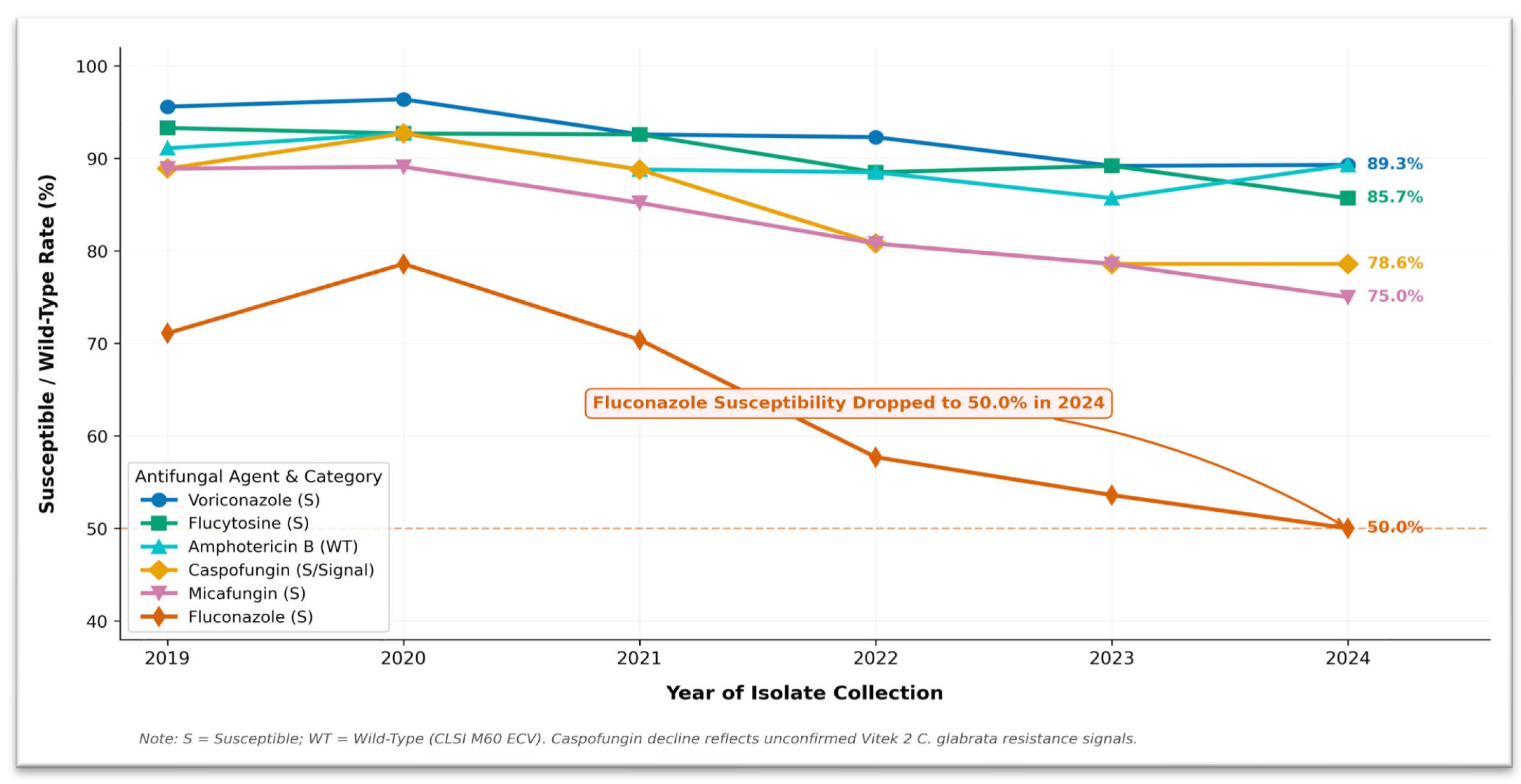
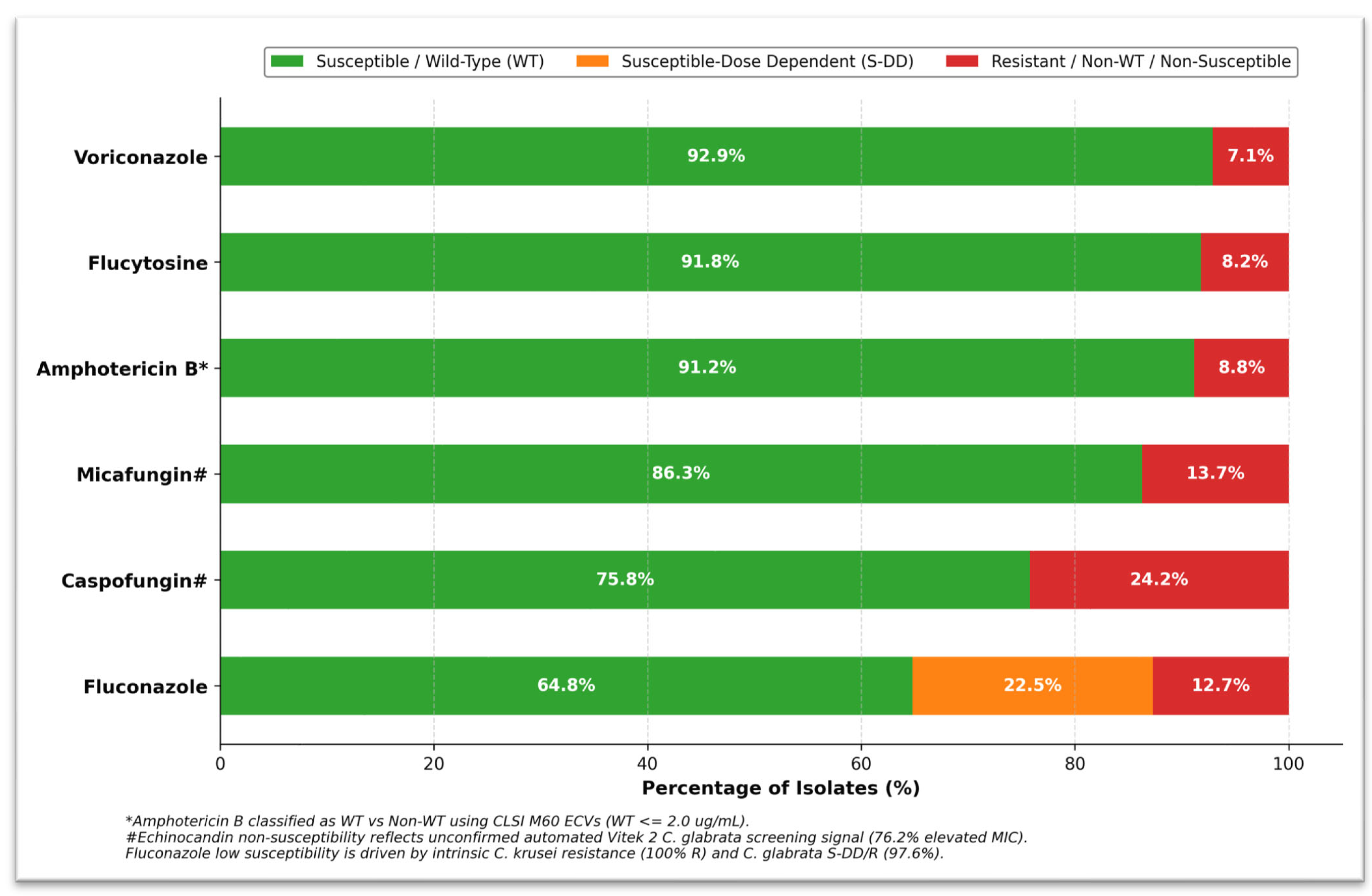
Across all 182 isolates, voriconazole demonstrated the highest overall susceptibility (92.9%, 169/182), followed by flucytosine (91.8%, 167/182), amphotericin B (91.2% Wild-Type, 166/182), micafungin (86.3%, 157/182), caspofungin (75.8%, 138/182), and fluconazole (64.8%, 118/182) (Figure 3 and Figure 4). Overall fluconazole non-susceptibility was heavily driven by intrinsic C. krusei resistance (100% R, n = 12) and C. glabrata non-susceptibility (97.6% S-DD/R, n = 41/42). When excluding intrinsically resistant C. krusei, acquired fluconazole susceptibility among inherently susceptible species was 69.4% (118/170).
Figure 3.
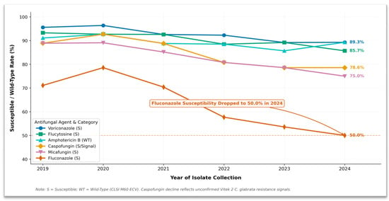
Annual antifungal susceptibility and Wild-Type trends in Jordan (2019–2024).
Figure 4.
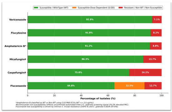
Overall antifungal susceptibility and resistance profile (n = 182). Interpreted according to CLSI M60 standards (S-DD for azoles; WT/NWT for amphotericin B).
When analyzed by species, the antimicrobial susceptibility testing (AST) profiles revealed significant clinical variations (see Table 3). C. albicans was highly susceptible to all tested antifungal agents (flucytosine 96.9%; amphotericin B 94.9% WT; micafungin 94.9%; voriconazole 94.9%; caspofungin 92.9%; fluconazole 92.9%). C. glabrata had a very low fluconazole susceptibility rate of only 2.4% (97.6% S-DD/R). Caspofungin automated susceptibility rates were also low in C. glabrata (n = 10/42; MIC ≤ 0.12 µg/mL) (19.0% n = 8; MIC > 0.25 µg/mL) and 57.1% (n = 24) ≥ 0.5 µg/mL (MIC). This high caspofungin MIC is an unverified screening signal which needs to be validated by reference broth microdilution.
Table 3.
Species-disaggregated antifungal susceptibility profiles across all isolates (n = 182).
3.5. Predictors of Prolonged Hospital Stay and Resistance
In multivariable logistic regression for prolonged hospital stay (≥21 days), patient age was identified as a continuous predictor (OR = 0.982, 95% CI: 0.971–0.993, p = 0.001; Table 4).
Table 4.
Univariable and multivariable logistic regression analysis for factors associated with prolonged hospitalization (length of stay ≥ 21 days).
4. Discussion
This study provides a six-year longitudinal surveillance of Candida species distribution, epidemiology and antifungal susceptibility patterns at JUH the pre-pandemic (2019), pandemic (2020–2021) and post-pandemic (2022–2024) periods. Invasive and localized candidiasis is the most common healthcare-associated infection worldwide [1], with high morbidity, prolonged hospitalization and attributable mortality rates from 30% to 50% in critically ill patients [1,3]. Using strict eligibility filtering on 182 non-duplicate clinical isolates, this surveillance shows major shifts in species ecology (most alarmingly, a rise in NACs) and critical vulnerabilities in current empirical antifungal regimens in Jordan.
4.1. Prevalence and Shifting Ecology of Non-Albicans Candida Species
A central finding of this investigation is the prominent proportion of non-albicans Candida species, which collectively accounted for 46.2% (n = 84/182) of all clinical isolates, dominated by C. glabrata (23.1% overall [42/182]; 50.0% of NAC), followed by C. parapsilosis (9.9% overall [18/182]; 21.4% of NAC), C. tropicalis (6.6% overall [12/182]; 14.3% of NAC), and C. krusei (6.6% overall [12/182]; 14.3% of NAC). Although C. albicans was still the most frequently isolated species overall (53.8%), the large proportion of NAC species is consistent with global surveillance networks (e.g., SENTRY Antifungal Surveillance Program, ARTEMIS DISK) showing epidemiological shift from C. albicans towards less susceptible NAC pathogens over the last two decades [2,14]. Our findings also corroborate recent single-center findings in Jordan [8,9,10], extending those observations by providing longitudinal multi-year stratification across distinct pandemic phases.
This ecological displacement is more pronounced in the Middle East and North Africa (MENA) region: studies from Saudi Arabia [15], Kuwait [16], the United Arab Emirates [17] and Egypt [18] have reported NAC prevalence > 50% in intensive care and immunocompromised patient cohorts, respectively.
In Jordan, prior single-center studies were mostly mucosal or genital tract isolates with higher baseline susceptibility for C. albicans [8]. The current longitudinal data from a major 600-bed tertiary care hospital, however, show that inpatients are increasingly exposed to high-risk NAC species with intrinsic or rapidly acquired resistance. The expansion of C. glabrata has been due to frequent azole exposure, widespread fluconazole prophylaxis in surgical and oncological units, and patient age-related changes in patients’ host mucosal microbiota [19,20]).
4.2. Antifungal Resistance Patterns: Azole and Echinocandin Vulnerabilities
Antifungal susceptibility profiles evaluated via Vitek 2 Compact and interpreted according to updated CLSI M60 standards revealed striking divergent activity across drug classes. Voriconazole demonstrated the highest overall susceptibility (92.9%), followed closely by flucytosine (91.8%) and amphotericin B (91.2% Wild-Type profile). Conversely, fluconazole exhibited poor overall activity (64.8% susceptibility), driven by C. glabrata non-susceptibility (97.6% S-DD/R) and intrinsic C. krusei resistance (100% R).
Most alarmingly, caspofungin susceptibility among C. glabrata isolates was restricted to 23.8% (76.2% resistance profile signal, MIC ≥ 0.25 ug/mL). While echinocandins represent first-line therapy for invasive candidiasis per IDSA and ESCMID guidelines [21,22], elevated caspofungin MICs in C. glabrata have been increasingly reported worldwide, mediated by FKS1 and FKS2 hot-spot mutations [6,23,24]. However, methodologists caution that automated Vitek 2 testing frequently overestimates caspofungin MICs in C. glabrata due to technical instrument calibration limits (e.g., Vitek 2 can exhibit elevated MIC higher and false-resistance rates for caspofungin than broth microdilution (CLSI M27)) [25]. Therefore, a confirmatory testing via reference broth microdilution, E-test and molecular FKS sequencing is required before altering institutional first-line treatment guidelines [6,21,22].
4.3. Temporal Dynamics and COVID-19 Pandemic Impact
Stratification across temporal phases provided unique insights into pandemic-related fungal dynamics. During peak COVID-19 pandemic years (2020–2021), C. albicans predominated (70.9%), whereas post-pandemic years witnessed a significant expansion of C. glabrata and C. tropicalis (p < 0.001). Similar transient surges toward C. albicans during pandemic surges, followed by post-pandemic NAC rebound, have been documented in Southern Europe [10], South Korea [11] and Latin America [13]. The high proportion of C. albicans during COVID-19 was likely facilitated by widespread immunomodulatory therapy (high-dose dexamethasone, tocilizumab), severe mucosal barrier disruption in mechanically ventilated patients, prolonged ICU stays, and empirical broad-spectrum antibacterial administration [26,27,28,29]. Crucially, absolute isolate numbers fluctuated across study periods (45.0/yr pre-COVID vs. 27.5/yr pandemic). Rather than reflecting a drop in true community infection incidence, this reduction reflects overall hospital admission volume, selective microbiological sampling, and infection control practices. This underscores the absolute necessity of denominator-adjusted proportion metrics in hospital epidemiological surveillance [24]. Furthermore, Candida auris was recently reported for the first time in Jordan [7], but no C. auris isolates were identified in this study cohort; therefore, surveillance should continue to detect undetected hospital outbreaks.
4.4. Clinical Specimen Spectrum and Colonization vs. Infection Differentiation
Differentiating true fungal infection from mucosal colonization remains a major diagnostic challenge in clinical mycology [30]. In our study, non-blood clinical specimens comprised 95.6% of isolates, including urine (37.4%) and respiratory aspirates/sputum (29.7%). Candida species frequently colonize the respiratory and urinary tracts of hospitalized patients, particularly those with endotracheal tubes or urinary catheters.
In accordance with IDSA/ESCMID guidelines, Candida isolated from respiratory secretions almost universally represents airway colonization rather than parenchymal pneumonia [21,22]. By enforcing strict clinical diagnostic criteria (purulent sputum, radiologic pulmonary infiltrates, fever, leukocytosis, and physician-initiated systemic antifungal therapy), we minimized misclassification risk while acknowledging that non-blood isolates reflect a combination of tissue infection and high-density colonization [28,29]. Clinical risk scores (e.g., Candida Score) combined with quantitative serum biomarkers (1,3-beta-D-glucan or T2 Candida magnetic resonance) in prospective studies will be needed to improve diagnostic specificity [29,30].
4.5. Hospital Length of Stay and Multivariable Predictor Dynamics
Hospital length of stay (LOS) in this cohort was long (median, 21.5 days; range, 0–402 days). LOS did not differ significantly across COVID-19 periods (Kruskal–Wallis p = 0.285). Dataset analysis shows that prolonged hospitalization (30–100 days) was highly concentrated in pediatric, adolescent, and young adult surgical/trauma patients requiring extended ICU care, mechanical ventilation, and multi-system rehabilitation [31,32]. However, among elderly patients (>70 years), acute medical crises were more frequent with early mortality or shorter acute care stays before discharge to step-down care. This illustrates the potential interaction between host age, comorbidities, surgical and trauma related factors and institutional care pathways in tertiary referral centers [32].
4.6. Methodological Limitations and Study Strengths
Several limitations of this study need to be highlighted. First, the retrospective single-center design at Jordan University Hospital limits generalizability of resistance rates to secondary community hospitals or private healthcare centers in Jordan. Second, the 14-day exclusion window for duplicate isolates from the same patient may be relatively brief given the prolonged median length of hospital stay (21.5 days, IQR: 10.75–43.5 days); extending the exclusion window to 30 days in future work could more effectively eliminate persistent colonizing or repeat strains during long-stay admissions. Third, non-blood specimens comprised 95.6% of isolates, with respiratory samples representing 29.7% (n = 54). Despite enforcing rigorous clinical treatment filtering, retrospective medical record reviews cannot fully exclude incidental airway colonization, as clinicians in real-world tertiary care settings may occasionally initiate empirical antifungal therapy for respiratory colonization in critically ill patients with unexplained fever or pulmonary infiltrates. Fourth, automated Vitek 2 Compact testing without broth microdilution or E-test confirmation means caspofungin MIC values should be interpreted with caution due to known platform overestimation. Molecular sequencing for key resistance determinants (FKS1, FKS2, ERG11, and PDR1) was not performed due to resource constraints. Finally, patient treatment regimens were recorded as primary single-agent therapy, whereas complex inpatient care often involves sequential drug switching due to clinical failure, organ dysfunction, or toxicity.
Despite these limitations the study has strengths too. It is one of very few multi-year longitudinal surveillance studies in Jordan with AST data based on updated CLSI M60 standards; duplicate records filtering, strict specimen selection for clinical specimens and denominator-adjusted temporal analysis give a good baseline for regional mycology surveillance.
4.7. Clinical Implications and Antifungal Stewardship Recommendations
Based on our findings, we propose four priority recommendations for hospital infection control and pharmacy committees in Jordan:
- Empirical Azole Protocols: Against fluconazole monotherapy in ICU, oncological and surgical units where C. glabrata (97.6% S-DD/R) and C. krusei (100% intrinsic resistance) are predominant.
- Echinocandin AST Confirmation: Require confirmatory reference testing (E-test or broth microdilution) or micafungin surrogate testing before discontinuing echinocandins based on automated caspofungin MIC elevations only.
- Institutional Antifungal Stewardship Programs (ASPs): Dedicated ASPs should be set up to audit antifungal prescribing, prevent inappropriate prophylaxis and selection pressure.
- Regional Reference Molecular Surveillance: Building centralized reference mycology capacity for FKS/ERG11 gene mutation screening and national Candida auris outbreak surveillance.
5. Conclusions
This six-year longitudinal surveillance study shows major ecological shift towards non-albicans Candida species in a large Jordanian tertiary care hospital and concerns about azole non-susceptibility and automated caspofungin resistance signals in C. glabrata. Species distribution changed significantly during the COVID-19 pandemic, highlighting the dynamic nature of hospital fungal epidemiology. Species identification, CLSI M60 breakpoint interpretation update, confirmatory AST reference testing and institutional antifungal stewardship programs are needed to improve clinical outcomes and preserve antifungal efficacy in Jordan.
Author Contributions
Conceptualization and Methodology: H.M. and L.M.M. Data curation: H.M. Formal analysis: H.M., L.G. and L.M.M. Writing—original draft preparation: H.M., L.G. and L.M.M. Review and editing: L.M.M. All authors have read and agreed to the published version of the manuscript.
Funding
This research received no external funding.
Institutional Review Board Statement
The study was conducted in accordance with the Declaration of Helsinki and approved by the Institutional Review Board (IRB) of Jordan University Hospital (protocol code 10/2024/7181; date of approval: 11 March 2024).
Informed Consent Statement
Not applicable. Informed consent was waived due to the retrospective, anonymized design.
Data Availability Statement
The datasets used and analyzed during the current study are available from the corresponding author upon reasonable request.
Acknowledgments
The authors thank the staff of the Central Microbiology Laboratory at Jordan University Hospital for technical support.
Conflicts of Interest
The authors declare no conflicts of interest.
References
- Pappas, P.G.; Lionakis, M.S.; Arendrup, M.C.; Ostrosky-Zeichner, L.; Kullberg, B.J. Invasive Candidiasis. Nat. Rev. Dis. Primers 2018, 4, 18026. [Google Scholar] [CrossRef] [Scilit] [PubMed]
- Fisher, M.C.; Alastruey-Izquierdo, A.; Berman, J.; Bicanic, T.; Bignell, E.M.; Bowyer, P.; Bromley, M.; Brüggemann, R.; Garber, G.; Cornely, O.A.; et al. Tackling the Emerging Threat of Antifungal Resistance to Human Health. Nat. Rev. Microbiol. 2022, 20, 557–571. [Google Scholar] [CrossRef] [Scilit] [PubMed]
- McCarty, T.P.; White, C.M.; Pappas, P.G. Candidemia and Invasive Candidiasis. Infect. Dis. Clin. N. Am. 2021, 35, 389–413. [Google Scholar] [CrossRef] [Scilit] [PubMed]
- Arendrup, M.C.; Prakash, A.; Meletiadis, J.; Sharma, C.; Chowdhary, A. Comparison of EUCAST and CLSI Reference Microdilution MICs of Eight Antifungal Compounds for Candida auris and Associated Tentative Epidemiological Cutoff Values. Antimicrob. Agents Chemother. 2017, 61, e00485-17. [Google Scholar] [CrossRef] [Scilit] [PubMed]
- CLSI. Performance Standards for Antifungal Susceptibility Testing of Yeasts, 2nd ed.; CLSI Supplement M60; Clinical and Laboratory Standards Institute: Wayne, PA, USA, 2020. [Google Scholar]
- Perlin, D.S. Echinocandin Resistance in Candida. Clin. Infect. Dis. 2015, 61, S612–S617. [Google Scholar] [CrossRef] [Scilit] [PubMed]
- Al-Ramahi, J.W.; Ghanem, R.A.; Sayyouh, O.H.; Al-Jammal, D.; Said, M.M.; Nasrat, S.; El-Shokry, M.; Gazo, M.; Osman, T.S. Report of the First Case of Candida auris Identified in Jordan. Microbiol. Res. 2023, 14, 1559–1567. [Google Scholar] [CrossRef] [Scilit]
- Al-Groom, R.M.; Ali, R.R.M.; Abu Shaqra, Q.M. Genotypes Analysis and Antifungal Susceptibility of Candida albicans Strains Isolated from Women with Vaginal Candidiasis in Jordan Using PCR Targeting 25SrDNA and ALT Repeat Sequences of the RPS. Pak. J. Med. Sci. 2024, 40, 1619–1624. [Google Scholar] [CrossRef] [Scilit] [PubMed]
- Abu-Humaidan, A.H.; Alshdaifat, A.; Awajan, D.; Abu-Hmidan, M.; Alshdifat, A.; Hasan, H.; Ahmad, F.M.; Alaridah, N.; Irshaid, A.; Yamin, D. The Antimicrobial Resistance of Candida: A 5-Year Retrospective Analysis at a Tertiary Hospital in Jordan. J. Fungi 2025, 11, 87. [Google Scholar] [CrossRef] [Scilit] [PubMed]
- Boattini, M.; Pinto, M.F.; Christaki, E.; Fasciana, T.; Falces-Romero, I.; Tofarides, A.; Bianco, G.; Cendejas-Bueno, E.; Tricoli, M.R.; Tsiolakkis, G.; et al. Multicentre Surveillance of Candida Species from Blood Cultures during the SARS-CoV-2 Pandemic in Southern Europe (CANCoVEU Project). Microorganisms 2023, 11, 560. [Google Scholar] [CrossRef] [Scilit] [PubMed]
- Won, E.J.; Sung, H.; Kim, M.N. Changing Epidemiology of Clinical Isolates of Candida Species during the Coronavirus Disease 2019 Pandemic: Data Analysis from a Korean Tertiary Care Hospital for 6 Years (2017–2022). J. Fungi 2024, 10, 193. [Google Scholar] [CrossRef] [Scilit] [PubMed]
- Díaz-García, J.; Gómez, A.; Machado, M.; Alcalá, L.; Reigadas, E.; Sánchez-Carrillo, C.; Pérez-Ayala, A.; de la Pedrosa, E.G.G.; González-Romo, F.; Cuétara, M.S.; et al. Candida Genotyping of Blood Culture Isolates from Patients Admitted to 16 Hospitals in Madrid: Genotype Spreading during the COVID-19 Pandemic Driven by Fluconazole-Resistant C. parapsilosis. J. Fungi 2022, 8, 1228, Erratum in J. Fungi 2023, 9, 196. [Google Scholar] [CrossRef] [Scilit] [PubMed]
- de Treviño-Rangel, R.J.; Villanueva-Lozano, H.; González-Lara, M.F.; Castaño-Jimenez, P.A.; Corte-Rojas, R.E.; Quevedo-Ramos, M.A.; Avilés-Benítez, L.K.; López-Gutiérrez, E.; Feliciano-Guzmán, J.M.; Dorado-del Río, M.; et al. Species-Specific Antifungal Resistance Trends in Yeast Bloodstream Isolates Causing Candidemia before, during, and after the COVID-19 Pandemic: Insights from the INVIFAR Mexican Multicenter Network. Eur. J. Clin. Microbiol. Infect. Dis. 2026, 45, 1693–1706. [Google Scholar] [CrossRef] [Scilit] [PubMed]
- Pfaller, M.A.; Diekema, D.J.; Turnidge, J.D.; Castanheira, M.; Jones, R.N. Twenty Years of the SENTRY Antifungal Surveillance Program: Results for Candida Species from 1997–2016. Open Forum Infect. Dis. 2019, 6, S79–S94. [Google Scholar] [CrossRef] [Scilit] [PubMed]
- Alkharashi, N.; Aljohani, S.; Layqah, L.; Masuadi, E.; Baharoon, W.; Al-Jahdali, H.; Baharoon, S. Candida Bloodstream Infection: Changing Pattern of Occurrence and Antifungal Susceptibility over 10 Years in a Tertiary Care Saudi Hospital. Can. J. Infect. Dis. Med. Microbiol. 2019, 2019, 2015692. [Google Scholar] [CrossRef] [Scilit] [PubMed]
- Mokaddas, E.M.; Al-Sweih, N.A.; Khan, Z.U. Species Distribution and Antifungal Susceptibility of Candida Bloodstream Isolates in Kuwait: A 10-Year Study. J. Med. Microbiol. 2007, 56, 255–259. [Google Scholar] [CrossRef] [Scilit] [PubMed]
- Thomsen, J.; Abdulrazzaq, N.M.; Oulhaj, A.; Nyasulu, P.S.; Alatoom, A.; Denning, D.W.; Al Dhaheri, F.; UAE AMR Surveillance Consortium; Menezes, G.A.; Moubareck, C.A.; et al. Emergence of Highly Resistant Candida auris in the United Arab Emirates: A Retrospective Analysis of Evolving National Trends. Front. Public Health 2024, 11, 1244358. [Google Scholar] [CrossRef] [Scilit] [PubMed]
- Aziz, H.S.A.; Ismail, D.K.; Mohammed, N.S.A.; Elgendy, M.O.; Bassiouny, D.M. Distribution and Antifungal Susceptibility Profiles of Candida Species Isolated from Candidemia Patients Admitted to Egyptian Tertiary Hospitals: A Cross-Sectional Study. BMC Infect. Dis. 2024, 24, 1177. [Google Scholar] [CrossRef] [Scilit] [PubMed]
- Cai, G.; Zhang, Z. Global Surveillance of Candida albicans: MIC Distributions, Resistance Patterns, and Temporal Trends from the Antimicrobial Testing Leadership and Surveillance Program (2015–2024). Antimicrob. Agents Chemother. 2026, 70, e01617-25. [Google Scholar] [CrossRef] [Scilit] [PubMed]
- Badrane, H.; Cheng, S.; Dupont, C.L.; Hao, B.; Driscoll, E.; Morder, K.; Liu, G.; Newbrough, A.; Fleres, G.; Kaul, D.; et al. Genotypic Diversity and Unrecognized Antifungal Resistance among Populations of Candida glabrata from Positive Blood Cultures. Nat. Commun. 2023, 14, 5918. [Google Scholar] [CrossRef] [Scilit] [PubMed]
- Pappas, P.G.; Kauffman, C.A.; Andes, D.R.; Clancy, C.J.; Marr, K.A.; Ostrosky-Zeichner, L.; Reboli, A.C.; Schuster, M.G.; Vazquez, J.A.; Walsh, T.J.; et al. Clinical Practice Guideline for the Management of Candidiasis: 2016 Update by the Infectious Diseases Society of America. Clin. Infect. Dis. 2016, 62, e1–e50. [Google Scholar] [CrossRef] [Scilit] [PubMed]
- Chen, S.C.; Perfect, J.; Colombo, A.L.; Cornely, O.A.; Groll, A.H.; Seidel, D.; Albus, K.; de Almedia, J.N.; Garcia-Effron, G.; Gilroy, N.; et al. Global Guideline for the Diagnosis and Management of Rare Yeast Infections: An Initiative of the ECMM in Cooperation with ISHAM and ASM. Lancet Infect. Dis. 2021, 21, e375–e386, Erratum in Lancet Infect. Dis. 2021, 21, e363; Erratum in Lancet Infect. Dis. 2024, 24, e485. [Google Scholar] [CrossRef] [Scilit] [PubMed]
- Al-Baqsami, Z.F.; Ahmad, S.; Khan, Z. Antifungal Drug Susceptibility, Molecular Basis of Resistance to Echinocandins and Molecular Epidemiology of Fluconazole Resistance among Clinical Candida glabrata Isolates in Kuwait. Sci. Rep. 2020, 10, 6238. [Google Scholar] [CrossRef] [Scilit] [PubMed]
- Chakrabarti, A.; Singh, S. Multidrug-Resistant Candida auris: An Epidemiological Review. Expert Rev. Anti-Infect. Ther. 2020, 18, 551–562. [Google Scholar] [CrossRef] [Scilit] [PubMed]
- Winkler, M.L.; Deshpande, L.; Kimbrough, J.H.; Karr, M.; Rhomberg, P.; Klauer, A.L.; Castanheira, M. Anidulafungin Is a Useful Surrogate Marker for Predicting in Vitro Susceptibility to Rezafungin among Five Candida Species Using CLSI Methods and Interpretive Criteria. J. Clin. Microbiol. 2025, 63, e01129-24. [Google Scholar] [CrossRef] [Scilit] [PubMed]
- Kaur, M.; Thakur, P.; Verma, N.; Choksket, S.; Harshvardhan; Korpole, S.; Bandarupalli, D.; Grover, V. Invasive Fungal Infections in Immunocompromised Conditions: Emphasis on COVID-19. Curr. Microbiol. 2024, 81, 400. [Google Scholar] [CrossRef] [Scilit] [PubMed]
- Eggimann, P.; Pittet, D. Candida Colonization Index and Subsequent Infection in Critically Ill Surgical Patients: 20 Years Later. Intensive Care Med. 2014, 40, 1429–1448. [Google Scholar] [CrossRef] [Scilit] [PubMed]
- Keighley, C.; Cooley, L.; Morris, A.J.; Ritchie, D.; Clark, J.E.; Boan, P.; Worth, L.J.; Australasian Antifungal Guidelines Steering Committee. Consensus Guidelines for the Diagnosis and Management of Invasive Candidiasis in Haematology, Oncology and Intensive Care Settings, 2021. Intern. Med. J. 2021, 51, 89–117. [Google Scholar] [CrossRef] [Scilit] [PubMed]
- Calandra, T.; Roberts, J.A.; Antonelli, M.; Bassetti, M.; Vincent, J.L. Diagnosis and Management of Invasive Candidiasis in the ICU: An Updated Approach to an Old Enemy. Crit. Care 2016, 20, 125. [Google Scholar] [CrossRef] [Scilit] [PubMed]
- Barantsevich, N.; Barantsevich, E. Diagnosis and Treatment of Invasive Candidiasis. Antibiotics 2022, 11, 718. [Google Scholar] [CrossRef] [Scilit] [PubMed]
- Lefering, R.; Waydhas, C.; TraumaRegister, D.G.U. Prediction of Prolonged Length of Stay on the Intensive Care Unit in Severely Injured Patients—A Registry-Based Multivariable Analysis. Front. Med. 2024, 11, 1358205. [Google Scholar] [CrossRef] [Scilit] [PubMed]
- Wozniak, H.; Beckmann, T.S.; Dos Santos Rocha, A.; Pugin, J.; Heidegger, C.P.; Cereghetti, S. Long-Stay ICU Patients with Frailty: Mortality and Recovery Outcomes at 6 Months. Ann. Intensive Care 2024, 14, 31. [Google Scholar] [CrossRef] [Scilit] [PubMed]

Disclaimer/Publisher’s Note: The statements, opinions and data contained in all publications are solely those of the individual author(s) and contributor(s) and not of MDPI and/or the editor(s). MDPI and/or the editor(s) disclaim responsibility for any injury to people or property resulting from any ideas, methods, instructions or products referred to in the content. |
© 2026 by the authors. Licensee MDPI, Basel, Switzerland. This article is an open access article distributed under the terms and conditions of the Creative Commons Attribution (CC BY) license.