


Abstract
Background/Objectives: The preservation of ancestors as mummies was a central element of many pre-Hispanic cultures of western South America. Here, we present our results from a bioarchaeological and anatomical investigation of a Peruvian mummy, curated today in the anatomical collection of the Charité, Berlin, Germany. The remains were collected by the Prussian physician, botanist, and explorer Franz Julius Ferdinand Meyen (1804–1840), who served as a ship’s doctor in South America from 1830 to 1832. According to Meyen, the mummy was found in a natural cave in the Andes, near Pasco, today Peru. As is often the case, all context and documentation of origin are lost today. Methods: We performed radiocarbon dating, external macroscopic examination, and computed tomography imaging. Results: According to skeletal anatomy, the mummy is that of an adult male who died in the late pre-Hispanic or Early Colonial Period. While the skin is largely well preserved, most internal organs could not be identified on CT and external genitalia are missing. An incision in the right flank communicating with the abdominal cavity and a large defect in the pelvic floor were observed. Conclusions: This Peruvian mummy represents a rare case of anthropogenic mummification with removal of the internal organs. Evisceration was likely performed through the right flank incision, with additional access routes via the pelvic floor and the mouth. However, since neither grave goods nor parts of the mummy bundle have survived, his culture of origin remains uncertain.
1. Introduction
The oldest discovered artificial mummies in the world may originate from Western South America: The earliest cases of elaborate artificial mummification are found in burial sites in what is now Northern Chile and Southern Peru, dating from 5000 to 1500 before the Common Era (BCE) [1]. However, the beginnings of mummification practices for those cases for which methods cannot be determined (and which are therefore usually described as “naturally mummified”) cannot be precisely dated.
In contrast to the relatively well-known Egyptian mummification practices, those of the pre-Hispanic cultures of South America remain largely unknown. Since these cultures had no written language and Spanish conquerors suppressed ancient funerary rituals during Christianization, much of the knowledge about mummification techniques has been lost. The most widespread method appears to have been “exposure to heat”, for example, through smoking [2]. Individuals were usually placed in a squatted position and tightly wrapped in multiple layers of textiles forming a so-called mummy bundle. In South American mummies, the internal organs are often preserved, allowing for diverse investigations into past diseases. Also, imprints of different textiles on the skin provide important clues that can sometimes help determine the original cultural background.
It was not until the 19th century that pre-Hispanic mummies attracted attention from European and North American traders, explorers, collectors, and museum networks. Within the expanding circuits of colonial extraction and the antiquities trade, mummies and other pre-Hispanic objects were transformed into commodities. They were removed from burial sites and incorporated into private and institutional collections. Mummy bundles were frequently opened at the burial site to assess and extract objects considered commercially valuable. Materials deemed suitable for sale were packaged and shipped to Europe and North America, while other remains and associated funerary materials were discarded or left behind. These practices fragmented burial assemblages, severed the dead from their cultural and archaeological contexts, and contributed to the formation of numerous museum and university collections in Western Europe and North America that continue to hold human remains from pre-Hispanic funerary contexts today.
Here, we present our results from an investigation of a Peruvian mummy preserved at the anatomical collection of the Charité Berlin, Germany. We have previously reported our results regarding the provenance of this mummy [3] and will focus here on bioarchaeological and anatomical results to elucidate the likely mummification procedures.
2. Materials and Methods
2.1. Human Remains
The mummy in question was collected by the Prussian physician, botanist, and explorer Franz Julius Ferdinand Meyen (1804–1840), who served as a ship’s doctor on a Prussian expedition to South America from 1830 to 1832. According to Meyen, the mummy was found in a natural cave in the region of Pasco, a department of Peru, but the specific context of acquisition remains uncertain (for more detail see [3]). Meyen sent this mummy to the Friedrich Wilhelm University in Berlin. Today, the mummy is cared for in the anatomical collection of the Charité, Berlin (inventory entry “Peruaner-Mumie. A.N.15657. N.C.1190.”). In the first part of his anthropological catalogue of the collection, the anatomist Gustav Broesike (1853–1922) wrote a short paragraph about this mummy (page 86): “List of various other anthropological objects. 405. 15657. Mummy in a seated position from a Peruvian grave. This includes two boxes containing fairly long plain black hair.” [4] (Translation from German by the authors). Both boxes seem lost today. This entry shows that the individual was bereft of his clothes and burial goods, which significantly hampers the determination of cultural specifics in social groups of the time of the individual’s burial. Our own scientific interest regarding this mummy must be balanced with the respect that all human remains deserve. As the origins of this mummy were largely unclear at the outset of our project, we felt justified to conduct an anthropological investigation, including minimally invasive procedures, as part of a provenance analysis. Only such an analysis can produce a basis for further considerations regarding the future handling of the remains, including possible repatriation (see discussion). The Institute of Neuroanatomy of Charité is the custodian of the mummy, and our plans have been discussed with and were approved by its director. We have kept the extent of damage as minimal as possible and have not interfered with the outer appearance of the mummy.
2.2. External Macroscopic Examination
External macroscopical examination and photo documentation were performed under daylight conditions and independently by three researchers (A.-M.B., S.S., A.W.).
2.3. Computed Tomography
Computed tomography (CT) imaging was performed at the Radiology Centre Heidelberg (MZV) using a high-performance dual-source CT scanner (SOMATOM Force, Siemens Healthineers, Forchheim, Germany).
Following careful positioning of the mummy and acquisition of topograms in both anteroposterior and lateral projections, a high-resolution spiral CT scan was performed. The slice thickness was 0.4 mm with a slice increment of 0.3 mm. The pitch factor was 0.85, and the table feed was 16.3 mm per rotation. The rotation speed was four revolutions per second with 384 slices acquired per rotation. The tube current-time product was set to 238 mAs, and the tube voltage to 120 kV. Additionally, a further scan was performed using material-selective dual-energy techniques with tube voltages of 100 kV and 150 kV. The resulting isotropic voxel size was 0.4 mm.
Axial datasets were generated from the raw data using various reconstruction kernels, which influence edge definition, image noise, and spatial resolution. Furthermore, different iterative reconstruction filters (ADMIRE levels I–V) were applied. The reconstructed datasets were compared regarding image quality. The sequences offering the best soft-tissue differentiation, as well as those providing optimal visualization of the skeletal system, were selected and analysed for subsequent multiplanar and three-dimensional reconstructions. Dual-energy CT technology has enabled improved differentiation of absorption values across various tissue structures.
In a standard CT protocol using a tube voltage of 120 kV, typical density values in living patients are well established in clinical literature. For example, −1000 Hounsfield units (HU) for air, −100 HU for adipose tissue, 0 HU for water, around 40 HU for soft tissue, 40–70 HU for blood, and over 300 HU for cortical bone could be measured. However, due to the pronounced tissue desiccation found in dry mummies, these reference values derived from clinical imaging of living patients can only be applied to a limited extent. Examples from an Ancient Egyptian mummy include 1400–1600 HU for compact bone, −400–−80 HU for spongy bone, 1700–2900 HU for enamel, 1600–1800 HU for dentin, −200–−120 HU for muscle, −70–210 HU for ligaments and −200–−230 HU for skin [5]. Post-processing of the CT datasets was performed on a MacBook Pro (2020, Cupertino, California, USA) using the commercially available DICOM workstation OsiriX MD (version 12.5.1). Multiplanar and three-dimensional reconstructions were generated from the axial datasets.
CT datasets were assessed independently by a medical specialist for radiology (R.S.) and a medical specialist for anatomy (S.S.).
2.4. Sex and Age Estimation
As the external genitalia are not preserved, biological sex was estimated from the morphology of the pelvis on CT, following the standards of Buikstra and Ubelaker [6]. In short, morphology of the greater sciatic notch (narrower in males, wider in females), the ventral arc (present in females, absent in males) and the subpubic concavity (present in females, absent in males) was analyzed. Additionally, the subpubic angle (narrower in males, wider in females) was assessed. Age at death was estimated from the fusion of the medial clavicular epiphysis and the spheno-occipital synchondrosis. Complete fusion of both structures was defined as an indicator of a minimum age of 18 years [6].
2.5. Radiocarbon Dating
For radiocarbon dating, tissue samples from the left thoracic wall (connective tissue and musculature, likely endothoracic fascia and intercostal muscles) and the left gluteal region (connective tissue and musculature, likely gluteus maximus and/or gluteus medius) were collected and sent to the Leibniz laboratory of the Christian Albrechts University in Kiel, Germany (sample numbers: KIA-59231 and KIA-59232). The samples underwent solvent cleaning. Amino acids were extracted from the cleaned tissue and dated using accelerator mass spectrometry (HVE 3MV Tandetron 4130, Amersfoort, The Netherlands). Calibration of conventional 14C ages to calendar years was performed using the OxCal v4.4.4 software package and the SHCal20 dataset for the southern hemisphere [7,8].
3. Results
3.1. External Inspection
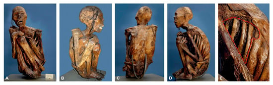
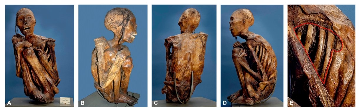
After death, the body was placed in an upright position, with the legs bent closely against the body, the right foot positioned over the left, and the hands resting on the knees. While the right hand remained on the knees, the left hand appears to have slipped down slightly. The body was secured in this tightly flexed position with broad cloth strips, which are no longer preserved but can be detected through traces and areas of discoloration on the skin. The mouth of the mummy is open (Figure 1A–D).
Figure 1.
External morphology of the Peruvian mummy. (A): Anterior. (B): Right side. (C): Posterior. (D): Left side. (E): Large skin defects (within the red dotted outline) at the left side of the chest (detail from image (D)).
Evidence of decomposition and/or later damage can be observed on the right lower lip, on the inner surfaces of the arms and legs towards the abdomen, on the left shoulder, and, most notably, on the left side and posterior surface of the body, where large areas of skin are missing (Figure 1E). Only strands of the individual’s hair have been preserved. The precise timing of the described damage cannot be determined. It may have occurred either while the body was enclosed within the mummy bundle, or after its excavation before 1832, or during transport to Europe or at a later stage while the mummy was housed in the collection. Traces of varnish indicate that the mummy was treated after entering the collection, possibly as part of an attempt to combat insect infestation.
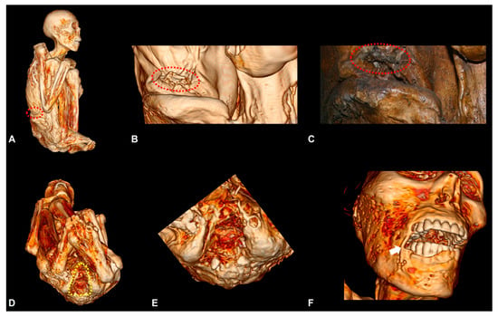
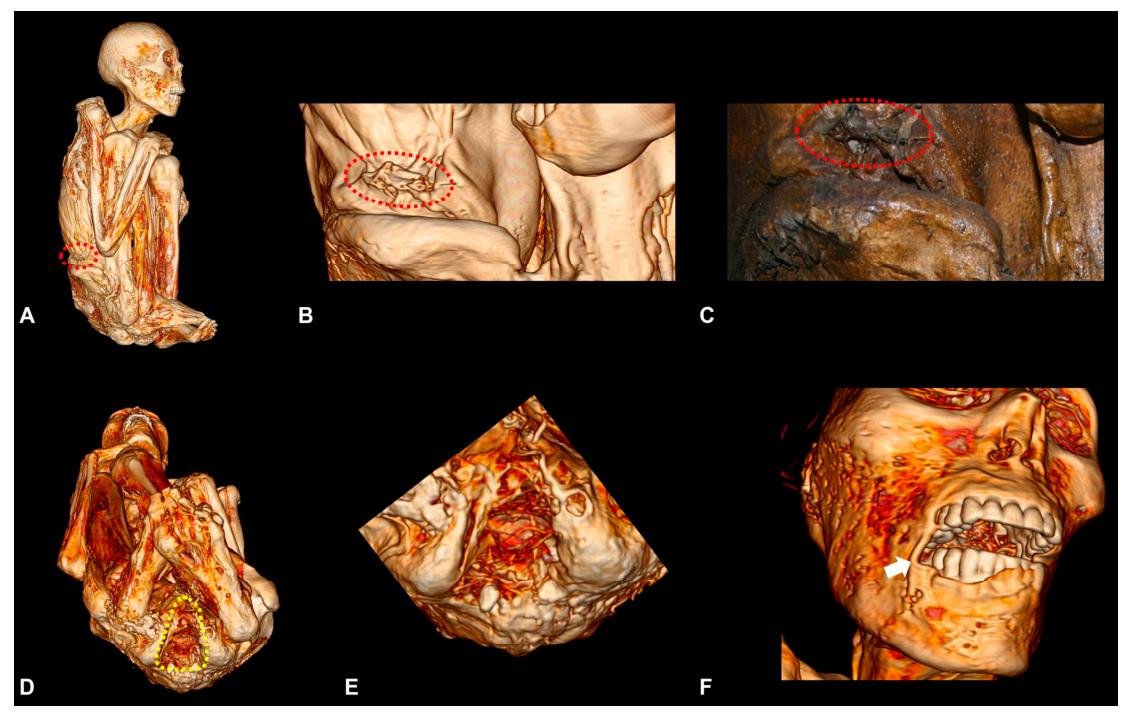
Traces of cuts made to remove organs are not easily identified, but neither can they be ruled out in the areas of postmortem skin defects. However, on re-examination guided by the CT findings (see below), a small, circumscribed opening was identified on the right flank (see Section 3.3.6).
3.2. Radiocarbon Dating
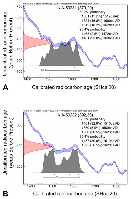
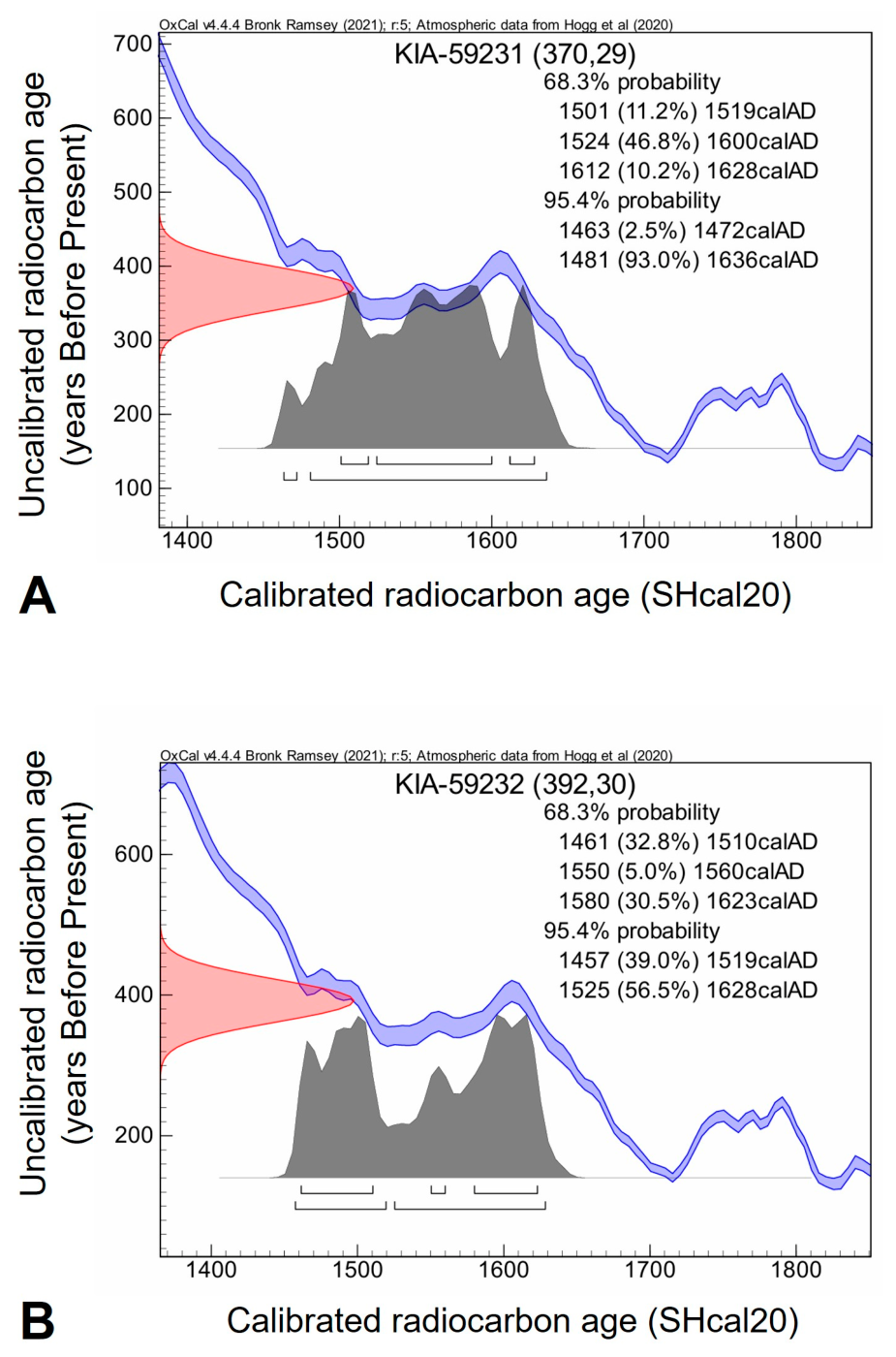
Results from radiocarbon dating of KIA-59231 and KIA-59232 are shown in Figure 2 and Table 1. KIA-59231 dates to 1463–1636 Common Era (CE) (SHcal20, 95.4% probability) and KIA-59232 to 1457–1628 CE (SHcal20, 95.4% probability). Both dates are consistent with each other and place the death of the individual in the late pre-Hispanic or Early Colonial Period.
Figure 2.
Histograms of radiocarbon dating calibration with the uncalibrated radiocarbon age curve (red), the calibration curve (blue) and the calibrated probability distribution (grey). The brackets mark the specific calendar date ranges for the different probability levels. (A): Sample from the thoracic wall (KIA-59231). (B): Sample from the gluteal region (KIA-59232).
Table 1.
Results of radiocarbon dating.
3.3. Anatomical Findings on Computed Tomography
3.3.1. Head and Neck
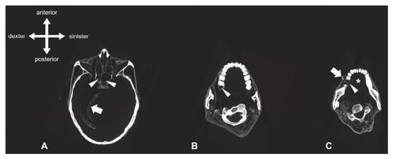
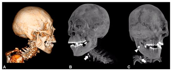
The calvaria is intact and the cranial sutures are closed. The skull base shows normal anatomical features with no evidence of bony destruction or traumatic changes. The cranial cavity appears largely empty. Preserved intracranial soft-tissue structures are identified as meninges including dura mater with falx cerebri and tentorium cerebelli (Figure 3A). Small, nodular soft-tissue remnants are present in the region of the left occipital lobe. The sella turcica and clivus are normally formed. The orbits exhibit smooth bony margins. Collapsed eyeballs are visible bilaterally. Additionally, the extraocular muscles and optic nerves seem delineated as far as the optic canal. Symmetrical conditions can be observed in the region of the temporal bones with normal visualization of the cochlea and vestibular apparatus. The mastoid cells are normally pneumatized. The internal and external auditory canals do show a normal configuration bilaterally. The auditory ossicles are not detectable on either side. The temporomandibular joints appear normal. The paranasal sinuses are normally pneumatized. Findings include symmetrically formed maxillary sinuses, hypoplasia of the frontal sinus, and asymmetrical configuration of the sphenoid sinuses. The uncinate process appears normally formed bilaterally. There is a slight deviation of the nasal septum to the right. The nasal conchae have a normal configuration. The ethmoid bone shows no defect areas. A circumscribed bony defect can be noted at the floor of the left sphenoid sinus, with a maximum diameter of approximately 1.3 cm. However, no extension of the defect toward the cranial cavity or direct communication with the intracranial space is detectable. The hard palate and the hyoid bone (Figure 3A) are intact. The tongue, the soft palate, and the oral floor musculature are not detectable (Figure 3B,C), while the epiglottis and the arytenoid cartilages are clearly identifiable. The cervical spine shows a marked angulation (Figure 4A), which we interpret as a post-mortem positional change related to the tightly flexed body position. There is no evidence of vertebral fracture, malformation or scoliosis. At the level of the fifth cervical vertebra (C5), a bipartite oval formation can be observed in the subcutaneous tissue on the right, with a maximum longitudinal dimension of approximately 25 mm and a width of approximately 8.2 mm (Figure 4B,C). The two components each have a diameter of approximately 9.7 mm. Between them lies a concave indentation with a diameter of approximately 7 mm, creating the impression of a central opening. This structure exhibits a mean density of approximately 86 HU. Based on its morphology and density, it is presumed to be an introduced foreign body or artifact.
Figure 3.
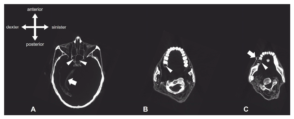
Transversal CT images of head and neck, inferior view. (A): Cranial cavity. While remnants of the dura mater are visible (arrow), no brain tissue is detectable. The sphenoid sinuses are marked with arrowheads. (B): Upper jaw. Note the empty socket of the right maxillary third molar (arrowhead). (C): Lower jaw. Note the empty double-rooted socket of the right mandibular first molar (arrowhead), the missing tongue (asterisk) and the distortion of the right cheek (arrow).
Figure 4.
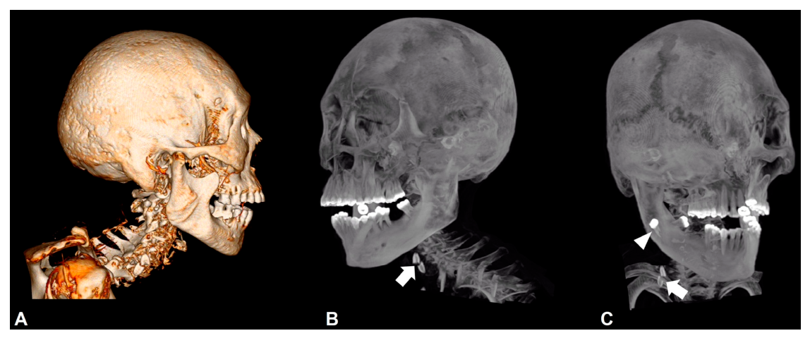
Three-dimensional computed tomography reconstruction of head and neck. (A): Right side view. No signs of artificial skull deformation are visible. The unusual curvature of the cervical spine is most likely a consequence of the packing of the mummy bundle: The space around the head was densely packed with filling material which fixed the head in an upright position despite the strongly flexed trunk. (B): Left-oblique view. Foreign body in the soft tissues of the neck (white arrow). (C): Right-oblique view. Foreign body in the soft tissues of the neck (white arrow) and post-mortem displaced tooth 18 (white arrowhead).
In the maxilla, the right third molar (18) is missing (Figure 3B). The corresponding socket presents as a smooth-margined, peripherally sclerotic single-rooted socket. The remaining maxillary teeth are intact. Enamel, dentin, and pulp chambers are clearly distinguishable, and the roots exhibit a normal morphology. In the mandible, edentulous spaces are present in the region of the second molars (37 and 47), accompanied by marked segmental alveolar ridge atrophy on both sides, consistent with long-standing tooth loss. The mandibular third molars (38 and 48) are positioned transversely on both sides and exhibit normal root morphology without discernible pathological changes. Tooth 46 is missing. The corresponding double-rooted socket is smooth-margined, peripherally sclerotic, and without atrophy (Figure 3C). On the medial aspect of the right mandible, an isolated, fully intact tooth can be observed, featuring normal enamel structure and a single root (Figure 4C). There is no evidence of carious changes or traumatic defects.
3.3.2. Thorax
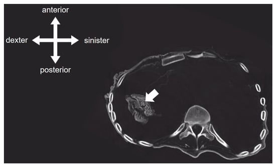
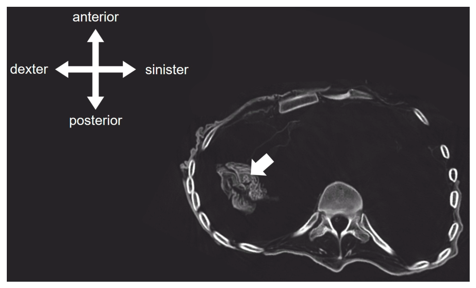
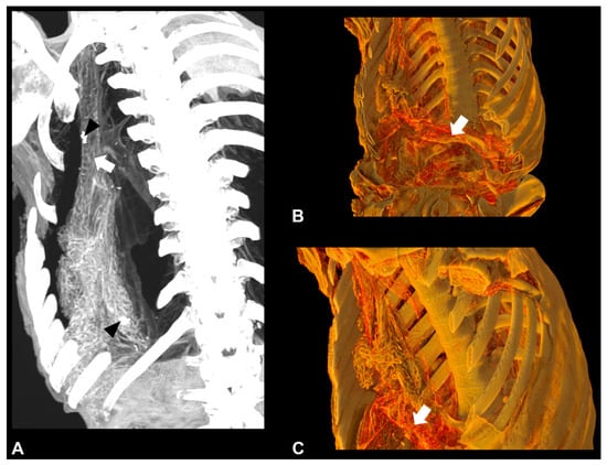
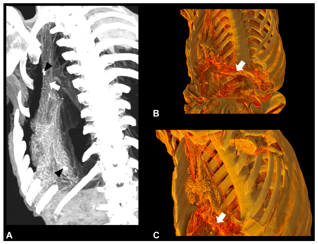
Longitudinal strands (approximately 12 cm in length) can be traced from the glottic region into the right hemithorax. Here, a spongy mass is visible lying against the right hemidiaphragm (Figure 5). Within this mass, there is an irregularly shaped foreign body featuring small spicule-like projections and a maximum diameter of approximately 6 mm. Its mean density is approximately 76 HU, with increased density at the periphery. The right hemidiaphragm appears tented (elevated cranially). Interruptions in the contour are visible in the posterior part. Mediastinal organs, such as the heart, could not be identified (Figure 5). However, an arc-shaped structure with cranially directed branches is notable. The structure ends in an oblique, smooth-edged termination. Supplementary maximum intensity projection (MIP) reconstructions clearly visualize the trachea and its cartilaginous rings within the elongated structural complex in the right thorax (Figure 6). Furthermore, the carina and the origins of the right and left main bronchi are identifiable. The cartilaginous rings of the larger segmental bronchi were also preserved and distinguishable. A triangular soft-tissue structure resting on the diaphragm is discussed as a possible remnant of the pericardium.
Figure 5.
Transversal CT image of the thorax, inferior view. Note the spongy mass in the right hemithorax (arrow) and the complete absence of the heart.
Figure 6.
Three-dimensional computed tomography reconstructions of the remaining thoracic structures. (A): Anterior view of the right thorax, showing remnants of the trachea and bronchi with visible cartilaginous rings (white arrow) and residual lung tissue. Foreign bodies are indicated by black arrowheads. (B,C): Left-oblique view on the remaining diaphragmatic structures (arrow). Note the complete absence of the heart.
3.3.3. Abdomen
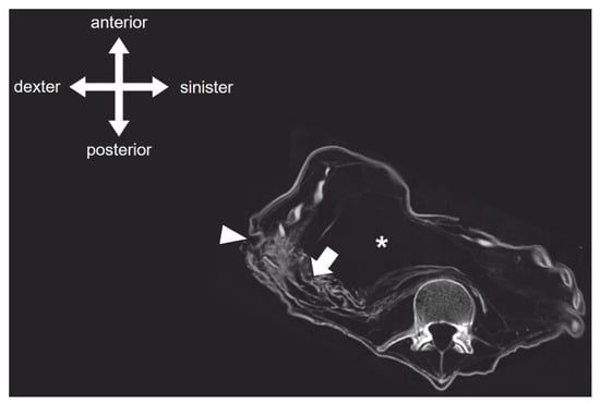
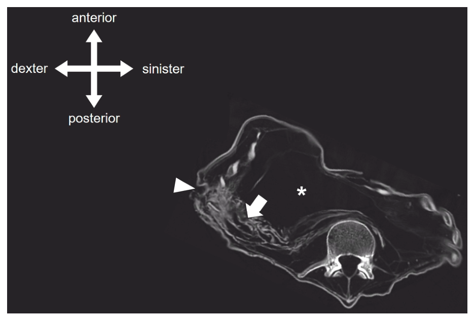
The parenchymal organs of the upper abdomen (liver, spleen, pancreas), the kidneys and the abdominal vascular structures could not be visualised on CT (Figure 7). Below the diaphragm sponge-like formations are visible predominantly in the right half of the abdomen. A cord-like or bundle-like structure (maybe including parts of the stomach and the intestines) extends between the elevated right hemidiaphragm and a point below the right costal margin.
Figure 7.
Transversal CT image of the abdomen, inferior view. The abdominal cavity appears largely empty (asterisk). Strands of tissue, which may contain portions of the stomach and intestines, extend toward the right abdominal wall below the lower rib cage (arrow). At the point of contact with the right abdominal wall, a circumscribed defect is visible (arrowhead).
3.3.4. Pelvis and Perineum
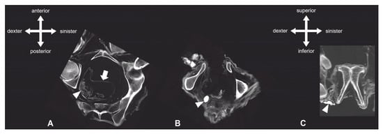
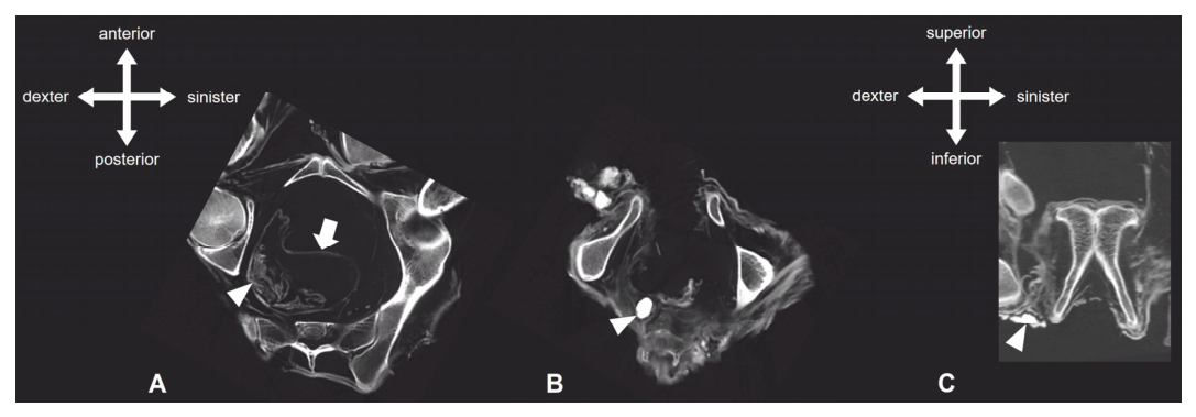
Portions of the described cord-like structure can be traced into the pelvis, extending on the right side into the presacral space. A fine, ring-like septation (probably including remnants of the peritoneum) is visible towards the anterior abdominal wall (Figure 8A). This septation can be traced down to the coccyx. At the level of the pelvic floor, this septation shows signs of destruction and can no longer be distinctly differentiated from the surrounding tissue.
Figure 8.
CT images of the pelvis. (A): Transversal section, inferior view. The pelvic cavity is largely empty. Possible remnants of the peritoneum (arrow) and the sigmoid/rectum (arrowhead) are visible. (B): Transversal section, inferior view. The pelvic floor shows a large defect. Note the foreign body (arrowhead). (C): Frontal section, anterior view. The narrow subpubic angle indicates male sex of the mummified person. Note the foreign body (arrowhead).
In addition to several smaller, homogeneous, and relatively smooth-bordered foreign bodies of increased density, there is a larger foreign body measuring approximately 1.5 cm in diameter (Figure 8B). This object has a mean density of approximately 140 HU and stands out clearly against the surrounding structures.
The pelvic floor shows a defect with a maximum diameter of approximately 8 cm. The external genitalia are absent (Figure 8B). Additional foreign bodies with a maximum diameter of up to 1.2 cm can be observed along the ischium on both sides (Figure 8C).
As no remnants of the external genitalia are visible, sex was determined based on the morphology of the pelvis. The morphology of the greater sciatic notch, the ventral arc, the subpubic concavity, and the subpubic angle (Figure 8C) is consistent with a male sex assessment.
3.3.5. Postcranial Skeletal System
There is no evidence of degenerative changes, sequelae of trauma, or destructive bone diseases. All epiphyseal and primary ossification centers appear completely fused, indicating an adult age. Radiological findings are summarized in Table 2.
Table 2.
Overview of the anatomical findings by body region.
3.3.6. Putative Sites of Evisceration
Three regions show findings that may represent access routes for the removal of internal organs:
- Right flank: Below the right costal margin, a cutaneous opening with a maximum longitudinal diameter of approximately 4 cm is visible (Figure 9A–C). The cord-like bundle of soft tissue described in Section 3.3.3, interpreted as remnants of the stomach and intestines, extends towards this opening. The morphology of the opening resembles a “tobacco-pouch” closure, characterised by the concentric retraction of the surrounding soft tissues. The opening was not recognised at the initial external inspection. However, on re-examination guided by the CT findings, a small, circumscribed opening was identified macroscopically at this site.Figure 9. Putative sites of evisceration. (A): Three-dimensional computed tomography reconstruction, right oblique view. Note the “tobacco-pouch” shaped opening below the right costal margin (red dotted outline). (B): Higher magnification of the opening (red dotted outline), detail from (A). (C): Corresponding photographic documentation of the opening (red dotted outline). (D): Three-dimensional computed tomography reconstruction, inferior view. Note the large defect of the pelvic floor (yellow dotted outline). (E): Higher magnification of the pelvic floor defect, detail from (D). (F): Three-dimensional computed tomography reconstruction, right oblique view. Note the distortion of the right cheek (white arrow), possibly related to trans-oral manipulation.
- Pelvic floor: The pelvic floor shows a large defect, while pelvic organs and the external genitalia are absent (Figure 9D,E). Several dense foreign bodies lie in the soft tissues of the pelvic outlet and along both ischia.
- Mouth: The mouth is open, and the soft tissues of the right cheek appear distorted (Figure 9F). Teeth 18 and 46 are missing without alveolar atrophy, and tooth 18 lies displaced on the medial aspect of the right mandible.
4. Discussion
4.1. Ancestor Cult, Mummification and Evisceration in Pre-Hispanic South America
As the investigated mummy shows signs of evisceration, we will first discuss the historical context of mummification techniques. The preservation of ancestors as mummies was a central feature of many pre-Hispanic societies along the west coast of South America. Deceased ancestors were understood to be reanimated through the creation of mummy bundles, which provided them with a new material embodiment. Rather than being regarded as permanently dead, they were understood to exist again, in a new shape, as active members of their communities, now residing in a different realm while remaining closely connected to their descendants and former communities through reciprocal relations of care and protection. Burial sites were often constructed in such a way that the ancestors could overlook the settlements and lands of their descendants. Spanish chroniclers’ descriptions of burial rituals suggest that “ancestor care” was understood as a reciprocal duty. Food offerings from cultivated fields served to nourish the ancestors in the afterlife while also demonstrating that the fields were being tended. Burial sites are often found near water sources, suggesting that the dead also watched over the essential resource of water for their living descendants [9]. These mummified ancestors, which were often accessible, displayed, and visited, may also have functioned as witnesses to land tenure, lineage continuity, and normative social order in societies without writing [10].
The Inca (1430–1530/70 CE) had perhaps the most pronounced ancestor cult of all pre-Hispanic societies. Once a small and insignificant group originating from Lake Titicaca, the Inca had created within only 100 years an empire that extended over more than 5000 km. During these campaigns of conquest, the Inca had become aware that they themselves did not possess a sufficiently developed ancestor cult with mummified ancestors to legitimize their power. This changed with the 9th Inca ruler, Inca Pachacutec Yupanqui. He established the cult surrounding the future mummies of Inca rulers as well as the remains of previous Inca ancestors. He ordered that all remains of the preceding Inca leaders be brought to the main temple in the Inca capital Cuzco, where they were to be wrapped in individual mummy bundles. He also decreed that, from then on, all mummies were to participate in public ceremonies visible to everyone. This practice visually reinforced the supremacy of the Inca conquerors [11]. Perhaps specialists from those groups encountered by the Inca that already practiced mummy cults had been incorporated into the Inca ruler’s sphere.
None of the mummies of the Inca rulers have survived to the present day. When the Spaniards conquered South America, they brutally advanced the destruction of the old cults, including those surrounding the mummies. Just as the Inca had once created their own mummy cult to secure their dominance over conquered groups, the Spaniards violently destroyed all “non-Christian ritual practices” to consolidate their own power. Many mummies fell victim to these actions [12]. With the Spanish conquest of the South American continent and its prolonged consequences, ancestor cults were increasingly suppressed, and the mummification of ancestors gradually ceased.
Thus, considerable uncertainty remains regarding the processes of mummification and their variation across different periods and geographical regions. This is primarily due to the absence of written sources from many Indigenous cultures prior to the Spanish conquest. Moreover, currently available scientific methods are often unable to verify suspected techniques, such as the possible smoking of corpses. Limited financial resources further restrict research on pre-Hispanic mummies, with many investigations still relying mainly on macroscopic observations. Advanced imaging techniques, including X-ray and CT, remain unavailable in many South American institutions, significantly limiting current knowledge regarding practices such as evisceration in pre-Hispanic societies. Although evidence remains scarce in general, several researchers have previously suggested or documented comparable procedures in different cultural and geographical contexts for pre-Hispanic mummies from the west coast of South America.
The first known artificially mummified individuals on the South American continent were taken completely apart after death and then reassembled. They were ancestors from the groups belonging to the Chinchorro culture that once lived along the coast of today’s northern Chile and southern Peru around 5000–1500 BCE. Towards the end of this culture, the climate changed and no such drastic methods were needed anymore [13]. Later cultures had very different but nevertheless very efficient methods for the preservation of their dead, leaving the outer body intact right from the beginning. An “exposure to heat” seems to have been the most widespread method for quick results [2].
Around 1500 BCE, two burial positions existed simultaneously throughout western South America: either the dead were lying extended on the back, clad in a burial shroud or leaf mat, or they were placed in a squatted position within a mummy bundle. The final resting place was either an underground chamber, a niche in the rocks or a funerary tower, depending on the individual’s status and the burial customs of the region. Mummy bundles were filled with grave goods. In this way, the dead were equipped with the necessities for their journey into the afterlife. To give the bundle a stable shape, it was stuffed with materials like raw cotton and/or leaves, materials that were also absorbent, thus helping to finalize the mummification process. As in pre-Columbian times in South America no wheel was known for any means of transport, intentional mummification may also have had the added benefit of reducing the body’s weight: an adult mummy could weigh under 10 kg. Also, the placement in a squatted position in a mummy bundle made it easier for the living to transport the deceased to the burial site.
For Southern “Ancient Peru”, Tello suspected complex mummification processes as a result of the investigation of mummies from the Paracas necropolis (400 BCE to 300 CE) of Cerro Colorado, on the southern central coast of what is today Peru [14]. Tello hypothesized that the methods applied to preserve venerated ancestors included the use of salt, chemicals, heat and the removal of internal organs via the pelvic floor (evisceration per anum) [14]. Later investigations by Vreeland and Cockburn do however suggest that Tello most probably did confuse taphonomic processes by decomposition with those of evisceration [15].
On the southern coast of Peru, in the lower part of the Osmore valley, the Chiribaya culture did flourish between 900 CE and 1350 CE. Some adult mummies from the sites of Chiribaya Baja, Loreto Viejo and Algodonal showed abdominal cuts by which the inner organs were taken out and then the body thoroughly cleaned, and its cavities were filled with cotton and even fruits and seeds [16]. Unfortunately, the examination itself is insufficiently published, making it difficult to assess the reported observations.
In the Central Highlands of what is today Bolivia, evisceration by abdominal cuts was confirmed in a study undertaken by Ponce Sanginés and Linares Iturralde [17]. They analysed ten mummies from the Carangas province, dating to the Late Horizon/Inca time and Early Colonial Period (1430–1570 CE). All ten (adult) mummies did show abdominal cuts through which the inner organs had been removed. This was also described in a Spanish document dating to 1580 for the Pacajes groups living then in that region.
Much further north, among the pre-Hispanic Muisca groups in the central Eastern Andes in what is today Colombia, evisceration per anum was done prior to an exposure to heat according to the Spanish chroniclers Fernández de Oviedo, 1548 [18] and Simón, 1621 [19] (both cited in [20]). However, this written description of a culture in its last phases, has not been confirmed on preserved Muisca mummies so far. A Muisca mummy from Gachantivá, cared for in the British Museum, has a cut on the side of the abdomen, by which the inner organs were taken out, thus helping the following drying process [20,21], but no evisceration per anum had been carried out.
However, despite the above-mentioned limitations, CT examinations have been possible in selected cases, particularly through international research collaborations. One such example is the investigation of Chachapoya mummies from the northeastern Andes of present-day Peru, where CT analyses enabled a more detailed examination of internal anatomical structures and proof of an evisceration seems to have been found. The mummies from the “Laguna de los Cóndores”, belonging to the Chachapoya-culture, were discovered in 1996. Of the 200 mummies found, six adults were CT-scanned and did show a possible evisceration per anum [16,22]. Nystrom investigated further mummies from that site and offers an alternative explanation for this practice. Firstly, he argues that the treatment may have represented a “normal cleaning process” carried out prior to burial, rather than an intentional procedure aimed at facilitating mummification. He emphasizes that “the modern Western definition of intentionality may not accurately reflect the concept in any number of pre-historic non-Western contexts” [23]. Secondly, Nystrom suggests that the presence of a prolapsed rectum does not necessarily indicate deliberate intervention, but may instead have resulted from post-mortem processes, such as the accumulation of intra-abdominal gases, as observed in mummy #7 [23].
Cultural practices as well as mummification techniques may have spread through trade connections, the effects of war, and migration of people and/or knowledge. However, such processes are difficult to trace, as our knowledge of pre-Hispanic mummification methods remains limited and reported cases of evisceration are rarely supported by imaging. Against this background, the present case is one of the few in which intentional evisceration of a pre-Hispanic individual, reportedly from the Andean highlands, can be documented by CT imaging.
4.2. General Morphology of the Present Case and Evidence of Intentional Evisceration
The tightly flexed body position of this mummy is characteristic of pre-Hispanic burial practices, as it facilitated the placement of the body within a mummy bundle and its subsequent transport to the place of burial. Irrespective of whether the individual died shortly before or after the arrival of the Spaniards, the flexed body position, the burial in a mummy bundle and the treatment of the body clearly follow pre-Hispanic mortuary tradition. Another characteristic feature is the open mouth. In contrast to Egyptian mummies, no particular attention has been paid to the preservation of the facial features.
CT reveals the extensive absence of internal organs. Different criteria were considered to distinguish deliberate post-mortem intervention from ante- or perimortem injuries and from taphonomic damage: First, the loss of soft tissue is selective. Decomposition alone cannot account for the selective loss of the heart and parenchymal organs while the trachea, bronchial cartilages and remnants of the diaphragm and the peritoneum remain preserved. Second, the right flank opening is circumscribed, provides direct access to the abdominal cavity, and the intestinal remnants are oriented towards it. Third, the skeleton shows no fractures, cut marks or other signs of trauma. Taken together, these findings suggest deliberate post-mortem manipulation, indicating an active form of mummification.
Apart from minimal tissue remnants in the occipital region, the brain was no longer detectable. However, the dura mater with falx cerebri and tentorium cerebelli were preserved and clearly distinguishable. Examination of the bony structures yielded no evidence of transnasal or transpharyngeal brain removal. Nevertheless, the circumscribed defect at the base of the left sphenoid sinus could be associated with an unsuccessful attempt to access the intracranial cavity. Loss of the brain with preservation of the dura mater is common in naturally desiccated mummies and does not speak against intentional evisceration in other body regions.
4.3. Trans-Oral Access for Evisceration of the Oral Cavity
Access to the upper aerodigestive tract was likely gained via the oral cavity. We hypothesise that teeth 18 and 46 were lost during this manipulation. However, to our knowledge, an intentional removal of teeth has not been described as part of Andean mummification practices, and post-mortem tooth loss during later handling of the body cannot be excluded. The missing maxillary right third molar (18) was found lying loose against the medial aspect of the right mandible, while the first molar was no longer present. In contrast, the missing teeth 37 and 47 show smooth-edged, atrophic alveoli, indicating tooth loss that occurred during life. The soft tissues of the oral cavity, including the tongue, the soft palate, and the oral floor musculature were largely absent. Since the right cheek looked distorted and no further defects of the skin of head and neck were observed, trans-oral access seems possible.
4.4. Evisceration of the Thoracic and Abdominal Cavity via the Right Flank
In the thorax, the trachea, carina, and the main and segmental bronchi were still detectable. The spongy mass in the right hemithorax, extending down to the diaphragm, likely corresponds to the collapsed lung. Within this formation, a small, irregularly shaped foreign body with spicule-like margins is visible. An interbronchial location cannot be ruled out, so this foreign body may have been introduced during mummification. The arc-shaped mediastinal structure likely corresponds to the remaining aortic arch. Its sharp ending is consistent with deliberate organ removal, since the heart itself is no longer detectable. The triangular structure resting on the diaphragm likely represents remnants of the pericardium. The interruptions in the posterior contour of the right hemidiaphragm could indicate that the thoracic organs were removed through the diaphragm from the abdominal cavity. The significance of the oval, centrally cleft structure in the soft tissues of the right neck remains unclear. Based on its shape, density, and location, it could be a foreign body which was placed during the thoracic evisceration process. Elongated, fibrous structures likely correspond to the remaining gastrointestinal tract (predominantly segments of the small intestine). They extend directly toward a cutaneous opening in the right flank. The portions of this bundle extending into the presacral space may represent remnants of the sigmoid and rectum. The opening below the right costal margin is the most convincing of the proposed access routes: it is circumscribed, it communicates with the abdominal cavity, and the intestinal remnants are oriented towards it. Lateral abdominal incisions for the removal of the internal organs have been reported for Chiribaya mummies from the Osmore valley [16], for mummies from the Carangas province in Bolivia [17] and for a Muisca mummy from Gachantivá [20,21], and they therefore represent the best-documented evisceration route in the Andes.
4.5. Evisceration of the Pelvic Cavity via the Pelvic Floor
The second defect zone is much more pronounced, encompassing the pelvic floor with a complete loss of external genital structures. This opening may have served as an additional access point for the removal of further abdominal organs. However, it cannot be determined which organs were removed through which opening. Additionally, the perineal region is also particularly prone to post-mortem damage. For example, scavengers preferentially attack the body orifices and the decomposition of the pelvic viscera with the accumulation of gases may lead to rupture of the perineum or prolapse of the rectum, as discussed by Nystrom et al. for Chachapoya mummies [23]. However, some observations argue against a purely taphonomic origin in the present case: No gnawing marks were found at the pelvic bone. Additionally, several dense foreign bodies, possibly stones, are located in the soft tissues of the pelvic floor, where they may have been placed deliberately during the mummification process. We therefore regard evisceration per anum as a plausible, but not unequivocally proven, interpretation of the pelvic floor defect. The same applies to the absence of the external genitalia, which may have been removed during this procedure or were unintentionally lost post-mortem.
4.6. Limitations
This study has important limitations. The provenance rests solely on Meyen’s account, precluding cultural attribution. While the selective absence of internal organs clearly indicates evisceration, the identification of the access routes is based on CT morphology alone, and post-mortem damage to the external openings cannot be fully excluded. Additionally, no cause of death could be determined morphologically.
5. Conclusions
In the present study, a Peruvian mummy currently curated in the anatomical collection of the Charité—Universitätsmedizin Berlin, Germany, was investigated using a combination of macroscopic examination, high-resolution computed tomography (CT), and radiocarbon dating. The mummy was collected by the Prussian physician, botanist, and explorer Franz Julius Ferdinand Meyen during his journey through South America between 1830 and 1832. According to Meyen’s records, it originated from a natural cave in the Andes in the region of present-day Pasco, Peru.
The examination identified the individual as an adult male. Radiocarbon dating yielded dates between the mid-fifteenth and early seventeenth centuries CE, placing the mummy within the late pre-Hispanic or Early Colonial Period. Anatomical findings provide evidence for deliberate post-mortem removal of the internal organs as part of an artificial mummification procedure. The reconstruction of the CT findings suggests that evisceration was performed through several access routes. The oral cavity appears to have been used to access and remove structures of the upper aerodigestive tract. In addition, a distinct cutaneous opening in the right flank, directly communicating with the abdominal cavity, appears to have provided access for the removal of thoracic and abdominal organs. A further and considerably larger defect was present in the pelvic floor, accompanied by complete absence of the external genitalia. The anatomical configuration suggests that this opening may have served as an additional route for removing organs. Thus, the overall findings indicate that the mummy underwent deliberate evisceration, probably through the mouth, the right flank, and the pelvic floor.
The present case is particularly important in the context of pre-Hispanic South American mummification, for which direct evidence of intentional evisceration remains scarce. The reconstruction of ancient mummification practices is hampered by the loss of Indigenous knowledge following the Spanish conquest, the absence of written sources for many pre-Hispanic societies, the fragmentation or loss of archaeological contexts, and the limited availability of advanced imaging techniques such as X-ray or even CT in many South American institutions. Consequently, previously reported cases of evisceration are relatively few and, in some instances, difficult to evaluate because of incomplete documentation or possible taphonomic explanations. Against this background, the mummy investigated in the present study represents an exceptional case in which intentional evisceration can be demonstrated on the basis of detailed modern CT imaging. At the same time, the absence of grave goods and the original mummy bundle prevents a secure attribution to a specific cultural group.
Given this insecure attribution, it remains difficult to contact a specific group for inquiries about an interest in repatriation. To our knowledge, there is no general interest in repatriating mummies in European collections to Peru, if they do not belong to very specific groups. Therefore, no efforts have been made so far to contact Peruvian institutions.
Author Contributions
A.-M.B., S.S. and R.S. contributed equally. S.S.: conceptualization, investigation, writing—original draft. A.-M.B.: investigation, writing—original draft. R.S.: investigation, formal analysis, visualization, writing—original draft. M.G.: investigation, writing—review and editing. E.H.: resources, writing—review and editing. A.W.: investigation, writing—review and editing. All authors have read and agreed to the published version of the manuscript.
Funding
This research received no external funding.
Institutional Review Board Statement
The study followed the codes of ethics of the British Association for Biological Anthropology and Osteoarchaeology and the American Association for Biological Anthropology, as well as the Declaration of Helsinki and the recommendations of the German Museums Association for the Care of Human Remains in Museums and Collections. According to the local Ethics Committee, no further formal approval was necessary for the present study. Independently of this, the examination of the mummy, including the removal of two small tissue samples for radiocarbon dating, was authorised the institutional board of the Center of Anatomy, Charité—Universitätsmedizin Berlin (28 July 2025).
Informed Consent Statement
Not applicable.
Data Availability Statement
The CT datasets supporting the findings of this study are available from the corresponding author upon reasonable request.
Acknowledgments
We are very grateful to Rubén H. Buitrón Picharde (Fundación Augusto N. Wiese) for generously sharing his expertise on the current state of research on Peruvian mummification techniques and for his valuable literature recommendations.
Conflicts of Interest
The authors declare no conflicts of interest.
References
- Arriaza, B. Chinchorro Bioarchaeology: Chronology and Mummy Seriation. Lat. Am. Antiq. 1995, 6, 35–55. [Google Scholar] [CrossRef] [Scilit]
- Barandiarán, A. Mitos, Leyendas y Tradiciones Lambayecanas; Club de Autores y Lectores: Lima, Peru, 1938. [Google Scholar]
- Schumann, S.; Begerock, A.-M.; González, M.; Heuckendorf, E.; Sokiranski, R.; Tessmann, B.; Winkelmann, A. Über den Erwerb und Verbleib der Mumien und Schädel, gesammelt von Franz Julius Ferdinand Meyen. HiN—Alexander von Humboldt im Netz. Int. Z. Für Humboldt.-Stud. 2026, 27, 139–154. [Google Scholar] [CrossRef] [PubMed]
- Broesike, G. Das Anthropologische Material des Anatomischen Museums der Königlichen Universität zu Berlin. Erster Theil; Friedrich Vieweg und Sohn: Braunschweig, Germany, 1881. [Google Scholar]
- Friedman, S.N.; Nguyen, N.; Nelson, A.J.; Granton, P.V.; MacDonald, D.B.; Hibbert, R.; Holdsworth, D.W.; Cunningham, I.A. Computed Tomography (CT) Bone Segmentation of an Ancient Egyptian Mummy: A Comparison of Automated and Semiautomated Threshold and Dual-Energy Techniques. J. Comput. Assist. Tomogr. 2012, 36, 616–622. [Google Scholar] [CrossRef] [Scilit] [PubMed]
- Buikstra, J.E.; Ubelaker, D.H. (Eds.) Standards for Data Collection from Human Skeletal Remains: Proceedings of a Seminar at the Field Museum of Natural History; Arkansas Archaeological Survey Research Series 44; Arkansas Archaeological Survey: Fayetteville, AR, USA, 1994. [Google Scholar]
- Bronk Ramsey, C.; Lee, S. Recent and Planned Developments of the Program OxCal. Radiocarbon 2013, 55, 720–730. [Google Scholar] [CrossRef] [Scilit]
- Hogg, A.G.; Heaton, T.J.; Hua, Q.; Palmer, J.G.; Turney, C.S.M.; Southon, J.; Bayliss, A.; Blackwell, P.G.; Boswijk, G.; Ramsey, C.B.; et al. SHCal20 Southern Hemisphere Calibration, 0–55,000 Years cal BP. Radiocarbon 2020, 62, 759–778. [Google Scholar] [CrossRef] [Scilit]
- Shimada, I.; Segura Llanos, R.; Goldstein, D.; Shimada, M.; Speakman, R.; Wagner, U.; Watanabe, H. What Did People Do at Pachacamac? Identity, Form, Timing and Meaning of Offerings. In Proceedings of the 34th Midwest Conference on Andean and Amazonian Archaeology and Ethnohistory, Nashville, TN, USA, 25–26 February 2006. [Google Scholar]
- Begerock, A.-M. Ein Leben mit den Ahnen—Der Tod als Inszenierung für die Lebenden: Eine Untersuchung Anhand Ausgewählter Kulturen des Westlichen Südamerika zu Hinweisen Einer Intentionellen Mumifizierung der Verstorbenen und der Kulturimmanenten Gründe. Ph.D. Thesis, Freie Universität Berlin, Berlin, Germany, 2015. Available online: http://www.diss.fu-berlin.de/diss/receive/FUDISS_thesis_000000101717 (accessed on 7 October 2026).
- Cobo, B. Inca Religion and Customs; Hamilton, R., Ed. and Translator; University of Texas Press: Austin, TX, USA, 1990. [Google Scholar]
- Garcilaso de la Vega, I. Wahrhaftige Kommentare zum Reich der Inka; Thiemer-Sachse, U., Ed.; Rütten & Loening: Berlin, Germany, 1983. [Google Scholar]
- Marquet, P.; Santoro, C.; Latorre, C.; Standen, V.; Abades, S.; Rivadeneira, M.; Arriaza, B.; Hochberg, M. Emergence of Social Complexity Among Coastal Hunter-Gatherers in the Atacama Desert of Northern Chile. Proc. Natl. Acad. Sci. USA 2012, 109, 14754–14760. [Google Scholar] [CrossRef] [Scilit] [PubMed]
- Tello, J.C. Antiguo Perú: Primera Época; Comisión Organizadora del Segundo Congreso Sudamericano de Turismo: Lima, Peru, 1929. [Google Scholar]
- Vreeland, J.M.; Cockburn, A. Mummies of Peru. In Mummies, Disease and Ancient Cultures; Cockburn, A., Cockburn, E., Eds.; Cambridge University Press: Cambridge, UK, 1985; pp. 135–174. [Google Scholar]
- Guillén, S. Artificial Mummies from the Andes. Coll. Antropol. 2004, 28, 141–157. [Google Scholar] [CrossRef] [Scilit]
- Ponce Sanginés, C.; Iturralde, E.L. Comentario Antropológico Acerca de la Determinación Paleoserológica de Grupos Sanguíneos en Momias Prehispánicas del Altiplano Boliviano; Publicación 15; Academia Nacional de Ciencias de Bolivia: La Paz, Bolivia, 1966. [Google Scholar]
- Fernández de Oviedo, G. Historia General y Natural de las Indias; Real Academia de la Historia: Madrid, Spain, 1851. [Google Scholar]
- Simón, P. Noticias Historiales de las Conquistas de Tierra Firme en las Indias Occidentales; Banco Popular: Bogota, Colombia, 1981; Volume 3. [Google Scholar]
- Martínez Martín, A.; Santamaría, L.M. Sobre la momificación y los cuerpos momificados de los muiscas. Rev. Salud Hist. Sanid. 2012, 7, 61–80. [Google Scholar]
- Cárdenas-Arroyo, F. La momificación indígena en Colombia. Bol. Mus. Oro 1989, 25, 121–123. [Google Scholar]
- Friedrich, K.M.; Nemec, S.; Czerny, C.; Fischer, H.; Plischke, S.; Gahleitner, A.; Viola, T.B.; Imhof, H.; Seidler, H.; Guillén, S. The Story of 12 Chachapoyan Mummies Through Multidetector Computed Tomography. Eur. J. Radiol. 2010, 76, 143–150. [Google Scholar] [CrossRef] [Scilit] [PubMed]
- Nystrom, K.C.; Buikstra, J.E.; Muscutt, K. Chachapoya Mortuary Behavior: A Consideration of Method and Meaning. Chungará 2010, 42, 477–495. [Google Scholar] [CrossRef] [Scilit]

Disclaimer/Publisher’s Note: The statements, opinions and data contained in all publications are solely those of the individual author(s) and contributor(s) and not of MDPI and/or the editor(s). MDPI and/or the editor(s) disclaim responsibility for any injury to people or property resulting from any ideas, methods, instructions or products referred to in the content. |
© 2026 by the authors. Licensee MDPI, Basel, Switzerland. This article is an open access article distributed under the terms and conditions of the Creative Commons Attribution (CC BY) license.