


Abstract
Background/Objectives: The brachial plexus is a complex network of nerves responsible for the sensory and motor functions of the upper limbs. While extensively studied, the typical configuration taught only sometimes matches what is found in individuals, as numerous variations have been documented. These variations can have significant clinical and surgical implications. This study explored the prevalence and types of brachial plexus variations in donor bodies from the Midwestern United States. Methods: Using bodies donated through the Gift of Body Donation program at A.T. Still University’s Kirksville College of Osteopathic Medicine, the brachial plexuses of 32 donors were analyzed. Results: Among the 64 plexuses examined, 33 (52%) had a typical M-shaped median nerve formation without additional variations, 18 (28%) had a typical M-shaped formation with additional variations, 6 (9%) had an abnormal M without other variations, and 7 (11%) had an abnormal M with other variations. Thus, 31 plexuses (48%) showed atypical branching patterns. The most common variations were extra communicating branches, observed in 17 (55%) of the 31 variant plexuses. These branches were most often found between the medial and lateral cords (13/17, 76%) and between the musculocutaneous and median nerves (4/17, 24%). Other, less common variations included variant nerves, small branches, and rejoining nerves. Conclusions: This study highlights the significant prevalence of variations in the brachial plexus, which may impact clinical and surgical practices. Further research is needed to fully understand the implications of these variations, especially in procedures like nerve blocks and reconstructive surgeries.
1. Introduction
The brachial plexus (BP) is a complex network of nerves responsible for providing motor and sensory innervation to the upper extremity. As the BP progresses distally from the neck to the hand, additional branches form that can be characterized as roots, trunks, divisions, cords, and terminal nerves [1,2,3,4].
This standardized branching pattern has been extensively studied; however, variations are present across patient populations [5,6,7]. Although BP variations have been documented worldwide, limited research has compared the distribution of specific variation patterns across different populations. The current literature suggests prevalence rates ranging from 30–60% in donor body specimens [6,7,8]. For example, variations in the musculocutaneous nerve (MCN) in relation with the median nerve (MN) are well documented and may account for branching variation [9,10]. Common variations include fewer or greater roots, atypical nerve branching, and extra communicating branches. An Iranian study identified pre-fixed and post-fixed plexuses—variants in which root contributions shift one spinal level rostrally or caudally, respectively—in 12% of donor bodies [5] while a Chinese study found that 33% of BPs displayed at least one variation, with many involving communicating branches between cords or terminal nerves [6]. A US study conducted in Arizona revealed a high frequency of variations when following the MCN and MN distally [7]. A more recent donor body study from Georgia similarly found a high rate of infraclavicular BP variation in 44.4% of plexuses [11]. This study also found bilateral variations to be more common than unilateral ones while identifying several previously undescribed branching patterns [11].
These anatomical differences have significant clinical implications, influencing the effectiveness of nerve blocks, surgical interventions, and injury management. If unrecognized, these deviations may lead to failed nerve blocks [12], misdiagnosis [7], or iatrogenic injury [12]. For example, aberrant courses of the MCN or MN have been directly associated with incomplete anesthesia during BP blocks in surgical and obstetric settings [4,13,14]. Awareness of such variations is essential for clinicians to minimize complications and improve procedural outcomes [15,16]. The present study builds on this existing body of research by examining the prevalence and types of BP variations in a Midwestern American donor population.
2. Materials and Methods
This study analyzed 32 donor body specimens (64 BP) obtained through A.T. Still University’s Gift of Body Program at A.T. Still University Kirksville College of Osteopathic Medicine.
All donor bodies were preserved using a formaldehyde-based embalming solution according to the institution’s standard protocol. Blunt dissection was performed to expose the cervical nerve roots. The BP and nearby vessels were carefully cleared of subcutaneous fat and fascia for adequate visualization. Each nerve root was followed distally to its terminal branches in the hand.
The formation of the MN, MCN, and ulnar nerve through the lateral and medial cords formed a characteristic M-shape on the anterior surface of the axillary artery [3]. This M-shape served as an anatomic landmark for orienting each BP and identifying anatomic variations. Each plexus was carefully photographed and analyzed from its origin to its termination.
Each BP was assigned to one of four mutually exclusive categories using a classification system developed for this study: normal plexus, normal M-shape with additional variation, abnormal M-shape with no variation, and abnormal M-shape with additional variation. These categories are consistent with previous anatomical definitions of the standard M-shape pattern and well-documented variations involving nerve communication or abnormal courses [4,7].
A typical M-shaped configuration with additional variation retained the expected formation of the major terminal nerves but exhibited variations elsewhere in the plexus. An atypical M-shaped configuration demonstrated altered formation of the MN without additional anatomical variation. An atypical M-shaped configuration with additional variation demonstrated both altered MN formation and variations elsewhere in the plexus.
All data was analyzed using Microsoft Excel 16.11 (Microsoft Corporation, Redmond, WA, USA). The prevalence and variation in the BP was summarized using descriptive statistics, including frequency and percentage. Fischer exact tests were performed to assess the associations between donor sex and laterality with the presence of anatomical variation. A p-value of <0.05 was considered statistically significant.
All procedures in the current study were considered exempt by the A.T. Still University Kirksville Institutional Review Board.
3. Results
A total of 64 BP from 32 donor bodies were examined. The sample was made up of 16 males and 16 females, aged 52 to 95 years. The average age of the sample population is 77 years, with a standard deviation of 10.3.
3.1. Classification of Variations
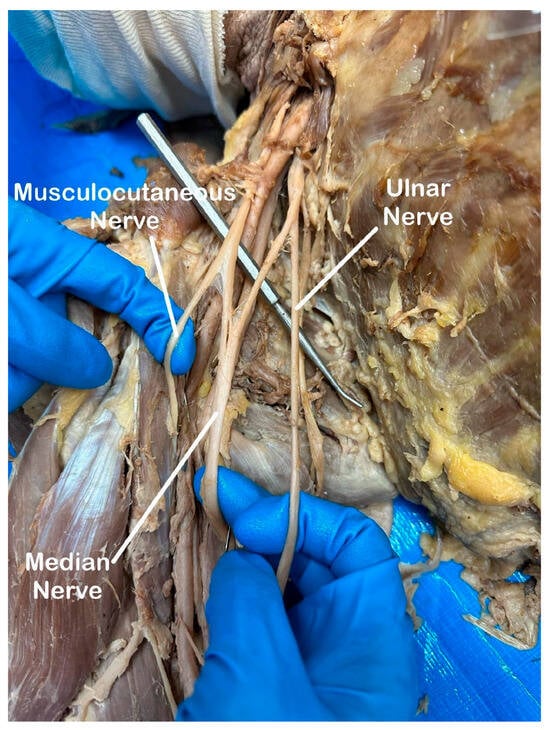
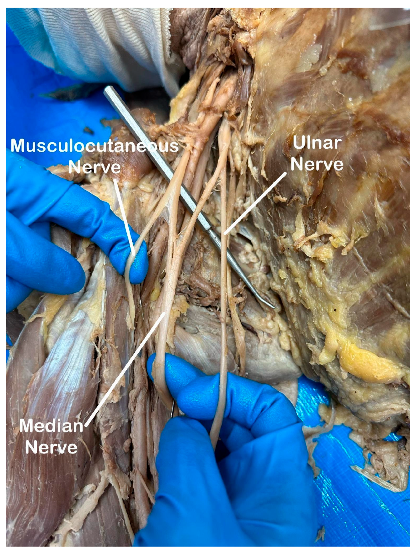
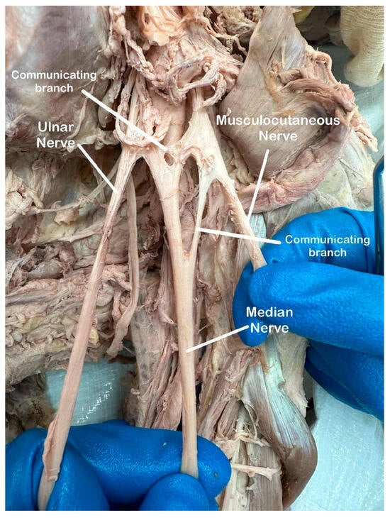
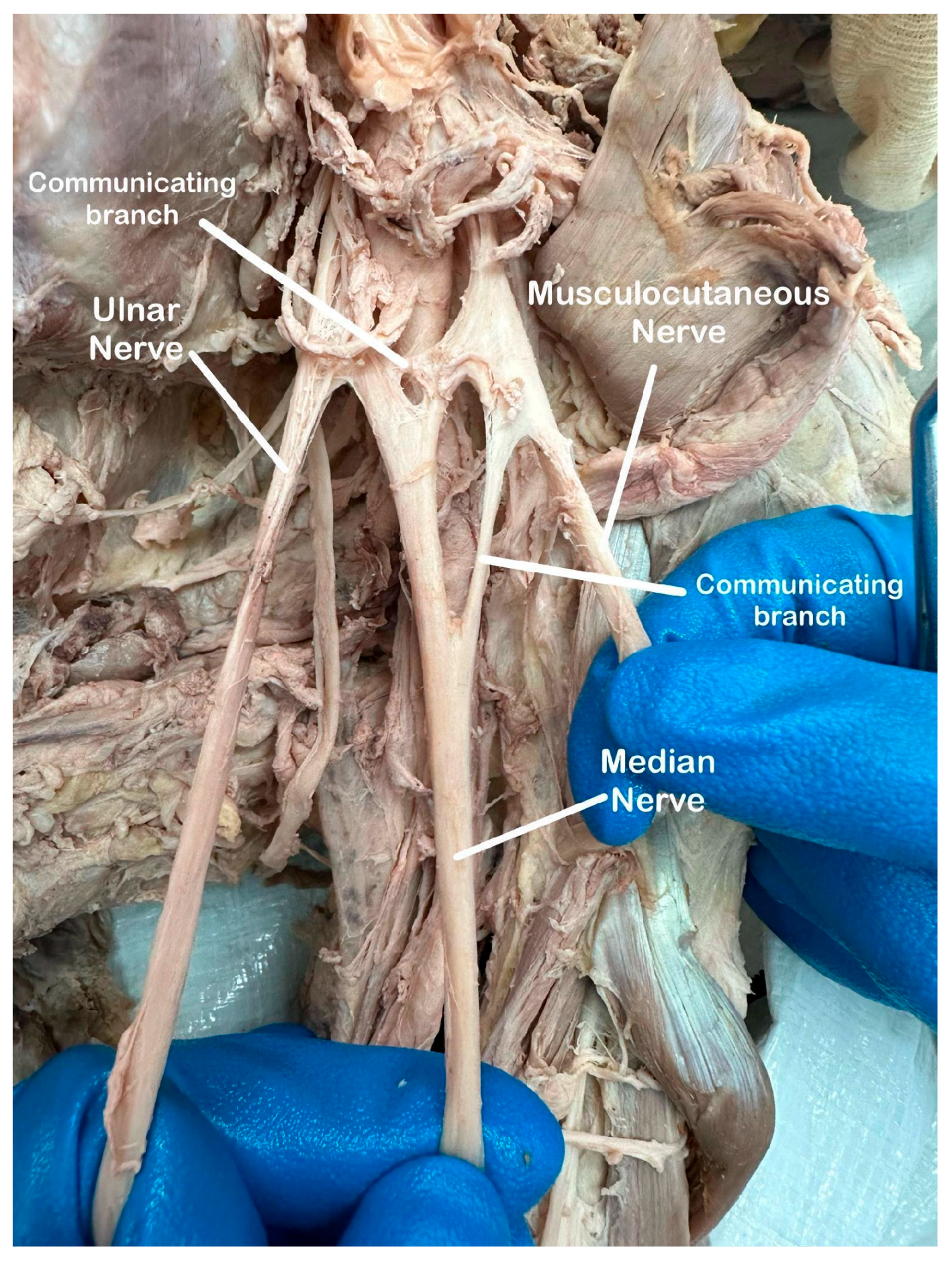
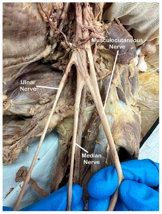
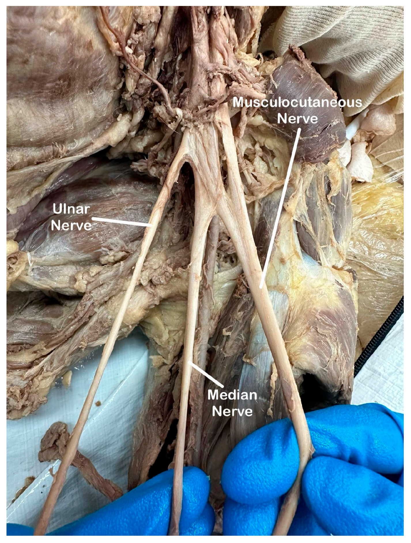
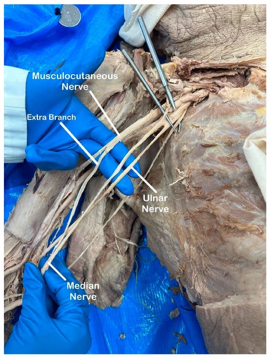
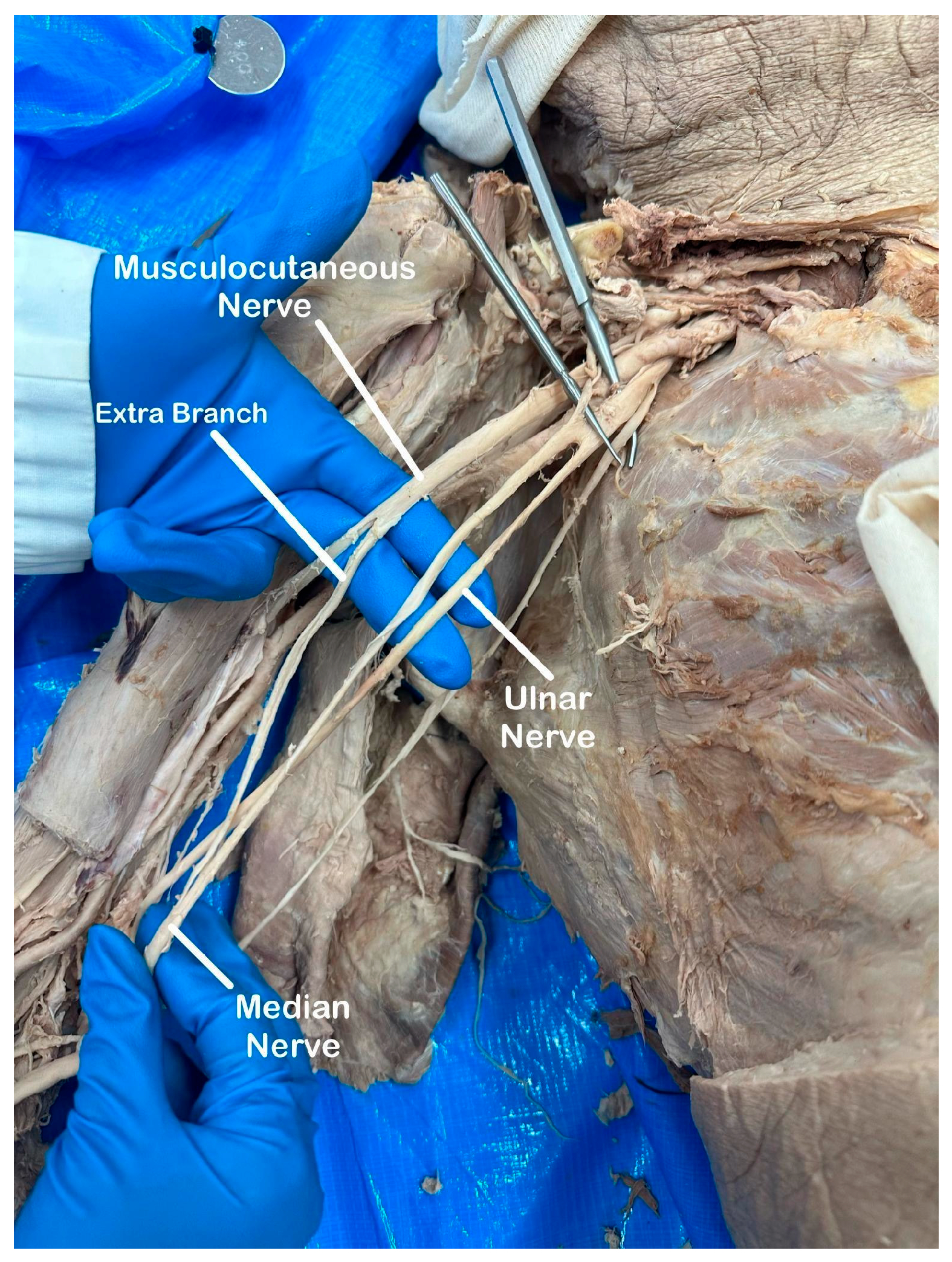
Of the 64 BP examined, 33 (52%) showed a normal M-shape plexus with no additional variation (Figure 1). The remaining 31 (48%) plexuses deviated from the normal M-shape and were classified in the following categories: normal M-shape plexus with variation, 18 (28%, Figure 2); abnormal M-shape with no variation, 6 (9%, Figure 3); and abnormal M-shape with variation, 7 (11%, Figure 4).
Figure 1.
Normal M-shape plexus with no variation.
Figure 2.
Normal M-shape plexus with variation.
Figure 3.
Abnormal M-shape with no variation.
Figure 4.
Abnormal M-shape with variation.
3.2. Normal M-Shape Plexus with Additional Variation
Of the 18 plexuses demonstrating a normal M-shape with variation, 7 plexuses showed extra communicating branches between the medial and lateral cords. Additionally, three plexuses showed communicating branches between the MN and MCN. Less frequent variations involving the MCN included one plexus with bifurcation proximal to its contribution to the biceps brachii muscle. Another MCN had a direct origin from the MN, coursing to the biceps brachii and brachialis muscles. Variations involving the MN included one plexus with extensive communicating branches proximal to its origin in the upper arm, one plexus displayed distal anastomosis between the ulnar and MCN in the distal arm, and one plexus identified the MN traveling posterior to the brachial artery. One plexus demonstrated an M-shaped configuration that was wrapped around the axillary artery more proximally, with shorter roots, trunks, divisions, and cords relative to the other plexuses.
3.3. Abnormal M-Shape Plexus with No Variation
In six female plexuses, the atypical formation of the MN altered the characteristic M-shaped configuration. However, no isolated abnormal M-shaped plexuses were found in male donor bodies.
3.4. Abnormal M-Shape Plexus with Additional Variation
Among the 7 plexuses with an abnormal M-shape with variation, 6 plexuses showed extra communicating branches between medial and lateral cords immediately proximal to its terminal branches. One plexus showed a communicating branch between the MN and MCN immediately distal to its terminal branches.
3.5. Variations by Sex and Side
The prevalence of BP variations was nearly equal between male (47%) and female (50%) donors. No statistically significant difference was observed (Fischer’s exact test, p = 0.054).
Variations were observed on both sides: 10 of the 31 variant plexuses were right-sided (32%), seven were left-sided (23%), and 14 occurred bilaterally in seven donor bodies (45%). No statistically significant difference was observed in left and right-sided plexuses (Fischer’s exact test, p = 0.79).
4. Discussion
4.1. Overview and Significance
This study demonstrates substantial anatomical variability in the BP configuration within a Midwestern donor body population. The most common variations showed the normal M-shape with variation, whereas abnormal formation of the MN occurred less frequently. By organizing these patterns into mutually exclusive categories, the current study provides a framework for comparing distinct variations and considering their anatomical and clinical relevance. Despite growing recognition of BP variability, systematic donor body studies characterizing these variations in U.S. donor populations remain relatively limited compared to the broader international literature, and few have examined a geographically distinct Midwestern cohort specifically. By documenting variation patterns in this population, the current study addresses this gap and provides a comparative dataset for future work assessing whether variation prevalence and type differ meaningfully across U.S. regions.
4.2. Variations Among Donor Populations
The most common variation identified was an M-shaped BP with additional communicating branches, particularly between the medial and lateral cords. Previous studies have reported similar communications between the cords, although their frequencies vary among donor populations. Han et al. previously identified communicating branch variations as the most common variation [6]; the overall prevalence of variation in their sample of 60 Chinese donor bodies was 66.7%, exceeding the 48% variation found in our sample, likely attributed to an increased sample size. Additionally, another study utilizing an Arizona donor population reported a 5% prevalence of communicating branches between the MCN and MN [7], which aligns with the results from our study suggesting a comparable frequency across U.S. samples. The axons contributing to the BP undergo extensive growth and reorganization during upper-limb development, creating opportunities for communication anastomosis to exist [2,9]. Differences in variation patterns may also reflect population characteristics [17], donor body preservation methods [6], and dissection techniques [18].
4.3. Distribution by Sex and Side
Variation prevalence was similar between male and female donor bodies. Although all six plexuses with an isolated abnormal M-shaped configuration were from female donors, this finding is not supported by the current literature [9,11] and should be interpreted cautiously given the small sample size. Variations also occurred both unilaterally and bilaterally within 31 variant plexuses. Unilateral variations (17, 55%) were more common than bilateral variations (14, 45%) in our study. This contrasts the findings of Bozhikova et. al, which reported a higher rate of bilateral variation (14, 87.5%) relative to unilateral variation (2, 12.5%) in a Georgia donor sample [11]. However, this discrepancy was likely attributed due to their small sample size (n = 16).
4.4. Clinical and Surgical Implications
Variation in the BP can directly affect diagnostic, anesthetic, and surgical processes. Fusion or absence of expected nerve components can complicate diagnostic interpretation [6] and surgical intervention [9,11]. For example, when the MCN arises directly from the MN, an MN lesion could manifest as arm flexor weakness and loss of sensation in the lateral forearm [6,19]. In infants with Erb or Klumke palsy resulting from excessive traction during childbirth [20], variant nerve branches could produce unexpected electromyography (EMG) findings and complicate localization of injury [9,21]. These variations are also relevant in procedures where nerve blocks are dependent on predictable nerve locations. Deviations from the expected anatomical course could lead to inadvertent nerve injury or incomplete anesthesia [9].
4.5. Limitations
All dissections were conducted using a standardized approach, with careful observations and documented as high-resolution images. Nevertheless, several limitations must be acknowledged. This study was limited by its relatively small sample of Midwestern donor bodies which may limit generalizability to a broader population. Additionally, the bilateral plexuses obtained from each donor may not represent statistically independent observations. Furthermore, donor information such as handedness or a history of trauma to the upper extremity or axilla was not provided and may also be contributing factors to variations observed. Lastly, the classification system developed for the current study has not been externally validated and may not encompass the full spectrum of BP variation.
4.6. Future Directions
Given the prevalence of BP variations observed in this study, future research should aim to expand sample size and demographic diversity to improve the generalizability of these findings. A more comprehensive assessment of the entire BP, rather than focusing on the MN and its characteristic M-shape formation, could provide a fuller picture of anatomical variability. Additional investigation into the clinical significance of specific variations, such as their impact on nerve conduction, susceptibility to injury, and surgical outcomes, would further enhance these findings in medical practice. Future research integrating embryological studies and genetic analyses could also provide further insight into why these variations occur. Moreover, studies combining MRI or ultrasound with nerve conduction studies and EMG in living patients could serve as a functional assessment.
5. Conclusions
This study highlights the high prevalence of anatomical variations in the BP, with nearly half (48%) of examined plexuses exhibiting deviations from the textbook norm. The most common variation involved extra communicating branches, particularly between the medial and lateral cords, reinforcing previous anatomical findings. These variations have important clinical implications, as unexpected nerve branching patterns may affect nerve block efficacy, surgical outcomes, and the management of BP injuries.
By identifying and classifying these variations, our findings contribute to a more comprehensive understanding of BP anatomy. While the precise causes of these deviations remain unclear, recognizing their existence is critical for medical education, surgical planning, and clinical practice. Further research, particularly involving larger sample sizes, functional assessments, and imaging studies in living patients, will be necessary to appreciate the significance of these variations in patient care.
Author Contributions
Conceptualization, K.K. and S.M.; Methodology, K.K. and S.M.; Investigation, U.N., P.G., A.C., S.P. and M.P.; Formal Analysis, K.K., U.N., S.P. and M.P.; Writing—Original Draft Preparation, K.K.; Writing—Review and Editing, U.N., P.G., A.C., S.P., M.P. and S.M.; Supervision, S.M.; Project Administration, S.M. All authors have read and agreed to the published version of the manuscript.
Funding
This research received no external funding.
Institutional Review Board Statement
This study was reviewed and considered exempt by the A.T. Still University Kirksville Institutional Review Board according to 45 CFR 46.104(d)(4)(ii) (IRB Protocol No. SM20240730-001, exemption granted 30 July 2024).
Informed Consent Statement
Consent obtained through the Gift of Body Donation program.
Data Availability Statement
Data available on request from the corresponding author.
Acknowledgments
We sincerely thank the individuals who generously donated their bodies through the Gift of Body Program at A.T. Still University; their selflessness made this research possible. We are also grateful to the Department of Anatomy at A.T. Still University for providing their resources, facilities, and support during our data collection. Special thanks to Deborah Goggin for her guidance with manuscript editing.
Conflicts of Interest
The authors declare that they have no known competing financial interests or personal relationships that could have appeared to influence the work reported in this paper.
References
- Johnson, E.O.; Vekris, M.; Demesticha, T.; Soucacos, P.N. Neuroanatomy of the brachial plexus: Normal and variant anatomy of its formation. Surg. Radiol. Anat. 2010, 32, 291–297. [Google Scholar] [CrossRef] [Scilit] [PubMed]
- Leijnse, J.N.; Bakker, B.S.; D’Herde, K. The brachial plexus—Explaining its morphology and variability by a generic developmental model. J. Anat. 2019, 236, 862–882. [Google Scholar] [CrossRef] [Scilit] [PubMed]
- Polcaro, L.; Daly, D.T. Anatomy, Head and Neck, Brachial Plexus; StatPearls Publishing: Treasure Island, FL, USA, 2019. Available online: https://www.ncbi.nlm.nih.gov/books/NBK531473/ (accessed on 14 August 2026).
- Choi, D.; Rodríguez-Niedenführ, M.; Vázquez, T.; Parkin, I.; Sañudo, J.R. Patterns of connections between the musculocutaneous and median nerves in the axilla and arm. Clin. Anat. 2002, 15, 11–17. [Google Scholar] [CrossRef] [Scilit] [PubMed]
- Emamhadi, M.; Yousefzadeh Chabok, S.; Samini, F.; Alijani, B.; Behzadnia, H.; Ayati Firozabadi, F.; Reihanian, Z. Anatomical Variations of Brachial Plexus in Adult Cadavers; A Descriptive Study. Arch. Bone Jt. Surg. 2016, 4, 253–258. [Google Scholar] [PubMed]
- Han, Y.; An, M.; Zilundu, P.L.M.; Zhuang, Z.; Chen, J.; Jiang, Z.; Gu, L.; Yang, J.; Wang, D.; Xu, D.; et al. Anatomical variations of the brachial plexus in adult cadavers: A descriptive study and clinical significance. Microsurgery 2024, 44, e31182. [Google Scholar] [CrossRef] [Scilit] [PubMed]
- Patel, N.T.; Smith, H.F. Clinically Relevant Anatomical Variations in the Brachial Plexus. Diagnostics 2023, 13, 830. [Google Scholar] [CrossRef] [Scilit] [PubMed]
- Uysal, İ.İ.; Şeker, M.; Karabulut, A.K.; Büyükmumcu, M.; Ziylan, T. Brachial Plexus Variations in Human Fetuses. Neurosurgery 2003, 53, 676–684. [Google Scholar] [CrossRef] [Scilit] [PubMed]
- Bozhikova, E.; Novakov, S.; Petleshkova, T.; Harizanova, Z.; Uzunov, N. Variations in the Terminal Branches of the Brachial Plexus in the Axillary Fossa. BioMed Res. Int. 2025, 2025, 5547451. [Google Scholar] [CrossRef] [Scilit] [PubMed]
- Astaneh, M.E.; Rezaei-Tazangi, F.; Astaneh, M.R. Abnormal Innervation of the Arm Muscles by the Median Nerve: A Case Report. Shiraz E-Med. J. 2023, 24, e134642. [Google Scholar] [CrossRef] [Scilit]
- Bozhikova, E.; Davis, J.C.; Moon, P.H.; Uzunov, N. A Cadaveric Study on the Variations in the Infraclavicular Part of Brachial Plexus in Axillary Region and Upper Arm. BioMed Res. Int. 2025, 2025, 6650184. [Google Scholar] [CrossRef] [Scilit] [PubMed]
- Kim, H.J.; Park, S.H.; Shin, H.Y.; Choi, Y.S. Brachial Plexus Injury as a Complication after Nerve Block or Vessel Puncture. Korean J. Pain 2014, 27, 210–218. [Google Scholar] [CrossRef] [Scilit] [PubMed]
- Mian, A.; Chaudhry, I.; Huang, R.; Rizk, E.; Tubbs, R.S.; Loukas, M. Brachial plexus anesthesia: A review of the relevant anatomy, complications, and anatomical variations. Clin. Anat. 2013, 27, 210–221. [Google Scholar] [CrossRef] [Scilit] [PubMed]
- Orebaugh, S.L.; Williams, B.A. Brachial Plexus Anatomy: Normal and Variant. Sci. World J. 2009, 9, 300–312. [Google Scholar] [CrossRef] [Scilit] [PubMed]
- Pantekidis, I.; Piagkou, M.; Koutserimpas, C.; Samolis, A.; Tsakotos, G.; Kostares, M.; Troupis, T. A Review of the Typical Course of the Musculocutaneous Nerve into the Coracobrachialis Muscle: Its Variability and Possible Clinical Implications. Acta Med. Acad. 2023, 52, 95–104. [Google Scholar] [CrossRef] [Scilit] [PubMed]
- Singh, R. Variations of Cords of Brachial Plexus and Branching Pattern of Nerves Emanating from Them. J. Craniofac. Surg. 2017, 28, 543–547. [Google Scholar] [CrossRef] [Scilit] [PubMed]
- Guday, E.; Bekele, A.; Muche, A. Anatomical study of prefixed versus postfixed brachial plexuses in adult human cadaver. ANZ J. Surg. 2017, 87, 399–403. [Google Scholar] [CrossRef] [Scilit] [PubMed]
- Leung, S.; Zlotolow, D.A.; Kozin, S.H.; Abzug, J.M. Surgical Anatomy of the Supraclavicular Brachial Plexus. J. Bone Jt. Surg. Am. 2015, 97, 1067–1073. [Google Scholar] [CrossRef] [Scilit] [PubMed]
- Saeed, M.; Rufai, A.A. Median and musculocutaneous nerves: Variant formation and distribution. Clin. Anat. 2003, 16, 453–457. [Google Scholar] [CrossRef] [Scilit] [PubMed]
- Doumouchtsis, S.K.; Arulkumaran, S. Are All Brachial Plexus Injuries Caused by Shoulder Dystocia? Obstet. Gynecol. Surv. 2009, 64, 615–623. [Google Scholar] [CrossRef] [Scilit] [PubMed]
- American College of Obstetricians and Gynecologists’ Task Force on Neonatal Brachial Plexus Palsy. Executive Summary: Neonatal Brachial Plexus Palsy. Obstet. Gynecol. 2014, 123, 902–904. [Google Scholar] [PubMed]
Disclaimer/Publisher’s Note: The statements, opinions and data contained in all publications are solely those of the individual author(s) and contributor(s) and not of MDPI and/or the editor(s). MDPI and/or the editor(s) disclaim responsibility for any injury to people or property resulting from any ideas, methods, instructions or products referred to in the content. |
© 2026 by the authors. Licensee MDPI, Basel, Switzerland. This article is an open access article distributed under the terms and conditions of the Creative Commons Attribution (CC BY) license.