


Abstract
Fruit softening in Carica papaya L. is a significant postharvest limitation, primarily driven by the dynamic remodeling of cell wall polysaccharides. In this study, we conducted a genome-wide identification and in silico characterization of gene families involved in cell wall assembly and disassembly in papaya. A total of 181 genes were identified and classified into metabolic pathways: hemicellulose (58), pectin (69), extensin (24), expansin (13), and cellulose (17). These genes encode 176 predicted proteins, ranging in size from 100 to 1093 amino acids, featuring family-specific catalytic domains, including glycosyl hydrolases, transferases, and serine/threonine kinases. Phylogenetic analyses revealed strong conservation within the expansin-A and pectin polygalacturonase subfamilies, while hemicellulose-related XTH genes exhibited significant diversification. Experimental validation of nine XTH members confirmed this diversification, with amplicons ranging from 322 to 1370 bp, consistent with computational predictions. Notably, CpXTH1 and CpXTH32 produced bands of approximately 1200 and 1400 bp, respectively. These findings underscore the complexity of papaya cell wall gene families and provide a molecular framework for understanding fruit softening. Given that postharvest losses of papaya in Mexico exceed 34.7% of production (approximately 150,000 tons annually), our results offer valuable genomic resources for biotechnological strategies aimed at extending shelf life and reducing economic losses.
1. Introduction
During the development and physiological ripening of fruits, a series of biochemical mechanisms and processes occur that remodel the assembly and disassembly of the cell wall components. This remodeling impacts the quality of the fruit, particularly in characteristics that determine physicochemical parameters (such as pH, soluble solids, acidity, water and fiber content) and organoleptic qualities (taste, aroma, texture, color, size and shape) [1]. The development of the fruit involves the expansion and synthesis of the cell wall, where the assembly of polymeric structures, composed of cellulose, hemicelluloses, pectins, and structural proteins, give the fruit its unique texture and firmness until harvest [2]. The assembly of these structures is mediated by the action of enzymes and non-enzymatic proteins, which are regulated by gene families. The enzymes encoding by these genes play a crucial role in the expansion and synthesis of the cell wall by breaking down hemicellulose or xylolucan chains, as well as pectins. Non-enzymatic proteins such as expansins and extensins, also contribute to this process [3].
On the other hand, fruit softening or loss of firmness is also caused by the depolymerization of polysaccharides in the fruit cell wall, which involve breaking the bonds between cellulose and hemicellulose, as well as hemicellulose and pectins, among other complex sugar bonds [4]. This carbohydrate modification is mediated by the action of hydrolytic enzymes [5], which also degrade the middle lamella by dissolving pectin polymers that hinder cell–cell adhesion [6], thereby causing fruit softening. Additionally, some glucosidase enzymes catalyze the hydrolysis of glycosidic bonds, resulting in the generation of simpler sugars [7].
All the enzymes involved in the assembly and disassembly of the fruit cell wall are encoded by large gene families. Depending on the type of sugar that the enzyme acts upon (such as cellulose, hemicellulose, pectins or structural polysaccharides), the responsible gene is given a specific name [8]. For example, these include polygalacturonases (PG), cellulases (CEL) or expansins (EXP). To date, key modulator genes involved in this remodeling have been identified in some fleshy fruit species, such as tomato (Solanum lycopersicum), strawberry (Fragaria × ananassa), and melon (Cucumis melo). Notably examples include PG2, CEL1, and EXP1, which regulate the degradation of cell wall components during ripening [9,10]. However, the complete genetic pathways and their regulatory regions (including promoters, cis-regulatory elements, and transcription factors) that control the spatial and temporal expression of these genes are still being defined. This knowledge is essential for designing biotechnological strategies that can delay the softening process at the post-harvest level, thereby mitigating the associated economic losses [11].
In the specific case of Carica papaya L., a fruit considered native from Mexico and of great economic importance both nationally and internationally, the post-harvest life time under controlled conditions (12–14 °C and 85–90% relative humidity) is limited to one to three weeks due to the rapid softening mediated by the coordinated activity of enzymes such as pectate lyases and β-galactosidases [11]. The most cultivated and commercial variety is Maradol. Recent studies estimate that post-harvest papaya losses in Mexico exceed 34.7% of total production, which is equivalent to more than 150,000 tons per year, significantly impacting producing states such as Veracruz, Chiapas, and Oaxaca [12]. This issue underscores the urgency of understanding in depth the biochemical and genetic mechanisms behind cell wall remodeling in papaya. The objective of this study was to identify in silico the number of genes associated with the remodeling of the fruit’s cell wall in the genome of C. papaya and to validate the variability of some of these members on variety Maradol.
2. Materials and Methods
2.1. Genome-Wide Identification of Cellulose, Hemicellulose, Pectin, Extensin and Expansin Genes Families in Carica papaya
The genome of C. papaya L., specifically the transgenic variety ‘SunUp’ [13] deposited in the gene bank from the NCBI database (RefSeq access: GCF_000150535.1) [14,15] was utilized in this study. An in silico analysis was conducted on the genome of C. papaya (taxid: 3649), the search by using the rentrez package for the annotated genes (Glucomannan 4-beta-mannosyltransferase, Alpha-galactosidase, Alpha-L-arabinofuranosidase, Beta-D-xylosidase, Beta-galactosidase, Beta-glucosidase, Cellulose synthase, Endoglucanase, Exopolygalacturonase, Expansin, Extensin, O-fucosyltransferase, Fucosyltransferase, Pectin acetylesterase, pectinesterase/pectinesterase, Pollen-specific leucine-rich repeat extensin-like protein, Polyamine transporter, Polygalacturonase, Xyloglucan 6-xylosyltransferase, Xyloglucan endotransglucosylase/hydrolase protein, and Xyloglucan galactosyltransferase) belonging to each family involved in the dynamic process of cell wall remodeling of other fleshy fruits was done. The GenInfo Identifiers of the obtained sequences were manually cured by revising their metadata in the GenBank to filter a final reference protein dataset for subsequent analyses. For each polysaccharide group, a name list was generated with nucleotide sequences accession, and sequences corresponding to the genomic DNA (gDNA), messenger RNA (mRNA) and coding regions (CDS) of each selected gene, as well as their protein sequences, were downloaded. The sequences were classified by gene family and grouped in Supplementary Table S1. In addition, protein sequences from Arabidopsis thaliana were retrieved from UniProt using gene family-specific terms (XTH, BGA, AGA, XGT, BG, XYL, PG, PAE, GMGT, PAE5X, RGL, PE, EG, EXP, LREXP, EXT, LREXT) via the UniProt REST API. Multiple sequence alignment of these sequences was conducted with the selected C. papaya sequences using ClustalW implemented in the msa package. Pairwise identity matrices were calculated using the seqinr::dist.alignment function with the identity matrix method. A high-identity subset (>50% identity) was extracted for downstream analysis. Sequence identifiers were standardized: C. papaya XP accessions were simplified, and A. thaliana identifiers were condensed to UniProt IDs and gene names. Final identity relationships were visualized using a clustered heatmap (pheatmap package) with blue-red color gradient representing 0–100% identity. Arabidopsis orthologues with high identity were summarized in the Supplementary Table S1.
2.2. Computational Analysis of Catalytic Domains in Papaya Cell Wall Gene Families
Each protein sequence corresponding to the retrieved genes was curated to remove non-canonical characters and stored in an optimized list structure for subsequent analyses. To ensure that only catalytic domains were included, each sequence was independently analyzed for conserved domain composition using a combined strategy. First, domain annotations were retrieved from Pfam and InterPro databases through automated queries to their REST APIs, which allowed the identification of characterized catalytic cores integrated into those databases. In parallel, local pattern searches were implemented to predict conserved catalytic motifs, including glycosyl hydrolases, kinases, transferases, oxidoreductases, proteases, and other enzyme-specific signatures. This local prediction was complemented by motif detection of catalytic triads, ATP-binding pockets, and metal-binding residues commonly present in active sites. Only the amino acid regions corresponding to annotated or predicted catalytic domains were retained for multiple sequence alignment. The resulting curated sequences were aligned using the ClustalOmega algorithm implemented in the msa package v1.28.0 [16], employing the Gonnet substitution matrix to optimize accuracy for evolutionarily distant sequences. Phylogenetic reconstruction was subsequently conducted using a maximum likelihood approach in the phangorn package v2.10.0 [17], with evolutionary distances calculated via the dist.ml function using default parameters for amino acid sequences. iTOL v.7 was used to visualize the phylogenetic trees.
2.3. Chromosomal Distribution of Cell Wall Remodeling Genes in Papaya SunUp
Protein sequences previously identified as putative cell wall remodeling genes were remapped onto the chromosome-level assembly of papaya SunUp (GCA_021527605.1), which is assembled but not annotated. Protein accessions were retrieved from NCBI using the rentrez [18] and seqinr [19] packages in R and compiled into a FASTA query set. The papaya SunUp genome was downloaded in FASTA format, and a local nucleotide BLAST database was generated with makeblastdb. Sequence alignments were performed with tblastn [20] using the rBLAST package [21], restricting results to the best hit per protein based on highest bit score and lowest e-value. BLAST outputs were processed in R with dplyr [18,22] to remove redundancies and to normalize gene positions relative to chromosome length. Chromosome size information was derived from the GenBank assembly records, and coordinates were expressed as percentages of chromosome length. To visualize the genomic organization of these genes, a density-based normalization was implemented to represent the relative abundance of mapped loci along each chromosome. The function lmvdencolor [23] was used with a sliding window of 4% of chromosome length and a bias parameter of 5 to generate a continuous color gradient from the Spectral palette (RColorBrewer). This approach highlights regions with locally higher concentrations of cell wall remodeling genes, providing a normalized representation of gene density across the papaya genome. Each gene was positioned along its corresponding chromosome using LinkageMapView [24] through the lmv.linkage.plot [25] function, preserving duplicate loci where applicable to display all mapped events. The resulting high-resolution chromosomal maps integrate gene position, relative density, and normalized spatial distribution, offering a comprehensive overview of the genomic landscape of cell wall remodeling genes in papaya.
2.4. Phylogenetic Analysis
A phylogenetic analysis was performed in order to analyze the hemicellulose, pectin, extension, expansin and cellulose family’s structure and clades, as well as their function. Neighbor-joining trees were generated from the distance matrices and subsequently optimized through maximum likelihood iteration with the optim.pml function, including topological optimization (optNni = TRUE) and branch length optimization (optEdge = TRUE). For each gene family, the resulting phylogenetic trees were visualized using iTOL v7 [26], incorporating bootstrap values when available and customized formatting for clarity of presentation. Sequence length distribution analysis was performed for each gene family, calculating descriptive statistics including mean, minimum, maximum, and standard deviation of protein lengths, with results visualized through ggplot2 v3.4.0 histograms and density plots [27]. Distance matrices were computed and stored for downstream comparative analysis, capturing pair-wise evolutionary distances between all sequences within each gene family. All analyses were executed in R v4.2.0 [28]. The trees were visualized and edited with iTOL software version 6, https://itol.embl.de/ (accessed on 15 October 2025).
2.5. XTH Family Genetic Validation
(a) Oligonucleotides design: In order to validate certain genes, the XTH family, which encodes enzymes that remodel hemicellulose and are frequently reported in fleshy fruits, were selected. Oligonucleotides were designed from the nucleotide sequences of XTH1, XTH2, XTH5, XTH6, XTH8, XTH23, XTH31, XTH32 and XTHβ (Suplementary Table S2), using the Primer 3 program, version 0.4.0 [29,30].
(b) DNA extraction: The CTAB method reported by Dellaporta et al. (1983) [31] was used for genomic DNA (gDNA) extraction from young leaves of papaya variety Maradol. The precipitate was then resuspended in 50 μL of 1X TE buffer. DNA concentration was determined using spectrophotometry (Nanodrop Lite Plus, Thermo Scientific, Waltham, MA, USA). The quality and integrity of the gDNA was verified on an agarose gel (0.8%) in a horizontal electrophoresis chamber (Bio-Rad® mini sub cell GT, Hercules, CA, USA), using 1X TAE buffer. The molecular weight marker O’GeneRuler DNA Ladder Mix (Thermo Scientific™) was employed, and samples were mixed with 1 μL of GelRed® (Biotium, Fremont, CA, USA). Electrophoresis was conducted at 80 V for 25 min, and the products were analyzed using a photodocumentation system (Bio-Rad® Gel Doc™ XR+, Hercules, CA, USA).
(c) CpXTH gene family amplification: Gene amplification was performed in a Veriti thermocycler (Applied Biosystems®, Waltham, MA, USA). Amplification of the CpXTH genes was carried out under the following conditions: 94 °C for 2 min, 30 cycles of 94 °C for 45 s, gene-specific annealing temperature for 30 s (Supplementary Table S2), 72 °C for 1 min for 30 s, and final extension phase at 72 °C for 10 min. PCR products for the genes were analyzed using the electrophoresis conditions previously described.
3. Results
3.1. Genome-Wide Identification of Cellulose, Hemicellulose, Pectin, Extensin and Expansin Genes Families in Carica papaya
Results identified a total of 181 genes involved in the dynamic process of cell wall remodeling (Supplementary Table S1). 58 genes related to hemicellulose metabolism were found, with sizes ranging from 303 to 3282 bp, encoding enzymes that vary in size from 100 to 1093 aa. Additionally, 69 genes associated with pectin metabolism were identified, with sizes ranging from 456 to 1857 bp, encoding proteins between 152 and 618 aa. Likewise, 24 genes related to extensins were found, with sizes between 413 and 3209 bp, encoding proteins ranging in size from 137 to 782 aa. Furthermore, 13 genes associated with expansins were identified, with sizes between 543 and 1494 bp, encoding proteins ranging from 180 and 497 aa. Finally, 17 genes related to cellulose metabolism were identified, with sizes ranging from 1164 and 1866 bp, encoding proteins that vary in size from 387 to 621 aa. The diversity in gene, mRNA, and protein size underscores the complexity of cell wall remodeling in papaya.
3.2. Computational Analysis of Catalytic Domains in Papaya Cell Wall Gene Families
Functional predictable proteins summarize 176 accessions. This result arises mainly because some genes produce multiple transcripts, and consequently, several proteins. For example, Beta-glucosidase 11 (gen ID: 110825627) appear in different RNA and protein accessions and have varying lengths. Similarly, some genes yield multiple protein products, likely as a result of alternative splicing. For instance, Alpha-galactosidase-1-like (gen ID: 110819856) produces three protein isoforms: XP_021901189.1, XP_021894779.1 and XP_021894780.1.
The 176 proteins are summarized in Table 1, which presents a computational analysis of catalytic domains for each gene family involved in plant cell wall-related processes in C. papaya: Hemicellulose, pectin, extensin, expansin, and cellulose. Each family is characterized by the total number of proteins, their mean length, size range (minimum and maximum length), and standard deviation, reflecting sequence variability. For example, the cellulose family contains the largest proteins on average (mean length ≈ 493 aa), while expansin proteins exhibited the most conserved lengths (SD ≈ 21.24). A key finding across all families is the widespread presence of the serine/threonine kinase domain, detected in nearly all proteins. This suggests a regulatory role via phosphorylation in cell wall biosynthesis and remodeling. Family-specific catalytic domains were also identified as follows: Hemicellulose, where three proteins contain a catalytic triad domain typical of hydrolases active in hemicellulose breakdown; and expansin and cellulose; each of which has one protein with an ATP-binding domain, indicating energy-dependent functions such as ATP-driven enzymatic activity or transport. Finally, Table 1 provides a list of proteins, including a column with accessions of domain-containing proteins, enabling further functional and evolutionary studies.
Table 1.
Computational analysis and catalytic domains in plant cell wall gene families that act in the metabolism of cell wall polysaccharides in C. papaya.
3.3. Chromosomal Distribution of Cell Wall Remodeling Genes in Papaya SunUp
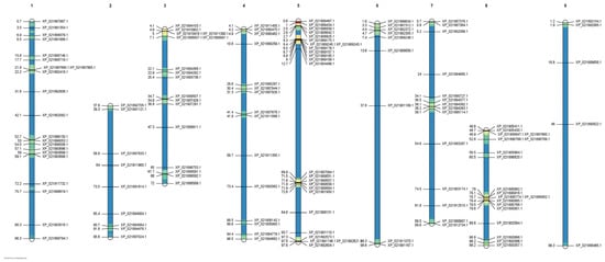
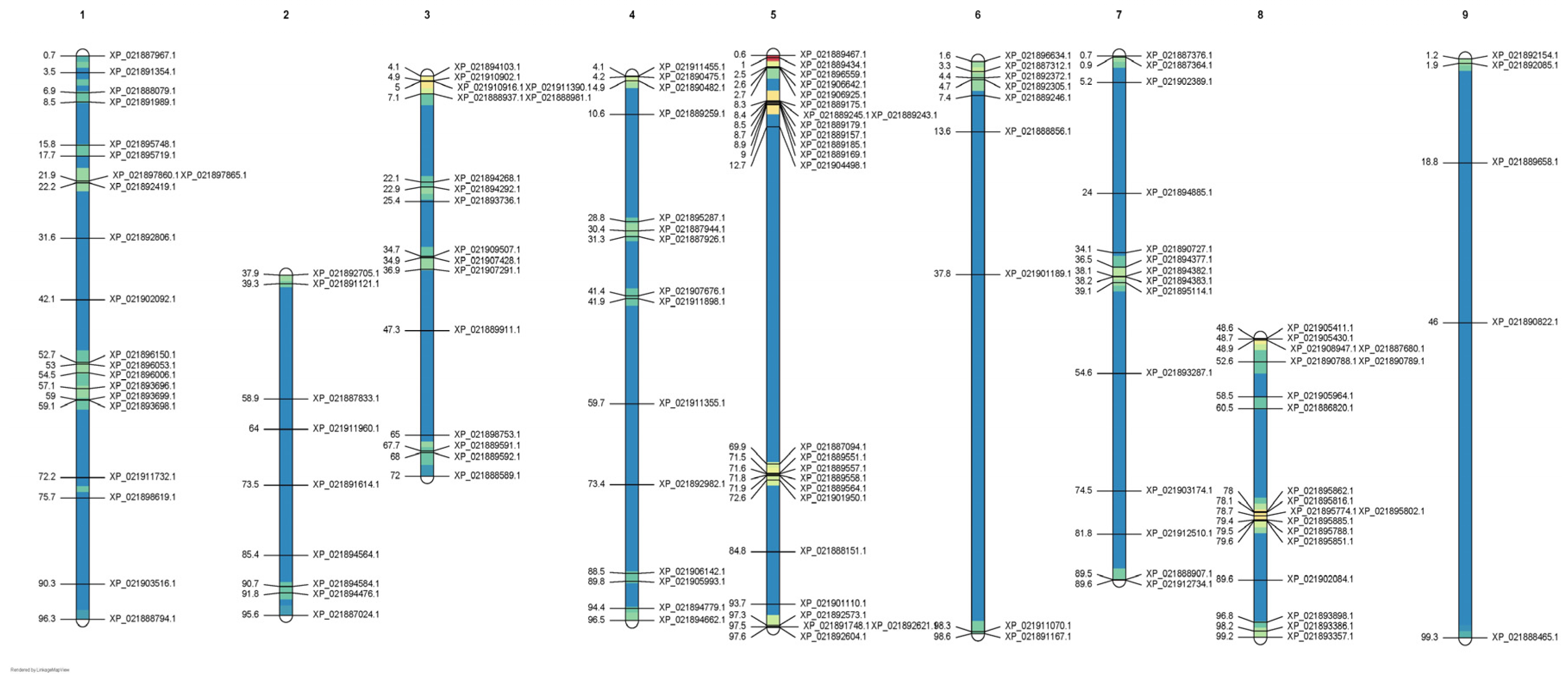
A total of candidate cell wall remodeling genes was successfully mapped onto the chromosome level assembly of papaya SunUp (GCA_021527605.1). The genomic map revealed that these genes are distributed across all nine chromosomes, showing distinct variation in both density and spatial organization along each chromosomal region (Figure 1). Gene coordinates were normalized relative to chromosome length, and local gene densities were computed and visualized using a color gradient derived from the Spectral palette, where warmer tones indicate regions of higher normalized density. The resulting distribution pattern showed that chromosomes 5 and 7 contain prominent gene-dense regions, suggesting local enrichment or clustering of cell wall related genes. In contrast, chromosomes 2 and 8 exhibited sparse distributions with low-density values across most of their length. Intermediate patterns were observed on chromosomes 3, 6, and 9, where gene clusters alternate with gene-poor segments. The inclusion of individual gene identifiers further highlights localized hotspots and the relative organization of gene families along each chromosome. Overall, this normalized chromosomal density map supports a non-random genomic organization of cell wall remodeling genes in papaya, suggesting that certain chromosomal regions may act as preferential loci for functional clustering or local duplication events related to cell wall metabolism.
Figure 1.
Chromosomal distribution and normalized density of cell wall remodeling genes in papaya (C. papaya L., SunUp). Putative cell wall remodeling genes were mapped onto the chromosome-level SunUp genome assembly (GCA_021527605.1) using tblastn, and genomic positions were expressed as percentages of chromosome length. The color gradient represents the normalized local gene density along each chromosome, with warmer colors (yellow–red) indicating regions of higher relative gene concentration and cooler colors (green–blue) denoting lower densities. Gene identifiers are shown at their corresponding chromosomal positions, illustrating both spatial clustering and overall genomic organization. Chromosome numbers are indicated at the top.
3.4. Phylogenetic Analysis
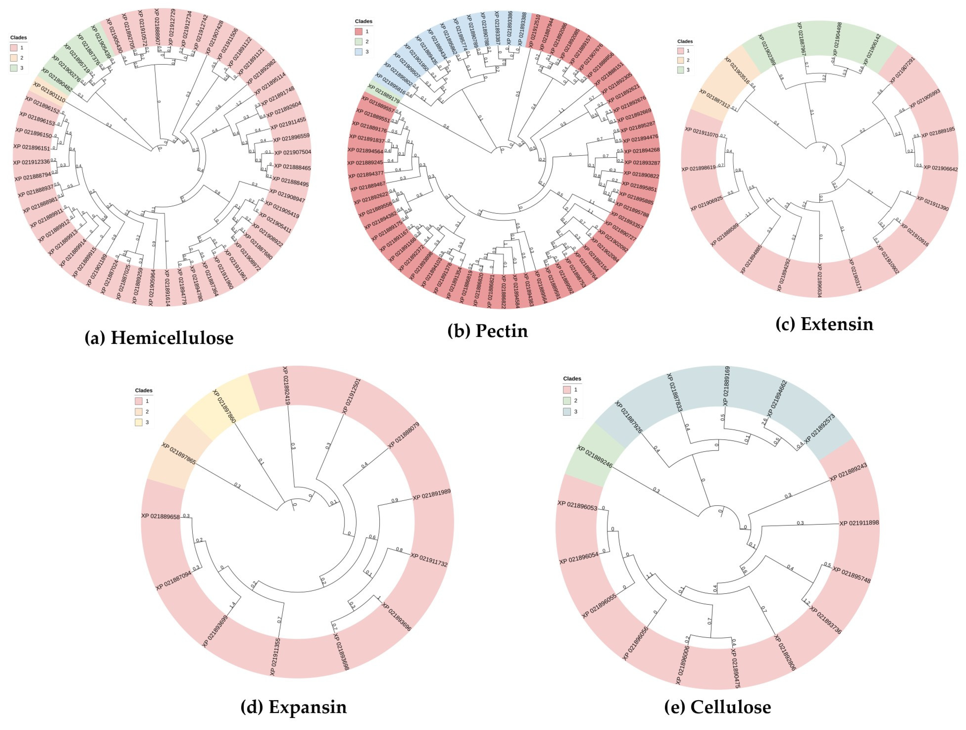
The phylogenetic reconstruction of the large hemicellulose family (58 genes) revealed a highly diversified structure with multiple distinct clades corresponding to different enzymatic functions. Major groups include xyloglucan endotransglucosylase/hydrolases (XTHs, e.g., XP_021907428, XP_021912729), beta-galactosidases, and B-D-xylosidases. The XTH proteins form a particularly cohesive and well supported cluster, while beta-galactosidases appear scattered across several clades, suggesting possible functional divergence among their members. This complex phylogeny accurately reflects the functional heterogeneity of enzymes required for hemicellulose synthesis and remodeling (Figure 2a).
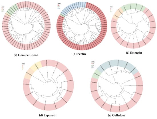
Figure 2.
Phylogenetic tree illustrating the protein families remodeling hemicellulose (a), pectin (b), extensin (c), expansin (d), and cellulose (e). Enzyme clades are numbered from 1 to 3 into each family.
The phylogeny of the pectin modification enzyme family, encompassing 69 genes, displays a structure that reflects its functional diversity. Major clades correspond to distinct enzymatic activities, including polygalacturonases (e.g., XP_021889434, XP_021901950.1), pectin acetylesterases (e.g., XP_021898753.1, XP_021886819.1), and pectinesterase inhibitors (e.g., XP_021889176.1, XP_021889564.1). The polygalacturonase clade shows particularly high consistency, suggesting possible evolutionary conservation. Furthermore, genes annotated as pectin acetylesterases form a distinct and diverse group, separate from the hydrolases, highlighting a probable evolutionary divergence between these functional classes within the pectin network (Figure 2b); all these statements should be validated on future studies.
The phylogenetic tree of the extensin family, comprising 24 genes, shows a pattern of divergence into two major groups. One clade is primarily composed of classical extensins (e.g., XP_021910902, XP_021910916), while the other major group contains leucine-rich repeat extensins (LRX proteins, e.g., XP_021905993, XP_021907291). The pollen specific LRX proteins (e.g., XP_021911732, XP_021902389) cluster together within the larger LRX clade, indicating a specialized evolutionary lineage. This clear separation between extensin types supports the hypothesis of neofunctionalization following gene duplication, correlating with their distinct roles in cell wall structure and signaling (Figure 2c).
The phylogeny of the expansin family, with 13 identified genes, is characterized by a deep split into two primary clades, corresponding to the expansin-A (EXPA) and Expansin-B (EXPB) subfamilies. The EXPA clade is larger and more diverse, consistent with its broader role in cell wall loosening, while the EXPB clade forms a distinct, smaller group. Within the EXPA cluster, members show further subdivision (e.g., XP_021892419.1, XP_021812501), suggesting functional specialization among these isoforms. The overall tree topology aligns with established expansin classification and provides insight into the evolutionary history that has shaped this family of key cell wall-loosening proteins (Figure 2d).
Finally, the phylogenetic of the cellulose family, comprising 17 genes, is characterized by the clad endoglucanase (e.g., XP_ 021911898.1, XP_021890475.1). This proposal phylogeny provides a robust evolutionary framework for understanding the functional specialization within this crucial cell wall synthesis machinery (Figure 2e).
3.5. XTH Family Genetic Validation
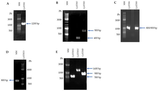
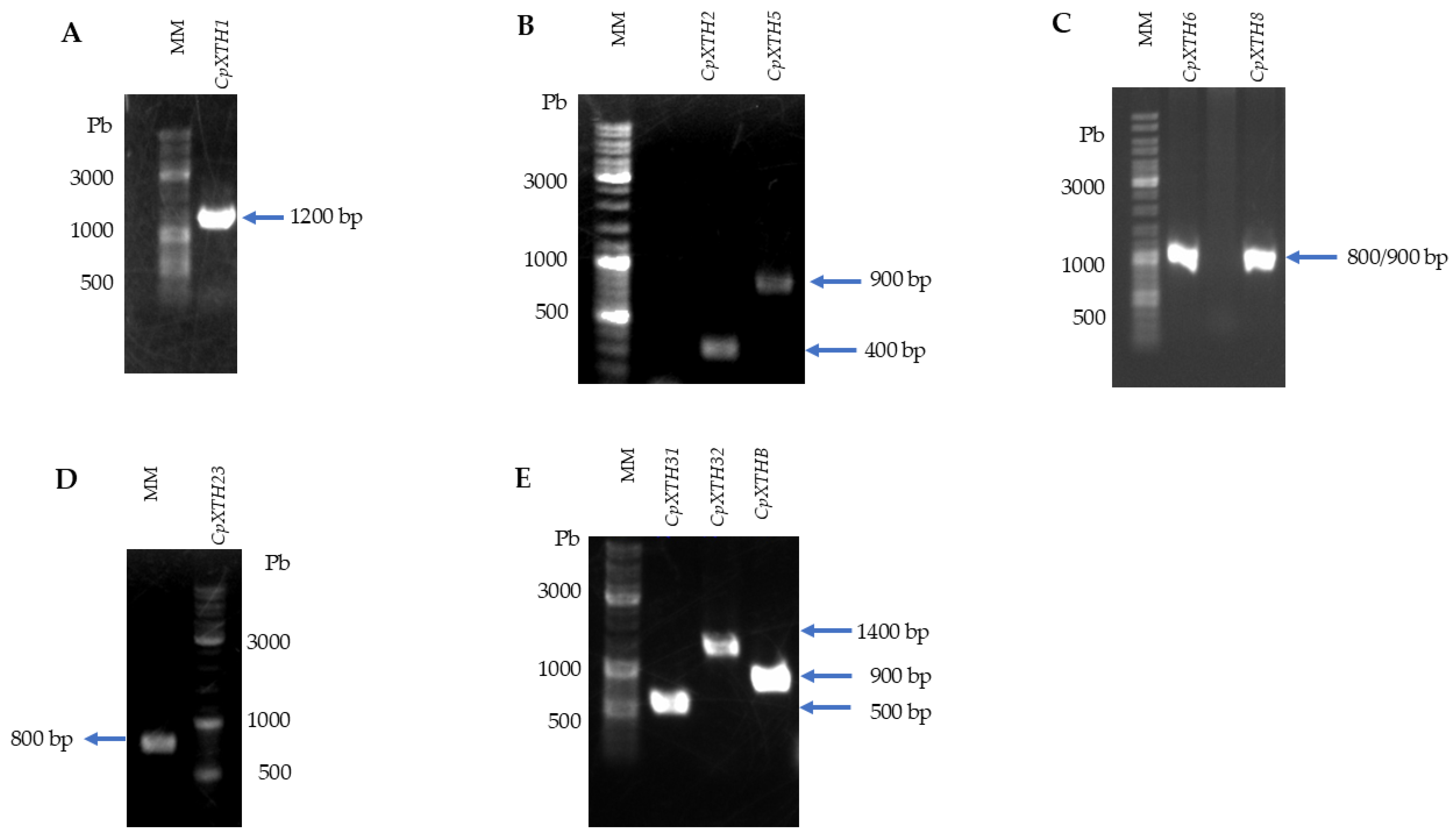
The result of the PCR analysis for the genes CpXTH1 and CpXTH32 presented amplicons with approximate sizes of 1200 and 1400 bp, respectively, while the genes CpXTH5, CpXTH6, CpXTH8, CpXTH23 and CpXTHβ obtained amplicons with sizes around 800 and 900 bp. The genes CpXTH2 and CpXTH31 exhibited amplicons with sizes of 400 and 500 bp, respectively (Figure 3). All the XTH gene members were identified on genomic DNA of papaya variety Maradol and confirming the in silico analysis.
Figure 3.
Agarose gel electrophoresis of CpXTH gene amplification products. Genomic DNA was extracted from young leaves of papaya cv. Maradol using the CTAB method [31]. DNA quality and integrity were verified on 0.8% agarose gels stained with GelRed® and visualized with a Gel Doc™ XR+ system. CpXTH genes were amplified by PCR under specific conditions (see Supplementary Table S2) and the products were analyzed under the same electrophoresis conditions. The O’GeneRuler DNA Ladder Mix (Thermo Scientific™) was used as molecular size marker. Panels correspond to amplification products of: (A) CpXTH1, (B) CpXTH2 and CpXTH5, (C) CpXTH6 and CpXTH8, (D) CpXTH23, (E) CpXTH31, CpXTH32 and CpXTHβ.
4. Discussion
In other fleshy fruits, various genes per family have been identified, although the specific number is not always provided. For example, in apples (Malus domestica), researchers identified 115 genes potentially involved in cell wall dynamics during fruit development, ripening and cold storage. They also observed structural modifications in hemicelluloses and the expression of β-galactosidases genes during fruit development and storage, which play an important role in texture [32]. In watermelon (Citrullus lanatus), an analysis of gene expression networks revealed 354 genes grouped into functional modules involved in the metabolism of pectins, hemicelluloses and cellulose. While the analysis specifies the number of genes by family, it highlights genes coding for cellulases and β-galactosidases, which are involved in the loss of fruit firmness [33]. Additionally, Tranbarger et al., 2017 [34] conducted a transcriptomic study during the maturation of the mesocarp of Elaeis guineensis, identifying 56 genes belonging to at least 20 functional families, including XTH, expansins and β-galactosidases, which are involved in tissue softening. Similarly, a global transcriptome study in peach (Prunus persica) revealed a total of 3553 differentially expressed genes during ripening, of which 1615 were overexpressed in ripe fruits. Among these genes, several related to the cell wall stood out, including polygalacturonases, pectate lyases, and xyloglucan endotransglycosylases/hydrolases (XTH33), which play a central role in fruit softening [35]. For Diospyros kaki (persimmon), Wang et al., 2021 [36] identified 221 differentially expressed genes during persimmon fruit ripening, grouped into 26 modules, several of which were involved in cell wall modification. Li et al., 2019 [37] identified 13 genes directly related to cell wall remodeling and degradation in banana (Musa spp.), with differential expression between the immature and ripe stages of the fruit. The most relevant genes include Expansin-A8, Expansin-A2, Pectate lyase 15 and 22, Polygalacturonase, β-galactosidase and Xyloglucan endotransglycosylase/hydrolase (XTH/XET). Other studies have reported the expression of the XTH6 and XTH23 genes in C. papaya, indicating that these genes are primarily activated during fruit development [30]. Additionally, the XTH2 and XTH5 genes were also expressed during both fruit development and ripening [38]. Future transcriptomic studies should be conducted to have a deeper understanding of the roles of all cell wall gene families in papaya
The functional proteins identified demonstrate the extensive complexity involved in cell wall remodeling. A key finding across all protein families is the widespread presence of the ser_thr_kinase domain, which play a basic role in regulation of various cellular functions through the phosphorylation of serine and threonine residues in other proteins activating or disactivating their function [39]; for example, when the plant receives external signal such as hormones, or environmental effects, the action of this domain could impact in cell wall biosynthesis and remodeling and in consequence on fruit development or softening [40,41]. In particular, on expansins and extensis this domain is the most predominant, but in hemicelluloses, the catalytic_triad domain is also significantly predominant.
It has been observed that enzymes containing a catalytic triad domain are capable of glycosylate and hydrolyze polysaccharides, such is the nature of hemicelluloses which are mostly glycosyl hydrolase enzymes, so this domain is predominant for this type of protein [42]. The triangle of residues in these enzymes allows the transfer of chemical groups that facilitate biochemical reactions, resulting in the restructuring of cell wall rigidity during fruit growth and ripening. In the C. papaya XTH gene family, the encoded proteins have been reported to contain the GH16_XET domain, which includes two subdomains: the GH16 domain and the XET_C domain [43]; this composition includes the catalytic triad domain of the enzyme, which acts on glycosidic bonds, specifically in xyloglucans and xylans. Therefore, this analysis confirms that the hemicellulose XTH proteins family presents the presence of this catalytic triad domain which helps in the glycosylase and hydrolase activity.
It’s interesting the finding of the domain ATP_binding on expansin and cellulose proteins. The ATP-binding domain enables proteins to bind ATP, signifying energy-dependent functions critical for modifying cellular structures or activate other proteins. This binding can impact on protein conformation by acting as an allosteric regulator; such interactions induce structural changes that can enhance or inhibit protein activity [44]. This mechanism allows cells to fine-tune their metabolic processes in response to environmental conditions. For instance, expansins and certain glycosyl hydrolases require ATP for their activity; they harness the energy derived from ATP to facilitate the cleavage of bonds in cell wall polysaccharides, thereby promoting their degradation and restructuring [45,46].
Most cell wall proteins are highly conserved; however, possible duplications were observed in several family genes, where one or more members are present [47]. This phenomenon may be explained by the evolutionary adaptations of C. papaya over time, giving a consequent complexity on cell wall remodeling. Nevertheless, studies as primary structure in promotor regions, and regulator elements controlling each gene, could help in having a deep understanding of the fruit softening process in papaya fruit. On the other hand, the loci abundance was observed to be higher in linkage groups 5 and 7, suggesting the importance of these chromosomal sites in cell wall remodeling. Although the other genes are distributed in other chromosomal regions, it is essential to emphasize that the remodeling process is complex, nevertheless these regions could be used for developing marker assisted selection. Therefore, other omics studies are needed to clearly decipher the role of these genes during fruit on each point of development and ripening, which will contribute to a deeper understanding of the key points in the control of fruit softening.
5. Conclusions
This study presents the first genome-wide identification and characterization of cell wall-related gene families in Carica papaya L. A total of 181 genes were identified and categorized into metabolic pathways: hemicellulose (58), pectin (69), extensin (24), expansin (13), and cellulose (17). These genes were analyzed through comparative genomics, phylogenetic reconstruction, and catalytic domain profiling. The analysis revealed both conserved and divergent evolutionary patterns across families, with expansin and polygalacturonase clades demonstrating strong conservation, while hemicellulose-related XTH genes exhibited significant diversification. In silico predictions were experimentally validated by amplifying nine CpXTH genes, confirming product sizes ranging from 322 to 1370 bp. These results underscore the structural and functional complexity of papaya cell wall remodeling and providing a reference dataset for future functional genomics studies. Understanding the regulatory basis of these gene families is essential for deciphering the molecular mechanisms underlying fruit softening. Beyond its fundamental significance, this knowledge establishes a framework for developing biotechnological strategies aimed at modulating cell wall remodeling in papaya, with the potential to mitigate postharvest losses and improve fruit quality.
Supplementary Materials
The following supporting information can be downloaded at: https://www.mdpi.com/article/10.3390/applbiosci5010002/s1, Supplementary Table S1: List of gene families involved in the dynamic process of cell wall remodeling in Carica papaya (NCBI, 2025). Supplementary Table S2: Selected oligonucleotides for amplification of CpXTH genes of C. papaya.
Author Contributions
Conceptualization, D.G.-L. and M.A.R.-L.; methodology, M.S.-A., R.R.-Q. and J.A.N.-Z.; software, J.A.N.-Z. and M.S.-A.; validation, J.A.N.-Z., R.R.-Q. and D.G.-L.; formal analysis, M.S.-A.; investigation and resources, D.G.-L.; data curation, J.A.N.-Z. and R.R.-Q.; writing—original draft preparation, D.G.-L. and M.S.-A.; writing—review and editing, M.A.R.-L. and R.R.-Q.; visualization, J.A.N.-Z.; supervision, R.R.-Q.; project administration, D.G.-L.; funding acquisition, D.G.-L., R.R.-Q., M.A.R.-L. and J.A.N.-Z. All authors have read and agreed to the published version of the manuscript.
Funding
This research received no external funding.
Institutional Review Board Statement
Not applicable.
Informed Consent Statement
Not applicable.
Data Availability Statement
The original contributions presented in this study are included in the article and Supplementary Materials. Further inquiries can be directed to the corresponding author.
Acknowledgments
Authors thanks to SECIHTI grant scholarship number 5f9cc7e05880d4846b4f759 for M.S.-A.
Conflicts of Interest
The authors declare no conflicts of interest.
References
- Zhang, C.; Xiong, Z.; Yang, H.; Wu, W. Changes in pericarp morphology, physiology and cell wall composition account for flesh firmness during the ripening of blackberry. Sci. Hortic. 2019, 250, 59–68. [Google Scholar] [CrossRef] [Scilit]
- Malacarne, G.; Lagreze, J.; Rojas San Martin, B.; Malnoy, M.; Moretto, M.; Moser, C.; Dalla Costa, L. Insights into the cell-wall dynamics in grapevine berries during ripening and in response to biotic and abiotic stresses. Plant Mol. Biol. 2024, 114, 38. [Google Scholar] [CrossRef] [Scilit]
- Castilleux, R.; Plancot, B.; Vicré, M.; Nguema-Ona, E.; Driouich, A. Extensin, an underestimated key component of cell wall defence? Ann. Bot. 2021, 127, 709–713. [Google Scholar] [CrossRef] [Scilit]
- Moya-León, M.A.; Mattus-Araya, E.; Herrera, R. Molecular events occurring during softening of strawberry fruit. Front. Plant Sci. 2019, 10, 615. [Google Scholar] [CrossRef] [Scilit] [PubMed]
- Peng, Z.; Liu, G.; Li, H.; Wang, Y.; Gao, H.; Jemrić, T.; Fu, D. Molecular and genetic events determining the softening of fleshy fruits: A comprehensive review. Int. J. Mol. Sci. 2022, 23, 12482. [Google Scholar] [CrossRef] [Scilit]
- Zheng, X.; Yuan, Y.; Huang, B.; Hu, X.; Tang, Y.; Xu, X.; Wu, M.; Gong, Z.; Luo, Y.; Gong, M.; et al. Control of fruit softening and ascorbic acid accumulation by manipulation of SlIMP3 in tomato. Plant Biotechnol. J. 2022, 20, 1213–1225. [Google Scholar] [CrossRef] [Scilit]
- Betulia de Morais Souto, B.; Barbosa, M.F.; Sales, R.M.M.; Moura, S.C.; Araújo, A.R.B.; Quirino, B.F. The potential of β-glucosidases for aroma and flavor improvement in the food industry. Microbe 2023, 1, 100004. [Google Scholar] [CrossRef] [Scilit]
- Robinson, P.K. Enzymes: Principles and biotechnological applications. Essays Biochem. 2015, 59, 1–41. [Google Scholar] [CrossRef] [Scilit]
- Shetty, N.; Dorairaj, D.; Sharma, S.; Mawale, K.; Puthusseri, B.; Giridhar, P. Determining the function of ripening-associated genes and biochemical changes during tomato fruit maturation. Preprints 2023. [Google Scholar] [CrossRef] [Scilit]
- Zhang, H.; Wang, H.; Yi, H.; Zhai, W.; Wang, G.; Fu, Q. Transcriptome profiling of Cucumis melo fruit development and ripening. Hortic. Res. 2016, 3, 16014. [Google Scholar] [CrossRef] [Scilit]
- Koul, B.; Pudhuvai, B.; Sharma, C.; Kumar, A.; Sharma, V.; Yadav, D.; Jin, J.-O. Carica papaya L.: A Tropical Fruit with Benefits beyond the Tropics. Diversity 2022, 14, 683. [Google Scholar] [CrossRef] [Scilit]
- Barro-Arroyo, A.; Morales-Pablo, R.; Aguilar-Gutiérrez, G.; Ramírez-Martínez, A. Estimation of papaya losses and the factors that cause them in the northern part of the state of Veracruz. Res. Dev. Food Sci. Technol. 2022, 7, 261–266. [Google Scholar]
- Ming, R.; Hou, S.; Feng, Y.; Yu, Q.; Dionne-Laporte, A.; Saw, J.H.; Senin, P.; Wang, W.; Ly, B.V.; Lewis, K.L.T.; et al. The draft genome of the transgenic tropical fruit tree papaya (Carica papaya Linnaeus). Nature 2008, 452, 991–996. [Google Scholar] [CrossRef] [Scilit] [PubMed]
- Magee, A.M.; Aspinall, S.; Rice, D.W.; Cusack, B.P.; Sémon, M.; Perry, A.S.; Stefanović, S.; Milbourne, D.; Barth, S.; Palmer, J.D.; et al. Localized hypermutation and associated gene losses in legume chloroplast genomes. Genome Res. 2010, 20, 1700–1710. [Google Scholar] [CrossRef] [Scilit]
- NCBI Resource Coordinators. National Center for Biotechnology Information (NCBI) [Internet]; National Library of Medicine (US): Bethesda, MD, USA, 1988. Available online: https://www.ncbi.nlm.nih.gov/ (accessed on 10 March 2025).
- Bodenhofer, U.; Bonatesta, E.; Horejs-Kainrath, C.; Hochreiter, S. msa: An R package for multiple sequence alignment. Bioinformatics 2015, 31, 3997–3999. [Google Scholar] [CrossRef] [Scilit]
- Schliep, K. phangorn: Phylogenetic analysis in R. Bioinformatics 2011, 27, 592–593. [Google Scholar] [CrossRef] [Scilit]
- Winter, D.J. rentrez: An R package for the NCBI eUtils API. R J. 2017, 9, 520–526. [Google Scholar] [CrossRef] [Scilit]
- Charif, D.; Lobry, J. SeqinR 1.0-2: A contributed package to the R project for biological sequence retrieval and analysis. In Structural Approaches to Sequence Evolution: Molecules, Networks, Populations; Bastolla, U., Porto, M., Roman, H., Vendruscolo, M., Eds.; Springer: New York, NY, USA, 2007; pp. 207–232. [Google Scholar]
- Camacho, C.; Coulouris, G.; Avagyan, V.; Ma, N.; Papadopoulos, J.; Bealer, K.; Madden, T.L. BLAST+: Architecture and applications. BMC Bioinform. 2009, 10, 421. [Google Scholar] [CrossRef] [Scilit]
- Hahsler, M.; Nagar, A. rBLAST: R Interface for the Basic Local Alignment Search Tool. 2024. Available online: https://bioconductor.org/packages/release/bioc/html/rBLAST.html (accessed on 20 November 2025).
- Wickham, H.; François, R.; Henry, L.; Müller, K.; Vaughan, D. dplyr: A Grammar of Data Manipulation. R Package 2025, version 1.1.4. Available online: https://dplyr.tidyverse.org (accessed on 20 November 2025).
- Narváez-Zapata, J.A.; Alaníz-Cuevas, Y.; Chávez-Medina, J.A.; Espinoza-Mancillas, M.G.; Maldonado-Mendoza, I.; Singh, R.K.; Quiroz-Figueroa, F.R. Unveiling the molecular arsenal: Genome sequencing and in silico secretome analysis of Fusarium verticillioides provide insights into maize root rot pathogenesis. Front. Plant Sci. 2025, 16, 1672761. [Google Scholar] [CrossRef] [Scilit]
- Singh, M.S.; Brar, J.S.; Mittal, A.; Gill, M.I.S.; Arora, N.K.; Sohi, H.S.; Chhuneja, P.; Dhillon, G.S.; Singh, N.; Thakur, S. Construction of a genetic linkage map and QTL mapping of fruit quality traits in guava (Psidium guajava L.). Front. Plant Sci. 2023, 14, 1123274. [Google Scholar] [CrossRef] [Scilit] [PubMed]
- Cappai, F.; Rampazo Amadeu, R.; Benevenuto, J.; Cullen, R.; Garcia, A.; Grossman, A.; Ferrão, L.F.V.; Munoz, P. High-resolution linkage map and QTL analyses of fruit firmness in autotetraploid blueberry. Front. Plant Sci. 2020, 11, 562171. [Google Scholar] [CrossRef] [Scilit]
- Letunic, I.; Bork, P. Interactive Tree of Life (iTOL) v5: An Online Tool for Phylogenetic Tree Display and Annotation. Nucleic Acids Res. 2021, 49, W293–W296. [Google Scholar] [CrossRef] [Scilit]
- Wickham, H. ggplot2: Elegant Graphics for Data Analysis; Springer: New York, NY, USA, 2016. [Google Scholar]
- Posit Team. RStudio: Integrated Development Environment for R. Posit Software; Posit Team: Boston, MA, USA, 2024; Available online: http://www.posit.co/ (accessed on 15 October 2025).
- Untergasser, A.; Cutcutache, I.; Koressaar, T.; Ye, J.; Faircloth, B.C.; Remm, M.; Rozen, S.G. Primer3—New capabilities and interfaces. Nucleic Acids Res. 2012, 40, e115. [Google Scholar] [CrossRef] [Scilit]
- Zúñiga-Hernández, M.E.; Rosas-Quijano, R.; Salvador-Figueroa, M.; Vázquez-Ovando, A.; Gálvez-López, D. Expression of the CpXTH6 and CpXTH23 Genes in Carica papaya Fruits. Int. J. Mol. Sci. 2025, 26, 4490. [Google Scholar] [CrossRef] [Scilit] [PubMed]
- Dellaporta, S.L.; Wood, J.; Hicks, J.B. A plant DNA minipreparation: Version II. Plant Mol. Biol. 1983, 1, 19–21. [Google Scholar] [CrossRef] [Scilit]
- Dheilly, E.; Le Gall, S.; Guillou, M.C.; Renard, C.M.G.C.; Bonnin, E.; Garnier, C. Cell wall dynamics during apple development and storage involves hemicellulose modifications and related enzymes. BMC Plant Biol. 2016, 16, 201. [Google Scholar] [CrossRef] [Scilit] [PubMed]
- Zhang, Z.; Zhao, L.; Wang, Z.; Wu, X.; Wang, J.; Yang, Y. Coexpression network analysis reveals genes related to cell wall metabolism during fruit softening in watermelon (Citrullus lanatus). Front. Plant Sci. 2021, 12, 630243. [Google Scholar] [CrossRef] [Scilit]
- Tranbarger, T.J.; Dussert, S.; Joët, T.; Argout, X.; Summo, M.; Champion, A.; Cros, D.; Omore, A.; Nouy, B. Transcriptome analysis of cell wall and lipid metabolism during fruit development and ripening in oil palm (Elaeis guineensis). Front. Plant Sci. 2017, 8, 603. [Google Scholar] [CrossRef] [Scilit]
- Tsalgatidou, P.C.; Boutsika, A.; Delis, C. Global transcriptome analysis of the peach (Prunus persica) in the interaction system of fruit–chitosan–Monilinia fructicola. Plants 2024, 13, 567. [Google Scholar] [CrossRef] [Scilit]
- Kou, J.; Zhao, Z.; Zhang, Q.; Wei, C.; Ference, C.M.; Guan, J.; Wang, W. Comparative transcriptome analysis reveals the mechanism involving ethylene and cell wall modification related genes in Diospyros kaki fruit firmness during ripening. Genomics 2021, 113, 552–563. [Google Scholar] [CrossRef] [Scilit]
- Li, T.; Yun, Z.; Wu, Q.; Qu, H.; Duan, X.; Jiang, Y. Combination of transcriptomic, proteomic, and metabolomic analysis reveals the ripening mechanism of banana pulp. Biomolecules 2019, 9, 523. [Google Scholar] [CrossRef] [Scilit] [PubMed]
- Aguilar-Velazquez, B.; Rosas-Quijano, R.; Vázquez-Ovando, A.; Miguel, S.; Adriano-Anaya, L.; Galvez-Lopez, D. CpXTH2 and CpXTH5 genes are expressed during fruit development and ripening in Carica papaya L. Fruits 2021, 76, 3–10. [Google Scholar] [CrossRef] [Scilit] [PubMed]
- Seok, S.H. Structural insights into protein regulation by phosphorylation and substrate recognition of protein kinases/phosphatases. Life 2021, 11, 957. [Google Scholar] [CrossRef] [Scilit]
- Shan, Y.; Zhang, D.; Luo, Z.; Li, T.; Qu, H.; Duan, X.; Jiang, Y. Advances in chilling injury of postharvest fruit and vegetable: Extracellular ATP aspects. Compr. Rev. Food Sci. Food Saf. 2022, 21, 4251–4273. [Google Scholar] [CrossRef] [Scilit]
- Wang, W.; Fan, D.; Hao, Q.; Jia, W. Signal transduction in non-climacteric fruit ripening. Hortic. Res. 2022, 9, uhac190. [Google Scholar] [CrossRef] [Scilit] [PubMed]
- Gilbert, H.J. The biochemistry and structural biology of plant cell wall deconstruction. Plant Physiol. 2010, 153, 444–455. [Google Scholar] [CrossRef] [Scilit]
- Broman, K.W.; Wu, H.; Sen, Ś.; Churchill, G.A. R/qtl: QTL mapping in experimental crosses. Bioinformatics 2003, 19, 889–890. [Google Scholar] [CrossRef] [Scilit]
- Rodríguez-Domínguez, R.; Rosas-Quijano, R.; Salvador-Figueroa, M.; Vazquez-Ovando, A.; Gálvez-López, D. Primary structure of the XTH gene family members from Carica papaya. Acta Hortic. 2023, 1362, 433–440. [Google Scholar] [CrossRef] [Scilit]
- Minic, Z.; Jouanin, L. Plant glycoside hydrolases involved in cell wall polysaccharide degradation. Plant Physiol. Biochem. 2006, 44, 435–449. [Google Scholar] [CrossRef] [Scilit]
- Minic, Z.; Jamet, E.; Négroni, L.; Der Garabedian, P.; Zivy, M.; Jouanin, L. A sub-proteome of Arabidopsis thaliana mature stems trapped on Concanavalin A is enriched in cell wall glycoside hydrolases. J. Exp. Bot. 2007, 58, 2503–2512. [Google Scholar] [CrossRef] [Scilit] [PubMed]
- Qiao, X.; Li, Q.; Yin, H.; Qi, K.; Li, L.; Wang, R.; Zhang, S.; Paterson, A.H. Gene duplication and evolution in recurring polyploidization–diploidization cycles in plants. Genome Biol. 2019, 20, 38. [Google Scholar] [CrossRef] [Scilit] [PubMed]

Disclaimer/Publisher’s Note: The statements, opinions and data contained in all publications are solely those of the individual author(s) and contributor(s) and not of MDPI and/or the editor(s). MDPI and/or the editor(s) disclaim responsibility for any injury to people or property resulting from any ideas, methods, instructions or products referred to in the content. |
© 2026 by the authors. Licensee MDPI, Basel, Switzerland. This article is an open access article distributed under the terms and conditions of the Creative Commons Attribution (CC BY) license.