


Simple Summary
Q fever is a bacterial infection caused by Coxiella burnetii that spreads from animals, mainly cattle, sheep and goats, to humans, typically through the inhalation of contaminated particles during animal births or contact with their products. Although it has been a notifiable disease in Portugal since 1999, the number of diagnosed cases has remained persistently low, despite evidence suggesting that a substantial proportion of the population, particularly farmers, shepherds, cheese producers and veterinarians, has already been exposed to the bacterium. This discrepancy points to widespread underdiagnosis and underreporting. The present review systematically gathered and analysed all available scientific evidence on this infection in Portugal across human, animal and environmental domains, with the aim of identifying what is known, where the main gaps lie, and what measures are needed to build a more integrated and effective national surveillance system.
Abstract
Q fever, caused by Coxiella burnetii, is a globally distributed zoonotic disease with significant public health and veterinary relevance, yet its epidemiology in Portugal remains fragmented. This systematic review aimed to assess the epidemiology of C. burnetii infection and Q fever in Portugal by compiling, critically analysing and integrating available evidence from animal, human, and environmental studies, adopting a One Health perspective. A comprehensive literature search was conducted in international databases and national repositories in accordance with PRISMA 2020 guidelines. Studies reporting serological, molecular or isolation-based detection of C. burnetii in humans, animals or environmental samples were eligible. A total of 24 studies were included, covering human populations, ruminants, companion animals, wildlife and environmental matrices. Evidence was predominantly focused on ruminants, with consistently high and heterogeneous levels of exposure reported across different species, regions and production systems. Human epidemiological studies were limited and largely restricted to occupational groups and blood donors, but consistently indicated substantial exposure, suggesting underdiagnosis and underreporting. Data on companion animals, wildlife and environmental sources were scarce and methodologically heterogeneous. This review confirms endemic circulation of C. burnetii in Portugal, but highlights pronounced geographic and methodological gaps, reinforcing the need for harmonised, integrated One Health surveillance to improve risk assessment and evidence-based prevention strategies.
1. Introduction
Q fever is a zoonosis caused by the intracellular bacterium C. burnetii; it is found worldwide and has a significant impact on both public health and livestock production [1,2,3,4]. In humans, the infection may be asymptomatic or present acutely, with varying severity (most commonly: non-specific febrile syndrome, with fever, headaches, myalgia, arthralgia, hepatitis, and/or pneumonia) and in a chronic, more severe form (endocarditis and/or vascular infection), and is presumed to have been underdiagnosed and underreported in Portugal over the years [1,3]. Diagnosis depends on the detection and titration of antibodies against C. burnetii: phase II antigens (in acute forms) or phase I antigens (in chronic forms). Diagnosis can also be made by detecting C. burnetii DNA in blood or other samples, such as sputum, liver biopsy and cardiac valve material. Culturing this pathogen, although technically possible, is not routinely performed in a hospital setting. Treatment depends on the form of the disease and consists of antibiotic therapy with doxycycline (and hydroxychloroquine). The duration of treatment and clinical follow-up is particularly prolonged for chronic forms (>18 months), with mortality approaching 20% in this context [3,5,6,7,8].
The main reservoir hosts are livestock, including cattle, sheep and goats [1,4,9]. The primary route of transmission to humans is the inhalation of contaminated aerosols, particularly those originating from the birth products of infected ruminants [3,9,10]. Other possible routes include the consumption of raw or unpasteurised dairy products [1] and transmission via ixodid ticks, the latter being indirect but less common [3].
In Portugal, epidemiological and clinical knowledge on Q fever in humans remains limited, fragmented and largely outdated. The few available studies have focused mainly on specific subpopulations, namely blood donors, hospitalised patients and occupational groups such as shepherds, cheesemakers and veterinarians, and consistently point to substantial exposure to C. burnetii across mainland Portugal, with seroprevalence estimates well above what would be expected from the number of officially notified cases [11,12,13,14,15]. Nationwide clinical characterisation is scarce and essentially restricted to a single retrospective analysis conducted by the National Institute of Health Dr. Ricardo Jorge (INSA) covering cases reported between 2004 and 2013 [12]; since then, the published evidence has been confined to isolated case series of acute and chronic Q fever [12,13,14,15,16]. Overall, this body of work suggests endemic circulation of C. burnetii in the Portuguese population but also highlights a marked lack of updated, geographically representative and clinically integrated data.
Q fever has been a notifiable disease in Portugal since 1999, and cases are reported via the National Epidemiological Surveillance System (SINAVE). These data contain limited clinical information and have not been published nationally since 2019. However, as they are reported to the ECDC (European Centre for Disease Prevention and Control), we can see from the latest report on zoonoses that 20–35 cases of Q fever were diagnosed in Portugal per year between 2020 and 2023 [17,18].
In the animal sector, evidence in Portugal is almost entirely concentrated on domestic ruminants, cattle, sheep and goats, which are consistently identified as the main reservoirs of C. burnetii worldwide [1,4,9]. National studies, mostly conducted in the Northern and Central regions, have repeatedly documented high levels of exposure in dairy herds and small ruminant flocks, using both serological and molecular approaches, and have also implicated C. burnetii in reproductive losses, particularly abortion episodes in sheep and goats [18,19,20,21,22,23,24,25,26,27,28]. Despite this accumulating evidence, data remain geographically uneven, methodologically heterogeneous and rarely linked to human surveillance, which limits their usefulness for an integrated One Health risk assessment [12,19].
The One Health approach, which encompasses human, animal and environmental health, has increasingly been recognised as essential for the surveillance of emerging zoonoses. However, in Portugal, there is still a lack of studies that bridge the gap between clinical data in humans and veterinary data over time. This gap is particularly relevant in a disease such as Q fever, whose transmission dynamics involve multiple species and environmental factors [12,14].
In this context, the present systematic review aims to compile, critically analyse, and integrate all available scientific evidence on C. burnetii infection and Q fever in Portugal, encompassing studies in humans, ruminants, companion animals, and the environment, in order to characterise its epidemiological distribution, diagnostic approaches, and the main existing knowledge gaps. This synthesis also provides a scientific basis for complementary prevalence and molecular characterisation studies in ruminants, particularly through the analysis of milk as a key matrix for animal surveillance.
2. Materials and Methods
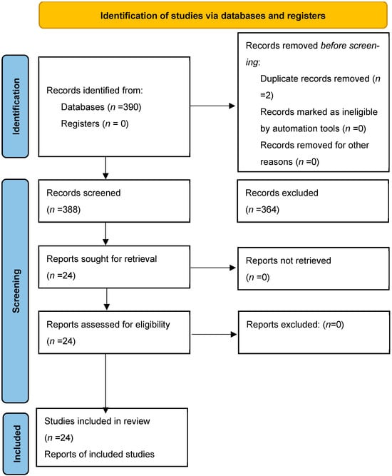
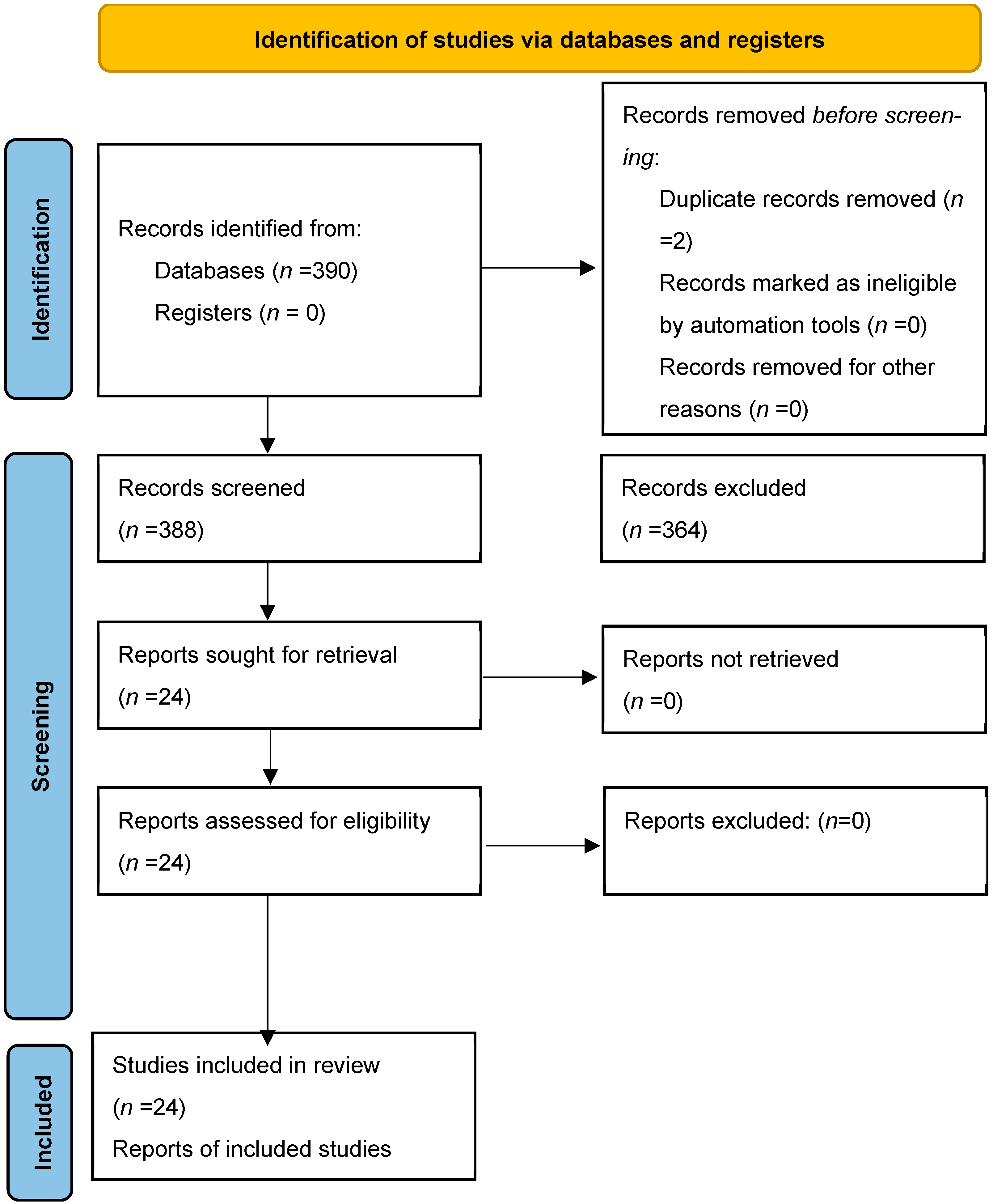
This study was conducted in accordance with the PRISMA 2020 guidelines [29]. This systematic review was not prospectively registered in PROSPERO or in any other systematic review registry. The review protocol, including the research question, eligibility criteria, search strategy, and data extraction plan, was developed a priori by the authors. Any amendments (if applicable) are described within this manuscript. The study selection process is presented in the PRISMA flow diagram shown in Figure 1.
Figure 1.
PRISMA flow diagram.
The primary aim of this study was to systematically compile and analyse the available scientific evidence on Q fever and C. burnetii in Portugal from a One Health perspective, with a focus on mapping the potential geographical spread of the pathogen. Specifically, we aimed to describe the occurrence of Q fever in humans, identify the presence of C. burnetii in ruminants and other animal species, characterise environmental studies conducted to date, and explore potential spatial and/or temporal associations between human cases, animal reservoirs, and environmental contamination.
A comprehensive literature search was performed in PubMed, Medline, Web of Science, RCAAP and Google Scholar using combinations of the keywords “Coxielosis”, “Q fever”, “C. burnetii” and “Portugal”. To minimise the risk of missing relevant studies, an additional targeted search was conducted using terms related to the main epidemiological manifestations and surveillance contexts of C. burnetii infection, including “abortion”, “reproductive disorders”, “livestock”, “dairy production”, “milk”, “occupational exposure”, “seroprevalence”, “molecular detection”, and “environmental contamination”, combined with “Portugal”. The last search was conducted on 14 August 2026.
Eligible studies included epidemiological investigations conducted in Portugal involving humans, animals, or environmental samples, written in English and Portuguese, with no restriction on start date, and reporting detection of C. burnetii through serological (e.g., ELISA, IFA), molecular (PCR), or culture methods. Original research articles, technical reports, and relevant academic theses were considered. Studies were excluded if they did not constitute epidemiological investigations conducted in Portugal, including purely theoretical studies, in vitro experimental studies, imported cases without evidence of infection in Portugal, reviews, and case reports. Study selection was conducted independently by two reviewers, who screened titles and abstracts, followed by full-text assessment of potentially eligible articles. Discrepancies were resolved by consensus. Duplicate records were identified and removed manually by the authors. Data extraction was performed independently by two reviewers using a standardised form. Disagreements were resolved by discussion and, when necessary, by consultation with a third senior reviewer.
Extracted variables included study identification (author and year), geographic location within Portugal, study design, population and species, sample size, sampling period, diagnostic methods (including assay characteristics and thresholds), number of positive cases and corresponding prevalence, molecular findings (e.g., target genes and Ct values), assessed risk factors, and reported funding sources or conflicts of interest.
Study quality was assessed using appropriate JBI critical appraisal tools. Due to heterogeneity in study design, populations, diagnostic approaches, and the limited availability of consistent effect size measures, a meta-analysis was not feasible. Therefore, findings are presented as a qualitative and descriptive synthesis of the available evidence.
3. Results
A total of 390 papers were identified through the database searches. After removing two duplicates, 388 papers were screened by title. Of these, 234 were excluded, leaving 154 for abstract review. Following assessment of the abstracts, 130 articles were excluded, resulting in 24 studies being included in the final review (Table 1).
Table 1.
Papers identified through the databases.
The quality assessment is presented in Supplementary Table S1. Most studies were classified as high quality, with moderate-quality ratings mainly observed in conference abstracts, historical reports and studies with limited methodological reporting.
The 24 studies included human populations, ruminants (cattle, sheep and goats), companion animals (dogs and cats), wild animals and environmental samples (ticks). A complete list of included studies is presented in Table 1.
Prevalence rates varied according to species, region and diagnostic method (ELISA/IFA/PCR) [11,14,15,21,22,23,24,25,26,27,28,30,31,32,33,34,35,36,37,38,39,40,41].
Human data were comparatively limited and mainly derived from occupational exposure studies and seroprevalence assessments in specific populations such as blood donors and veterinarians. According to the available studies, the seroprevalence in the human population ranges from 28.7 to 40%, depending on the population studied [11,14,15].
3.1. Prevalence and Occupational Risk Groups
Veterinarians presented one of the highest rates of infection risk, with a seroprevalence of 33.7% (IgG antibodies), compared with 17.4% in the control group (blood donors with no known direct or indirect exposure to farm animals or agricultural environments were selected as the control group, as these settings are considered the main sources of C. burnetii exposure). The risk of exposure for these professionals is more than double (OR: 2.41) that of the population with no contact with animals [15]. Shepherds and cheese producers in the Serra da Estrela region showed a seroprevalence of 28.1%, significantly higher than the 8.1% observed in the control group, meaning that these professionals are 4.45 times more likely to be infected than the general population [14].
In the general population, a study of 150 blood donors from the Lisbon and Tagus Valley region revealed a seropositivity rate of 28.7% [11], with marked regional variation between Setúbal (48.6%), Lisboa (24.2%) and Santarém (15%) [11]. Notably, this same donor study identified one case with titres suggestive of active acute Q fever in an individual who had passed the medical examination for donation.
3.2. Ruminants
The majority of the available evidence focused on ruminants, representing the most extensively studied group. Serological and molecular approaches were commonly applied in cattle, sheep and goats. However, the reported estimates were not pooled or interpreted as a continuous prevalence range because they derived from distinct epidemiological units and sampling frameworks, including individual animals, herds, bulk tank milk, reproductive tissues and clinically selected abortion submissions [21,22,23,24,25,26,27,28,30,31,32].
Reported seroprevalence varied across regions, with some studies identifying significant levels of infection, particularly in sheep populations [21,26,27,28,30,32]. The results also varied significantly depending on the diagnostic method (serology vs. PCR) and the type of sample (milk tank samples vs. individual samples).
3.2.1. Cattle
Prevalence in cattle was primarily focused on the dairy sector through the analysis of bulk tank milk (BTM). In terms of seroprevalence assessed by ELISA in bulk milk, values of 37.8% were reported in dairy cattle herds from the central region between 2009 and 2013 [25], rising to 61.1% on farms in Northwest Portugal (Barcelos) [23] and reaching 90.5% on dairy farms in the Central Coastal region (Aveiro district), with 58% showing strongly positive antibody levels (ODR > 100%), based on 2021 bulk tank milk sampling [31]. A more recent study conducted in 2021 in the Central region reported an even higher seroprevalence of 96.7% [22].
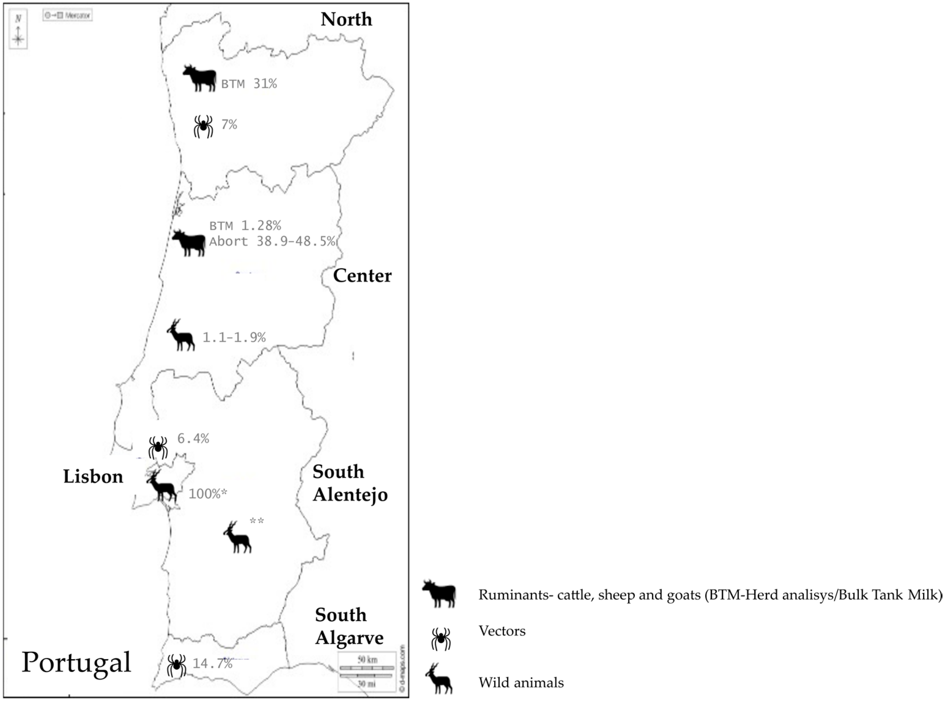
Regarding herd-level molecular detection in bulk tank milk (BTM), C. burnetii DNA was detected in 20.0% of dairy herds in Central Portugal between 2009 and 2013 [25] and in 31.03% of dairy herds in the North region between 2020 and 2021 [22]. In contrast, a more recent BTM study conducted in Central Portugal reported only one positive sample among 156 herds tested, corresponding to a prevalence of 0.64% [32].
Molecular investigations based on clinically selected abortion material showed considerably higher detection rates. C. burnetii was identified in 17.2% of aborted foetuses [21], while an Iberian study including Portuguese submissions reported positivity rates of 38.9% in sheep and 48.5% in goat abortion cases [30].
Tissue-based molecular studies also confirmed pathogen circulation, with C. burnetii DNA detected in 20.83% of tissue samples collected from domestic ruminants at the national level [24].
3.2.2. Sheep
Data for sheep were available from both individual-level and herd-level serological studies. At the individual level, seroprevalence ranged from 8.6% in the Central region [21] to 11.4% nationally, with regional variation across the Centre (17.9%), North (11.2%), Alentejo (9.6%) and Algarve (4.2%) [27]. At the herd level, seroprevalence reached 37.5% in flocks from the Central region [21] and 51.3% in dairy sheep herds from Northern and Central Portugal based on bulk tank milk testing [25]. A longitudinal study conducted in the Serra da Estrela region reported an increase in herd seroprevalence from 10.2% in 2015 to 25.6% in 2016 [28].
PCR-based molecular investigations should be interpreted separately according to the sampling framework. In clinically selected abortion material, C. burnetii DNA was detected in 36.0% of abortion cases across Portugal [20]. Similarly, a multicentre study including Portuguese sheep abortion submissions reported molecular detection in 35.29% (438/1241) of abortion episodes [30].
Tissue-based molecular investigations also confirmed pathogen circulation. In samples including liver, spleen, lymph nodes, mammary gland and uterine tissues collected from domestic ruminants, as well as foetal samples obtained through surveillance programmes and abortion investigations, C. burnetii DNA was detected in 10.53% of samples analysed [24].
3.2.3. Goats
Goats frequently presented the highest exposure levels among small ruminants. At the individual level, seroprevalence reached 10.4% in the Central region [21]. At the herd level, seroprevalence was 28.8% in goat herds from the Central region [21] and 46.2% in dairy goat herds from Northern and Central Portugal based on bulk tank milk testing [25].
Molecular findings should be interpreted according to the sampling framework. Herd-level molecular detection based on bulk tank milk samples did not identify C. burnetii DNA in goats in studies conducted in Northern and Central Portugal (0%) [25].
In contrast, investigations based on clinically selected abortion material reported substantially higher detection rates. C. burnetii DNA was identified in 40.5% of goat abortion cases in Portugal [20]. Similarly, in a multicentre Iberian study including Portuguese submissions, C. burnetii was detected in 48.5% (180/371) of goat abortion cases and was significantly more frequent than Chlamydia abortus in this species (p < 0.001) [30].
Tissue-based molecular studies also confirmed pathogen circulation, with C. burnetii DNA detected in 23.53% of tissue samples collected from domestic ruminants at the national level [24].
3.3. Companion Animals
Epidemiological evidence regarding C. burnetii in companion animals in Portugal is scarce and fragmented when compared with the data available for ruminants. The studies identified focused predominantly on the Lisbon, Northern and Central regions.
In dogs, serological analysis dates back to 1995, with a seroprevalence of 4.8% in Setúbal [33]. Subsequent studies in Lisbon (2014) indicated similar figures (4.3%) [34], while in the Central region, between 2011 and 2014, significantly higher exposure was observed, reaching 12.6% [35]. However, more recent data (2020–2021) revealed a sharp drop in seropositivity to 1.7% [36]. Data on cats are even more limited: in 2012, a seroprevalence of 17.2% was recorded in the Central region [35], whereas studies conducted in 2021 detected no seropositive animals (0%) in this species [35]. As for molecular detection by PCR, recent investigations of uterine tissues, endometrial swabs and ticks collected from these animals did not detect C. burnetii DNA, suggesting the absence of active infection or shedding in these specific samples [35,36].
3.4. Environmental and Vector-Related Studies
Environmental evidence was limited to studies investigating ticks collected in Portugal as potential vectors or environmental indicators of C. burnetii circulation, although most of these studies primarily targeted other zoonotic agents such as Rickettsia spp.
In urban and recreational parks, a study carried out in the Lisbon metropolitan area (2018), specifically in Monsanto Forest Park (PFM), revealed the presence of C. burnetii DNA in 6.4% of ticks collected from vegetation [37]. The bacterium was detected in five of the eight species found in the park, including Ixodes ricinus, Ixodes frontalis and, for the first time, Ixodes ventalloi [37].
At the interface with livestock farming in Northern Portugal, a recent study (2022–2023) focusing on native breeds in the district of Bragança investigated the role of ticks in transmission among ruminants [38]. An infection rate of 7% was found in ticks of the species Rhipicephalus sanguineus collected from sheep of the Churra Galega Mirandesa breed [38], whereas none of the Rhipicephalus bursa ticks collected from Garrano horses tested positive [38].
The highest rate of molecular detection in ticks was recorded in south-eastern Portugal (habitat of the Iberian lynx), covering areas such as Mértola, Moura, Serpa and Loulé, in 2017 [39]. The prevalence reached 14.7% (34/231) of the ticks analysed [39], with most detections occurring in Hyalomma lusitanicum ticks collected from vegetation, the first record of this association in Portugal [39]. Infected ticks were also found on hosts such as cats and Egyptian mongooses, reinforcing the complexity of the transmission cycle [39].
3.5. Wildlife
Evidence regarding wildlife is extremely limited, with only a small number of studies identifying the presence of the pathogen in animals from a zoological setting [42]. No large-scale surveillance studies in free-ranging wildlife were identified.
A comprehensive study in eastern Central Portugal (2016–2022) detected a seroprevalence of 1.9% (5/259) in free-ranging red deer (Cervus elaphus) [40] and a slightly lower seroprevalence of 1.1% (4/358) in wild boar (Sus scrofa) from the same region [40]. Seropositive cases were detected mainly in municipalities such as Monte Fidalgo, Alcafozes, Sarnadas de Ródão, Rosmaninhal and Vila Velha de Ródão [40].
Among wild carnivores, the Egyptian mongoose (Herpestes ichneumon) stands out with the highest molecular (DNA) detection rate among wildlife species studied, at 11.1% (5/45) [24], suggesting a degree of exposure to C. burnetii among wild carnivores in Portugal. In spot checks of other species, no bacterial DNA was detected in foxes (0/4), genets (0/3), weasels (0/2) or badgers (0/1) [23]. Regarding wild birds, a recent study (2025) conducted on birds admitted to rehabilitation centres in mainland Portugal (n = 92 for serology, n = 103 for PCR), including representative species such as common buzzard, tawny owl and white stork, detected no positive animals, neither by ELISA (0/92) nor by qPCR (no confirmed positive samples) [41]. Similarly, no evidence of DNA was found in cloacal samples from 19 griffon vultures (Gyps fulvus) in a 2015 study [23].
4. Discussion
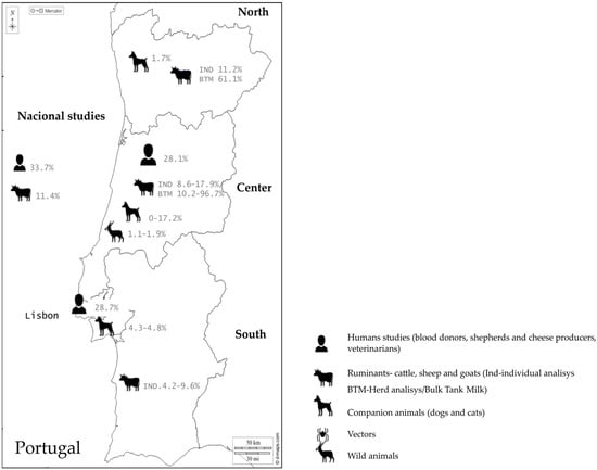
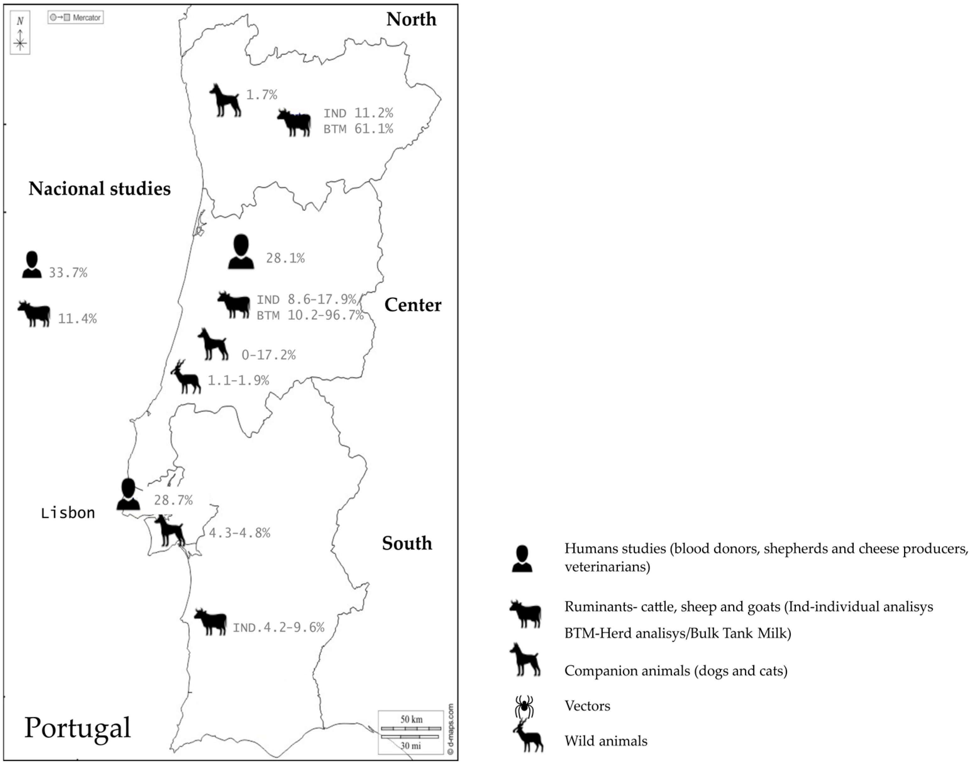
This systematic review provides a comprehensive and up-to-date overview of the epidemiology of C. burnetii infection in Portugal, confirming the endemic and persistent circulation of the pathogen across human, animal and environmental domains. The following sections examine how these realms intersect, drawing out the cross-cutting patterns and the gaps in cross-referencing them that define the current state and the current limitations of a One Health approach to Q fever surveillance in Portugal. Geographic distributions of serological and molecular evidence of C. burnetii in Portugal identified through the studies included in this review are presented in Figure 2 and Figure 3.
Figure 2.
Seroprevalence of Coxiella burnetii in domestic ruminants in Portugal, by study, species, and level of analysis (individual vs. herd/bulk tank milk, BTM). Icons indicate regions where serological evidence of exposure to Coxiella burnetii was reported in the studies included in this review. Note: Human studies were conducted in different population groups and therefore, reported prevalence estimates are not directly comparable. Map adapted from d-maps.com, available at: https://www.d-maps.com/carte.php?lang=en&num_car=61202 (accessed on 7 September 2026).
Figure 3.
Molecular detection of Coxiella burnetii DNA in domestic ruminants in Portugal, by study, species, and sample type (bulk tank milk, tissue, abortion products). Icons indicate regions where serological evidence of exposure to Coxiella burnetii was reported in the studies included in this review. * Zoo animals n = 2; ** Wildlife investigated but no evidence detected. Map adapted from d-maps.com, available at: https://www.d-maps.com/carte.php?lang=en&num_car=61202 (accessed on 7 September 2026).
4.1. The Reservoir-to-Human Exposure Gradient
The available evidence demonstrates the central role of domestic ruminants (cattle, sheep, and goats) as the main reservoir of C. burnetii in Portugal, with individual seroprevalence ranging from 8.6% to 17.3% [21,26] and molecular detection ranging from 16.5% in reproductive tissues, in epidemiological surveys, to 48.5% in abortion products when clinical suspicion exists [24,30]. In milk, antibody prevalence ranges from 25.6% to 96.7% by ELISA [22,28], and the detection of the bacteria varies from 0.64% to 31% by PCR [21,32]. This is consistent with the epidemiological role traditionally attributed to these species internationally [2,4,9], though it also reflects a bias in national research toward the dairy sector. Evidence is more robust in the Northern and Central regions, where seroprevalence in dairy herds assessed by ELISA on tank milk ranges from 37.8% [25] to 90.5% [31], though direct temporal comparisons should be interpreted cautiously given differences in study populations, sampling strategies and diagnostic approaches. PCR-detected co-infections are also frequent [30]; the same study found that 66% of C. burnetii-positive sheep and 36% of positive goats in Central Portugal also tested positive for C. abortus or other abortive agents, highlighting the etiological complexity of abortion outbreaks and justifying a parallel, syndromic diagnostic approach rather than single-agent testing. Conversely, the failure to detect C. burnetii DNA in tank milk from some goat herds [28] should not be read as absence of risk, but may reflect the intermittent shedding pattern characteristic of this species.
This animal-level exposure translates directly into occupational risk at the human–animal interface. Shepherds and cheese producers in the Serra da Estrela region showed a seroprevalence of 28.1%, significantly higher than the 8.1% observed in controls (OR 4.45) [14]; veterinarians showed 33.7% seroprevalence (OR 2.41 versus individuals without animal contact) [15]. The magnitude of these figures, comparable to, and in the case of veterinarians exceeding, the seroprevalence documented in dairy herds themselves, indicates that occupational exposure is not a marginal phenomenon but a direct extension of the reservoir prevalence described above. Notably, the high seropositivity among cheese producers raises questions about exposure routes that remain unexplored in Portugal, particularly aerosols generated during dairy processing and consumption of unpasteurised products [13], a route distinct from the birth-product aerosolisation classically emphasised.
Importantly, exposure is not confined to this occupational gradient. A study among blood donors in the Lisbon and Tagus Valley region found 28.7% seropositivity, with marked regional variation—Setúbal 48.6%, Lisboa 24.2%, Santarém 15.0%, including one case with serological titres consistent with active acute infection in an individual approved for donation [11]. This reverses the gradient one would expect for a zoonosis traditionally associated with rural livestock areas: Setúbal, a more peri-urban district, showed over three times the seropositivity of the more rural Santarém. Read alongside the reservoir and occupational data above, this pattern suggests that non-livestock exposure routes, wind-borne aerosolisation from peri-urban farms, contact with companion animals, or consumption of artisanal dairy products, may play an underestimated role in more densely populated areas, extending the exposure gradient well beyond the classic occupational risk groups. This hypothesis is reinforced, rather than resolved, by the companion animal and environmental evidence discussed in Section 4.3 and Section 4.4, the documented association between rural habitats, proximity to domestic ruminants and seropositivity in dogs [32], and the tick-borne evidence of environmental circulation in recreational and peri-urban areas [34,36], both of which are consistent with pathogen spread reaching populations outside the traditional occupational sphere. Taken together, these findings indicate that C. burnetii circulates along a continuous gradient, from the ruminant reservoir, through occupational groups, into the general peri-urban population, rather than being confined to a narrow, easily surveilled risk group.
4.2. The Seroprevalence–Notification Paradox
Set against this evidence of widespread exposure, Portugal’s notified case counts remain persistently low. SINAVE recorded 19 cases in 2001, declining to six cases annually by 2005, then rising to approximately 10–14 cases per year through 2010. Peaks of 25 cases occurred in 2012 and 2015, and 2017 recorded the highest figure on record, with 48 cases reported according to the Directorate-General for Health and the EFSA; between 2018 and 2020, 39, 32 and 22 cases were reported, respectively. The contrast between these figures, in the tens per year, nationally, and the seroprevalence rates of 28–34% documented among occupational groups and blood donors alike (Section 4.1) constitutes a true epidemiological paradox [17], and is, in our view, the central empirical finding that a One Health framing of Portuguese Q fever epidemiology must explain.
Clinical and surveillance data help account for part of this gap. The hospital series from Santa Maria Hospital (2001–2010) found that only 47% of confirmed cases were reported to the Directorate-General for Health [12], directly illustrating the invisibility of the disease to the formal notification system. The clinical profile captured at the hospital level, predominantly male (91%), with liver involvement in 84.4% of cases, isolated hepatitis in 53.1%, and a seasonal peak in May [6,7,12], suggests that SINAVE primarily captures the most severe presentations (hepatitis and chronic forms such as endocarditis and vascular infection, with mortality approaching 20%). Milder or clinically nonspecific presentations, consistent with the high background seroprevalence documented in Section 4.1, are likely misattributed to nonspecific febrile syndromes, especially outside rural settings, and thus remain largely invisible to the reporting system.
This paradox is not purely a diagnostic or clinical phenomenon; it is also structural. Farmers’ low level of knowledge about Q fever as a zoonosis [30], combined with the absence of systematic biosafe disposal practices for birthing and abortion products, perpetuates environmental circulation of C. burnetii at the source, feeding the occupational risk already quantified among shepherds [13] and veterinarians [14]. Conversely, veterinary data show that simple biosafety practices, wearing gloves (adjusted OR 0.28) and including Q fever in the differential diagnosis of animal abortions, substantially reduce infection risk [25], indicating that the paradox is, at least partly, modifiable through awareness and protective behaviour at the animal–human interface rather than solely through improvements in human clinical diagnosis.
4.3. Environmental and Wildlife Circulation: Evidence Beyond the Farm
If exposure indeed extends beyond the classic occupational and rural gradient, as Section 4.1 suggests, environmental and wildlife data provide independent, indirect support for this pattern. Most Portuguese tick studies were originally designed to investigate other zoonotic agents, particularly Rickettsia spp., so evidence regarding C. burnetii is largely incidental [37,38,39]; nonetheless, the findings are consistent with active pathogen circulation in vector populations: 6.4% positivity in the Monsanto Forest Park, a recreational area frequented by humans and pets in Lisbon [34], including the first detection of the pathogen in I. ventalloi; 7% positivity in R. sanguineus s.l. collected from Churra Galega Mirandesa sheep in Bragança; and 14.7% in ticks from southeastern Portugal, habitat of the Iberian lynx, with H. lusitanicum as the predominant vector [36]. Although ticks are not considered essential vectors for human transmission, which occurs primarily via aerosols [3], their positivity in a peri-urban recreational park frequented by the general public is notable in light of the Setúbal blood donor findings (Section 4.1): both point toward pathogen presence in environments outside the traditional farm setting. Detection of C. burnetii DNA in ticks is best interpreted as evidence of environmental contamination and circulation rather than proof of a major transmission role to humans.
Wildlife evidence, though extremely limited, tells a similar story of circulation beyond domestic herds. Red deer (C. elaphus) and wild boar (S. scrofa) show low seroprevalence (1.9% and 1.1%, respectively) in eastern Central Portugal [40]. More striking is the Egyptian mongoose (H. ichneumon), with the highest molecular detection rate among wildlife species studied, at 11.1% [23], suggesting that Q fever exposure among wild carnivores in Portugal warrants further investigation.
This is, to our knowledge, the most direct molecular link in the current Portuguese literature between a wildlife carnivore and human clinical disease, and it deserves more prominence than its position in the primary literature might suggest; it is a genuinely One Health finding, connecting wildlife ecology to human clinical genotypes via shared circulating strains. Other carnivores sampled (foxes, genets, weasels, and badgers) were negative, though sample sizes were minimal [23], and no evidence of the bacterium was found in wild birds screened by ELISA or qPCR [41]. Overall, wildlife species are best regarded as ecological sentinels of pathogen circulation in natural ecosystems, rather than confirmed transmission reservoirs for human infection, but the mongoose genotype finding indicates that the boundary between “sentinel” and “source” may be less settled than the sparse sampling to date has allowed us to determine.
4.4. Methodological Heterogeneity as a Barrier to Integration
A recurring theme across all domains reviewed is that methodological heterogeneity does not merely limit comparability within a domain; it actively obstructs the cross-domain integration that a One Health approach requires. In ruminants, comparative studies show discrepancies between ELISA and PCR results in tank milk [21,23], indicating that serological detection reflects cumulative herd exposure rather than active infection or current shedding; this dissociation between seropositivity and molecular shedding means that seroprevalence and molecular prevalence data cannot be combined or averaged across studies without distorting the picture of actual transmission risk. In companion animals, evidence is similarly constrained by heterogeneous serological cutoffs (IFA vs. ELISA) and convenience sampling [33,34,35,36]: reported canine seroprevalence has ranged from 3.3% in Setúbal in 1995 [33] to 7.7% in Lisbon in 2014 [34] and 1.7% in the North-Center region in 2020–2021 [36], while the only available feline study reported 20% seropositivity in the Central region [35]. These figures cannot be interpreted as evidence of a consistent temporal trend because the underlying studies differ in population, region, sampling strategy and design.
In the environmental domain, the tick studies reviewed in Section 4.3 were not designed with C. burnetii as their primary target [37,38,39], meaning the true extent of vector-borne circulation is likely underestimated rather than fully characterised. Across domains, this heterogeneity in diagnostic platform, cutoff threshold, sampling design and study objective is the single largest obstacle to constructing an integrated, quantitative One Health risk model for Q fever in Portugal, and is a central reason a meta-analysis was not feasible in the present review.
4.5. Geographic and Species Gaps as Symptoms of Fragmented Surveillance
The geographic distribution of the available evidence is markedly uneven, concentrated in the Central, Lisbon, and Tagus Valley regions, with very limited published evidence from the Alentejo and Algarve, regions where extensive livestock systems and climatic conditions (high temperatures, dry winds) would be expected to favour aerosol-borne spread of the bacterium. This imbalance likely reflects the distribution of research activity rather than actual epidemiological patterns, and should not be read as evidence of lower risk in these regions.
This geographic gap does not exist in isolation: it compounds the species-level gaps identified above. Companion animals, wildlife, and environmental matrices (dust, soil) remain characterised by scarce, geographically uneven, and methodologically heterogeneous evidence (Section 4.3 and Section 4.4), and notably, the Alentejo and Algarve gap recurs specifically in the companion animal literature as well [32], where extensive livestock production is predominant, but no studies were identified. In other words, the same regions that are undersampled for human and ruminant data are also undersampled for the sentinel species (dogs, wildlife, and ticks) that would otherwise help detect circulation independent of the notification system described in Section 4.2. This is not a coincidence: it reflects the absence of a shared, cross-sectoral research and surveillance agenda in Portugal, rather than independent gaps in six unrelated fields. Despite the theoretical relevance of the One Health approach, its effective implementation in Q fever surveillance in Portugal remains minimal; human, veterinary and environmental data lack spatial or temporal integration. Ruminants serve as the primary reservoir, the environment and vectors as amplifiers, wildlife as an ecological maintenance sphere, companion animals as a domestic interface, and humans as accidental hosts; as this review demonstrates, these dimensions have never been systematically cross-referenced in the Portuguese literature.
4.6. Strengths and Limitations of the Review
The main strength of this review lies in its systematic approach (PRISMA 2020) and the inclusion of gray literature, academic theses and technical reports, which made it possible to identify gaps that would have remained hidden in an analysis limited to indexed articles. The cross-cutting integration of human, animal and environmental evidence attempted here also constitutes an original contribution in the national context. The main limitations stem from the high methodological heterogeneity discussed in Section 4.4, variation in diagnostic tests (ELISA, IFA, PCR), different positivity thresholds, and limited molecular characterisation (genotyping), which made a formal meta-analysis unfeasible. The possibility of publication bias, and the outdated nature of some human population data, should also be considered.
4.7. Towards Effective One Health Surveillance in Portugal
Taken together, the evidence reviewed here supports a coherent, if currently unproven, One Health circulation pattern for C. burnetii in Portugal: a ruminant reservoir with well-documented, high-magnitude exposure (Section 4.1); occupational and blood donor human data, indicating that this exposure extends beyond farm boundaries, including into peri-urban populations (Section 4.1 and Section 4.2); environmental and wildlife sentinel data that independently corroborate circulation outside the domestic sphere (Section 4.3); and a persistent notification paradox that is best explained by clinical under-recognition and structural gaps in biosafety awareness, rather than by genuinely low exposure (Section 4.2). What is missing is not evidence of connection between these domains, but the infrastructure to demonstrate and monitor it systematically.
5. Conclusions
This systematic review confirms the endemic circulation of C. burnetii in Portugal, with consistent evidence of high exposure among domestic ruminants and humans directly involved in the animal sector. The contrast between the high seroprevalence in animals and the low number of reported human cases indicates substantial exposure to C. burnetii in Portugal and suggests that the burden of human Q fever may be underestimated because of underdiagnosis and underreporting.
Methodological heterogeneity, as well as geographical and species-specific gaps, particularly regarding companion animals, wildlife, and environmental matrices, limit the accurate quantification of risk and undermine true One Health integration in national surveillance. To transition from fragmented surveillance to effective control, priorities include: harmonising diagnostic protocols across sectors; implementing an integrated surveillance system that links human (SINAVE) and animal (REVIVE) data, ideally with shared alerting; targeted research in the Alentejo and Algarve regions to close the geographic blind spot that recurs across every domain reviewed; broader application of molecular genotyping to trace transmission chains between wildlife, livestock and human cases; and systematic characterisation of companion animals, wildlife and environmental matrices (dust, soil) as potential links in the transmission cycle, rather than as peripheral topics addressed by isolated, small studies.
Supplementary Materials
The following supporting information can be downloaded at: https://www.mdpi.com/article/10.3390/zoonoticdis6040041/s1, Table S1: Quality assessment of included studies using Joanna Briggs Institute (JBI) critical appraisal tools. References [11,14,15,21,22,23,24,25,26,27,28,30,31,32,33,34,35,36,37,38,39,40,41,42] are cited in the Supplementary Materials.
Author Contributions
Conceptualisation, R.C.; methodology, R.C. and J.M.d.S.; software, R.C.; validation, R.C. and J.M.d.S.; formal analysis, R.C.; investigation, R.C.; resources, R.C.; data curation, R.C.; writing—original draft preparation, R.C.; writing—review and editing, R.C.; visualisation, J.M.d.S., F.C., P.P. and R.R.; supervision J.M.d.S., F.C., P.P. and R.R. All authors have read and agreed to the published version of the manuscript.
Funding
This research was funded by the projects UIDB/CVT/00772/2020 (DOI:10.54499/UIDB/00772/2020) and LA/P/0059/2020 of the Portuguese Science and Technology Foundation (FCT).
Institutional Review Board Statement
Not applicable.
Data Availability Statement
All data analysed in this systematic review are available in the studies cited in the reference list. No new datasets were generated or analysed during the current study.
Acknowledgments
During the preparation of this manuscript, the author(s) used Claude Opus 4.5 for the purpose of assisting with language editing, improving clarity and readability, and shortening sections of the text. The authors have reviewed and edited the output and take full responsibility for the content of this publication.
Conflicts of Interest
The authors declare no conflicts of interest.
Abbreviations
The following abbreviations are used in this manuscript:
| WOAH | World Organisation for Animal Health |
| INSA | National Institute of Health Dr Ricardo Jorge |
| SINAVE | National Epidemiological Surveillance System |
| ECDC | European Center for Disease Prevention and Control |
| BTM | Bulk Tank Milk |
| LVT | Lisboa e Vale do Tejo (“Lisbon and Tagus Valley”) |
References
- Maurin, M.; Raoult, D. Q fever. Clin. Microbiol. Rev. 1999, 12, 518–553. [Google Scholar] [CrossRef] [Scilit] [PubMed]
- Angelakis, E.; Raoult, D. Q fever. Vet. Microbiol. 2010, 140, 297–309. [Google Scholar] [CrossRef] [Scilit] [PubMed]
- Eldin, C.; Mélenotte, C.; Mediannikov, O.; Ghigo, E.; Million, M.; Edouard, S.; Mege, J.-L.; Maurin, M.; Raoult, D. From Q fever to Coxiella burnetii infection: A paradigm change. Clin. Microbiol. Rev. 2017, 30, 115–190. [Google Scholar] [CrossRef] [Scilit] [PubMed]
- Porter, S.R.; Czaplicki, G.; Mainil, J.; Guattéo, R.; Saegerman, C. Q Fever: Current state of knowledge and perspectives of research of a neglected zoonosis. Int. J. Microbiol. 2011, 2011, 248418. [Google Scholar] [CrossRef] [Scilit] [PubMed]
- Hellenbrand, W.; Breuer, T.; Petersen, L. Changing epidemiology of Q fever in Germany, 1947 to 1999. Emerg. Infect. Dis. 2001, 7, 789–796. [Google Scholar] [CrossRef] [PubMed]
- Delahaye, A.; Eldin, C.; Bleibtreu, A.; Djossou, F.; Marrie, T.J.; Ghanem-Zoubi, N.; Roeden, S.; Epelboin, L. Treatment of persistent focalized Q fever: Time has come for an international randomized controlled trial. J. Antimicrob. Chemother. 2024, 79, 1725–1747. [Google Scholar] [CrossRef] [Scilit] [PubMed]
- Lipman-Arens, S.; Finn, T.; Istomin, V.; Cohen, R.; Reisfeld, S. The Prognostic Value of Serology in Persistent Q Fever Infection. Vector Borne Zoonotic Dis. 2024, 24, 293–298. [Google Scholar] [CrossRef] [Scilit] [PubMed]
- Zhan, S.; Jin, H.; Ji, H.; Hou, X.; Li, J.; Zhang, Y.; Zheng, J.; Cui, L. Clinical diagnosis of Q fever by targeted next-generation sequencing for identification of Coxiella burnetii. BMC Infect. Dis. 2025, 25, 190, Correction in BMC Infect. Dis. 2025, 25, 339. https://doi.org/10.1186/s12879-025-10674-3. [Google Scholar] [CrossRef] [Scilit] [PubMed]
- Roest, H.I.J.; van Gelderen, B.; Dinkla, A.; Frangoulidis, D.; van Zijderveld, F.G.; Rebel, J.M.J.; Van Keulen, L. Q fever in pregnant goats: Pathogenesis and excretion of Coxiella burnetii. PLoS ONE 2011, 7, e48949. [Google Scholar] [CrossRef] [Scilit] [PubMed]
- Van der Hoek, W.; Morroy, G.; Renders, N.H.M.; Weers-Pothoff, I.G.M.; Schneeberger, P.M.; Leenders, A.C.A.P. Epidemic Q fever in humans in the Netherlands. Adv. Exp. Med. Biol. 2012, 984, 329–364. [Google Scholar] [CrossRef] [Scilit] [PubMed]
- Parreira, P. Estudo da Prevalência de Anticorpos Anti Coxiella burnetii Numa Amostra de Dadores de Sangue de uma Região Portuguesa. Master’s Thesis, Faculdade de Medicina da Universidade de Lisboa, Lisbon, Portugal, 2008. [Google Scholar]
- Santos, A.S. Febre Q: Do diagnóstico à investigação ecoepidemiológica de Coxiella burnetii no contexto da infeção humana. Bol. Epidemiológico Obs. 2015, 4, 20–24. [Google Scholar]
- Palmela, C.; Badura, R.; Valadas, E. Acute Q fever in Portugal. Epidemiological and clinical features of 32 hospitalized patients. Germs 2012, 2, 43–59. [Google Scholar] [CrossRef] [Scilit] [PubMed] [PubMed Central]
- Cruz, R.; Santos, C.; Esteves, F.; Ferreira, A.S.; Vasconcelos-Nóbrega, C.; Mega, C.; Albuquerque, C.; Teixeira-De-Lemos, E.; Coelho, A.C.; Vala, H.; et al. Occupationally Acquired Q Fever in Shepherds and Sheep Milk Cheesemakers. EcoHealth 2020, 17, 255–257. [Google Scholar] [CrossRef] [Scilit] [PubMed]
- Moreira, G.; Ribeiro, M.; Martins, M.; Cardoso, J.M.; Esteves, F.; Anastácio, S.; Duarte, S.; Vala, H.; Cruz, R.; Mesquita, J.R. Assessing Q Fever Exposure in Veterinary Professionals: A Study on Seroprevalence and Awareness in Portugal, 2024. Vet. Sci. 2025, 12, 512. [Google Scholar] [CrossRef] [Scilit] [PubMed] [PubMed Central]
- Alves, J.; Almeida, F.; Duro, R.; Ferraz, R.; Silva, S.; Sobrinho-Simões, J.; Sarmento, A. Presentation and diagnosis of acute Q fever in Portugal—A case series. IDCases 2016, 7, 34–37. [Google Scholar] [CrossRef] [Scilit] [PubMed] [PubMed Central]
- European Food Safety Authority; ECDC. The European Union One Health 2023 zoonoses report 2023. EFSA J. 2024, 22, e9106. [Google Scholar] [CrossRef] [Scilit] [PubMed]
- European Food Safety Authority; ECDC. The European Union summary report on zoonoses, zoonotic agents and food-borne outbreaks 2020. EFSA J. 2021, 19, e06951. [Google Scholar] [PubMed]
- Barreiro, L.; Figueiredo, T.; Catarino, L.; Tavares, L. Seroprevalência de Coxiella burnetii em bovinos leiteiros do Norte de Portugal. In Proceedings of the Actas do Congresso Internacional de Produção Animal Sustentável; University of Trás os Montes e Alto Douro: Vila Real, Portugal, 2017. [Google Scholar]
- Clemente, L.; Barahona, M.J.; Andrade, M.F.; Botelho, A. Diagnosis by PCR of Coxiella burnetii in aborted fetuses of domestic ruminants in Portugal. Vet. Rec. 2009, 164, 373–374. [Google Scholar] [CrossRef] [Scilit] [PubMed]
- Anastácio, S.; Tavares, N.; Carolino, N.; Sidi-Boumedine, K.; da Silva, G.J. Serological evidence of exposure to Coxiella burnetii in sheep and goats in central Portugal. Vet. Microbiol. 2013, 167, 500–505. [Google Scholar] [CrossRef] [Scilit] [PubMed] [PubMed Central]
- Coelho, P.; Silveira, E.; Da Silva, G.; Anastácio, S. Coxielosis in dairy cattle herds; serological and molecular study. Int. J. Infect. Dis. 2022, 116, S66. [Google Scholar] [CrossRef] [Scilit]
- Pimenta, L.; Alegria, N.; Anastácio, S.; Sidi-Boumedine, K.; da Silva, G.; Rabiço, Â.; Simões, J. Prevalence of Coxiella burnetii antibodies in Portuguese dairy cattle herds. Trop. Anim. Health Prod. 2015, 47, 227–230. [Google Scholar] [CrossRef] [Scilit] [PubMed]
- Cumbassá, A.; Barahona, M.J.; Cunha, M.V.; Azórin, B.; Fonseca, C.; Rosalino, L.M.; Tilburg, J.; Hagen, F.; Santos, A.S.; Botelho, A. Coxiella burnetii DNA detected in domestic ruminants and wildlife from Portugal. Vet. Microbiol. 2015, 180, 136–141. [Google Scholar] [CrossRef] [Scilit] [PubMed]
- Anastácio, S.; Carolino, N.; Sidi-Boumedine, K.; da Silva, G.J. Q Fever Dairy Herd Status Determination Based on Serological and Molecular Analysis of Bulk Tank Milk. Transbound. Emerg. Dis. 2016, 63, e293–e300. [Google Scholar] [CrossRef] [Scilit] [PubMed]
- Cruz, R.; Esteves, F.; Vasconcelos-Nóbrega, C.; Santos, C.; Ferreira, A.S.; Mega, A.C.; Coelho, A.C.; Vala, H.; Mesquita, J.R. Prospective Serosurvey of Coxiella burnetii Antibodies in Selected Sheep of Portugal. EcoHealth 2018, 15, 871–874. [Google Scholar] [CrossRef] [Scilit] [PubMed]
- Cruz, R.; Esteves, F.; Vasconcelos-Nóbrega, C.; Santos, C.; Ferreira, A.S.; Mega, C.; Coelho, A.C.; Vala, H.; Mesquita, J.R. A Nationwide Seroepidemiologic Study on Q Fever Antibodies in Sheep of Portugal. Vector Borne Zoonotic Dis. 2018, 18, 601–604. [Google Scholar] [CrossRef] [Scilit] [PubMed]
- Cruz, R.; Esteves, F.; Vasconcelos-Nóbrega, C.; Santos, C.; Ferreira, A.S.; Mega, C.; Coelho, A.C.; Vala, H.; Mesquita, J.R. Outbreaks of abortions by Coxiella burnetii in small ruminant flocks and a longitudinal serological approach on archived bulk tank milk suggest Q fever emergence in Central Portugal. Transbound. Emerg. Dis. 2018, 65, 972–975. [Google Scholar] [CrossRef] [Scilit] [PubMed]
- Page, M.J.; Moher, D.; Bossuyt, P.M.; Boutron, I.; Hoffmann, T.C.; Mulrow, C.D.; Shamseer, L.; Tetzlaff, J.M.; Akl, E.A.; Brennan, S.E. PRISMA 2020 explanation and elaboration: Updated guidance and exemplars for reporting systematic reviews. BMJ 2021, 372, n160. [Google Scholar] [PubMed]
- Ramo, M.L.A.; Benito, A.A.; Quílez, J.; Monteagudo, L.V.; Baselga, C.; Tejedor, M.T. Coxiella burnetii and Co-Infections with Other Major Pathogens Causing Abortion in Small Ruminant Flocks in the Iberian Peninsula. Animals 2022, 12, 3454. [Google Scholar] [CrossRef] [Scilit] [PubMed] [PubMed Central]
- Leitão, M.T.P.C. Prevalência da Febre Q em Leite de Tanque de Vacarias do Centro Litoral de Portugal. Master’s Thesis, FMV-Universidade de Lisboa, Lisbon, Portugal, 2023. [Google Scholar]
- Pires, H.; Santos-Silva, S.; Cruz, A.V.S.; Cardoso, L.; Lopes, A.P.; Pereira, M.A.; Nóbrega, C.; Mega, A.C.; Santos, C.; Cruz, R.; et al. Molecular evidence of sporadic Coxiella burnetii excretion in sheep milk, central Portugal. Vet. Res. Commun. 2024, 48, 2713–2719. [Google Scholar] [CrossRef] [Scilit] [PubMed] [PubMed Central]
- Bacellar, F.; Dawson, J.E.; Silveira, C.A.; Filipe, A.R. Antibodies against Rickettsiaceae in dogs of Setúbal, Portugal. Cent. Eur. J. Public Health 1995, 3, 100–102. [Google Scholar] [PubMed]
- Duarte, L. Pesquisa de Coxiella burnetii em Animais de Companhia em Contexto Urbano. Master’s Thesis, Faculdade de Medicina Veterinária de Lisboa, Universidade Lusófona de Humanidades e Tecnologias, Lisbon, Portugal, 2014. [Google Scholar]
- Anastácio, S.; Anjos, S.; Neves, S.; Neves, T.; Esteves, P.; Craveiro, H.; Madeira, B.; Pires, M.D.A.; Sousa, S.; da Silva, G.; et al. Coxiella burnetii in Dogs and Cats from Portugal: Serological and Molecular Analysis. Pathogens 2022, 11, 1525. [Google Scholar] [CrossRef] [Scilit] [PubMed] [PubMed Central]
- Anjos, S.C. Coxiella burnetii in Small Animals: Serological and Molecular Study in Female Dogs. Master’s Thesis, Escola Universitária Vasco da Gama Mestrado Integrado em Medicina Veterinária, Coimbra, Portugal, 2021. [Google Scholar]
- Santos, A.S.; de Bruin, A.; Veloso, A.R.; Marques, C.; Pereira da Fonseca, I.; de Sousa, R.; Sprong, H.; Santos-Silva, M.M. Detection of Anaplasma phagocytophilum, Candidatus neoehrlichia sp., Coxiella burnetii and Rickettsia spp. in questing ticks from a recreational park, Portugal. Ticks Tick. Borne Dis. 2018, 9, 1555–1564. [Google Scholar] [CrossRef] [Scilit] [PubMed]
- Gomes Gonçalves, S.; Mesquita, J.R.; Ruano, Z.M.; Barradas, P.F. Coxiella burnetii detected in Rhipicephalus sanguineus ticks colected in portuguese autochothonous livestock. Acta Trop. 2025, 268, 107697. [Google Scholar] [CrossRef] [Scilit] [PubMed]
- Santos-Silva, M.M.; Melo, P.; Santos, N.; Antunes, S.; Duarte, L.R.; Ferrolho, J.; Milhano, N.; Santos, P.T.; Domingos, A.; Santos, A.S. PCR screening of tick-borne agents in sensitive conservation areas, Southeast Portugal. Mol. Cell. Probes 2017, 31, 42–45. [Google Scholar] [CrossRef] [Scilit] [PubMed]
- Pires, H.; Cardoso, L.; Lopes, A.P.; Fontes, M.D.C.; Matos, M.; Pintado, C.; Figueira, L.; Mesquita, J.R.; Matos, A.C.; Coelho, A.C. Seropositivity for Coxiella burnetii in Wild Boar (Sus scrofa) and Red Deer (Cervus elaphus) in Portugal. Pathogens 2023, 12, 421. [Google Scholar] [CrossRef] [Scilit] [PubMed]
- Loureiro, F.; Mesquita, J.R.; Cardoso, L.; Pintado, C.; Gomes Gonçalves, S.; Matos, A.C.; Soeiro, V.; Gonçalves, A.; Silva, F.; Matos, M.; et al. Molecular and Serological screening Support the lack of Coxiella burnetii circulation in Wild birds of Portugal. Birds 2025, 6, 3. [Google Scholar] [CrossRef] [Scilit]
- Clemente, L.; Fernandes, T.L.; Barahona, M.J.; Bernardino, R.; Botelho, A. Confirmation by PCR of Coxiella burnetii in animals at a zoo in Lisbon, Portugal. Vet. Rec. 2008, 163, 221–222. [Google Scholar] [CrossRef] [Scilit] [PubMed]

Disclaimer/Publisher’s Note: The statements, opinions and data contained in all publications are solely those of the individual author(s) and contributor(s) and not of MDPI and/or the editor(s). MDPI and/or the editor(s) disclaim responsibility for any injury to people or property resulting from any ideas, methods, instructions or products referred to in the content. |
© 2026 by the authors. Licensee MDPI, Basel, Switzerland. This article is an open access article distributed under the terms and conditions of the Creative Commons Attribution (CC BY) license.