


Abstract
Poultry meat producers face pressure to ensure both safety and efficiency as food systems expand in scale and complexity. However, existing diagnostic tools often force a trade-off between speed, accuracy, and cost, leaving limited options for real-time, on-site pathogen detection. This study evaluated the performance of a novel point-of-use rapid diagnostic tool combining filtration and lyophilized isothermal amplification for detecting and semi-quantifying Salmonella in chicken carcass rinse samples. Targeted DNA (invA) was amplified at 65 °C for 60 min, and fluorescence was measured over time. Salmonella was successfully detected in all spiked samples (1.0- to 7.5-log10 CFU/mL; n = 57). An association was observed between the isothermal detection speed data and Salmonella load in the sample (r2 = 0.90; p < 0.001), particularly at concentrations ≥ 1.5-log10 CFU/mL. Repeatable data were obtained across three operators and samples of multiple origins (Georgia, Illinois, Nebraska; 0.16-log10; 95%CI: 0.11–0.21). Importantly, complete inclusivity for all tested Salmonella serotypes (n = 46/46) and exclusivity against non-target organisms (n = 0/37) was validated in this study. In conclusion, the technological combination of filtration and lyophilized isothermal amplification enabled the point-of-use detection and quantification of Salmonella in chicken carcass samples within 60 min and minimal lab infrastructure.
1. Introduction
In the United States alone, the incidence of foodborne illness has remained consistently high over the past several decades, with an estimated 48 million cases, 128,000 hospitalizations, and 3000 deaths occurring each year in the U.S. alone [1,2]. Beyond public health consequences, foodborne pathogens impose substantial economic burdens on the food industry. It was estimated that foodborne illnesses cost the national economy over $75 billion in 2023 [3]. In 2024 alone, the U.S. experienced three multi-state foodborne outbreaks: Listeria monocytogenes linked to Boar’s Head deli meats [4], Escherichia coli associated with McDonald’s onions [5], and Salmonella contamination traced to cucumbers [6]. The increasing frequency and severity of such incidents underscore the urgent need for improved pathogen detection methods that are rapid, reliable, and deployable at the point of need.
The poultry industry faces increasing challenges in ensuring both food safety and economic sustainability as production systems intensify to meet rising global demand [7]. Poultry products, particularly raw chicken, are leading sources of foodborne illness due to contamination by Salmonella enterica and Campylobacter [1,3,8]. Within poultry systems, contamination can originate at the farm level (e.g., litter, water, feed, and flock colonization) and subsequently persist or amplify during transport and processing (e.g., during defeathering, evisceration, and chilling steps) [9,10]. During these stages, pathogens are frequently exposed to environmental and processing-associated stressors such as temperature fluctuations, oxidative compounds, sanitizer treatments, osmotic stress, nutrient limitation, and desiccation [11,12,13]. These conditions can induce physiological adaptations and sublethal injury responses that enhance pathogen survival while reducing recovery efficiency with standard diagnostic protocols, thus potentially affecting food safety monitoring and pathogen detection performance [14,15,16]. Consequently, rapid molecular approaches capable of detecting stressed or injured cells may provide complementary tools to traditional microbiological methods in poultry processing environments.
A wide variety of diagnostic technologies are commercially available to detect Salmonella in products and the associated environment [17]; however, most of them cannot provide the detection accuracy while keeping pace with the speed and operational demands of the food industry [18]. Affordable point-of-use tests such as lateral flow immunoassays (LFA, rapid test strips) [19], adenosine triphosphate (ATP) [20], microscopy [21], and microfluidic lab-on-a-chip [22] are rapid, but not specific and not sensitive enough, leading to false results (cross-reactivity, detection of non-viable organisms, environmental conditions, and sample matrix interference) [23,24,25]. Spectroscopic methods [26] offer rapid and high-resolution analysis; however, their application is limited by high equipment costs and reduced sensitivity in complex food samples. Culture-based methods [27] are cost-effective and provide sensitive and accurate results, but they are labor-intensive and require expensive infrastructure (e.g., a Biosafety Level 2 facility, autoclaves, decontamination facilities, incubators, etc.) and trained operators. Critically, culturing results may take up to seven days to obtain due to lengthy incubation steps, making them poorly aligned with the fast-paced decision timelines of modern food production, where actions must be taken within hours. On the other hand, molecular (real-time polymerase chain reaction [28], sequencing [29], and DNA microarrays [30]) and immunologic (enzyme-linked immunosorbent assay, ELISA [31]) methods provide sensitive and specific results within a short period of time, but they are expensive, require highly trained operators and a unique working environment (e.g., clean room and cold storage), and can yield false results (e.g., detection of non-viable organisms) [32]. Next-generation detection technologies are consistently being evaluated (e.g., biosensor-based techniques [33], CRISPR-based diagnostics [34], and next-generation sequencing (NGS) [35]) to rapidly detect low concentrations of pathogens; however, these methods may take more time to be approved by governmental agencies. Furthermore, some of these detection methods must be conducted in specific environmental conditions (light, temperature, humidity, pH, pressure, airborne contaminants), since reagents have limited shelf-life and restricted storage conditions and cannot be used for the detection of pathogens directly on premises [30]. Consequently, the food industry is left with only two options: using rapid tests that are not accurate or reliable, or shipping samples to expensive specialized laboratories to obtain delayed but accurate results [36]. In the latter, additional logistical burdens (e.g., cold storage, inventory cost, and reduced shelf-life) and falsely reported results may apply (e.g., sample leakage, improper handling during transport, or exposure to contaminated surfaces) [37]. Altogether, there is an urgent need for improved pathogen detection solutions that enable real-time, on-site testing while ensuring accurate and reliable results [38].
The objective of this study was to evaluate the semi-quantitative accuracy and detection specificity of a novel point-of-use diagnostic kit for the rapid detection of pathogens in field samples. Given that Salmonella is considered by the USDA Food Safety and Inspection Service (FSIS) as an adulterant in the U.S. poultry industry [39], this study focused on the detection and quantification of Salmonella in chicken carcass rinse (CCR). Unlike conventional laboratory-based isothermal assays, which typically require dedicated sample preparation procedures, specialized equipment, and trained personnel [40,41], the filtration-isothermal kit tested in this study integrates rapid sample concentration and isothermal amplification into a simple point-of-use workflow using shelf-stable and non-hazardous reagents with minimal laboratory infrastructure. As a proof of concept, this study validated the technological combination of filtration and lyophilized isothermal amplification as a suitable point-of-use test kit to detect and quantify Salmonella directly from CCR samples within 60 min.
2. Materials and Methods
2.1. Bacterial Strains and Culture Conditions
Bacterial strains used in this study were cultured under optimal growing conditions as recommended by the institutes from which the strains were obtained: US department of Agriculture National Center for Agricultural Utilization Research (USDA-NRRL, Peoria, IL, USA), National Institutes of Health Biological and Emerging Infections Research Resources Program (NIH BEI, Manassas, VA, USA), and American Type Culture Collection (ATCC, Manassas, VA, USA). Details about the bacterial strains used in this study are listed in Appendix A Table A1. A cocktail of three Salmonella serotypes commonly encountered in poultry (Typhimurium, Enteritidis, and Heidelberg; ratio 1:1:1) was used as a model for the spiking of the CCR samples. The Salmonella inocula were prepared by 10-fold dilution of an overnight culture grown in Buffered Peptone Water (BPW, pH 7.2; Neogen, Lansing, MI, USA) at 37 °C in cold fresh BPW until reaching the desired bacterial concentrations depending on the validation requirement. The Salmonella inocula and Salmonella-spiked samples were plated on selective media (Xylose Lysine Tergitol 4 [XLT-4] agar; Neogen, Lansing, MI, USA) and incubated at 42 °C for up to 48 h to enumerate Salmonella load. For the inclusivity/exclusivity study, all strains were grown in an appropriate medium using pure stocks kept at −80 °C in 27% glycerol. All Enterobacteriaceae strains (e.g., Salmonella, Escherichia coli, and Shigella) were cultured in Luria broth (LB; BD Life Sciences, Franklin Lakes, NJ, USA) in aerobic conditions at 37 °C. Other aerobic bacteria with lower optimal growing temperature (e.g., Bacillus, Clavibacter, Listeria, Xanthomonas) were cultured in LB in aerobic conditions at 28–30 °C. Lactobacillus strains were cultured in De Man–Rogosa–Sharpe (MRS; Sigma-Aldrich, Saint-Louis, MO, USA) medium at 37 °C with anaerobic conditions (BD GasPak anaerobic sachets; BD Life Sciences, Franklin Lakes, NJ, USA).
2.2. Preparation and Spiking of Salmonella in Chicken Carcass Rinse Samples
CCR samples were prepared according to the USDA FSIS compliance guideline [42]. One kilogram of chicken primals (breast meat and thigh) purchased at the local grocery shop (Champaign, IL, USA) was resuspended in cold BPW (2–4 °C; pH 7.2; ratio 5 g of primal per ml of broth) and homogenized for one minute in a 90° arcing motion [43]. The CCR was collected into sterile containers (e.g., 50 mL conical tubes). CCR samples were confirmed free of Salmonella using a microbiology approach (plating 1 mL of sample onto XLT-4 agar) before spiking them with the Salmonella cocktail. The experimental design was based on the Association of Official Analytical Chemists International Research Institute’s Performance Tested Methods (AOAC PTM) guidelines [44]. A total of 64 CCR samples (57 spiked and seven blank samples) were tested in this study across five days. Final Salmonella concentrations between 101–107 CFU/mL were tested (n = 4–11 samples per spiking dose level; nine spiking levels tested). Each CCR sample was spiked with 0.25–0.5 mL of inoculum (i.e., <1% of final volume) to preserve the physicochemical integrity of the samples (e.g., pH, concentration of salts, debris, and other inhibitors). Both the spiking dose and final concentration of Salmonella in the spiked CCR were confirmed by plating on XLT-4 agar. Ten-fold dilution in fresh BPW was conducted before plating to ensure proper enumeration of Salmonella. Spiked samples were kept at 4 °C until being used for further testing.
2.3. Detection and Quantification of Salmonella in Artificial Chicken Carcass Rinse Samples
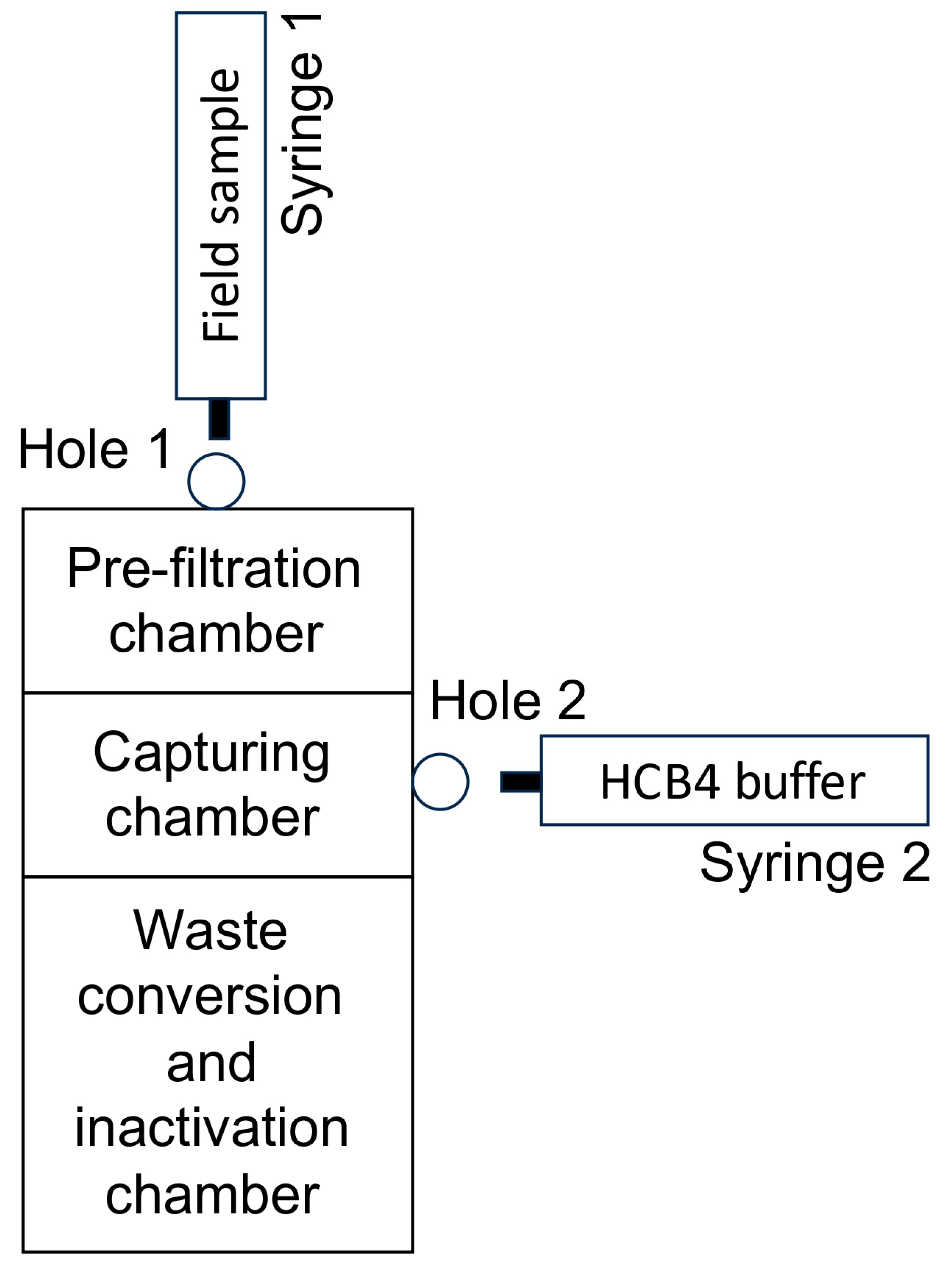
The detection and quantification of Salmonella in the CCR was conducted using a novel test kit based on filtration and lyophilized isothermal detection (HyperKit Total Salmonella; Hypercell, Ithaca, NY, USA; Appendix B Figure A1). This dose-response assay was used to generate an algorithm to predict the concentration of Salmonella in CCR samples based on the isothermal data. Briefly, an aliquot of the CCR sample (10 mL) was collected using a 10 mL Luer lock syringe (BH supplies, Berwick, PA, USA). The aliquot was first injected inside the pre-filter using the top male Luer lock (Hole 1; Appendix B Figure A1). Using a combination of multiple filtration modules located inside the HyperKit, bacteria including Salmonella were separated from large (>20 µm) and small (<0.22 µm) particles present inside the CCR samples and concentrated inside a “capturing chamber”. Then, bacterial cells were cleaned inside the capturing chamber by injecting 3 mL of HCB4 buffer (Hypercell, Ithaca, NY, USA) inside a 3 mL Luer lock syringe using the male Luer lock adapter connected to the capturing chamber (Hole 2; Appendix B Figure A1). Purified bacterial cells were collected inside a 3 mL syringe by pulling the plunger of the syringe to extract the purified product (c.a., 150–200 µL). The recovered liquid was transferred into a 1.5 mL collection tube. Fifty microliters of the extracted product was transferred into the 0.2 mL high-profile PCR tube pre-filled with lyophilized isothermal reagents (e.g., Bst DNA polymerase enzyme, invA primers, dNTPs, MgSO4, reaction buffer, and cryoprotectants; Hypercell, Ithaca, NY, USA). The inoculated tube was incubated at 65 °C for 60 min in the HyperReader-R8 (isothermal reader; Hypercell, Ithaca, NY, USA; Appendix B Figure A1). Fluorescence signal (Sybr Green; excitation: 495 nm; emission: 530 nm) was recorded every minute to monitor DNA amplification over time. A sample was considered positive for Salmonella when the relative fluorescence unit (RFU) exceeded 103 within 60 min of incubation at 65 °C. A result was considered negative for Salmonella when the RFU was under 103 within the same incubation period. Each assay was complemented with positive and negative controls (i.e., one tube spiked with 103 genomic copies of Salmonella and one tube spiked with HCB4, respectively). The time needed for the RFU to reach 103 was used to assess the detection speed of Salmonella in the sample (i.e., time-to-results [TTR] in minutes). Using a cubic polynomial regression (JMP software version 18; SAS Institute, Cary, NC, USA), TTR data obtained from the 57 spiked CCR samples were compared to the Salmonella counts obtained from the same CCR samples using the microbiology approach (XLT-4 agar at 42 °C). The generated equation (Log10 [Salmonella concentration/mL] = 8−5 × TTR4 − 0.0069 × TTR3 + 0.216 × TTR2 − 2.9027 × TTR + 15.82) was used to convert TTR data into Salmonella concentration data. This equation was used to assess the Salmonella load in commercial CCR samples obtained from multiple commercial plants. A Grubbs’ outlier test was conducted to identify outliers between the predicted Salmonella counts obtained using the filtration-isothermal kit and Salmonella counts obtained using the microbiology method.
2.4. Detection and Quantification of Salmonella in Commercial Chicken Carcass Rinse Samples
Two independent poultry processing facilities provided CCR samples collected under real-world conditions to conduct an external validation of the predictive model. A poultry processing facility located in Georgia (Company G) collected and shipped CCR samples (n = 10) to conduct direct detection and quantification of Salmonella without enrichment (>10 CFU/mL) using both microbiological and filtration-isothermal approaches. The CCR samples were collected in accordance with the USDA FSIS compliance guidelines, directly from the processing line prior to the chilling step (cooling of chicken carcasses at 4 °C to prevent microbial growth) [42]. Aliquots of the CCR samples (20–30 mL stored in 100 mL plastic bottles) were shipped the same day on ice and processed within 24 h of collection for the detection of Salmonella using the HyperKit (10 mL of CCR processed per reaction, as described above) and microbiology approach (XLT-4 agar). For culture confirmation, aliquots of samples (0.1, 0.5, and 1 mL per plate) were plated on XLT-4 agar and incubated at 42 °C for up to 48 h. An equal volume of 1X BPW (pH 7.2) was used as a blank control, and 1X BPW (pH 7.2) spiked with 103 CFU/mL was used as a positive control. TTR data obtained from the 10 commercial CCR were converted into Log10 (Salmonella cell/mL) using the equation mentioned above. Salmonella data estimated using HyperKit were compared to the Salmonella counts obtained from the same CCR samples using the microbiology approach.
A poultry processing facility located in Nebraska (Company N) shipped carcass samples (n = 11) collected using MicroTally® Mitt (Fremonta; San Jose, CA, USA) to conduct 1 CFU detection of Salmonella post-enrichment. These samples were collected based on the manufacturer’s recommendation (https://microtally.com/wp-content/uploads/2024/11/Validation-of-MT-Mitt-sampling-for-poultry.pdf, accessed on 15 April 2026), directly from the processing line after the chilling step. The used MicroTally® Mitts were shipped the same day on ice and processed within 24 h of collection. MicroTally® Mitts were resuspended in cold 1X BPW (pH 7.2) and manually homogenized for at least 30 s. The resuspended MicroTally® Mitts were processed based on the ISO 6579 procedure [45]. Briefly, the resuspended MicroTally® Mitt were enriched at 35 °C for 24 h (first non-selective enrichment). Then, 1 mL of the enriched BPW was transferred into 9 mL of RV broth (Neogen, Lansing, MI, USA) and incubated at 42 °C for 24 h (second selective enrichment). During the enrichment process, four collection time points (TP) were selected. TP1 was collected before enrichment; TP2 was collected 8 h after the first enrichment started; TP3 was collected 24 h after the first enrichment started; and TP4 was collected 24 h after the second enrichment started. Each TP was processed using filtration-isothermal (as described above), microbiology (0.01, 0.1, and 1 mL plating on XLT-4 agar), and USDA Bacteriological Analytical Manual (BAM) PrepMan-Loop-mediated isothermal amplification (LAMP; as recommended by USDA) [40,41] methods for the detection of Salmonella in the CCR. Qualitative results (presence/absence) were recorded for each method used.
2.5. Inclusivity and Exclusivity Testing
The inclusivity and exclusivity of the HyperKit were evaluated according to the AOAC Performance Tested MethodsSM (PTM) validation guidelines to assess detection specificity for the method [46]. Briefly, inclusivity was determined by testing the HyperKit against a panel of targeted organisms (c.a., 103 CFU per reaction), ensuring all strains were reliably detected without false negatives. Exclusivity was assessed by challenging the kit with high concentrations (>106 CFU per reaction) of non-target organisms with distinct phylogenetic differences to confirm the absence of cross-reactivity and false-positive results. The complete list of target and non-target organisms used in the validation is provided in Appendix A Table A1. The detection of the target organisms was based on a real-time fluorescence signal measured by the CFX96 Touch Real-Time PCR System (Bio-Rad; Hercules, CA, USA). A sample was considered positive if the RFU exceeded 103 within 60 min of incubation at 65 °C. A result was considered negative if no increase in fluorescence above the threshold was observed within the same incubation period. Each assay was complemented with a tube inoculated using only HCB4 (blank negative control) and another tube spiked with a standardized concentration of Salmonella genomic DNA (positive control; c.a., 103 genomic copies) extracted from a known bacterial strain detected by the designated kit. The quality and concentration of the extracted genomic DNA were confirmed using Nanodrop 2000C (Thermo Fisher Scientific, Waltham, MA, USA). Assays were considered valid if the positive control yielded results within 10 min and the RFU was above 3 × 10³. Two independent experiments were conducted.
2.6. Reproducibility, Repeatability, Shelf-Life, Robustness, and Matrix Testing
The reproducibility of the filtration-isothermal kit was evaluated by three independent users (scientific staff members of Dr. Rajashekara’s lab, University of Illinois [Champaign, IL, USA] with varying expertise and levels of laboratory experience) following the recommended procedure. The reproducibility of the technique was assessed based on the TTR obtained by the operators when processing the same sample. The repeatability of the filtration-isothermal kit was evaluated across the 11-spiking concentrations tested in this study (between 1.0-log10 and 7.5-log10 CFU.mL; n = 2–4 replicates per concentration) following the recommended procedure. The repeatability of the technique was assessed based on the TTR obtained by a given operator when processing the same sample multiple times. The shelf-life of the lyophilized isothermal reagents was validated using ASTM F1980 accelerated aging standard study [47,48]. Briefly, lyophilized isothermal reagents in the PCR tubes were incubated inside a food dehydrator (EFD770WD; Elite Gourmet, City of Industry, CA, USA) for up to 69 days at 52 ± 3 °C (ambient relative humidity between 12–30%). A Q10 factor of 2.0 was used to simulate the accelerated aging of the reagents at 52 °C. Aliquots (n = 3 tubes) were collected every 11 days and spiked with a standardized concentration of Salmonella Typhimurium genomic DNA (c.a., 103 genomic copies). The detection speed data (TTR and maximum RFU) obtained for each aliquot were compared against the same lot of reagents stored at 4 °C (control group). The robustness of the filtration-isothermal kit was evaluated by transferring different volumes of the extract product using the filtration kit into the PCR tube pre-filled with the lyophilized isothermal reagents (final volume between 20 and 150 µL). The effect of sample matrices on the detection of Salmonella with the filtration-isothermal kit was evaluated using CCR, BPW, and 1X Phosphate Buffered Saline (PBS; pH 7.4; Thermo Fisher Scientific, Waltham, MA, USA). Sample matrices were spiked with a known concentration of Salmonella (c.a., 156 ± 5 CFU/mL) and processed with the filtration-isothermal kit, as described above (n = 7 reps per group). For all experiments mentioned above, a sample was considered positive for Salmonella when the RFU exceeded 103 within 60 min of incubation at 65 °C. A result was considered negative for Salmonella when the RFU was under 103 within the same incubation period. The standard procedure recommended for the HyperKit was used as baseline reference (as described above) to assess whether the induced changes in the procedure led to false results (e.g., false-negative or improper semi-quantitative data). A Bland-Altman analysis was used to assess the agreement in Salmonella counts between the filtration-isothermal kit and the microbiology method across the 11 spiking groups.
2.7. Statistical Analyses
Bacterial data were log10 transformed. Fluorescent data (relative fluorescent unit; RFU) were recorded using the HyperReader software (v4.0), Laboao software (v.H8801_V1004), or CFX Maestro Software (v2.3) and were used to determine the detection speed (time-to-results in minutes; TTR) required to detect a specific concentration of Salmonella in the samples. The detection speed was inversely proportional to the quantity of Salmonella present in the sample. A sample was considered positive if the RFU reached at least 103 within 60 min at 65 °C incubation. TTRs were converted into predicted Salmonella concentration data using the following algorithm (Log10 [Salmonella concentration/mL] = 8−5 × TTR4 − 0.0069 × TTR3 + 0.216 × TTR2 − 2.9027 × TTR + 15.82). The formula was generated using a cubic polynomial regression based on the Salmonella bacterial counts and TTR data obtained using the 57 spiked CCR. This equation was then used to assess the Salmonella load in the commercial CCR samples. Statistical analyses were performed using JMP 18 software (SAS Institute, Cary, NC, USA). A Grubbs’ outlier test was conducted to identify outliers between the predicted Salmonella counts obtained using the filtration-isothermal kit and Salmonella counts obtained using the microbiology method. A Bland-Altman plot was used to assess the agreement in Salmonella counts between the filtration-isothermal kit and the microbiology method across the 11 spiking groups.
3. Results
3.1. Associations Between the Detection Speed and Salmonella Load in Chicken Carcass Rinse
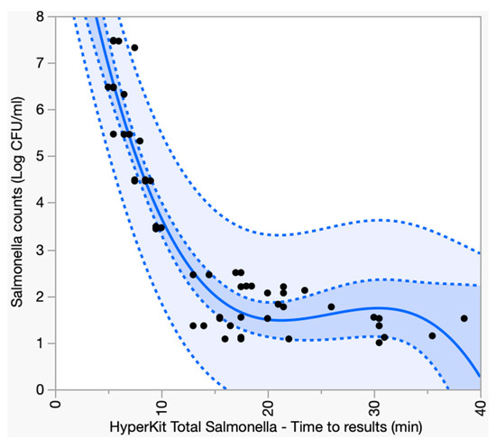
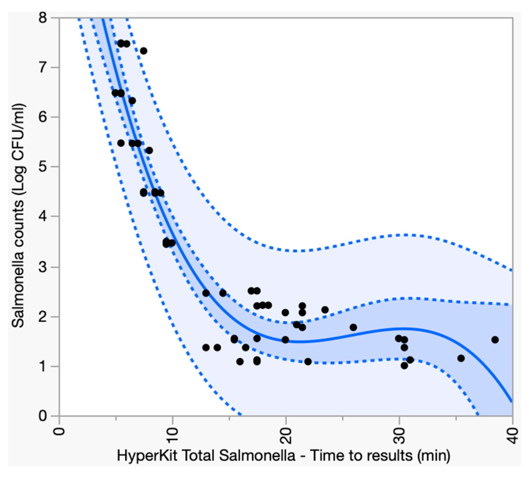
All CCR samples spiked with a concentration of between 1.0-log10 and 7.5-log10 CFU/mL (n = 57) were detected as positive in both the filtration-isothermal and the microbiological approaches (Table 1). With the filtration-isothermal kit, Salmonella concentrations between 5.5-log10 and 7.5-log10 CFU/mL (n = 13/57) yielded positive results within 4.6 ± 0.5 min, between 3.4-log10 and 5.3-log10 CFU/mL (n = 13/57) yielded positive results within 7 ± 0.7 min, between 2.1-log10 and 2.5-log10 CFU/mL (n = 11/57) yielded positive results within 13.5 ± 2.6 min, and between 1.0-log10 and 1.8-log10 CFU/mL (n = 13/57) yielded positive results within 18.4 ± 7.4 min (Figure 1). No fluorescent signal above 103 RFU was recorded from the blank samples (n = 7). A cubic polynomial regression demonstrated an association between the detection speed and the Salmonella concentration in the sample (r2 = 0.90; root mean square error (RMSE) = 0.67; p < 0.001). The semi-quantitative predictive accuracy of the filtration-isothermal kit was 0.2-log10 ± 0.1-log10 precision between 1.5-log10 and 7.5-log10 CFU/mL compared to Salmonella counts obtained by the microbiology plating method (n = 39/57; Spiking groups E-L; Table 1, Appendix C Figure A2). On the other hand, its semi-quantitative predictive accuracy was reduced when spiking concentrations were under 1.5-log10 CFU/mL (Salmonella population overestimated by 0.6-log10 ± 0.1-log10 compared to Salmonella counts obtained by the microbiology plating method; n = 18/57; Spiking groups B-D; Table 1, Appendix C Figure A2). The reduced accuracy of the model was explained by early TTR when Salmonella concentration was under 2-log10 CFU/mL (TTR between 13 and 22 min instead of TTR > 30; n = 11/21).
Table 1.
Association between detection speed and Salmonella concentration in chicken carcass rinse samples using the filtration-isothermal detection method.
Figure 1.
Association between the detection speed of the filtration-isothermal method and the concentration of Salmonella in chicken carcass rinse samples. Y-axis data obtained by microbiology plating after 24 h incubation at 37 °C. X-axis data were obtained using the filtration-isothermal method (detection of fluorescent signal over time at 65 °C). The solid blue line indicates the cubic polynomial regression model (r2 = 0.90; RMSE = 0.67). The dark blue halo indicates the 95% confidence interval. The light blue halo indicates where single future observations are likely to fall. Black dots indicate chicken carcass rinse samples (n = 57). Blank samples (Salmonella-free) were not plotted in this graph.
3.2. Rapid Detection and Quantification of Salmonella in Commercial Chicken Carcass Rinse
All CCR samples (#1–10) received by Company G were detected positive within 40 min with the filtration-isothermal kit and after 24 h with the microbiology plating method (Table 2). Overall, the predicted Salmonella counts fluctuated between 1.96-log10 and 3.47-log10 CFU/mL for the filtration-isothermal kit, and between 1.91-log10 and 2.36-log10 CFU/mL for the microbiology plating method across the 10 CCR samples tested. Only one sample (sample #2) was significantly overestimated with the filtration-isothermal kit compared to the microbiology plating method (3.47-log10 CFU/mL versus 2.02-log10 CFU/mL, respectively; p < 0.05). Excluding sample #2 as an outlier, the Salmonella data for both approaches were within 0.24 ± 0.18-log10 CFU/mL agreement.
Table 2.
Pre-enrichment quantification of Salmonella in commercial chicken carcass rinse samples from Company G using the filtration-isothermal and microbiology approaches.
For the 11 MicroTally® Mitt samples received by Company N, a few samples were positive before enrichment across the three Salmonella detection methods used (sample #4 and #9 using the filtration-isothermal approach; sample #3, #4, and #9 using direct plating; and sample #4 using USDA BAM PrepMan-LAMP; Table 3). After 8 h of enrichment in the non-selective broth, all samples (11/11) were positive for Salmonella using the filtration-isothermal approach, while only 45% (5/11) were positive for Salmonella using the USDA BAM PrepMan-LAMP, and none of the samples were determined positive for Salmonella after plating on XLT-4 agar due to the predominant growth of non-Salmonella-like colonies that altered the interpretation of the results. After 24 h of enrichment in the non-selective broth, all samples (11/11) were positive for Salmonella using filtration-isothermal and USDA BAM PrepMan-LAMP approaches. Interpretations on XLT-4 agar were still compromised due to the predominant background noise. Only after the 24-h enrichment in selective RV broth, all three methods detected all 11 samples as positive for Salmonella.
Table 3.
Post-enrichment detection of Salmonella in commercial MicroTally® Mitt samples from Company N using the filtration-isothermal and microbiology approaches.
3.3. HyperKit Total Salmonella Only Detected Salmonella Serotypes and None of the Other Prokaryotes and Eukaryotes Tested
The lyophilized isothermal master mix detected all Salmonella serotypes within 10 min (n = 46/46), while none of the strains belonging to the exclusive list (non-Salmonella organisms; n = 0/37) were detected after 60 min incubation at 65 °C (Appendix A Table A1). Among the exclusive list, 10 Enterobacteriaceae genus closely related to Salmonella (e.g., Citrobacter, Enterobacter, Escherichia, Hafnia, Klebsiella, Pantoea, Proteus, Shewanella, Shigella, and Yersinia), Gram-positive bacteria (Staphylococcus), and non-prokaryotic organisms (Saccharomyces) tested negative. Blank controls (DNA-free) were also negative. Furthermore, the lyophilized isothermal master mix also detected Salmonella when co-inoculated with a predominant concentration of E. coli (Ratio 1:1000, respectively). Tests conducted with Salmonella alone yielded a TTR of 12 ± 2 min, while tests conducted with a mix of Salmonella-E. coli yielded a TTR of 11 ± 2 min.
3.4. Repeatability, Reproducibility, Shelf-Life, Robustness, and Matrix Testing with HyperKit Total Salmonella
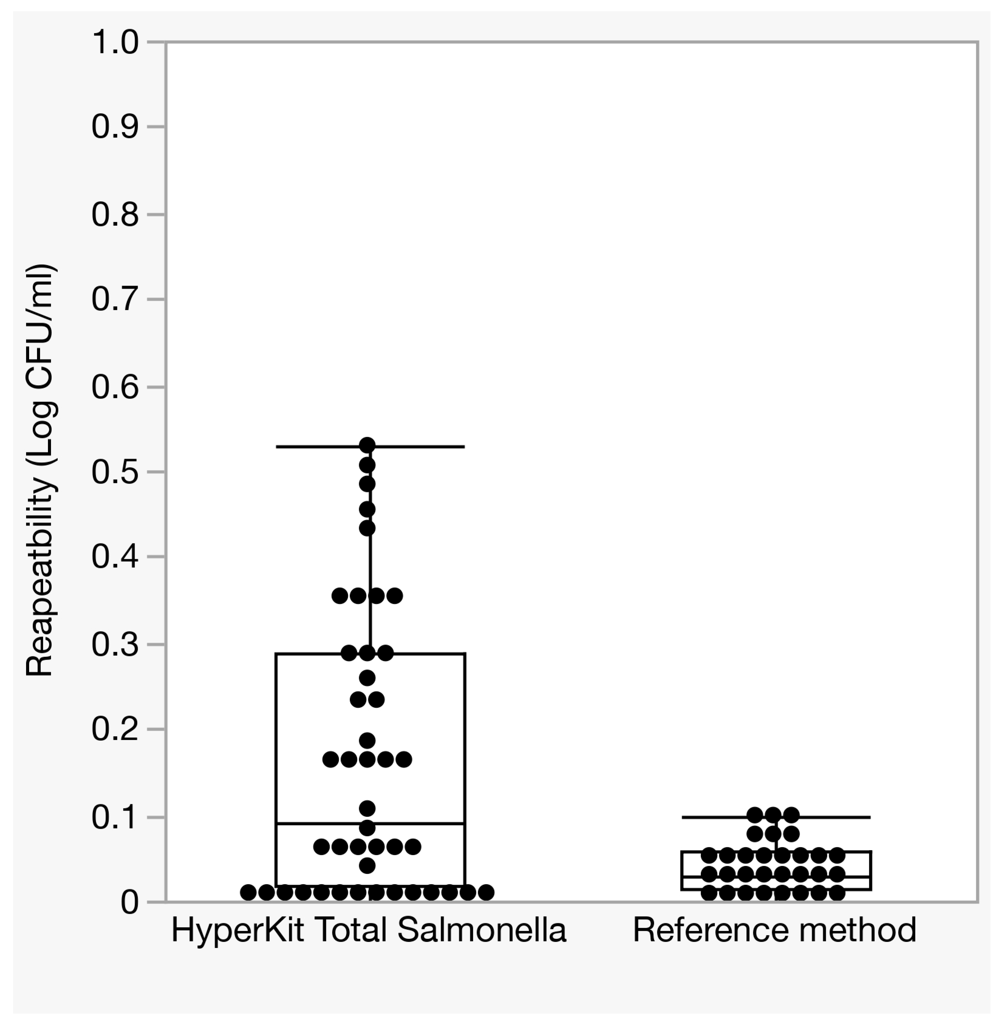
Across the 12-spiking concentrations of Salmonella tested in this study, a variability in predicted Salmonella counts of 0.16-log10 (95% CI: 0.11–0.21) was obtained using the filtration-isothermal kit (Appendix C Figure A3). The variation was independent of the Salmonella concentration in the CCR samples. As a point of comparison, the microbiology plating method was 0.04-log10 (95% CI: 0.03–0.05; Appendix C Figure A3). The reproducibility of the filtration-isothermal kit was validated by three operators. All operators detected Salmonella in the spiked samples with equivalent detection speed (TTR between 13.5 and 14.5 min) and predicted counts (Salmonella counts between 1.65-log10 and 1.68-log10 CFU/mL).
The accelerated stability study showed that the lyophilized isothermal reagents maintained their detection speed until 34 days of storage at 52 ± 3 °C (c.a., 9 months at 22 °C and 33 months at 4 °C; Appendix D Table A2). From 0 to 34 days of storage, the TTR were approx. 11 ± 1.1 min with a consistent fluorescent signal intensity (maximum RFU) between 3500 and 4000 for both groups incubated at 52 °C and 4 °C. On the other hand, a progressive decay of the detection speed and fluorescent signal intensity was observed after 34 days of storage at 52 ± 3 °C (Appendix D Table A2). The detection speed was reduced by 3.3 min at 46 days, followed by 13 min at 57 days, and over 27 min at 69 days of storage at 52 ± 3 °C. Similarly, a gradual reduction of the fluorescent signal intensity was observed over time after 34 days of storage at 52 ± 3 °C. The fluorescent signal went from 3500 and 4000 to 1700 and 2100 at 69 days of storage at 52 ± 3 °C. No decrease in fluorescent signal intensity or delayed detection speed was detected when the lyophilized reagents were stored at 4 °C.
The robustness studies demonstrated that the detection of Salmonella was not compromised when the lyophilized isothermal reagents were resuspended with a final volume between 40 and 60 µL of extract product from the filtration system (Appendix D Table A3; p > 0.05). On the other hand, substantial reductions of detection speed (until not being detected) were recorded when the final volumes were under 40 µL or exceeded 60 µL. It was also demonstrated that the detection of Salmonella with the lyophilized isothermal reagents was possible in the real-time thermocycler with filters compatible with SYBR green (Appendix D Table A4). Two additional units were tested in this study and provided equivalent detection efficacy: CFX96 Touch Real-Time PCR System (Bio-Rad; Hercules, CA, USA) and Model LPCR-H800 (Laboao, Zhengzhou, China).
The effect of sample matrices on the detection speed of Salmonella demonstrated equivalent detection speed using the filtration-isothermal kit between the spiked CCR, BPW, and 1X PBS groups (Appendix D Table A5). At a spiking dose of 156 ± 5 CFU/mL, all three groups showed a detection speed between 14.6 and 15.7 min (p > 0.05).
4. Discussion
As food production systems become faster and more decentralized, the need for diagnostic tools that are equally rapid, accurate, and easy to use at the point of production is more critical than ever to consistently maintain high standards of food safety and quality [7]. The data presented in this study demonstrate that the combination of a point-of-use filtration-based method with isothermal amplification was able to extract and detect low concentrations of Salmonella (<10 CFU) within 60 min in CCR without standard scientific equipment (e.g., centrifuge, vortex, incubator, and biosafety hood). Diagnostic kits capable of combining speed, sensitivity, and specificity play a key role in quality control in the food processing industry [49]. Such technology could allow chicken meat processors to make on-site, real-time decisions to mitigate contamination and optimize production processes.
From a performance standpoint, the detection and quantification of Salmonella was not affected by the nature of the samples (i.e., artificially spiked CCR versus commercial CCR naturally contaminated with Salmonella). No false-positive or false-negative results were recorded under the tested conditions. Furthermore, the utilization of Salmonella-specific genes (i.e., invA) for the kit ensured the detection of all Salmonella serotypes tested in this study (n = 46), while preventing cross-reactivity toward non-target or closely related organisms (n = 37) [50,51]. In terms of operational practicality, repeatable semi-quantitative data were obtained between technological replicates and across users. Furthermore, the lyophilized status of the isothermal master mix enabled the storage of the reagents at ambient temperature during the period of time required to conduct all the experiments described in this study (up to nine months) [52]. It is also important to mention that 10 mL of CCR was used for the extraction of Salmonella using the filtration-isothermal kit, which is substantially higher compared to standard detection methods (e.g., molecular and microbiology), with limited sample input size (<1 mL) [53,54]. Future study will assess whether the processing capacity of the filtration kit could be extended up to 200 mL input volume for bacterial extraction. Such achievement might enhance the sensitivity of the test for the detection of key organisms present at low concentrations in large field samples with short enrichment times (e.g., E. coli O157 in beef; Listeria monocytogenes in ready-to-eat product) [55,56]. The development of a point-of-use technology capable of detecting 1 CFU in a few hours would enable same-shift decision-making for food processors [57,58,59]. Furthermore, processing larger volumes (up to 200 mL) could mitigate the reduced quantitative accuracy observed in this study when samples were spiked below 1.5-log10 CFU/mL. The reduced quantitative accuracy of isothermal methods was previously reported near the detection limit [60,61]. At low copy numbers, stochastic variation in target distribution among aliquots, combined with variability in amplification initiation, can result in disproportionally early time-to-results (TTR) values and subsequent overestimation of pathogen concentrations.
Overall, this study demonstrated the ability of the HyperKit Total Salmonella to rapidly detect and quantify Salmonella in CCR using a simple filtration-isothermal workflow. The analytical sensitivity and specificity observed in this study were comparable to those reported for commercially available molecular platforms (BAX®, Gene-Up®, SureTect™, and iQ-Check™), which are widely used in food safety testing [62,63]. Similar to these systems, the filtration-isothermal method accurately detected 10 CFU and above, but an enrichment is required to reliably detect lower concentrations. Nevertheless, this method demonstrated substantial benefits favoring field deployability (reduced infrastructure and operational requirements, simplified sample preparation, rapid time-to-results, low-cost fluorescent reader, and no hazardous chemical handling compared to other molecular platforms), which would facilitate earlier intervention and more immediate decision-making during poultry production and processing. Commercial molecular platforms are generally optimized for high-throughput testing and can process up to 96 samples simultaneously, making them well-suited for a centralized laboratory environment. In contrast, the filtration-isothermal method is a rapid (i.e., <1 min processing time; up to 60 min detection time) and simple (one-step tool) point-of-use kit, but it has limited throughput sample processing capacity (up to 20 samples), which may limit its utility for large-scale batch testing. Further validation studies must be conducted to identify bottleneck conditions that could affect the performance of the filtration-isothermal method of the HyperKit. To overcome these issues, an electro-mechanical device can be developed to automate the processing of multiple samples simultaneously. This approach will minimize the need for trained operators, reduce risks for operator errors, and enhance the accuracy of the results [64]. Similarly, the lyophilized isothermal master mix enabled the detection of only one pathogen per reaction. Studies demonstrated that multiplexing can be used with molecular techniques (qPCR and isothermal reactions) to facilitate the detection of multiple organisms within one reaction tube to answer industry needs [65,66,67,68]. During the study, the lyophilized isothermal reagents did not allow distinguishing between live and dead cells, which could explain the quantitative discrepancies observed between DNA-based detection (detecting both live and dead cells) and culturing (detecting only live cells) when processing CCR. Adding a PMAxx treatment (propidium monoazide) between the extraction step and isothermal incubation would prevent the detection of non-viable cells [69].
5. Conclusions
This study demonstrated that the technological combination of filtration with lyophilized isothermal detection could enhance real-time microbial surveillance at critical control points across the food supply chain, using a simplified workflow that avoids complex sample preparation or extended incubation periods. This technology could be applied to food safety testing, environmental monitoring, supply chain quality control, and emergency response conditions. The analytical sensitivity, operational simplicity, and field readiness make it particularly well suited for rapid screening applications in food processing environments. Future studies should focus on expanded validation on other pathogens of significance, comparison with existing AOAC-certified methods across a wider range of commodities and matrix types, and integration into routine food safety management systems.
Author Contributions
Conceptualization, L.D. and G.R.; methodology, L.D. and A.S.K.; validation, A.S.K. and R.S.; formal analysis, A.S.K.; resources, L.D. and G.R.; data curation, A.S.K.; writing—original draft preparation, A.S.K.; writing—review and editing, L.D., A.S.K., R.S. and G.R.; supervision, L.D. and G.R.; project administration, L.D. and G.R. All authors have read and agreed to the published version of the manuscript.
Funding
This research received no external funding.
Institutional Review Board Statement
Not applicable.
Informed Consent Statement
Not applicable.
Data Availability Statement
The original contributions presented in this study are included in the article. Further inquiries can be directed to the corresponding authors.
Acknowledgments
We thank Shrestha Prajjwal, Steven Smalley, and Elizabeth Ducharme for their technical support.
Conflicts of Interest
Author Loïc Deblais was employed by the company Hypercell Technologies, Ithaca, USA. The products used for the detection and quantification of Salmonella in artificial chicken carcass rinse samples are also from Hypercell Technologies. The remaining authors declare that the research was conducted in the absence of any commercial or financial relationships that could be construed as a potential conflict of interest.
Abbreviations
The following abbreviations are used in this manuscript:
| AOAC PTM | Association of Official Analytical Chemists Performance Tested Methods |
| ATCC | American type culture collection |
| ATP | adenosine triphosphate |
| BAM | bacteriological analytical manual |
| BPW | buffered peptone water |
| CCR | chicken carcass rinse |
| CFU | Colony-forming unit |
| CI | confidence interval |
| CRISPR | clustered regularly interspaced short palindromic repeats |
| DNA | deoxyribonucleic acid |
| ELISA | enzyme-linked immunosorbent assay |
| FSIS | Food Safety and Inspection Service |
| LAMP | loop-mediated isothermal amplification |
| LB | Luria broth |
| LFA | lateral flow immunoassays |
| LOD | limit of detection |
| min | minutes |
| mL | milliliter |
| MRS | De Man–Rogosa–Sharpe |
| NGS | next-generation sequencing |
| NIH BEI | National Institutes of Health: Biodefense and Emerging Infections |
| NRRL | National Center for Agricultural Utilization Research |
| °C | degree Celsius |
| PCR | polymerase chain reaction |
| RFU | relative fluorescent units |
| RMSE | root mean square error |
| TP | time point |
| TTR | time-to-results |
| µL | microliter |
| USDA | United States Department of Agriculture |
| XLT-4 | xylose lysine tergitol 4 |
Appendix A. Inclusivity—Exclusivity of the Filtration-Isothermal Detection Kit
Table A1.
Inclusivity (n = 46) and exclusivity (n = 37) of HyperKit total Salmonella.
Appendix B. Details About the Sample Purification Procedure Using the Filtration-Isothermal Detection Kit
Figure A1.
Description of the filtration-isothermal detection kit. Syringe 1 (3 mL graduated syringe with male Luer lock adapter) is used to collect the liquid field samples. Hole 1 (entry port with female Luer lock adapter) is used to inject the liquid field samples inside the filtration system. The filtration system is divided into three organized modules. First, the large particles (>20 µm) inside the field samples will be separated from the rest of the liquid using a succession of mesh with low protein retention and fast flow rate. Then, the small particles (between 20 and 0.22 µm) are collected inside the capturing chamber via a membrane with smaller pores. The collected bacteria are purified by injecting the Hypercell buffer (prefilled syringe 2–3 mL graduated syringe with male Luer lock adapter) directly inside the capturing chamber using Hole 2 (entry port with female Luer lock adapter). Purified bacteria are retrieved inside syringe 2 (c.a., 150–200 µL) when creating negative pressure by pulling the plunger of syringe 2. The waste (liquid and particles under <0.22 µm) is collected inside the bottom chamber, where the waste is converted into a solid gel using 1 g of sodium polyacrylate and inactivated with 0.04 g of dimethyl benzyl ammonium chloride.
Appendix C. Semi-Quantitative Performance of the Filtration-Isothermal Detection Kit in Chicken Carcass Rinse
Figure A2.
Differences in predicted Salmonella concentration obtained with the filtration-isothermal method compared to the counts obtained with the microbiology method. The boxplot is separated by spiking groups (B-K), proportionally organized based on Salmonella concentration in chicken carcass rinse samples (Table 1). Black dots indicate chicken carcass rinse samples (n = 57).
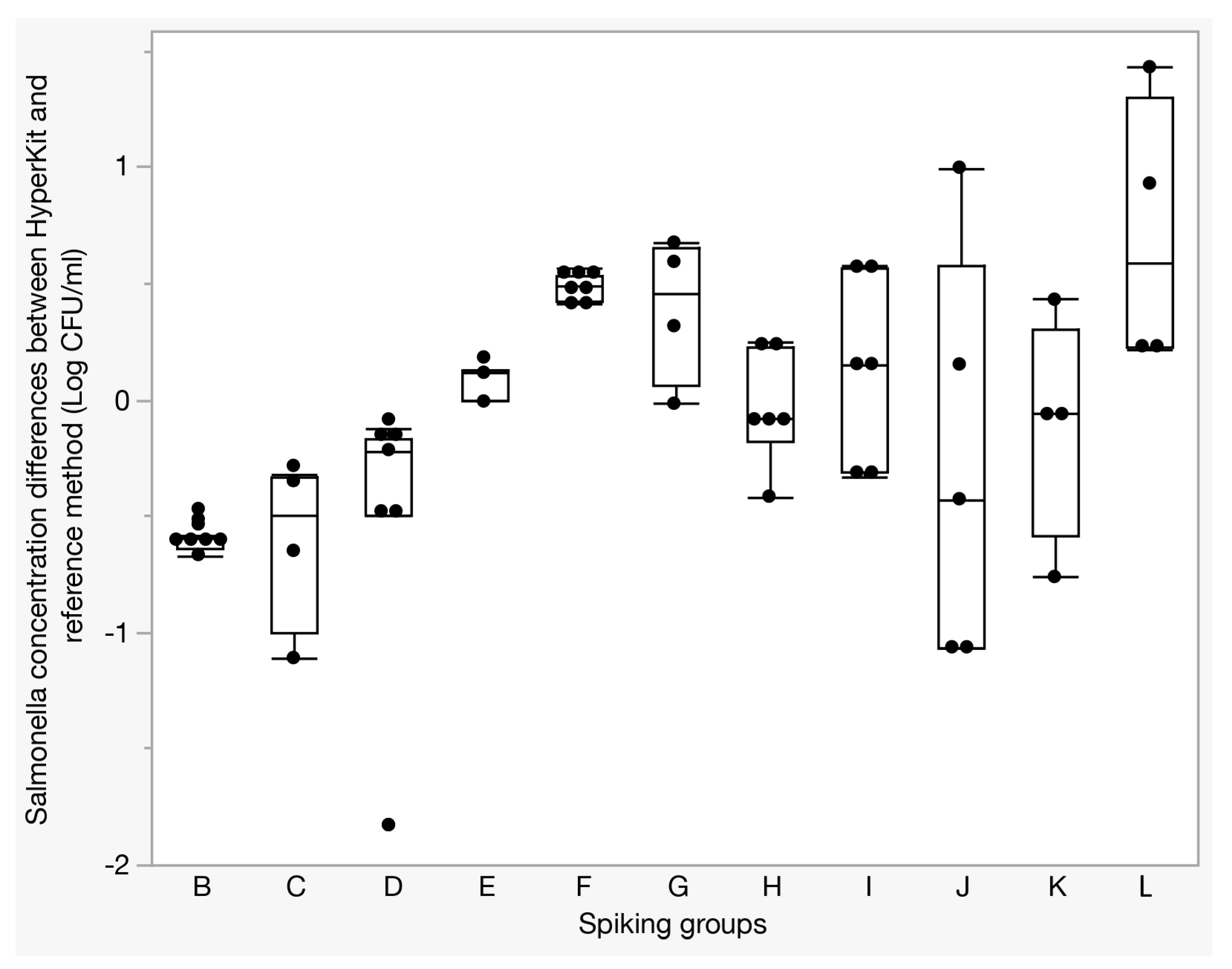
Figure A3.
Quantitative accuracy observed with the filtration-isothermal method compared to the counts obtained with the microbiology method. The quantitative accuracy of each method was measured by processing several times (n = 2–4 times) the same chicken carcass rinse samples spiked with a known concentration of Salmonella. Black dots indicate the variability in Salmonella concentration obtained when processing the same chicken carcass rinse samples compared to the average of the concentrations obtained with the same chicken carcass rinse sample (n = 44 replicates total).
Appendix D. Detection Performance of the Lyophilized Isothermal Master Mix
Table A2.
Accelerated aging study of lyophilized isothermal master mix at 52 °C.
Table A3.
Impact of reaction volume on the capacity of the lyophilized isothermal master mix to detect Salmonella.
Table A4.
Impact of real-time PCR machine models on the detection of Salmonella using lyophilized isothermal master mix.
Table A5.
Impact of chicken carcass rinse on the detection speed of Salmonella after extraction using the filtration-isothermal kit.
References
- CDC. Estimates: Burden of Foodborne Illness in the United States. Available online: https://www.cdc.gov/food-safety/php/data-research/foodborne-illness-burden/index.html (accessed on 1 July 2025).
- World Health Organization. Food Safety. Available online: https://www.who.int/news-room/fact-sheets/detail/food-safety (accessed on 1 July 2025).
- Hoffmann, S.; White, A.E.; McQueen, R.B.; Ahn, J.-W.; Gunn-Sandell, L.B.; Scallan Walter, E.J. Economic Burden of Foodborne Illnesses Acquired in the United States. Foodborne Pathog. Dis. 2025, 22, 4–14. [Google Scholar] [CrossRef] [Scilit] [PubMed]
- CDC. Investigation Update: Listeria Outbreak, Meats Sliced at Delis. Available online: https://www.cdc.gov/listeria/outbreaks/delimeats-7-24/investigation.html (accessed on 1 July 2025).
- CDC. E. coli Outbreak Linked to Onions Served at McDonald’s. Available online: https://www.cdc.gov/ecoli/outbreaks/e-coli-O157.html (accessed on 1 July 2025).
- CDC. Investigation Update: Salmonella Outbreak, Cucumbers—June 2024. Available online: https://www.cdc.gov/salmonella/outbreaks/africana-06-24/investigation.html (accessed on 1 July 2025).
- Mahony, J.; van Sinderen, D. Virome Studies of Food Production Systems: Time for ‘Farm to Fork’ Analyses. Curr. Opin. Biotechnol. 2022, 73, 22–27. [Google Scholar] [CrossRef] [Scilit] [PubMed]
- Scallan, E.; Hoekstra, R.M.; Angulo, F.J.; Tauxe, R.V.; Widdowson, M.-A.; Roy, S.L.; Jones, J.L.; Griffin, P.M. Foodborne Illness Acquired in the United States—Major Pathogens. Emerg. Infect. Dis. 2011, 17, 7–15. [Google Scholar] [CrossRef] [PubMed]
- Zhang, Y.; Liu, J.; Pan, Y.; Shi, K.; Mai, P.; Li, X.; Shen, S. Progress on the Prevention of Poultry Salmonella with Natural Medicines. Poult. Sci. 2025, 104, 104603. [Google Scholar] [CrossRef] [Scilit] [PubMed]
- Shaji, S.; Selvaraj, R.K.; Shanmugasundaram, R. Salmonella Infection in Poultry: A Review on the Pathogen and Control Strategies. Microorganisms 2023, 11, 2814. [Google Scholar] [CrossRef] [Scilit] [PubMed]
- Marmion, M.; Macori, G.; Ferone, M.; Whyte, P.; Scannell, A.G.M. Survive and Thrive: Control Mechanisms That Facilitate Bacterial Adaptation to Survive Manufacturing-Related Stress. Int. J. Food Microbiol. 2022, 368, 109612. [Google Scholar] [CrossRef] [Scilit] [PubMed]
- Betiku, E.; Ogundipe, T.T.; Kalapala, T.; Obe, T. A Mini-Review on Multi-Hurdle Control of Salmonella Along Poultry Production Continuum. Animals 2025, 15, 875. [Google Scholar] [CrossRef] [Scilit] [PubMed]
- González, R.J.; Sampedro, F.; Feirtag, J.M.; Sánchez-Plata, M.X.; Hedberg, C.W. Prioritization of Chicken Meat Processing Interventions on the Basis of Reducing the Salmonella Residual Relative Risk. J. Food Prot. 2019, 82, 1575–1582. [Google Scholar] [CrossRef] [Scilit] [PubMed]
- Soni, A.; Bremer, P.; Brightwell, G. A Comprehensive Review of Variability in the Thermal Resistance (D-Values) of Food-Borne Pathogens—A Challenge for Thermal Validation Trials. Foods 2022, 11, 4117. [Google Scholar] [CrossRef] [Scilit] [PubMed]
- Shao, L.; Sun, Y.; Zou, B.; Zhao, Y.; Li, X.; Dai, R. Sublethally Injured Microorganisms in Food Processing and Preservation: Quantification, Formation, Detection, Resuscitation and Adaption. Food Res. Int. 2023, 165, 112536. [Google Scholar] [CrossRef] [Scilit] [PubMed]
- Kim, U.; Moon, Y.-J.; Kim, J.-H.; Lee, S.-Y.; Oh, S.-W. Development of Modified Enrichment Broth for Short Enrichment and Recovery of Filter-Injured Salmonella Typhimurium. Int. J. Food Microbiol. 2022, 362, 109497. [Google Scholar] [CrossRef] [Scilit] [PubMed]
- O’Bryan, C.A.; Ricke, S.C.; Marcy, J.A. Public Health Impact of Salmonella spp. on Raw Poultry: Current Concepts and Future Prospects in the United States. Food Control 2022, 132, 108539. [Google Scholar] [CrossRef] [Scilit]
- Law, J.W.F.; Ab Mutalib, N.S.; Chan, K.G.; Lee, L.H. Rapid Methods for the Detection of Foodborne Bacterial Pathogens: Principles, Applications, Advantages and Limitations. Front. Microbiol. 2015, 5, 770. [Google Scholar] [CrossRef] [Scilit] [PubMed]
- Chen, K.; Ma, B.; Li, J.; Chen, E.; Xu, Y.; Yu, X.; Zhang, M. A Rapid and Sensitive Europium Nanoparticle-Based Lateral Flow Immunoassay Combined with Recombinase Polymerase Amplification for Simultaneous Detection of Three Food-Borne Pathogens. Int. J. Environ. Res. Public Health 2021, 18, 4574. [Google Scholar] [CrossRef] [Scilit] [PubMed]
- Sun, Z.; Guo, J.; Wan, W.; Wang, C. A System of Rapidly Detecting Escherichia coli in Food Based on a Nanoprobe and Improved ATP Bioluminescence Technology. Nanomaterials 2022, 12, 2417. [Google Scholar] [CrossRef] [Scilit] [PubMed]
- Ismail, B.B.; Wang, W.; Ayub, K.A.; Guo, M.; Liu, D. Advances in Microscopy-Based Techniques Applied to the Antimicrobial Resistance of Foodborne Pathogens. Trends Food Sci. Technol. 2024, 152, 104674. [Google Scholar] [CrossRef] [Scilit]
- Lonchamps, P.L.; He, Y.; Wang, K.; Lu, X. Detection of Pathogens in Foods Using Microfluidic “Lab-on-Chip”: A Mini Review. J. Agric. Food Res. 2022, 10, 100430. [Google Scholar] [CrossRef] [Scilit]
- Jayamohan, H.; Romanov, V.; Li, H.; Son, J.; Samuel, R.; Nelson, J.; Gale, B.K. Chapter 11—Advances in Microfluidics and Lab-on-a-Chip Technologies. In Molecular Diagnostics, 3rd ed.; Patrinos, G.P., Ed.; Academic Press: Cambridge, MA, USA, 2017; pp. 197–217. [Google Scholar]
- Maze, M.J.; Sharples, K.J.; Allan, K.J.; Rubach, M.P.; Crump, J.A. Diagnostic Accuracy of Leptospirosis Whole-Cell Lateral Flow Assays: A Systematic Review and Meta-Analysis. Clin. Microbiol. Infect. 2019, 25, 437–444. [Google Scholar] [CrossRef] [Scilit] [PubMed]
- Shama, G.; Malik, D.J. The Uses and Abuses of Rapid Bioluminescence-Based ATP Assays. Int. J. Hyg. Environ. Health 2013, 216, 115–125. [Google Scholar] [CrossRef] [Scilit] [PubMed]
- Hussain, M.; Zou, J.; Zhang, H.; Zhang, R.; Chen, Z.; Tang, Y. Recent Progress in Spectroscopic Methods for the Detection of Foodborne Pathogenic Bacteria. Biosensors 2022, 12, 869. [Google Scholar] [CrossRef] [Scilit] [PubMed]
- Fusco, V.; Quero, G.M. Culture-dependent and Culture-independent Nucleic-acid-based Methods Used in the Microbial Safety Assessment of Milk and Dairy Products. Compr. Rev. Food Sci. Food Saf. 2014, 13, 493–537. [Google Scholar] [CrossRef] [Scilit] [PubMed]
- Du, M.; Li, J.; Liu, Q.; Wang, Y.; Chen, E.; Kang, F.; Tu, C. Rapid Detection of Trace Salmonella in Milk Using an Effective Pretreatment Combined with Droplet Digital Polymerase Chain Reaction. Microbiol. Res. 2021, 251, 126838. [Google Scholar] [CrossRef] [Scilit] [PubMed]
- Sekse, C.; Holst-Jensen, A.; Dobrindt, U.; Johannessen, G.S.; Li, W.; Spilsberg, B.; Shi, J. High Throughput Sequencing for Detection of Foodborne Pathogens. Front. Microbiol. 2017, 8, 2029. [Google Scholar] [CrossRef] [Scilit] [PubMed]
- Panwar, S.; Duggirala, K.S.; Yadav, P.; Debnath, N.; Yadav, A.K.; Kumar, A. Advanced Diagnostic Methods for Identification of Bacterial Foodborne Pathogens: Contemporary and Upcoming Challenges. Crit. Rev. Biotechnol. 2023, 43, 982–1000. [Google Scholar] [PubMed]
- Li, Q.; Qin, D.; Zhu, J.; Yang, X.; Lu, Z.; Ye, S.; Wen, K. Development and Validation of an ELISA Kit for the Detection of Staphylococcus Aureus Enterotoxin A. Food Control 2024, 166, 110630. [Google Scholar] [CrossRef] [Scilit]
- Notermans, S.; Wernars, K. Immunological Methods for Detection of Foodborne Pathogens and Their Toxins. Int. J. Food Microbiol. 1991, 12, 91–102. [Google Scholar] [CrossRef] [Scilit] [PubMed]
- Escobar, V.; Scaramozzino, N.; Vidic, J.; Buhot, A.; Mathey, R.; Chaix, C.; Hou, Y. Recent Advances on Peptide-Based Biosensors and Electronic Noses for Foodborne Pathogen Detection. Biosensors 2023, 13, 258. [Google Scholar] [CrossRef] [Scilit] [PubMed]
- Lu, Y.; Yang, H.; Bai, J.; He, Q.; Deng, R. CRISPR-Cas Based Molecular Diagnostics for Foodborne Pathogens. Crit. Rev. Food Sci. Nutr. 2024, 64, 5269–5289. [Google Scholar] [PubMed]
- Lewis, E.; Hudson, J.A.; Cook, N.; Barnes, J.D.; Haynes, E. Next-Generation Sequencing as a Screening Tool for Foodborne Pathogens in Fresh Produce. J. Microbiol. Methods 2020, 171, 105840. [Google Scholar] [CrossRef] [Scilit] [PubMed]
- Bhowmik, D.; Oppenheimer, P.G.; Rickard, J.J.S.; Jelinek, R. Resilient Sustainable Current and Emerging Technologies for Foodborne Pathogen Detection. Sustain. Food Technol. 2025, 3, 10–31. [Google Scholar] [CrossRef] [Scilit] [PubMed]
- Osorio, A.E.; Corradini, M.G.; Dewi, G. In-Store Cold Chain Failures: Food Safety Considerations. J. Mark. Channels 2017, 24, 153–170. [Google Scholar] [CrossRef] [Scilit]
- Ye, Y.; Li, L.; Chen, Y.; Li, B.; Xu, Z. Molecular Methods for Rapid Detection and Identification of Foodborne Pathogenic Bacteria. World J. Microbiol. Biotechnol. 2025, 41, 175. [Google Scholar] [CrossRef] [Scilit] [PubMed]
- USDA. Finalizes Policy to Protect Consumers from Salmonella in Raw Breaded Stuffed Chicken Products|USDA. Available online: https://www.usda.gov/about-usda/news/press-releases/2024/04/26/usda-finalizes-policy-protect-consumers-salmonella-raw-breaded-stuffed-chicken-products (accessed on 22 December 2025).
- Domesle, K.; Yang, Q.; Hammack, T.; Ge, B. Validation of a Salmonella Loop-Mediated Isothermal Amplification Assay in Animal Food. Int. J. Food Microbiol. 2018, 264, 63–76. [Google Scholar] [CrossRef] [Scilit] [PubMed]
- Wallace, H.A.; Wang, H.; Jacobson, A.; Ge, B.; Zhang, G.; Hammack, T. BAM Chapter 5: Salmonella. In FDA’s Bacteriological Analytical Manual (BAM), 2024 Edition; FDA: Silver Spring, MD, USA, 2024. [Google Scholar]
- USDA Food Safety and Inspection Service. Salmonella and Campylobacter Verification Program for Raw Meat and Poultry Products. FSIS Dir. 2013, 10, 2501. [Google Scholar]
- Wages, J.A.; Dittoe, D.K.; Feye, K.M.; Ricke, S.C. Consequences of implementing neutralizing buffered peptone water in commercial poultry processing on the microbiota of whole bird carcass rinses and the subsequent microbiological analyses. Front. Microbiol. 2022, 13, 813461. [Google Scholar] [CrossRef] [Scilit] [PubMed]
- Rosauer, M.L.; Lopez-Velasco, G.; Silbernagel, K.M.; Horine, L.; Tudor, A. Validation of the 3MTM Molecular Detection Assay 2—Salmonella for Detection of Salmonella in Dried Cannabis Flower and Dried Hemp Flower: AOAC Performance Tested Methods SM 091501. J. AOAC Int. 2022, 105, 1684–1690. [Google Scholar] [CrossRef] [Scilit] [PubMed]
- Eyigor, A.; Temelli, S.; Carli, K.T. Evaluation of ISO 6579 and FDA-BAM Methods to Complement Real-Time Polymerase Chain Reaction for the Detection of Salmonella in Naturally Contaminated Poultry Meat and Red Meat. Foodborne Pathog. Dis. 2010, 7, 921–927. [Google Scholar] [CrossRef] [Scilit] [PubMed]
- Lonczynski, T.; Cowin, L. The Validation of the MEMP Salmonella Assay: AOAC Performance Tested MethodSM 042002. J. AOAC Int. 2021, 104, 1109–1118. [Google Scholar] [CrossRef] [Scilit] [PubMed]
- Pavčnik, L.; Prunk, M.; Trdan Lušin, T.; Roškar, R. Accelerated Predictive Stability Testing: Accelerating Registration Phase and Application of Reduced Designs for Shelf-Life Determination of Parenteral Drug Product. Pharmaceutics 2025, 17, 160. [Google Scholar] [CrossRef] [Scilit] [PubMed]
- Standard Guide for Accelerated Aging of Sterile Barrier Systems and Medical Devices. Available online: https://store.astm.org/f1980-21.html (accessed on 22 March 2026).
- Liu, H.B.; Zang, Y.X.; Du, X.J.; Li, P.; Wang, S. Development of an Isothermal Amplification-Based Assay for the Rapid Visual Detection of Salmonella Bacteria. J. Dairy Sci. 2017, 100, 7016–7025. [Google Scholar] [CrossRef] [Scilit] [PubMed]
- Dmitric, M.; Vidanovic, D.; Matovic, K.; Tesovic, B.; Sekler, M.; Vicic, I.; Karabasil, N. Development of a Novel invA Gene-Based Real-Time PCR Assay for the Detection of Salmonella in Food. Czech J. Food Sci. 2023, 41, 287–294. [Google Scholar] [CrossRef] [Scilit]
- Wang, C.; Xu, Z.; Hou, X.; Wang, M.; Zhou, C.; Liang, J.; Wei, P. Rapid, Sensitive, Specific, and Visual Detection of Salmonella in Retail Meat with Loop-Mediated Isothermal Amplification, Targeting the invA Gene. J. Food Prot. 2022, 85, 6–12. [Google Scholar] [CrossRef] [Scilit] [PubMed]
- Kumar, S.; Sharma, S.; Bhardwaj, N.; Pande, V.; Savargaonkar, D.; Anvikar, A.R. Advanced Lyophilised Loop Mediated Isothermal Amplification (L-LAMP) Based Point of Care Technique for the Detection of Dengue Virus. J. Virol. Methods 2021, 293, 114168. [Google Scholar] [CrossRef] [Scilit] [PubMed]
- Shin, J.H. Nucleic Acid Extraction Techniques. In Advanced Techniques in Diagnostic Microbiology; Springer: Boston, MA, USA, 2012; pp. 209–225. [Google Scholar] [CrossRef] [Scilit] [PubMed]
- Pennisi, I.; Cavuto, M.L.; Miglietta, L.; Malpartida-Cardenas, K.; Stringer, O.W.; Mantikas, K.-T.; Reid, R.; Frise, R.; Moser, N.; Randell, P.; et al. Rapid, Portable, and Electricity-Free Sample Extraction Method for Enhanced Molecular Diagnostics in Resource-Limited Settings. Anal. Chem. 2024, 96, 11181–11188. [Google Scholar] [CrossRef] [Scilit] [PubMed]
- Currie, A.; Honish, L.; Cutler, J.; Locas, A.; Lavoie, M.-C.; Gaulin, C.; Galanis, E.; Tschetter, L.; Chui, L.; Taylor, M.; et al. Outbreak of Escherichia coli O157:H7 Infections Linked to Mechanically Tenderized Beef and the Largest Beef Recall in Canada, 2012. J. Food Prot. 2019, 82, 1532–1538. [Google Scholar] [CrossRef] [Scilit] [PubMed]
- Kurpas, M.; Wieczorek, K.; Osek, J. Ready-to-Eat Meat Products As a Source of Listeria Monocytogenes. J. Vet. Res. 2018, 62, 49–55. [Google Scholar] [CrossRef] [Scilit] [PubMed]
- Tsai, K.; Nonnenmann, M.W.; Rohlman, D.; Baker, K.K. Development of Shortened Enrichment Methods for Detection of Salmonella Typhimurium Spiked in Milk. ACS Food Sci. Technol. 2023, 3, 831–837. [Google Scholar] [CrossRef] [Scilit] [PubMed]
- Murakami, T. Filter-Based Pathogen Enrichment Technology for Detection of Multiple Viable Foodborne Pathogens in 1 Day. J. Food Prot. 2012, 75, 1603–1610. [Google Scholar] [CrossRef] [Scilit] [PubMed]
- Kabiraz, M.P.; Majumdar, P.R.; Mahmud, M.M.C.; Bhowmik, S.; Ali, A. Conventional and Advanced Detection Techniques of Foodborne Pathogens: A Comprehensive Review. Heliyon 2023, 9, e15482. [Google Scholar] [CrossRef] [Scilit] [PubMed]
- Dangerfield, T.L.; Paik, I.; Bhadra, S.; Johnson, K.A.; Ellington, A.D. Kinetics of Elementary Steps in Loop-Mediated Isothermal Amplification (LAMP) Show That Strand Invasion during Initiation Is Rate-Limiting. Nucleic Acids Res. 2023, 51, 488–499. [Google Scholar] [CrossRef] [Scilit] [PubMed]
- Hardinge, P.; Murray, J.A.H. Full Dynamic Range Quantification Using Loop-Mediated Amplification (LAMP) by Combining Analysis of Amplification Timing and Variance between Replicates at Low Copy Number. Sci. Rep. 2020, 10, 916. [Google Scholar] [CrossRef] [Scilit] [PubMed]
- The National Advisory Committee on Microbiological Criteria for Foods (NACMCF). Response to Questions Posed by the Food Safety and Inspection Service: Enhancing Salmonella Control in Poultry Products. J. Food Prot. 2024, 87, 100168. [Google Scholar] [CrossRef] [Scilit] [PubMed]
- Applegate, S.F.; Englishbey, A.K.; Stephens, T.P.; Sanchez-Plata, M.X. Development and Verification of a Poultry Management Tool to Quantify Salmonella from Live to Final Product Utilizing RT-PCR. Foods 2023, 12, 419. [Google Scholar] [CrossRef] [Scilit] [PubMed]
- Oh, S.J.; Park, B.H.; Choi, G.; Seo, J.H.; Jung, J.H.; Choi, J.S.; Seo, T.S. Fully Automated and Colorimetric Foodborne Pathogen Detection on an Integrated Centrifugal Microfluidic Device. Lab. Chip 2016, 16, 1917–1926. [Google Scholar] [CrossRef] [Scilit] [PubMed]
- Agel, E.; Altın, K.H. Field-Applicable Simultaneous Multiplex LAMP Assay for Screening HBV and HCV Co-Infection in a Single Tube. BMC Infect. Dis. 2024, 24, 805. [Google Scholar] [CrossRef] [Scilit] [PubMed]
- Jang, W.S.; Park, S.; Bae, J.H.; Yoon, S.Y.; Lim, C.S.; Cho, M.C. Development of a Multiplex Loop-Mediated Isothermal Amplification (LAMP) for the Diagnosis of Bacterial Periprosthetic Joint Infection. PLoS ONE 2024, 19, 0302783. [Google Scholar] [CrossRef] [Scilit] [PubMed]
- Kasahara, K.; Ishikawa, H.; Sato, S.; Shimakawa, Y.; Watanabe, K. Development of Multiplex Loop-Mediated Isothermal Amplification Assays to Detect Medically Important Yeasts in Dairy Products. FEMS Microbiol. Lett. 2014, 357, 208–216. [Google Scholar] [CrossRef] [Scilit] [PubMed]
- Kline, E.C.; Panpradist, N.; Hull, I.T.; Wang, Q.; Oreskovic, A.K.; Han, P.D.; Lutz, B.R. Multiplex Target-Redundant RT-LAMP for Robust Detection of SARS-CoV-2 Using Fluorescent Universal Displacement Probes. Microbiol. Spectr. 2022, 10, e01583-21. [Google Scholar] [CrossRef] [Scilit] [PubMed]
- Chen, S.; Wang, F.; Beaulieu, J.C.; Stein, R.E.; Ge, B. Rapid Detection of Viable Salmonellae in Produce by Coupling Propidium Monoazide with Loop-Mediated Isothermal Amplification. Appl. Environ. Microbiol. 2011, 77, 4008–4016. [Google Scholar] [CrossRef] [Scilit] [PubMed]

Disclaimer/Publisher’s Note: The statements, opinions and data contained in all publications are solely those of the individual author(s) and contributor(s) and not of MDPI and/or the editor(s). MDPI and/or the editor(s) disclaim responsibility for any injury to people or property resulting from any ideas, methods, instructions or products referred to in the content. |
© 2026 by the authors. Licensee MDPI, Basel, Switzerland. This article is an open access article distributed under the terms and conditions of the Creative Commons Attribution (CC BY) license.