


Abstract
The clinical and pathogenetic intersection of Familial Mediterranean Fever (FMF) and axial spondyloarthritis (axSpA) represents a compelling frontier in modern rheumatology, challenging the traditional binary classification of inflammatory diseases. FMF is a monogenic autoinflammatory disorder caused by mutations in the MEFV gene, characterized by dysregulation of the pyrin inflammasome and surges in interleukin-1β (IL-1β), while axSpA is an immune-mediated condition linked to the HLA-B27 antigen and the IL-23/IL-17 axis. Following a structured search of PubMed/MEDLINE and Scopus, this review integrates PRISMA-informed screening of epidemiological data with a comprehensive narrative synthesis of the molecular pathogenesis and clinical management of this association. The analysis demonstrates a substantially elevated prevalence of spondyloarthritis among FMF patients compared to the general population. It explores the molecular “bridge” where innate immune activation provides the requisite cytokine milieu for the expansion of Th17 cells that drive spinal inflammation. Clinical evidence defines a distinct FMF-associated spondyloarthritis phenotype, characterized by a balanced sex distribution, early onset, and high risk of destructive hip involvement and AA amyloidosis, particularly in M694V carriers. Management strategies focus on dual biologic blockade in refractory cases, targeting both the upstream IL-1 pathway and downstream TNF or IL-17 effectors. This report identifies critical knowledge gaps, emphasizing the need for large-scale clinical trials to optimize outcomes for this complex patient population.
1. Introduction
The traditional conceptual framework of rheumatology has long differentiated between autoimmune and autoinflammatory disorders based on the primary branch of the immune system involved. Autoinflammatory diseases, a term coined in the 1990s following the discovery of the genetic basis of TNF receptor-associated periodic syndrome (TRAPS) and familial Mediterranean fever (FMF), are characterized by seemingly unprovoked paroxysms of systemic inflammation driven by the innate immune system [1,2]. These disorders generally lack high-titer autoantibodies or antigen-specific autoreactive T cells and are instead mediated by abnormal activation of molecular scaffolds known as inflammasomes [3]. FMF is the prime example of this group, originating from mutations in the MEFV gene on chromosome 16p13.3 [4].
In contrast, autoimmune diseases have historically been defined by a loss of self-tolerance in the adaptive immune system, resulting in the activation of B and T lymphocytes against self-antigens [5]. Axial spondyloarthritis (axSpA), encompassing ankylosing spondylitis (AS) and non-radiographic axial spondyloarthritis (nr-axSpA), has traditionally been viewed through this autoimmune lens due to its robust association with the MHC class I molecule HLA-B27 [6]. However, the emergence of the “immunological continuum” model suggests that many conditions, including axSpA, reside between these two poles, exhibiting features of both innate and adaptive immune inflammation [7].
The co-occurrence of FMF and spondyloarthritis provides a unique clinical window into this continuum. Epidemiological investigations consistently reveal that FMF patients exhibit a disproportionately high frequency of sacroiliitis and chronic axial inflammation, suggesting that the pyrin-mediated autoinflammatory state serves as a potent modifier or even a primary driver of spondyloarthritis in genetically susceptible individuals [8]. This association might not merely be a matter of clinical coincidence; it is thought to be rooted in deep molecular synergies. The activation of the pyrin inflammasome leads to the secretion of IL-1β, which has been identified as a critical upstream catalyst for the IL-23/IL-17 pathway that underpins the pathogenesis of spondyloarthritis [9].
From a clinical perspective, the FMF-associated spondyloarthritis phenotype (FMF-SpA) deviates from idiopathic AS in several ways, including a shift toward equal prevalence in both sexes and a higher incidence of destructive hip arthritis. Furthermore, the coexistence of these conditions exponentially increases the risk of AA amyloidosis, the most devastating long-term complication of chronic inflammation [10].
The aim of this review is to bridge the gap between these two traditionally distinct fields by synthesizing the current evidence on epidemiology, genetics, pathogenesis, and clinical management. By examining the “cross-over” between the pyrin inflammasome and the Th17 axis, this analysis seeks to provide a comprehensive understanding of FMF-SpA as a distinct clinical entity and offer a roadmap for future research and therapeutic intervention.
2. Search Strategy
In accordance with the principles of high-quality narrative reviews [11], we conducted a structured search to synthesize current evidence on axSpA in the context of FMF, framing the manuscript as a structured narrative review. We queried PubMed/MEDLINE and Scopus for articles without language restrictions up to 20 February 2026. To ensure an exhaustive capture of evidence regarding this rare clinical phenotype, the search was not limited to full-text journal articles but also integrated high-quality data from conference proceedings and abstracts.
For the epidemiological synthesis (Section 4), we followed a workflow inspired by the PRISMA guidelines [12]. The systematic search was executed using the following Boolean search string: “Familial Mediterranean Fever” AND (“Axial spondyloarthritis” OR “ankylosing spondylitis”) AND (“MEFV” OR “M694V”) AND (“imaging” OR “diagnosis” OR “management” OR “treatment” OR “epidemiology”). To evaluate records objectively, eligibility criteria were established. Studies were included if they were original research articles—encompassing retrospective and prospective cohort studies, cross-sectional analyses, longitudinal and case–control studies—or peer-reviewed conference abstracts that directly investigated the prevalence, incidence, genetic background, or clinical features of axSpA/AS in patients diagnosed with FMF. Conversely, records were excluded if they consisted of single case reports with limited clinical utility or sample sizes insufficient for epidemiological synthesis. Furthermore, studies were excluded if they lacked standardized diagnostic validation criteria for either FMF (such as the Tel-Hashomer criteria) or axSpA (such as the ASAS or modified New York criteria), or if they presented a clear phenotype or outcome mismatch relative to the target FMF-SpA syndrome. The initial screening of titles and abstracts, followed by the full-text eligibility assessment, was conducted independently by two authors (S.D. and Y.S.). Any discrepancies, conflicting evaluations, or doubts regarding study inclusion were discussed and resolved through consensus, under the direct supervision and arbitration of a senior investigator (Z.K.). The initial search yielded 108 records, from which 9 duplicates were removed. Of the remaining 99 records subjected to initial screening, 73 were excluded based on title and abstract evaluation, and 3 reports were not retrieved. Consequently, 23 were assessed for eligibility through full-text review, and 9 original studies were ultimately included in the qualitative analysis of prevalence and incidence (Figure 1). Sources for the remaining thematic sections—such as pathogenesis, clinical features, and management—were derived from the primary search results and supplemented by manual reference tracking of key identified articles to ensure a comprehensive overview.
Figure 1.
PRISMA flow diagram of the study selection process. The flowchart illustrates the systematic identification, screening, and inclusion of records specifically for the epidemiological synthesis of axial Spondyloarthritis in Familial Mediterranean Fever. From an initial pool of 108 records, 9 original studies met the predefined eligibility criteria, ensuring a high-fidelity assessment of the clinical phenotype.
Given the significant heterogeneity of the included literature—which consists predominantly of retrospective cohort studies, case series, and expert consensus—a formal critical appraisal of evidence quality was not performed. Instead, data were extracted and synthesized narratively to provide a holistic overview of the field’s evolution and current clinical standards.
3. Etiology
3.1. Familial Mediterranean Fever
FMF is the oldest and most frequent autoinflammatory disease, historically recognized as an autosomal recessive disorder predominantly affecting populations from the Mediterranean basin, including Turks, Armenians, Jews, and Arabs [13]. The causative locus, the MEFV (MEditerranean FeVer) gene, was identified in 1997 on the short arm of chromosome 16 (16p13.3) by the International and French FMF Consortia [2,13]. This gene contains 10 exons and encodes a 781-amino acid protein known as pyrin or marenostrin, which is primarily expressed in innate immune cells such as granulocytes, monocytes, and dendritic cells [14]. Pyrin functions as a pattern recognition receptor that monitors cytosolic homeostasis; pathogenic variants lead to a gain-of-function effect, resulting in the constitutive overactivation of the pyrin inflammasome and the subsequent release of proinflammatory cytokines, specifically interleukin (IL)-1β and IL-18, the detailed downstream molecular cascade of which is further delineated within the mechanistic framework of Section 5 [14].
The mutational spectrum of MEFV is extensive, with over 300 variants reported in the Infevers database [13,15]. Most pathogenic mutations cluster in the C-terminal B30.2 domain (exon 10), which is critical for regulating inflammasome activation [16]. The five most common variants—M694V, M694I, M680I, and V726A in exon 10, and E148Q in exon 2—account for approximately 70–85% of cases in endemic areas [17]. Among these, the M694V mutation is associated with high penetrance, earlier disease onset, and a significant risk of renal amyloidosis [14,17].
While FMF is classically viewed as recessive, clinical practice has revealed a more complex inheritance landscape. In Middle Eastern populations, nearly one-third of symptomatic patients carry only a single MEFV mutation [18]. Furthermore, specific variants such as p.H478Y and p.T577N have been linked to true autosomal dominant inheritance patterns, often characterized by a longer attack duration [19,20]. This “genotype–phenotype gap” suggests that MEFV variants represent a spectrum of genetic risk rather than a simple Mendelian switch, with penetrance influenced by modifier genes and environmental factors [18].
Beyond classical genetics, epigenetic mechanisms—including DNA methylation, histone modifications, and non-coding RNAs—are increasingly recognized as potential determinants of the clinical heterogeneity in FMF [4].
DNA methylation at the MEFV locus has been the focus of conflicting reports. The gene contains a 998 bp CpG island spanning part of the first intron and the entire second exon [17,21]. Some studies have reported higher methylation levels in FMF patients compared to healthy controls, correlating this hypermethylation with reduced MEFV expression and increased disease severity [22]. It is hypothesized that such methylation may lead to exon 2 skipping during alternative splicing, producing the MEFV-d2 transcript. This isoform lacks the second exon and encodes a protein that localizes to the nucleus rather than the cytoplasm, potentially disrupting the anti-inflammatory functions of full-length pyrin [21]. However, other investigations have found no significant correlation between methylation levels and clinical symptoms or gene expression, suggesting that the role of methylation as a primary driver of disease severity remains inconclusive and may be affected by sample size or tissue specificity [14,17].
Histone modifications represent another layer of regulation, though direct evidence in FMF is currently sparse. While alterations in histone marks are known to modulate the NLRP3 inflammasome in other autoinflammatory conditions like Behçet’s disease, specific studies characterizing the histone landscape in FMF are still needed to confirm their functional impact [17,23].
MicroRNAs (miRNAs) serve as critical post-transcriptional regulators of the innate immune response in FMF. Various miRNAs have been shown to target inflammasome components; for example, miR-223 targets NLRP3, miR-142 targets ASC, and miR-446 targets pro-IL-1β [24,25,26]. In FMF cohorts, dysregulated miRNA profiles (such as downregulated miR-197-3p and miR-204-3p) have been observed, which may contribute to the persistent subclinical inflammation seen during attack-free periods [27,28]. However, because many of these studies are restricted by small cohorts and the confounding effects of colchicine treatment, these epigenetic signatures are currently viewed as suggestive hypotheses rather than established clinical biomarkers.
3.2. Axial Spondyloarthritis
The etiology of axSpA is defined by a complex interplay between a powerful genetic foundation and various environmental and host-specific risk factors. While radiographic axSpA (AS) is the most recognized form, the broader axSpA concept also includes nonradiographic disease (nr-axSpA), both of which share a clinical predisposition for spinal and pelvic joint dysfunction [29].
Axial spondyloarthritis is widely regarded as one of the most heritable immune-mediated conditions, with over 90% of disease risk attributed to genetic factors as estimated by twin studies [30]. The primary genetic determinant remains the MHC class I allele HLA-B27*, which is present in 85–95% of White and Han Chinese patients with AS [31]. Despite this powerful association, only approximately 5% of HLA-B27-positive individuals in the general population develop the disease, suggesting that additional genetic and environmental factors are required for disease onset [29].
The genetic predisposition linked to HLA-B27 is conceptually anchored in three primary etiological models: the arthritogenic peptide hypothesis, heavy-chain misfolding with endoplasmic reticulum stress, and cell-surface homodimer formation [32,33,34]. Furthermore, host susceptibility involves HLA-B27-mediated alterations in the gut microbiome and subsequent interleukin-23 dysregulation [35]. While these genetic and environmental links establish the etiological architecture of axSpA, their active molecular pathways and downstream cellular cascades are reserved entirely for the comprehensive mechanistic discussion in Section 5.
Genome-wide association studies (GWASs) have identified more than 100 susceptibility loci outside the MHC region that contribute to the remaining genetic risk [31]. Two of the most significant non-HLA associations are ERAP1 and IL23R [36]. ERAP1 is responsible for trimming peptides to an optimal length for MHC-I presentation [37]. Specific ERAP1 variants (e.g., rs30187, rs27044) are strongly associated with AS, but notably, this association is primarily restricted to HLA-B27-positive individuals, suggesting a critical functional interaction between the two genes in the antigen-processing pathway [36,37]. Polymorphisms in the IL-23 receptor gene, such as the rs11209026 G allele and rs11209032, highlight the central role of the IL-23/IL-17 axis in the disease [38]. Meta-analyses have confirmed that specific IL23R variants confer risk in European populations, although results in Asian populations have been less consistent [39].
Beyond classical genetics, several environmental and host factors significantly influence disease susceptibility and clinical manifestations. The “enthesitis-based” model suggests that biomechanical loading acts as a primary trigger [40]. Mechanical stress at the entheses—regions where tendons and ligaments insert into bone—can cause microtrauma and activate tissue-resident immune cells, such as group 3 innate lymphoid cells (ILC3) and γδ T cells, to produce IL-17 independently of classical IL-23 signaling [31,40].
Up to 70% of patients with AS exhibit subclinical gut inflammation, prompting the hypothesis that axSpA may originate in the gut [41]. Patients often display a distinct gut microbiome characterized by a loss of diversity and shifts in specific bacterial families (e.g., increase in Faecalibacterium prausnitzi with decrease in Bacteroides fragilis, etc.) [42]. This dysbiosis, coupled with increased intestinal permeability, may lead to the translocation of microbial products into the systemic circulation, triggering a persistent hyperinflammatory state [35].
A notable disparity exists in the clinical presentation of axSpA. Men are approximately three times more likely than women to fulfill the modified New York criteria for AS, whereas the male-to-female ratio for nr-axSpA is roughly 1:1. Furthermore, radiographic sacroiliitis is more strongly associated with HLA-B27 in men than in women, suggesting that sex-specific factors influence the progression of structural damage [43].
Smoking is a well-established risk factor that correlates with higher disease activity and worse radiographic progression [44]. Conversely, some evidence suggests that breastfeeding may have a protective effect, potentially by modulating early gut microbial development [45]. Vitamin D deficiency has also been linked to both increased susceptibility and disease severity in axSpA [46].
4. Epidemiology: The Incidence of Spondyloarthritis in FMF Patients
The current epidemiological understanding of this overlap syndrome is predominantly shaped by data from Mediterranean and Middle Eastern cohorts, where FMF is highly endemic [8]. Furthermore, interpreting these data requires acknowledging the significant nomenclatural heterogeneity across the published literature; the included studies often investigate distinct disease subsets, varying from historical seronegative spondyloarthropathies (SNSA) and peripheral SpA to modern classifications of axSpA and classic AS [8,10,47,48]. To maintain diagnostic precision, these specific phenotypes are strictly differentiated throughout this synthesis and are individually mapped in Table 1. Consequently, the prevalence and risk estimates synthesized below should be interpreted with caution regarding their immediate generalizability to broader, non-endemic global populations.
Table 1.
Comparative Analysis of Epidemiological, Genetic, and Clinical Parameters Across Key FMF and axial Spondyloarthritis Cohorts.
4.1. Prevalence and Risk Estimates
The reported incidence of co-occurrence between FMF and axSpA in adult populations ranges from 0.5% to 7.5% [47]. Early epidemiological screenings estimated the prevalence of seronegative spondyloarthropathy (SNSA) among FMF patients at approximately 0.4%, based on a large cohort of 3000 individuals [48]. However, contemporary research indicates higher frequencies, with sacroiliitis identified in 7% of a 256-patient FMF study group [49]. In a cohort of 201 unrelated FMF patients, the frequency of AS was 7.5%, while the frequency of axSpA reached 8.9% [8]. Furthermore, a large-scale evaluation of 971 FMF patients identified SpA as the most prevalent associated inflammatory condition, with a prevalence of 12.9% [10]. In contrast, data from pediatric and adolescent FMF populations suggest that axial involvement remains rare, with a reported sacroiliitis frequency of only 0.87% [50]. Generally, spondylitis is observed in approximately 2–3% of FMF cases [51].
The risk of developing spondyloarthritis is also notably increased among relatives of FMF patients; the risk ratios for SpA and AS in first-degree relatives are 3.3 and 2.9, respectively, compared to the general population [8]. Conversely, in patients already diagnosed with AS, the carriage rate of at least one Mediterranean fever (MEFV) gene mutation is reported to be 30.5% [52]. Table 1 summarizes the common metrics evaluated across the cohorts, comparing the clinical and genetic findings between the studies.
4.2. Ethnic Predisposition and Geographic Trends
FMF is predominantly found in populations of Mediterranean and Middle Eastern descent, including Turks, Arabs, Armenians, and Sephardic Jews [48]. The disease is particularly prevalent in Turkey, where the estimated prevalence is 1/1000 and the carrier rate for MEFV mutations is approximately 1:5 [52]. In healthy populations within these endemic regions, carrier rates for MEFV missense variations can be as high as 20% [53] or even 39% in specific cohorts [8].
Geographic trends suggest that MEFV gene variations may serve as a region-specific pathogenetic link between FMF and SpA, particularly in the Turkish population where both disorders are common [8]. In clinical studies conducted in Israel, FMF patients identified with SNSA were found to be Sephardic Jews of North African, Turkish, or Iraqi origin [48]. The prevalence of SpA and AS in the general Turkish population is estimated at 1.09% and 0.49%, respectively [8].
4.3. Genotype–Phenotype Correlation in Epidemiology
The M694V mutation is the most frequent MEFV variant identified in patients suffering from both FMF and axSpA [47]. There is a strong correlation between the M694V variant and the development of sacroiliitis; its frequency in FMF patients with sacroiliitis reached 93.7%, compared to 44.5% in those without [49]. Homozygosity for the M694V mutation is associated with more severe inflammatory phenotypes and chronic joint involvement [50]. In a cohort of 127 FMF-SpA patients, homozygous M694V was the most prevalent genotype, accounting for 51% of cases [10].
Exon 10 variations, particularly M694V, are significantly more frequent in patients with AS than in healthy control subjects [53]. Patients with AS who carry these MEFV variants tend to be younger at the time of disease onset compared to those without variations. Similarly, in FMF-axSpA co-occurrence, M694V positivity is linked to an earlier age of symptom onset and diagnosis for both FMF and axSpA [47]. While classic axSpA is characterized by high HLA-B27 positivity, FMF-related SpA often exhibits lower HLA-B27 frequencies, ranging from 30.4% [10] to complete negativity in certain FMF-AS subgroups [8]. Nevertheless, the presence of HLA-B27 may contribute to the severity of spondyloarthropathy in FMF patients who also carry MEFV mutations [49]. In rare cases, sacroiliitis has also been associated with other genotypes, such as the compound heterozygous R202Q-M694V mutation [50].
5. Pathogenesis: The Molecular Bridge
5.1. Pathogenesis of Axial Spondyloarthritis
The pathogenesis of axSpA is strongly linked to HLA-B27, which remains the most significant genetic risk factor for the disease [54]. Three primary hypotheses attempt to explain its role: the “arthritogenic peptide” model suggests that HLA-B27 presents self-peptides that mimic microbial antigens, leading to the activation of autoreactive CD8+ T cells. Alternatively, HLA-B27 has a tendency to form cell-surface homodimers that can interact with killer immunoglobulin-like receptors (KIRs) on CD4+ Th17 cells, potentially triggering pathogenic cytokine production [55,56]. A third hypothesis focuses on the tendency of HLA-B27 to misfold within the endoplasmic reticulum (ER), which activates the unfolded protein response (UPR) and increases the expression of IL-23. Furthermore, HLA-B27 may interact with the bone morphogenetic protein (BMP) pathway receptor subunit ALK2, augmenting signaling pathways that increase responsiveness to Activin A and TGF-β, thereby promoting both Th17 development and aberrant bone formation [32]. In early osteoblasts, HLA-B27 misfolding has been shown to activate X-box binding protein-1 (XBP1) mRNA splicing, which is linked to increased mineralization and structural damage [32].
The IL-23/IL-17 axis is recognized as a critical driver of inflammation in SpA [57]. IL-23 is primarily produced by myeloid cells, such as macrophages and dendritic cells, in response to various triggers, including ER stress or microbial stimuli. This cytokine promotes the survival and expansion of IL-17-producing cells, including Th17 cells, γδT cells, and type 3 innate lymphoid cells (ILC3s) [57]. IL-17A, the signature cytokine of this pathway, acts on various target cells like fibroblasts and osteoblasts to induce the production of pro-inflammatory mediators and contribute to tissue remodeling [58]. While IL-17 is central to the development of enthesitis and peripheral arthritis, its role in the axial skeleton is highlighted by the efficacy of IL-17 inhibitors in clinical trials for both radiographic and non-radiographic axSpA [59,60,61].
Mechanical stress plays a pivotal role in the localization of SpA, particularly at the enthesis, where tendons and ligaments attach to the bone [62]. This “mechanoinflammation” is thought to function as a Koebner-like phenomenon, where repetitive physical strain at entheseal sites triggers local innate immune responses [63]. This microdamage can trigger the activation of local mesenchymal cells and the recruitment of innate immune cells. These processes subsequently drive the production of IL-17A—independent of IL-23 signaling—alongside osteoblast activation and the eventual formation of new bone [64]. Such mechanisms effectively bridge the gap between mechanical strain and chronic immune-mediated inflammation.
5.2. Pathogenesis of Familial Mediterranean Fever
FMF is the prototypical autoinflammatory disease caused by mutations in the MEFV gene, which encodes the pyrin protein [13]. Pyrin is a component of the innate immune system that functions as a pattern-recognition receptor, specifically sensing modifications or inactivation of Rho GTPases by bacterial toxins. Under normal conditions, pyrin is kept in an inactive state through phosphorylation by kinases like PKN1 and PKN2, which facilitates its binding to 14-3-3 proteins [65]. Pathogenic MEFV mutations, particularly those in the B30.2 domain, can disrupt this autoinhibitory structure or interfere with phosphorylation, lowering the threshold for pyrin inflammasome activation [65].
Once activated, the pyrin inflammasome recruits the adapter protein ASC and pro-caspase-1, leading to the proteolytic cleavage and activation of caspase-1 [66]. Active caspase-1 then processes the precursors of IL-1β and IL-18 into their mature, bioactive forms, which are secreted from the cell to initiate a potent inflammatory response. IL-1β is considered the central mediator of the clinical manifestations of FMF, including recurrent fever and serositis [66,67]. Beyond genetic mutations, physiological stress and catecholamines have been implicated in triggering attacks [68]. Emotional or physical stress can activate the sympathoadrenal system, leading to the release of epinephrine and norepinephrine; these mediators may increase intracellular cAMP levels or influence signaling pathways that further promote inflammasome activation and IL-1β synthesis [68,69,70].
5.3. The Cross-Over: Bridging the Gap
The coexistence of axSpA and FMF suggests a significant pathogenic overlap where autoinflammatory mechanisms are hypothesized to interact with and potentially modulate the development of spondyloarthropathies [71]. This potential bridge is characterized by shared cytokine pathways, common genetic predispositions, and a localized response to tissue-level stress (Figure 2).
Figure 2.
Pathogenic crossover between FMF and axSpA. (1) Constitutive IL-1β from the FMF pyrin inflammasome, augmented by HLA-B27 and ER stress and cellular stress, is postulated to act as an upstream contributor to the IL-23/IL-17 axis and Th17 cell expansion, promoting joint inflammation. (2) MEFV is a non-MHC susceptibility gene, where mutations may lower the threshold for chronic, SpA-related inflammation. (3) Both diseases share a localized response to site-specific physiological stress, which can be conceptualized as innate immune failure to regulate microdamage and trigger potential mechanical inflammation.
5.3.1. IL-1β as a Catalyst for the Th17 Axis
While the individual pathways of both diseases are well-characterized, the direct cross-talk where the overproduction of IL-1β, a hallmark of the pyrin inflammasome activation in FMF, may serve as a critical upstream driver for the IL-23/IL-17 axis central to axSpA, remains a plausible hypothesis supported primarily by preliminary associative evidence [72,73]. Dendritic cells and macrophages are major sources of IL-23, and its secretion—alongside IL-1β and IL-6—is augmented by microbial products and cellular stress signals. In FMF, the constitutive activation of the pyrin inflammasome results in a surplus of mature IL-1β, which is thought to act as a potential upstream contributor by promoting the differentiation and expansion of Th17 cells and other IL-17-producing innate-like T cells [66,74]. Furthermore, the misfolding of HLA-B27 within the endoplasmic reticulum has been shown to augment the production of both IL-23 and IL-1, creating a synergistic pro-inflammatory environment that potentially links autoinflammatory signaling with the type 17 immune response [75,76].
A notable translational parallel to this autoinflammatory–autoimmune crossover is hypothesized to exist in Still’s disease, where a secondary activation of type 17 immune pathways may potentially contribute to an overlapping spondyloarthritis phenotype or a subsequent phenotype shift. Clinical observations suggest that the hyperinflammatory, systemic cytokine environment of IL-1β, IL-18, and IL-6 characteristic of adult-onset Still’s disease might promote downstream Th17 cell polarization, which can be associated with large-joint arthritis and, in certain cohorts, sacroiliitis [77]. This conceptual framework is further illuminated by recent immunophenotyping evidence indicating that upstream autoinflammatory dysregulation could interface with adaptive immune responses, potentially expanding circulating central memory Th17 cell reservoirs that may subsequently be recruited to peripheral inflamed tissues. [78].
5.3.2. MEFV as a Spondyloarthritis Susceptibility Gene
Genetic evidence increasingly identifies MEFV as a possible non-MHC susceptibility gene for spondyloarthritis, independent of HLA-B27 status [79]. A large-scale genome-wide association study (GWAS) involving Turkish and Iranian populations identified rare MEFV polymorphisms that are significantly associated with an increased risk of developing AS. Clinical data support these genetic findings, as patients with FMF exhibit a significantly higher prevalence of MHC Class I-associated disorders, specifically axial SpA and psoriatic arthritis, compared to the general population [71]. This suggests that MEFV mutations may not only contribute to the classic episodic fevers of FMF but may also modulate the threshold for chronic, SpA-related inflammatory processes.
5.3.3. The Tissue-Specific Microdamage Model
From a conceptual standpoint, the localization of inflammation in both FMF and axSpA may be understood through a proposed model of tissue-specific responses to mechanical stress and microdamage, representing a plausible paradigm that warrants further empirical validation [71]. In axSpA, inflammation is typically localized to the enthesis, where mechanical strain triggers a Koebner-like phenomenon characterized by innate immune activation [63,80]. Similarly, FMF involves recurrent inflammation of serosal membranes, which are tissues subjected to constant mechanical movement and potential microtrauma. This “mechanistic” classification suggests that both diseases may represent a shared underlying vulnerability of the innate immune system to properly regulate responses to site-specific physiological stress [71]. The presence of MEFV mutations is postulated to hypersensitize these tissues, potentially predisposing them to full-blown inflammatory cascades following routine physiological strain at entheseal or serosal sites.
6. Clinical and Imaging Features
FMF is characterized by recurrent episodes of fever and polyserositis, with musculoskeletal involvement being the second most common manifestation [81]. While the joint symptoms of FMF often present as acute, self-limiting monoarthritis, a significant subset of patients develops features resembling SpA, including sacroiliitis and spondylitis. The musculoskeletal signs of FMF encompass a wide spectrum, including arthralgia, arthritis, enthesitis, and sacroiliitis [51].
6.1. Demographic Shifts
The onset of FMF typically occurs at a young age, with 90% of patients experiencing their first symptoms before age 20 [81]. In pediatric cohorts with sacroiliitis, the mean age of symptom onset is reported around 7.2 to 7.7 years [82]. In adult cohorts with coexistent FMF and SpA, the median age of the population is approximately 39 years, with a median age at SpA diagnosis of 28 years [10].
Although FMF itself does not show a gender preference, earlier studies of coexisting SNSA in FMF reported a notable male predominance, with some series showing nearly 90% of cases were male [48,81]. However, more recent evaluations of FMF-SpA cohorts suggest a more balanced sex distribution, with some studies reporting 48% females [10] or even a higher prevalence of females (61.4%) in FMF-AS groups compared to pure AS groups [83]. Sacroiliitis is estimated to occur in approximately 7% of the general FMF population. Among FMF patients specifically presenting with musculoskeletal symptoms, the prevalence of sacroiliitis can be as high as 32.7% [49].
6.2. Clinical Manifestations
Musculoskeletal involvement occurs in roughly 75% of FMF patients during the course of the disease [82]. A large single-center cohort found that 40% of FMF patients had arthritis or arthralgia [84]. The hallmark articular manifestation is acute, recurrent monoarthritis, which predominantly affects large joints of the lower extremities, particularly the ankle and knee [51,81]. A monoarticular pattern is significantly more frequent than oligo- or polyarticular involvement, and “red arthritis”—characterized by an erysipelas-like rash over the involved joint—is a hallmark feature occurring in 64% of patients. In pediatric patients, arthritis may remain the only symptom of the disease for years, which can lead to significant diagnostic delays [84]. In cases of FMF-SpA coexistence, joint involvement is typically intermittent (90.8%) and oligoarticular (76.7%), though chronic arthritis is observed in approximately 23.6% to 23.8% of these patients [10]. Chronic arthritis in FMF usually lasts three months or more and most frequently involves the hips or knees [48,81].
Inflammatory back pain (IBP) is a frequent symptom, reported by over 78% of FMF patients in some cohorts [8]. Enthesitis is another common feature, affecting more than one-fifth of FMF patients [85]. It often manifests as heel pain or lower-extremity enthesopathy and is frequently associated with a more severe FMF phenotype and higher disease severity scores. Lower extremity enthesitis may also present as exertional leg pain, which was the most common finding accompanying attacks in a large arthritis cohort [84]. When compared to pure AS, FMF patients with AS show a higher frequency of peripheral arthritis and enthesitis but a lower frequency of uveitis [83]. Axial symptoms are the dominant symptom type in the majority (81.9%) of patients with coexistent FMF and SpA [10].
6.3. Imaging Modalities and Findings
Sacroiliitis is the hallmark of axial involvement and is graded on radiographs using the modified New York criteria. In unselected FMF cohorts, the frequency of sacroiliitis is estimated at approximately 7% to 10.5% [8,49]. Magnetic resonance imaging (MRI) is a highly sensitive tool for depicting active sacroiliitis, which is characterized by bone marrow edema (BME) in the subchondral bone [81,86]. In patients with FMF-SpA, active sacroiliac joint lesions are evident in approximately 65% of cases, and the involvement is bilateral in nearly 70% of these patients [87].
A distinct imaging characteristic of the FMF-SpA phenotype is the relative sparsity of chronic spinal lesions on MRI, even in patients with substantial disease duration and severe sacroiliac joint involvement [87]. Specifically, syndesmophytes are found to be less frequent in FMF-related spondylitis compared to pure AS [81]. The most common axial skeleton finding on MRI for patients with FMF-SpA is facet joint arthritis [87]. Standardized MRI definitions for lesions such as capsulitis, enthesitis, fat metaplasia, and ankylosis are utilized to improve diagnostic and classification confidence in axial SpA [86].
Ultrasonography (US) is utilized to evaluate enthesitis using the OMERACT scoring system, which assesses vascularity and structural changes like tendon thickening [88]. FMF patients exhibit significantly higher OMERACT scores compared to healthy controls, with the Achilles tendon being a frequently affected site. Additionally, radiographic hip joint involvement is a common musculoskeletal manifestation in FMF that can lead to a destructive course requiring early prosthetic joint replacement [81].
7. Treatment and Management Recommendations
7.1. General Management of Familial Mediterranean Fever and Colchicine-Resistant Cases
Colchicine remains the gold standard and cornerstone for the treatment of FMF, with the primary goals being the complete resolution of inflammatory attacks, the normalization of subclinical inflammation between attacks, and the prevention of long-term complications, specifically AA amyloidosis [89,90,91]. A “treat-to-target” approach is recommended, where treatment is initiated as soon as a clinical diagnosis is made [89,90]. In the context of monitoring subclinical inflammation and minimizing the long-term risk of AA amyloidosis, serum amyloid A (SAA) represents a relevant, though underutilized, biomarker that remains elevated during attack-free periods in a substantial portion of FMF patients, particularly those carrying homozygous M694V mutations [92]. Interestingly, SAA has also been shown to function as a reliable indicator of disease activity that correlates significantly with BASDAI scores in classical ankylosing spondylitis cohorts [93]. Therefore, while direct comparative literature for the combined FMF-SpA overlap phenotype remains sparse, longitudinal tracking of SAA levels might theoretically offer complementary clinical utility in evaluating the cumulative inflammatory burden and guiding therapy titration in these concomitant cases.
According to the 2024 EULAR/PReS recommendations, the starting dose of colchicine should be 0.5 mg/day for children under 5 years, 0.5–1.0 mg/day for children aged 5–10 years, and 1.0–1.5 mg/day for adults and children over 10 years [90]. The dose should be titrated based on clinical response and biochemical markers, such as C-reactive protein (CRP) and SAA, without exceeding 2 mg/day in children and 3 mg/day in adults [89,90].
Colchicine resistance, affecting approximately 5–15% of patients, is generally defined as the occurrence of one or more attacks per month over a three-month period despite the regular use of the maximum tolerated colchicine dose [91,94]. For these patients, interleukin-1 (IL-1) inhibitors—including anakinra, canakinumab, and rilonacept—are the first-line biological treatment choice and have demonstrated high efficacy and safety in preventing acute flares [90,94,95,96]. In pediatric cases, canakinumab has been shown to be highly effective, achieving complete remission in the majority of patients and allowing for the safe extension of dosing intervals. For patients who are refractory or intolerant to IL-1 inhibitors, interleukin-6 (IL-6) inhibitors, such as tocilizumab, may offer an acceptable alternative for controlling attacks and reducing proteinuria in those with amyloidosis [91].
7.2. Management of FMF-Associated Arthritis and Spondyloarthritis
Arthritis is the second most common manifestation of FMF, often presenting as acute, self-limited monoarthritis involving large lower-extremity joints like the ankle and knee [84,89]. Acute attacks are managed with analgesics, antipyretics, and non-steroidal anti-inflammatory drugs (NSAIDs). However, chronic or destructive arthritis occurs in 2–5% of patients and is frequently resistant to colchicine [84,97]. Chronic musculoskeletal involvement or protracted febrile myalgia may require the addition of biological disease-modifying antirheumatic drugs (bDMARDs) to achieve disease control [90,97].
Axial musculoskeletal involvement can develop in already diagnosed FMF patients despite regular colchicine therapy [98]. For FMF patients with predominant articular symptoms or axial involvement, TNF inhibitors, such as etanercept and infliximab, have been shown to effectively control both FMF attacks and musculoskeletal symptoms [97,98]. Anti-TNF agents are considered a therapeutic alternative when IL-1 blockers are unavailable or when joint involvement is the primary feature.
7.3. Emerging Strategies: Dual Biologic Therapy and JAK Inhibitors
Achieving remission in patients with concomitant FMF and spondyloarthritis who fail standard therapies remains a significant challenge [99]. Recently, the combination of two biological agents—such as an IL-1 inhibitor with a TNFi or an IL-17 inhibitor—has been explored as a highly selected, experimental strategy. While conceptually compelling, the clinical validation of this approach is currently restricted to limited, small-scale observations. A case series of four patients resistant to standard treatments reported successful remission with dual biologic therapy, though the authors caution that evidence is limited and there is a risk of serious infectious complications [99].
Additionally, Janus kinase (JAK) inhibitors, specifically tofacitinib, have been investigated as an alternative off-label option in patients refractory to multiple biological agents [100]. Similarly to dual biologics, the clinical evidence supporting JAK inhibitors in this specific population remains strictly anecdotal. A case review of four patients reported clinical and laboratory improvements with tofacitinib, including a substantial reduction in proteinuria in a patient with AA amyloidosis. While these findings suggest that tofacitinib may theoretically modulate the inflammatory pathways involved in FMF, extrapolating these preliminary outcomes to broader clinical guidelines remains constrained by the absence of randomized controlled trials. In this context, incorporating JAK inhibitors into the management of this specific phenotype necessitates careful patient selection and close safety monitoring. Further prospective, multi-center studies are essential to fully elucidate the long-term efficacy and definitive safety profile of JAK inhibition in this complex patient population [100]. The presented therapeutic strategies are summarized in Table 2.
Table 2.
Overview of therapeutic options, targeted molecular mechanisms, and clinical considerations for the FMF-SpA overlap phenotype.
8. Knowledge Gaps and Future Perspectives
Despite significant progress in understanding the intersection of autoinflammation and autoimmunity, several critical areas remain unexplored to optimize the management of patients with coexistent FMF and axSpA.
There is currently no internationally accepted definition of minimal disease activity or sustained remission specific to patients with the FMF-SpA phenotype [90]. Future research must focus on developing a standardized minimum outcome set that integrates clinical attack frequency, patient-reported outcome measures, and sensitive biochemical markers like SAA to guide treat-to-target strategies.
While dual biologic therapy—combining IL-1 inhibitors with TNF or IL-17 inhibitors—has shown promise in refractory cases, existing evidence is largely limited to small case series [99]. Large-scale, multicenter clinical trials are urgently needed to establish the long-term safety and efficacy of these combinations, as well as the potential role of JAK inhibitors in this population.
Most genetic data regarding the association between MEFV mutations and spondyloarthritis are derived from Mediterranean and Middle Eastern populations [101]. Further investigation is warranted to explore these associations in diverse ethnic groups to assess the impact of environmental factors and determine if other MEFV variants beyond the M694V mutation contribute significantly to axSpA susceptibility.
Significant delays between disease onset and diagnosis persist, particularly for associated spondyloarthritis features which may be overshadowed by typical FMF attacks [96]. There is a pressing need for the identification of sensitive biomarkers and the implementation of early diagnostic imaging protocols, such as sacroiliac MRI, to detect axial involvement before irreversible structural damage or AA amyloidosis occur.
The frequent occurrence of spondyloarthritis in HLA-B27-negative FMF patients suggests the presence of alternative genetic drivers or complex interactions between MEFV and other non-MHC loci [79]. Genome-wide association studies are needed to better map the molecular connection between the pyrin inflammasome and the Th17 axis, which may, in turn, unveil new therapeutic targets.
9. Strengths and Limitations
This review provides a comprehensive synthesis of the co-occurrence of FMF and axSpA, spanning from underlying etiopathogenesis and molecular “bridging” to epidemiological trends, clinical characterization, and therapeutic management. A major strength is the application of a structured, systematic search approach to characterize a distinct FMF-associated spondyloarthritis phenotype, which deviates from classic idiopathic ankylosing spondylitis by its balanced sex distribution and high risk of destructive hip involvement. To ensure an exhaustive capture of evidence regarding this rare clinical phenotype, the analysis was not restricted to full-length journal articles but also integrated high-quality data from conference proceedings and abstracts. These contributions provide a valuable source of contemporary data while maintaining scientific integrity. This approach, combined with the absence of language restrictions, was essential for minimizing publication bias and providing a truly global perspective on the FMF-SpA association.
Despite the comprehensive nature of the review, several methodological and inherent limitations must be considered. First, while the search strategy was structured, the inherent heterogeneity of the included literature—consisting mainly of small cohorts and retrospective series—precluded a formal meta-analysis or a universal critical appraisal of all sources. Consequently, a risk of selection bias remains in the narrative extraction of data for certain thematic sections. Furthermore, a significant portion of the current literature is geographically and ethnically concentrated, with most studies focusing on Mediterranean and Middle Eastern populations where FMF is endemic. This regional focus, characterized by a relatively homogenous genetic profile, may limit the generalizability of these findings to other ethnic groups with different environmental exposures and MEFV variant distributions. The clinical evidence is further constrained by the rarity of the FMF-SpA phenotype, resulting in a literature base largely comprising small patient cohorts, case reports, and retrospective series. Such study designs are often prone to incomplete longitudinal data and may not provide the high-level evidence required for definitive management guidelines. This challenge is compounded by the current lack of internationally standardized criteria for defining minimal disease activity or sustained remission specific to this complex patient group, making it difficult to objectively compare outcomes across different therapeutic trials. Acknowledging the methodological constraints of this structured review is essential for an accurate interpretation of its findings. A notable limitation is the intentional inclusion of conference abstracts and proceedings alongside fully published, peer-reviewed journal articles. While this broad-spectrum approach was clinically necessary to maximize the capture of scarce data regarding the rare FMF-SpA overlap phenotype, it introduces an inherent risk of reporting bias due to the brief and less granular nature of abstract-level methodologies. Consequently, the synthesized data should be interpreted as a comprehensive map of emerging, real-world clinical experience rather than a collection of heavily controlled, bias-free trial outcomes. Finally, the majority of available data originates from tertiary referral centers, which potentially introduces a selection bias toward more severe or refractory cases, including patients with a higher risk of complications like AA amyloidosis or destructive hip involvement.
10. Conclusions
AxSpA occurring in the context of FMF is a distinct clinical and genetic phenotype that occupies the crossroads of autoinflammation and autoimmunity. The hyperactive pyrin inflammasome fuels an IL-1-rich environment that acts as a fundamental catalyst for the Th17 axis, driving axial and peripheral inflammation. This condition is characterized by a balanced sex distribution, sacroiliitis, frequent lower limb involvement, and a markedly elevated risk of amyloidosis, particularly when the M694V mutation and HLA-B27 coexist.
Clinicians should adopt a personalized management approach for these patients, employing early MRI for diagnosis and aggressive therapeutic strategies where necessary. While colchicine remains essential for systemic control, the management of the axial skeleton often requires the introduction of bDMARDs. In refractory cases, dual biologic therapy targeting both autoinflammatory and autoimmune pathways represents a promising, albeit high-risk, frontier. Future research should prioritize the identification of specific biomarkers and the conduct of dedicated clinical trials to optimize care for this unique patient population.
Author Contributions
Conceptualization, S.D. and Y.S.; methodology, S.D.; software, N/A; validation, S.D., Y.S. and Z.K.; formal analysis, S.D., Y.S. and Z.K.; investigation, S.D. and Y.S.; resources, S.D.; data curation, S.D., Y.S. and Z.K.; writing—original draft preparation, S.D., Y.S. and Z.K.; writing—review and editing, S.D., Y.S. and Z.K.; visualization, S.D.; supervision, Z.K. All authors have read and agreed to the published version of the manuscript.
Funding
This research received no external funding.
Institutional Review Board Statement
Not applicable.
Informed Consent Statement
Not applicable.
Data Availability Statement
No new data were created in this study. The search strategy was described in the methodology section.
Acknowledgments
The authors acknowledge the support of the University Hospital “St. Marina”-Varna and University Hospital “St. Ivan Rilski”—Sofia. Figure 1 was generated using the PRISMA2020 Shiny App [12]. Figure 2 is created in BioRender. Dimitrov, S. (2026) https://BioRender.com/cxrfz1x (accessed on 27 March 2026). Generative AI (Gemini 3 Flash, Google, 2026) was used as a language-editing and formatting assistant during manuscript preparation. It helped refine English phrasing, improve clarity, and align the structure with MDPI formatting requirements. No AI tools were used for data generation, analysis, interpretation, or reference fabrication. All scientific content, interpretation, and conclusions are entirely the authors’ own. The authors take full responsibility for the accuracy and integrity of the submitted work.
Conflicts of Interest
The authors declare no conflicts of interest.
References
- McDermott, M.F.; Aksentijevich, I.; Galon, J.; McDermott, E.M.; Ogunkolade, B.W.; Centola, M.; Mansfield, E.; Gadina, M.; Karenko, L.; Pettersson, T.; et al. Germline Mutations in the Extracellular Domains of the 55 kDa TNF Receptor, TNFR1, Define a Family of Dominantly Inherited Autoinflammatory Syndromes. Cell 1999, 97, 133–144. [Google Scholar] [CrossRef] [Scilit] [PubMed]
- French FMF Consortium. A Candidate Gene for Familial Mediterranean Fever. Nat. Genet. 1997, 17, 25–31. [Google Scholar] [CrossRef] [Scilit] [PubMed]
- Doria, A.; Zen, M.; Bettio, S.; Gatto, M.; Bassi, N.; Nalotto, L.; Ghirardello, A.; Iaccarino, L.; Punzi, L. Autoinflammation and Autoimmunity: Bridging the Divide. Autoimmun. Rev. 2012, 12, 22–30. [Google Scholar] [CrossRef] [Scilit] [PubMed]
- Ugurlu, S.; Kilinc, O.C.; Karacan, I.; Parlar, K. Familial Mediterranean Fever: A Comprehensive Review of Pathogenesis, Genetics, and Epigenetic Regulation. Balk. Med. J. 2026, 43, 3–14. [Google Scholar] [CrossRef] [Scilit] [PubMed]
- Yasmeen, F.; Pirzada, R.H.; Ahmad, B.; Choi, B.; Choi, S. Understanding Autoimmunity: Mechanisms, Predisposing Factors, and Cytokine Therapies. Int. J. Mol. Sci. 2024, 25, 7666. [Google Scholar] [CrossRef] [Scilit] [PubMed]
- Sibley, C.H. Autoinflammation and HLA-B27: Beyond Antigen Presentation. Ocul. Immunol. Inflamm. 2016, 24, 460–469. [Google Scholar] [CrossRef] [Scilit] [PubMed]
- Tchitchek, N.; Binvignat, M.; Roux, A.; Pitoiset, F.; Dubois, J.; Marguerit, G.; Saadoun, D.; Cacoub, P.; Sellam, J.; Berenbaum, F.; et al. Deep Immunophenotyping Reveals That Autoimmune and Autoinflammatory Disorders Are Spread along Two Immunological Axes Capturing Disease Inflammation Levels and Types. Ann. Rheum. Dis. 2024, 83, 638–650. [Google Scholar] [CrossRef] [Scilit] [PubMed]
- Akar, S.; Soysal, O.; Balci, A.; Solmaz, D.; Gerdan, V.; Onen, F.; Tunca, M.; Akkoc, N. High Prevalence of Spondyloarthritis and Ankylosing Spondylitis among Familial Mediterranean Fever Patients and Their First-Degree Relatives: Further Evidence for the Connection. Arthritis Res. Ther. 2013, 15, R21. [Google Scholar] [CrossRef] [Scilit] [PubMed]
- Smith, J.A.; Colbert, R.A. Review: The Interleukin-23/Interleukin-17 Axis in Spondyloarthritis Pathogenesis: Th17 and Beyond. Arthritis Rheumatol. 2014, 66, 231–241. [Google Scholar] [CrossRef] [Scilit] [PubMed]
- Yuce Inel, T.; Avanoglu Guler, A.; Yasar Bilge, N.S.; Kasifoglu, T.; Tufan, A.; Sari, I. Effect of MEFV Mutations and HLA-B27 on Clinical Findings of Familial Mediterranean Fever and Spondyloarthritis. Medicine 2025, 104, e45344. [Google Scholar] [CrossRef] [Scilit] [PubMed]
- Gasparyan, A.Y.; Ayvazyan, L.; Blackmore, H.; Kitas, G.D. Writing a Narrative Biomedical Review: Considerations for Authors, Peer Reviewers, and Editors. Rheumatol. Int. 2011, 31, 1409–1417. [Google Scholar] [CrossRef] [Scilit] [PubMed]
- Haddaway, N.R.; Page, M.J.; Pritchard, C.C.; McGuinness, L.A. PRISMA2020: An R Package and Shiny App for Producing PRISMA 2020-Compliant Flow Diagrams, with Interactivity for Optimised Digital Transparency and Open Synthesis. Campbell Syst. Rev. 2022, 18, e1230. [Google Scholar] [CrossRef] [Scilit] [PubMed]
- Lancieri, M.; Bustaffa, M.; Palmeri, S.; Prigione, I.; Penco, F.; Papa, R.; Volpi, S.; Caorsi, R.; Gattorno, M. An Update on Familial Mediterranean Fever. Int. J. Mol. Sci. 2023, 24, 9584. [Google Scholar] [CrossRef] [Scilit] [PubMed]
- Tufan, A.; Lachmann, H.J. Familial Mediterranean Fever, from Pathogenesis to Treatment: A Contemporary Review. Turk. J. Med. Sci. 2020, 50, 1591–1610. [Google Scholar] [CrossRef] [Scilit] [PubMed]
- Milhavet, F.; Cuisset, L.; Hoffman, H.M.; Slim, R.; El-Shanti, H.; Aksentijevich, I.; Lesage, S.; Waterham, H.; Wise, C.; Sarrauste de Menthiere, C.; et al. The Infevers Autoinflammatory Mutation Online Registry: Update with New Genes and Functions. Hum. Mutat. 2008, 29, 803–808. [Google Scholar] [CrossRef] [Scilit] [PubMed]
- Park, Y.H.; Wood, G.; Kastner, D.L.; Chae, J.J. Pyrin Inflammasome Activation and RhoA Signaling in the Autoinflammatory Diseases FMF and HIDS. Nat. Immunol. 2016, 17, 914–921. [Google Scholar] [CrossRef] [Scilit] [PubMed]
- Chaaban, A.; Salman, Z.; Karam, L.; Kobeissy, P.H.; Ibrahim, J.-N. Updates on the Role of Epigenetics in Familial Mediterranean Fever (FMF). Orphanet J. Rare Dis. 2024, 19, 90. [Google Scholar] [CrossRef] [Scilit] [PubMed]
- Ben-Chetrit, E.; Touitou, I. The Significance of Carrying MEFV Variants in Symptomatic and Asymptomatic Individuals. Clin. Genet. 2024, 106, 217–223. [Google Scholar] [CrossRef] [Scilit] [PubMed]
- Aldea, A.; Campistol, J.M.; Arostegui, J.I. A Severe Autosomal-Dominant Periodic Inflammatory Disorder with Renal AA Amyloidosis and Colchicine Resistance Associated to the MEFV H478Y Variant in a Spanish Kindred: An Unusual Familial Mediterranean Fever Phenotype or Another MEFV-Associated Periodic Inflammatory Disorder? Am. J. Med. Genet. 2004, 124, 67–73. [Google Scholar]
- Stoffels, M.; Szperl, A.; Simon, A.; Netea, M.G.; Plantinga, T.S.; van Deuren, M.; Kamphuis, S.; Lachmann, H.J.; Cuppen, E.; Kloosterman, W.P.; et al. MEFV Mutations Affecting Pyrin Amino Acid 577 Cause Autosomal Dominant Autoinflammatory Disease. Ann. Rheum. Dis. 2014, 73, 455–461. [Google Scholar] [CrossRef] [Scilit] [PubMed]
- Kirectepe, A.K.; Kasapcopur, O.; Arisoy, N.; Celikyapi Erdem, G.; Hatemi, G.; Ozdogan, H.; Tahir Turanli, E. Analysis of MEFV Exon Methylation and Expression Patterns in Familial Mediterranean Fever. BMC Med. Genet. 2011, 12, 105. [Google Scholar] [CrossRef] [Scilit] [PubMed]
- Zekry, M.E.; Sallam, A.-A.M.; AbdelHamid, S.G.; Zarouk, W.A.; El-Bassyouni, H.T.; El-Mesallamy, H.O. Genetic and Epigenetic Regulation of MEFV Gene and Their Impact on Clinical Outcome in Auto-Inflammatory Familial Mediterranean Fever Patients. Curr. Issues Mol. Biol. 2023, 45, 721–737. [Google Scholar] [CrossRef] [Scilit] [PubMed]
- Ma, X.; Wang, X.; Zheng, G.; Tan, G.; Zhou, F.; Wei, W.; Tian, D.; Yu, H. Critical Role of Gut Microbiota and Epigenetic Factors in the Pathogenesis of Behçet’s Disease. Front. Cell Dev. Biol. 2021, 9, 719235. [Google Scholar] [CrossRef] [Scilit] [PubMed]
- Haneklaus, M.; Gerlic, M.; Kurowska-Stolarska, M.; Rainey, A.-A.; Pich, D.; McInnes, I.B.; Hammerschmidt, W.; O’Neill, L.A.J.; Masters, S.L. Cutting Edge: miR-223 and EBV miR-BART15 Regulate the NLRP3 Inflammasome and IL-1β Production. J. Immunol. 2012, 189, 3795–3799. [Google Scholar] [CrossRef] [Scilit] [PubMed]
- Zhang, D.; Lee, H.; Wang, X.; Groot, M.; Sharma, L.; Cruz, D. A Potential Role of Microvesicle-Containing miR-223/142 in Lung Inflam- Mation. Thorax 2019, 74, 865–874. [Google Scholar] [CrossRef] [Scilit] [PubMed]
- Shikano, S.; Gon, Y.; Maruoka, S.; Shimizu, T.; Kozu, Y.; Iida, Y.; Hikichi, M.; Takahashi, M.; Okamoto, S.; Tsuya, K.; et al. Increased Extracellular Vesicle miRNA-466 Family in the Bronchoalveolar Lavage Fluid as a Precipitating Factor of ARDS. BMC Pulm. Med. 2019, 19, 110. [Google Scholar] [CrossRef] [Scilit] [PubMed]
- Akkaya-Ulum, Y.Z.; Akbaba, T.H.; Tavukcuoglu, Z.; Chae, J.J.; Yilmaz, E.; Ozen, S.; Balci-Peynircioglu, B. Familial Mediterranean Fever-Related miR-197-3p Targets IL1R1 Gene and Modulates Inflammation in Monocytes and Synovial Fibroblasts. Sci. Rep. 2021, 11, 685. [Google Scholar] [CrossRef] [Scilit] [PubMed]
- Koga, T.; Migita, K.; Sato, T.; Sato, S.; Umeda, M.; Nonaka, F.; Fukui, S.; Kawashiri, S.-Y.; Iwamoto, N.; Ichinose, K.; et al. MicroRNA-204-3p Inhibits Lipopolysaccharide-Induced Cytokines in Familial Mediterranean Fever via the Phosphoinositide 3-Kinase γ Pathway. Rheumatology 2018, 57, 718–726. [Google Scholar] [CrossRef] [Scilit] [PubMed]
- Brown, M.A.; Kenna, T.; Wordsworth, B.P. Genetics of Ankylosing Spondylitis--Insights into Pathogenesis. Nat. Rev. Rheumatol. 2016, 12, 81–91. [Google Scholar] [CrossRef] [Scilit] [PubMed]
- Brown, M.A.; Kennedy, L.G.; MacGregor, A.J.; Darke, C.; Duncan, E.; Shatford, J.L.; Taylor, A.; Calin, A.; Wordsworth, P. Susceptibility to Ankylosing Spondylitis in Twins: The Role of Genes, HLA, and the Environment. Arthritis Rheum. 1997, 40, 1823–1828. [Google Scholar] [CrossRef] [Scilit] [PubMed]
- Hwang, M.C.; Ridley, L.; Reveille, J.D. Ankylosing Spondylitis Risk Factors: A Systematic Literature Review. Clin. Rheumatol. 2021, 40, 3079–3093. [Google Scholar] [CrossRef] [Scilit] [PubMed]
- Navid, F.; Holt, V.; Colbert, R.A. The Enigmatic Role of HLA-B*27 in Spondyloarthritis Pathogenesis. Semin. Immunopathol. 2021, 43, 235–243. [Google Scholar] [CrossRef] [Scilit] [PubMed]
- Jah, N.; Jobart-Malfait, A.; Emroza, K.; Noteuil, A.; Chiocchia, G.; Breban, M.; André, C. HLA-B27 Subtypes Predisposing to Ankylosing Spondylitis Accumulate in an Endoplasmic Reticulum-Derived Compartment Apart From the Peptide-Loading Complex. Arthritis Rheumatol. 2020, 72, 1534–1546. [Google Scholar] [CrossRef] [Scilit] [PubMed]
- Jeanty, C.; Sourisce, A.; Noteuil, A.; Jah, N.; Wielgosik, A.; Fert, I.; Breban, M.; André, C. HLA-B27 Subtype Oligomerization and Intracellular Accumulation Patterns Correlate with Predisposition to Spondyloarthritis. Arthritis Rheumatol. 2014, 66, 2113–2123. [Google Scholar] [CrossRef] [Scilit] [PubMed]
- Yemula, N.; Sheikh, R. Gut Microbiota in Axial Spondyloarthritis: Genetics, Medications and Future Treatments. ARP Rheumatol. 2024, 3, 216–225. [Google Scholar] [CrossRef] [Scilit] [PubMed]
- Haroon, N. Endoplasmic Reticulum Aminopeptidase 1 and Interleukin-23 Receptor in Ankylosing Spondylitis. Curr. Rheumatol. Rep. 2012, 14, 383–389. [Google Scholar] [CrossRef] [Scilit] [PubMed]
- Kochan, G.; Krojer, T.; Harvey, D.; Fischer, R.; Chen, L.; Vollmar, M.; von Delft, F.; Kavanagh, K.L.; Brown, M.A.; Bowness, P.; et al. Crystal Structures of the Endoplasmic Reticulum Aminopeptidase-1 (ERAP1) Reveal the Molecular Basis for N-Terminal Peptide Trimming. Proc. Natl. Acad. Sci. USA 2011, 108, 7745–7750. [Google Scholar] [CrossRef] [Scilit] [PubMed]
- Duan, Z.; Pan, F.; Zeng, Z.; Zhang, T.; Wang, S.; Li, G.; Xu, S.; Xu, J.; Zhang, L. Interleukin-23 Receptor Genetic Polymorphisms and Ankylosing Spondylitis Susceptibility: A Meta-Analysis. Rheumatol. Int. 2012, 32, 1209–1214. [Google Scholar] [CrossRef] [Scilit] [PubMed]
- Han, R.; Xia, Q.; Xu, S.; Fan, D.; Pan, F. Interleukin-23 Receptor Polymorphism (Rs10889677 A/C) in Ankylosing Spondylitis: Meta-Analysis in Caucasian and Asian Populations. Clin. Chim. Acta 2018, 477, 53–59. [Google Scholar] [CrossRef] [Scilit] [PubMed]
- Gracey, E.; Burssens, A.; Cambré, I.; Schett, G.; Lories, R.; McInnes, I.B.; Asahara, H.; Elewaut, D. Tendon and Ligament Mechanical Loading in the Pathogenesis of Inflammatory Arthritis. Nat. Rev. Rheumatol. 2020, 16, 193–207. [Google Scholar] [CrossRef] [Scilit] [PubMed]
- Mielants, H.; De Vos, M.; Goemaere, S.; Schelstraete, K.; Cuvelier, C.; Goethals, K.; Maertens, M.; Ackerman, C.; Veys, E.M. Intestinal Mucosal Permeability in Inflammatory Rheumatic Diseases. II. Role of Disease. J. Rheumatol. 1991, 18, 394–400. [Google Scholar] [PubMed]
- Cardoneanu, A.; Cozma, S.; Rezus, C.; Petrariu, F.; Burlui, A.M.; Rezus, E. Characteristics of the Intestinal Microbiome in Ankylosing Spondylitis. Exp. Ther. Med. 2021, 22, 676. [Google Scholar] [CrossRef] [Scilit] [PubMed]
- Li, Z.; van der Linden, S.M.; Khan, M.A.; Baumberger, H.; van Zandwijk, H.; Khan, M.K.; Villiger, P.M.; Brown, M.A. Heterogeneity of Axial Spondyloarthritis: Genetics, Sex and Structural Damage Matter. RMD Open 2022, 8, e002302. [Google Scholar] [CrossRef] [Scilit] [PubMed]
- Fatica, M.; Çela, E.; Ferraioli, M.; Costa, L.; Conigliaro, P.; Bergamini, A.; Caso, F.; Chimenti, M.S. The Effects of Smoking, Alcohol, and Dietary Habits on the Progression and Management of Spondyloarthritis. J. Pers. Med. 2024, 14, 1114. [Google Scholar] [CrossRef] [Scilit] [PubMed]
- Alonso, S.; Braña, I.; Pardo, E.; Burger, S.; González Del Pozo, P.; Alperi, M.; Queiro, R. Are Patients with Axial Spondyloarthritis Who Were Breastfed Protected against the Development of Severe Disease? J. Clin. Med. 2023, 12, 1863. [Google Scholar] [CrossRef] [Scilit] [PubMed]
- Zhao, S.; Duffield, S.J.; Moots, R.J.; Goodson, N.J. Systematic Review of Association between Vitamin D Levels and Susceptibility and Disease Activity of Ankylosing Spondylitis. Rheumatology 2014, 53, 1595–1603. [Google Scholar] [CrossRef] [Scilit] [PubMed]
- Kiraci, M.; Bilgin, E.; Duran, E.; Farisogullari, B.; Bolek, E.C.; Yardimci, G.K.; Özsoy, Z.; Ayan, G.; Sandal Uzun, G.; Akbaba, T.H.; et al. Pos0977 Association of Demographic and Clinical Characteristics of Patients with Familial Mediterranean Fever and Axial Spondyloarthritis with the M694v Mutation. Ann. Rheum. Dis. 2022, 81, 797. [Google Scholar] [CrossRef] [Scilit]
- Langevitz, P.; Livneh, A.; Zemer, D.; Shemer, J.; Pras, M. Seronegative Spondyloarthropathy in Familial Mediterranean Fever. Semin. Arthritis Rheum. 1997, 27, 67–72. [Google Scholar] [CrossRef] [Scilit] [PubMed]
- Kaşifoğlu, T.; Calişir, C.; Cansu, D.U.; Korkmaz, C. The Frequency of Sacroiliitis in Familial Mediterranean Fever and the Role of HLA-B27 and MEFV Mutations in the Development of Sacroiliitis. Clin. Rheumatol. 2009, 28, 41–46. [Google Scholar] [CrossRef] [Scilit] [PubMed]
- Shotorbani, B.S.; Mahboobi, L. The Role of FMF Subtypes in the Development of Sacroiliitis in Patients with Familial Mediterranean Fever. Med. Psychol. Health Res. J. (MPHRJ) 2025, 1, 257–262. [Google Scholar] [CrossRef]
- Sarikaya, S.; Özdolap, Ş.; Maraşli, E. Spondylitis and Arthritis in Familial Mediterranean Fever. Arch. Rheumatol. 2012, 27, 241–247. [Google Scholar] [CrossRef] [Scilit]
- Cinar, M.; Dinc, A.; Simsek, I.; Erdem, H.; Koc, B.; Pay, S.; Tunca, Y.; Kilic, S.; Gul, D. The Rate and Significance of Mediterranean Fever Gene Mutations in Patients with Ankylosing Spondylitis: A Three-Month, Longitudinal Clinical Study. Rheumatol. Int. 2008, 29, 37–42. [Google Scholar] [CrossRef] [Scilit] [PubMed]
- Cosan, F.; Ustek, D.; Oku, B.; Duymaz-Tozkir, J.; Cakiris, A.; Abaci, N.; Ocal, L.; Aral, O.; Gül, A. Association of Familial Mediterranean Fever-Related MEFV Variations with Ankylosing Spondylitis. Arthritis Rheum. 2010, 62, 3232–3236. [Google Scholar] [CrossRef] [Scilit] [PubMed]
- Navid, F.; Chen, L.; Bowness, P.; Colbert, R.A. HLA-B27 and Spondyloarthritis: At the Crossroads of Innate and Adaptive Immunity. Nat. Rev. Rheumatol. 2025, 21, 77–87. [Google Scholar] [CrossRef] [Scilit] [PubMed]
- Chan, A.T.; Kollnberger, S.D.; Wedderburn, L.R.; Bowness, P. Expansion and Enhanced Survival of Natural Killer Cells Expressing the Killer Immunoglobulin-like Receptor KIR3DL2 in Spondylarthritis. Arthritis Rheum. 2005, 52, 3586–3595. [Google Scholar] [CrossRef] [Scilit] [PubMed]
- Bowness, P.; Ridley, A.; Shaw, J.; Chan, A.T.; Wong-Baeza, I.; Fleming, M.; Cummings, F.; McMichael, A.; Kollnberger, S. Th17 Cells Expressing KIR3DL2+ and Responsive to HLA-B27 Homodimers Are Increased in Ankylosing Spondylitis. J. Immunol. 2011, 186, 2672–2680. [Google Scholar] [CrossRef] [Scilit] [PubMed]
- Tsukazaki, H.; Kaito, T. The Role of the IL-23/IL-17 Pathway in the Pathogenesis of Spondyloarthritis. Int. J. Mol. Sci. 2020, 21, 6401. [Google Scholar] [CrossRef] [Scilit] [PubMed]
- Ceribelli, A.; Motta, F.; Vecellio, M.; Isailovic, N.; Ciccia, F.; Selmi, C. Clinical Trials Supporting the Role of the IL-17/IL-23 Axis in Axial Spondyloarthritis. Front. Immunol. 2021, 12, 622770. [Google Scholar] [CrossRef] [Scilit] [PubMed]
- van der Heijde, D.; Deodhar, A.; Baraliakos, X.; Brown, M.A.; Dobashi, H.; Dougados, M.; Elewaut, D.; Ellis, A.M.; Fleurinck, C.; Gaffney, K.; et al. Efficacy and Safety of Bimekizumab in Axial Spondyloarthritis: Results of Two Parallel Phase 3 Randomised Controlled Trials. Ann. Rheum. Dis. 2023, 82, 515–526. [Google Scholar] [CrossRef] [Scilit] [PubMed]
- Deodhar, A.; Blanco, R.; Dokoupilová, E.; Hall, S.; Kameda, H.; Kivitz, A.J.; Poddubnyy, D.; van de Sande, M.; Wiksten, A.S.; Porter, B.O.; et al. Improvement of Signs and Symptoms of Nonradiographic Axial Spondyloarthritis in Patients Treated with Secukinumab: Primary Results of a Randomized, Placebo-Controlled Phase III Study. Arthritis Rheumatol. 2021, 73, 110–120. [Google Scholar] [CrossRef] [Scilit] [PubMed]
- van der Heijde, D.; Cheng-Chung Wei, J.; Dougados, M.; Mease, P.; Deodhar, A.; Maksymowych, W.P.; Van den Bosch, F.; Sieper, J.; Tomita, T.; Landewé, R.; et al. Ixekizumab, an Interleukin-17A Antagonist in the Treatment of Ankylosing Spondylitis or Radiographic Axial Spondyloarthritis in Patients Previously Untreated with Biological Disease-Modifying Anti-Rheumatic Drugs (COAST-V): 16 Week Results of a Phase 3 Randomised, Double-Blind, Active-Controlled and Placebo-Controlled Trial. Lancet 2018, 392, 2441–2451. [Google Scholar] [CrossRef] [Scilit] [PubMed]
- Jacques, P.; McGonagle, D. The Role of Mechanical Stress in the Pathogenesis of Spondyloarthritis and How to Combat It. Best Pract. Res. Clin. Rheumatol. 2014, 28, 703–710. [Google Scholar] [CrossRef] [Scilit] [PubMed]
- Nagit, R.-E.; Rezus, E.; Cianga, P. Exploring the Pathogenesis of Spondylarthritis beyond HLA-B27: A Descriptive Review. Int. J. Mol. Sci. 2024, 25, 6081. [Google Scholar] [CrossRef] [Scilit] [PubMed]
- Lopalco, G.; Cito, A.; Iannone, F.; Diekhoff, T.; Poddubnyy, D.; Proft, F. Beyond Inflammation: The Molecular Basis of Bone Remodeling in Axial Spondyloarthritis and Psoriatic Arthritis. Front. Immunol. 2025, 16, 1599995. [Google Scholar] [CrossRef] [Scilit] [PubMed]
- Wang, Q.; Jin, T.; Jian, S.; Han, X.; Song, H.; Zhou, Q.; Yu, X. A Dominant Pathogenic MEFV Mutation Causes Atypical Pyrin-Associated Periodic Syndromes. JCI Insight 2023, 8, e172975. [Google Scholar] [CrossRef] [Scilit] [PubMed]
- Chaaban, A.; Yassine, H.; Hammoud, R.; Kanaan, R.; Karam, L.; Ibrahim, J.-N. A Narrative Review on the Role of Cytokines in the Pathogenesis and Treatment of Familial Mediterranean Fever: An Emphasis on Pediatric Cases. Front. Pediatr. 2024, 12, 1421353. [Google Scholar] [CrossRef] [Scilit] [PubMed]
- Heilig, R.; Broz, P. Function and Mechanism of the Pyrin Inflammasome. Eur. J. Immunol. 2018, 48, 230–238. [Google Scholar] [CrossRef] [Scilit] [PubMed]
- Korkmaz, C.; Cansu, D.U.; Cansu, G.B. Familial Mediterranean Fever: The Molecular Pathways from Stress Exposure to Attacks. Rheumatology 2020, 59, 3611–3621. [Google Scholar] [CrossRef] [Scilit] [PubMed]
- Maydych, V.; Claus, M.; Dychus, N.; Ebel, M.; Damaschke, J.; Diestel, S.; Wolf, O.T.; Kleinsorge, T.; Watzl, C. Impact of Chronic and Acute Academic Stress on Lymphocyte Subsets and Monocyte Function. PLoS ONE 2017, 12, e0188108. [Google Scholar] [CrossRef] [Scilit] [PubMed]
- Steptoe, A.; Hamer, M.; Chida, Y. The Effects of Acute Psychological Stress on Circulating Inflammatory Factors in Humans: A Review and Meta-Analysis. Brain Behav. Immun. 2007, 21, 901–912. [Google Scholar] [CrossRef] [Scilit] [PubMed]
- Watad, A.; Bragazzi, N.L.; Adawi, M.; Shoenfeld, Y.; Comaneshter, D.; Cohen, A.D.; McGonagle, D.; Amital, H. FMF Is Associated with a Wide Spectrum of MHC Class I- and Allied SpA Disorders but Not with Classical MHC Class II-Associated Autoimmune Disease: Insights from a Large Cohort Study. Front. Immunol. 2019, 10, 2733. [Google Scholar] [CrossRef] [Scilit] [PubMed]
- Gaffen, S.L.; Jain, R.; Garg, A.V.; Cua, D.J. The IL-23-IL-17 Immune Axis: From Mechanisms to Therapeutic Testing. Nat. Rev. Immunol. 2014, 14, 585–600. [Google Scholar] [CrossRef] [Scilit] [PubMed]
- Sutton, C.E.; Lalor, S.J.; Sweeney, C.M.; Brereton, C.F.; Lavelle, E.C.; Mills, K.H.G. Interleukin-1 and IL-23 Induce Innate IL-17 Production from Γδ T Cells, Amplifying Th17 Responses and Autoimmunity. Immunity 2009, 31, 331–341. [Google Scholar] [CrossRef] [Scilit] [PubMed]
- Revu, S.; Wu, J.; Henkel, M.; Rittenhouse, N.; Menk, A.; Delgoffe, G.M.; Poholek, A.C.; McGeachy, M.J. IL-23 and IL-1β Drive Human Th17 Cell Differentiation and Metabolic Reprogramming in Absence of CD28 Costimulation. Cell Rep. 2018, 22, 2642–2653. [Google Scholar] [CrossRef] [Scilit] [PubMed]
- Colbert, R.A.; Tran, T.M.; Layh-Schmitt, G. HLA-B27 Misfolding and Ankylosing Spondylitis. Mol. Immunol. 2014, 57, 44–51. [Google Scholar] [CrossRef] [Scilit] [PubMed]
- DeLay, M.L.; Turner, M.J.; Klenk, E.I.; Smith, J.A.; Sowders, D.P.; Colbert, R.A. HLA–B27 Misfolding and the Unfolded Protein Response Augment Interleukin-23 Production and Are Associated with Th17 Activation in Transgenic Rats. Arthritis Rheum. 2009, 60, 2633–2643. [Google Scholar] [CrossRef] [Scilit] [PubMed]
- Mitrovic, S.; Hassold, N.; Kamissoko, A.; Rosine, N.; Mathian, A. Adult-Onset Still’s DiseaseorSystemic-OnsetJuvenileIdiopathicArthritisandSpondyloarthritis: OverlappingSyndromeorPhe- Notype Shift? Rheumatology 2022, 61, 2535–2547. [Google Scholar] [CrossRef] [Scilit] [PubMed]
- Myachikova, V.; Kudryavtsev, I.; Rubinstein, A.; Aquino, A.; Isakov, D.; Golovkin, A.; Maslyanskiy, A. Deep Immunophenotyping of Circulating T and B Cells in Relapsing Adult-Onset Still’s Disease. Curr. Issues Mol. Biol. 2024, 46, 1177–1191. [Google Scholar] [CrossRef] [Scilit] [PubMed]
- Li, Z.; Akar, S.; Yarkan, H.; Lee, S.K.; Çetin, P.; Can, G.; Kenar, G.; Çapa, F.; Pamuk, O.N.; Pehlivan, Y.; et al. Genome-Wide Association Study in Turkish and Iranian Populations Identify Rare Familial Mediterranean Fever Gene (MEFV) Polymorphisms Associated with Ankylosing Spondylitis. PLoS Genet. 2019, 15, e1008038. [Google Scholar] [CrossRef] [Scilit] [PubMed]
- Xynogalas, I.; Karamanakos, A.; Galani, A.; Kougkas, N.; Vassilakis, K.D.; Baraliakos, X.; Fragoulis, G.E. Enthesitis in Spondyloarthritis. From Pathogenesis to Clinical Presentation and Imaging: Similarities and Differences between PsA and axSpA. Autoimmun. Rev. 2026, 25, 104014. [Google Scholar] [CrossRef] [Scilit] [PubMed]
- Akkoc, N.; Gul, A. Familial Mediterranean Fever and Seronegative Arthritis. Curr. Rheumatol. Rep. 2011, 13, 388–394. [Google Scholar] [CrossRef] [Scilit] [PubMed]
- Kaçmaz, H.; Aldemir, E.; Tanatar, A.; Karadağ, Ş.G.; Çakan, M.; Sönmez, H.E.; Ayaz, N.A. Sacroiliitis in Children and Adolescents with Familial Mediterranean Fever. Adv. Rheumatol. 2021, 61, 29. [Google Scholar] [CrossRef] [Scilit] [PubMed]
- Yazici, A.; Ozdemir Isik, O.; Temiz Karadag, D.; Cefle, A. Are There Any Clinical Differences between Ankylosing Spondylitis Patients and Familial Mediterranean Fever Patients with Ankylosing Spondylitis? Int. J. Clin. Pract. 2021, 75, e13645. [Google Scholar] [CrossRef] [Scilit] [PubMed]
- Yenigun, S.; Ayla, A.Y.; Yuzbasioglu, M.B.; Baspinar, S.N.; Ergun, S.; Karabicek, A.; Belli, C.; Demirkol, F.; Ozdogan, H.; Ugurlu, S. Characteristics of Arthritis in Patients with Familial Mediterranean Fever. Intern. Med. J. 2024, 54, 1802–1808. [Google Scholar] [CrossRef] [Scilit] [PubMed]
- Sen, N.; Yilmaz, M.; Mercan, R.; Volkan, O.; Yilmaz-Oner, S.; Tukel, E.; Tezcan, M.E. Enthesitis May Be One of the Signs of Severe Disease in Familial Mediterranean Fever. Clin. Rheumatol. 2021, 40, 1479–1485. [Google Scholar] [CrossRef] [Scilit] [PubMed]
- Maksymowych, W.P.; Lambert, R.G.; Østergaard, M.; Pedersen, S.J.; Machado, P.M.; Weber, U.; Bennett, A.N.; Braun, J.; Burgos-Vargas, R.; de Hooge, M.; et al. MRI Lesions in the Sacroiliac Joints of Patients with Spondyloarthritis: An Update of Definitions and Validation by the ASAS MRI Working Group. Ann. Rheum. Dis. 2019, 78, 1550–1558. [Google Scholar] [CrossRef] [Scilit] [PubMed]
- Turan, A.; Mercan, R.; Bitik, B.; Kucuk, H.; Ozturk, M.A.; Tufan, A. Magnetic Resonance Imaging Features of Familial Mediterranean Fever Associated Spondyloarthritis. Pediatr. Rheumatol. Online J. 2015, 13, P114. [Google Scholar] [CrossRef] [Scilit]
- Yilmaz, Ö.; Kısacık, B.; Ozkan, F.; Güven, G.; Unlü, E.N.; Pehlivan, Y.; Onat, A.M. Does Enthesopathy Relate to M694V Gene Mutation in Patients with Familial Mediterranean Fever? Clin. Rheumatol. 2013, 32, 1593–1598. [Google Scholar] [CrossRef] [Scilit] [PubMed]
- Cherqaoui, B.; Rossi-Semerano, L.; Georgin-Lavialle, S.; Dusser, P.; Galeotti, C.; Piram, M.; Hentgen, V.; Touitou, I.; Koné-Paut, I. Boundaries between Familial Mediterranean Fever and Juvenile Spondyloarthritis: Analysis of Three French Retrospective Cohorts. Jt. Bone Spine 2018, 85, 733–739. [Google Scholar] [CrossRef] [Scilit] [PubMed]
- Ozen, S.; Sağ, E.; Oton, T.; Gül, A.; Sieiro Santos, C.; Bayraktar, D.; Proft, F.N.; Lachmann, H.J.; Kuemmerle Deschner, J.; Gattorno, M.; et al. EULAR/PReS Endorsed Recommendations for the Management of Familial Mediterranean Fever (FMF): 2024 Update. Ann. Rheum. Dis. 2025, 84, 899–909. [Google Scholar] [CrossRef] [Scilit] [PubMed]
- Saidane, O.; Bouden, S.; Jerbi, A.; Rouached, L.; Mahmoud, I.; Tekaya, R.; Tekaya, A.B.; Abdelmoula, L. Efficacy and Safety of Anti-Interleukin-6 Treatment in Familial Mediterranean Fever: A Systematic Literature Review. Reumatologia 2025, 63, 337–347. [Google Scholar] [CrossRef] [Scilit] [PubMed]
- Hu, Q.-L.; Fu, S.; Huang, R.; Zhang, L.; Wu, L.-F.; Lv, Y.-J. The Value of Serum Amyloid A in the Diagnosis and Management of Ankylosing Spondylitis. Int. J. Gen. Med. 2021, 14, 2715–2719. [Google Scholar] [CrossRef] [Scilit] [PubMed]
- Berkun, Y.; Padeh, S.; Reichman, B.; Zaks, N.; Rabinovich, E.; Lidar, M.; Shainberg, B.; Livneh, A. A Single Testing of Serum Amyloid a Levels as a Tool for Diagnosis and Treatment Dilemmas in Familial Mediterranean Fever. Semin. Arthritis Rheum. 2007, 37, 182–188. [Google Scholar] [CrossRef] [Scilit] [PubMed]
- Yücel, B.B.; Aydog, O.; Nalcacioglu, H.; Yılmaz, A. Effectiveness of Canakinumab Treatment in Colchicine Resistant Familial Mediterranean Fever Cases. Front. Pediatr. 2021, 9, 710501. [Google Scholar] [CrossRef] [Scilit] [PubMed]
- Arnold, D.D.; Yalamanoglu, A.; Boyman, O. Systematic Review of Safety and Efficacy of IL-1-Targeted Biologics in Treating Immune-Mediated Disorders. Front. Immunol. 2022, 13, 888392. [Google Scholar] [CrossRef] [Scilit] [PubMed]
- Ozen, S.; Kuemmerle-Deschner, J.B.; Cimaz, R.; Livneh, A.; Quartier, P.; Kone-Paut, I.; Zeft, A.; Spalding, S.; Gul, A.; Hentgen, V.; et al. International Retrospective Chart Review of Treatment Patterns in Severe Familial Mediterranean Fever, Tumor Necrosis Factor Receptor-Associated Periodic Syndrome, and Mevalonate Kinase Deficiency/Hyperimmunoglobulinemia D Syndrome: Real-World Experience in Three Periodic Fever Syndromes. Arthritis Care Res. 2017, 69, 578–586. [Google Scholar] [CrossRef] [Scilit] [PubMed]
- Bilgen, S.A.; Kilic, L.; Akdogan, A.; Kiraz, S.; Kalyoncu, U.; Karadag, O.; Ertenli, I.; Dogan, I.; Calguneri, M. Effects of Anti-Tumor Necrosis Factor Agents for Familial Mediterranean Fever Patients with Chronic Arthritis and/or Sacroiliitis Who Were Resistant to Colchicine Treatment. J. Clin. Rheumatol. 2011, 17, 358–362. [Google Scholar] [CrossRef] [Scilit] [PubMed]
- Dhahri, R.; Boussaid, S.; Ammar, L.B.; Fenniche, I.; Ben Ayed, H.; Rahmouni, S.; Zouaoui, K.; Rekik, S.; Amri, K.; Sahli, H.; et al. Effectiveness of “Tumor Necrosis Factor Inhibitors” in Monogenic Hereditary Recurrent Fevers in Children and Adolescents: A Systematic Review. Front. Immunol. 2025, 16, 1710180. [Google Scholar] [CrossRef] [Scilit] [PubMed]
- Yıldırım, D.; Kardaş, R.C.; Özkızıltaş, B.; Vasi, İ.; Küçük, H.; Öztürk, M.A.; Tufan, A. Dual Biologic Therapy in Patients with Familial Mediterranean Fever and Spondyloarthritis: Case-Based Review. Eur. J. Rheumatol. 2023, 10, 29–33. [Google Scholar] [CrossRef] [Scilit] [PubMed]
- Karadeniz, H.; Güler, A.A.; Atas, N.; Satış, H.; Salman, R.B.; Babaoglu, H.; Tufan, A. Tofacitinib for the Treatment for Colchicine-Resistant Familial Mediterranean Fever: Case-Based Review. Rheumatol. Int. 2020, 40, 169–173. [Google Scholar] [CrossRef] [Scilit] [PubMed]
- Zhong, L.; Song, H.; Wang, W.; Li, J.; Ma, M. MEFV M694V Mutation Has a Role in Susceptibility to Ankylosing Spondylitis: A Meta-Analysis. PLoS ONE 2017, 12, e0182967. [Google Scholar] [CrossRef] [Scilit] [PubMed]
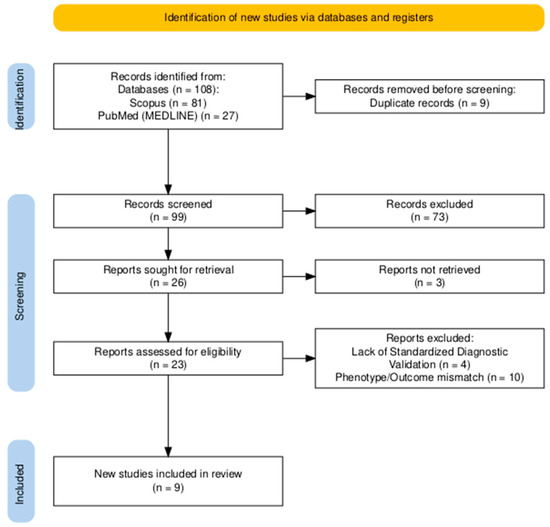
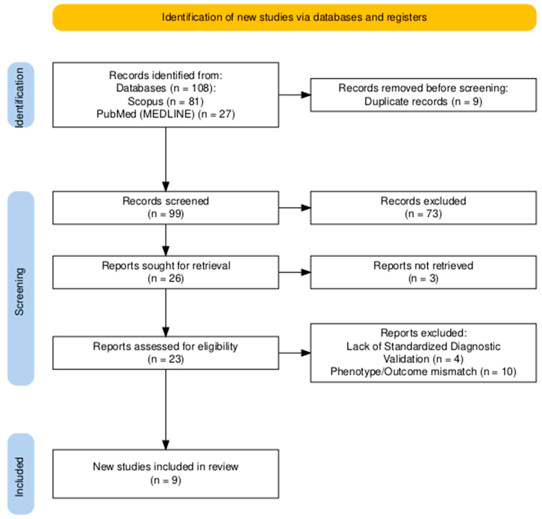
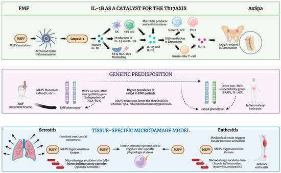
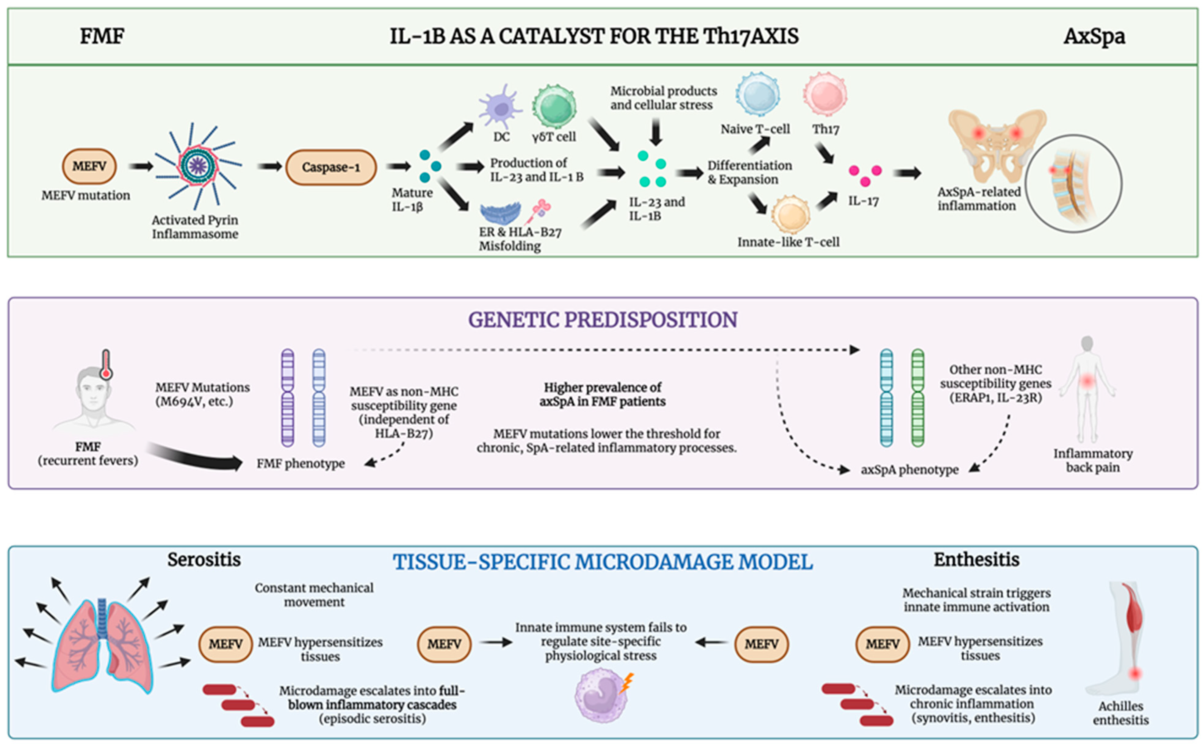

Disclaimer/Publisher’s Note: The statements, opinions and data contained in all publications are solely those of the individual author(s) and contributor(s) and not of MDPI and/or the editor(s). MDPI and/or the editor(s) disclaim responsibility for any injury to people or property resulting from any ideas, methods, instructions or products referred to in the content. |
© 2026 by the authors. Licensee MDPI, Basel, Switzerland. This article is an open access article distributed under the terms and conditions of the Creative Commons Attribution (CC BY) license.