
Review

Gut Microbiota and Psoriatic Arthritis: From Pathogenesis to Microbiota-Targeted Therapies
Rheumatology Unit, Department of Medicine, Surgery and Neurosciences, University of Siena, 53100 Siena, Italy
*
Author to whom correspondence should be addressed.
Rheumato 2026, 6(3), 18; https://doi.org/10.3390/rheumato6030018

Submission received: 18 June 2026 / Revised: 23 July 2026 / Accepted: 29 July 2026 / Published: 4 August 2026

Abstract

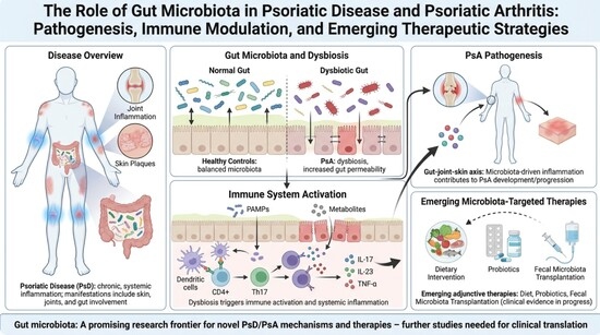
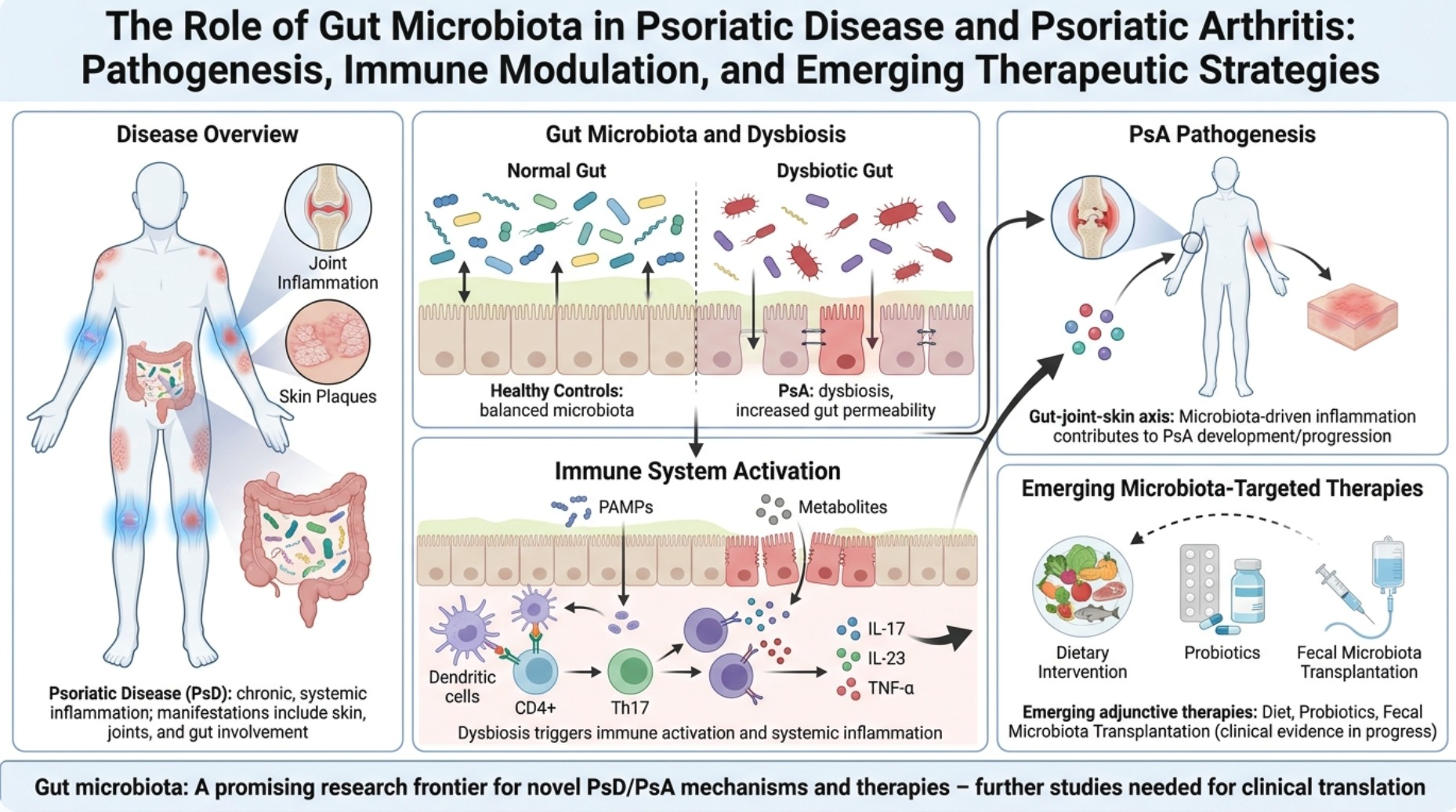
Background/Objectives: Psoriatic disease (PsD) is a chronic, systemic inflammatory disorder characterized by a wide spectrum of clinical manifestations extending beyond skin and joint involvement. Increasing evidence suggests a relevant role of the gut microbiota in the pathogenesis of psoriatic arthritis (PsA), although a clear causal relationship remains to be fully established. The aim of this review is to summarize current evidence on the role of the gut microbiome in PsD, with a focus on immune modulation, disease pathogenesis and potential therapeutic implications. Methods: A narrative review of the literature was conducted, focusing on studies investigating gut microbiota composition, dysbiosis patterns, and microbiome-related mechanisms in PsD and PsA, as well as emerging microbiota-targeted interventions. Results: Recent advances in high-throughput sequencing technologies have identified distinct microbial signatures associated with PsA onset and progression. Dysbiosis appears to influence immune system regulation, contributing to systemic inflammation through mechanisms involving intestinal barrier dysfunction and immune activation. Patients with PsA exhibit specific alterations in gut microbial composition compared to healthy controls. Emerging therapeutic strategies targeting the microbiota—including dietary interventions, probiotics, and fecal microbiota transplantation—show potential as adjunctive approaches in disease management, although clinical evidence remains limited. Conclusions: The gut microbiome represents a promising area of research in PsD, offering novel insights into disease mechanisms and potential therapeutic targets. Further studies are needed to clarify causal relationships and to define the clinical applicability of microbiota-based interventions.

Graphical Abstract

1. Introduction

Psoriatic arthritis is a chronic and progressive inflammatory disease within the spondyloarthritis spectrum, which also includes ankylosing spondylitis, reactive arthritis, arthritis associated with inflammatory bowel disease (IBD) and undifferentiated arthritis. It affects skin, nails, peripheral and axial joints, as well as the enthesis, with a prevalence from 0.1% to 1% in the general population [1].
The significant overlap between PsA and psoriasis, with estimates of PsA development ranging from 9% to 41% of psoriasis patients, has led to the concept of “Psoriatic Disease”. This term highlights a systemic condition that, beyond cutaneous and articular manifestations, encompasses a broad spectrum of shared clinical features and pathogenetic mechanisms [2].
Both PsO and PsA are characterized by elevated levels of pro-inflammatory cytokines, including tumor necrosis factor (TNF), interferon (IFN)-γ, interleukin (IL)-17, IL-22 and IL-23. Their pathogenesis, still not fully understood, reflects a complex interplay between genetic predisposition, environmental factors and activation of innate and adaptive immune responses [3]. Among these, gut microbiota has emerged as a potential mediator of gene–environmental interaction in PsO and PsA, as well as in immune-mediated disease generally, drawing growing attention in recent research.
The microbiota refers to the diversity of microorganisms - including bacteria, eukaryotes, viruses - that persist at the mucosal surfaces of the human body. In particular, the gut microbiota is influenced by a wide range of factors, including age, anatomical site, genetics, diet, geographic environment, resulting in marked interindividual variability [4]. Host–microbiota interactions are critical for individual health. At mucosal sites, T and B lymphocytes exhibit distinct phenotypes and functional properties that are modulated by the microbiota. Disruption of microbial community homeostasis can lead to an aberrant host–microbe relationship, potentially contributing to disease development [5]. Indeed, growing evidence indicates that the gut microbiota plays a role in various autoimmune and inflammatory diseases, including IBD [6], diabetes [7], cardiovascular disease [8], anxiety and depression [9], many of which are commonly observed as comorbidities in patients with PsA, and concomitant medications.
Emerging evidence suggests that alterations in gut microbiota composition may contribute to the pathogenesis of PsA, although the underlying mechanisms remain only partially understood. Microbial dysbiosis has been proposed to influence disease development through several pathways, including changes in the production of microbial metabolites and the modulation of host immune responses. This review aims to provide an overview of the current literature addressing the relationship between gut microbiota and PsA, focusing on the mechanisms through which intestinal microbial imbalance may be involved in disease pathogenesis and on the available studies evaluating the potential clinical impact of interventions targeting gut dysbiosis in patients with PsA. A better understanding of gut microbiome’s role in these diseases may provide novel perspectives for the management of PsD.

2. Materials and Methods

A comprehensive database search was conducted in PubMed, Scopus and Google Scholar covering the period from inception (January 1996, November 2004 and January 2004, respectively) to May 2026, particularly for the terms “psoriatic arthritis”, “gut microbiota”, “microbiome”, and “autoimmune diseases”. The inclusion criteria encompassed English-language publications or publications with an English translation available, including conference abstract, observational studies, randomized controlled trials (RCTs) and meta-analyses.
The present review focused on three main topics: the pathogenic role of the gut microbiota in the development of autoimmune diseases, with particular emphasis on PsA; evaluation and characterization of gut microbiota composition in patients with PsA; and the emerging therapeutic perspectives targeting the gut microbiota in the management of psoriatic arthritis.
The study selection process was conducted in two stages: an initial evaluation of title/abstract followed by full-text review. Some of the authors conducted the selection process. Studies were subsequently selected based on their relevance to the three main topics mentioned above. Studies were included if they investigated the composition, diversity, or functional role of the microbiota in patients with psoriatic arthritis, explored the relationship between microbial dysbiosis and disease pathogenesis, immune mechanisms, disease activity, biomarkers, or therapeutic interventions targeting the microbiome. Original research articles, including observational studies, case–control studies, cohort studies, and clinical trials, were considered eligible. The exclusion criteria comprised: (i) articles unrelated to psoriatic arthritis or the microbiota; (ii) studies focusing exclusively on other rheumatic or autoimmune diseases without separate data for PsA; (iii) animal or in vitro studies (unless included to discuss relevant pathogenic mechanisms); (iv) conference abstracts without full-text availability; (v) editorials, letters, commentaries, and expert opinions lacking original data; (vi) duplicate publications; and (vii) articles published in languages other than English. Narrative reviews, systematic reviews, and meta-analyses were not included in the qualitative synthesis of original evidence but were screened to identify additional relevant primary studies. Conference abstracts were included only when they reported novel or emerging findings that were not yet available in full-text peer-reviewed publications. Given their preliminary nature and limited methodological detail, evidence from conference abstracts was interpreted with caution and assigned less weight than that derived from peer-reviewed observational studies, randomized controlled trials, and systematic reviews or meta-analyses. This review has some limitations that should be considered when interpreting the findings. Although the literature search was conducted across three major databases (PubMed, Scopus, and Google Scholar) to maximize the identification of relevant studies, it is possible that some eligible publications were not retrieved. In addition, given the narrative nature of this review, article selection was based on relevance to the topic rather than on a predefined systematic review protocol. The available evidence is also characterized by substantial heterogeneity in study design, patient populations, microbiota profiling methodologies, and reported outcomes, which may account for some of the inconsistencies observed across studies. Consequently, the conclusions presented should be interpreted in the context of the current body of evidence, which continues to evolve.

3. The Pathogenic Role of Gut Microbiota in the Development of Autoimmune Diseases

The term microbiome refers to the collective genetic repertoire of a microbial community in a reasonably well-defined habitat which has distinct physio-chemical properties [10]; whereas microbiota describes the community of living microorganisms inhabiting a given environment [11]. The microbiota population colonize several body sites including gut, skin, respiratory tract and reproductive system, influencing immune homeostasis. Its composition changes throughout life depending on various factor - diet, type of delivery, BMI, exercise and use of drugs - and may vary even among genetically identical twins [12]. The human gut microbiota is largely dominated by Bacteroidetes and Firmicutes (90%), with the remaining phyla, including Actinobacteria, Proteobacteria, Fusobacteria and Verrucomicrobia, representing a minor fraction [13].

3.1. Gut Microbiota and Innate Immunity

The gut microbiota plays a central role in shaping the innate immune system, primarily through its interactions with gut-associated lymphoid tissue (GALT) and mucosa-associated lymphoid tissue (MALT). As GALT and MALT represent the frontline of gut mucosal defense, the principal functions of GALT include pathogen recognition, initiation of innate immune responses, antigen presentation to activate adaptive immunity, and maintenance of immune tolerance toward commensal microorganisms [14]. GALT cells form a critical interface linking local immune responses to the gut microbiota with systemic immune regulation, yet their potential role in the development of autoimmune diseases remains incompletely understood. Within this framework, pattern recognition receptor (PRR)–pathogen-associated molecular pattern (PAMP) interactions drive the development and organization of GALT structures; while microbial metabolites, particularly short-chain fatty acids (SCFAs), emerge as key modulators of GALT immune responses through epigenetic mechanisms [15]. Thus, SCFAs are associated with the regulation of innate lymphoid cells (ILCs) by affecting G-protein-coupled receptor activity (GPCR) [16] and interleukin-22 (IL-22) production of ILC, enhancing epithelial barrier integrity and antimicrobial defenses [17].
The development of innate lymphoid cells (ILCs), key components of GALT, occurs independently of gut microbiota, whereas their specific functions are shaped by commensal microbiota signals [18]. The microbiota not only modulates the cytotoxic function of natural killer (NK) cells but also guides the development of myeloid-derived macrophages, which are essential for preserving intestinal mucosal integrity, through the regulation of myeloid cell hematopoiesis and migration [19]. Furthermore, disruption of microbial communities can alter Toll-like receptor (TLR) signaling, inflammasome activation and ILC composition, contributing to systemic inflammation and autoimmune susceptibility.
Gut microbiota is implicated in dendritic cells (DC) development and trafficking. Gut microbiota regulate DC maturation through two principal mechanisms: microbial pattern-recognition signals and microbiota-derived metabolites. Microbial ligands activate TLRs and PRRs, promoting cytokine production and the expression of co-stimulatory molecules that drive DC maturation. Under dysbiotic conditions, enhanced LPS–TLR4 signaling favors a pro-inflammatory DC phenotype. In parallel, microbial metabolites—including SCFAs, bile acids, and tryptophan derivatives—modulate DC development and function through metabolic and epigenetic mechanisms. Although the effects of SCFAs are context-dependent, they generally contribute to the regulation of DC inflammatory responses and the maintenance of immune homeostasis [20]. Germ-free and antibiotic-treated mice exhibit reduced DC numbers, impaired migration, and defective T-cell priming, whereas microbial colonization promotes DC recruitment to the intestinal mucosa. Notably, microbiota-derived signals also shape extraintestinal DC compartments, as early-life colonization increases thymic DC numbers, while antibiotic treatment during weaning reduces thymic DC populations. The effects of the microbiota are also subset-specific. Microbiota-derived butyrate suppresses DC2 differentiation through HDAC3 inhibition, reducing co-stimulatory molecule expression and limiting naïve T-cell priming. In contrast, specific gut bacterial strains have been shown to enhance the maturation and migration of CD103+CD11b conventional DCs, thereby promoting CD8+ T-cell activation, highlighting the context-dependent effects of microbiota-derived signals on DC biology [21].

3.2. Gut Microbiota and Adaptive Immunity

The gut microbiota also plays also a central role in the development and maturation of the host adaptive immune system, contributing to immune homeostasis through a long-standing co-evolution with the host. Early-life microbial colonization represents a “window of opportunity” for immune education, promoting lymphoid tissue development, T- and B-cell maturation and the establishment of tolerance toward commensal microbes [22].
Key immune mediators involved in the bidirectional interaction between the gut microbiota and the host immune system include immunoglobulin A (IgA), T helper 17 (Th17) cells and regulatory T (Treg) cells.
Mucosal IgA Secretory IgA (sIgA) is transported across the intestinal epithelium via the polymeric immunoglobulin receptor (pIgR) and bind to microbes, dietary antigens, and other luminal components. By coating and agglutinating microorganisms, IgA limits their direct interaction with the epithelium and prevents excessive immune activation while contributing to the regulation of microbial composition and activity. Importantly, IgA production is strongly influenced by the gut microbiota, as germ-free animals display markedly reduced IgA+ cells in Peyer’s patches and in the lamina propria, whereas microbial colonization rapidly induces their expansion and secretion [23]. The repertoire of IgA in the gut is dynamically modulated in response to changes in microbial community composition. This process is primarily mediated by the re-entry of B-cell clones into germinal centers, enabling further somatic hypermutation and diversification of plasma cell clones established in the intestinal mucosa [24].
Th17 cells, characterized by the expression of transcription factor RAR-related orphan receptor (RORγt), are highly abundant in the lamina propria and represent a major subset of memory CD4 T-cells [25]. Th17 cells contribute to mucosal defense against extracellular pathogens by promoting antimicrobial responses and strengthening epithelial barrier integrity through the production of interleukin (IL) 17A, IL-17F and IL-22. However, upon stimulation by pro-inflammatory cytokines such as interleukin (IL)-23 and interleukin (IL) 1β, they can acquire pathogenic properties and contribute to the development of autoimmune diseases. The microbiota represents one of the main drivers of Th17 cell differentiation, together with diet and lipid metabolism [26,27]. Thus, segmented filamentous bacteria (SFB) are important inducers of Th17 responses and IgA stimulation through their direct interaction with the intestinal epithelium and activation of immune signaling pathways. Evidence from experimental mice models shows that epithelial adhesion by bacteria such as Citrobacter rodentium and Escherichia coli O157:H7 promotes Th17 responses, whereas adhesion-defective mutants fail to induce Th17 cell differentiation [28].
Regulatory T-cells (Treg), particularly abundant in the colon, are essential in maintaining immune tolerance to dietary antigens and gut microbiota [29]. The intestinal microbiota plays a key role in Treg induction. While Clostridia species (clusters IV, XIVa and XVIII) contribute to Treg function by promoting interleukin (IL) 10 and cytotoxic T-lymphocyte antigen 4 (CTLA-4) expression and reinforcing epithelial barrier integrity, SCFAs derived from the fermentation of dietary fibres by Clostridia species exert an even more significant impact, strongly suppressing pro-inflammatory cytokine production in dendritic cells and driving Treg differentiation and proliferation through epigenetic modifications and activation of G-protein-coupled receptors (GPCRs) [30].

3.3. Microbiota and Autoimmune Disease - Leaky Gut Syndrome and Dysbiosis

Gut health critically depends on the integrity and proper functioning of the intestinal barrier, a complex system that encompasses the gut microbiota, the mucosal layer, the intestinal epithelium and immune components that can be classified into extracellular and cellular elements.
The first line of defense is provided by extracellular components, including bile, gastric and pancreatic secretions [31] as well as a mucosal layer composed of water, electrolytes, proteins and bacteria with the function to trap pathogens whose function is to trap pathogens, thereby preventing microbial colonization [32]. Regarding cellular components, immune cells such as Paneth cells and mechanisms of mucosal immunity including IgA secretion regulate microbial density and maintain barrier integrity. In intestinal epithelial cells, intercellular junctions, particularly tight junctions composed of proteins such as claudins, occludin, and junctional adhesion molecules, allow nutrient absorption while preventing the passage of harmful microbes and antigens [33]. Under physiological conditions, tight junction (TJ) opening is transient and tightly regulated, allowing controlled paracellular transport across intestinal epithelium. However, excessive zonulin release can disrupt TJ integrity, increasing intestinal permeability, resulting in a condition commonly referred to as “leaky gut syndrome” [34], characterized by the translocation of microorganisms, toxins and undigested food particles into the circulation, potentially leading to systemic effects. Factors that can alter intestinal permeability include physical [35] and psychological [36] stress, alcohol abuse [37], antibiotics [38], air pollution and food additives [39].
Gut microbiota may contribute to the onset and progression of autoimmune disease (AID) through three principal mechanisms: translocation of gut bacteria, immune system modulation by bacterial metabolites and molecular mimicry [40]. Gut bacteria can modulate TJ expression and intestinal barrier integrity, as dysbiosis may disrupt intercellular junctions, facilitating the translocation of microbial products which can activate MALT and trigger inflammatory response [32]. Gut microbiota products, including their metabolites, can directly modulate the mucosal immune system eliciting either pro- or anti-inflammatory effects. For instance, high dietary salt intake as in the Western diet has been shown to promote Th17 expansion along with depletion of Lactobacillus species [41]. Furthermore, molecular mimicry represents a mechanism whereby the immune system fails to discriminate between self-antigens and structurally similar microbial peptides, leading to the activation of immune responses against host tissues. In this way, the gut microbiota may drive the generation of cross-reactive lymphocytes, involving both the innate and adaptive immune systems, thereby contributing to the onset of systemic autoimmune responses [42].
Cross-sectional human studies, with additional support from approaches such as Mendelian randomization, have identified disease-specific gut microbiota signatures in autoimmune diseases (AIDs), some of which demonstrate potential diagnostic value through machine learning approaches. However, their clinical applicability is limited by inconsistencies across studies, methodological heterogeneity, and the difficulty in distinguishing causation from association, further compounded by confounding factors such as ongoing therapies [43]. Stronger evidence supporting a causal relationship between gut microbiota and aberrant immune system activation is primarily derived from experimental models, whereas robust evidence in human studies remains limited.

4. The Role of Microbiota in the Development of PsO

Since approximately one-third of patients with PsO will develop PsA over the course of their lifetime, a potential causal contribution of cutaneous microbiome alteration to PsD has been proposed [44], although its influence appears to be less pronounced than that of the gut microbiota [45]. Similarly to the gut, the skin hosts a distinct and complex microbiota whose composition varies according to the anatomical site, with Propionibacterium species predominating in sebaceous regions, whereas Staphylococcus and Corynebacterium more common in elbows and feet [46]. The cutaneous microbiome can potentially play a role in the onset and progression of psoriasis, as skin lesions resulting from intense itching may facilitate the penetration of epidermal bacteria into deeper dermal layers [47]. 16s rRNA sequencing has demonstrated that psoriatic lesions are characterized by a more abundant and diverse microbial community compared with general population, with a relative decrease in Actinobacteria, Proteobacteria and Propionibacterium and a concomitant increase in Firmicutes [48].
A more prominent role in the development of PsD appears to be attributed to the gut microbiota, although a clear causal relationship remains to be established. Emerging evidence has proposed the concept of “gut–skin axis”, which consists of a bidirectional communication and interdependence between the gastrointestinal tract and the skin, representing a fundamental mechanism linking the intestinal microbiota to the development of systemic autoimmune disease, such as psoriasis. Supporting the role of the microbiota in maintaining the balance between regulatory (Treg) and effector T-cells, reduced gut beta-diversity has been observed in patients with psoriasis, characterized by a decrease in Treg cells and an overproduction of Th17 cells [49]. Moreover, the loss of intestinal barrier integrity allows the translocation of lipopolysaccharides (LPSs) into the bloodstream, leading to excessive keratinocyte proliferation through activation of Toll-like receptor 4 (TLR-4) and increased production of pro-inflammatory cytokines such as IL-6 and IL-8 [50]. Recent studies employing Mendelian randomization (MR) analyses, leveraging genome-wide association study (GWAS) data from large cohorts, have identified associations between specific gut bacterial taxa and psoriasis susceptibility. In particular, taxa such as Mollicutes, Holdemania, and Tenericutes were positively correlated with increased psoriasis risk, whereas Victivallacae, Lachnospiraceae, Lactococcus and Eubacterium exhibited potential protective effects [51]. Emerging tissue-resolved technologies, particularly spatial transcriptomics, are providing unprecedented opportunities to dissect the microbiota–immune axis at cellular resolution [52]. By preserving the spatial organization of tissues, these approaches enable the identification of discrete immune niches and cell–cell interactions within inflamed skin and intestinal mucosa, allowing microbiota-responsive pathways to be mapped to specific anatomical microenvironments rather than inferred from bulk tissue analyses [53]. When integrated with complementary approaches such as single-cell transcriptomics, microbial sequencing, and spatial proteomics, spatial transcriptomics may reveal how microbial-derived signals shape local immune circuits involving keratinocytes, dendritic cells, neutrophils, and T-cells, thereby offering new mechanistic insights into the gut–skin axis in psoriatic disease. Such multimodal strategies are expected to facilitate the identification of spatially restricted pathogenic pathways and may ultimately support the development of more targeted therapeutic interventions.

5. The Role of Microbiota in PsA

5.1. The SpA Model of the “Gut–Joint Axis”

Concerning the skin, it seems to host an autonomous microbiome that primarily modulates local inflammatory responses, with a less well-established influence on distant anatomical sites [54]. In contrast, the gut microbiota has been shown to influence both innate and adaptive immunity at a systemic level [55]. Although joints have long been considered sterile environments, bacterial components have been identified in synovial fluid of patients with rheumatoid arthritis (RA) [56], likely reaching the joint through circulation. In this context, gut dysbiosis may contribute to joint involvement through the hematogenous dissemination of microbial products [57].
The interaction between PsA and gut microbiota has been primarily investigated within the spondyloarthritis (SpA) spectrum, to which PsA belongs. Concerning SpA, the enthesis has been recognized as the key site of inflammation and the concept of the gut–enthesis axis has been proposed, supporting a potential role of gut microbiota as a “hidden engine” of the disease [58]. Notably, current evidence suggests that the overall composition of the microbiota may be less relevant than the localization of bacteria and their ability to translocate across the intestinal epithelial barrier, thereby establishing close interactions with immune cells in the lamina propria [59]. In this context, dysbiosis has been shown to promote IL-23 production by intestinal Paneth cells and to induce the upregulation of zonulin, a modulator of tight junctions, leading to increased intestinal permeability. IL-23 acts synergically with other innate cytokines (i.e., IL-17) to alter the immune tolerance and to drive the expansion of type 3 innate lymphoid cells (ILC3s), which are highly represented in the gut of patients affected by SpA [60]. Although demonstrating a definitive intestinal origin and subsequent migration of immune cells from the gut to the enthesis remains challenging, recent technologies using photoconvertible mice models have enabled the tracking of immune cell trafficking from the gut to secondary lymphoid organs, providing the most compelling evidence to date supporting the existence of a gut–enthesis axis [61,62].
A “gut–joint axis” has been proposed in SpA, including PsA, although the directionality of this relationship remains unclear: whether intestinal inflammation precedes and drives joint inflammation (causal hypothesis) or whether the two processes co-exist (correlative hypothesis), with a high microbial load potentially accelerating the onset of joint symptoms [63]. In this context, IL-23 emerges as a key mediator of the “skin–gut–joint axis”, as its increased production has been linked to impaired intestinal barrier integrity in IBD [64], as well as to articular and entheseal inflammation in SpA, particularly PsA [65]. In IBD, neutrophils have been identified as a major cellular source of IL-23 within the intestinal mucosa [66]. A similar pattern has been observed in murine models affected by SpA and IBD, in which skeletal neutrophils represent an early source of entheseal inflammation and IL-23 transcript expression at the enthesis [67], pointing to a shared pathogenic pathway across tissues and highlighting the similarity in the inflammatory profiles of the intestine and joints in SpA patients. In both settings, IL-23 promotes Th17 activation through the IL-23/IL-17 axis. On the one hand, dysbiosis promotes immune activation by increasing intestinal permeability and allowing the translocation of microbial products into the bloodstream [68]; on the other hand, short-chain fatty acids regulate intestinal Th17 and Treg cell responses, thereby modulating the IL-23/IL-17 axis [69,70].
Gut-derived inflammation may represent an early event preceding or amplifying joint inflammation, linking these two anatomical sites in the context of SpA. Supporting this hypothesis, Ciccia et al. reported an increased frequency of intestinally derived IL-17+ and IL-22+ ILC3 in the peripheral blood, synovial fluid and inflamed bone marrow of patients with AS, suggesting an active homing pathway between gut and inflamed sacroiliac joints [71]. However, although the SpA–gut axis has historically focused on the relationship between the synovium and the gut, there is strong evidence that experimental arthritis may concomitantly affect the gut and the enthesis in both TNF-transgenic and SKG mouse models [72,73].
Furthermore, the inflammasome has been a topic of interest in efforts to elucidate the gut–joint axis, with evidence indicating that its activation in both murine models and patients with SpA is associated with dysbiosis and that it is involved in triggering ileitis in SKG mice [74].

5.2. Studies About the Role of Gut Microbiota in PsA

Retrospective and cross-sectional microbiota studies in inflammatory diseases are limited by reverse causality, confounding (including treatment, diet and antibiotics) and early-life microbial influences, thereby constraining causal inference and obscuring the directionality of observed associations. To overcome these limitations, Mendelian randomization leverages host genetic variants associated with microbiome composition as instrumental variables to strengthen causal inference, though its validity depends on strict adherence to the assumptions of relevance, independence and exclusion restriction [75]. Recent studies employing Mendelian randomization analysis have provided valuable insights into the role of the gut microbiota in the development of PsA. A Mendelian randomization study demonstrated a causal effect between inflammatory bowel disease and both psoriasis and psoriatic arthritis, whereas no evidence of a reverse causal effect was observed [76]. It is well known that the gut microbiota significantly influences inflammatory arthritis, including PsA, with evidence suggesting its contribution to psoriatic bone remodeling, even without articular symptoms [77]. In addition, microscopic intestinal abnormalities have been observed in PsA patients, even in the absence of overt gastrointestinal involvement [78]. A two-sample Mendelian randomization study conducted analysis of 13,266 gut microbiota samples and 3186 patients with PsA and identified that bacteria such as Rikenellaceae and Ruminococcaceae UCG011 were associated with PsA, while Methanobacteria and Eubacterium fissicatena were found to be protective [79]. This result suggests that alterations in gut microbiota may influence the severity of PsA.
Barrier dysregulation appears to be a key driver of pro-inflammatory cell recruitment and translocation of metabolites and microorganisms due to increased intestinal permeability, as already noticed in patients affected by SpA [80]. Thus, HLA-B27-positive patients showed an upregulation of zonulin, an epithelial-derived regulator of tight junctions, and consequently, of intestinal permeability [59].
Collectively, these findings support the existence of a mechanistic link between gut dysbiosis, impaired barrier integrity, and systemic immune activation in PsA. However, despite growing evidence from genetic and observational studies, the extent to which microbiota alterations represent a primary pathogenic driver rather than a consequence of chronic inflammation remains to be fully elucidated.
Neutrophil Extracellular Traps (NETs) are implicated in autoimmune diseases, including psoriasis. NET-associated RNA complexed with LL37 triggers TLR8/TLR13-dependent cytokine release and further NET formation, creating a self-amplifying inflammatory loop in psoriatic skin [81]. In the gut, NETs contribute to host defense at mucosal surfaces by trapping and eliminating invading microorganisms. However, excessive or persistent NET formation can also amplify inflammation, promote tissue injury and compromise epithelial barrier integrity. Consistent with this dual role, increased NET levels and abundant tissue-associated NETs have been reported in inflammatory bowel disease, indicating that the inflamed intestinal mucosa represents a NET-rich microenvironment [82,83]. Microbiota-targeted interventions have been shown to suppress NET formation in experimental models of intestinal inflammation and arthritis. For example, Lactobacillus rhamnosus GG inhibited PMA- and Staphylococcus aureus-induced NET formation by attenuating protein kinase C (PKC) signaling and reducing reactive oxygen species (ROS) production. These findings suggest a potential mechanism through which the gut microbiota may modulate neutrophil function [84]. The most compelling mechanistic evidence linking the gut microbiota, NET formation, and arthritis derives from rheumatoid arthritis models rather than psoriatic arthritis. For instance, Parabacteroides goldsteinii and its outer membrane vesicles were shown to attenuate NET formation and alleviate arthritis severity [85]. Although these studies provide compelling evidence supporting a functional link between the gut microbiota and NET formation in inflammatory arthritis, they do not demonstrate that the same mechanism operates in psoriatic arthritis. Platelet–neutrophil niches are associated with the development of PsA. A 2026 transcriptomic study demonstrated the selective expansion of platelet–leukocyte niches within psoriatic skin lesions, with pronounced platelet-neutrophil colocalization in inflammatory epidermal regions. These platelet–neutrophil niches were spatially associated with enhanced dendritic cell and T-cell activation signatures, indicating that they function as sites of local immune amplification rather than reflecting simple cellular proximity [86]. Mechanistically, platelet–neutrophil interactions are well positioned to promote NET-driven inflammation, as platelet–neutrophil aggregates are increasingly recognized as key contributors to a wide range of inflammatory diseases. Platelets enhance neutrophil activation through both direct cell–cell contact and the release of soluble mediators, thereby promoting NETosis [87]. Once released, NETs further activate platelets through NET-associated damage-associated molecular patterns (DAMPs), reinforcing platelet activation, coagulation, and additional NET formation. This reciprocal crosstalk establishes a self-perpetuating thromboinflammatory circuit that sustains inflammation, exacerbates vascular and tissue injury, and is increasingly recognized as a central pathogenic mechanism in immune-mediated inflammatory diseases [88]. Peripheral multiomics showed enhanced platelet–neutrophil coupling and increased neutrophil activation in PsA, consistent with greater systemic inflammation than skin-limited Pso [86]. NET dysregulation is established across inflammatory diseases, including psoriasis; the PsA literature supports a gut metabolite role but not a PsA-specific NET mechanism [89].
Furthermore, it is challenging to discuss whether microbiota-conditioned DCs may influence local immune response in PsD by migrating to skin or joints. Microbiota-derived signals modulate DCs, which are intrinsically migratory, and both migrated and tissue-resident DC subsets can amplify IL-23/IL-17 axis-driven inflammation in psoriatic disease. However, evidence directly linking DC migration to inflamed joints in PsA remains largely indirect. Microbiota-derived products regulate DC maturation and migratory competence, including the upregulation of CCR7 and the production of T cell-polarizing cytokines, thereby providing a biologically plausible basis for systemic immune effects [90]. In psoriasis, distinct DC subsets undergo dynamic redistribution within the skin, including migration from the dermis to the epidermis and the accumulation of inflammatory DC populations within psoriatic lesions, where they sustain local inflammatory responses. Accordingly, microbiome dysbiosis is thought to promote both cutaneous and articular inflammation through DC-centered immune circuits, although direct evidence demonstrating the migration of microbiota-conditioned DCs into PsA joints is still limited. DCs are inherently equipped to traffic between different anatomical compartments. Pre-conventional DCs (pre-cDCs) and monocytes seed non-lymphoid tissues, including the skin, whereas tissue-resident DCs migrate to peripheral lymphoid organs [91]. Distinct DC subsets rely on specific chemokine receptor programs, including CCR7, CCR6, and CCR10, which direct their trafficking toward inflamed tissues, particularly the skin. Microbiota-derived signals can further shape this migratory phenotype. Commensal microorganisms establish a basal activation program in conventional DCs (cDCs) through tonic type I interferon (IFN-I) signaling, while bacterial products modulate DC maturation and functional polarization. For example, Staphylococcus aureus-derived peptidoglycan induces CCR7 expression together with the production of IL-1β, IL-6, and IL-23, thereby promoting Th17-skewed immune responses [92]. Evidence that microbiota-conditioned DCs can migrate to distant tissues is provided by studies showing that a defined intestinal bacterium enhances CCR7 expression in cDCs, promoting their migration from gut-associated sites to tumor tissue, where they augment local T-cell activation. Although this observation derives from a non-psoriatic setting, it supports the concept that microbiota-driven DC programming can influence immune responses at distal anatomical sites. Psoriatic skin harbors pathogenic DC subsets producing IL-23, TNF-α, IL-1β, and IL-23A, thereby sustaining Th17-mediated inflammation. Dynamic redistribution of DCs within the skin has also been well documented, including the migration of cDC2 into the epidermis before overt lesion development and the marked accumulation of epidermal DCs in established psoriasis. Moreover, intestinal dysbiosis exacerbates psoriasis-like inflammation in experimental models by increasing cutaneous infiltration of Th17 cells and monocyte-derived DCs and by enhancing DC-derived IL-23 production through fatty acid-dependent mechanisms [93]. In a murine model of PsA, DC-specific deletion of TNFR2 attenuated both psoriatic skin lesions and joint inflammation, indicating that DCs can coordinate inflammatory responses across the skin–joint axis, although this study did not directly demonstrate the migration of microbiota-conditioned DCs into joint tissues [94]. Collectively, these findings support the hypothesis that microbiota-conditioned DCs may migrate to the joints and contribute to the immunopathogenesis of PsA. Nevertheless, direct evidence demonstrating the homing of these cells into synovial tissues and their capacity to initiate or sustain local immune responses remains limited. Therefore, although biologically plausible, the contribution of microbiota-conditioned DC migration to joint inflammation in PsA has yet to be conclusively established (Table A1).

5.3. The Role of Intestinal Metabolites in PsA

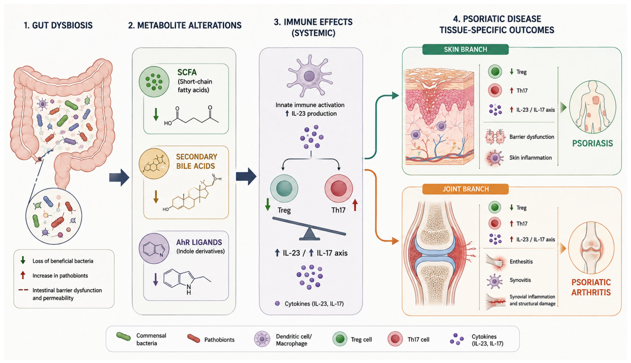
Encouraging findings on the role of the gut microbiota in the development of PsA have emerged from studies investigating intestinal metabolites, focusing on SCFAs, bile acids and bacterial-derived tryptophan metabolites (Figure 1) (Table 1).

5.3.1. Short-Chain Fatty Acids (SCFAs)

SCFAs encompass a class of fatty acids containing fewer than six carbons atoms, including acetate, propionate and butyrate, derived through microbial fermentation of dietary fibers in the gut. Their production is shaped by gut predominant bacterial phyla: bacteria of the Bacteroidetes phylum mainly produce acetate and propionate, while those of the Firmicutes phylum generate butyrate [95]. SCFAs exert multiple functions: in addition to serving as an energy source for colonocytes, they promote IgA response and the induction of genes encoding mucins and tight junctions, essential for preserving intestinal barrier integrity [96]. Depending on the predominant metabolite, their biological effects may differ [97]; for instance, butyrate has been reported to suppress pro-inflammatory cytokines and reactive oxygen species (ROS) [98], whereas acetate enhances their release, thereby contributing to the elimination of harmful bacteria and to protection against infections [99]. In recent years, SCFAs have also emerged as immunomodulatory mediators. They promote the differentiation of Treg through the induction of FoxP3 expression [100]. In particular, butyrate stimulates intestinal epithelial cells to produce retinoic acid, which acts synergistically to further enhance Treg differentiation [101]. Moreover, SCFAs increase IL-10 production in T helper 1 cells [102]. Conversely, distinct SCFAs exert differential effects on Th17 cells: acetate and propionate enhance IL-17 production, whereas butyrate inhibits its expression [103]. Evidence regarding alterations in SCFA production is increasing in psoriasis, whereas studies in PsA are emerging but remain limited. Several SCFA-producing bacterial species, including Eubacterium rectale, were consistently found to be depleted, with even lower abundances observed in PsA compared with cutaneous psoriasis alone [104]. Overall, depletion of SCFA and SCFA-producing bacteria is well documented in psoriasis and other inflammatory diseases, suggesting a broader pattern of reduced SCFA tone in these patients [105].

5.3.2. Bile Acids (BAs)

Bile acids (BAs) are cholesterol-derived molecules produced in liver and secreted into duodenum as primary bile acids [106]. The two main primary BAs, chenodeoxycholic acid and cholic acid, are converted into secondary bile acids, lithocolic acid and deoxycholic acid, respectively, through the metabolic activity of intestinal bacteria. However, the interaction between gut microbiota and bile acids is bidirectional: BAs shape the intestinal microbial community by promoting the growth of BA-metabolizing bacteria while inhibiting BA-sensitive species [89]. Importantly, BAs exert their biological effects through receptors including the farnesoid X receptor (FXR) and the membrane receptor TGR5, both of which are also expressed on immune cells such as macrophages, dendritic cells and NK cells, thereby suggesting an immunomodulatory and anti-inflammatory role [107]. BAs have been shown to inhibit NF-kB activation, suppress the transcription of pro-inflammatory genes and interfere with NLRP3 inflammasome and caspase-1 signaling pathways. Moreover, lithocholic acid appears to inhibit the transcription factor RORγt, a key regulator of Th17 cells, that play a central role in the pathogenesis of Pso and PsA [108]. Consistently, serum metabolomic analyses have demonstrated a dysregulated BA profile in patients with Pso and PsA [109]. Furthermore, in a longitudinal psoriasis cohort, patients who progressed to PsA had lower serum primary and secondary BA than non-progressors, suggesting a potential predicting role for BA profiles in disease progression. The authors proposed that these alterations in BA metabolism reflect gut dysbiosis and dysregulated hepatic metabolism, contributing to a “metabolic–inflammatory axis” that may drive joint inflammation [110].
Although current evidence supports a role for SCFAs and bile acids as important mediators of the gut–joint axis, most available data remain observational. Therefore, whether these metabolic alterations represent causal drivers of PsA development or secondary consequences of chronic inflammation remains to be determined.

5.3.3. Bacterial-Derived Tryptophan Metabolites

Tryptophan, one of the nine essential amino acids, partially escapes absorption in the small intestine and reaches the colon [111], where it can be catabolized by gut microbiota into indole-derived compounds. These molecules contribute to the maintenance of intestinal barrier integrity by enhancing tight junction (TJ) protein expression and, through activation of the aryl hydrocarbon receptor (AhR), promote IL-22 production [112]. Although evidence linking tryptophan catabolites to psoriasis remains limited, reduced levels of indole derivatives have been observed in psoriatic skin lesions [113] and exacerbation of psoriasis-like inflammation has been reported in AhR-deficient murine models [114].

5.3.4. Trimethylamin N-Oxide (TMAO)

Patients with psoriasis exhibited significantly higher plasma levels of trimethylamine N-oxide (TMAO), a gut microbiota-derived metabolite generated from dietary phosphatidylcholine [115] and implicated in the pathogenesis of cardiovascular diseases, compared with healthy controls. TMAO levels were significantly associated with disease activity in both Pso and PsA [116], suggesting a potential link between gut microbial metabolism, systemic inflammation, and the increased cardiovascular risk observed in patients with psoriatic disease. These findings support the hypothesis that TMAO may represent both a biomarker of disease activity and a potential mediator of the cardiometabolic comorbidities frequently associated with PsA. Nevertheless, whether elevated TMAO levels directly contribute to disease pathogenesis or simply reflect underlying inflammatory and metabolic disturbances remains uncertain (Table 2 and Table 3).

6. Gut Microbiota Characterization in PsA

To better understand the features of gut microbiota, it is fundamental to introduce two key concepts: alpha (α)- and beta (β)-diversity. The former refers to the richness and abundance of bacterial species within a single sample (i.e., an individual), whereas the latter assesses the differences and dissimilarities in microbial composition between different samples. A systematic review including 92 observational studies demonstrated an association between gut microbiota dysbiosis and several rheumatic diseases. Overall, these patients showed reduced α-diversity, indicating decreased microbial heterogeneity compared with healthy controls. Significant alterations in β-diversity were also reported. Notably, no disease-specific microbial signature emerged [117]. The Firmicutes/Bacteroidetes (F/B) ratio is considered a key indicator of gut microbiota health [118]. Alterations in the F/B ratio have been reported in both psoriasis and psoriatic arthritis. In particular, Pso patients characterized by enterotype 2 (Prevotella-predominant) exhibited a significantly higher F/B ratio compared with individuals with enterotype 1 (Bacteroides-predominant) or enterotype 3 (Ruminococcus-predominant) [119].
A pioneering study investigating gut microbiota composition in psoriatic arthritis (PsA), which still emerges as part of the limited evidence currently available in the literature, was conducted by Scher et al. The authors aimed to characterize microbial diversity and relative taxonomic abundance through high-throughput 16S ribosomal RNA pyrosequencing in treatment-naïve patients with newly diagnosed PsA, compared with patients with Pso and healthy controls. The study demonstrated a reduced α-diversity in PsA patients compared with healthy subjects, together with a lower relative abundance of Akkermansia and Ruminococcus, including in comparison with Pso patients. Conversely, the Bacteroidetes phylum and the Coprobacillus genus were less abundant in Pso patients. More specifically, PsA was characterized by reduced abundance of Coprococcus, Pseudobutyrivibrio, Alistipes, Akkermansia, Parabacteroides, and Ruminococcaceae. Notably, the latter two taxa were also depleted in Pso patients, suggesting the existence of a potential shared gut microbiota signature, although unproven [120].
To further characterize this microbial profile, a Chinese research group subsequently employed metagenome sequencing analysis. Their findings revealed an increased abundance Bacteroidetes spp., particularly Bacteroidetes sp. 3_1_19, as well as Blautia AF14-40. Moreover, a positive correlation was observed between K07114 (calcium-activated chloride channel homolog) and disease activity assessed by the Disease Activity in Psoriatic Arthritis (DAPSA) score, whereas Tet32 (an antibiotic resistance gene) and carbohydrate-binding module family 50 showed negative correlations with erythrocyte sedimentation rate (ESR) [121].
A reduction in fecal IgA levels as well as in Receptor Activator of Nuclear factor Kappa-B ligand (RANKL) and osteoprotegerin (OPG) concentration was observed, similarly to what has been reported in psoriasis [120]. RANKL, which is overexpressed in the serum and synovial tissue of patients with PsA, acts as an osteoclast-activating factor promoting arthritis development, whereas at the intestinal level it contributes to the differentiation of the lamina propria. Alterations in RANKL expression may be attributable to specific effects of the microbial profile associated with PsO and PsA or may reflect a modulatory role of these molecules in the development of systemic inflammation [122]. Scher et al. further reported reduced levels of medium-chain fatty acids (MCFAs), particularly heptanoate and hexanoate, whereas no significant differences in SCFAs were observed in fecal samples from patients with PsA and psoriasis [120].
Furthermore, fecal calprotectin, a recognized marker of intestinal inflammation, was assessed in patients with PsA, Pso and irritable bowel syndrome (IBS). Patients with PsA exhibited a higher prevalence and greater concentrations of fecal calprotectin compared with those with Pso. In addition, calprotectin levels showed a positive correlation with body surface area (BSA) in PsA [123]. These findings support the idea that gastrointestinal inflammation may contribute to PsA pathogenesis and to the progression from Pso to PsA, potentially constituting a risk factor for PsA development in patients with psoriasis [122]. Significant differences in gut microbiota composition between patients with psoriatic arthritis (PsA) and those with undifferentiated arthritis have also been reported. Notably, Megasphaera elsdenii was found to be approximately 10,000-fold more abundant in patients with PsA than in non-PsA subjects and demonstrated a clinical association with the presence of enthesitis, thereby suggesting a potential contribution of gut microbial dysbiosis to the pathogenesis of this musculoskeletal manifestation of PsA [124].
The findings discussed thus far reflect the characterization of gut microbiota in psoriatic arthritis independently of potential therapeutic influences. However, immunosuppressive agents, including cyclophosphamide and methotrexate, may significantly reduce gut microbial diversity, promoting depletion of commensal anaerobes and expansion of potential pathogens [125,126]. Evidence derived from studies in spondyloarthritis has suggested that anti-TNF therapy may contribute to a partial restoration of gut microbial dysbiosis, with increased relative abundances of Lachnospiraceae family and the Coprococcus genus reported after six months of treatment [127]. Nevertheless, no significant differences in either alpha- or beta-diversity were identified between anti-TNF responders and non-responders. In contrast, other investigations have described a substantially neutral effect of anti-TNF therapy on gut microbiota composition [128]. Another study investigated the evolution of gut microbiota taxonomy and functionality in patients with PsA undergoing treatment with IL-17 inhibitors, comparing microbial profiles before treatment and after one and three months of therapy. Following both one and three months of treatment, a significant increase in microbial alpha-diversity was observed, whereas no substantial changes in beta-diversity were detected [129]. In addition, treatment was associated with an increased abundance of the Bacteroidota phylum, consistently with previous observations in psoriasis [130], as well as an increase in the Phocaeicola genus, which in turn showed a positive correlation with an increase in alpha-diversity. One study evaluated the correlation between gut microbial alterations and disease activity in patients with PsA undergoing treatment with tofacitinib. At baseline, several bacterial families previously associated with inflammatory conditions, including Selenomonadaceae, Prevotellaceae, and Eggerthellaceae, showed a positive correlation with DAPSA scores. Following tofacitinib treatment, a reduction in the Actinobacteria phylum, together with a decreased abundance of the Megamonas genus and related species—microorganisms previously implicated in autoimmune diseases—was observed. In addition, an enrichment of butyrate-producing bacterial species, including Coprococcus comes, Ruminococcus bicirculans, and Butyricimonas sp. AT11, was reported. Notably, butyrate has been described as reduced in patients with psoriasis progressing to psoriatic arthritis [131].
Overall, current evidence supports an association between PsA and gut dysbiosis, although no disease-specific microbial signature has been consistently identified. Whether these alterations represent causal drivers or biomarkers of disease remains to be established.

7. Future Perspective: Microbiota as a Therapeutic Target

Given the impact of gut dysbiosis on PsA, several adjunctive therapeutic strategies have been proposed for disease management, including probiotics, prebiotics, fecal microbiota transplantation and phage therapy. However, most available evidence remains preliminary, and no microbiota-based intervention can currently be recommended as part of routine PsA management.

7.1. Probiotics

Probiotics are live microorganisms generally regarded as safe, which remain metabolically active within the gastrointestinal tract. They exert multiple beneficial functions, including the production of SCFAs, the synthesis of vitamins B and K, and the metabolism of carcinogenic compounds. In addition, probiotics modulate the immune response through the stimulation of immunoglobulin and cytokine production, as well as indirectly by strengthening the intestinal epithelial barrier [122]. Probiotics may be employed both to prevent and to restore intestinal homeostasis, as well as to maintain the integrity of the intestinal epithelial barrier [132]. The probiotic species most commonly used include bacteria belonging to the genera Lactobacillus, Bifidobacterium and Saccharomyces boulardii [133].
An association between probiotic use and disease activity in patients with PsA was investigated, including a comparison with patients affected by rheumatoid arthritis, a condition in which the role of the gut microbiota is considered less clearly established than in PsA. Probiotic use was found to be more prevalent among patients with a greater symptomatic burden at baseline. However, no clinically significant improvements in patient outcomes were observed among PsA subjects receiving probiotics. The authors suggested that these findings might reflect the worse baseline disease status of probiotic users rather than a limited therapeutic effect of probiotics themselves, thereby highlighting the need for prospective randomized studies to better define their efficacy [134]. In a pilot open-label study involving patients with psoriatic arthritis, multi-strain probiotic supplementation administered for 12 weeks was associated with a reduction in disease activity scores and improvements in biomarkers of intestinal permeability and gut inflammation, including zonulin and fecal calprotectin levels. The study also reported correlations between intestinal permeability markers and Th17-related immune responses. Given the small sample size and absence of a placebo-controlled design, the finding should be considered preliminary [135]. A pilot double-blind, randomized, placebo-controlled trial in 14 patients with mild-to-moderate PsA showed that 12-week supplementation with a multi-strain probiotic (1 × 10⁹ CFU/day) significantly reduced pro-inflammatory markers (CD4+ IFN-γ T-cells, B-cells, and IFN-γ) and increased regulatory cytokines (IL-10, TGF-β, and IL-4) compared with placebo, suggesting potential immunomodulatory benefits of probiotics as an adjunctive strategy in PsA management [136].

7.2. Prebiotics

Prebiotics, including inulin, fructooligosaccharides (FOSs) and galactooligosaccharides (GOSs), are selectively indigestible fibers that modulate the composition and function of gastrointestinal microbiota and are metabolized into SCFAs, thereby supporting gut barrier integrity and exerting anti-inflammatory effects beneficial to host health [137]. Prebiotics could therefore be considered a subject of further research to explore their potential application in adjunctive therapeutic strategies.

7.3. Diet

Owing to the recognized inflammatory and metabolic activity of adipose tissue, weight reduction in obese individuals with PsA has been associated with significant improvements in both clinical outcomes and systemic inflammatory parameters [138], as reflected by lower 66/68 swollen and tender joint counts, reduced LEI and CRP values and improved HAQ scores [139]. Emerging evidence suggest that glucagon-like peptide 1 (GLP-1) receptor agonists, through their effects on weight reduction, may have a potential role in the management of patients with PsA [140]. Indeed, based on the available literature, vitamin D supplementation and dietary weight reduction through a hypocaloric diet in overweight and obese individuals are weakly recommended [141]. A gluten-free diet has shown benefits only in patients with PsA who also present anti-gliadin antibodies [142]. Therefore, nutritional intervention should be considered as a complementary approach, alongside pharmacological therapy, with the potential to enhance symptom control and reduce disease activity. The DIETA trial, a randomized, double-blind, placebo-controlled study, evaluated the effect of a hypocaloric dietary regimen, including a subgroup receiving both diet and omega-3 supplementation, on disease activity in patients with psoriatic arthritis. The hypocaloric diet was associated with improvements in DAS28-CRP and BASDAI, whereas omega-3 supplementation led to significant improvement in body composition, despite the absence of a corresponding reduction in disease activity. Indeed, omega-3 fatty acids, polyunsaturated fats associated with beneficial cardiovascular effects, have previously been shown to be associated with clinical improvement in RA, including reductions in morning stiffness and number of swollen joints [143]. In PsA, however, there is a lack of strong evidence supporting a clear therapeutic role for omega-3 fatty acids. One study reported improvement in psoriatic plaques and subjective improvement in joint pain in most patients with PsA, suggesting that polyunsaturated fatty acids may be useful as potential adjunctive agents to standard therapy in Pso and PsA [144]. A 12-week hypocaloric diet improved joint disease activity, even in the absence of a correlation between weight loss and clinical response, suggesting that dietary quality—characterized by a lower Dietary Inflammatory Index, higher intake of fibers, omega-3 fatty acids, and antioxidant vitamins—may be relevant beyond caloric restriction alone [145]. Furthermore, studies suggested that the Mediterranean diet may serve as an adjunctive therapy in patients with PsA. Caso et al. assessed adherence to the Mediterranean diet in a cohort of patients with PsA and observed that higher DAPSA scores were correlated with lower adherence to the above-mentioned diet [146].

7.4. Fecal Microbiota Transplantation

Fecal microbiota transplantation (FMT) is a therapeutic intervention involving the transfer of fecal microbial communities from a healthy, rigorously screened donor to a recipient to restore the composition and functions of intestinal microbiota. Originally developed for the treatment of Clostridioides difficile infection and typically performed with colonoscopy, FMT has been subsequently been investigated in a broader range of clinical contexts, including auto-inflammatory rheumatic disease [147]. Several studies have evaluated the effects of probiotic supplementation delivered via fecal microbiota transplantation; nevertheless, no differences were identified between treated and untreated patients. An increase in microbiota beta-diversity was reported in only one study [148]. This observation may be explained by limitation of the studies, such as the heterogeneity of bacterial strains belonging to different species used for probiotic supplementation and the application of diverse microbiota assessment methodologies, although all studies employed metabolomics-based approaches [133]. Indeed, a meta-analysis demonstrated that FMT can significantly improve outcomes in ulcerative colitis (UC), in liver and metabolic diseases, including obesity [149]. Encouraging evidence has been described in UC [150] and Crohn’s disease (CD) [151], where FMT has been associated with improvements in both clinical and endoscopic remission rates. Conversely, only limited benefits, mainly involving gastrointestinal symptom relief, have been observed in systemic sclerosis (SSc) [152]. No clear therapeutic effects of FMT in type 1 diabetes mellitus have emerged, although additional well-designed studies are needed to further explore its potential role [153].
Fecal microbiota has been explored as a method to modify the gut microbiome in psoriatic arthritis, based on the link between gut dysbiosis and inflammation. The first interventional randomized controlled trial investigating the use of FMT in psoriatic arthritis was the FLORA study, a double-blind, placebo-controlled trial involving patients with peripheral psoriatic arthritis inadequately controlled by methotrexate therapy. Participants were randomly assigned to receive either FMT from a healthy donor or a sham transplantation procedure to evaluate the safety and clinical effects of the intervention. Therapeutic failure was found to be significantly more frequent in the FMT group, while greater improvements in the Health Assessment Questionnaire Disability Index (HAQ-DI) were observed in the placebo group. No significant differences were reported between groups regarding ACR20 response rates. Although no serious adverse events were reported, mild gastrointestinal symptoms were more frequent following [154]. To account for the unfavorable results of the trial, McGonagle et al. hypothesized that FMT may have paradoxically triggered a reactive arthritis-like disease flare. Based on the observation of a greater improvement in SPARCC scores observed in the sham group compared with the FMT group, the authors suggested that FMT could transiently disrupt intestinal homeostasis, thereby promoting a temporary exacerbation of disease activity before the re-establishment of immune homeostasis [155]. Despite the negative clinical results of the FLORA trial, several exploratory analyses have provided important mechanistic insight. Proteomic analysis of the FLORA trial, patients with PsA exhibited higher baseline levels of IL-6, IL-2, IL-18R1 and IFN-γ compared with healthy controls. Following FMT, the most notable cytokine changes included a reduction in IL-6 levels and a persistent increase in IFN-γ, whereas no significant longitudinal changes were observed for IL-2 or IL-18R1. Although these changes were not accompanied by measurable clinical improvement: the reduction in IL-6 may suggest a potential attenuation of inflammatory activity, no corresponding clinical benefit was observed and the biological significance of the persistent elevation of IFN-γ remains unclear [156].
Another exploratory metagenomic analysis of FLORA trial suggested that baseline gut microbiota composition may influence long-term response to FMT in PsA. Patients with a Bacteroides-dominated microbiota showed greater microbial diversity, more successful donor-strain engraftment and improved clinical outcomes compared with patients harboring a Prevotella-dominated microbiota, suggesting the recipient microbial profiles may affect treatment efficacy [157]. A further exploratory analysis of the FLORA trial investigated whether small intestinal barrier permeability, assessed by the lactulose-to-mannitol ratio (LMR), and fecal, plasma, and urinary metabolomic profiles were associated with clinical response to FMT or sham transplantation in patients with PsA. Treatment failures exhibited higher LMR values, suggesting impaired intestinal barrier integrity. Distinct metabolomic signatures were also observed between healthy donors and patients with PsA, with butyrate and valine predominating in donor samples, whereas glutamate, tyrosine, alanine, and tryptophan were more abundant in patients. Moreover, treatment failure was associated with increased fecal levels of tyrosine, leucine, and phenylalanine, as well as elevated plasma glucose concentrations. FMT induced a significant increase in fecal butyrate, a short-chain fatty acid known for its anti-inflammatory and immunomodulatory properties. Specific metabolites, including propionate and GlycA, were associated with favorable clinical outcomes and functional improvement, supporting a potential role of microbiota-derived metabolites in modulating disease activity and treatment response in PsA [158].

8. Conclusions

Accumulating evidence supports a relevant role of the gut microbiota in the pathophysiology of PsA, highlighting the potential existence of a gut–joint–skin axis, as previously proposed in SpA, and in the modulation of both innate and adaptive immune responses, although a clear cause–effect relationship remains to be fully understood. In this context, intestinal alterations associated with dysbiosis and with the onset and progression of autoimmune diseases, including PsA, are characterized by impaired intestinal barrier integrity (“leaky gut”) and dysregulated production of metabolites that are physiologically generated by the gut microbiota, such as SCFAs. Existing evidence supporting the gut–enthesis axis in spondyloarthritis has further strengthened the concept of shared immunopathogenic mechanisms across intestinal and musculoskeletal inflammation. In this context, several interleukins are being investigated as key mediators of convergent inflammatory pathways affecting both systems, with particular focus on the IL-17/IL-23 axis.
Despite the increasing number of studies characterizing the gut microbiome in PsA, a reproducible disease-specific microbial signature has not yet been identified. Differences in study design, sequencing methodologies, geographic and dietary factors, disease stage, and concomitant therapies likely account for much of the heterogeneity observed across studies. Nevertheless, recurring findings, including depletion of SCFA-producing bacteria, alterations in taxa such as Akkermansia, Ruminococcus, and Parabacteroides, and evidence of subclinical intestinal inflammation, suggest that gut dysbiosis represents a relevant component of disease pathobiology rather than a mere epiphenomenon. Recent advances in metagenomics, multi-omics technologies, and Mendelian randomization analyses are providing increasingly robust tools to investigate causal relationships between the microbiome and PsA. These approaches may facilitate the identification of microbial biomarkers associated with disease susceptibility, progression, and treatment response, while also improving our understanding of the immunological mechanisms linking intestinal and musculoskeletal inflammation.
From a therapeutic perspective, microbiota-targeted interventions, including probiotics, dietary strategies, and fecal microbiota transplantation, remain largely exploratory. Although preliminary studies have demonstrated immunomodulatory effects and potential improvements in biomarkers of gut inflammation and permeability, clinical benefits on articular disease activity remain inconsistent and insufficient to support routine implementation in clinical practice. The growing interest and enthusiasm are reflected by the increasing number of ongoing clinical trials currently recruiting patients. Future large-scale, longitudinal, and methodologically standardized studies will be essential to define the clinical relevance of microbiome alterations in PsA and to determine whether modulation of the gut microbiota can evolve from an intriguing biological concept into an effective adjunctive therapeutic strategy within precision medicine approaches.
The challenge ahead will be to translate the rapidly expanding knowledge of the PsA microbiome into clinically meaningful biomarkers and targeted therapeutic strategies.

Author Contributions

Conceptualization, C.B. and S.L.; methodology, S.V.; software, S.G.; validation, S.V. and C.B.; formal analysis, S.V.; investigation, S.V.; resources, S.V.; data curation, C.B.; writing—original draft preparation, S.V.; writing—review and editing, C.B.; visualization, B.F. and L.C.; supervision, C.B.; project administration, C.B. All authors have read and agreed to the published version of the manuscript.

Funding

This research received no external funding.

Institutional Review Board Statement

Not applicable.

Informed Consent Statement

Not applicable.

Data Availability Statement

No new data were created or analyzed in this study. Data sharing is not applicable to this article.

Acknowledgments

During the preparation of this manuscript/study the authors used ChatGPT (GPT-5.6 Thinking, OpenAI, San Francisco, CA, USA; accessed on 3 August 2026) for English-language editing, stylistic refinement, table creation and Figure 1 creation. The authors have reviewed and edited the output and take full responsibility for the content of this publication.

Conflicts of Interest

The authors declare no conflicts of interest.

Abbreviations

The following abbreviations are used in this manuscript:
PsAPsoriatic arthritis
PsDPsoriatic disease
SpASpondyloarthritis

Appendix A

In this section, the main studies investigating gut microbiota alterations in psoriatic arthritis are reported in Table A1.
Table A1. Main studies investigating gut microbiota alterations in psoriatic arthritis.
StudyYearDesignPopulationMain Microbiota FindingsClinical Relevance
Scher et al. [120]2015Cross-sectional, treatment-naïve patientsNew-onset PsA, Pso, healthy controlsReduced α-diversity in PsA; depletion of Akkermansia, Ruminococcus, Parabacteroides, Coprococcus, Pseudobutyrivibrio, and RuminococcaceaeFirst study demonstrating gut dysbiosis in PsA, resembling patterns observed in IBD
Liu et al. [121]2024Metagenomic sequencingPsA patients vs. controlsIncreased abundance of Bacteroidetes spp. and Blautia AF14-40; functional microbial signatures correlated with DAPSA and ESRSuggested links between microbiome composition and disease activity
Gan et al. [79]2024Two-sample Mendelian randomization13,266 microbiota samples; 3186 PsA patientsRikenellaceae and Ruminococcaceae UCG011 associated with increased PsA risk; Methanobacteria and Eubacterium fissicatena showed protective associationsProvided evidence supporting a potential causal role of gut microbiota in PsA susceptibility
Lin et al. [124]2022Pilot comparative studyPsA vs. undifferentiated arthritisMarked enrichment of Megasphaera elsdenii (~10,000-fold higher in PsA)Associated with enthesitis, suggesting a potential role in musculoskeletal manifestations
Liu et al. [129]2025Longitudinal metagenomic studyPsA patients treated with IL-17 inhibitorsIncreased α-diversity after treatment; enrichment of Bacteroidota and PhocaeicolaSuggested partial restoration of gut dysbiosis following IL-17 blockade
Picchianti Diamanti et al. [131]2024Prospective case seriesPsA patients treated with tofacitinibReduction in Actinobacteria, Megamonas; enrichment of butyrate-producing bacteria (Coprococcus comes, Ruminococcus bicirculans, Butyricimonas sp.)Improvement in microbial profile associated with anti-inflammatory bacterial species

References

  1. Karmacharya, P.; Chakradhar, R.; Ogdie, A. The epidemiology of psoriatic arthritis: A literature review. Best Pract. Res. Clin. Rheumatol. 2021, 35, 101692. [Google Scholar] [CrossRef] [PubMed]
  2. Ritchlin, C. Psoriatic disease—From skin to bone. Nat. Clin. Pract. Rheumatol. 2007, 3, 698–706. [Google Scholar] [CrossRef] [PubMed]
  3. Veale, D.J.; Fearon, U. The pathogenesis of psoriatic arthritis. Lancet 2018, 391, 2273–2284. [Google Scholar] [CrossRef] [PubMed]
  4. Ursell, L.K.; Metcalf, J.L.; Parfrey, L.W.; Knight, R. Defining the human microbiome. Nutr. Rev. 2012, 70, S38–S44. [Google Scholar] [CrossRef] [PubMed]
  5. Honda, K.; Littman, D.R. The microbiota in adaptive immune homeostasis and disease. Nature 2016, 535, 75–84. [Google Scholar] [CrossRef] [PubMed]
  6. Qiu, P.; Ishimoto, T.; Fu, L.; Zhang, J.; Zhang, Z.; Liu, Y. The gut microbiota in inflammatory bowel disease. Front. Cell. Infect. Microbiol. 2022, 12, 733992. [Google Scholar] [CrossRef] [PubMed]
  7. Li, W.-Z.; Stirling, K.; Yang, J.-J.; Zhang, L. Gut microbiota and diabetes: From correlation to causality and mechanism. World J. Diabetes 2020, 11, 293. [Google Scholar] [CrossRef] [PubMed]
  8. Xu, H.; Wang, X.; Feng, W.; Liu, Q.; Zhou, S.; Liu, Q.; Cai, L. The gut microbiota and its interactions with cardiovascular disease. Microb. Biotechnol. 2020, 13, 637–656. [Google Scholar] [CrossRef] [PubMed]
  9. Simpson, C.A.; Diaz-Arteche, C.; Eliby, D.; Schwartz, O.S.; Simmons, J.G.; Cowan, C.S. The gut microbiota in anxiety and depression–a systematic review. Clin. Psychol. Rev. 2021, 83, 101943. [Google Scholar] [CrossRef] [PubMed]
  10. Berg, G.; Rybakova, D.; Fischer, D.; Cernava, T.; Vergès, M.-C.C.; Charles, T.; Chen, X.; Cocolin, L.; Eversole, K.; Corral, G.H.; et al. Microbiome definition re-visited: Old concepts and new challenges. Microbiome 2020, 8, 103. [Google Scholar] [CrossRef] [PubMed]
  11. Marchesi, J.R.; Ravel, J. The vocabulary of microbiome research: A proposal. Microbiome 2015, 3, 31. [Google Scholar] [CrossRef] [PubMed]
  12. Xie, H.; Guo, R.; Zhong, H.; Feng, Q.; Lan, Z.; Qin, B.; Ward, K.J.; Jackson, M.A.; Xia, Y.; Chen, X.; et al. Shotgun metagenomics of 250 adult twins reveals genetic and environmental impacts on the gut microbiome. Cell Syst. 2016, 3, 572–584. [Google Scholar] [PubMed]
  13. Patloka, O.; Komprda, T.; Franke, G. Review of the relationships between human gut microbiome, diet, and obesity. Nutrients 2024, 16, 3996. [Google Scholar] [PubMed]
  14. Jiao, Y.; Wu, L.; Huntington, N.D.; Zhang, X. Crosstalk between gut microbiota and innate immunity and its implication in autoimmune diseases. Front. Immunol. 2020, 11, 282. [Google Scholar] [CrossRef] [PubMed]
  15. Woo, V.; Alenghat, T. Host–microbiota interactions: Epigenomic regulation. Curr. Opin. Immunol. 2017, 44, 52–60. [Google Scholar] [PubMed]
  16. Sepahi, A.; Liu, Q.; Friesen, L.; Kim, C.H. Dietary fiber metabolites regulate innate lymphoid cell responses. Mucosal Immunol. 2021, 14, 317–330. [Google Scholar] [CrossRef] [PubMed]
  17. Yang, W.; Yu, T.; Huang, X.; Bilotta, A.J.; Xu, L.; Lu, Y.; Sun, J.; Pan, F.; Zhou, J.; Zhang, W.; et al. Intestinal microbiota-derived short-chain fatty acids regulation of immune cell IL-22 production and gut immunity. Nat. Commun. 2020, 11, 4457. [Google Scholar] [PubMed]
  18. Constantinides, M.G. Interactions between the microbiota and innate and innate-like lymphocytes. J. Leukoc. Biol. 2018, 103, 409–419. [Google Scholar] [PubMed]
  19. Wang, X.; Yuan, W.; Yang, C.; Wang, Z.; Zhang, J.; Xu, D.; Sun, X.; Sun, W. Emerging role of gut microbiota in autoimmune diseases. Front. Immunol. 2024, 15, 1365554. [Google Scholar] [PubMed]
  20. Malik, J.A.; Zafar, M.A.; Lamba, T.; Nanda, S.; Khan, M.A.; Agrewala, J.N. The impact of aging-induced gut microbiome dysbiosis on dendritic cells and lung diseases. Gut Microbes 2023, 15, 2290643. [Google Scholar] [CrossRef] [PubMed]
  21. Andrusaite, A.; Lewis, J.; Frede, A.; Farthing, A.; Kästele, V.; Montgomery, J.; Mowat, A.; Mann, E.; Milling, S. Microbiota-derived butyrate inhibits cDC development via HDAC inhibition, diminishing their ability to prime T cells. Mucosal Immunol. 2024, 17, 1199–1211. [Google Scholar] [PubMed]
  22. Zhao, Q.; Elson, C.O. Adaptive immune education by gut microbiota antigens. Immunology 2018, 154, 28–37. [Google Scholar] [PubMed]
  23. Wei, M.; Shinkura, R.; Doi, Y.; Maruya, M.; Fagarasan, S.; Honjo, T. Mice carrying a knock-in mutation of Aicda resulting in a defect in somatic hypermutation have impaired gut homeostasis and compromised mucosal defense. Nat. Immunol. 2011, 12, 264–270. [Google Scholar] [PubMed]
  24. Lindner, C.; Thomsen, I.; Wahl, B.; Ugur, M.; Sethi, M.K.; Friedrichsen, M.; Smoczek, A.; Ott, S.; Baumann, U.; Suerbaum, S.; et al. Diversification of memory B cells drives the continuous adaptation of secretory antibodies to gut microbiota. Nat. Immunol. 2015, 16, 880–888. [Google Scholar] [CrossRef] [PubMed]
  25. Hirota, K.; Turner, J.-E.; Villa, M.; Duarte, J.H.; Demengeot, J.; Steinmetz, O.M.; Stockinger, B. Plasticity of TH17 cells in Peyer’s patches is responsible for the induction of T cell–dependent IgA responses. Nat. Immunol. 2013, 14, 372–379. [Google Scholar] [PubMed]
  26. Atarashi, K.; Nishimura, J.; Shima, T.; Umesaki, Y.; Yamamoto, M.; Onoue, M.; Yagita, H.; Ishii, N.; Evans, R.; Honda, K.; et al. ATP drives lamina propria TH17 cell differentiation. Nature 2008, 455, 808–812. [Google Scholar] [PubMed]
  27. Ivanov, I.I.; de Llanos Frutos, R.; Manel, N.; Yoshinaga, K.; Rifkin, D.B.; Sartor, R.B.; Finlay, B.B.; Littman, D.R. Specific microbiota direct the differentiation of IL-17-producing T-helper cells in the mucosa of the small intestine. Cell Host Microbe 2008, 4, 337–349. [Google Scholar] [PubMed]
  28. Atarashi, K.; Tanoue, T.; Ando, M.; Kamada, N.; Nagano, Y.; Narushima, S.; Suda, W.; Imaoka, A.; Setoyama, H.; Nagamori, T.; et al. Th17 cell induction by adhesion of microbes to intestinal epithelial cells. Cell 2015, 163, 367–380. [Google Scholar] [PubMed]
  29. Bilate, A.M.; Lafaille, J.J. Induced CD4+ Foxp3+ regulatory T cells in immune tolerance. Annu. Rev. Immunol. 2012, 30, 733–758. [Google Scholar] [CrossRef] [PubMed]
  30. Narushima, S.; Sugiura, Y.; Oshima, K.; Atarashi, K.; Hattori, M.; Suematsu, M.; Honda, K. Characterization of the 17 strains of regulatory T cell-inducing human-derived Clostridia. Gut Microbes 2014, 5, 333–339. [Google Scholar] [CrossRef] [PubMed]
  31. Martinsen, T.C.; Fossmark, R.; Waldum, H.L. The phylogeny and biological function of gastric juice—Microbiological consequences of removing gastric acid. Int. J. Mol. Sci. 2019, 20, 6031. [Google Scholar] [CrossRef] [PubMed]
  32. Marcari, A.B.; Paiva, A.D.; Simon, C.R.; Dos Santos, M.E.S.M. Leaky gut syndrome: An interplay between nutrients and dysbiosis. Curr. Nutr. Rep. 2025, 14, 25. [Google Scholar] [PubMed]
  33. Paray, B.A.; Albeshr, M.F.; Jan, A.T.; Rather, I.A. Leaky gut and autoimmunity: An intricate balance in individuals health and the diseased state. Int. J. Mol. Sci. 2020, 21, 9770. [Google Scholar] [PubMed]
  34. Hollander, D.; Kaunitz, J.D. The “leaky gut”: Tight junctions but loose associations? Dig. Dis. Sci. 2020, 65, 1277–1287. [Google Scholar] [PubMed]
  35. Wegierska, A.E.; Charitos, I.A.; Topi, S.; Potenza, M.A.; Montagnani, M.; Santacroce, L. The connection between physical exercise and gut microbiota: Implications for competitive sports athletes. Sports Med. 2022, 52, 2355–2369. [Google Scholar] [PubMed]
  36. Liu, S.; Gao, J.; Zhu, M.; Liu, K.; Zhang, H.-L. Gut microbiota and dysbiosis in Alzheimer’s disease: Implications for pathogenesis and treatment. Mol. Neurobiol. 2020, 57, 5026–5043. [Google Scholar] [CrossRef] [PubMed]
  37. Pohl, K.; Moodley, P.; Dhanda, A.D. Alcohol’s Impact on the Gut and Liver. Nutrients 2021, 13, 3170. [Google Scholar] [CrossRef] [PubMed]
  38. Mamieva, Z.; Poluektova, E.; Svistushkin, V.; Sobolev, V.; Shifrin, O.; Guarner, F.; Ivashkin, V. Antibiotics, gut microbiota, and irritable bowel syndrome: What are the relations? World J. Gastroenterol. 2022, 28, 1204. [Google Scholar] [CrossRef] [PubMed]
  39. Di Tommaso, N.; Gasbarrini, A.; Ponziani, F.R. Intestinal barrier in human health and disease. Int. J. Environ. Res. Public Health 2021, 18, 12836. [Google Scholar] [PubMed]
  40. Shaheen, W.A.; Quraishi, M.N.; Iqbal, T.H. Gut microbiome and autoimmune disorders. Clin. Exp. Immunol. 2022, 209, 161–174. [Google Scholar] [CrossRef] [PubMed]
  41. Wilck, N.; Matus, M.G.; Kearney, S.M.; Olesen, S.W.; Forslund, K.; Bartolomaeus, H.; Haase, S.; Mähler, A.; Balogh, A.; Markó, L.; et al. Salt-responsive gut commensal modulates TH17 axis and disease. Nature 2017, 551, 585–589. [Google Scholar] [PubMed]
  42. Li, B.; Selmi, C.; Tang, R.; Gershwin, M.; Ma, X. The microbiome and autoimmunity: A paradigm from the gut–liver axis. Cell. Mol. Immunol. 2018, 15, 595–609. [Google Scholar] [CrossRef] [PubMed]
  43. Walter, J.; Armet, A.M.; Finlay, B.B.; Shanahan, F. Establishing or exaggerating causality for the gut microbiome: Lessons from human microbiota-associated rodents. Cell 2020, 180, 221–232. [Google Scholar] [CrossRef] [PubMed]
  44. Castelino, M.; Eyre, S.; Upton, M.; Ho, P.; Barton, A. The bacterial skin microbiome in psoriatic arthritis, an unexplored link in pathogenesis: Challenges and opportunities offered by recent technological advances. Rheumatology 2014, 53, 777–784. [Google Scholar] [PubMed][Green Version]
  45. Eppinga, H.; Konstantinov, S.R.; Peppelenbosch, M.P.; Thio, H.B. The microbiome and psoriatic arthritis. Curr. Rheumatol. Rep. 2014, 16, 407. [Google Scholar] [PubMed]
  46. Zhou, Y.; Mihindukulasuriya, K.A.; Gao, H.; La Rosa, P.S.; Wylie, K.M.; Martin, J.C.; Kota, K.; Shannon, W.D.; Mitreva, M.; Sodergren, E.; et al. Exploration of bacterial community classes in major human habitats. Genome Biol. 2014, 15, R66. [Google Scholar] [PubMed]
  47. Celoria, V.; Rosset, F.; Pala, V.; Dapavo, P.; Ribero, S.; Quaglino, P.; Mastorino, L. The skin microbiome and its role in psoriasis: A review. Psoriasis Targets Ther. 2023, 13, 71–78. [Google Scholar]
  48. Gao, Z.; Tseng, C.; Strober, B.E.; Pei, Z.; Blaser, M.J. Substantial alterations of the cutaneous bacterial biota in psoriatic lesions. PLoS ONE 2008, 3, e2719. [Google Scholar] [PubMed]
  49. Thye, A.Y.-K.; Bah, Y.-R.; Law, J.W.-F.; Tan, L.T.-H.; He, Y.-W.; Wong, S.-H.; Thurairajasingam, S.; Chan, K.-G.; Lee, L.-H.; Letchumanan, V. Gut–skin axis: Unravelling the connection between the gut microbiome and psoriasis. Biomedicines 2022, 10, 1037. [Google Scholar] [PubMed]
  50. Pachauri, A.; Sharma, S. Unravelling the gut–skin axis: The role of gut microbiota in pathogenesis and management of psoriasis. Inflammopharmacology 2025, 33, 3671–3678. [Google Scholar] [PubMed]
  51. Li, Y.; Chen, G.; Hu, X.; Bao, Y.; Wu, C.; Zeng, N.; Jiang, F. Assessing causal relationships between gut microbiota and psoriasis: Evidence from two sample Mendelian randomization analysis. Sci. Rep. 2024, 14, 8831. [Google Scholar] [PubMed]
  52. Mayassi, T.; Li, C.; Segerstolpe, Å.; Brown, E.M.; Weisberg, R.; Nakata, T.; Yano, H.; Herbst, P.; Artis, D.; Graham, D.B.; et al. Spatially restricted immune and microbiota-driven adaptation of the gut. Nature 2024, 636, 447–456. [Google Scholar] [CrossRef] [PubMed]
  53. Minton, K. Spatial resolution of host–microbiome interactions. Nat. Rev. Genet. 2024, 25, 79. [Google Scholar] [PubMed]
  54. Naik, S.; Bouladoux, N.; Wilhelm, C.; Molloy, M.J.; Salcedo, R.; Kastenmuller, W.; Deming, C.; Quinones, M.; Koo, L.; Conlan, S.; et al. Compartmentalized control of skin immunity by resident commensals. Science 2012, 337, 1115–1119. [Google Scholar] [PubMed]
  55. Yeoh, N.; Burton, J.P.; Suppiah, P.; Reid, G.; Stebbings, S. The role of the microbiome in rheumatic diseases. Curr. Rheumatol. Rep. 2013, 15, 314. [Google Scholar] [CrossRef] [PubMed]
  56. Kempsell, K.E.; Cox, C.J.; Hurle, M.; Wong, A.; Wilkie, S.; Zanders, E.D.; Gaston, J.H.; Crowe, J.S. Reverse transcriptase-PCR analysis of bacterial rRNA for detection and characterization of bacterial species in arthritis synovial tissue. Infect. Immun. 2000, 68, 6012–6026. [Google Scholar] [CrossRef] [PubMed]
  57. Chimenti, M.S.; Perricone, C.; Novelli, L.; Caso, F.; Costa, L.; Bogdanos, D.; Conigliaro, P.; Triggianese, P.; Ciccacci, C.; Borgiani, P.; et al. Interaction between microbiome and host genetics in psoriatic arthritis. Autoimmun. Rev. 2018, 17, 276–283. [Google Scholar] [CrossRef] [PubMed]
  58. Mauro, D.; Nakamura, A.; Haroon, N.; Ciccia, F. The gut-enthesis axis and the pathogenesis of Spondyloarthritis. In Seminars in immunology; Elsevier: Amsterdam, The Netherlands, 2021; Volume 58, p. 101607. [Google Scholar]
  59. Ciccia, F.; Guggino, G.; Rizzo, A.; Alessandro, R.; Luchetti, M.M.; Milling, S.; Saieva, L.; Cypers, H.; Stampone, T.; Di Benedetto, P.; et al. Dysbiosis and zonulin upregulation alter gut epithelial and vascular barriers in patients with ankylosing spondylitis. Ann. Rheum. Dis. 2017, 76, 1123–1132. [Google Scholar] [PubMed]
  60. Mauro, D.; Cai, B.; Ciancio, A.; Forte, G.; Gandolfo, S.; Thomas, R.; Bergot, A.-S.; Ciccia, F. The role of the gut and intestinal dysbiosis in the pathogenesis of Spondyloarthritis. Jt. Bone Spine 2025, 92, 105923. [Google Scholar] [CrossRef] [PubMed]
  61. Teng, F.; Klinger, C.N.; Felix, K.M.; Bradley, C.P.; Wu, E.; Tran, N.L.; Umesaki, Y.; Wu, H.-J.J. Gut microbiota drive autoimmune arthritis by promoting differentiation and migration of Peyer’s patch T follicular helper cells. Immunity 2016, 44, 875–888. [Google Scholar] [CrossRef] [PubMed]
  62. Tajik, N.; Frech, M.; Schulz, O.; Schälter, F.; Lucas, S.; Azizov, V.; Dürholz, K.; Steffen, F.; Omata, Y.; Rings, A.; et al. Targeting zonulin and intestinal epithelial barrier function to prevent onset of arthritis. Nat. Commun. 2020, 11, 1995. [Google Scholar] [CrossRef] [PubMed]
  63. Gracey, E.; Vereecke, L.; McGovern, D.; Fröhling, M.; Schett, G.; Danese, S.; De Vos, M.; Van den Bosch, F.; Elewaut, D. Revisiting the gut–joint axis: Links between gut inflammation and spondyloarthritis. Nat. Rev. Rheumatol. 2020, 16, 415–433. [Google Scholar] [PubMed]
  64. Sewell, G.W.; Kaser, A. Interleukin-23 in the pathogenesis of inflammatory bowel disease and implications for therapeutic intervention. J. Crohn’s Colitis 2022, 16, ii3–ii19. [Google Scholar]
  65. Schett, G.; Rahman, P.; Ritchlin, C.; McInnes, I.B.; Elewaut, D.; Scher, J.U. Psoriatic arthritis from a mechanistic perspective. Nat. Rev. Rheumatol. 2022, 18, 311–325. [Google Scholar] [PubMed]
  66. Kvedaraite, E.; Lourda, M.; Ideström, M.; Chen, P.; Olsson-Åkefeldt, S.; Forkel, M.; Gavhed, D.; Lindforss, U.; Mjösberg, J.; Henter, J.-I.; et al. Tissue-infiltrating neutrophils represent the main source of IL-23 in the colon of patients with IBD. Gut 2016, 65, 1632–1641. [Google Scholar] [PubMed]
  67. Stavre, Z.; Bridgewood, C.; Zhou, Q.; Maeda, Y.; Huang, T.; Karman, J.; Khan, A.; Giryes, S.; Sharif, K.; McGonagle, D.; et al. A role for neutrophils in early enthesitis in spondyloarthritis. Arthritis Res. Ther. 2022, 24, 24. [Google Scholar] [PubMed]
  68. Di Vincenzo, F.; Del Gaudio, A.; Petito, V.; Lopetuso, L.R.; Scaldaferri, F. Gut microbiota, intestinal permeability, and systemic inflammation: A narrative review. Intern. Emerg. Med. 2024, 19, 275–293. [Google Scholar] [CrossRef] [PubMed]
  69. Tan, J.; McKenzie, C.; Potamitis, M.; Thorburn, A.N.; Mackay, C.R.; Macia, L. The role of short-chain fatty acids in health and disease. Adv. Immunol. 2014, 121, 91–119. [Google Scholar] [PubMed]
  70. Arpaia, N.; Campbell, C.; Fan, X.; Dikiy, S.; Van Der Veeken, J.; Deroos, P.; Liu, H.; Cross, J.R.; Pfeffer, K.; Coffer, P.J.; et al. Metabolites produced by commensal bacteria promote peripheral regulatory T-cell generation. Nature 2013, 504, 451–455. [Google Scholar] [CrossRef] [PubMed]
  71. Ciccia, F.; Guggino, G.; Rizzo, A.; Saieva, L.; Peralta, S.; Giardina, A.; Cannizzaro, A.; Sireci, G.; De Leo, G.; Alessandro, R.; et al. Type 3 innate lymphoid cells producing IL-17 and IL-22 are expanded in the gut, in the peripheral blood, synovial fluid and bone marrow of patients with ankylosing spondylitis. Ann. Rheum. Dis. 2015, 74, 1739–1747. [Google Scholar] [PubMed]
  72. Armaka, M.; Apostolaki, M.; Jacques, P.; Kontoyiannis, D.L.; Elewaut, D.; Kollias, G. Mesenchymal cell targeting by TNF as a common pathogenic principle in chronic inflammatory joint and intestinal diseases. J. Exp. Med. 2008, 205, 331–337. [Google Scholar] [PubMed]
  73. Rahman, M.A.; Thomas, R. The SKG model of spondyloarthritis. Best Pract. Res. Clin. Rheumatol. 2017, 31, 895–909. [Google Scholar] [PubMed]
  74. Guggino, G.; Mauro, D.; Rizzo, A.; Alessandro, R.; Raimondo, S.; Bergot, A.-S.; Rahman, M.A.; Ellis, J.J.; Milling, S.; Lories, R.; et al. Inflammasome activation in ankylosing spondylitis is associated with gut dysbiosis. Arthritis Rheumatol. 2021, 73, 1189–1199. [Google Scholar] [PubMed]
  75. Stoll, M.L.; Appah, M.; Tiwari, H.K. The microbiota in axial spondyloarthritis: What have we learned from Mendelian randomisation studies? Clin. Exp. Rheumatol. 2025, 44, 1–10. [Google Scholar] [PubMed]
  76. Freuer, D.; Linseisen, J.; Meisinger, C. Association between inflammatory bowel disease and both psoriasis and psoriatic arthritis: A bidirectional 2-sample Mendelian randomization study. JAMA Dermatol. 2022, 158, 1262–1268. [Google Scholar] [PubMed]
  77. Paine, A.; Ritchlin, C. Bone remodeling in psoriasis and psoriatic arthritis: An update. Curr. Opin. Rheumatol. 2016, 28, 66–75. [Google Scholar] [PubMed]
  78. Scarpa, R.; Manguso, F.; D’Arienzo, A.; D’Armiento, F.P.; Astarita, C.; Mazzacca, G.; Ayala, F. Microscopic inflammatory changes in colon of patients with both active psoriasis and psoriatic arthritis without bowel symptoms. J. Rheumatol. 2000, 27, 1241–1246. [Google Scholar] [PubMed]
  79. Gan, Y.; Liu, J.; Jin, M.; Zhang, Y.; Huang, S.; Ma, Q.; Wu, Y.; Xu, L.; Bao, J.; Fan, Y. The role of the gut-joint axis in the care of psoriatic arthritis: A two-sample bidirectional Mendelian randomization study. Dermatol. Ther. 2024, 14, 713–728. [Google Scholar] [CrossRef] [PubMed]
  80. Van Hooste, W.; Gray, E.; Parsa, S.; Elewaut, D.; Lubberts, E.; Taams, L.S.; Venken, K. Barrier breakdown: Insights into the skin–gut axis in psoriatic arthritis. Trends Immunol. 2026, 47, 600–614. [Google Scholar] [CrossRef] [PubMed]
  81. Herster, F.; Bittner, Z.; Archer, N.K.; Dickhöfer, S.; Eisel, D.; Eigenbrod, T.; Knorpp, T.; Schneiderhan-Marra, N.; Löffler, M.W.; Kalbacher, H.; et al. Neutrophil extracellular trap-associated RNA and LL37 enable self-amplifying inflammation in psoriasis. Nat. Commun. 2020, 11, 105. [Google Scholar] [CrossRef] [PubMed]
  82. Drury, B.; Hardisty, G.; Gray, R.D.; Ho, G. Neutrophil extracellular traps in inflammatory bowel disease: Pathogenic mechanisms and clinical translation. Cell. Mol. Gastroenterol. Hepatol. 2021, 12, 321–333. [Google Scholar] [CrossRef] [PubMed]
  83. Chen, K.; Shao, L.-H.; Wang, F.; Shen, X.-F.; Xia, X.-F.; Kang, X.; Song, P.; Wang, M.; Lu, X.-F.; Wang, C.; et al. Netting gut disease: Neutrophil extracellular trap in intestinal pathology. Oxidative Med. Cell. Longev. 2021, 2021, 5541222. [Google Scholar] [CrossRef] [PubMed]
  84. Mutua, V.; Gershwin, L.J. A review of neutrophil extracellular traps (NETs) in disease: Potential anti-NETs therapeutics. Clin. Rev. Allergy Immunol. 2021, 61, 194–211. [Google Scholar] [CrossRef] [PubMed]
  85. Ye, H.; Wang, H.; Han, B.; Chen, K.; Wang, X.; Ma, F.; Cheng, L.; Zheng, S.; Zhao, X.; Zhu, J.; et al. Guizhi Shaoyao Zhimu decoction inhibits neutrophil extracellular traps formation to relieve rheumatoid arthritis via gut microbial outer membrane vesicles. Phytomedicine 2025, 136, 156254. [Google Scholar] [CrossRef] [PubMed]
  86. Bonilha, C.S. Platelet–neutrophil niches associate with epidermal immune activation and systemic inflammation in psoriatic disease. J. Mol. Med. 2026, 104, 74. [Google Scholar] [CrossRef] [PubMed]
  87. Li, J.; Geng, Y.; Luo, Y.; Sun, X.; Guo, Y.; Dong, Z. Pathological roles of NETs-platelet synergy in thrombotic diseases: From molecular mechanisms to therapeutic targeting. Int. Immunopharmacol. 2025, 159, 114934. [Google Scholar] [CrossRef] [PubMed]
  88. Wang, H.; Kim, S.J.; Lei, Y.; Wang, S.; Wang, H.; Huang, H.; Zhang, H.; Tsung, A. Neutrophil extracellular traps in homeostasis and disease. Signal Transduct. Target. Ther. 2024, 9, 235. [Google Scholar] [CrossRef] [PubMed]
  89. Lai, Y.; Wu, X.; Chao, E.; Bloomstein, J.D.; Wei, G.; Hwang, S.T.; Shi, Z. Impact of gut bacterial metabolites on psoriasis and psoriatic arthritis: Current status and future perspectives. J. Investig. Dermatol. 2023, 143, 1657–1666. [Google Scholar] [CrossRef] [PubMed]
  90. Liu, J.; Zhang, X.; Cheng, Y.; Cao, X. Dendritic cell migration in inflammation and immunity. Cell. Mol. Immunol. 2021, 18, 2461–2471. [Google Scholar] [CrossRef] [PubMed]
  91. Schaupp, L.; Muth, S.; Rogell, L.; Kofoed-Branzk, M.; Melchior, F.; Lienenklaus, S.; Ganal-Vonarburg, S.C.; Klein, M.; Guendel, F.; Hain, T.; et al. Microbiota-induced type I interferons instruct a poised basal state of dendritic cells. Cell 2020, 181, 1080–1096. [Google Scholar] [CrossRef] [PubMed]
  92. Nakamizo, S.; Dutertre, C.-A.; Khalilnezhad, A.; Zhang, X.M.; Lim, S.; Lum, J.; Koh, G.; Foong, C.; Yong, P.J.A.; Tan, K.J.; et al. Single-cell analysis of human skin identifies CD14+ type 3 dendritic cells co-producing IL1B and IL23A in psoriasis. J. Exp. Med. 2021, 218, e20202345. [Google Scholar] [CrossRef] [PubMed]
  93. Zhao, Q.; Yu, J.; Zhou, H.; Wang, X.; Zhang, C.; Hu, J.; Hu, Y.; Zheng, H.; Zeng, F.; Yue, C.; et al. Intestinal dysbiosis exacerbates the pathogenesis of psoriasis-like phenotype through changes in fatty acid metabolism. Signal Transduct. Target. Ther. 2023, 8, 40. [Google Scholar] [CrossRef] [PubMed]
  94. Kaur, R.; Lin, J.; Harvey, J.E.; Brambilla, R.; Chandrasekharan, U.M.; Husni, M.E. Targeting dendritic cell-specific TNFR2 improves skin and joint inflammation in a murine model of psoriatic arthritis. Sci. Rep. 2025, 15, 31574. [Google Scholar] [CrossRef] [PubMed]
  95. Macfarlane, S.; Macfarlane, G.T. Regulation of short-chain fatty acid production. Proc. Nutr. Soc. 2003, 62, 67–72. [Google Scholar] [CrossRef] [PubMed]
  96. Parada Venegas, D.; De la Fuente, M.K.; Landskron, G.; González, M.J.; Quera, R.; Dijkstra, G.; Harmsen, H.J.; Faber, K.N.; Hermoso, M.A. Short chain fatty acids (SCFAs)-mediated gut epithelial and immune regulation and its relevance for inflammatory bowel diseases. Front. Immunol. 2019, 10, 277. [Google Scholar] [CrossRef] [PubMed]
  97. González-Bosch, C.; Boorman, E.; Zunszain, P.A.; Mann, G.E. Short-chain fatty acids as modulators of redox signaling in health and disease. Redox Biol. 2021, 47, 102165. [Google Scholar] [CrossRef] [PubMed]
  98. Chang, P.V.; Hao, L.; Offermanns, S.; Medzhitov, R. The microbial metabolite butyrate regulates intestinal macrophage function via histone deacetylase inhibition. Proc. Natl. Acad. Sci. USA 2014, 111, 2247–2252. [Google Scholar] [CrossRef] [PubMed]
  99. Schlatterer, K.; Beck, C.; Schoppmeier, U.; Peschel, A.; Kretschmer, D. Acetate sensing by GPR43 alarms neutrophils and protects from severe sepsis. Commun. Biol. 2021, 4, 928. [Google Scholar] [CrossRef] [PubMed]
  100. Duscha, A.; Gisevius, B.; Hirschberg, S.; Yissachar, N.; Stangl, G.I.; Dawin, E.; Bader, V.; Haase, S.; Kaisler, J.; David, C.; et al. Propionic acid shapes the multiple sclerosis disease course by an immunomodulatory mechanism. Cell 2020, 180, 1067–1080. [Google Scholar] [CrossRef] [PubMed]
  101. Schilderink, R.; Verseijden, C.; Seppen, J.; Muncan, V.; Van Den Brink, G.R.; Lambers, T.T.; van Tol, E.A.; de Jonge, W.J. The SCFA butyrate stimulates the epithelial production of retinoic acid via inhibition of epithelial HDAC. Am. J. Physiol.-Gastrointest. Liver Physiol. 2016, 310, G1138–G1146. [Google Scholar] [CrossRef] [PubMed]
  102. Sun, M.; Wu, W.; Chen, L.; Yang, W.; Huang, X.; Ma, C.; Chen, F.; Xiao, Y.; Zhao, Y.; Ma, C.; et al. Microbiota-derived short-chain fatty acids promote Th1 cell IL-10 production to maintain intestinal homeostasis. Nat. Commun. 2018, 9, 3555. [Google Scholar] [CrossRef] [PubMed]
  103. Chen, L.; Sun, M.; Wu, W.; Yang, W.; Huang, X.; Xiao, Y.; Ma, C.; Xu, L.; Yao, S.; Liu, Z.; et al. Microbiota metabolite butyrate differentially regulates Th1 and Th17 cells’ differentiation and function in induction of colitis. Inflamm. Bowel Dis. 2019, 25, 1450–1461. [Google Scholar] [CrossRef] [PubMed]
  104. Xiao, Y.; Wang, Y.; Tong, B.; Gu, Y.; Zhou, X.; Zhu, N.; Xu, X.; Yin, X.; Kou, Y.; Tan, Y.; et al. Eubacterium rectale is a potential marker of altered gut microbiota in psoriasis and psoriatic arthritis. Microbiol. Spectr. 2024, 12, e01154-23. [Google Scholar] [CrossRef] [PubMed]
  105. Xiao, X.; Hu, X.; Yao, J.; Cao, W.; Zou, Z.; Wang, L.; Qin, H.; Zhong, D.; Li, Y.; Xue, P.; et al. The role of short-chain fatty acids in inflammatory skin diseases. Front. Microbiol. 2023, 13, 1083432. [Google Scholar] [CrossRef] [PubMed]
  106. Jia, W.; Xie, G.; Jia, W. Bile acid–microbiota crosstalk in gastrointestinal inflammation and carcinogenesis. Nat. Rev. Gastroenterol. Hepatol. 2018, 15, 111–128. [Google Scholar] [CrossRef] [PubMed]
  107. Perino, A.; Demagny, H.; Velazquez-Villegas, L.; Schoonjans, K. Molecular physiology of bile acid signaling in health, disease, and aging. Physiol. Rev. 2021, 101, 683–731. [Google Scholar] [CrossRef] [PubMed]
  108. Song, X.; Sun, X.; Oh, S.F.; Wu, M.; Zhang, Y.; Zheng, W.; Geva-Zatorsky, N.; Jupp, R.; Mathis, D.; Benoist, C.; et al. Microbial bile acid metabolites modulate gut RORγ+ regulatory T cell homeostasis. Nature 2020, 577, 410–415. [Google Scholar] [CrossRef] [PubMed]
  109. Chen, D.; He, J.; Li, J.; Zou, Q.; Si, J.; Guo, Y.; Yu, J.; Li, C.; Wang, F.; Chan, T.; et al. Microbiome and metabolome analyses reveal novel interplay between the skin microbiota and plasma metabolites in psoriasis. Front. Microbiol. 2021, 12, 643449. [Google Scholar] [CrossRef] [PubMed]
  110. Paine, A.; Brookes, P.S.; Bhattacharya, S.; Li, D.; De La Luz Garcia-Hernandez, M.; Tausk, F.; Ritchlin, C. Dysregulation of bile acids, lipids, and nucleotides in psoriatic arthritis revealed by unbiased profiling of serum metabolites. Arthritis Rheumatol. 2023, 75, 53–63. [Google Scholar] [CrossRef] [PubMed]
  111. Platten, M.; Nollen, E.A.; Röhrig, U.F.; Fallarino, F.; Opitz, C.A. Tryptophan metabolism as a common therapeutic target in cancer, neurodegeneration and beyond. Nat. Rev. Drug Discov. 2019, 18, 379–401. [Google Scholar] [CrossRef] [PubMed]
  112. Ye, X.; Li, H.; Anjum, K.; Zhong, X.; Miao, S.; Zheng, G.; Liu, W.; Li, L. Dual role of indoles derived from intestinal microbiota on human health. Front. Immunol. 2022, 13, 903526. [Google Scholar] [CrossRef] [PubMed]
  113. Qiao, P.; Zhang, C.; Yu, J.; Shao, S.; Zhang, J.; Fang, H.; Chen, J.; Luo, Y.; Zhi, D.; Li, Q.; et al. Quinolinic acid, a tryptophan metabolite of the skin microbiota, negatively regulates NLRP3 inflammasome through AhR in psoriasis. J. Investig. Dermatol. 2022, 142, 2184–2193. [Google Scholar] [CrossRef] [PubMed]
  114. Di Meglio, P.; Duarte, J.H.; Ahlfors, H.; Owens, N.D.; Li, Y.; Villanova, F.; Tosi, I.; Hirota, K.; Nestle, F.O.; Mrowietz, U.; et al. Activation of the aryl hydrocarbon receptor dampens the severity of inflammatory skin conditions. Immunity 2014, 40, 989–1001. [Google Scholar] [CrossRef] [PubMed]
  115. Sikora, M.; Kiss, N.; Stec, A.; Giebultowicz, J.; Samborowska, E.; Jazwiec, R.; Dadlez, M.; Olszewska, M.; Rudnicka, L. Trimethylamine N-oxide, a gut microbiota-derived metabolite, is associated with cardiovascular risk in psoriasis: A cross-sectional pilot study. Dermatol. Ther. 2021, 11, 1277–1289. [Google Scholar] [CrossRef] [PubMed]
  116. Coras, R.; Kavanaugh, A.; Boyd, T.; Huynh, D.; Lagerborg, K.A.; Xu, Y.-J.; Rosenthal, S.B.; Jain, M.; Guma, M. Choline metabolite, trimethylamine N-oxide (TMAO), is associated with inflammation in psoriatic arthritis. Clin. Exp. Rheumatol. 2019, 37, 481. [Google Scholar] [PubMed]
  117. Wang, Y.; Wei, J.; Zhang, W.; Doherty, M.; Zhang, Y.; Xie, H.; Li, W.; Wang, N.; Lei, G.; Zeng, C. Gut dysbiosis in rheumatic diseases: A systematic review and meta-analysis of 92 observational studies. EBioMedicine 2022, 80, 104055. [Google Scholar] [CrossRef] [PubMed]
  118. Stojanov, S.; Berlec, A.; Štrukelj, B. The influence of probiotics on the firmicutes/bacteroidetes ratio in the treatment of obesity and inflammatory bowel disease. Microorganisms 2020, 8, 1715. [Google Scholar] [CrossRef] [PubMed]
  119. Codoñer, F.M.; Ramírez-Bosca, A.; Climent, E.; Carrión-Gutierrez, M.; Guerrero, M.; Pérez-Orquín, J.M.; Horga de la Parte, J.; Genovés, S.; Ramón, D.; Navarro-López, V.; et al. Gut microbial composition in patients with psoriasis. Sci. Rep. 2018, 8, 3812. [Google Scholar] [CrossRef] [PubMed]
  120. Scher, J.U.; Ubeda, C.; Artacho, A.; Attur, M.; Isaac, S.; Reddy, S.M.; Marmon, S.; Neimann, A.; Brusca, S.; Patel, T.; et al. Decreased bacterial diversity characterizes the altered gut microbiota in patients with psoriatic arthritis, resembling dysbiosis in inflammatory bowel disease. Arthritis Rheumatol. 2015, 67, 128–139. [Google Scholar] [CrossRef] [PubMed]
  121. Liu, W.; Li, C.; Xie, W.; Fan, Y.; Zhang, X.; Wang, Y.; Li, L.; Zhang, Z. The signature of the gut microbiota associated with psoriatic arthritis revealed by metagenomics. Ther. Adv. Musculoskelet. Dis. 2024, 16, 1759720X241266720. [Google Scholar] [CrossRef] [PubMed]
  122. Bonomo, M.G.; D’Angelo, S.; Picerno, V.; Carriero, A.; Salzano, G. Recent advances in gut microbiota in psoriatic arthritis. Nutrients 2025, 17, 1323. [Google Scholar] [CrossRef] [PubMed]
  123. Vidyapeeth, S.B.; Subramanian, B. Isolation and Characterization of Aerobic Gut Microbiome of Psoriatic Arthritic and Psoriasis Patients. SBV J. Basic Clin. Appl. Health Sci. 2018, 21, 20–24. [Google Scholar] [CrossRef]
  124. Lin, C.-Y.; Hsu, C.-Y.; He, H.-R.; Chiang, W.-Y.; Lin, S.-H.; Huang, Y.-L.; Kuo, Y.-H.; Su, Y.-J. Gut microbiota differences between psoriatic arthritis and other undifferentiated arthritis: A pilot study. Medicine 2022, 101, e29870. [Google Scholar] [CrossRef] [PubMed]
  125. Ciążyńska, M.; Olejniczak-Staruch, I.; Sobolewska-Sztychny, D.; Narbutt, J.; Skibińska, M.; Lesiak, A. The Role of NLRP1, NLRP3, and AIM2 Inflammasomes in Psoriasis: Review. Int. J. Mol. Sci. 2021, 22, 5898. [Google Scholar] [CrossRef] [PubMed]
  126. Thio, H.B. The microbiome in psoriasis and psoriatic arthritis: The skin perspective. J. Rheumatol. Suppl. 2018, 94, 30–31. [Google Scholar] [CrossRef] [PubMed]
  127. Ditto, M.C.; Parisi, S.; Landolfi, G.; Borrelli, R.; Realmuto, C.; Finucci, A.; Caviglia, G.P.; Ribaldone, D.G.; Astegiano, M.; Zanetti, A.; et al. Intestinal microbiota changes induced by TNF-inhibitors in IBD-related spondyloarthritis. RMD Open 2021, 7, e001755. [Google Scholar] [CrossRef] [PubMed]
  128. Bazin, T.; Hooks, K.B.; Barnetche, T.; Truchetet, M.-E.; Enaud, R.; Richez, C.; Dougados, M.; Hubert, C.; Barré, A.; Nikolski, M.; et al. Microbiota composition may predict anti-TNF alpha response in spondyloarthritis patients: An exploratory study. Sci. Rep. 2018, 8, 5446. [Google Scholar] [CrossRef] [PubMed]
  129. Liu, W.; Geng, Y.; Wang, Y.; Zhao, J.; Fan, Y.; Zhang, X.; Xie, W.; Zhang, L.; Zhang, Z. Evolution of gut microbiota in psoriatic arthritis treated with IL-17 inhibitor revealed by metagenomics. Jt. Bone Spine 2025, 92, 105868. [Google Scholar] [CrossRef] [PubMed]
  130. Zhao, H.; Shang, L.; Zhang, Y.; Liang, Z.; Wang, N.; Zhang, Q.; Gao, C.; Luo, J. IL-17A inhibitors alleviate Psoriasis with concomitant restoration of intestinal/skin microbiota homeostasis and altered microbiota function. Front. Immunol. 2024, 15, 1344963. [Google Scholar] [CrossRef] [PubMed]
  131. Picchianti Diamanti, A.; Panebianco, C.; Di Gioia, V.; Bellofatto, I.A.; Salemi, S.; Di Rosa, R.; Sesti, G.; Nalli, G.; Salerno, G.; Finocchiaro, E.; et al. A case series report on the effect of tofacitinib on joint inflammation and gut microbiota composition in psoriatic arthritis patients naive to biologic agents. Microorganisms 2024, 12, 2387. [Google Scholar] [CrossRef] [PubMed]
  132. Hsieh, C.-Y.; Osaka, T.; Moriyama, E.; Date, Y.; Kikuchi, J.; Tsuneda, S. Strengthening of the intestinal epithelial tight junction by Bifidobacterium bifidum. Physiol. Rep. 2015, 3, e12327. [Google Scholar] [CrossRef] [PubMed]
  133. Kristensen, N.B.; Bryrup, T.; Allin, K.H.; Nielsen, T.; Hansen, T.H.; Pedersen, O. Alterations in fecal microbiota composition by probiotic supplementation in healthy adults: A systematic review of randomized controlled trials. Genome Med. 2016, 8, 52. [Google Scholar] [CrossRef] [PubMed]
  134. Grinnell, M.; Ogdie, A.; Wipfler, K.; Michaud, K. Probiotic use and psoriatic arthritis disease activity. ACR Open Rheumatol. 2020, 2, 330–334. [Google Scholar] [CrossRef] [PubMed]
  135. Haidmayer, A.; Bosch, P.; Lackner, A.; D’Orazio, M.; Fessler, J.; Stradner, M.H. Effects of probiotic strains on disease activity and enteric permeability in psoriatic arthritis—A pilot open-label study. Nutrients 2020, 12, 2337. [Google Scholar] [CrossRef] [PubMed]
  136. Alshihmani, A.H.H.; Kolahdooz, H.; Mahmoudi, M.; Rezaieyazdi, Z.; Kamal Kheder, R.; Tabasi, N.S.; Fadaee, A.; Esmaeili, S.-A. Immunomodulatory Effects of Multi-Strain Probiotic Capsules for Psoriatic Arthritis: A Pilot Double-Blind Randomized Controlled Trial. Food Sci. Nutr. 2025, 13, e71132. [Google Scholar] [CrossRef] [PubMed]
  137. Flint, H.J.; Duncan, S.H.; Scott, K.P.; Louis, P. Links between diet, gut microbiota composition and gut metabolism. Proc. Nutr. Soc. 2015, 74, 13–22. [Google Scholar] [CrossRef] [PubMed]
  138. Klingberg, E.; Bilberg, A.; Björkman, S.; Hedberg, M.; Jacobsson, L.; Forsblad-d’Elia, H.; Carlsten, H.; Eliasson, B.; Larsson, I. Weight loss improves disease activity in patients with psoriatic arthritis and obesity: An interventional study. Arthritis Res. Ther. 2019, 21, 17. [Google Scholar] [CrossRef] [PubMed]
  139. Klingberg, E.; Björkman, S.; Eliasson, B.; Larsson, I.; Bilberg, A. Weight loss is associated with sustained improvement of disease activity and cardiovascular risk factors in patients with psoriatic arthritis and obesity: A prospective intervention study with two years of follow-up. Arthritis Res. Ther. 2020, 22, 254. [Google Scholar] [CrossRef] [PubMed]
  140. Buonanno, S.; Gaggiano, C.; Terribili, R.; Cantarini, L.; Frediani, B.; Gentileschi, S. The potential role of GLP-1 receptor agonists in the management of psoriatic disease: A scoping review. Inflamm. Res. 2025, 74, 167. [Google Scholar] [CrossRef] [PubMed]
  141. Ford, A.R.; Siegel, M.; Bagel, J.; Cordoro, K.M.; Garg, A.; Gottlieb, A.; Green, L.J.; Gudjonsson, J.E.; Koo, J.; Lebwohl, M.; et al. Dietary recommendations for adults with psoriasis or psoriatic arthritis from the medical board of the national psoriasis foundation: A systematic review. JAMA Dermatol. 2018, 154, 5–21. [Google Scholar] [CrossRef] [PubMed]
  142. Bhatia, B.K.; Millsop, J.W.; Debbaneh, M.; Koo, J.; Linos, E.; Liao, W. Diet and psoriasis, part II: Celiac disease and role of a gluten-free diet. J. Am. Acad. Dermatol. 2014, 71, 350–358. [Google Scholar] [CrossRef] [PubMed]
  143. Akbar, U.; Yang, M.; Kurian, D.; Mohan, C. Omega-3 fatty acids in rheumatic diseases: A critical review. JCR J. Clin. Rheumatol. 2017, 23, 330–339. [Google Scholar] [CrossRef] [PubMed]
  144. Lassus, A.; Dahlgren, A.-L.; Halpern, M.; Santalahti, J.; Happonen, H.-P. Effects of dietary supplementation with polyunsaturated ethyl ester lipids (Angiosan®) in patients with psoriasis and psoriatic arthritis. J. Int. Med. Res. 1990, 18, 68–73. [Google Scholar] [CrossRef] [PubMed]
  145. Leite, B.F.; Morimoto, M.A.; Gomes, C.M.; Klemz, B.N.; Genaro, P.S.; Shivappa, N.; Hebert, J.R.; Damasceno, N.R.; Pinheiro, M.M. Dietetic intervention in psoriatic arthritis: The DIETA trial. Adv. Rheumatol. 2022, 62, 12. [Google Scholar] [CrossRef] [PubMed]
  146. Caso, F.; Navarini, L.; Carubbi, F.; Picchianti-Diamanti, A.; Chimenti, M.S.; Tasso, M.; Currado, D.; Ruscitti, P.; Ciccozzi, M.; Annarumma, A.; et al. Mediterranean diet and psoriatic arthritis activity: A multicenter cross-sectional study. Rheumatol. Int. 2020, 40, 951–958. [Google Scholar] [CrossRef] [PubMed]
  147. Gupta, S.; Allen-Vercoe, E.; Petrof, E.O. Fecal microbiota transplantation: In perspective. Ther. Adv. Gastroenterol. 2016, 9, 229–239. [Google Scholar] [CrossRef] [PubMed]
  148. Ferrario, C.; Taverniti, V.; Milani, C.; Fiore, W.; Laureati, M.; De Noni, I.; Stuknyte, M.; Chouaia, B.; Riso, P.; Guglielmetti, S. Modulation of fecal Clostridiales bacteria and butyrate by probiotic intervention with Lactobacillus paracasei DG varies among healthy adults. J. Nutr. 2014, 144, 1787–1796. [Google Scholar] [CrossRef] [PubMed]
  149. Green, J.E.; Davis, J.A.; Berk, M.; Hair, C.; Loughman, A.; Castle, D.; Athan, E.; Nierenberg, A.A.; Cryan, J.F.; Jacka, F.; et al. Efficacy and safety of fecal microbiota transplantation for the treatment of diseases other than Clostridium difficile infection: A systematic review and meta-analysis. Gut Microbes 2020, 12, 1854640. [Google Scholar] [CrossRef] [PubMed]
  150. Zeng, L.; Deng, Y.; Yang, K.; Chen, J.; He, Q.; Chen, H. Safety and efficacy of fecal microbiota transplantation for autoimmune diseases and autoinflammatory diseases: A systematic review and meta-analysis. Front. Immunol. 2022, 13, 944387. [Google Scholar] [CrossRef] [PubMed]
  151. Sokol, H.; Landman, C.; Seksik, P.; Berard, L.; Montil, M.; Nion-Larmurier, I.; Bourrier, A.; Le Gall, G.; Lalande, V.; De Rougemont, A.; et al. Fecal microbiota transplantation to maintain remission in Crohn’s disease: A pilot randomized controlled study. Microbiome 2020, 8, 12. [Google Scholar] [CrossRef] [PubMed]
  152. Fretheim, H.; Chung, B.K.; Didriksen, H.; Bækkevold, E.S.; Midtvedt, Ø.; Brunborg, C.; Holm, K.; Valeur, J.; Tennøe, A.H.; Garen, T.; et al. Fecal microbiota transplantation in systemic sclerosis: A double-blind, placebo-controlled randomized pilot trial. PLoS ONE 2020, 15, e0232739. [Google Scholar] [CrossRef] [PubMed]
  153. De Groot, P.; Nikolic, T.; Pellegrini, S.; Sordi, V.; Imangaliyev, S.; Rampanelli, E.; Hanssen, N.; Attaye, I.; Bakker, G.; Duinkerken, G.; et al. Faecal microbiota transplantation halts progression of human new-onset type 1 diabetes in a randomised controlled trial. Gut 2021, 70, 92–105. [Google Scholar] [CrossRef] [PubMed]
  154. Kragsnaes, M.S.; Kjeldsen, J.; Horn, H.C.; Munk, H.L.; Pedersen, J.K.; Just, S.A.; Ahlquist, P.; Pedersen, F.M.; de Wit, M.; Möller, S.; et al. Safety and efficacy of faecal microbiota transplantation for active peripheral psoriatic arthritis: An exploratory randomised placebo-controlled trial. Ann. Rheum. Dis. 2021, 80, 1158–1167. [Google Scholar] [CrossRef] [PubMed]
  155. McGonagle, D.G.; Bridgewood, C.; Marzo-Ortega, H. Correspondence on ‘Safety and efficacy of faecal microbiota transplantation for active peripheral psoriatic arthritis: An exploratory randomised placebo-controlled trial’. Ann. Rheum. Dis. 2023, 82, e164. [Google Scholar] [CrossRef] [PubMed]
  156. Kragsnaes, M.S.; Jensen, J.R.B.; Nilsson, A.C.; Malik, M.I.; Munk, H.L.; Pedersen, J.K.; Horn, H.C.; Kruhøffer, M.; Kristiansen, K.; Mullish, B.H.; et al. Dynamics of inflammation-associated plasma proteins following faecal microbiota transplantation in patients with psoriatic arthritis and healthy controls: Exploratory findings from the FLORA trial. RMD Open 2024, 10, e003750. [Google Scholar] [CrossRef] [PubMed]
  157. Qin, P.; Kragsnaes, M.S.; Holm, D.K.; Horn, H.C.; Nilsson, A.C.; Kjeldsen, J.; Kristiansen, K.; Ellingsen, T. Clinical significance of gut microbiota community types for long-term response to fecal microbiota transplantation in patients with psoriatic arthritis. Arthritis Rheumatol. 2025, 78, 320. [Google Scholar] [CrossRef] [PubMed]
  158. Kragsnaes, M.S.; Miguens Blanco, J.; Mullish, B.H.; Serrano-Contreras, J.I.; Kjeldsen, J.; Horn, H.C.; Pedersen, J.K.; Munk, H.L.; Nilsson, A.C.; Salam, A.; et al. Small intestinal permeability and metabolomic profiles in feces and plasma associate with clinical response in patients with active psoriatic arthritis participating in a fecal microbiota transplantation trial: Exploratory findings from the FLORA trial. ACR Open Rheumatol. 2023, 5, 583–593. [Google Scholar] [CrossRef] [PubMed]
Figure 1. Gut dysbiosis and metabolite-mediated immune dysregulation in psoriatic arthritis. Altered production of SCFAs, secondary bile acids, AhR ligands, and TMAO may contribute to intestinal barrier dysfunction, activation of the IL-23/IL-17 pathway, and chronic inflammation in PsA.
Rheumato 06 00018 g001
Table 1. Proposed metabolite-mediated mechanisms linking gut dysbiosis and psoriatic arthritis.
Dysbiosis-Associated AlterationMetabolic ConsequenceImmunological EffectPotential Clinical ConsequenceEvidence Level
Depletion of SCFA-producing bacteria↓ Butyrate↓ Treg, ↑ Th17 responsesSynovitis, enthesitis, chronic inflammationModerate
Altered bile acid-metabolizing bacteria↓ Secondary bile acids↑ IL-23/IL-17 axis activityDisease progression and persistent inflammationModerate
Reduced tryptophan-metabolizing bacteria↓ AhR ligands↓ IL-22 production and barrier protectionIncreased intestinal permeabilityLow
Increased microbial choline metabolism↑ TMAOEnhanced systemic inflammatory toneIncreased cardiovascular comorbidity burdenModerate
Gut dysbiosis and barrier dysfunctionAltered metabolite profile + microbial translocationActivation of innate and adaptive immunityGut–joint axis activationModerate
↓: decrease, ↑: increase
Table 2. Microbiota-derived metabolites implicated in the pathogenesis of psoriatic arthritis.
MetaboliteMain Microbial ProducersPhysiological FunctionsAlterations Reported in PsA/PsDProposed Pathogenic ConsequencesStrength of Evidence
ButyrateFaecalibacterium, Eubacterium, Roseburia, CoprococcusPromotes Treg differentiation, enhances epithelial barrier integrity, suppresses NF-κB activation, reduces pro-inflammatory cytokine productionReduced abundance of butyrate-producing bacteria, including Eubacterium rectale, in Pso and particularly in PsA [86,113]Loss of immune tolerance, impaired barrier function, enhanced Th17 polarization and chronic inflammationModerate
PropionateBacteroides, Prevotella spp.Regulates Treg expansion and immune homeostasis; modulates inflammatory responsesIndirect evidence of reduced production in dysbiotic microbiota associated with PsA [77,82]Altered Treg/Th17 balance and increased inflammatory responsesLow
AcetateMainly Bacteroidetes spp.Supports antimicrobial defense and neutrophil activation through GPR43 signalingNo direct PsA-specific evidence; alterations inferred from dysbiosis studies [77,81]Potential contribution to altered innate immune responsesVery low
Secondary bile acids (deoxycholic acid, lithocholic acid)Clostridium, Eubacterium, Bacteroides spp.Activation of FXR and TGR5 pathways; inhibition of NF-κB and NLRP3 inflammasome; suppression of RORγt-mediated Th17 differentiationAltered serum BA profiles reported in Pso and PsA; lower BA levels observed in patients progressing from Pso to PsA [92,93]Enhanced IL-23/IL-17 signaling, systemic inflammation and disease progressionModerate
Indole derivatives (tryptophan metabolites)Lactobacillus, Clostridium, Peptostreptococcus spp.Activation of AhR signaling, induction of IL-22 production, maintenance of epithelial integrityReduced indole metabolites and impaired AhR signaling described mainly in Pso and experimental models [95,96,97]Impaired mucosal defense, increased permeability and dysregulated immune responsesLow
Trimethylamine-N-oxide (TMAO)Produced from microbial metabolism of dietary choline and phosphatidylcholineNo established anti-inflammatory role; involved in cardiometabolic pathwaysIncreased plasma levels observed in Pso and PsA, positively associated with disease activity [98,99]Systemic inflammation, endothelial dysfunction and increased cardiovascular riskModerate
Table 3. Strength of evidence supporting microbiota-derived metabolites in PsA.
Metabolite PathwayHuman PsA StudiesLongitudinal DataMechanistic EvidenceOverall Evidence
SCFAsYesLimitedStrongModerate–High
Bile acidsYesLimitedModerateModerate
TMAOYesLimitedModerateModerate
Tryptophan/AhR metabolitesMostly Pso and animal modelsNoStrong experimental evidenceLow–Moderate
Other microbial metabolitesSparseNoPreliminaryLow
Disclaimer/Publisher’s Note: The statements, opinions and data contained in all publications are solely those of the individual author(s) and contributor(s) and not of MDPI and/or the editor(s). MDPI and/or the editor(s) disclaim responsibility for any injury to people or property resulting from any ideas, methods, instructions or products referred to in the content.