


Abstract
This study is a narrative review of natural killer (NK) cells in Behcet disease (BD). BD is an inflammatory disorder with manifestations in mucosal tissues. Unlike autoimmune diseases that generate autoantibodies, BD is believed to be an autoinflammatory disease triggered by innate immune cells rather than adaptive cells. Hyperactivation of neutrophils causes vasculitis and thrombosis, and they migrate into cutaneous and ocular lesions. Dominance of M1 macrophages promotes the differentiation of Th1 cells. Moreover, the cross-reaction of bacterial heat shock proteins induces production of cytokines such as IL-4 and IFN-γ in γδT cells, which alters the balance between Th1 and Th2 phenotypes. Nevertheless, NK cells play more critical roles in BD pathogenesis than other innate immune cells because not only is their activity precisely controlled by the interaction between ligands and receptors, but NK1 shift also elicits Th1 dominance. The genetic factors associated with BD are HLA-B51 and major histocompatibility complex class I-related chain A (MICA), which stimulate NK receptors as ligands. Improperly processed peptides dysregulate their interaction with NK receptors, triggering the inflammatory response. NK1 and NK2 subsets represent cytokine production in relapse and remission periods; however, the cytotoxicity of NK cells in relapse is lower than that in remission periods. It still remains unclear how NK cells are activated recurrently and expand cytokine production. This review highlights the regulation of gene expression encoding NK receptors, tissue-resident NK cells, and adaptive NK cells to discuss their potential for relapse. Splicing variants and readthrough genes encoding NK receptors easily alter cytokine production. Moreover, tissue-resident NK cells in mucosal tissues and adaptive NK cells that memorize the virus infection have the potential to trigger hyperactivation in relapse.
1. Introduction
Behcet disease (BD) is a chronic and recurrent inflammatory disorder. Due to its high incidence in regions including East Asia, the Middle East, and Mediterranean countries, environmental factors and genetic association have been considered as causes of the disease. Although no specific bacterium or virus that triggers the onset of BD has been identified, some microorganisms that associate with abnormal immune responses have been listed [1]. For instance, the frequency of Streptococcus sanguinis in oral flora correlates with the activation of neutrophils [2]. Regarding genetic factors, a higher prevalence of human leukocyte antigen (HLA) -B51 in patients with BD than healthy controls (HCs) was reported in Japan from a serological analysis [3] followed by genotyping studies including disequilibrium with major histocompatibility complex class I-related chain A (MICA) [4,5,6,7]. Inflammatory diseases such as ankylosing spondylitis, colitis, and psoriasis are strongly associated with HLA class 1 alleles and are known as major histocompatibility complex class I (MHC-I)-opathies [8]. In this concept, although autoimmune diseases exhibit common pathogenesis, they are attributed to the contacts of HLA class 1 molecules with NK cells and CD8-positive T cells.
The primary manifestations of BD are cutaneous lesions, oral aphthous ulcers, uveitis, genital ulcers, and vascular and neurological complications. BD affects both sexes; however, the mortality rate in men is higher than in women. Ocular and vascular symptoms are more frequently observed in men, whereas arthralgia, arthritis, and gastrointestinal involvement occur more frequently in women [9]. A genetic association study comparing men with women demonstrated that the association with HLA-B and MICA in men is higher than in women [10].
Although autoimmune and autoinflammatory diseases with similar phenotypes in the same tissues have a complicated diagnosis, network modularity analyses have identified 10 diseases [11] in which BD is different from systemic lupus erythematosus (SLE) and rheumatoid arthritis. The International Criteria for Behcet’s Disease (ICBD) has established a guide for the accurate diagnosis and classification of BD [12]. Nonetheless, no single definitive laboratory test has been available to date. A diagnostic model for vascular BD based on an increase in the level of inflammatory, hematological, and thrombosis parameters in vascular BD compared with those in nonvascular BD was recently proposed [13].
Lack of evidence concerning autoantibodies in BD has directed interest in the pathogenesis of innate immune cells. Previous research has shown that serum isolated from patients with BD improved the adherence of neutrophils to a human umbilical vein endothelial cell monolayer in vitro [14]. Another study showed that activated neutrophils migrate into cutaneous lesions in pathergy tests [15]. Moreover, they form neutrophil extracellular traps (NETs) to eliminate pathogens and activate other immune cells, releasing inflammatory cytokines. NETs damage endothelial cells and contribute to thrombosis via the production of reactive oxygen species [16]. In uveitis, the feedback loop between NETs and IL-17A maintains the hyperactivation and infiltration of neutrophils [17].
In general, M1 macrophages play a proinflammatory role and eliminate pathogens via the release of IL-12, TNF-α, IL-β, and IL-6, whereas M2 macrophages play an anti-inflammatory role for wound repair. In BD, macrophages are polarized due to a shift toward the proinflammatory M1 phenotype and impairment of the M2 phenotype [18]. Decrease in IL-10 secretion along with the dominance of proinflammatory cytokine production causes M1 polarization [5,19], which consequently induces differentiation of Th1 cells.
Despite a quite low frequency of cell numbers among the entire T cell population, a higher portion of γδT cells was detected in mucocutaneous lesions [20]. These cells respond to antigens directly, unlike CD8-positive T cells that recognize antigens presented by HLA class 1 molecules. Bacterial infection induces cross-reaction of heat shock proteins in γδT cells, triggering proliferation [21]. Moreover, γδT cells produce IFN-γ and promote the differentiation of Th1 cells, along with producing IL-4 for Th2 cells, thereby altering the balance between Th1 and Th2 phenotypes [22]. TNF-α secreted from γδT cells increases IL-8 secretion and recruits additional neutrophils to the lesions [23].
The role of natural killer (NK) cells in BD pathogenesis has not been explored in detail because of limited information regarding their migration. As HLA class 1 molecules control the function of NK cells through their receptors, their dysregulation could cause hyperactivation. Immature NK cells generally develop into cytokine-producing cells (CD56bright) and finally acquire cytolytic functions (CD56dim) [24,25], which comprise the majority of peripheral NK cells. CD56bright NK cells secrete IFN-γ by recycling endosomes [26]. NK cells undergo a licensing process in which they correctly recognize self-HLA through killer cell immunoglobulin-like receptor (KIR) [27]. Then, they acquire cytotoxicity and eliminate only unnecessary cells that do not possess self-HLA without damaging their own cells [28]. Perforin forms pores in the membranes that deliver granzyme into target cells, resulting in cell death. Secretory lysosomes store perforin and granzyme [29], but LAMP1 (CD107a) is expressed on the cell surface, and it reduces the binding of perforin to the NK cell’s own membrane [30].
This narrative review aims to summarize the function of NK cells and their potential in the pathogenesis of BD, where cytokine production and cytotoxicity are dysregulated by the interaction between ligands and receptors due to genetic polymorphism. The potential pathogenesis of BD with transcriptional control of NK receptors, tissue-resident NK cells, and adaptive NK cells is also discussed. Altered cell surface expression of NK receptors could easily modify the responses to ligands. Furthermore, some tissue-resident NK cells in mucosal tissues and adaptive NK cells are focused as latent populations to be involved in relapse.
2. Search Methods
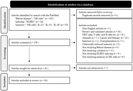
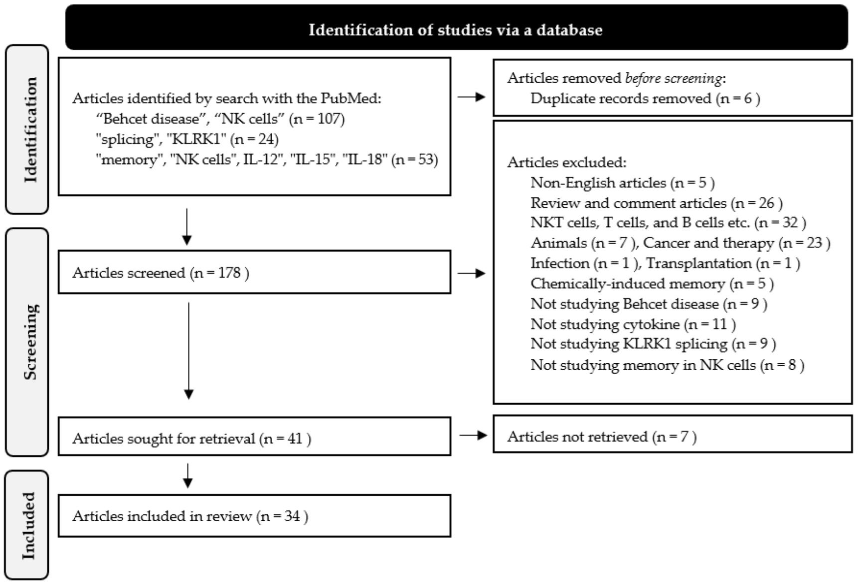
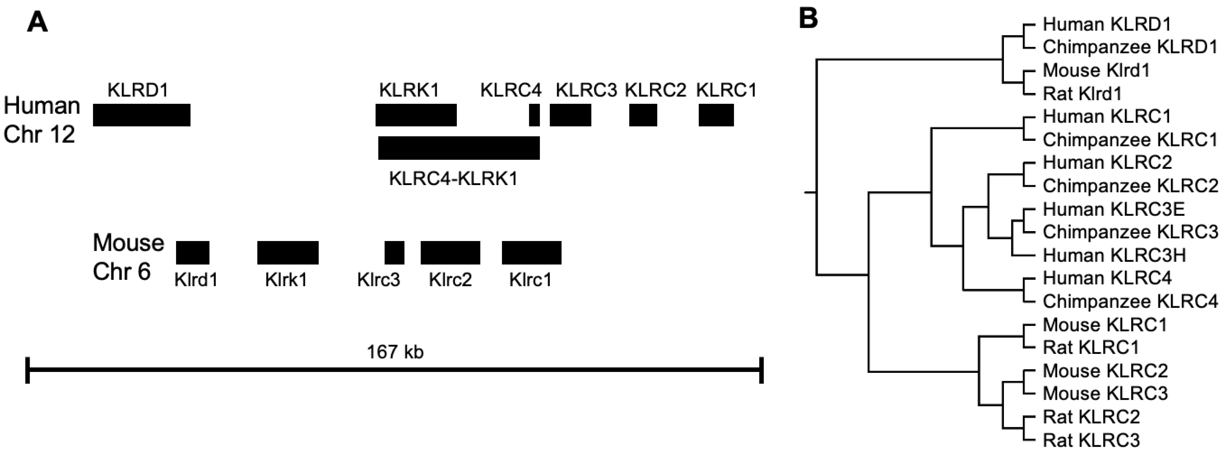
A literature review was performed using the following terms with the PubMed: “Behcet disease”, “NK cells” (identified 107 articles), “splicing”, “KLRK1” (identified 24 articles), “memory”, “NK cells”, IL-12”, “IL-15”, “IL-18” (identified 53 articles). Conjunctions were not used. Articles associated with NKT cells, T cells, B cells, mouse immune cells, cancer therapy, review articles, and non-English articles were excluded. Finally, 34 articles were obtained (Figure 1). Moreover, relevant original and review articles were collected through PubMed and MEDLINE between January and February 2026.
Figure 1.
Articles selection process according to PRISMA 2020.
3. NK Cells in BD
3.1. Numbers, Cytokine Production, and Cytotoxicity
The number of NK cells obtained from patients with BD is conflicting. Nevertheless, two common points have been identified: an NK1/NK2 shift based on cytokine production and lower cytotoxicity in NK cells obtained from BD compared with those in HCs.
Table 1 lists the studies regarding the population and capability of peripheral NK cells in BD. An increase in the number of cytotoxic NK cells (CD16+CD56+) was detected in peripheral blood collected from patients with BD compared with that in HCs [20,31]; however, other studies demonstrated no significant differences in the number of NK cells [32,33,34]. A decrease in the number of NK cells was reported by Hamzaoui et al. [35]. As BD is a recurrent disorder, NK subsets during relapse and remission were analyzed. Patients exhibited an increase in the number of terminally differentiated NK cells (CD16+CD57+) during relapse compared with that during remission [36]; however, the other study showed a decrease in the number of NK cells (CD56dim) [37]. Temporal changes and individual differences in the total number of PBMCs may be responsible for the conflicting data.
Unlike the number of NK cells, cytotoxicity is relatively reproducible. The cytotoxicity of peripheral NK cells against K562 cells during relapse was lower compared with that during remission [36]. Moreover, NK cells (CD3-CD56+, CD16+CD56+) showed decreased cytotoxicity during relapse [35,38]. Because BD patients exhibit increased cytokine production during relapse, different NK cell types may produce high levels of cytokines during relapse. Splicing variants and a readthrough gene produce distinct receptors, which may markedly change the response of NK cells (Section 4.2 and Section 4.3). Alternatively, although they are a minor population during remission, memory-like NK cells proliferate and are hyperactivated by specific antigens (Section 5.2).
NK1 and NK2 subsets were originally investigated to determine whether they produce IFN-γ or not. IFN-γ non-secreting NK cells produce IL-4, IL-5, and IL-13 [39]. NK cells produce proinflammatory cytokines, such as IFN-γ, with their NK1 profile during the relapse period and promote Th1 inflammation. Conversely, NK cells produce IL-4 and IL-10 with their NK2 profile during the remission period, which suppresses Th1 response. An increased NK1/NK2 ratio was detected in patients with uveitis [40] and mucocutaneous BD [35]. Remarkably, one study classified NK cells based on the data of single-cell RNA sequencing (scRNA-seq) and cellular indexing of transcriptomes and epitopes by sequencing (CITE-seq) [41]. The NK1 subset includes cytotoxic cells (CD56dim), whereas NK2 subset represents cytokine-producing cells (CD56bright). As a distinct population from peripheral NK cells, NK3 cells include memory-like NK cells that express KLRC2.
Table 1.
NK subsets in BD pathogenesis.
Cytotoxicity against K562 cells is also estimated by means of LAMP1 expression; however, it does not conform to earlier studies (Table 2). The expression level of LAMP1 in NK cells (CD16+) isolated from BD cultured with K562 cells is comparable to HCs [34], whereas NK cells (CD3−CD56+) isolated from patients with BD exhibited higher expression of LAMP1 than those obtained from HCs [33]. The expression level of LAMP1 in NK cells (CD3−CD16+) during relapse period is higher than that during remission [42]. Therefore, studies concerning the number of NK cells and their capability indicate that the activities of NK cells are not proportionate to the number of NK cells. Further functional assays are required to understand the dysregulation of NK cells.
Table 2.
The expression of marker genes.
3.2. NK Cell Receptors and Their Ligands
Activities of NK cells are regulated by activating and inhibitory receptors. Moreover, precisely processed peptides adjust the affinity to ligands. Unbalance of signals from receptors and processing errors are involved in pathogenesis.
3.2.1. HLA-B and KIRs
HLA-B51 is the highest genetic risk factor for BD. Furthermore, HLA-B15 and HLA-B27 are associated with HLA-B51-positive BD [7]. Patients with HLA-A02, HLA-A24, and HLA-B57 are at risk for developing BD, whereas HLA-A03, HLA-B35, and HLA-B58 are protective [44].
NK cells recognize their own cells through the interaction of HLA-B with KIR3DL1, by which normal cells are protected from cytolysis [45]. HLA-B variants are determined by specific residues at 77–83. Bw4 recognizes KIR3DL1, which possesses two immunoreceptor tyrosine-based inhibitory motifs (ITIMs) in the cytoplasmic region, and inhibits cytolytic signals, whereas Bw6 does not. Positions at 82 and 83 were identified as essential residues using HLA-B78 (HLA-B*1513), HLA-B75 (HLA-B*1502), and nonfunctional Bw6 motif [46].
The presence of KIR3DL1Low/DS1 increased the risk of BD, whereas KIR3DL1High/DL1Null decreased the risk [47]. The frequency of KIR3DL1*004 encoding a misfolded protein [48] is lower in patients with BD than in HCs in both HLA-B51-positive and -negative individuals [49]. The other component that influences the binding of HLA-B to KIR3DL1 is HLA class 1-bound peptides. The peptides bind to HLA class 1 molecules, and they exhibit different affinities [50,51]. Insufficient licensing of NK cells may cause hyperactivation.
HLA-B27 is normally expressed as HLA-B27 in association with β2-microglobulin (β2m) in BD patients. Both HLA-B51 and HLA-B27 are Bw4, which can bind to KIR3DL1. Inaccurate processing of peptides could alter binding to the HLA pocket and attenuate the affinity of HLA-B51 to KIR3DL1, which is likely to disrupt the self-inhibition [22]. HLA-B27 may form peptidomes; however, the peptides have not been identified in BD patients [52]. At present, common events in both HLA-B51 and HLA-B27 carriers are increases of IL-17 and IL-23, which activate neutrophils and damage mucosal tissues [53].
Finally, KIR2DL4 possesses unique features because it contains a charged arginine in the transmembrane [54] and couples with an FcRγ adaptor [55]. KIR2DL4 induces IFN-γ production in NK cells, whereas CD16 and 2B4 induce cytolysis [56]. Although earlier studies found that HLA-G binds to KIR2DL4 and inhibits NK cells in the maternal vasculature during the initial period of pregnancy [57,58], KIR2DL4 was later identified as a novel susceptible gene in patients with severe uveitis [59].
3.2.2. MICA and KLRK1 (NKG2D)
MICA is an MHC class 1 chain-related protein that does not require β2m to form a surface structure. KLRK1 homodimers bind monomeric MICA on the surface of cancer cells and cause cytolysis [60]. Pairs of KLRK1 and the adaptor protein HCST (DAP10) activate signaling molecules such as phosphatidylinositol 3-kinase (PI3K), GRB2, and VAV1 [61,62]. MICA gene has diverse polymorphisms, and its variance is associated with BD. In Slovak, Spanish, and other Eurasian populations, MICA*008 exhibited the highest frequency [63,64]. MICA exhibits strong linkage disequilibrium with HLA-B51 due to its proximity on the genome. MICA*009 is associated with HLA-B51 in Caucasian, Spanish, and Turkish patients [65]. MICA A6, which is a microsatellite polymorphism coding the transmembrane region, was detected in Tukey (MICA*006) and Chinese patients (MICA*049) [66,67]. MICA alleles are classified into two groups [68], which indicates that they cause different immunopathologies. Type 1 (MICA*006, MICA*009, and MICA*049) exhibits high binding affinity for KLRK1, which enhances cytotoxicity, whereas type 2 (MICA*008) shows low affinity and is likely to cause unnecessary cells to expand and activate other immune cells to complement NK cells that are not activated.
3.2.3. HLA-E and KLRD1/KLRC1 (CD94/NKG2A)
HLA-E is a nonclassical MHC class 1 molecule that loads nonapeptides processed from other classical HLA class 1 molecules. Heterodimers of KLRD1 with KLRC1 recognize HLA-E and inhibitory cytolysis. ITIM recruits SHP-1 and SHP-2, whereas it inhibits activation mediators such as VAV1 and ZAP-70/SYK [69]. No single polymorphism of HLA-E was associated with BD; however, specific combinations of SNPs show susceptibility. Individuals with HLA-E*0101 have a decreased risk of developing BD, whereas individuals without HLA-E*0101, NKG2A c.-4258*G/*G, and c.338-90*G have an increased risk of developing BD [70,71]. HLA-E*0101 enhances cytotoxicity, whereas NKG2A c.-4258*G/*G and c.338-90*G reduce cytotoxicity. It remains unclear how they regulate the activity of NK cells entirely in patients with BD.
Nonapeptides presented by HLA-E were classified into 10 groups based on sequence, and their effect on genetic association was investigated. The frequency of N2: VMAPRTLVL and N7: VTAPRTVLL was found to be higher in patients with BD than in controls, indicating that these nonapeptides confer inaccurate presentation and BD risk [72].
3.2.4. Expression of NK Receptors
In the past decade, there has been increasing research on the expression of cell surface markers. Genome editing has become available for knock-out experiments using human cells. For instance, knockout of CD56 does not affect cytotoxicity against cell lines [73]; however, loss of KLRC1 improves cytolytic function against myeloma and solid tumors [74,75,76].
The interaction of ligands with receptors is a trigger for an immediate response. KIR3DL1 is an inhibitory NK receptor. NKB1 antibody detects variant surface expression of KIR3DL1 among patients with BD [77]. Conversely, KLRK1 (NKG2D), NCR1 (NKp46), NCR2 (NKp44), and NCR3 (NKp30) activate NK receptors and induce cytolysis. No significant differences were detected in cell surface expression in NK cells (CD16+CD56dim) obtained from patients with BD and HCs [43]; however, increases in KLRK1 expression were detected in NK cells (CD3−CD56+, CD3−CD16+) obtained from patients with BD [33,42] (Table 2). An approach using genome editing to eliminate or improve the recognition of KLRK1 was proposed [78]. Mimicking altered expression of NK receptors using NK cells obtained from healthy donors through genome editing is helpful to understand the mechanism by which substantial IFN-γ is produced during relapse.
3.2.5. Other Genetic Factors
ERAP1 trims precursor peptides in ER to the correct length for binding to HLA class 1 molecules [79]. Genome-wide association study (GWAS) demonstrated association at ERAP1 rs17482078 (R725Q) in patients with uveitis from a Turkish population [80]. ERAP1 rs17482078 fails to trim peptides due to decreased stability and enzymatic activity, suggesting that unprocessed longer peptides interfere with the binding of HLA class 1 molecules and KIR.
IL-23 maintains Th17 cells and induces the production of IL-17 in patients with uveitis [81]. The feedback loop between NETs and Th17 cells underlies the activation of neutrophils and infiltration [17]. Moreover, the expression of IL-12β2 mRNA in remission is lower than that in relapse. The shift to NK2 decreases IFN-γ production in NK cells [82]. However, IL23R/IL12Rβ2 rs924080 is a polymorphism in the noncoding region [5]. It remains unclear how these receptors mediate ligand signaling in BD pathogenesis.
HLA-A26 is associated with BD in the Japanese patient population. The frequency of HLA-A26 within HLA-B51-positive patients is higher than that within HLA-B51-negative patients [83].
4. Regulation of NK Receptor Genes
4.1. Genomic Organization and Gene Expression of the NK Complex
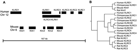
NKG2 receptors are expressed on NK cells and possess killer cell lectin-like domains. Liganded with MICA or HLA-E, they regulate cytokine production and cytotoxic function. They are located on human chromosome 12 and consist of KLRD1, KLRK1, and KLRC1–4 (Figure 2A). KLRD1 is a common counterpart for KLRC1, KLRC2, and KLRC3 to form heterodimers, whereas KLRK1 forms homodimers. KLRC1 with the ITIM motif in its cytoplasmic region works as an inhibitory receptor, whereas KLRC2 pairs with TYROBP (DAP12) to activate receptors [84,85]. KLRC3 pairs with TYROBP; however, they are retained in the endoplasmic reticulum rather than being expressed on the cell surface [86]. KLRC4 does not pair with KLRD1 but can pair with TYROBP, suggesting that the cytoplasmic distribution of KLRC4 suppresses the activating signal by depriving KLRC2 of TYROBP instead of forming extracellular structures that are stimulated by ligands [87].
Figure 2.
NKC and phylogenetic relationship between primate and rodent NKC genes. (A) Genomic organization of humans and mice is shown. (B) Phylogenetic relationship of amino acid sequences from humans, chimpanzees, mice, and rats is analyzed. Sequences were obtained from NCBI.
The amino acid sequences of KLRC4 are conserved between humans and other primates but not between rodents [86,88,89] (Figure 2A). Tree analysis of amino acid sequence clearly separates rodents Klrc1-3 from primates KLRC1–4 (Figure 2B). Judging from the similarity of KLRC1–4 amino acids and the proximate location on the genome, errors during DNA replication and recombination with transposable elements would have produced paralogs, and accumulation of mutations provides a functional difference.
4.2. Splicing of KLRK1 (NKG2D) and Its Function
Mice Klrk1 has short (NKG2D-S) and long (NKG2D-L) isoforms. NKG2D-S is expressed only in activated NK cells pairing with HCST (DAP10) and TYROBP (DAP12), whereas NKG2D-L is found in both resting and activating NK cells pairing with HCST [90]. TYROBP includes the ITAM motif in its cytoplasmic region and activates SYK and ZAP70 [91].
In humans, NK cells possess full-length KLRK1, and it does not interact with TYROBP but HCST [92], which activates PI3K, GRB2, and VAV1 and initiates tyrosine-phosphorylation events [61]. In contrast, TYROBP mediates KIR2DS2 stimulation and activates downstream tyrosine kinases, such as SYK and ZAP70 [93]. Truncated KLRK1 (KLRK1TR) is a human isoform that does not form an extracellular structure, unlike mouse NKG2D-S. Splicing failure leaves intron four that provides a stop codon, generating KLRK1TR. KLRK1TR is bound to HCST, thereby interfering with the pairing of full-length KLRK1 (KLRK1Full) and HCST [94]. Splicing isoforms of KLRK1 are not conserved among species.
4.3. KLRK1-KLRC4 Readthrough Gene
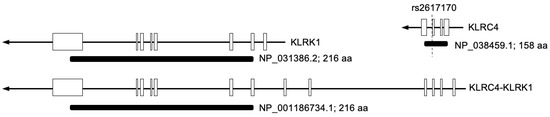
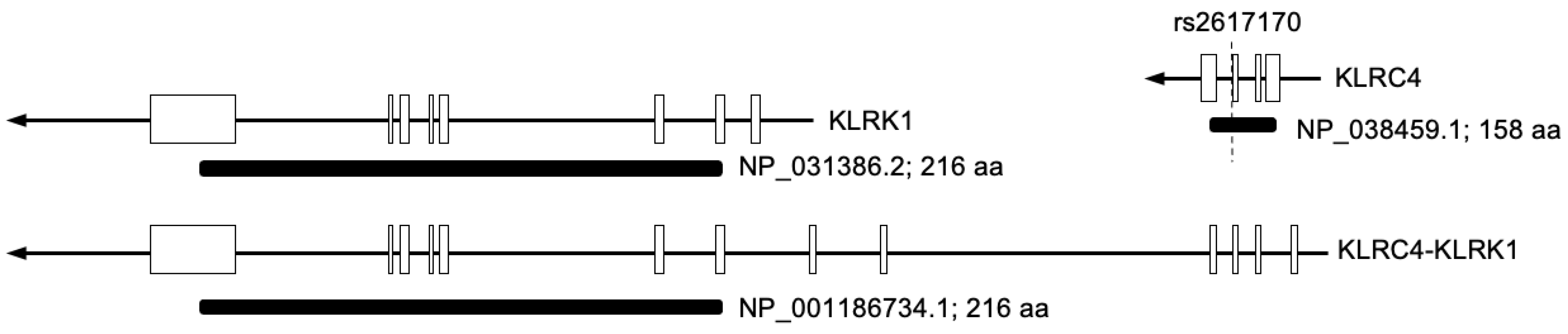
Human KLRK1 and KLRC4 are located next to each other on chromosome 12. According to the NCBI reference sequence, they transcribe NM_007360.4 and NM_013431.2, respectively. In addition, they also form a readthrough gene, KLRC4–KLRK1 (NM_001199805.1), which entirely covers both KLRC4 and KLRK1. Readthrough transcription is an event where transcription machinery fails to find the termination site in the transcription process under stress [95]. Therefore, KLRC4–KLRK1 could encode a hybrid protein. Alternatively, it is possible for KLRC4–KLRK1 to produce several variants using different promoters and start codons. However, only a translation product (NP_001186734.1), which is equivalent to KLRK1 translation (NP_031386.2), has been registered in the database (Figure 3). KLRC4 protein is detectable using an antibody because it has a shorter C-type lectin-like domain than KLRC1, KLRC2, and KLRC3. Nonetheless, it will be hard to identify each isoform using antibodies that recognize the structure unique to KLRC4 if KLRC4–KLRK1 uses multiple start codons and produces multiple isoforms. Cap analysis of gene expression sequencing (CAGE-seq) that covers isoforms more efficiently than conventional RNA-seq analysis will be useful to explore the transcription of KLRK1-KLRC4 thoroughly. Moreover, identification of transcription initiation sites could help predict translation.
Figure 3.
Gene structure of human KLRK1, KLRC4, and KLRC4-KLRK1. Transcripts of these genes, along with their translation, are depicted.
4.4. Dysregulation of KLRC4 in BD
KLRC4 rs2617170 (N104S) was identified in patients with BD as a nonsynonymous variant (NSV) [81,96]. It remains unclear how the lack of fucosylation affects the partnering of KLRC4 N104S with KLRD1. Gene Chip data obtained from the peripheral blood mononuclear cells (PBMCs) of patients with BD (mucocutaneous manifestations, ocular involvement, and large vein thrombosis) categorized genes into the following four groups: negative regulators of inflammation, neutrophil granule proteins, antigen processing and presentation proteins, and regulators of immune response. Regarding the role of KLRC4, it is a regulator of immune response [97]. Interestingly, the effect of polymorphisms on KLRC4 mRNA expression and cytokine production was analyzed, which revealed that the mRNA expression of KLRC4 rs2617170 from CC carriers was significantly higher than that in CT and TT individuals. Furthermore, PBMCs collected from patients with TT and stimulated with LPS secrete more IL-8 than PBMCs collected from CC and CT carriers [98].
4.5. Fucosylation of CD16a
FUT2 plays critical roles in the gut epithelium. FUT2 dysregulation impairs mucosal fucosylation. The weak barrier function increases gut inflammation. FUT2 variants are associated with inflammatory bowel disease (IBD) and Crohn’s disease [99]. In mice, chronic stress causes gut dysfunction and decreases in Fut2 expression and fucosylation on the intestinal epithelium that tethers microbiota [100]. Single nucleotide polymorphism (SNP) array analysis found FUT2 variants rs601338 (W143X) and rs602662 (G258S) [101]. Fucose-deficient IgG1 bound more strongly to CD16a than fucosylated IgG1 and elevated antibody-dependent cell-mediated cytotoxicity (ADCC) [102].
5. Distinct NK Subsets
5.1. Circulating and Tissue-Resident NK Cells
It has been believed that circulating and resident NK cells are fundamentally separated during embryonic stages. CD16−CD56bright is a major population in tissue-resident NK cells, whereas peripheral NK cells predominantly include CD16+CD56dim [103]. Moreover, NK cells in the intestines decrease effector function and exhibit adaptation in their environment [103]. Despite these relevant observations, there has been limited evidence showing that tissue-resident NK cells are activated in the relapse period.
The other possibility is that some peripheral NK cells in patients with BD acquire tissue residency, although this assumption has not been supported. CD4+ T cells infiltrate predominantly; however, NK cells are infrequent in patients with uveitis [104]. Nevertheless, several studies have recently referred to the plasticity of peripheral NK cells. First, circulating NK cells acquire tissue residency when encountering acute infection [105]. Second, murine cytomegalovirus infection recruits NK cells to the salivary gland and forms a long-lived, tissue-resident, memory-like population (NKRM cells) [106]. NK cells appear to possess the ability to change their fate through virus infection and adapt to the tissue environment.
5.2. NK Cell Memory
Immediate immune response without prior sensitization is the capability of NK cells, although they can be sensitized. Some studies using anti-CD16a/CD30 bispecific antibodies, cancer cells, LPS, and cytokines have demonstrated the presence of memory-like NK cells [107]. Both mouse and human NK cells cultured with IL-12, IL-15, and IL-18 produce substantial IFN-γ and persist for a long time [108,109]. Moreover, these cytokine-induced memory-like (CIML) NK cells acquire cytotoxicity against myeloid leukemia [110]. Despite decreased expression of CD16 in CIML NK cells, they maintain the capacity to trigger antibody-dependent cell-mediated cytotoxicity (ADCC) [111].
The other type of NK cells is memory/adaptive NK cells that have been infected by HCMV [112]. Human HCMV infection expands KLRC2-positive cells with increased cytokine production. These cells produce higher levels of IFN-γ in recipients with HCMV infection than in recipients without HCMV infection. KLRC2-positive NK cells maintain memory when they are transplanted to the recipient with antigen [113]. Furthermore, NK cells with a previous HMCV infection are FcRγ-deficient and have distinct memory features. These cells persist for the long term and produce IFN-γ against HCMV [113]. Memory/adaptive NK cells lack the expression of FcRγ and SYK due to epigenetic regulation and expand in an antibody-dependent manner [114].
Nevertheless, there is some ambiguous evidence when discussing the potential involvement of NK cell memory in BD. Firstly, HCMV IgG levels in patients with BD are lower than those in HCs [115], which does not ensure that patients with BD have undergone the infection and generate memory. Secondly, periods of relapse alternate with periods of remission from weeks to years. NK cells in relapse exhibit lower cytotoxicity than those in remission [36,38], consistent with the dominance of CD56bright in relapse. Substantial IFN-γ production is common in both peripheral NK cells in BD and memory-like NK cells; however, there are some differences in the population. CIML NK cells include both CD56bright and CD56dim, and memory/adaptive NK cells predominantly have CD56dim. Seeking local environments where cytokine cocktails develop CIML NK cells or tracking cells infected with HCMV for identifying memory/adaptive NK cells among peripheral blood is difficult. Therefore, further studies using scRNA-seq data on CIML NK cells or memory/adaptive NK cells as reference will be effective in identifying original cells with memory that can be hyperactivated by specific stimulation and developing therapy to target those cells.
6. Future Perspectives
Future research must focus on NK cell receptors as their diversity affects the response to ligands. For instance, KLRK1 is a key receptor that could interact with HLA class 1 molecules. New findings on the splicing of KLRK1 and the transcriptional regulation of the KLRC4–KLRK1 readthrough gene will elucidate individual differences in immune responses. The functions of human NK cells can be modified by genome editing, and their functions can be validated experimentally, overcoming the limitations in mouse-based studies aimed at characterizing polymorphisms in human immune response genes.
Studies aimed at identifying memory-like NK cells should also be encouraged. Comparative analyses of scRNA-seq data between NK cells obtained from patients with BD in remission and those in relapse could identify the specific markers, which will aid healthcare workers in predicting prognosis. Additionally, the population of adaptive NK cells that could expand substantially in response to a specific antigen might be identified. Furthermore, it is necessary to explore the possibility of tissue-resident NK cells infiltrating into mucosal lesions in patients with BD. Once their involvement is established, eliminating those cells may represent a new therapy.
7. Conclusions
BD is a chronic autoinflammatory disorder characterized by unpredictable relapses. Innate immune cells contribute to its pathogenesis. The number of peripheral NK cells varies among patients with BD, and the cytotoxicity of peripheral NK cells tends to be lower during relapse than during remission. NK1/NK2 subsets clearly depict cytokine production in relapse and remission. Moreover, the strongest association with HLA-B51 indicates that the interaction with NK receptors is critical to BD pathogenesis. Polymorphisms and unprocessed peptides impair the accuracy of recognition and cause the hyperactivation of NK cells.
NK cells exhibit an immediate response, including cytokine production and cytolysis, without prior sensitization, and their lifespan is short. However, recent studies have demonstrated that NK cells are able to acquire memory through exposure to cytokine cocktails or HCMV infection and produce IFN-γ against specific stimulation. Although neither CIML NK cells nor memory/adaptive NK cells have been identified from patients with BD, hyperactivation in relapse is similar to the response of these adaptive NK cells. Alternatively, activation of tissue-resident NK cells and migration of peripheral NK cells to mucosal tissues are anticipated episodes. Moreover, transcriptional regulation of activating receptors, such as KLRK1 and KLRC4, could easily modify the responses to ligands and contribute to hyperactivation. Further studies focusing on adaptive NK cells among skewed innate NK cells in BD will clarify the mechanism underlying chronic and recurrent manifestations.
Funding
This work was partly supported by the Grants-in-Aid for Scientific Research (C) (No. 23K09156).
Institutional Review Board Statement
Not applicable.
Informed Consent Statement
Not applicable.
Data Availability Statement
The data regarding sequences and genomic locations are available at NCBI (https://www.ncbi.nlm.nih.gov/gene, accessed on 13 May 2026).
Conflicts of Interest
The author declares no conflict of interest.
Abbreviations
The following abbreviations are used in this review.
| ADCC | Antibody-Dependent Cell-mediated Cytotoxicity |
| BD | Behcet Disease |
| β2m | β2-microglobulin |
| CAGE-seq | Cap Analysis of Gene Expression Sequencing |
| CIML NK cells | Cytokine-Induced memory-like NK cells |
| CITE-seq | Cellular Indexing of Transcriptomes and Epitopes by sequencing |
| GWAS | Genome-Wide Association Study |
| HC | Healthy Control |
| HCMV | Human Cytomegalovirus |
| HLA | Human Leukocyte Antigen |
| IBD | Inflammatory Bowel Disease |
| ICBD | The International Criteria for Behcet’s Disease |
| ITIM | Immunoreceptor Tyrosine-Based Inhibitory Motif |
| KIR | Killer Cell Immunoglobulin-like Receptor |
| MHC-I | Major histocompatibility complex class 1 |
| MICA | Major histocompatibility complex class 1-related protein A |
| NETs | Neutrophil Extracellular Traps |
| NK | Natural Killer |
| NKRM cells | Tissue-Resident Memory-like Natural Killer cells |
| NSV | Nonsynonymous Variant |
| PBMCs | Peripheral Blood Mononuclear Cells |
| PI3K | Phosphatidylinositol 3-Kinase |
| scRNA-seq | Single-Cell RNA Sequencing |
| SNP | Single Nucleotide Polymorphism |
References
- Moghoofei, M.; Pajavand, H.; Shahbazi, R.; Rezaei, M.; Taki, E. The Role of Viral and Bacterial Infections in the Etiology of Behçet’s Disease. J. Clin. Lab. Anal. 2026, 40, e70133. [Google Scholar] [CrossRef] [Scilit]
- Perazzio, S.F.; Andrade, L.E.C.; de Souza, A.W.S. Understanding Behçet’s Disease in the Context of Innate Immunity Activation. Front. Immunol. 2020, 11, 586558. [Google Scholar] [CrossRef] [Scilit]
- Ohno, S.; Ohguchi, M.; Hirose, S.; Matsuda, H.; Wakisaka, A.; Aizawa, M. Close association of HLA-Bw51 with Behcet’s disease. Arch. Ophthalmol. 1982, 100, 1455–1458. [Google Scholar] [CrossRef] [Scilit]
- Mizuki, N.; Meguro, A.; Tohnai, I.; Gül, A.; Ohno, S.; Mizuki, N. Association of Major Histocompatibility Complex Class I Chain-Related Gene A and HLA-B Alleles with Behçet’s Disease in Turkey. Jpn. J. Ophthalmol. 2007, 51, 431–436. [Google Scholar] [CrossRef] [Scilit] [PubMed]
- Remmers, E.F.; Cosan, F.; Kirino, Y.; Ombrello, M.J.; Abaci, N.; Satorius, C.; Le, J.M.; Yang, B.; Korman, B.D.; Cakiris, A.; et al. Genome-wide association study identifies variants in the MHC class I, IL10, and IL23R-IL12RB2 regions associated with Behçet’s disease. Nat. Genet. 2010, 42, 698–702. [Google Scholar] [CrossRef] [Scilit] [PubMed]
- Hughes, T.; Coit, P.; Adler, A.; Yilmaz, V.; Aksu, K.; Düzgün, N.; Keser, G.; Cefle, A.; Yazici, A.; Ergen, A.; et al. Identification of multiple independent susceptibility loci in the HLA region in Behçet’s disease. Nat. Genet. 2013, 45, 319–324. [Google Scholar] [CrossRef] [Scilit] [PubMed]
- Ombrello, M.J.; Kirino, Y.; de Bakker, P.I.; Gül, A.; Kastner, D.L.; Remmers, E.F. Behçet disease-associated MHC class I residues implicate antigen binding and regulation of cell-mediated cytotoxicity. Proc. Natl. Acad. Sci. USA 2014, 111, 8867–8872. [Google Scholar] [CrossRef] [Scilit]
- Kuiper, J.J.; Prinz, J.C.; Stratikos, E.; Kuśnierczyk, P.; Arakawa, A.; Springer, S.; Mintoff, D.; Padjen, I.; Shumnalieva, R.; Vural, S.; et al. EULAR study group MHC-I-opathies. EULAR study group on ‘MHC-I-opathy’: Identifying disease-overarching mechanisms across disciplines and borders. Ann. Rheum. Dis. 2023, 82, 887–896. [Google Scholar] [CrossRef] [Scilit]
- Sota, J.; Ragab, G.; AlMaglouth, I.; Lopalco, G.; Tufan, A.; Direskeneli, H.; Hinojosa-Azaola, A.; Mayrink Giardini, H.A.; Guerriero, S.; Triggianese, P.; et al. Influence of gender on Behçet’s disease phenotype and irreversible organ damage: Data from the International AIDA Network Behçet’s Disease Registry. Jt. Bone Spine 2025, 92, 105819. [Google Scholar] [CrossRef] [Scilit]
- Jo, Y.G.; Ortiz-Fernández, L.; Coit, P.; Yilmaz, V.; Yentür, S.P.; Alibaz-Oner, F.; Aksu, K.; Erken, E.; Düzgün, N.; Keser, G.; et al. Sex-specific analysis in Behcet’s disease reveals higher genetic risk in male patients. J. Autoimmun. 2022, 132, 102882. [Google Scholar] [CrossRef] [Scilit]
- Liu, A.; Su, Y.; Zhu, J.; Li, Y.Y. Disease association study of Autoimmune and autoinflammatory diseases by integrating multi-modal data and hierarchical ontologies. Front. Immunol. 2025, 16, 1575490. [Google Scholar] [CrossRef] [Scilit]
- Davatchi, F.; Assaad-Khalil, S.; Calamia, K.T.; Crook, J.E.; Sadeghi-Abdollahi, B.; Schirmer, M.; Tzellos, T.; Zouboulis, C.C.; Akhlagi, M.; Al-Dalaan, A.; et al. The International Criteria for Behcet’s Disease (ICBD): A collaborative study of 27 countries on the sensitivity and specificity of the new criteria. J. Eur. Acad. Dermatol. Venereol. 2014, 28, 338–347. [Google Scholar]
- Zhan, H.; Cheng, L.; Chen, H.; Liu, Y.; Feng, X.; Li, H.; Li, Z.; Li, Y. Evaluation of inflammatory-thrombosis panel as a diagnostic tool for vascular Behcet’s disease. Clin. Rheumatol. 2025, 44, 1279–1291. [Google Scholar] [CrossRef] [Scilit]
- Sahin, S.; Akoğlu, T.; Direskeneli, H.; Sen, L.S.; Lawrence, R. Neutrophil adhesion to endothelial cells and factors affecting adhesion in patients with Behçet’s disease. Ann. Rheum. Dis. 1996, 55, 128–133. [Google Scholar] [CrossRef] [Scilit]
- Rahman, S.; Daveluy, S. Pathergy Test. In StatPearls [Internet]; StatPearls Publishing LLC: Treasure Island, FL, USA, 2023. [Google Scholar]
- Le Joncour, A.; Martos, R.; Loyau, S.; Lelay, N.; Dossier, A.; Cazes, A.; Fouret, P.; Domont, F.; Papo, T.; Jandrot-Perrus, M.; et al. Critical role of neutrophil extracellular traps (NETs) in patients with Behcet’s disease. Ann. Rheum. Dis. 2019, 78, 1274–1282. [Google Scholar] [CrossRef] [Scilit]
- Wu, Y.; Ning, K.; Huang, Z.; Chen, B.; Chen, J.; Wen, Y.; Bu, J.; Hong, H.; Chen, Q.; Zhang, Z.; et al. NETs-CD44-IL-17A Feedback Loop Drives Th17-Mediated Inflammation in Behcet’s Uveitis. Adv. Sci. 2025, 12, e2411524. [Google Scholar] [CrossRef] [Scilit]
- Hirahara, L.; Takase-Minegishi, K.; Kirino, Y.; Iizuka-Iribe, Y.; Soejima, Y.; Yoshimi, R.; Nakajima, H. The Roles of Monocytes and Macrophages in Behçet’s Disease With Focus on M1 and M2 Polarization. Front. Immunol. 2022, 13, 852297. [Google Scholar] [CrossRef] [Scilit]
- Nakano, H.; Kirino, Y.; Takeno, M.; Higashitani, K.; Nagai, H.; Yoshimi, R.; Yamaguchi, Y.; Kato, I.; Aoki, I.; Nakajima, H. GWAS-identified CCR1 and IL10 loci contribute to M1 macrophage-predominant inflammation in Behcet’s disease. Arthritis Res. Ther. 2018, 20, 124. [Google Scholar] [CrossRef] [Scilit]
- Suzuki, Y.; Hoshi, K.; Matsuda, T.; Mizushima, Y. Increased peripheral blood gamma delta+ T cells and natural killer cells in Behçet’s disease. J. Rheumatol. 1992, 19, 588–592. [Google Scholar]
- Lule, S.; Colpak, A.I.; Balci-Peynircioglu, B.; Gursoy-Ozdemir, Y.; Peker, S.; Kalyoncu, U.; Can, A.; Tekin, N.; Demiralp, D.; Dalkara, T. Behçet Disease serum is immunoreactive to neurofilament medium which share common epitopes to bacterial HSP-65, a putative trigger. J. Autoimmun. 2017, 84, 87–96. [Google Scholar] [CrossRef] [Scilit]
- van der Houwen, T.B.; van Hagen, P.M.; van Laar, J.A.M. Immunopathogenesis of Behçet’s disease and treatment modalities. Semin. Arthritis Rheum. 2022, 52, 151956. [Google Scholar] [CrossRef] [Scilit]
- Hasan, M.S.; Bergmeier, L.A.; Petrushkin, H.; Fortune, F. Gamma Delta (γδ) T Cells and Their Involvement in Behçet’s Disease. J. Immunol. Res. 2015, 2015, 705831. [Google Scholar] [CrossRef] [Scilit]
- Freud, A.G.; Mundy-Bosse, B.L.; Yu, J.; Caligiuri, M.A. The Broad Spectrum of Human Natural Killer Cell Diversity. Immunity 2017, 47, 820–833. [Google Scholar] [CrossRef] [Scilit]
- Gianchecchi, E.; Delfino, D.V.; Fierabracci, A. NK cells in autoimmune diseases: Linking innate and adaptive immune responses. Autoimmun. Rev. 2018, 17, 142–154. [Google Scholar] [CrossRef] [Scilit]
- Reefman, E.; Kay, J.G.; Wood, S.M.; Offenhäuser, C.; Brown, D.L.; Roy, S.; Stanley, A.C.; Low, P.C.; Manderson, A.P.; Stow, J.L. Cytokine secretion is distinct from secretion of cytotoxic granules in NK cells. J. Immunol. 2010, 184, 4852–4862. [Google Scholar] [CrossRef] [Scilit]
- Boudreau, J.E.; Hsu, K.C. Natural killer cell education in human health and disease. Curr. Opin. Immunol. 2018, 50, 102–111. [Google Scholar] [CrossRef] [Scilit]
- Stokic-Trtica, V.; Diefenbach, A.; Klose, C.S.N. NK Cell Development in Times of Innate Lymphoid Cell Diversity. Front. Immunol. 2020, 11, 813. [Google Scholar] [CrossRef] [Scilit]
- Prager, I.; Watzl, C. Mechanisms of natural killer cell-mediated cellular cytotoxicity. J. Leukoc. Biol. 2019, 105, 1319–1329. [Google Scholar] [CrossRef] [Scilit]
- Cohnen, A.; Chiang, S.C.; Stojanovic, A.; Schmidt, H.; Claus, M.; Saftig, P.; Janßen, O.; Cerwenka, A.; Bryceson, Y.T.; Watzl, C. Surface CD107a/LAMP-1 protects natural killer cells from degranulation-associated damage. Blood 2013, 122, 1411–1418. [Google Scholar] [CrossRef] [Scilit]
- Sakly, K.; Lahmar, R.; Nefzi, F.; Hammami, S.; Harzallah, O.; Sakly, N.; Sakly, W.; Hassine, M.; Mahjoub, S.; Ghedira, I.; et al. Phenotypic abnormalities of peripheral blood mononuclear cells in patients with Behçet’s disease and association with HLA-B51 expression. Immunol. Investig. 2014, 43, 463–478. [Google Scholar] [CrossRef] [Scilit]
- Saruhan-Direskeneli, G.; Uyar, F.A.; Cefle, A.; Onder, S.C.; Eksioglu-Demiralp, E.; Kamali, S.; Inanç, M.; Ocal, L.; Gül, A. Expression of KIR and C-type lectin receptors in Behcet’s disease. Rheumatology 2004, 43, 423–427. [Google Scholar] [CrossRef] [Scilit]
- Bonacini, M.; Soriano, A.; Zerbini, A.; Calò, E.; Cimino, L.; Muratore, F.; Fontana, L.; Braglia, L.; Parmeggiani, M.; Salvarani, C.; et al. Higher Frequencies of Lymphocytes Expressing the Natural Killer Group 2D Receptor in Patients with Behcet Disease. Front. Immunol. 2018, 9, 2157. [Google Scholar] [CrossRef] [Scilit] [PubMed]
- Cosan, F.; Aktas Cetin, E.; Akdeniz, N.; Emrence, Z.; Cefle, A.; Deniz, G. Natural Killer Cell Subsets and Their Functional Activity in Behcet’s Disease. Immunol. Investig. 2017, 46, 419–432. [Google Scholar] [CrossRef] [Scilit]
- Hamzaoui, K.; Berraies, A.; Kaabachi, W.; Ammar, J.; Hamzaoui, A. Pulmonary manifestations in Behcet disease: Impaired natural killer cells activity. Multidiscip. Respir. Med. 2013, 8, 29. [Google Scholar] [CrossRef] [Scilit] [PubMed]
- Kaneko, F.; Takahashi, Y.; Muramatsu, R.; Adachi, K.; Miura, Y.; Nakane, A.; Minagawa, T. Natural killer cell numbers and function in peripheral lymphoid cells in Behcet’s disease. Br. J. Dermatol. 1985, 113, 313–318. [Google Scholar] [CrossRef] [Scilit]
- Hasan, M.S.; Ryan, P.L.; Bergmeier, L.A.; Fortune, F. Circulating NK cells and their subsets in Behçet’s disease. Clin. Exp. Immunol. 2017, 188, 311–322. [Google Scholar] [CrossRef] [Scilit]
- Onder, M.; Bozkurt, M.; Gürer, M.A.; Gülekon, A.; Sezgin, P.; Imir, T. Natural cellular cytotoxicity in Behcet’s disease. J. Dermatol. 1994, 21, 239–243. [Google Scholar] [CrossRef] [Scilit]
- Deniz, G.; Akdis, M.; Aktas, E.; Blaser, K.; Akdis, C.A. Human NK1 and NK2 subsets determined by purification of IFN-gamma-secreting and IFN-gamma-nonsecreting NK cells. Eur. J. Immunol. 2002, 32, 879–884. [Google Scholar] [CrossRef] [Scilit]
- Kucuksezer, U.C.; Aktas-Cetin, E.; Bilgic-Gazioglu, S.; Tugal-Tutkun, I.; Gül, A.; Deniz, G. Natural killer cells dominate a Th-1 polarized response in Behcet’s disease patients with uveitis. Clin. Exp. Rheumatol. 2015, 33, S24–S29. [Google Scholar]
- Rebuffet, L.; Melsen, J.E.; Escalière, B.; Basurto-Lozada, D.; Bhandoola, A.; Björkström, N.K.; Bryceson, Y.T.; Castriconi, R.; Cichocki, F.; Colonna, M.; et al. High-dimensional single-cell analysis of human natural killer cell heterogeneity. Nat. Immunol. 2024, 25, 1474–1488. [Google Scholar] [CrossRef] [Scilit] [PubMed]
- Sallakci, N.; Tahrali, I.; Kucuksezer, U.C.; Cetin, E.A.; Gul, A.; Deniz, G. Effect of different cytokines in combination with IL-15 on the expression of activating receptors in NK cells of patients with Behcet’s disease. Immunol. Res. 2022, 70, 654–666. [Google Scholar] [CrossRef] [Scilit]
- Gelmez, M.Y.; Cinar, S.; Cetin, E.A.; Ozcit-Gürel, G.; Babuna-Kobaner, G.; Erdugan, M.; Gul, A.; Akdag-Kose, A.; Deniz, G. Inflammatory status might direct ILC and NK cells to IL-17 expressing ILC3 and NK subsets in Behcet’s disease. Immunol. Lett. 2021, 235, 1–8. [Google Scholar] [CrossRef] [Scilit]
- Montes-Cano, M.A.; Conde-Jaldón, M.; García-Lozano, J.R.; Ortiz-Fernández, L.; Ortego-Centeno, N.; Castillo-Palma, M.J.; Espinosa, G.; Graña-Gil, G.; González-Gay, M.A.; Barnosi-Marín, A.C.; et al. HLA and non-HLA genes in Behçet’s disease: A multicentric study in the Spanish population. Arthritis Res. Ther. 2013, 15, R145. [Google Scholar] [CrossRef] [Scilit]
- Pugh, J.; Nemat-Gorgani, N.; Djaoud, Z.; Guethlein, L.A.; Norman, P.J.; Parham, P. In vitro education of human natural killer cells by KIR3DL1. Life Sci. Alliance 2019, 2, e201900434. [Google Scholar] [CrossRef] [Scilit]
- Sanjanwala, B.; Draghi, M.; Norman, P.J.; Guethlein, L.A.; Parham, P. Polymorphic sites away from the Bw4 epitope that affect interaction of Bw4+ HLA-B with KIR3DL1. J. Immunol. 2008, 181, 6293–6300. [Google Scholar] [CrossRef] [Scilit]
- Petrushkin, H.; Norman, P.J.; Lougee, E.; Parham, P.; Wallace, G.R.; Stanford, M.R.; Fortune, F. KIR3DL1/S1 Allotypes Contribute Differentially to the Development of Behçet Disease. J. Immunol. 2019, 203, 1629–1635. [Google Scholar] [CrossRef] [Scilit]
- Pando, M.J.; Gardiner, C.M.; Gleimer, M.; McQueen, K.L.; Parham, P. The protein made from a common allele of KIR3DL1 (3DL1*004) is poorly expressed at cell surfaces due to substitution at positions 86 in Ig domain 0 and 182 in Ig domain 1. J. Immunol. 2003, 171, 6640–6649. [Google Scholar] [CrossRef] [Scilit]
- Castaño-Núñez, Á.; Montes-Cano, M.A.; García-Lozano, J.R.; Ortego-Centeno, N.; García-Hernández, F.J.; Espinosa, G.; Graña-Gil, G.; Sánchez-Bursón, J.; Juliá, M.R.; Solans, R.; et al. Association of Functional Polymorphisms of KIR3DL1/DS1 with Behçet’s Disease. Front. Immunol. 2019, 10, 2755. [Google Scholar] [CrossRef] [Scilit]
- Saunders, P.M.; Illing, P.T.; Coin, L.; Wong, S.C.; Oates, C.V.L.; Purcell, A.W.; Brooks, A.G. Peptide selectivity of killer cell immunoglobulin-like receptors differs with allotypic variation in HLA class I. J. Immunol. 2025, 214, 747–761. [Google Scholar] [CrossRef] [Scilit]
- Maiers, M.; Louzoun, Y.; Pymm, P.; Vivian, J.P.; Rossjohn, J.; Brooks, A.G.; Saunders, P.M. Prediction of KIR3DL1 and human leukocyte antigen binding. J. Biol. Chem. 2025, 301, 110437. [Google Scholar] [CrossRef] [Scilit]
- López de Castro, J.A. How ERAP1 and ERAP2 Shape the Peptidomes of Disease-Associated MHC-I Proteins. Front Immunol. 2018, 9, 2463. [Google Scholar] [CrossRef] [Scilit]
- Naffaa, M.E.; Hassan, F.; Omar, M.; Abacar, K.; McGonagle, D. The IL-23/IL-17 axis in Behçet’s syndrome pathogenesis: From immunological perspectives to therapeutic implications. Front Immunol. 2026, 17, 1761519. [Google Scholar] [CrossRef] [Scilit]
- Pollock, N.R.; Harrison, G.F.; Norman, P.J. Immunogenomics of Killer Cell Immunoglobulin-Like Receptor (KIR) and HLA Class I: Coevolution and Consequences for Human Health. J. Allergy Clin. Immunol. Pract. 2022, 10, 1763–1775. [Google Scholar] [CrossRef] [Scilit]
- Kikuchi-Maki, A.; Catina, T.L.; Campbell, K.S. Cutting edge: KIR2DL4 transduces signals into human NK cells through association with the Fc receptor gamma protein. J. Immunol. 2005, 174, 3859–3863. [Google Scholar] [CrossRef] [Scilit]
- Rajagopalan, S.; Fu, J.; Long, E.O. Cutting edge: Induction of IFN-gamma production but not cytotoxicity by the killer cell Ig-like receptor KIR2DL4 (CD158d) in resting NK cells. J. Immunol. 2001, 167, 1877–1881. [Google Scholar] [CrossRef] [Scilit]
- Bespalova, O.; Bakleicheva, M.; Ivashchenko, T.; Tral, T.; Tolibova, G.; Kogan, I. Expression of HLA-G and KIR2DL4 receptor in chorionic villous in missed abortion. Gynecol. Endocrinol. 2020, 36, 43–47. [Google Scholar] [CrossRef] [Scilit]
- Rajagopalan, S.; Majumder, S.; Wang, C.; Hibler, W.; Shamsaddini, A.; Gardina, P.; Long, E.O. The fetal trophoblast cell marker HLA-G activates a type I interferon response in primary NK cells through the receptor KIR2DL4. Sci. Signal. 2026, 19, eadv2400. [Google Scholar] [CrossRef] [Scilit]
- Kim, S.J.; Lee, S.; Park, C.; Seo, J.S.; Kim, J.I.; Yu, H.G. Targeted resequencing of candidate genes reveals novel variants associated with severe Behçet’s uveitis. Exp. Mol. Med. 2013, 45, e49. [Google Scholar] [CrossRef] [Scilit]
- Ferrari de Andrade, L.; Tay, R.E.; Pan, D.; Luoma, A.M.; Ito, Y.; Badrinath, S.; Tsoucas, D.; Franz, B.; May KFJr Harvey, C.J.; Kobold, S.; et al. Antibody-mediated inhibition of MICA and MICB shedding promotes NK cell-driven tumor immunity. Science 2018, 359, 1537–1542. [Google Scholar] [CrossRef] [Scilit]
- Wilton, K.M.; Overlee, B.L.; Billadeau, D.D. NKG2D-DAP10 signaling recruits EVL to the cytotoxic synapse to generate F-actin and promote NK cell cytotoxicity. J. Cell Sci. 2019, 133, jcs230508. [Google Scholar] [CrossRef] [Scilit]
- Lanier, L.L. Up on the tightrope: Natural killer cell activation and inhibition. Nat. Immunol. 2008, 9, 495–502. [Google Scholar] [CrossRef] [Scilit]
- Muñoz-Saá, I.; Cambra, A.; Pallarés, L.; Espinosa, G.; Juan, A.; Pujalte, F.; Matamoros, N.; Milà, J.; Julià, M.R. Allelic diversity and affinity variants of MICA are imbalanced in Spanish patients with Behçet’s disease. Scand. J. Immunol. 2006, 64, 77–82. [Google Scholar] [CrossRef] [Scilit]
- Durmanová, V.; Tirpakova, J.; Stuchlikova, M.; Shawkatova, I.; Kuba, D.; Sapak, M.; Buc, M. Characterization of MICA gene polymorphism of HLA complex in the Slovak population. Ann. Hum. Biol. 2011, 38, 570–576. [Google Scholar] [CrossRef] [Scilit]
- Hughes, E.H.; Collins, R.W.; Kondeatis, E.; Wallace, G.R.; Graham, E.M.; Vaughan, R.W.; Stanford, M.R. Associations of major histocompatibility complex class I chain-related molecule polymorphisms with Behcet’s disease in Caucasian patients. Tissue Antigens 2005, 66, 195–199. [Google Scholar] [CrossRef] [Scilit]
- Eyerci, N.; Balkan, E.; Akdeniz, N.; Keleş, S. Association of MICA Alleles and Human Leukocyte Antigen B in Turkish Patients Diagnosed with Behçet’s Disease. Arch. Rheumatol. 2018, 33, 352–357. [Google Scholar] [CrossRef] [Scilit]
- Zhu, W.; Deng, Y.; Wang, J.; Guo, X.; Ding, W.; Chao, J.; Lin, D.; Wang, Y.; Zhou, X. MICA*049, not MICA*009, is associated with Behçet’s disease in a Chinese population. Sci. Rep. 2019, 9, 10856. [Google Scholar] [CrossRef] [Scilit]
- Luo, Q.; Yin, X.; Zhu, Q.; Luo, W.; Liu, R.; Wei, L.; Zou, Y. Two major human phenotypes of MICA molecules and their differential activation to NK cells via NKG2D receptor. Front. Immunol. 2025, 16, 1563872. [Google Scholar] [CrossRef] [Scilit]
- Fisher, J.G.; Doyle, A.D.P.; Graham, L.V.; Khakoo, S.I.; Blunt, M.D. Disruption of the NKG2A:HLA-E Immune Checkpoint Axis to Enhance NK Cell Activation against Cancer. Vaccines 2022, 10, 1993. [Google Scholar] [CrossRef] [Scilit]
- Seo, J.; Park, J.S.; Nam, J.H.; Bang, D.; Sohn, S.; Lee, E.S.; Park, K.S. Association of CD94/NKG2A, CD94/NKG2C, and its ligand HLA-E polymorphisms with Behcet’s disease. Tissue Antigens 2007, 70, 307–313. [Google Scholar] [CrossRef] [Scilit]
- Park, K.S.; Park, J.S.; Nam, J.H.; Bang, D.; Sohn, S.; Lee, E.S. HLA-E*0101 and HLA-G*010101 reduce the risk of Behcet’s disease. Tissue Antigens 2007, 69, 139–144. [Google Scholar] [CrossRef] [Scilit]
- Castaño-Núñez, Á.L.; Montes-Cano, M.A.; García-Lozano, J.R.; Ortego-Centeno, N.; García-Hernández, F.J.; Espinosa, G.; Graña-Gil, G.; Sánchez-Bursón, J.; Juliá, M.R.; Solans, R.; et al. The complex HLA-E-nonapeptide in Behçet disease. Front. Immunol. 2023, 14, 1080047. [Google Scholar] [CrossRef] [Scilit]
- Picard, L.K.; Claus, M.; Fasbender, F.; Watzl, C. Human NK cells responses are enhanced by CD56 engagement. Eur. J. Immunol. 2022, 52, 1441–1451. [Google Scholar] [CrossRef] [Scilit]
- Bexte, T.; Alzubi, J.; Reindl, L.M.; Wendel, P.; Schubert, R.; Salzmann-Manrique, E.; von Metzler, I.; Cathomen, T.; Ullrich, E. CRISPR-Cas9 based gene editing of the immune checkpoint NKG2A enhances NK cell mediated cytotoxicity against multiple myeloma. Oncoimmunology 2022, 11, 2081415. [Google Scholar] [CrossRef] [Scilit]
- Kaulfuss, M.; Mietz, J.; Fabri, A.; Vom Berg, J.; Münz, C.; Chijioke, O. The NK cell checkpoint NKG2A maintains expansion capacity of human NK cells. Sci. Rep. 2023, 13, 10555. [Google Scholar] [CrossRef] [Scilit]
- Mac Donald, A.; Guipouy, D.; Lemieux, W.; Harvey, M.; Bordeleau, L.J.; Guay, D.; Roméro, H.; Li, Y.; Dion, R.; Béland, K.; et al. KLRC1 knockout overcomes HLA-E-mediated inhibition and improves NK cell antitumor activity against solid tumors. Front. Immunol. 2023, 14, 1231916. [Google Scholar] [CrossRef] [Scilit]
- Takeno, M.; Shimoyama, Y.; Kashiwakura, J.; Nagafuchi, H.; Sakane, T.; Suzuki, N. Abnormal killer inhibitory receptor expression on natural killer cells in patients with Behcet’s disease. Rheumatol. Int. 2004, 24, 212–216. [Google Scholar] [CrossRef] [Scilit]
- Alves, E.; McLeish, E.; Blancafort, P.; Coudert, J.D.; Gaudieri, S. Manipulating the NKG2D Receptor-Ligand Axis Using CRISPR: Novel Technologies for Improved Host Immunity. Front. Immunol. 2021, 12, 712722. [Google Scholar] [CrossRef] [Scilit]
- Li, L.; Batliwala, M.; Bouvier, M. ERAP1 enzyme-mediated trimming and structural analyses of MHC I-bound precursor peptides yield novel insights into antigen processing and presentation. J. Biol. Chem. 2019, 294, 18534–18544. [Google Scholar] [CrossRef] [Scilit]
- Kirino, Y.; Bertsias, G.; Ishigatsubo, Y.; Mizuki, N.; Tugal-Tutkun, I.; Seyahi, E.; Ozyazgan, Y.; Sacli, F.S.; Erer, B.; Inoko, H.; et al. Genome-wide association analysis identifies new susceptibility loci for Behçet’s disease and epistasis between HLA-B*51 and ERAP1. Nat. Genet. 2013, 45, 202–207. [Google Scholar] [CrossRef] [Scilit]
- Pepple, K.L.; Lin, P. Targeting Interleukin-23 in the Treatment of Noninfectious Uveitis. Ophthalmology 2018, 125, 1977–1983. [Google Scholar] [CrossRef] [Scilit]
- Yamaguchi, Y.; Takahashi, H.; Satoh, T.; Okazaki, Y.; Mizuki, N.; Takahashi, K.; Ikezawa, Z.; Kuwana, M. Natural killer cells control a T-helper 1 response in patients with Behcet’s disease. Arthritis Res. Ther. 2010, 12, R80. [Google Scholar] [CrossRef] [Scilit]
- Nakamura, J.; Meguro, A.; Ishii, G.; Mihara, T.; Takeuchi, M.; Mizuki, Y.; Yuda, K.; Yamane, T.; Kawagoe, T.; Ota, M.; et al. The association analysis between HLA-A*26 and Behçet’s disease. Sci. Rep. 2019, 9, 4426. [Google Scholar] [CrossRef] [Scilit]
- López-Botet, M.; De Maria, A.; Muntasell, A.; Della Chiesa, M.; Vilches, C. Adaptive NK cell response to human cytomegalovirus: Facts and open issues. Semin. Immunol. 2023, 65, 101706. [Google Scholar] [CrossRef] [Scilit]
- Siemaszko, J.; Marzec-Przyszlak, A.; Bogunia-Kubik, K. Activating NKG2C Receptor: Functional Characteristics and Current Strategies in Clinical Applications. Arch. Immunol. Ther. Exp. 2023, 71, 9. [Google Scholar] [CrossRef] [Scilit]
- Orbelyan, G.A.; Tang, F.; Sally, B.; Solus, J.; Meresse, B.; Ciszewski, C.; Grenier, J.C.; Barreiro, L.B.; Lanier, L.L.; Jabri, B. Human NKG2E is expressed and forms an intracytoplasmic complex with CD94 and DAP12. J. Immunol. 2014, 193, 610–616. [Google Scholar] [CrossRef] [Scilit]
- Kim, D.K.; Kabat, J.; Borrego, F.; Sanni, T.B.; You, C.H.; Coligan, J.E. Human NKG2F is expressed and can associate with DAP12. Mol. Immunol. 2004, 41, 53–62. [Google Scholar] [CrossRef] [Scilit]
- Shum, B.P.; Flodin, L.R.; Muir, D.G.; Rajalingam, R.; Khakoo, S.I.; Cleland, S.; Guethlein, L.A.; Uhrberg, M.; Parham, P. Conservation and variation in human and common chimpanzee CD94 and NKG2 genes. J. Immunol. 2002, 168, 240–252. [Google Scholar] [CrossRef] [Scilit]
- Averdam, A.; Kuhl, H.; Sontag, M.; Becker, T.; Hughes, A.L.; Reinhardt, R.; Walter, L. Genomics and diversity of the common marmoset monkey NK complex. J. Immunol. 2007, 178, 7151–7161. [Google Scholar] [CrossRef] [Scilit]
- Gilfillan, S.; Ho, E.L.; Cella, M.; Yokoyama, W.M.; Colonna, M. NKG2D recruits two distinct adapters to trigger NK cell activation and costimulation. Nat. Immunol. 2002, 3, 1150–1155. [Google Scholar] [CrossRef] [Scilit]
- Wensveen, F.M.; Jelenčić, V.; Polić, B. NKG2D: A Master Regulator of Immune Cell Responsiveness. Front. Immunol. 2018, 9, 441. [Google Scholar] [CrossRef] [Scilit]
- André, P.; Castriconi, R.; Espéli, M.; Anfossi, N.; Juarez, T.; Hue, S.; Conway, H.; Romagné, F.; Dondero, A.; Nanni, M.; et al. Comparative analysis of human NK cell activation induced by NKG2D and natural cytotoxicity receptors. Eur. J. Immunol. 2004, 34, 961–971. [Google Scholar] [CrossRef] [Scilit]
- Aguilar, O.A.; Fong, L.K.; Lanier, L.L. ITAM-based receptors in natural killer cells. Immunol. Rev. 2024, 323, 40–53. [Google Scholar] [CrossRef] [Scilit]
- Karimi, M.A.; Aguilar, O.; Zou, B.; Bachmann, M.H.; Carlyle, J.R.; Baldwin, C.L.; Kambayashi, T. A truncated human NKG2D splice isoform negatively regulates NKG2D-mediated function. J. Immunol. 2014, 193, 2764–2771. [Google Scholar] [CrossRef] [Scilit]
- Caldas, P.; Luz, M.; Baseggio, S.; Andrade, R.; Sobral, D.; Grosso, A.R. Transcription readthrough is prevalent in healthy human tissues and associated with inherent genomic features. Commun. Biol. 2024, 7, 100. [Google Scholar] [CrossRef] [Scilit]
- Padula, M.C.; Leccese, P.; Lascaro, N.; Padula, A.A.; Carbone, T.; Martelli, G.; D’Angelo, S. A First Step for the Molecular Characterization of Neurological Involvement of Behcet Syndrome: An Italian Pivotal Study. J. Mol. Neurosci. 2021, 71, 1284–1289. [Google Scholar] [CrossRef] [Scilit]
- Kucuksezer, U.C.; Aktas Cetin, E.; Esen, F.; Tahrali, I.; Akdeniz, N.; Gelmez, M.Y.; Deniz, G. The Role of Natural Killer Cells in Autoimmune Diseases. Front. Immunol. 2021, 12, 622306. [Google Scholar] [CrossRef] [Scilit]
- Yang, Y.; Tan, H.; Deng, B.; Yu, H.; Su, G.; Hu, J.; Cao, Q.; Yuan, G.; Kijlstra, A.; Yang, P. Genetic polymorphisms of C-type lectin receptors in Behcet’s disease in a Chinese Han population. Sci. Rep. 2017, 7, 5348. [Google Scholar] [CrossRef] [Scilit]
- Lv, X.; Huang, Z.; Li, S.; Xu, X.; Chen, D.; Han, L.; He, Y.; Huang, S.; Wu, H.; Qi, Y.; et al. Correlation of Fut2 and Fut3 Gene Polymorphisms With Inflammatory Bowel Disease In Guangxi Zhuang Population. Int. J. Gen. Med. 2025, 18, 1217–1230. [Google Scholar] [CrossRef] [Scilit]
- Omata, Y.; Aoki, R.; Aoki-Yoshida, A.; Hiemori, K.; Toyoda, A.; Tateno, H.; Suzuki, C.; Takayama, Y. Reduced fucosylation in the distal intestinal epithelium of mice subjected to chronic social defeat stress. Sci. Rep. 2018, 8, 13199. [Google Scholar] [CrossRef] [Scilit]
- Xavier, J.M.; Shahram, F.; Sousa, I.; Davatchi, F.; Matos, M.; Abdollahi, B.S.; Sobral, J.; Nadji, A.; Oliveira, M.; Ghaderibarim, F.; et al. FUT2: Filling the gap between genes and environment in Behçet’s disease? Ann. Rheum. Dis. 2015, 74, 618–624. [Google Scholar] [CrossRef] [Scilit]
- Golay, J.; Andrea, A.E.; Cattaneo, I. Role of Fc Core Fucosylation in the Effector Function of IgG1 Antibodies. Front. Immunol. 2022, 13, 929895. [Google Scholar] [CrossRef] [Scilit] [PubMed]
- Dogra, P.; Rancan, C.; Ma, W.; Toth, M.; Senda, T.; Carpenter, D.J.; Kubota, M.; Matsumoto, R.; Thapa, P.; Szabo, P.A.; et al. Tissue Determinants of Human NK Cell Development, Function, and Residence. Cell 2020, 180, 749–763.e13. [Google Scholar] [CrossRef] [Scilit] [PubMed]
- Charteris, D.G.; Barton, K.; McCartney, A.C.; Lightman, S.L. CD4+ lymphocyte involvement in ocular Behcet’s disease. Autoimmunity 1992, 12, 201–206. [Google Scholar] [CrossRef] [Scilit]
- Torcellan, T.; Friedrich, C.; Doucet-Ladevèze, R.; Ossner, T.; Solé, V.V.; Riedmann, S.; Ugur, M.; Imdahl, F.; Rosshart, S.P.; Arnold, S.J.; et al. Circulating NK cells establish tissue residency upon acute infection of skin and mediate accelerated effector responses to secondary infection. Immunity 2024, 57, 124–140.e7. [Google Scholar] [CrossRef] [Scilit]
- Schuster, I.S.; Sng, X.Y.X.; Lau, C.M.; Powell, D.R.; Weizman, O.E.; Fleming, P.; Neate, G.E.G.; Voigt, V.; Sheppard, S.; Maraskovsky, A.I.; et al. Infection induces tissue-resident memory NK cells that safeguard tissue health. Immunity 2023, 56, 531–546.e6. [Google Scholar] [CrossRef] [Scilit]
- Terrén, I.; Orrantia, A.; Astarloa-Pando, G.; Amarilla-Irusta, A.; Zenarruzabeitia, O.; Borrego, F. Cytokine-Induced Memory-Like NK Cells: From the Basics to Clinical Applications. Front. Immunol. 2022, 13, 884648. [Google Scholar] [CrossRef] [Scilit]
- Pahl, J.H.W.; Cerwenka, A.; Ni, J. Memory-Like NK Cells: Remembering a Previous Activation by Cytokines and NK Cell Receptors. Front. Immunol. 2018, 9, 2796. [Google Scholar] [CrossRef] [Scilit]
- Romee, R.; Schneider, S.E.; Leong, J.W.; Chase, J.M.; Keppel, C.R.; Sullivan, R.P.; Cooper, M.A.; Fehniger, T.A. Cytokine activation induces human memory-like NK cells. Blood 2012, 120, 4751–4760. [Google Scholar] [CrossRef] [Scilit]
- Romee, R.; Rosario, M.; Berrien-Elliott, M.M.; Wagner, J.A.; Jewell, B.A.; Schappe, T.; Leong, J.W.; Abdel-Latif, S.; Schneider, S.E.; Willey, S.; et al. Cytokine-induced memory-like natural killer cells exhibit enhanced responses against myeloid leukemia. Sci. Transl. Med. 2016, 8, 357ra123. [Google Scholar] [CrossRef] [Scilit]
- Carreira-Santos, S.; González-Sánchez, M.; López-Sejas, N.; Hassouneh, F.; González-Fernández, L.; Jorge, I.; Durán, E.; Pera, A.; Vázquez, J.; Solana, R.; et al. Phenotypic, proteomic, and functional analyses of cytokine-induced memory-like NK cells show two distinct subsets based on CD16 expression. Sci. Rep. 2025, 15, 37053. [Google Scholar] [CrossRef] [Scilit] [PubMed]
- Sheppard, S.; Sun, J.C. Virus-specific NK cell memory. J. Exp. Med. 2021, 218, e20201731. [Google Scholar] [CrossRef] [Scilit] [PubMed]
- Gao, F.; Zhou, Z.; Lin, Y.; Shu, G.; Yin, G.; Zhang, T. Biology and Clinical Relevance of HCMV-Associated Adaptive NK Cells. Front. Immunol. 2022, 13, 830396. [Google Scholar] [CrossRef] [Scilit]
- Muccio, L.; Falco, M.; Bertaina, A.; Locatelli, F.; Frassoni, F.; Sivori, S.; Moretta, L.; Moretta, A.; Della Chiesa, M. Late Development of FcεRγneg Adaptive Natural Killer Cells Upon Human Cytomegalovirus Reactivation in Umbilical Cord Blood Transplantation Recipients. Front. Immunol. 2018, 9, 1050. [Google Scholar] [CrossRef] [Scilit]
- Oner, R.I.; Sayıner, S.H.; Akgun, S. Cytomegalovirus Antibody Titers in Patients with Behcet’s Disease. Intern. Med. 2018, 8, 1. [Google Scholar]

Disclaimer/Publisher’s Note: The statements, opinions and data contained in all publications are solely those of the individual author(s) and contributor(s) and not of MDPI and/or the editor(s). MDPI and/or the editor(s) disclaim responsibility for any injury to people or property resulting from any ideas, methods, instructions or products referred to in the content. |
© 2026 by the author. Licensee MDPI, Basel, Switzerland. This article is an open access article distributed under the terms and conditions of the Creative Commons Attribution (CC BY) license.