


Abstract
Optical spectroscopy provides useful information about polymeric ultrathin films by combining interferometric and optical absorption data contained in the UV-Vis-NIR spectra. In particular, the UV-Vis-NIR spectrum of an ultrathin polymeric film contains information about the film thickness, structural disorder, bandgap energy, type of electron transition model (direct/indirect, allowed/forbidden), cutoff wavelength (i.e., the opaque/transparent switching wavelength), etc. Here, these properties have been determined for a model semi-crystalline polymer (polyethylene terephthalate, PET) in the form of ultrathin film before and after a mild mechanical deformation treatment (manual stretching). It has been found that EU and Eg parameters are not strictly dependent on mechanical deformation due to their main dependence on the chemical composition/constitution of the polymer.
1. Introduction
Absorption optical spectroscopy (UV-Vis-NIR spectroscopy) provides important physical information about dielectric polymers at solid-state [1,2]. The electronic structure of amorphous or semi-crystalline dielectric polymers is approximately described by a model of band structure characterized by a high content of interstitial levels (i.e., localized tail states usually named trap states) [3,4,5]. Bandgap energy (Eg) is the fundamental feature of band structures (it is equivalent to Lewis shell excitation energies in atoms) and its numerical value can be determined with high accuracy by using absorption optical spectroscopy data [6]. Moreover, Urbach energy, EU, which measures electronic disorder in solids with a band structure, can also be determined by the same optical spectroscopy approach [6]. Important polymer behaviors can be understood on the basis of these two physical quantities deriving from optical spectra. For example, Eg is correlated to relevant polymer characteristics like photostability, thermal stability, dielectric strength, electrical polarizability [7], and various optical parameters like the cutoff wavelength, λg = hc/Eg. On the other hand, EU measures structural disorder in polymers (i.e., a quantity depending on crystallinity degree, level of chain entanglement in the amorphous phase, impurity content, porosity, surface roughness, etc.). Both parameters are obtained by analyzing the behavior of the polymer optical absorption coefficient with photon energy in the fundamental absorption edge spectral region. This absorption coefficient is a determining factor for optical material applications (e.g., optical limiters) and has a key role in the optical analysis of polymers and other optical-grade solid media (e.g., silica glass, semiconductor oxides), since it measures how strongly a material absorbs photons of a specific energy. According to the Lambert exponential law for homogeneous optical solids (i.e., I(d) = I0 · exp(−α · d) = Iinc · (1 − R)2∙exp(−α · d), where Iinc is the incident light intensity, R the reflectance (Fresnel loss), I(d) is the light intensity for a d-thick film, and α is the optical absorption coefficient [6,8]), the optical absorption coefficient quantifies the fraction of light intensity lost per distance unit, as radiation propagates through the material. Since the beam positioning inside an UV-Vis spectrophotometer is always perpendicular to the sample (thin film) surface, reflectance R is given by the Fresnel equation R = (n1 − n2/n1 + n2)2, that provides, for example, for an air–PET interface, a Fresnel loss value of 0.04 (with nair = 1.0 and nPET = 1.575). Therefore, the quantity, 1 − R, is close to one (1 − R = 0.96) and the Lambert law can be simplified (also scattering is assumed as negligible). In particular, introducing a physical parameter named total absorbance, Atot, defined as Atot = Log(Iinc/I), the absorption coefficient at a specific wavelength is given by the following logarithmic relationship: α(λ) = (ln10) · [Atot/d]. Both Atot and d can be obtained from optical absorption data. Indeed, total absorbance data are directly recorded in UV-Vis spectral measurements or can be calculated from the transmittance values (i.e., Atot = −Log(T%/100), where T% is the transmittance percentage), which are also provided as spectrophotometer output. In addition, interferometric analysis of spectral oscillations contained in the transparence region of the spectrum allows for very accurate film thickness (d) calculation (in the case that the polymer refractive index is known). These optical data are combined together and photon wavelength is converted to its energy by the modified Planck equation (i.e., E(eV) = 1240/λ(nm), where λ is the radiation wavelength) to achieve the α(E) graph. In the EU and Eg determination, the behavior of α(E) is analyzed by using some special types of graphical representations, named Urbach and Tauc plots, respectively.
Here, as an example of this general analysis method, a model system of ultrathin polymeric film consisting of optical-grade Mylar (polyethylene terephthalate, PET) was investigated. Bi-axially oriented ultrathin PET films allow for the fabrication of highly transparent and mechanically robust plastic devices with very low X-ray fluorescence that are widely used as windows in disposable sample cups for X-Ray Fluorescence Spectroscopy (XRF), substrates for electrodes/sensors in wearable electronics, insulator tapes, packaging material for optoelectronic devices, etc. This type of polymer has quite a high refractive index (1.575) and Mylar films for XRF applications have enough flat/smooth surfaces, which increases visibility of spectral oscillations. The stretching of these PET films generates thinner layers of uniform thickness that can be analyzed by the same approaches. The optical characterization of these ultrathin films by UV-Vis-NIR absorbance spectroscopy before and after the stretching treatment allows for obtaining useful information on the possible film changes in geometric, electronic, and structural disorder properties as a consequence of this mechanical deformation.
2. Materials and Methods
XRF-grade Mylar films (PANalytical B.V., X-Ray film Polyesterpetp, 943050007191, Almelo, The Netherlands) were investigated as a model of ultrathin polymer films. This type of film is made of bi-axially oriented polyethylene terephthalate (PET) and are used as windows in disposable sample cups for X-Ray Fluorescence Spectroscopy (XRF), since they allow for maximum transmission of low energy X-rays, high mechanical strength, low X-ray fluorescence, etc. The optical spectra were acquired at room temperature using a double beam UV-Vis-NIR spectrophotometer (VWR, UV-6300PC Spectrophotometer, VWR International Europe bvba, Leuven, Belgium). Spectra were recorded in the 190–1100 nm wavelength range, using a low scan speed and a scan step (resolution) of 0.5 nm (2 mm slit). The optical spectra were processed by a devoted analysis software (UV-Vis Analyst, Version 5.44), which allowed for automatic reading of peak and valley wavelengths of interferometric fringes (spectral oscillations) present in the spectrum (high transparence region) and the corresponding full-width at half-maximum (FWHM) values. The software also allowed smoothing operations for cleaning the spectrum from these interference fringes and measuring the average total absorbance value in such part of the optical spectrum. In particular, the PET film was optically characterized without blanking, and both reflectance (Fresnel loss) and scattering phenomena were assumed as negligible because of the normal incidence of the spectrophotometer beam on the film surface and low amount of surface defects, respectively. In particular, in order to have a perfectly perpendicular incidence of the spectrophotometer light beam, the films were accurately fixed to the instrument cuvette holder in a planar manner by using adhesive tape.
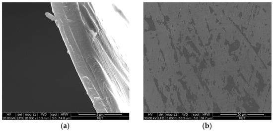
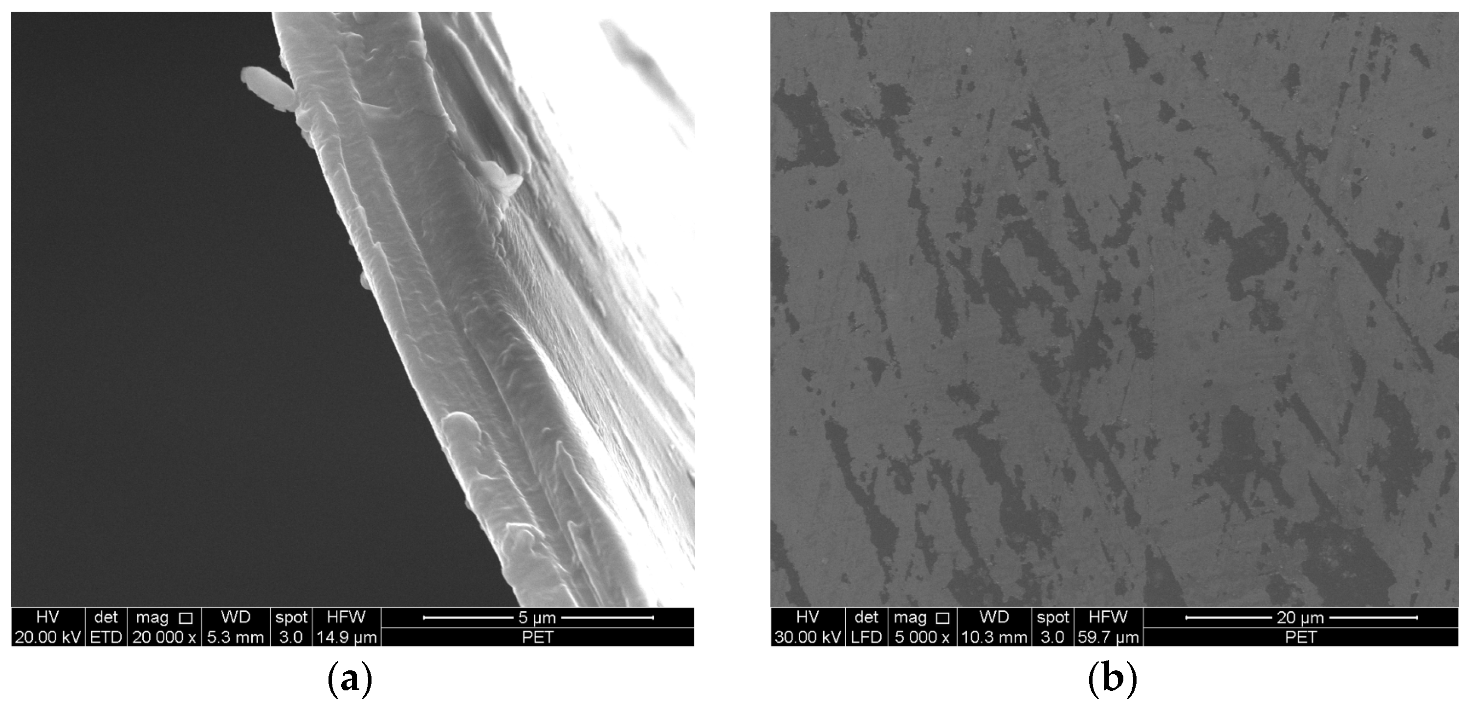
The ultrathin PET film cross-section and surface were morphologically characterized by using a scanning electron microscope (SEM, FEI Quanta 200 FEG, FEI Company - Thermo Fisher Scientific, Hillsboro, USA). According to the SEM characterization of ultrathin Mylar films, they showed uniform thickness and the presence of some surface defects, probably generated during post-processing treatments like rolling and cutting (see Figure 1a,b). The type of defect present on the film surface, namely terraces only a few nanometers thick, makes light scattering relatively ineffective. Scattering is more pronounced at interfaces with a significant refractive index mismatch; in the present case, such shallow terraces are unlikely to induce substantial light diffusion or diffuse reflection. Indeed, ultrathin PET films exhibit very high transparency in the visible region, with a transmittance of approximately 87.4%.
Figure 1.
SEM micrographs of ultrathin PET film showing cross-section (a) and surface (b).
The semi-crystalline nature of ultrathin PET film was established by X-ray diffraction (XRD, X’Expert PRO, PANalytical, Oxford, UK), and crystallinity degree was measured by differential scanning calorimetry (Discovery DSC 2500, Waters TA Instrument, New Castle, DE, USA).
3. Results
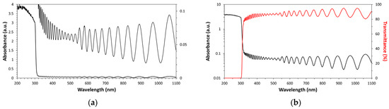
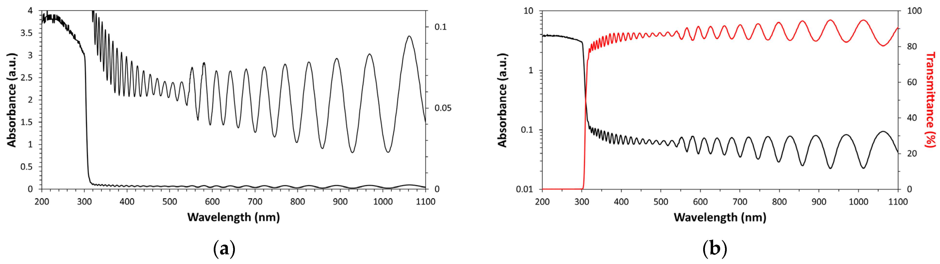
The interference phenomenon can be clearly detected in the high transparence region of the UV-Vis-NIR spectrum of ultrathin Mylar windows (see Figure 2). This physical phenomenon can be used for accurately measuring the film thickness (d is of the same order as the light wavelength). In particular, the following equation, derived from the interference maxima condition (i.e., 2n · d = m · λm) [9] in the case of a normally incident light beam, can be adopted [10]:
where d is the film thickness (in nm), Δm is the number of fringes (oscillations maxima) contained in the measurement interval, n is the average polymer refractive index, and λ1, λ2 are the wavelength limits of the measurement interval. According to the spectral oscillations in the ultrathin PET film optical spectrum shown in Figure 2, the film thickness obtained by using the above equation is 3.665 µm (an average refractive index value of 1.575 was used for calculation [11]).
d = Δm/2n(1/λ1 − 1/λ2),
Figure 2.
Optical spectrum of PET thin film expressed as absorbance (a) and in comparison with transmittance (b).
Since the PET films have a regular shape (i.e., thin circular disks), this optically measured thickness value was confirmed by a simple geometrical calculation based on density [12]. In particular, the following expression was used: d = m/(π · R2 · ρ), where m is the film weight, R the dish radius, and ρ the PET density. A value of 3.59 µm was found by using a PET density value of 1.3 g/cm3 [13]. This thickness value perfectly agrees with the value obtained by the interferometric measurement.
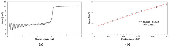
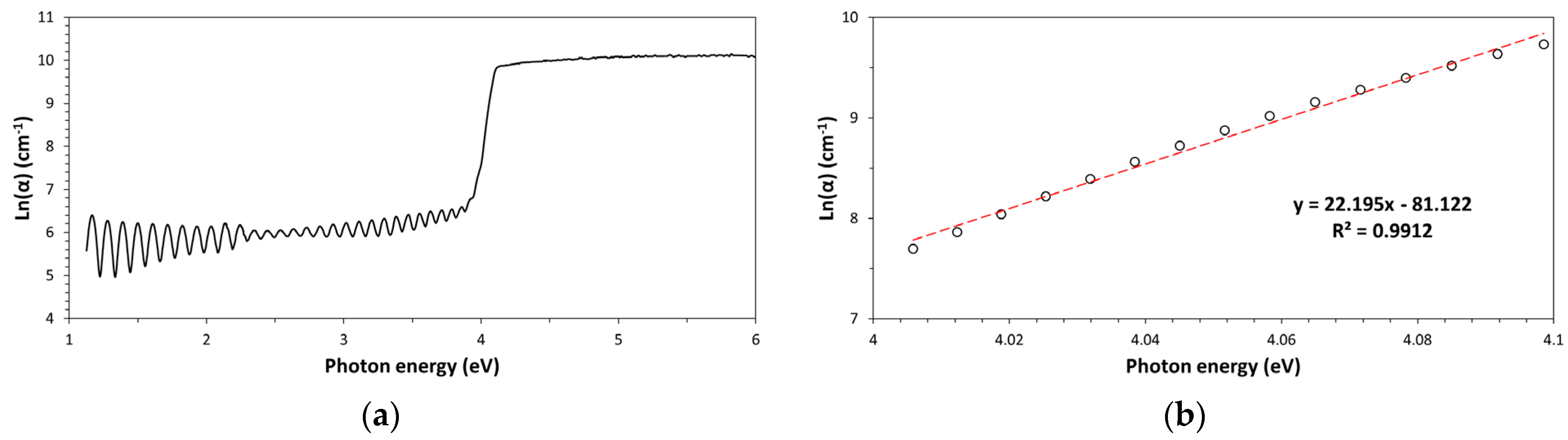
The interferometrically measured film thickness was used for calculating the optical absorption coefficient value. It must be pointed out that the absorbance data in the fundamental absorption edge (required for both Urbach and Tauc analysis) are readily accessible for the absorption coefficient calculation because spectral oscillations are present only in the high transparency region of the UV-Vis spectrum. In addition, the UV-Vis Analyst software allowed for the application of spectral smoothing, which reduced fringes in the spectrum for obtaining the total absorbance in this spectral region [14]. A graph of ln(α) vs. the photon energy is named the Urbach plot (see Figure 3a) and this special graph can be used to determine the amount of structural disorder in this ultrathin PET film [15]. Structural disorder is proportional to the Urbach energy, EU, which is given by the inverse of the slope of the linear part of the curve in the Urbach plot. In particular, this quantity can be obtained by linear regression analysis of this straight part of the graph; the least squares method was applied in the photon energy range: 3.942–4.119 eV (see Figure 3b). In general, the structural disorder obtained by the Urbach plot depends on all types of defects contained in this thin PET film like, for example, air bubbles (porosity), surface roughness, chemical impurities (molecular additives like plasticizers, UV-stabilizers, etc.) dissolved in the polymer, percentage of the amorphous phase, etc. However, with respect to perfectly processed films, most contributions can be neglected and the measured structural disorder corresponds mostly to the content of amorphous phase and to the degree of chain entanglement characterizing this amorphous phase. In the case of the ultrathin PET film, such calculation led to a value of (45.0 ± 0.03)meV (with R2 = 0.9912), which indicates quite low disorder, probably due to the semi-crystalline nature of this polymer.
Figure 3.
PET ultrathin film Urbach plot (a) and best fitting linear portion of the curve (b).
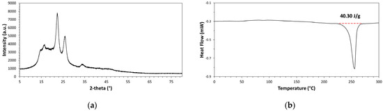
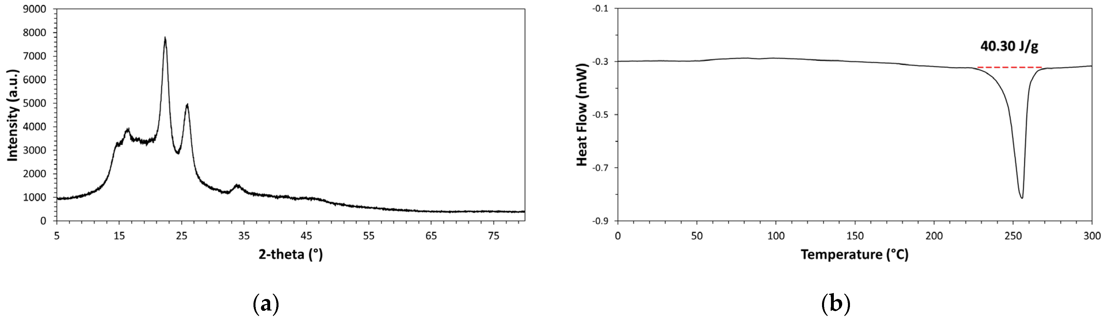
The small value of EU that was found by applying the Urbach approach to the spectral data of PET films can be well justified on the basis of the semi-crystalline nature of this thermoplastic polymer. Indeed, according to the XRD diffractogram shown in Figure 4a, the polymer contains an amorphous fraction, which causes the presence of a broad diffuse halo in the diffractogram, and a crystalline part, which causes the presence of three main peaks overlapped to the diffuse halo. These peaks are located at 2θ values of 16.5°, 22.4°, and 25.8°; such diffraction peak positioning is in good agreement with the literature information [16]. The FWHM of diffraction peaks is inversely proportional to the crystallite size (Scherrer equation [16]); consequently, the observed broad diffraction peaks suggest the presence of very small-sized crystallites in the polymer, which justifies the observed high optical transparency in the visible spectral region.
Figure 4.
XRD diffractogram (a) and DSC thermogram (b) of the ultrathin PET film.
The semi-crystalline nature of the ultrathin PET films was further confirmed by differential scanning calorimetry (DSC). According to the DSC thermogram shown in Figure 4b, the polymer presents an endothermic signal due to the melting of the crystalline phase and such a melting signal appears above the glass transition temperature (Tg), which is located at ca. 80.91 °C. The PET crystallites’ melting signal has an onset temperature at ca. 240 °C and a minimum at 255.59 °C. The DSC characterization of the polymer allows for measuring the melting enthalpy, which corresponded ca. 40.30 J/g. The calorimetric crystallinity degree, XC, of the polymeric sample can be determined by comparing the specimen melting enthalpy with the theoretical melting enthalpy of a hypothetical fully crystalline PET sample (ca. 140 J/g [17]) and such a ratio corresponded to ca. 28.8%. This crystallinity value agrees with the value characteristic of bi-axially oriented PET films [18]. Therefore, the small EU value experimentally found for the PET sample by the Urbach approach seems to be completely justified by its semi-crystalline nature.
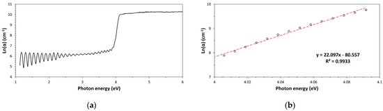
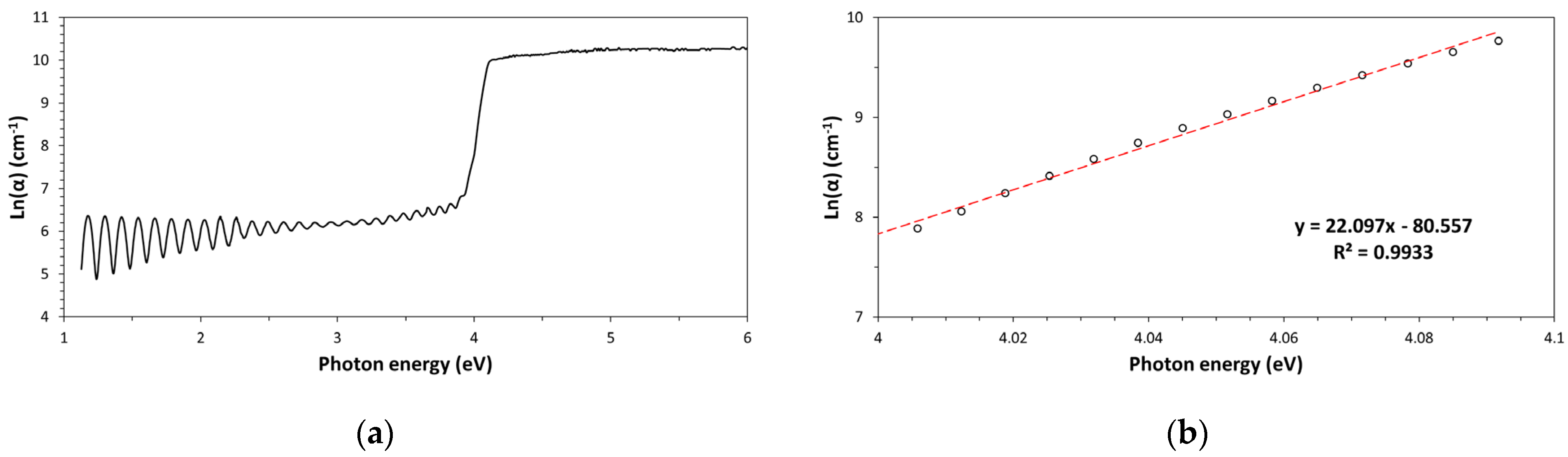
The same type of analysis (Urbach plot) was applied to a slightly mechanically deformed (i.e., manually stretched) ultrathin PET film (see Figure 5a,b). Manual stretching of ultrathin PET films caused plastic deformation characterized by a strain percentage of ca. 13.0% under approximately 12–14 N of applied load (films were deformed without breaking). Although mechanical deformation had the ability to modify the film thickness (it became 3.245 µm, corresponding to a decrease of 11.5%), the resulting Urbach energy value (EU = (45.2 ± 0.02) meV, with R2 = 0.9933) remained practically unchanged (see Figure 5b). Therefore, structural order did not change appreciably in this system as a result of the applied stress. Typically, polymer stretching has the effect of changing its crystalline morphology, causing transition from spherulitic to fibrous morphology. However, in the case of an ultrathin PET film sample for XRF application, the polymer is bi-axially oriented, which means it already has a fibrous morphology generated by the extrusion undergone during the film processing stage. Therefore, the subsequent manual stretching treatment should primarily lead to further increase macromolecular-chain orientation along the stretching direction; however, given the very small change observed in the Urbach energy, this effect does not occur to an appreciable extent.
Figure 5.
Urbach plot (a) and Urbach energy evaluation by linear regression analysis (b) for uniaxially stretched ultrathin PET film.
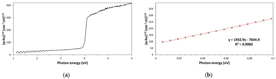
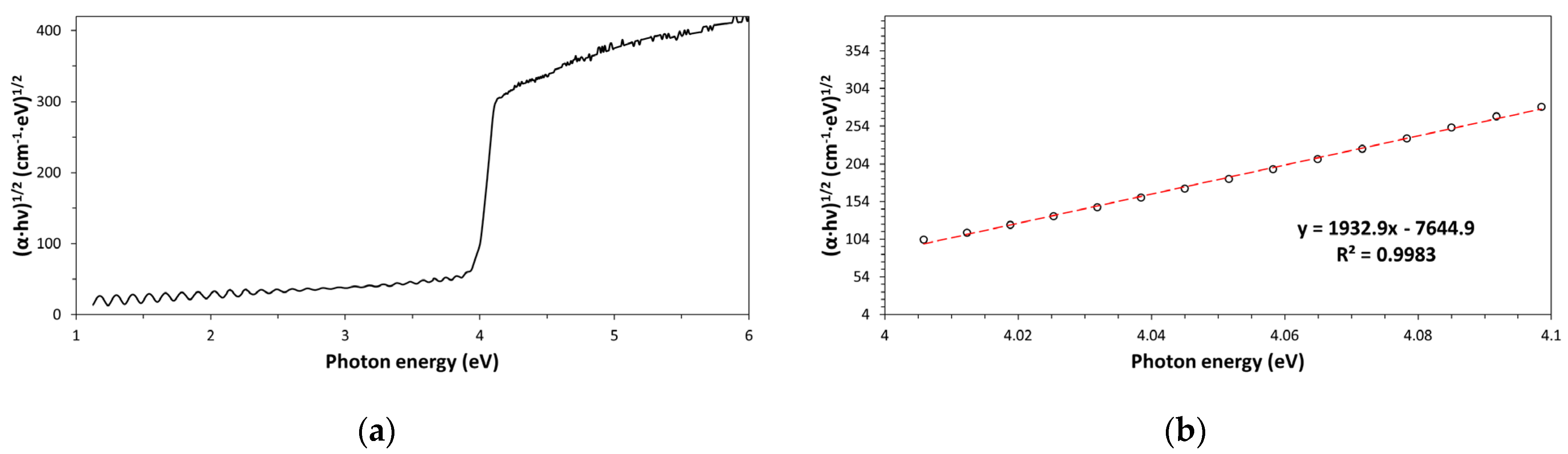
Ultraviolet radiation interacts with dielectric polymers causing photoexcitation (i.e., optical absorption due to electronic transitions). For some polymers like Kapton-H and polyetherimide (PEI), even visible photons may cause photoexcitations [19]. The cutoff wavelength was observed for these ultrathin PET films at ca. 300 nm. Consequently, photons with a wavelength lower than 300 nm are completely absorbed by the film, while transparency is close to 87.4% at wavelengths above 300 nm. The type of photoexcitation model and bandgap energy value (Eg) can be established optically by using the Tauc plot method. Eg is an important parameter for polymers since it is related to other physical properties like, for example, permittivity (Eg is inversely proportional to material permittivity, which is the capability of electric field instauration in a material and corresponds to the material polarizability [7]). In addition, accurate Eg determination could allow for dielectric polymer to be identified (however, identification is not possible with conjugated polymers like polyacetylene because of the Eg dependence on the conjugation extension and therefore on molecular weight). The bandgap energy was easily and accurately calculated by using the Tauc plot (see Figure 6a,b), which is a special graphical representation obtained by graphing the quantity (α · E)n as a function of the photon energy, E. The n value must be selected to allow for the linearization of the (α · E)n function, and n is 0.5 for an indirect electron transition model and 2 for a direct electron transition model (both models are for allowed electron transitions). Depending on the most convenient value found for n (the model with the best correlation factor), the type of electron transition model can be established. According to the following equation
the value of the intercept with the energy axis corresponds to Eg (indeed, for (α · E)n = 0, it results E = Eg). The intercept value is obtained from the best fitting equation of the linear portion of curve by using the following expression: -intercept/slope. In our case, an Eg value of (3.96 ± 0.002) eV (with R2 = 0.9995, in the photon energy range: 3.948–4.126 eV) was found for an indirect electron transition model (n = 0.5), which perfectly agrees with information provided in the literature [20,21].
(α · E)n = B · (E − Eg),
Figure 6.
PET ultrathin film Tauc plot (a) and best fitting linear section of the curve (b).
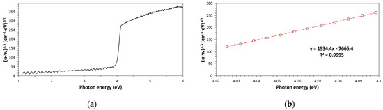
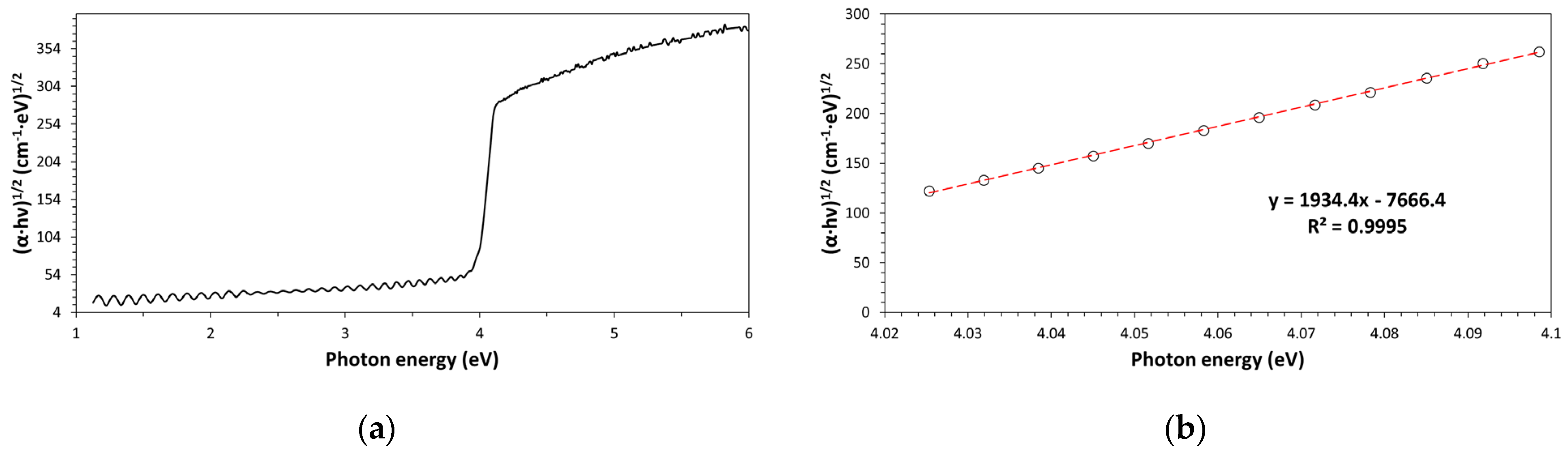
Manual stretching allowed for simulating mild uniaxial mechanical stresses that polymeric films typically experience in service or during industrial uses. This investigation has shown that Eg is a polymer physical characteristic, which remains practically unchanged after the mild mechanical damage undergone by this material (see Figure 7a,b); in particular, according to this least square analysis, Eg = (3.96 ± 0.02) eV after stretching. Indeed, Eg depends mainly on atom types (composition) and the way atoms are chemically bonded together (constitution) in the repeating unit.
Figure 7.
Tauc plot (a) and bandgap energy estimation by linear regression analysis (b) for a stretched ultrathin PET film.
According to the achieved experimental results, dielectric polymers at solid-state have a band structure principally depending on the polymer chemical composition/constitution and only marginally on crystallinity, molecular weight, and texture (i.e., fibrous/spherulitic morphology).
4. Conclusions
The optical absorption coefficient of dielectric polymers quantifies how much light these synthesis materials absorb per unit distance. This important optical parameter allows for analyzing band structure and structural disorder in dielectric polymers and provides many other important physical insights into these materials (e.g., cutoff wavelength, transparency percentage, spectral opacity range, etc.). In particular, the behavior of absorption coefficient with photon energy, E = hν, allows for measuring the optical bandgap energy, Eg, and the Urbach energy, EU, by using the Tauc method and Urbach plot, respectively. The behavior of the α(E) function can be obtained by combining interferometric measurements based on the analysis of spectral oscillations present in the high transparency region of the UV-Vis-NIR spectra with absorbance data contained in the area of fundamental absorption edge in the same spectra. This approach is quite general since spectral oscillations are of low intensity but can always be detected in the UV-Vis spectra of ultrathin films. Owing to the convenient refractive index value and high surface flatness/smoothness, ultrathin Mylar (PET) films were selected as model polymeric material for this type of analysis and the changes in the optical bandgap and Urbach energy upon stretching were investigated, showing a scarce dependence of these parameters on mechanical damage of films.
Funding
This research received no external funding.
Institutional Review Board Statement
Not applicable.
Informed Consent Statement
Not applicable.
Data Availability Statement
Data are available from the author.
Conflicts of Interest
The authors declare no conflicts of interest.
References
- Mohammed, H.A.; Mohammed, P.A.; Aziz, S.B. Investigation of optical band gap in PEO-based polymer composites doped with green-synthesized metal complexes using various models. RSC Adv. 2025, 15, 23319–23341. [Google Scholar] [CrossRef] [Scilit] [PubMed]
- Migahed, M.D.; Zidan, H.M. Influence of UV-irradiation on the structure and optical properties of polycarbonate films. Curr. Appl. Phys. 2006, 6, 91–96. [Google Scholar] [CrossRef] [Scilit]
- Chen, L.; Batra, R.; Ranganathan, R.; Sotzing, G.; Cao, Y.; Ramprasad, R. Electronic structure of polymer dielectrics: The role of chemical and morphological complexity. Chem. Mater. 2018, 30, 7699–7706. [Google Scholar] [CrossRef] [Scilit]
- Chen, L.; Huan, T.D.; Ramprasad, R. Electronic structure of polyethylene: Role of chemical, morphological and interfacial complexity. Sci. Rep. 2017, 7, 6128. [Google Scholar] [CrossRef] [Scilit] [PubMed]
- Hoffmann, R. How chemistry and physics meet in the solid states. Angew. Chem. Int. Ed. Engl. 1987, 26, 846–878. [Google Scholar] [CrossRef] [Scilit]
- Zanatta, A.R. Revisiting the optical bandgap of semiconductors and the proposal of a unified methodology to its determination. Sci. Rep. 2019, 9, 11225. [Google Scholar] [CrossRef] [Scilit] [PubMed]
- Lamichhane, A. Absorption in narrow and wide gap materials. Heliyon 2023, 9, e21507. [Google Scholar] [CrossRef] [Scilit] [PubMed]
- Wada, O.; Ramachari, D.; Yang, C.-S.; Pan, C.-L. Interrelationship among dielectric constant, energy band parameters and iconicity in multi-component oxide glasses revealed by optical- and THz-band spectroscopy. J. Non-Cryst. Solids 2021, 573, 121135. [Google Scholar] [CrossRef] [Scilit]
- Dahshan, A.; Aly, K.A. Determination of the thickness and optical constants of amorphous Ge-Se-Bi thin films. Philos. Mag. 2009, 89, 1005–1016. [Google Scholar] [CrossRef] [Scilit]
- Swanepoel, R. Determination of the thickness and optical constant of amorphous silicon. J. Phys. E Sci. Instrum. 1983, 16, 1214–1222. [Google Scholar] [CrossRef] [Scilit]
- Zhang, X.; Qiu, J.; Li, X.; Zhao, J.; Liu, L. Complex refractive indices measurements of polymers in visible and near-infrared bands. Appl. Opt. 2020, 59, 2337–2344. [Google Scholar] [CrossRef] [Scilit] [PubMed]
- Hulbers, P.D.T.; Shah, D.O. Multispectral determination of soap film thickness. Langumir 1997, 13, 5995–5998. [Google Scholar] [CrossRef] [Scilit]
- Thompson, A.B.; Woods, D.W. Density of amorphous polyethylene terephthalate. Nature 1955, 176, 78–79. [Google Scholar] [CrossRef] [Scilit]
- Schade, U.; Ritter, E.; Bawagan, A.D.O.; Beckmann, J.; Aziz, E.F. Far-infrared absorption measurements on thin polymer films. In 2014 39th International Conference on Infrared Millimiter, and Terahertz waves (IRMMW-THz); IEEE: Piscataway, NJ, USA, 2014. [Google Scholar]
- Okutan, M. The effect of grinding on optical band gap and urbach energy of polypyrrole/graphene composites. Eskiseh. Tech. Univ. J. Sci. Tech. A-Appl. Sci. Eng. 2023, 24, 309–323. [Google Scholar] [CrossRef] [Scilit]
- Johnson, J.E. X-ray diffraction studies of the crystallinity in polyethylene terephthalate. J. Appl. Sci. 1959, 2, 205–209. [Google Scholar] [CrossRef] [Scilit]
- Viora, L.; Combeau, M.; Pucci, M.F.; Perrin, D.; Liotier, P.-J.; Bouvard, J.-L.; Combeaud, C. A comparative study on crystallization for virgin and recycled polyethylene terephthalate (PET), multiscala effects on physico-mechanical properties. Polymers 2023, 15, 4613. [Google Scholar] [CrossRef] [Scilit] [PubMed]
- Bandla, S.; Allahkarami, M.; Hanan, J.C. Out-of-plane orientation and crystallinity of biaxially stretched polyethylene terephthalate. Powder Diffr. 2014, 29, 123–126. [Google Scholar] [CrossRef] [Scilit]
- Carotenuto, G. Study of the Kapton-H fundamental absorption edge and tailing behavior. Eng. Proc. 2025, 105, 7. [Google Scholar] [CrossRef] [Scilit]
- Musiatowicz, M.; Turek, M.; Drozdziel, A.; Pyszniak, K.; Grudzinski, W. Modification of optical, electronic and microstructural properties of PET by 150 keV Cs+ irradiation. Adv. Sci. Technol. Res. J. 2022, 16, 11–19. [Google Scholar] [CrossRef] [Scilit] [PubMed]
- Sadoh, A.; Hossain, S.; Fereira, S.; Ravindra, N.M. Optical properties of low-refractive index polymers. Mater. Sci. Eng. 2022, 6, 68–76. [Google Scholar] [CrossRef] [Scilit]
Disclaimer/Publisher’s Note: The statements, opinions and data contained in all publications are solely those of the individual author(s) and contributor(s) and not of MDPI and/or the editor(s). MDPI and/or the editor(s) disclaim responsibility for any injury to people or property resulting from any ideas, methods, instructions or products referred to in the content. |
© 2026 by the author. Licensee MDPI, Basel, Switzerland. This article is an open access article distributed under the terms and conditions of the Creative Commons Attribution (CC BY) license.