


Abstract
In small ruminants, up to 40% of fertilized ova are lost during early gestation due to inadequate progesterone. This study evaluated post-transfer progestogen supplementation in goats. A total of 207 thawed embryos were transferred into 111 recipients assigned to progestogen-supplemented (n = 37) or non-progestogen-supplemented (n = 74) groups. Pregnancy and embryo survival did not differ between treatments (p > 0.05). However, fetal and neonatal survival were significantly lower in supplemented goats (29%) compared with non-supplemented goats (100%; p < 0.05). Infectious causes were excluded. Possible mechanisms postulated in fetal losses comprise a sharp decline in progesterone after sponge removal or increased oxytocin and prostaglandin due to vaginal mechanical stimulation, inducing luteolysis. In conclusion, intravaginal progestogen supplementation is not recommended for recipient goats in embryo transfer programs.
1. Introduction
In sheep and goats, it has been estimated that 30–40% of fertilized ova are lost during the first three weeks of gestation [1]. Most embryonic mortality is believed to occur during the peri-implantation period and may be associated with inadequate circulating progesterone concentration and its effect on uterine function, failures in conceptus development, or deficiencies in maternal recognition of pregnancy, implantation, and/or placentation [2]. Different therapeutic strategies aimed at improving embryo survival during early gestation include progesterone supplementation through intravaginal devices or the induction of accessory corpora lutea via administration of hCG or GnRH [3].
Luteal insufficiency has also been described as a major cause of reduced pregnancy rates following embryo transfer [4]. In goats, information regarding the effects of progesterone supplementation on embryo survival is still limited [5]. In that study, the authors performed embryo transfer using fresh and frozen demi-embryos into recipient does. Recipients were treated with norgestomet implants (3 mg) on Day 10 after embryo transfer and again two weeks later. In 6 out of 55 treated females, luteal regression occurred despite the presence of a viable conceptus; however, 5 of these pregnancies were carried to term due to the norgestomet implants, resulting in five live-born kids. Another study [6] showed that supplementation with fluorogestone acetate devices (45 mg), from the time of embryo transfer until Day 45 post-estrus, resulted in pregnancy and embryo survival rates of 69% and 73%, respectively, compared with 23% and 19% in untreated females.
Based on these findings, the aim of the present study was to evaluate the effect of prolonged progestogen treatment on pregnancy rate and embryo, fetal, and neonatal survival in goats receiving cryopreserved embryos.
2. Materials and Methods
2.1. Study Site and Experimental Animals
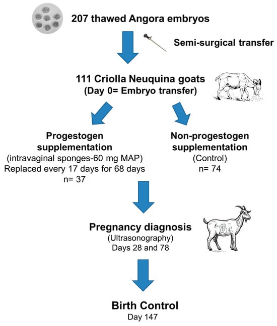
At the Experimental Field of Pilcaniyeu, Río Negro, Argentina (41°07′23″ S, 71°15′12″ W), during the breeding season, a total of 207 Angora embryos imported from the Republic of South Africa were semi-surgically transferred into 111 Criolla Neuquina recipient does (Figure 1). Recipients were 4–6 years old, with a mean body weight of 34.0 ± 1.7 kg and a body condition score of 2.6 ± 0.1 (subjective scale: 1 = emaciated, 5 = obese) [7]. All animals were clinically healthy, maintained under natural grazing conditions, and had free access to water.
Figure 1.
Schematic diagram representing the procedures used in embryo transfer and progestogen supplementation (intravaginal sponges containing progestogen) in recipient does.
2.2. Estrus Synchronization, Embryo Thawing, and Transfer
Estrus synchronization, embryo thawing, and transfer procedures were performed as described by [8]. Briefly, recipients received a 17-day progestogen treatment (60 mg MAP, Progespon®, Zoetis, Villa Adelina, Argentina) and a dose of 100 IU eCG (Novormon®, Zoetis, Argentina) at sponge removal.
For embryo thawing, each straw was removed from liquid nitrogen, shaken in air for 10 s, and immersed in a water bath at 30 °C for 1 min. The straw content was then expelled into a Petri dish, and embryos were transferred to another Petri dish containing holding medium (Syngro® Holding, Vetoquinol, Brisbane, Australia) for 5 min.
Embryo transfer was performed 8.5 days after sponge removal and eCG administration (Day 0 = day of embryo transfer). Prior to the procedure, recipient females were fasted for 24 h. Before transfer, the presence of at least one corpus luteum (CL) per doe was confirmed by laparoscopy, and only females with at least one CL were used.
Embryo transfer was carried out using a semi-surgical technique. Under general anesthesia with xylazine (4 mg, IM; Kensol®, König, Canelones, Uruguay) and ketamine (12.5 mg, IM; Ketonal®, Richmond, Grand Bourg, Argentina) and local anesthesia with lidocaine hydrochloride (2 mL, 2%; Over, San Vicente, Argentina), a small laparotomy (≈1 cm) was performed along the midline, cranial to the udder. The uterine horn ipsilateral to the ovary bearing the CL was exteriorized using atraumatic forceps. After puncture with an 18 G needle, one or two embryos were deposited into the uterine lumen using a piston-type embryo transfer pipette (Assipettor®, Minitüb, Tiefenbach, Germany) [9]. The uterine horn was then returned to the abdominal cavity and the incision was closed by suture.
Embryos were transferred within 20 min after thawing. Single or double transfers were performed without prior recipient selection due to logistical constraints during thawing.
All thawed embryos were used, as morphological evaluation [10] classified them as grade 1 (excellent) or grade 2 (good). All procedures were conducted under standardized conditions and by the same operators.
Finally, a prophylactic antibiotic treatment was administered, consisting of penicillin–streptomycin (10,000 IU/kg, IM; Estreptopendiben®, Biogénesis Bagó, Garin, Argentina), along with local application of gentamicin (Genmicin®, Over, Argentina) at the incision site.
2.3. Experimental Treatments
Immediately after embryo transfer, recipient does were randomly assigned to one of two treatments:
- -
- Progestogen supplementation: intravaginal sponges containing progestogen (60 mg MAP, Progespon®, Zoetis, Argentina) for 68 days, replaced every 17 days, according to a modified protocol described by [6]; (n = 37). The 68-day progestogen supplementation period (approximately 75 days after estrus) was established considering that placentome growth, in terms of mass and net cellular proliferation, reaches a maximum by approximately day 80 of gestation, suggesting that placental size and function during later gestation may thereafter be only minimally influenced in goats [11]. This duration also considered the permanence time of the sponge in the vagina, thus avoiding replacement with a new one.
- -
- No progestogen supplementation: no supplemented progestogen treatment; (n = 74) (Figure 1).
In the supplemented group, 30 recipients received double transfers and 7 recipients received single transfers, while in the non-supplemented group, 66 and 8 recipients received double or single transfers, respectively.
The unequal number of females per treatment was due to the assignment of a larger proportion of recipients to the conventional embryo transfer protocol used at the farm.
Pregnancy diagnosis was performed by transrectal ultrasonography on Days 28 and 78 after embryo transfer (Aloka SSD-500, Tokio, Japan) using a 5 MHz linear-array transducer, to assess embryo and fetal survival and identifying twin pregnancies. Neonatal survival was monitored at kidding.
Pregnancy rate was calculated as pregnant recipients per total recipients, embryo survival as viable embryos per transferred embryos, fetal survival as viable fetuses per viable embryos, and neonatal survival as kids born alive per viable embryos.
2.4. Statistical Analysis
Statistical analyses were performed using R software (2020). Proportions were compared using Fisher’s exact test, and pairwise comparisons were conducted with Holm correction for multiple testing. A value of p < 0.05 was considered statistically significant.
3. Results
Pregnancy rate and embryo survival did not differ significantly between treatments (p > 0.05). However, fetal and neonatal survival were lower in the progestogen-supplemented group compared to the non-supplemented group (Table 1).
Table 1.
Effect of intravaginal progestogen supplementation on pregnancy rate and embryo, fetal, and neonatal survival in recipient goats transferred with cryopreserved embryos (progestogen replaced every 17 days for 68 days vs. no progestogen).
Unexpected fetal losses were observed between 7 and 10 days after intravaginal sponge removal. Among the 11 fetal losses recorded in the progestogen-supplemented group, three abortions were directly observed at 7, 9, and 10 days after sponge removal. The remaining fetal losses were inferred from the absence of viable fetuses at Day 78 post embryo transfer, indicating that all losses occurred between days 28 and 78 post transfer.
No definitive cause of fetal loss could be determined based on clinical history, necropsy findings, or laboratory analyses performed by the Animal Health Group (INTA Bariloche). Serum samples tested negative for CpHV-1 (Animal Health, INTA Balcarce) and Brucella melitensis (Animal Health, INTA Bariloche).
4. Discussion
The present study shows that long-term progestogen supplementation did not affect pregnancy establishment or early embryo survival but was associated with a marked reduction in fetal and neonatal survival. Notably, fetal losses occurred shortly after progestogen withdrawal, suggesting a potential relationship between treatment cessation and pregnancy failure during mid-gestation.
Previous studies in different species have reported reduced pregnancy rates following progesterone supplementation, particularly during early gestation. In those cases, supplementation was applied shortly after mating or artificial insemination (approximately Days 1–9), and the proposed mechanisms included impaired ovulation, altered sperm transport, and negative effects on corpus luteum (CL) development [12,13]. In humans, supraphysiological progesterone levels in early pregnancy (e.g., >20 ng/dL on Day 19 of the cycle) have been associated with estrogen–progesterone imbalance [14] and embryo–uterine asynchrony [15].
In contrast, in the present study, progesterone supplementation was initiated later (around Day 7 of gestation) and maintained for a prolonged period (up to Day 74). To our knowledge, no similar reports have been described in the literature, since most earlier studies evaluated progesterone supplementation only during early gestation and for substantially shorter periods. In the present study, however, circulating progesterone concentration was not measured; therefore, it could not be confirmed whether progestogen supplementation effectively increased or maintained systemic progesterone levels, nor whether individual variation in hormonal response existed among recipient goats.
Another limitation of the present study is that not all abortion events could be directly observed, since recipient goats grazed under extensive field conditions during the day and were confined only at night. Consequently, although three abortions were directly detected 7–10 days after sponge removal (Days 81–84 of gestation), the exact timing of the remaining fetal losses could not be determined and was inferred from the absence of viable fetuses at Day 78 post embryo transfer. Therefore, individual variation in the timing of fetal losses could not be fully assessed.
Finally, although the proportion of single and double embryo transfers was not completely balanced between groups (19% vs. 11% single transfers in progestogen-supplemented and non-supplemented goats, respectively), this difference was relatively small and is unlikely to fully explain the marked difference observed in fetal survival, particularly considering that no fetal losses occurred in the non-supplemented group. Nevertheless, a potential effect of embryo number per recipient cannot be completely excluded and should be considered a limitation of the study.
In goats, the relatively high incidence of abortion compared to other species has been linked to their strong dependence on the CL for pregnancy maintenance [16], a feature also associated with late-term pregnancy losses [17].
Several mechanisms may explain the observed fetal losses. These include stress associated with animal handling, an abrupt decline in progesterone levels, and/or alterations in oxytocin and prostaglandin secretion induced by mechanical vaginal stimulation during sponge removal.
Stress factors-nutritional, management-related (e.g., confinement), or climatic (e.g., storms, snowfall)-may increase maternal cortisol levels and trigger abortions [18,19]. This explanation appears unlikely in the present study, as animals were accustomed to handling and confinement, and sponge removal was conducted carefully, minimizing stress (short confinement time, no use of dogs).
Another possible mechanism is a sharp decline in progesterone levels following sponge removal. This could result from interference of exogenous progesterone with the development and function of the endogenous CL, as previously suggested [12,13]. Nevertheless, evidence remains inconclusive, since [5] reported successful term pregnancies in 5 out of 6 goats supplemented with progesterone between Days 10 and 25 of the estrous cycle, despite CL regression.
In goats, rapid declines in progesterone concentrations are known to compromise pregnancy maintenance. Induced luteolysis with PGF2α causes a marked decrease in circulating progesterone within 24 h, followed by abortion or premature parturition [20,21]. Therefore, the abrupt withdrawal of exogenous progesterone supplementation, as performed in the present study, could be considered a plausible mechanism contributing to the observed fetal losses.
Since an abrupt withdrawal of exogenous progesterone supplementation was implemented in this work, it could be hypothesized that a gradual reduction in progestogen exposure may allow progressive endocrine adaptation and reduce potential impairment of CL function associated with sudden withdrawal. To our knowledge, studies directly comparing the effects of different progesterone withdrawal strategies during pregnancy are not currently available. Therefore, future studies comparing abrupt vs. gradual reduction in progestogen exposure, together with monitoring of circulating progesterone concentrations, may help clarify whether progesterone withdrawal dynamics may influence fetal viability.
A third explanation involves increased circulating oxytocin levels in response to mechanical vaginal stimulation during sponge removal [22]. Small amounts of exogenous oxytocin have been shown to induce uterine prostaglandin release, which acts as a luteolytic agent in sheep and goats [16,23]. However, oxytocin response to vaginal stimulation has been reported to be reduced under gestational progesterone concentrations [24], which may attenuate this mechanism.
5. Conclusions
Although intravaginal progestogen supplementation post embryo transfer did not affect early pregnancy or embryo survival, it was associated with significantly lower fetal and neonatal survival. This effect may be related to the abrupt withdrawal of exogenous progesterone in mid-gestation. Therefore, this treatment is not recommended for routine use in goat embryo transfer programs.
Author Contributions
Conceptualization, M.C. and A.G.; methodology, M.B.-G., A.M. and J.F.; formal analysis, N.G.; investigation, M.C. and A.G.; writing—original draft preparation, A.M. and M.B.-G.; writing—review and editing, J.F., N.G. and M.C. All authors have read and agreed to the published version of the manuscript.
Funding
This research received no external funding.
Institutional Review Board Statement
The present study complies with national and international standards regarding the use of animals for experimentation [CICUAE INTA—PATNOR-Res. 533/16 Disp. CRPN 66/17].
Informed Consent Statement
Not applicable.
Data Availability Statement
Data is unavailable due to privacy restrictions.
Acknowledgments
The authors have reviewed and edited the output and take full responsibility for the content of this publication. The authors thank the INTA Pilcaniyeu staff for their assistance with animal care.
Conflicts of Interest
The authors declare no conflicts of interest.
References
- Nancarrow, C.D. Embryonic mortality in the ewe and doe. In Embryonic Mortality in Domestic Species; Zavy, M.T., Geisert, R.D., Eds.; CRC Press: Boca Raton, FL, USA, 1994; pp. 79–97. [Google Scholar]
- Kittok, R.J.; Stellflug, J.N.; Lowry, S.R. Enhanced progesterone and pregnancy rate after gonadotropin administration in lactating ewes. J. Anim. Sci. 1983, 56, 652–655. [Google Scholar] [CrossRef] [Scilit] [PubMed]
- Fernández, J.; Bruno-Galarraga, M.; Soto, A.; de la Sota, R.; Cueto, M.; Lacau, I.; Gibbons, A. Hormonal therapeutic strategy on the induction of accessory corpora lutea in relation to follicle size and on the increase of progesterone in sheep. Theriogenology 2018, 105, 184–188. [Google Scholar] [CrossRef] [Scilit] [PubMed]
- Armstrong, D.T.; Pfitzner, A.P.; Warnes, G.M.; Ralph, M.M.; Seamark, R.F. Endocrine responses of goats after induction of superovulation with PMSG and FSH. J. Reprod. Fertil. 1983, 67, 395–401. [Google Scholar] [CrossRef] [Scilit] [PubMed]
- Beckett, D.M.; Oppenheim, S.M.; Moyer, A.L.; Bondurant, R.H.; Rowe, J.D.; Anderson, G.B. Progestin implants can rescue demi-embryo pregnancies in goats: A case study. Theriogenology 1999, 51, 1505–1511. [Google Scholar] [CrossRef] [Scilit] [PubMed]
- D’Alessandro, A.G.; Martemucci, G. Superovulatory response to gonadotrophin FSH/LH treatment and effect of progestin supplement to recipients on survival of transferred vitrified embryos in goats. Theriogenology 2016, 85, 296–301. [Google Scholar] [CrossRef] [Scilit] [PubMed]
- Russel, A.J.F.; Doney, J.M.; Gunn, R.G. Subjective assessment of body fat in live sheep. J. Agric. Sci. 1969, 72, 451–454. [Google Scholar] [CrossRef] [Scilit]
- Cueto, M.; Bruno-Galarraga, M.; Fernandez, J.; Giovannini, N.; Lentini-Ordoqui, A.; Gibbons, A. Reproductive efficiency of the embryo transfer of frozen Angora embryos imported from South Africa. In Proceedings of the III Congreso Argentino de Producción Caprina, La Rioja, Argentina, 4–6 November 2021. [Google Scholar]
- Green, R.E.; Santos, B.F.S.; Sicherle, C.C.; Landim-Alvarenga, F.C.; Bicudo, S.D. Viability of OPS vitrified sheep embryos after direct transfer. Reprod. Domest. Anim. 2009, 44, 406–410. [Google Scholar] [CrossRef] [Scilit] [PubMed]
- Stringfellow, D.A.; Givens, M.D. Manual of the International Embryo Transfer Society (IETS), 4th ed.; IETS: Champaign, IL, USA, 2010. [Google Scholar]
- Yan, Q.; Xu, J.; Wu, X.; Su, D.; Tan, Z. Stage-specific feed intake restriction differentially regulates placental traits and proteome of goats. Br. J. Nutr. 2018, 119, 1119–1132. [Google Scholar] [CrossRef] [Scilit] [PubMed]
- van Cleeff, J.; Macmillan, K.L.; Drost, M.; Lucy, M.C.; Thatcher, W.W. Effects of administering progesterone at selected intervals after insemination of synchronized heifers on pregnancy rates. Theriogenology 1996, 46, 1117–1130. [Google Scholar] [CrossRef] [Scilit] [PubMed]
- Soede, N.M.; Bouwman, E.G.; Van der Laan, I.; Hazeleger, W.; Jourquin, J.; Langendijk, P.; Kemp, B. Progestagen supplementation during early pregnancy does not improve embryo survival in pigs. Reprod. Domest. Anim. 2012, 47, 835–841. [Google Scholar] [CrossRef] [Scilit]
- Harini, C.; Sainath, S.B.; Reddy, P.S. Progesterone administration induces preimplantation embryonic loss in mice. Fertil. Steril. 2009, 91, 2137–2141. [Google Scholar] [CrossRef] [Scilit]
- Kofinas, J.D.; Blakemore, J.; McCulloh, D.H.; Grifo, J. Serum progesterone levels and pregnancy outcomes. J. Assist. Reprod. Genet. 2015, 32, 1395–1399. [Google Scholar] [CrossRef] [Scilit] [PubMed]
- Purohit, G.N.; Shekher, C.; Kumar, P.; Solanki, K. Induced termination of pregnancy in domestic farm animals. Iran. J. Appl. Anim. Sci. 2012, 2, 1–12. [Google Scholar]
- Cueto, M.; Gibbons, A.; Abad, M. Reproducción en caprinos. In Módulo de la Materia de Pequeños Rumiantes; FAUBA: Buenos Aires, Argentina, 2000. [Google Scholar]
- van Rensburg, S. Reproductive Physiology and Endocrinology of Normal and Habitually Aborting Angora Goats. Ph.D. Thesis, University of Pretoria, Pretoria, South Africa, 1970. Available online: http://hdl.handle.net/2263/50966 (accessed on 1 March 2023).
- Bedotti, D.; Rossanigo, C. Manual de Reconocimiento de Enfermedades del Caprino; Ediciones INTA: Ciudad Autónoma de Buenos Aires, Argentina, 2011. [Google Scholar]
- Bosu, W.T.K.; Garibay, J.S.; Barker, C.A.V. Peripheral plasma levels of progesterone in pregnant goats and in pregnant goats treated with prostaglandin F2α. Theriogenology 1979, 11, 131–148. [Google Scholar] [CrossRef] [Scilit]
- Bretzlaff, K.N.; Weston, P.G.; Hixon, J.E.; Ott, R.S. Plasma luteinizing hormone and progesterone concentrations in goats with estrous cycles of normal or short duration after prostaglandin F2α administration during diestrus or pregnancy. Am. J. Vet. Res. 1988, 49, 939–943. [Google Scholar] [CrossRef] [Scilit]
- Roberts, J.S. Cyclical fluctuations in reflexive oxytocin release. Biol. Reprod. 1975, 13, 314–317. [Google Scholar] [CrossRef] [Scilit]
- McCracken, J.A.; Barcikowski, B.; Carlson, J.C.; Green, K.; Samuelsson, B. Prostaglandin F2α in luteum regression. Adv. Biosci. 1972, 9, 599–624. [Google Scholar] [CrossRef] [Scilit]
- Roberts, J.S.; Share, L. Oxytocin response to vaginal stimulation. Endocrinology 1968, 83, 272–278. [Google Scholar] [CrossRef] [Scilit] [PubMed]

Disclaimer/Publisher’s Note: The statements, opinions and data contained in all publications are solely those of the individual author(s) and contributor(s) and not of MDPI and/or the editor(s). MDPI and/or the editor(s) disclaim responsibility for any injury to people or property resulting from any ideas, methods, instructions or products referred to in the content. |
© 2026 by the authors. Licensee MDPI, Basel, Switzerland. This article is an open access article distributed under the terms and conditions of the Creative Commons Attribution (CC BY) license.