


Abstract
Dairy cows are frequently affected by postpartum uterine infections, such as metritis and endometritis, which negatively impact fertility and cause significant economic losses. The extensive use of antibiotics may alter the reproductive microbiota and contribute to the emergence of antibiotic resistance. This study aimed to isolate lactic acid bacteria (LAB) from healthy vaginal samples of cows and evaluate their antimicrobial activity against uterine pathogens such as Escherichia coli and Salmonella spp. Phenotypic characterization and antibiotic susceptibility testing were performed, along with antagonistic assays using both overlay and well diffusion methods. The results demonstrated strain-dependent inhibitory activity against E. coli, with inhibition zones ranging from 2 to 4 mm depending on the LAB strain and the method used, whereas no inhibitory activity was observed against Salmonella. These findings support the potential application of selected LAB strains in bovine reproductive health management. However, further in vitro and in vivo studies are required to confirm their efficacy, safety, and stability under physiological conditions.
1. Introduction
Livestock breeding represents a fundamental pillar of agricultural systems in Algeria, playing a crucial socio-economic role for farmers. Beyond production, livestock contributes to resilience against biological, climatic, and economic risks [1]. In this context, bovine production, particularly milk and meat, remains essential for human nutrition, public health, and rural livelihoods, especially in low- and middle-income countries [2].
Over the past decade, the Algerian cattle population has shown a gradual increase, rising from 1,607,890 heads in 2006 to 2,171,633 heads in 2017. However, this growth has been accompanied by a concerning decline in the proportion of dairy cows, decreasing from 52.71% to 44.74% during the same period [3]. This imbalance reflects underlying challenges affecting the productivity and sustainability of the dairy sector.
Among these challenges, reproductive disorders represent a major constraint. High rates of abortion and genital tract pathologies significantly impact herd performance and profitability. Notably, studies have reported that 16.49% of slaughtered cows were pregnant, most at early gestation stages, highlighting reproductive mismanagement [4].
Postpartum uterine infections constitute one of the most critical issues in dairy cattle. Indeed, bacterial contamination of the uterine lumen is almost inevitable after calving, affecting more than 90% of cows within the first two weeks [5]. Although most animals eliminate these microorganisms within five weeks, persistence occurs in 10–17% of cases, leading to pathological conditions [6].
The primary pathogens involved include E. coli, Trueperella pyogenes, Fusobacterium necrophorum, and Prevotella spp. [7,8,9]. These infections are classified as metritis when occurring within the first 20 days postpartum, and as endometritis after 21 days [10]. Such conditions significantly impair reproductive performance, resulting in reduced conception rates, prolonged calving intervals, and increased culling risk [11]. Furthermore, pathogenic bacteria such as E. coli can disrupt the uterine microbiota, leading to dysbiosis, which exacerbates reproductive inefficiency [12,13].
Conventional treatments, including antibiotics, antiseptics, and hormonal therapies, have been widely used to control these infections. However, their effectiveness is often limited by side effects, antimicrobial resistance by reducing Virulence Factor Genes (VFGs), and increasing Antibiotic Resistance Genes (ARGs), and the presence of drug residues in animal products [14,15]. These limitations highlight the urgent need for safer and more sustainable alternatives.
In this regard, probiotics have emerged as a promising approach. Defined as live microorganisms that confer health benefits to the host [16], probiotics, particularly lactic acid bacteria (LAB), have demonstrated significant potential in modulating the reproductive tract microbiota. Intravaginal administration of LAB has been shown to reduce E. coli populations and improve the microbial balance, thereby limiting pathogen colonization [17]. In addition, LAB exhibit antagonistic activity against uterine pathogens in vitro [18].
Despite these advances, studies addressing the dysbiosis of the bovine genital microbiota remain limited, particularly in Algeria. Given the critical role of microbial balance in reproductive health, exploring innovative and natural approaches is of great interest.
Therefore, the objectives of this study are to: (i) isolate and characterize lactic acid bacteria and pathogenic bacteria from bovine vaginal samples, (ii) evaluate the antibiotic susceptibility profiles of both pathogenic bacteria and LAB isolates, and (iii) assess the in vitro antimicrobial activity of LAB strains against selected pathogenic bacteria associated with bovine uterine infections.
2. Materials and Methods
2.1. Study Design
This in vitro experimental study was conducted at the Microbiology and Plant Biology Laboratory of University Abdelhamid Ibn Badis Mostaganem, Algeria.
It aims to evaluate:
- The effect of antibiotics on infectious pathogens and even on the normal microbiota of lactic acid-producing cows compared to strains weakened by infection and other strains in a normal state, starting with clinically healthy cows.
- The antimicrobial activity of lactic acid bacteria (LAB).
Therefore, this work is divided into three main parts: First, sample collection, including pathogen samples and lactic acid bacteria samples, is conducted. Second, we study the effect of antibiotics on pathogenic bacteria and on lactic acid bacteria as a model of normal vaginal microbiota. Third, the effect of both lactic acid bacteria and their metabolites individually on isolated pathogenic bacteria as a potential model for probiotics and postbiotics is studied.
2.2. Sample Collection
Clinical samples were collected from the Province of Mostaganem over a three-month period, from March to May 2024, by a licensed and experienced veterinarian as part of routine farm examinations.
The study was then expanded to include additional samples collected between March and May 2025 from the same region to ensure reproducibility across years and to guarantee the representativeness of our samples while maintaining the same phenotypic and zoological characteristics in the cattle populations.
The limited sample size was mainly due to restricted access to clinically diagnosed cows during the sampling period and the exploratory nature of this preliminary in vitro study.
Vaginal sampling was selected as a minimally invasive and practical approach for preliminary screening of indigenous LAB strains. However, vaginal microbiota may not fully reflect the microbial composition of the uterus or cervix. Therefore, future investigations should include uterine or cervical sampling to improve the clinical relevance of the findings.
2.2.1. Ethical Statement
No special ethical approval was required, as clinical and LAB samples were collected by a licensed veterinarian, and all procedures adhered to standard animal care guidelines during routine clinical examinations. No experimental interventions were performed beyond standard veterinary practice.
2.2.2. General Examination
This examination is conducted in two stages:
The first stage includes collecting information about the cow’s reproductive history, including previous pregnancies, abortions, estrus cycles, and any previous treatments.
The second stage includes measuring respiratory rate and heart rate; examining mucous membranes; assessing behavior, appetite, weight gain and loss, the presence or absence of lameness, breast health, body condition, and the presence of abnormal secretions.
2.2.3. Clinical Examination and Ultrasound
Before proceeding with the collection of vaginal samples, whether lactating or pathogenic, a clinical examination and ultrasound examination are necessary.
The latter was performed transrectally to assess the condition of the cow’s reproductive system by inserting a lubricated probe into the rectum after emptying it, which allowed observation of the diameter of the uterine horns, the thickness of the uterine wall, the structure of the ovary, and the contents of the uterus (type and quantity).
2.2.4. Sampling
Vaginal swabs were collected from five healthy cows, one cow with endometritis, and one cow with metritis.
These were subsequently followed by nine vaginal swabs from healthy heifers in the breeding cycle (aged 10–15 months, free of genital infections, and with no history of antibiotic or hormonal treatment) according to the sampling guidelines.
Priority sampling involved washing the perineum with water, disinfecting it with povidone–iodine solution, and then washing it again with water. Samples were taken from each animal using a long (5–10 cm) sterile cotton swab. Each swab was placed in a transport medium, and the samples were immediately delivered to the laboratory.
The study was subsequently supplemented with 12 crossbred cows aged between three and six years from the Province of Relizane to ensure diversity in the zootechnical conditions of the samples.
2.2.5. Isolation and Purification of Microorganisms
Samples were inoculated onto various media: Man, Rogosa, Sharpe media (MRS) for lactic acid bacteria [19]; Nutritive Agar [20], Chapman [21], Salmonella Shigella Agar (SS) [22], and Violet Red Bile Lactose (VRBL) media [23] for pathogenic bacteria; and Sabouraud agar for yeasts [24]. Colonies were purified by successive subculturing.
MRS plates were incubated anaerobically at 37 °C for 24–48 h, while pathogenic bacteria were incubated aerobically at 37 °C for 24 h, except for yeasts which were incubated aerobically for 24–48 h at 30 °C.
2.2.6. Identification of Isolates
Pathogenic bacteria identification was performed using a horizontal method involving morphological analysis and biochemical tests such as the catalase test, and Simmons citrate test [25].
Lactic acid bacteria samples were identified according to a protocol by: nitrate reduction test [26], carbohydrate fermentation test [27], as well as physiological tests such as gas production test, growth tests at different sodium chloride concentrations and different pH levels (4 and 9.6), growth tests at different temperatures, and alcohol dehydrogenase test [27]. To ensure data reproducibility, independent manipulations were carried out with three experimental repetitions per test.
2.3. Antibiogram
Antibiotic susceptibility testing was performed according to CLSI (2023) [28] standards on pathogenic strains. The antibiotic susceptibility of the LAB was also tested to assess their safety of use.
2.4. Evaluation of Antimicrobial Activity
2.4.1. Overlay Method for the Probiotic Antimicrobial Capacity Test
To test the effect of lactic acid bacteria against the two infectious bacteria, the spot-on-lawn method was used. In total, 3 μL of each lactic acid bacterium, in spot form, was deposited on the surface of modified MRS agar containing 0.2% glucose and 1.2% agar and incubated anaerobically for 24 h at 37 °C. Then 100 μL of an overnight culture of the indicator bacteria was mixed with 7 mL of MH semi-solid (0.7% agar) and added to the Petri dish containing the grown culture. The activity of the lactic acid bacteria against the indicator bacteria was determined after 24 h of aerobic incubation at 37 °C by measuring the zones of inhibition [29].
Negative controls consisting of uninoculated MRS medium were included in antimicrobial activity assays to exclude inhibition unrelated to LAB metabolites.
The experiment was repeated 3 times for each sample.
2.4.2. Well Diffusion Method for the Secondary Metabolites (Postbiotics) Antimicrobial Capacity Test
This method makes it possible to bring the supernatant of the lactic strain producing antimicrobial substance with the strain test. Strains previously selected for their production of antimicrobial substance are concerned with this test.
Strains are grown in MRS medium liquid and incubated for 18 h. After incubation, the medium is centrifuged (8000 rpm/10 min) and the supernatant is preserved. In a Petri dish containing MRS solid seeded with the indicator strain, wells are made with a well punch and sealed with 10 μL of MRS agar. They receive 100 μL of the supernatant of the strain to be tested and the boxes are incubated for 24 to 48 h [30].
3. Results
3.1. Ultrasound Examination Results
Abnormal thickening of the uterine wall in some cows was observed as well as the presence of fluid, appearing as dark areas (hypoprosthenic or non-echoic), light-colored granular areas (residual tissue or echogenic material), and possibly thickening of the uterine wall.
After incubation, the pathogenic isolates were distributed according to the source of isolation and initial identification of the bacterial strains, as shown in Table 1.
Table 1.
Source of isolation and presumptive identification of bacterial strains.
3.2. Effect of Antibiotics on Infectious Bacteria
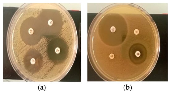
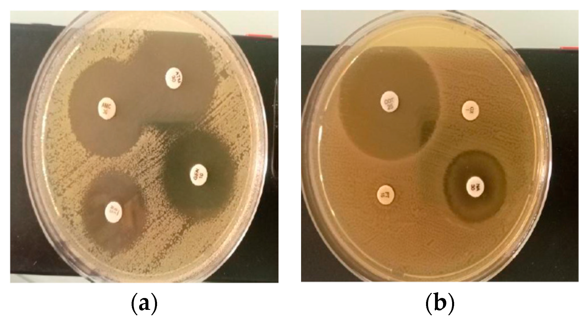
E. coli exhibited high susceptibility to aztreonam (32 mm) and gentamicin (28 mm), whereas Salmonella showed resistance to piperacillin–tazobactam and oxacillin, as described in Table 2 and Figure 1, which show the results of the antibiotic susceptibility of isolated pathogenic bacteria, obtained via the diffusion method on Mueller-Hinton agar (MH).
Table 2.
Antibiotic susceptibility profiles of E. coli and Salmonella isolates (in mm).
Figure 1.
The inhibition zone diameters (mm) obtained from the antibiogram of the two pathogenic bacteria are presented. (a) corresponds to a replicate of the antibiogram test performed on the E. coli isolate, while (b) corresponds to a replicate performed on the Salmonella isolate.
3.3. Effect of Antibiotics on Lactic Acid Bacteria
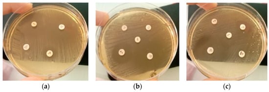
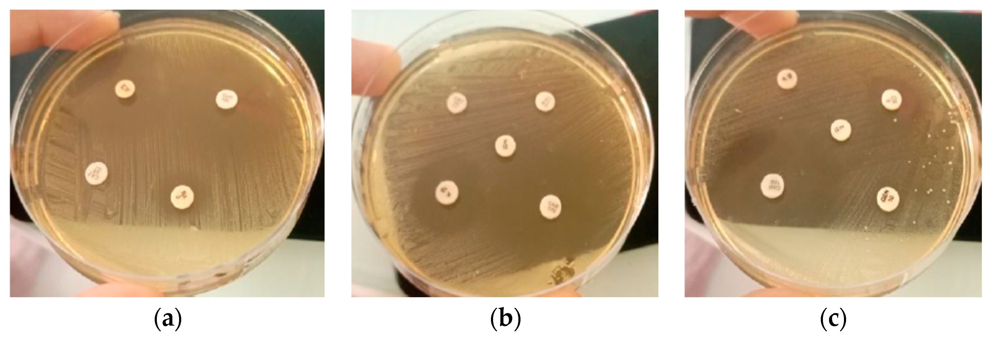
Table 3 and Figure 2 show the results of the antibiotic susceptibility of lactic acid bacteria, obtained via the MRS agar diffusion method.
Table 3.
Inhibition zone diameters (mm) of antibiotics against lactic acid bacteria (E3 and E4) using the MRS agar diffusion method.
Figure 2.
Antibiograms of lactic acid bacteria isolates (E3 and E4). (a,b) E3; (c) E4. Tested antibiotics include RA, CAZ, CAR, PT, CN/GEN, E, OX, and P.
3.4. Results of the Antimicrobial Activity of Lactobacilli: Antagonism
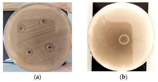
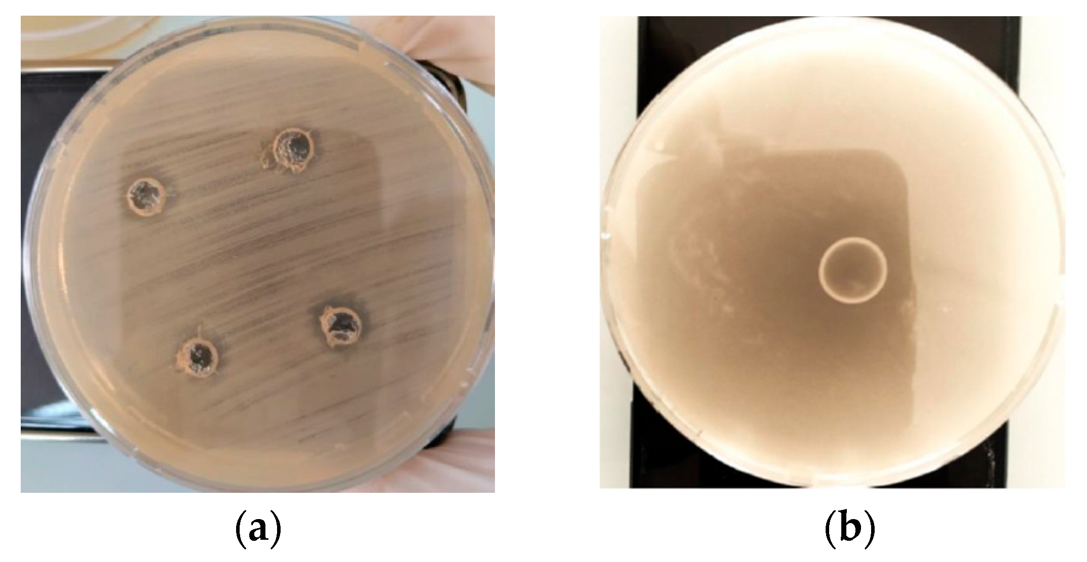
The results of in vitro inhibition of three lactic acid bacteria indicate antimicrobial potential against two tested infectious agents (Table 4 and Figure 3). The results in the table show that the highest inhibition activity was observed for isolate E5 against E. coli, with inhibition zones of 4 mm using the double-layer method and 2.5 mm using the well diffusion method.
Table 4.
Inhibition zones (mm) of the studied lactobacilli against the tested organisms using two methods: double-layer method and well method.
Figure 3.
Antimicrobial activity of the studied lactobacilli against target organisms, expressed as inhibition zones (mm), using two techniques: (a) agar well diffusion and (b) double-layer agar (spot-on-lawn) method.
4. Discussion
Postpartum uterine infections caused by pathogenic bacteria such as E. coli and Salmonella spp. remain a major problem in dairy cattle, requiring effective and targeted treatment strategies. In the present study, antibiotic susceptibility profiles revealed distinct responses between the two isolated pathogenic strains.
A limitation of the present study is the relatively small sample size, which may restrict the generalization of the findings. Therefore, further studies including larger cattle populations and molecular identification methods are required.
The E. coli strain exhibited high susceptibility to all tested antibiotics, particularly aztreonam (32 mm) and gentamicin (28 mm), consistent with previous studies reporting the persistent efficacy of these molecules against susceptible enteric strains [28,31]. The amoxicillin–clavulanic acid combination also showed high activity (28 mm), highlighting its efficacy against β-lactamase-producing strains [32]. In contrast, Salmonella displayed resistance to piperacillin–tazobactam and oxacillin, suggesting the presence of resistance mechanisms such as extended-spectrum β-lactamases (ESBLs) or alterations in penicillin-binding proteins [33]. However, its marked susceptibility to cotrimoxazole (35 mm) remains significant and is consistent with observations reporting preserved susceptibility in some strains despite the emergence of new resistances [34]. This heterogeneity in antibiotic response underscores the importance of antimicrobial susceptibility testing to guide appropriate treatment decisions, particularly in the context of increasing global antimicrobial resistance [35].
Beyond pathogenic bacteria, the evaluation of lactic acid bacteria (LAB) is essential not only for their antimicrobial potential but also for their safety as probiotic candidates. Unlike pathogenic bacteria, LAB require specific growth conditions, and standard culture media such as Mueller–Hinton agar are not always suitable. The use of MRS agar in this study allowed for optimal growth and more reliable interpretation of zones of inhibition, although it may require interpretation criteria adapted from CLSI/EUCAST standards [35,36].
The lactic acid bacteria isolates (E3 and E4) exhibited distinct antibiotic susceptibility profiles. Both strains were resistant to gentamicin, a resistance considered an intrinsic characteristic of Lactobacillus species due to their low membrane permeability [37]. However, E3 was susceptible to several β-lactam antibiotics, while E4 displayed broader resistance, possibly related to alterations in penicillin-binding proteins or the presence of resistance genes such as erm(B) [36,38]. Interestingly, E4 remained highly susceptible to carbenicillin (35 mm), suggesting selective susceptibility to certain semi-synthetic β-lactams [39].
From a safety perspective, antibiotic susceptibility testing is a crucial step in probiotic selection. The presence of transferable resistance genes (e.g., plasmids or transposons) in probiotic strains can contribute to the spread of antimicrobial resistance within the microbiota [40]. Therefore, the absence of acquired resistance to clinically important antibiotics (such as erythromycin, ampicillin, tetracycline, and chloramphenicol) is an essential condition for probiotic validation. Intrinsic resistance, such as that observed for gentamicin in lactobacilli, is generally acceptable provided it is not associated with mobile genetic elements [36,37].
In addition to safety, the selection of probiotic strains relies on multiple functional criteria, including survival in the gastrointestinal environment, adhesion to epithelial surfaces, and antimicrobial activity against pathogens [41,42,43]. In this context, the present study demonstrated that the use of MRS medium allowed for a more precise assessment of the susceptibility of lactic acid bacteria, revealing profiles consistent with the expected probiotic characteristics.
In vitro antagonism tests confirmed the probiotic potential of the studied isolates. Among the strains tested, E4 and E5 exhibited moderate antimicrobial activity against E. coli, while no inhibitory effect was observed against Salmonella spp. The largest zones of inhibition were recorded for E5, with 4 mm by the agar double-layer method and 2.5 mm by the well diffusion method. E4 also demonstrated moderate activity (2 mm), while E3 showed no inhibitory effect. These results confirm that antimicrobial activity is highly strain-dependent.
The inhibitory effects observed against E. coli can be attributed to the production of antimicrobial metabolites such as organic acids (notably lactic acid), bacteriocins, and hydrogen peroxide [44,45]. Acidification of the medium is considered a major mechanism of inhibition [46]. Differences between methods were also noted: the well diffusion method suggests efficient diffusion of antimicrobial compounds, while the double-layer method reveals more pronounced localized activity for E5, possibly indicating a higher concentration or stability of the active metabolites [47].
The lack of inhibitory activity against Salmonella spp. may be explained by insufficient production of active compounds or by the intrinsic resistance of this pathogen to metabolites derived from lactic acid bacteria [48]. Previous studies have reported considerable variability in Salmonella susceptibility depending on the lactic acid bacteria strain and experimental conditions [49,50].
5. Conclusions
Overall, the combination of antimicrobial activity against E. coli, acceptable antibiotic susceptibility profiles, and the absence of concerning resistance genes suggests the probiotic potential of the selected strains, particularly E4 and E5. However, further in vitro and vivo studies are needed to confirm their efficacy, safety, and stability under physiological conditions.
The absence of statistical analysis has been justified by the exploratory nature of the study and the limited number of isolates. As a result, the study focused on descriptive analysis, with experiments performed in triplicate and results reported as mean inhibition zone diameters (mm).
The present work should be considered a preliminary exploratory study due to the limited sample size and the restricted number of isolates analyzed.
Only two pathogenic bacterial isolates were included in the antimicrobial assays. Future studies should include additional uterine pathogens commonly associated with bovine endometritis, such as Trueperella pyogenes, Fusobacterium necrophorum, and Prevotella spp.
Bacterial identification in this study was based on phenotypic and biochemical characterization. Molecular identification techniques such as 16S rRNA sequencing and PCR-based detection of antimicrobial resistance genes would provide more precise characterization and should be included in future studies.
Author Contributions
Conceptualization, N.B.; methodology, H.M.; investigation, N.B. and H.M.; formal analysis, N.B. and H.M.; writing—original draft preparation, N.B. and H.M.; writing—review and editing, N.B. and H.M. All authors have read and agreed to the published version of the manuscript.
Funding
This research received no external funding.
Institutional Review Board Statement
The animal study protocol was conducted in accordance with institutional and national guidelines for the care and use of animals. Ethical review and approval were waived because vaginal samples were collected during routine veterinary procedures without causing additional harm or distress to the animals.
Informed Consent Statement
Not applicable.
Data Availability Statement
The data presented in this study are available on request from the corresponding author.
Conflicts of Interest
The authors declare no conflicts of interest.
Abbreviations
| LAB | lactic acid bacteria |
| MADR | Ministry of Agriculture and Rural Development |
| ISO | International Organization for Standardization |
| CLSI | Clinical and Laboratory Standards Institute |
| ESBLs | extended-spectrum β-lactamases |
| WHO | World Health Organization |
| EUCAST | European Committee on Antimicrobial Susceptibility Testing |
| EFSA | European Food Safety Authority |
References
- Kerbache, I. Etude socio-économique de l’élevage bovin à l’est algérien. Econ. J. 2019, 3, 208–234. [Google Scholar]
- Tona, G. Impact of beef and milk sourced from cattle production on global food security. In Bovine Science—Challenges and Advances; IntechOpen: London, UK, 2021. [Google Scholar] [CrossRef] [Scilit]
- MADR Ministry of Agriculture and Rural Development. Statistique Agricole: Superficies et Productions; Direction des Statistiques Agricoles et Des Systèmes D’information: Algiers, Algeria, 2018.
- Mimoune, N.; Kaidi, R.; Azzouz, M.Y.; Keddour, R.; Belarbi, A.; Derdour, S.Y. Genital tract pathologies of cows slaughtered at El-Harrach abattoir in Algeria. Kafkas Üniversitesi Vet. Fakültesi Derg. 2016, 22, 639–646. [Google Scholar] [CrossRef] [Scilit]
- Paisley, L.G.; Mickelsen, W.O.D.; Anderson, P.B. Mechanisms and therapy for retained fetal membranes and uterine infections of cows: A review. Theriogenology 1986, 25, 353–381. [Google Scholar] [CrossRef] [Scilit]
- Fourichon, C.; Seegers, H.; Malher, X. Effect of disease on reproduction in the dairy cow: A meta-analysis. Theriogenology 2000, 53, 1729–1759. [Google Scholar] [CrossRef] [Scilit]
- Williams, E.J.; Fischer, D.P.; Noakes, D.E.; England, G.C.W.; Rycroft, A.; Dobson, H.; Sheldon, I.M. The relationship between uterine pathogen growth 69 density and ovarian function in the postpartum dairy cow. Theriogenology 2007, 68, 549–559. [Google Scholar] [CrossRef] [Scilit]
- Adnane, M.; Chapwanya, A.; Kaidi, R.; Meade, K.G.; O’Farrelly, C. Profiling inflammatory biomarkers in cervico-vaginal mucus (CVM) postpartum: Potential early indicators of bovine clinical endometritis? Theriogenology 2017, 103, 117–122. [Google Scholar] [CrossRef] [Scilit] [PubMed]
- Okawa, H.; Goto, A.; Wijayagunawardane, M.M.P.; Vos, P.L.A.M.; Yamato, O.; Taniguchi, M.; Takagi, M. Risk factors associated with reproductive performance in Japanese dairy cows: Vaginal discharge with flecks of pus or calving abnormality extend time to pregnancy. J. Vet. Med. Sci. 2019, 81, 95–99. [Google Scholar] [CrossRef] [Scilit]
- Sheldon, I.M.; Williams, E.J.; Miller, A.N.A.; Nash, D.M.; Herath, S. Uterine diseases in cattle after parturition. Vet. J. 2008, 176, 115–121. [Google Scholar] [CrossRef] [Scilit] [PubMed]
- Guy, B.; Jocelyn, D. L’endométrite, Son Impact et Traitements; Le producteur de Lait Québécois: Longueuil, QC, Canada, 2011. [Google Scholar]
- Dohmen, M.J.; Joop, K.; Sturk, A.; Bols, P.E.; Lohuis, J.A. Relationship between intra-uterine bacterial contamination, endotoxin levels and the development of endometritis in postpartum cows with dystocia or retained placenta. Theriogenology 2000, 54, 1019–1032. [Google Scholar] [CrossRef] [Scilit]
- Santos, T.M.; Gilbert, R.O.; Bicalho, R.C. Metagenomic analysis of the uterine bacterial microbiota in healthy and metritic postpartum dairy cows. J. Dairy Sci. 2011, 94, 291–302. [Google Scholar] [CrossRef] [Scilit] [PubMed]
- Zhou, Z.; Gomes, M.S.; Canisso, I.F.; Garrett, E.F.; Stewart, J.S.; Lima, F.S. Uterine Microbiome, Antibiotic Resistance Genes and Virulence Factors of Metritic Treated Cows that Cure or Failed to Cure from Metritis. Clin. Theriogenology 2017, 9, 436. [Google Scholar]
- Pennehouat, G.; Joly-Guillou, M.; Mohseni, H.; Taarji, H.; Madélénat, P. Treatment of genital infections in gynecology. Gynécol. Obs. Fertil. 2001, 105, 345–352. [Google Scholar]
- Madsen, K.L. The use of probiotics in gastrointestinal disease. Can. J. Gastroenterol. Hepatol. 2001, 15, 817–822. [Google Scholar] [CrossRef] [Scilit] [PubMed]
- Genís, S.; Bach, A.; Arís, A. Effects of intravaginal lactic acid bacteria on bovine endometrium: Implications in uterine health. Vet. Microbiol. 2017, 203, 133–139. [Google Scholar] [CrossRef] [Scilit]
- Amarloie, O.A.; Dini, P.; Mousakhani, F.; Moghaddam, M.F.; Mottaghian, P. In Vitro Preventive Effects of Normal Vaginal Lactobacillus on Uterine Pathogens in Dairy Cows; American Association of Bovine Practitioners: Ashland, OH, USA, 2010. [Google Scholar]
- De Man, J.C.; Rogosa, M.; Sharpe, M.E. A medium for the cultivation of lactobacilli. J. Appl. Bacteriol. 1960, 23, 130–135. [Google Scholar] [CrossRef] [Scilit]
- Atlas, R.M. Handbook of Microbiological Media, 4th ed.; CRC Press: Boca Raton, FL, USA, 2010. [Google Scholar]
- Bio-Rad. Chapman–Mannitol Salt Agar: Instructions for Use; Bio-Rad Laboratories: Hercules, CA, USA, 2007. [Google Scholar]
- ifco Laboratories. Difco & BBL Manual: Manual of Microbiological Culture Media, 2nd ed.; BD Diagnostics: Franklin Lakes, NJ, USA, 2009. [Google Scholar]
- ISO 4832; Microbiology of Food and Animal Feeding Stuffs—Horizontal Method for the Enumeration of Coliforms—Colony-count Technique. International Organization for Standardization, ISO: Geneva, Switzerland, 2006.
- Martin, M.; White, D. Fungal culture media. In Manual of Clinical Microbiology, 8th ed.; Coyle, M.B., Ed.; ASM Press: Washington, DC, USA, 2004; pp. 1655–1658. [Google Scholar]
- Denis, F.; Poly, M.-C.; Martin, C.; Bingen, E.; Quentin, R. Bactériologie Médicale, 2nd ed.; Elsevier: Amsterdam, The Netherlands, 2011. [Google Scholar]
- Mirlohi, M.; Sabihe, S.Z.; Mahmoud, S.Z. Identification of Lactobacilli from fecal flora of some Iranian infants. Iran. J. Pediatr. 2008, 18, 357–363. [Google Scholar]
- Mahmoudi, A.; et Mameche, K. Les Infections Urinaires et les Infections Vaginales Caractérisées Dans un Laboratoire Médical du Dr. Boudissa à Boumerdès. Master’s Thesis, Université AKLI Mohand Oulhadj de Bouira, Bouïra, Algeria, 2019; pp. 3–60. [Google Scholar]
- Clinical and Laboratory Standards Institute (CLSI). Performance Standards for Antimicrobial Susceptibility Testing, 33rd ed.; CLSI supplement M100; Clinical and Laboratory Standards Institute: Wayne, PA, USA, 2023. [Google Scholar]
- Jacobsen, C.N.; Nielsen, V.R.; Hayford, A.E.; Møller, P.L.; Michaelsen, K.F.; Pærregaard, A.; Sandström, B.; Tvede, M.; Jakobsen, M. Screening of probiotic activities of fortyseven strains of Lactobacillus spp. by in vitro techniques and evaluation of the colonization ability of five selected strains in humans. Appl. Environ. Microbiol. 1999, 65, 4949–4956. [Google Scholar] [CrossRef] [Scilit]
- Thompson, J.; Johannsen, E.; Murray, R.G. Bacterial interactions and antimicrobial activity in vitro. J. Appl. Bacteriol. 1996, 81, 349–358. [Google Scholar]
- Kaper, J.B.; Nataro, J.P.; Mobley, H.L.T. Pathogenic Escherichia coli. Nat. Rev. Microbiol. 2004, 2, 123–140. [Google Scholar] [CrossRef] [Scilit]
- Livermore, D.M. β-Lactamases in laboratory and clinical resistance. Clin. Microbiol. Rev. 1995, 8, 557–584. [Google Scholar] [CrossRef] [PubMed]
- Threlfall, E.J. Antimicrobial drug resistance in Salmonella: Problems and perspectives in food- and water-borne infections. FEMS Microbiol. Rev. 2002, 26, 141–148. [Google Scholar] [CrossRef]
- Hendriksen, R.S.; Vieira, A.R.; Karlsmose, S.; Lo Fo Wong, D.M.; Jensen, A.B.; Wegener, H.C.; Aarestrup, F.M. Global monitoring of Salmonella serovar distribution from the World Health Organization Global Foodborne Infections Network Country Data Bank: Results of quality assured laboratories from 2001 to 2007. Foodborne Pathog. Dis. 2011, 8, 887–900. [Google Scholar] [CrossRef] [Scilit]
- World Health Organization (WHO). Global Antimicrobial Resistance and Use Surveillance System (GLASS) Report 2021; World Health Organization: Geneva, Switzerland, 2021; Available online: https://www.who.int/publications/i/item/9789240027336 (accessed on 21 February 2026).
- Charteris, W.P.; Kelly, P.M.; Morelli, L.; Collins, J.K. Antibiotic susceptibility of potentially probiotic Lactobacillus species. J. Food Prot. 1998, 61, 1636–1643. [Google Scholar] [CrossRef] [Scilit] [PubMed]
- Danielsen, M.; Wind, A. Susceptibility of Lactobacillus spp. to antimicrobial agents. Int. J. Food Microbiol. 2003, 82, 1–11. [Google Scholar] [CrossRef] [Scilit]
- Hummel, A.S.; Hertel, C.; Holzapfel, W.H.; Franz, C.M.A.P. Antibiotic resistances of starter and probiotic strains of lactic acid bacteria. Appl. Environ. Microbiol. 2007, 73, 730–739. [Google Scholar] [CrossRef] [Scilit]
- Kalka-Moll, W.M.; Rieß, M.; Gutsmann, T.; Lüders, C.; Gatermann, S. Activity of β-lactam antibiotics against lactobacilli. Antimicrob. Agents Chemother. 2002, 46, 2784–2786. [Google Scholar]
- European Food Safety Authority (EFSA). Guidance on the characterisation of microorganisms used as feed additives or as production organisms. EFSA J. 2018, 16, e05206. [Google Scholar] [CrossRef] [Scilit] [PubMed]
- FAO; WHO. Evaluation of Health and Nutritional Properties of Probiotics in Food Including Powder Milk with Live Lactic Acid Bacteria: Report of a Joint FAO/WHO Expert Consultation, Córdoba, Argentina, 1–4 October 2001; Food and Agriculture Organization of the United Nations (FAO): Rome, Italy; World Health Organization (WHO): Geneva, Switzerland, 2001; Available online: https://www.fao.org/3/a0512e/a0512e.pdf (accessed on 21 February 2026).
- Hill, C.; Guarner, F.; Reid, G.; Gibson, G.R.; Merenstein, D.J.; Pot, B.; Morelli, L.; Canani, R.B.; Flint, H.J.; Salminen, S.; et al. The International Scientific Association for Probiotics and Prebiotics consensus statement on the scope and appropriate use of the term probiotic. Nat. Rev. Gastroenterol. Hepatol. 2014, 11, 506–514. [Google Scholar] [CrossRef] [Scilit] [PubMed]
- Sanders, M.E.; Guarner, F.; Guerrant, R.; Holt, P.R.; Quigley, E.M.; Sartor, R.B.; Sherman, P.M.; Mayer, E.A. An update on the use and investigation of probiotics in health and disease. Gut 2010, 59, 285–289. [Google Scholar] [CrossRef] [Scilit]
- Reid, G.; Bruce, A. Selection of Lactobacillus strains for urogenital probiotic applications. J. Infect. Dis. 2001, 183, S77–S80. [Google Scholar] [CrossRef] [Scilit]
- Servin, A.L. Antagonistic activities of lactobacilli and bifidobacteria against microbial pathogens. FEMS Microbiol. Rev. 2004, 28, 405–440. [Google Scholar] [CrossRef] [Scilit] [PubMed]
- Otero, M.C.; Nader-Macías, M.E.F. Inhibition of Staphylococcus aureus by Lactobacillus isolated from the bovine vagina. FEMS Immunol. Med. Microbiol. 2006, 46, 337–344. [Google Scholar]
- Schillinger, U.; Lücke, F.K. Antibacterial activity of Lactobacillus sake isolated from meat. Appl. Environ. Microbiol. 1989, 55, 1901–1906. [Google Scholar] [CrossRef] [Scilit]
- Brashears, M.M.; Jaroni, D.; Trimble, J. Isolation, selection, and characterization of lactic acid bacteria for a competitive exclusion product to reduce shedding of Escherichia coli O157:H7 in cattle. J. Food Prot. 2003, 66, 355–363. [Google Scholar] [CrossRef] [Scilit]
- Tannock, G.W. Probiotics and Prebiotics: Scientific Aspects; Caister Academic Press: Poole, UK, 2002. [Google Scholar]
- Parvez, S.; Malik, K.A.; Kang, S.A.; Kim, H.Y. Probiotics and their fermented food products are beneficial for health. J. Appl. Microbiol. 2006, 100, 1171–1185. [Google Scholar] [CrossRef] [Scilit] [PubMed]

Disclaimer/Publisher’s Note: The statements, opinions and data contained in all publications are solely those of the individual author(s) and contributor(s) and not of MDPI and/or the editor(s). MDPI and/or the editor(s) disclaim responsibility for any injury to people or property resulting from any ideas, methods, instructions or products referred to in the content. |
© 2026 by the authors. Licensee MDPI, Basel, Switzerland. This article is an open access article distributed under the terms and conditions of the Creative Commons Attribution (CC BY) license.