


Abstract
Background/Objectives: Osteoporosis remains a clinically important metabolic bone disorder with limited bone-forming therapeutic options. SET domain bifurcated protein 1 (SETDB1) is involved in osteogenic epigenetic regulation, but small-molecule discovery guided by SETDB1-associated structural regions remains limited. This study aimed to identify a candidate compound with in silico relevance to a SETDB1-associated ligand-bound pocket and assess its association with early osteogenic readouts. Methods: A computational–experimental workflow was used, including hierarchical molecular docking, MM-GBSA rescoring, ADMET-based prioritization, redocking validation, molecular dynamics simulations, and preliminary in vitro evaluation in MC3T3-E1 cells. Compound 271 (C271) was selected based on structure-based screening results and predicted developability-related properties. Cytocompatibility, alkaline phosphatase (ALP) activity and staining, selected molecular markers, and SETDB1–H3 molecular dynamics behavior were evaluated. Results: Redocking reproduced the reference binding mode, and molecular dynamics simulations indicated that C271 maintained a relatively persistent conformation around the predicted SETDB1-associated pocket. Comparative SETDB1–H3 simulations showed altered H3 dynamics and SETDB1–H3 contact patterns in the C271-containing system. In cell-based assays, C271 showed no appreciable cytotoxicity within the tested concentration range and was associated with increased ALP activity and staining. C271 treatment was accompanied by higher global H3K9me3 and Runx2 levels, whereas SETDB1 protein abundance remained largely unchanged. Conclusions: C271 was identified as a computationally prioritized SETDB1-related candidate compound associated with early osteogenic-associated cellular responses. The evidence supports computational plausibility and cell-level association, but does not establish direct SETDB1 engagement, SETDB1 enzymatic modulation, SETDB1-dependent causality, or late-stage osteogenic maturation/mineralization. Given the single-compound evaluation, further target-engagement, enzymatic, and functional studies are needed.
1. Introduction
Osteoporosis is a systemic skeletal metabolic disorder characterized by diminished bone mineral density and microarchitectural deterioration, which collectively precipitate abnormally elevated bone fragility and fracture susceptibility [1,2,3]. The core pathophysiological mechanism involves an uncoupling imbalance between bone formation, mediated by osteoblasts, and bone resorption, mediated by osteoclasts, during dynamic bone remodeling [4,5,6,7]. This imbalance manifests as a relative predominance of bone resorption over formation, resulting in sustained net bone loss [8,9]. Consequently, therapeutic strategies that support osteoblast differentiation and bone-forming activity remain of considerable interest for restoring bone mass, improving bone quality, and mitigating fracture risk [10,11,12].
Epigenetic regulation, particularly histone modification, has emerged as an important regulatory layer in osteoblast biology [13,14]. Among such regulators, the histone H3 lysine 9 (H3K9)-specific methyltransferase SET domain bifurcated protein 1 (SETDB1) has been reported as an important epigenetic regulator involved in bone metabolism [15,16]. Mechanistically, SETDB1 directly catalyzes histone H3 lysine 9 trimethylation (H3K9me3) at the promoter region of the developmental homeobox transcription factor Orthodenticle Homeobox 2 (Otx2) gene, establishing a repressive chromatin environment that suppresses Otx2 transcription [16]. As Otx2 functions as a transcriptional repressor, its downregulation consequently alleviates its inhibitory effect on downstream osteogenic target genes, such as runt-related transcription factor 2 (Runx2). This regulatory cascade has been associated with the activation of osteogenesis-related signaling pathways, including Wnt/β-catenin and bone morphogenetic protein (BMP)-Smad pathways, thereby contributing to osteoblast differentiation [16,17,18,19,20]. Peroxisome proliferator-activated receptor γ (PPARγ) serves as a master transcriptional regulator governing the differentiation of mesenchymal stem cells (MSCs), specifically driving adipocyte lineage commitment [21]. In this lineage-allocation context, SETDB1 has been linked to PPARγ-related adipogenic regulation, as SETDB1 knockdown has been associated with enhanced adipogenesis and increased expression of adipogenic regulators, including PPARγ and C/EBPα [22]. Consistently, suppression of PPARγ transcriptional activity has been reported to favor osteogenic differentiation over adipogenic differentiation in MSCs [23]. Concurrently, studies demonstrate that mono-ADP-ribosylhydrolase 2 (Macrod2) inhibits glycogen synthase kinase 3β (GSK3β) activity, leading to destabilization of β-catenin—the core effector of Wnt signaling—and attenuation of its downstream transcriptional activity, which ultimately suppresses osteoblast proliferation [24,25]. SETDB1 directly catalyzes H3K9 trimethylation (H3K9me3) at the Macrod2 gene promoter region, establishing a transcriptionally repressive epigenetic mark that represses Macrod2 gene expression, thereby facilitating osteoblast differentiation [25]. Collectively, these studies indicate that SETDB1 is involved in osteogenesis-related epigenetic regulation. However, small-molecule discovery guided by SETDB1-associated structural regions remains limited.
Based on the reported involvement of SETDB1 in osteogenesis-related epigenetic regulation, an integrated computational–experimental workflow was established to identify a small-molecule candidate with potential relevance to a SETDB1-associated structural region and to preliminarily evaluate its association with early osteogenic-related cellular responses. First, a structure-based virtual screening campaign was performed against the TopScience commercial database (D001, comprising a diverse library of drug-like molecules; TopScience Co., Ltd., Shanghai, China) using the co-crystallized ligand-defined pocket of the human SETDB1 tandem Tudor domain as the structural reference [26,27]. Candidate compounds were then evaluated through hierarchical molecular docking and MM-GBSA rescoring, followed by ADMET-based prioritization to balance structure-derived screening results and predicted developability-related properties [28,29]. An auxiliary ligand-based pharmacophore model derived from the co-crystallized native ligand was used to provide an additional spatial-feature reference for candidate assessment [27,30]. Molecular dynamics simulations were subsequently performed to examine the conformational behavior of the prioritized candidate around the SETDB1-associated pocket, and comparative SETDB1–H3 simulations were used to provide computational context for possible changes at the SETDB1–H3 interaction interface [31,32]. The prioritized candidate was further evaluated in MC3T3-E1 cells for cytocompatibility, alkaline phosphatase (ALP) activity and staining, and selected molecular changes under osteogenic induction conditions [33,34] (Figure 1). Overall, this work focuses on identifying a small-molecule candidate with potential relevance to a SETDB1-associated structural region and preliminarily evaluating its association with early osteogenic-related cellular responses.
Figure 1.
Schematic workflow of the integrated computational–experimental strategy for identifying a SETDB1-related candidate compound associated with preliminary early osteogenic-associated cellular responses. The workflow includes protein and ligand preparation, hierarchical virtual screening using HTVS, SP, and XP docking followed by MM-GBSA rescoring, ADMET normalization-based prioritization, molecular dynamics simulation of the SETDB1–C271 computational complex, comparative molecular dynamics analysis of SETDB1–H3 systems in the absence and presence of C271, and subsequent in vitro evaluation in MC3T3-E1 cells.
2. Materials and Methods
2.1. Molecular Docking
2.1.1. Protein Structure Preparation
The published X-ray co-crystal structure of the human SETDB1 tandem Tudor domain (PDB ID: 7C9N) was retrieved from the Protein Data Bank [27]. This structure was selected as the receptor model because its co-crystallized small molecule defines a co-crystallized ligand-bound pocket that can serve as a structural reference for virtual screening. To generate a reliable receptor model for virtual screening, the raw structural data were meticulously processed using the Protein Preparation Wizard (PPW) module in Schrödinger Suite Release 2024-1 (Schrödinger, LLC, New York, NY, USA) [35]. Initially, redundant components, including crystallographic water molecules and non-essential cofactors, were deleted. Missing side chains and loop segments were subsequently modeled using Prime. Hydrogen atoms were added, bond orders were assigned, and formal charges were adjusted to reflect physiological pH (7.4) using the Epik module [36]. Furthermore, the hydrogen-bond network was optimized via comprehensive sampling of hydroxyl, asparagine, glutamine, and histidine orientations. Finally, a restrained energy minimization was executed using the OPLS4 force field until the root-mean-square deviation (RMSD) of the heavy atoms converged to 0.30 Å, yielding an energy-minimized receptor conformation for subsequent docking calculations [37].
2.1.2. Binding Site Definition and Grid Generation
Potential binding cavities on the SETDB1 surface were evaluated using the SiteMap module of Schrödinger, which identifies and scores topologically and physicochemically favorable binding regions [38]. The highest-scoring binding site (SiteScore) spatially overlapped with the co-crystallized native ligand-defined pocket. Consequently, the receptor grid was generated using the Receptor Grid Generation tool in Glide. The grid box was centered on the centroid of the native ligand, with the bounding box dimensions set to 16 × 16 × 16 Å to ensure comprehensive coverage of the SETDB1-associated ligand-bound pocket for subsequent ligand docking [39,40].
2.1.3. Ligand Database Preparation
A commercial compound library containing 1,648,191 distinct structural entities was procured from the TopScience database (D001). To construct a standardized screening dataset, structural standardization and 3D coordinate generation were performed using the LigPrep module in Schrödinger. For each ligand, Epik was employed to generate plausible ionization states and tautomers at a target physiological pH of 7.0 ± 2.0. Stereoisomers were retained per the source geometries, and low-energy ring conformations were generated. Subsequently, to enrich the library with molecules possessing basic drug-like physicochemical properties, a stringent pre-docking filter was applied based on Lipinski’s Rule of Five. Compounds violating the criteria of molecular weight ≤ 500 Da, hydrogen bond donors ≤ 5, hydrogen bond acceptors ≤ 10, and calculated logP ≤ 5 were systematically excluded [41].
2.1.4. Semi-Flexible Molecular Docking
Hierarchical precision docking calculations were performed using the Glide module, maintaining a rigid receptor-binding pocket conformation while permitting ligand conformational sampling through free rotation of rotatable bonds. A progressive filtering strategy was employed—initial high-throughput virtual screening (HTVS) followed by standard precision (SP) and extra precision (XP) scoring. The docking score function was used to rank candidate poses based on predicted pocket compatibility and interaction plausibility, including van der Waals interactions, hydrogen bonding, hydrophobic contacts, and steric complementarity [39,40,42].
2.1.5. Binding Free Energy Calculation
To further evaluate the shortlisted docking poses, the binding free energies (ΔG_bind) of the top-ranked ligand-receptor complexes were calculated using the Molecular Mechanics/Generalized Born Surface Area (MM-GBSA) method via the Prime module. Calculations were performed using the OPLS4 force field to compute the molecular mechanics energies, while the solvation free energy was modeled employing the Variable Dielectric Generalized Born (VSGB) 2.0 implicit solvent model. The relative binding free energy was calculated using the equation: ΔG_bind = E_complex − (E_protein + E_ligand), incorporating both polar and non-polar solvation terms. During the calculation, the ligands and the protein residues within a 5.0 Å radius of the ligand were allowed to undergo conformational relaxation, providing a complementary structure-based estimate for candidate comparison rather than direct experimental evidence of binding affinity [43,44].
2.1.6. Redocking Procedure for Docking Workflow Validation
To assess the reliability of the docking protocol, a redocking procedure was performed using the co-crystallized ligand in the selected SETDB1 tandem Tudor domain structure (PDB ID: 7C9N). The redocking ligand was the native co-crystallized ligand rather than C271. The native ligand was first extracted from the crystal complex and prepared using the same LigPrep workflow described above. Subsequently, the ligand was re-docked into the original binding pocket employing the identical receptor preparation, grid settings, and docking parameters used in the virtual screening workflow. The reproduced binding pose was then evaluated by comparing its spatial overlap with the crystallographic conformation. Specifically, a heavy-atom root-mean-square deviation (RMSD) of ≤2.0 Å was adopted as the criterion for acceptable pose reproduction, together with the retention of major protein–ligand interactions. This procedure was used to evaluate whether the docking setup could reproduce the known crystallographic pose before applying the same protocol to the comparative analysis of shortlisted candidates.
2.2. ADMET Prediction Screening
To evaluate the pharmacokinetic profiles and safety of the shortlisted compounds, comprehensive Absorption, Distribution, Metabolism, Excretion, and Toxicity (ADMET) predictions were performed using the ADMETlab 3.0 platform [28,29]. The shortlisted compounds obtained from docking and MM-GBSA screening were further assessed by integrating multiple ADMET-related endpoints to prioritize candidates with balanced predicted developability profiles. Rather than relying on a single parameter, compounds were comparatively evaluated according to their overall absorption, distribution, metabolism, excretion, and toxicity characteristics. In this workflow, docking and MM-GBSA calculations were used to define a structurally plausible candidate set, whereas ADMET normalization was used as a subsequent prioritization step to compare the overall developability balance among shortlisted candidates.
2.2.1. Absorption
Optimal oral bioavailability and intestinal absorption are prerequisites for systemic drug exposure [28]. The apparent permeability coefficient (Papp) in the Caco-2 cell model was calculated to estimate passive intestinal permeability [45]. Concurrently, the likelihood of compounds acting as substrates for P-glycoprotein (P-gp), an ATP-dependent efflux pump, was predicted to evaluate the risk of reduced bioavailability or tissue-specific accumulation [46].
2.2.2. Distribution
The volume of distribution at steady state (Vdss) was predicted to assess the distribution equilibrium of the candidates between the systemic circulation and peripheral tissues [47]. Because the intended biological evaluation focused on osteogenic cells rather than central nervous system activity, candidates with lower predicted Blood–Brain Barrier (BBB) penetration were considered preferable to reduce potential central nervous system (CNS)-related liability [48]. Additionally, the inhibitory activity against Multidrug Resistance Protein 1 (MRP1) was evaluated, as MRP1-mediated efflux may influence intracellular compound accumulation [49].
2.2.3. Metabolism
Cytochrome P450 3A4 (CYP3A4) is involved in the biotransformation of a large proportion of clinical drugs [50]. The probabilities of the candidate molecules acting as either substrates or inhibitors of CYP3A4 were computed to predict their susceptibility to first-pass metabolism and to evaluate the potential risk of drug–drug interactions (DDIs).
2.2.4. Excretion
The elimination half-life (T1/2) and human hepatocyte clearance (CL_hep) were calculated to evaluate the duration of drug action and the hepatic metabolic capacity, respectively. Compounds exhibiting excessively high clearance or suboptimal half-lives were flagged, as these properties may limit sustained exposure [51].
2.2.5. Toxicity
Toxicity-related endpoints were included to reduce the likelihood of selecting candidates with obvious predicted safety liabilities. The nephrotoxicity risk of the compounds was predicted to evaluate potential kidney-related liability [52]. Cardiac safety was assessed by predicting the inhibitory potential against the Human Ether-à-go-go-Related Gene (hERG) potassium channel, a major indicator associated with drug-induced QT prolongation and torsades de pointes (TdP) [53]. Furthermore, mutagenicity and carcinogenicity risks were evaluated via the predicted Ames test outcomes and the Stress Response p53 (SR-p53) activation status [54]. Compounds with lower predicted toxicity-related risks were considered more favorable during the integrated prioritization process [55].
2.2.6. Integrated ADMET-Based Prioritization
Following docking/MM-GBSA-based pre-screening, the remaining candidates were further prioritized through a normalized multi-parameter ADMET evaluation rather than by relying on any single structural score alone. In the present study, docking score and MM-GBSA binding free energy were used to establish a structurally plausible shortlisted set, whereas the final prioritization was based on the overall balance of developability-related properties across absorption, distribution, metabolism, excretion, and toxicity dimensions. Importantly, the normalization procedure was performed according to the endpoint classification framework and risk interpretation logic provided by the ADMETlab 3.0 platform, rather than by arbitrary empirical assignment [29]. To improve the transparency and comparability of the prioritization process, all evaluated ADMET endpoints were converted into a unified semi-quantitative scoring system. In this scoring system, a score of 1 indicated an excellent or favorable predicted property, a score of 0.5 indicated a medium or intermediate predicted property, and a score of 0 indicated a poor or unfavorable predicted property. Therefore, the presence of a score of 0 was regarded as a limiting ADMET-related liability during candidate prioritization. For Caco-2 permeability, values > −5.15 were classified as excellent and assigned a score of 1, whereas lower values were classified as poor and assigned a score of 0. For logVDss, values within the acceptable distribution interval corresponding to VDss = 0.04–20 L/kg were classified as excellent and assigned a score of 1, whereas values outside this interval were classified as poor and assigned a score of 0. For CYP3A4 inhibition probability and CYP3A4 substrate probability, lower predicted probabilities were considered more favorable, and values within 0–0.3, 0.3–0.7, and 0.7–1.0 were assigned scores of 1, 0.5, and 0, respectively. Plasma clearance was normalized as follows: 0–5, excellent (score 1); 5–15, medium (score 0.5); >15, poor (score 0). The elimination half-life was normalized as follows: >8, excellent (score 1); 1–8, medium (score 0.5); <1, poor (score 0). For the remaining probability-based endpoints, including P-gp substrate probability, f20, BBB penetration, MRP1 inhibition, hERG liability, Ames mutagenicity, neurotoxicity, and SR-p53 activation, values within 0–0.3, 0.3–0.7, and 0.7–1.0 were classified as excellent, medium, and poor, and assigned scores of 1, 0.5, and 0, respectively. Representative candidates among the top MM-GBSA-ranked compounds were subsequently compared at the normalized multi-endpoint level to evaluate their overall developability balance, and compounds without obvious limiting liabilities were retained for subsequent molecular dynamics simulations and biological evaluation.
The final prioritization followed a stepwise decision strategy. First, docking and MM-GBSA results were used to retain compounds with plausible structural compatibility with the SETDB1-associated pocket. Second, representative candidates among the top MM-GBSA-ranked compounds were compared using the normalized ADMET endpoint scores. Third, candidates showing a more balanced multi-endpoint profile and lacking obvious limiting liabilities were prioritized for subsequent molecular dynamics simulations and preliminary cell-based evaluation. Therefore, the final selection was not based on the highest docking score or the most favorable MM-GBSA value alone, but on the combined consideration of structural plausibility and normalized multi-endpoint developability balance.
2.3. Molecular Dynamics Simulations
To examine the conformational behavior of the prioritized compound around the SETDB1-associated pocket under solvated conditions, two independent 200 ns all-atom molecular dynamics (MD) simulations of the SETDB1–C271 computational complex were performed using the Desmond module in Schrödinger [32]. The optimized docking pose of C271 in complex with SETDB1, obtained from the preceding virtual screening workflow, was used as the initial conformation. The system was solvated in an orthorhombic box with a 10 Å buffer distance from the complex surface using the Transferable Intermolecular Potential 3-Point (TIP3P) water model. To mimic physiological ionic strength and neutralize the system, 0.15 M NaCl and appropriate counterions (Na+/Cl−) were incorporated. Prior to the production runs, the system was relaxed using the default Desmond multi-step relaxation protocol, including conjugate gradient energy minimization. To assess the reproducibility of the computational trajectory behavior, two independent 200 ns production simulations were carried out using different initial random seeds. The simulations were run in the isothermal-isobaric (NPT) ensemble at 300 K and 1.01325 bar, regulated by the Nosé–Hoover chain thermostat and the Martyna–Tobias–Klein barostat. The equations of motion were integrated using the RESPA algorithm with a 2-fs time step, and trajectory coordinates were recorded every 100 ps for subsequent conformational and interaction analyses [56,57].
To further examine the computational behavior of the SETDB1–H3 interaction interface, comparative MD simulations were performed for SETDB1–H3 systems in the absence and presence of C271. The initial SETDB1–H3 complex was prepared based on the reported structure with PDB ID: 6BHI [58], and the two simulation systems were constructed as SETDB1–H3 and SETDB1–H3–C271 complexes, respectively. In the C271-containing system, C271 was placed according to the predicted SETDB1-associated ligand-bound pocket used in the preceding structure-based screening workflow. Each system was prepared using the same Desmond-based procedure, including solvation with the TIP3P water model in an orthorhombic box, addition of 0.15 M NaCl and counterions for system neutralization, and pre-production relaxation using the default multi-step protocol. Production simulations were conducted for 100 ns in the NPT ensemble at 300 K and 1.01325 bar using the Nosé–Hoover chain thermostat and Martyna–Tobias–Klein barostat. Trajectory coordinates were saved every 100 ps. Protein RMSD, H3 RMSD, H3 RMSF, and SETDB1–H3 contact profiles were analyzed to compare the dynamic features of the SETDB1–H3 interface in the two computational systems.
2.4. Auxiliary Pharmacophore Analysis
A ligand-based pharmacophore analysis was performed using the Phase module of Schrödinger as an auxiliary spatial-feature reference [30,59]. A pharmacophore hypothesis was derived from the SETDB1 co-crystallized native ligand to represent the major steric and hydrophobic features of the co-crystallized ligand-defined pocket. The prioritized candidate compound, obtained from the preceding screening workflow, was subsequently mapped onto this model. The spatial alignment and feature-matching score, reported as the Phase Screen Score, were used for supplementary assessment of spatial-feature consistency with the pharmacophore model. Detailed model construction parameters and mapping results are provided in the Supplementary Materials.
2.5. Cell Culture
Murine pre-osteoblastic MC3T3-E1 cells (Cat. No. CL-0378, Procell Life Science & Technology Co., Ltd., Wuhan, China) were cultured in α-Minimum Essential Medium (α-MEM; Cat. No. 12571063, Gibco, Thermo Fisher Scientific, Waltham, MA, USA) supplemented with 10% fetal bovine serum (FBS; Cat. No. 10099-141, Gibco, Thermo Fisher Scientific, Waltham, MA, USA) and 1% penicillin-streptomycin (Cat. No. 15140-122, Gibco, Thermo Fisher Scientific, Waltham, MA, USA) [60]. Cells were maintained in a humidified atmosphere containing 5% CO2 at 37 °C [61]. The culture medium was replenished every 2–3 days, and subculturing was performed using 0.25% trypsin-EDTA upon reaching 80–90% confluence.
2.6. Cell Viability Assay
The cytocompatibility of the selected candidate compound, compound 271 (hereafter referred to as C271; Cat. No. T2982, purity 99.86%, TargetMol, Shanghai, China), was evaluated using a Cell Counting Kit-8 (CCK-8) assay (Cat. No. CK04, Dojindo, Shanghai, China) [62]. MC3T3-E1 cells were seeded into 96-well plates at a density of 5 × 10³ cells/well and incubated overnight for adhesion. Subsequently, the cells were treated with gradient concentrations of C271 (0, 5, 10, 50, 100, and 200 μM) for 24 h. Post-treatment, 10 μL of CCK-8 reagent was added to each well, followed by a 2 h incubation at 37 °C in the dark. The optical density (OD) was measured at 450 nm using a SpectraMax M2e microplate reader (Molecular Devices, San Jose, CA, USA). Cell viability was calculated as a percentage relative to the vehicle-treated control group.
2.7. Osteogenic Induction and Early Alkaline Phosphatase Readout Analysis
MC3T3-E1 cells were seeded in 12-well plates (2 × 10⁴ cells/cm2) and cultured to 70–80% confluence. Osteogenic induction was initiated by replacing the basal medium with an osteogenic induction medium (OIM), comprising α-MEM supplemented with 10% FBS, 10 mM β-glycerophosphate, 50 μg/mL ascorbic acid, and 100 nM dexamethasone. Concurrently, cells were exposed to C271 at predetermined concentrations (0, 20, 40, 60, 80, and 100 μM). The OIM and compound treatments were replenished every 2–3 days. After 7 days of continuous induction, qualitative ALP staining and quantitative ALP activity assays were performed to evaluate early osteogenic-associated readouts [60,63].
For qualitative visualization of ALP-positive staining, cells were fixed with 4% paraformaldehyde (PFA) for 15 min, washed with PBS, and stained using a BCIP/NBT ALP Color Development Kit strictly following the manufacturer’s protocol. Images were captured using a digital imaging system. For quantitative ALP activity assessment, cells were lysed on ice in 0.1 M glycine buffer (pH 10.4) containing 1% Triton X-100. The lysates were centrifuged, and the collected supernatants were reacted with 1 mg/mL p-nitrophenyl phosphate (pNPP) substrate at 37 °C for 30 min in the dark. The reaction was terminated with 0.1 M NaOH, and the absorbance was recorded at 405 nm. ALP activity was normalized to total protein content and expressed as nmol of p-nitrophenol produced per minute per milligram of protein [61].
2.8. Western Blot Analysis
MC3T3-E1 cells were treated with C271 at 0, 20, 40, and 60 μM under osteogenic induction conditions for 3 days before protein extraction. To examine selected molecular changes associated with C271 treatment under osteogenic induction conditions, Western blot analysis was performed. MC3T3-E1 cells were lysed in ice-cold RIPA buffer supplemented with a protease inhibitor cocktail. Total protein concentrations were determined using a BCA protein assay kit. Equivalent amounts of protein samples were resolved via SDS-PAGE on precast gels (Cat. No. G2312-10, Servicebio, Wuhan, China) and subsequently transferred onto PVDF membranes. The membranes were blocked with Rapid Blocking Buffer (Cat. No. P0252, Beyotime, Shanghai, China) for 30 min at room temperature, followed by overnight incubation at 4 °C with the following primary antibodies: anti-SETDB1 (1:10,000, Cat. No. 11231-1-AP, Proteintech Group, Inc., Rosemont, IL, USA), anti-Runx2 (1:300, Cat. No. 20700-1-AP, Proteintech Group, Inc., Rosemont, IL, USA), anti-histone H3 (1:4000, Cat. No. A22348, ABclonal Biotechnology Co., Ltd., Wuhan, Hubei, China), anti-H3K9me3 (1:2000, Cat. No. A22295, ABclonal Biotechnology Co., Ltd., Wuhan, Hubei, China), and anti-GAPDH (1:20,000, Cat. No. AC035, ABclonal Biotechnology Co., Ltd., Wuhan, Hubei, China) [64,65]. After three sequential washes with TBST, the membranes were incubated with HRP-conjugated goat anti-rabbit secondary antibodies (1:10,000, Cat. No. AS014, ABclonal Biotechnology Co., Ltd., Wuhan, Hubei, China) for 1.5 h at room temperature. Immunoreactive bands were visualized using an enhanced chemiluminescence (ECL) imaging system. Densitometric quantification was performed using the Gel Analyzer function in ImageJ software (version 1.53t, National Institutes of Health, Bethesda, MD, USA). Briefly, the corresponding lane regions were selected for each target protein and loading control, lane-profile plots were generated using the Plot Lanes function, and the peak areas were quantified after background correction. SETDB1 and Runx2 protein levels were normalized to GAPDH, whereas H3K9me3 levels were normalized to total histone H3. The normalized values were then expressed relative to the control group.
2.9. Statistical Analysis
Unless otherwise stated, all quantitative cell-based experiments were performed with three independent biological replicates. The value of n represents the number of independent biological replicates rather than technical replicates. Data are presented as mean ± standard deviation (SD). Statistical analyses were carried out using GraphPad Prism 8.0.2 (GraphPad Software, San Diego, CA, USA). Comparisons among multiple groups with a single experimental variable were performed using ordinary one-way analysis of variance (ANOVA), followed by Dunnett’s multiple-comparison test, with the untreated or vehicle-treated control group used as the reference as appropriate. A value of p < 0.05 was considered statistically significant. Statistical significance and the corresponding biological replicate numbers are indicated in the corresponding figure panels or figure legends.
2.10. Ethics Statement
This study did not involve human participants, human-derived materials, or animals. Therefore, ethical approval and informed consent were not required.
3. Results
3.1. Molecular Docking, MM-GBSA Screening, and Initial Candidate Identification
A hierarchical structure-based docking workflow was applied to the TopScience D001 database containing 1,648,191 compounds. Initial high-throughput virtual screening (HTVS) retained 1266 candidate molecules with favorable HTVS docking scores, and all 1266 candidates were subsequently subjected to standard precision (SP) docking. After SP docking, 1221 compounds were retained, from which the top 500 compounds ranked by SP docking score were selected for extra precision (XP) docking. The resulting candidates were further evaluated by Prime MM-GBSA rescoring. To improve the structural tractability of the shortlisted molecules, additional filtering criteria were applied, including exclusion of compounds containing imine or unstable N–S/N–O bonds, a requirement of negative MM-GBSA ΔGbind values, and a molecular weight range of 150–250 Da. Through this sequential screening workflow, 25 candidate compounds were retained for further analysis. Table 1 summarizes the top 10 candidates ranked by MM-GBSA ΔGbind values, whereas the complete structural and docking/MM-GBSA information for all 25 shortlisted compounds is provided in Supplementary Table S5.
Table 1.
Information on the top 10 small molecule candidates from the final screening.
Among the retained candidates, the top six molecules ranked by MM-GBSA rescoring were selected for representative docking-pose analysis (Figure 2). In the docking models, these compounds were positioned within the same co-crystallized ligand-defined SETDB1-associated pocket, while displaying distinct predicted interaction patterns with surrounding residues. As shown in Figure 2, the predicted docking poses were mainly characterized by hydrophobic and aromatic contacts involving residues such as Tyr268, Trp275, Tyr277, Phe297, Asp299, and Tyr301, with additional predicted contacts involving residues such as Thr212 or Asp270 observed in some cases. Collectively, these results indicate that the shortlisted compounds showed predicted structural compatibility with the co-crystallized ligand-defined SETDB1-associated pocket, thereby providing a structure-based rationale for subsequent prioritization using ADMET-related criteria.
Figure 2.
Representative predicted docking poses of the top six candidate molecules within the co-crystallized ligand-defined SETDB1-associated pocket, ranked by Prime MM-GBSA ΔGbind values. The panels show the predicted 3D and 2D interaction patterns for (A) compound 96, (B) compound 428, (C) compound 1043, (D) compound 129, (E) compound 303, and (F) compound 271. In the 2D interaction diagrams, red arrows indicate predicted π–cation interactions, whereas green arrows indicate predicted π–π stacking interactions. These annotations were automatically generated by the docking visualization software and are intended to facilitate visualization of the predicted docking poses rather than to provide experimental evidence of binding.
3.2. ADMET-Based Prioritization of Shortlisted Candidates
Following the identification of 25 shortlisted compounds from the docking/MM-GBSA screening workflow, systematic ADMET profiling was performed using the ADMETlab 3.0 platform to support subsequent multi-parameter prioritization. This assessment covered absorption- and distribution-related properties, metabolism and excretion characteristics, and toxicity-associated endpoints. To improve the transparency and comparability of the prioritization process, the raw ADMET prediction values were normalized according to the endpoint-specific classification framework and risk interpretation logic provided by ADMETlab 3.0, as described in Section 2.2.6. Table 1 summarizes the top 10 candidates ranked by MM-GBSA ΔGbind values after the docking/MM-GBSA workflow, whereas the full ADMET prediction results for the same 25 shortlisted compounds are provided in Supplementary Tables S2–S4. The complete structural and docking/MM-GBSA information for these 25 compounds is provided in Supplementary Table S5. Supplementary Tables S2–S5 contain the same 25 compounds, but they are arranged according to different criteria: Supplementary Tables S2–S4 follow the ADMET-based prioritization logic, whereas Table S5 is ordered according to MM-GBSA ΔGbind values.
To further clarify the prioritization rationale, representative candidates among the top MM-GBSA-ranked compounds were compared at the normalized multi-parameter ADMET level. As shown in Figure 3, the normalized ADMET radar plots of the top six MM-GBSA-ranked candidates revealed observable differences in their overall predicted developability balance. In the normalized radar plots, a score of 0 represented a poor or unfavorable predicted property. Most of the representative candidates with more favorable docking or MM-GBSA scores displayed one or more score-0 endpoints, indicating potential short-board liabilities in absorption/distribution-, metabolism/excretion-, or toxicity-related dimensions. These limiting features were mainly distributed among absorption/distribution-, metabolism/excretion-, or toxicity-related descriptors, indicating the presence of potential developability-related liabilities despite favorable structure-based ranking.
Figure 3.
Normalized ADMET comparison of the top six MM-GBSA-ranked shortlisted compounds. Radar plots show the normalized multi-parameter ADMET profiles of (A) compound 96, (B) compound 428, (C) compound 1043, (D) compound 129, (E) compound 303, and (F) compound 271. The compared endpoints include Caco-2 permeability, P-gp substrate probability, f20, BBB penetration, MRP1 inhibition, logVDss, CYP3A4 inhibition, CYP3A4 substrate probability, plasma clearance, half-life, hERG liability, Ames mutagenicity, neurotoxicity, and SR-p53 activation. In each radar plot, the blue line represents the normalized score of the indicated compound for each ADMET endpoint, whereas the yellow line represents the mean normalized score of all 25 shortlisted compounds for the corresponding endpoint. Higher normalized scores indicate more favorable predicted properties under the ADMETlab 3.0-based scoring framework adopted in this study. In the normalized scoring system, 1, 0.5, and 0 indicate excellent/favorable, medium/intermediate, and poor/unfavorable predicted properties, respectively.
In contrast, C271 did not show any score-0 endpoint among the evaluated ADMET dimensions, suggesting a more balanced predicted developability profile. Specifically, although C271 was not the top-ranked compound in docking or MM-GBSA rescoring, it maintained a relatively consistent normalized profile across absorption/distribution-, metabolism/excretion-, and toxicity-related dimensions without an obvious poor or unfavorable endpoint. Accordingly, C271 was not prioritized because it ranked first in any isolated metric, but because it showed a favorable overall balance among the representative top-ranked candidates. On this basis, C271 was selected for subsequent molecular dynamics simulations and preliminary cell-based evaluation. Auxiliary pharmacophore mapping provided an additional spatial-feature comparison between C271 and the pharmacophore model derived from the co-crystallized native ligand, and the detailed model construction parameters, mapping results, and key pharmacophore matching data are provided in the Supplementary Materials (Figure S1 and Table S1).
3.3. Docking Protocol Validation by Redocking
To evaluate whether the hierarchical docking workflow could reproduce the crystallographic binding pose before its application to shortlisted compounds, the co-crystallized native ligand in the selected SETDB1 structure was extracted and re-docked into the co-crystallized ligand-defined pocket using the identical receptor preparation, grid settings, and docking parameters as those used in the preceding virtual screening procedure. Accordingly, the ligand shown in Figure 4 is not C271, but the native co-crystallized ligand used specifically for redocking-based protocol evaluation. The reproduced pose showed substantial spatial overlap with the crystallographic conformation, with a heavy-atom root-mean-square deviation (RMSD) of 1.18 Å (Figure 4). In addition, the major protein–ligand interactions were generally retained after redocking. These results do not constitute validation of C271 itself; rather, they support the applicability of the docking setup for subsequent comparative analysis and prioritization of the shortlisted candidates, including C271.
Figure 4.
Redocking-based evaluation of the docking protocol using the co-crystallized native ligand in the co-crystallized ligand-defined SETDB1-associated pocket. (A) Crystallographic binding mode and corresponding 2D interaction pattern of the native ligand from the 7C9N structure. (B) Reproduced binding mode and corresponding 2D interaction pattern of the redocked native ligand generated using the same receptor preparation, grid settings, and docking parameters as those applied in the hierarchical virtual screening workflow. In the 2D interaction diagrams, purple arrows/lines indicate predicted hydrogen-bonding interactions, red arrows indicate predicted π–cation interactions, and green arrows indicate predicted π–π stacking interactions. These annotations were automatically generated by the docking visualization software and are intended to facilitate visualization of the predicted interaction patterns rather than to provide experimental evidence of binding. This figure is intended to evaluate the docking protocol and does not depict C271.
3.4. Independent 200 ns Molecular Dynamics Simulations of C271
To further examine the conformational behavior of C271 around the SETDB1-associated pocket, two independent 200 ns all-atom molecular dynamics simulations were performed. The protein Cα RMSD in both runs remained within a moderate fluctuation range after the initial equilibration stage, suggesting that the overall SETDB1 structural framework did not undergo large-scale destabilization during the simulations (Figure 5). In contrast, the ligand RMSD relative to the protein backbone increased during the early phase of both trajectories, indicating that C271 did not rigidly retain its initial docking pose. The ligand RMSD subsequently fluctuated within relatively stable ranges, consistent with pose rearrangement followed by partial conformational stabilization within the computational model.
Figure 5.
Two independent 200 ns molecular dynamics simulations of C271 around the co-crystallized ligand-defined SETDB1-associated pocket. (A) Protein Cα RMSD and ligand RMSD relative to the protein backbone during the first independent 200 ns simulation, together with the superposition of the initial and final ligand conformations in the aligned protein framework. (B) Protein Cα RMSD and ligand RMSD relative to the protein backbone during the second independent 200 ns simulation, together with the corresponding superposition of the initial and final ligand conformations. In the structural superposition panels, the yellow stick representation indicates the initial C271 conformation at 0 ns, whereas the green stick representation indicates the final C271 conformation at 200 ns. The red atoms in the ligand represent oxygen atoms of C271 according to the atom-type coloring scheme. The aligned protein framework is shown in white, and the binding-site region is highlighted in cyan. In both runs, C271 underwent positional rearrangement while remaining around the same SETDB1-associated pocket region within the computational model.
Superposition of the initial and final conformations showed that C271 remained around the same SETDB1-associated pocket region while adopting a different orientation by the end of the trajectories (Figure 5). These observations suggest that the initial docking pose provided a starting configuration for MD analysis, whereas C271 underwent positional adaptation around the pocket environment during simulation. Additional analyses of protein flexibility and secondary-structure evolution did not indicate large-scale structural disruption of the protein framework (Figure S2). Ligand-centered analyses also suggested that the internal geometry of C271 remained comparatively stable during the simulations, indicating that the RMSD shift mainly reflected positional rearrangement relative to the protein rather than major internal deformation of the compound (Figure S3).
Protein–ligand contact analysis showed that the predicted contacts of C271 during the simulations were mainly associated with hydrophobic and aromatic residues around the SETDB1-associated pocket (Figure S4). Contacts involving Tyr277 and Trp275 were recurrent in the trajectories, whereas interactions involving residues such as Thr212, Tyr268, and Asp299 appeared more dynamic. Overall, these MD results provide computational information on the conformational behavior of C271 around the SETDB1-associated pocket, but they should not be interpreted as direct experimental evidence of SETDB1 target engagement.
To provide a practical reference for computational workload, the main calculations were performed on servers equipped with two AMD EPYC 7H12 CPUs, one NVIDIA RTX PRO 5000 48 GB GPU, 128 GB DDR4-3200 RAM, and a 4 TB M.2 SSD. The initial 1,648,191-candidate virtual screening workflow was divided into independent batches and distributed across four servers with the same configuration. One representative Glide screening batch containing 8635 structures required approximately 6 h 23 min 30 s of wall-clock time. The two independent 200 ns Desmond simulations of the SETDB1–C271 computational complex required approximately 32 h 52 min 8 s and 32 h 52 min 21 s, respectively, including relaxation and production stages.
3.5. Comparative Molecular Dynamics Analysis of SETDB1–H3 Systems in the Absence and Presence of C271
To examine whether the presence of C271 was associated with changes in the dynamic features of the SETDB1–H3 interface at the computational level, comparative molecular dynamics simulations were performed using SETDB1–H3 and SETDB1–H3–C271 systems. The initial SETDB1–H3 complex was prepared based on the reported structure with PDB ID: 6BHI, and the same simulation and analysis workflow was applied to both systems.
In the SETDB1–H3 system without C271, H3 displayed a relatively lower and more stable RMSD profile relative to SETDB1 during the simulation (Figure S5A). In the C271-containing system, H3 showed a higher RMSD relative to SETDB1, indicating a change in the relative positioning of H3 within the computational model (Figure S5B). Consistently, H3 RMSF analysis showed increased local fluctuations in the C271-containing system compared with the system without C271 (Figure S5C,D).
The SETDB1–H3 contact timelines also differed between the two systems. In the absence of C271, SETDB1–H3 contacts were more sustained over the trajectory, whereas the C271-containing system showed an altered and more discontinuous contact pattern (Figure S5E,F). Together, these comparative simulations indicate that the presence of C271 was associated with changes in H3 dynamics and SETDB1–H3 contact patterns within the computational model. These results provide interface-level computational information and should not be interpreted as direct biochemical evidence of SETDB1 target engagement or SETDB1 enzymatic modulation.
3.6. In Vitro Evaluation of Cytocompatibility and Early Osteogenic-Associated Readouts of C271
The in vitro cytocompatibility of C271 and its association with early osteogenic-associated readouts were evaluated using MC3T3-E1 cells. After 24 h of C271 treatment, cell viability remained above 95% relative to the vehicle control at concentrations up to 100 μM, indicating no appreciable reduction in viability within this concentration range (Figure 6A). Based on this cytocompatibility profile, C271 was further assessed under osteogenic induction conditions at concentrations of 20, 40, 60, 80, and 100 μM. Quantitative analysis showed that C271 treatment was associated with increased alkaline phosphatase (ALP) activity, with the highest value observed in the 40 μM group at approximately 1.33-fold of the control level (Figure 6B). Consistent with the ALP activity results, BCIP/NBT staining showed increased ALP-positive staining in the C271-treated groups (Figure 6C), supporting an association between C271 treatment and early osteogenic-associated ALP readouts under the tested conditions. Collectively, these results indicate that C271 showed acceptable cytocompatibility within the tested concentration range and was associated with increased early osteogenic-associated ALP readouts in MC3T3-E1 cells.
Figure 6.
In vitro evaluation of cytocompatibility and early osteogenic-associated ALP readouts of C271 in MC3T3-E1 cells. (A) Cell viability determined by CCK-8 assay after 24 h treatment with C271 at the indicated concentrations. (B) Quantitative alkaline phosphatase (ALP) activity analysis under osteogenic induction conditions. (C) Representative BCIP/NBT staining images showing ALP-positive staining under osteogenic induction conditions after treatment with C271 at the indicated concentrations. Data are presented as mean ± SD (n = 3 independent biological replicates). Compared with the control group, * p < 0.05, ** p < 0.01, **** p < 0.0001; ns indicates no significant difference. Scale bar = 200 μm.
3.7. Molecular Changes Associated with C271 Treatment Under Osteogenic Induction Conditions
To further examine selected molecular changes associated with C271 treatment, Western blot analysis was performed in MC3T3-E1 cells under osteogenic induction conditions. The blot image is shown in Figure 7A, and the corresponding densitometric analyses of SETDB1, global H3K9me3, and Runx2 are presented in Figure 7B–D. The protein abundance of SETDB1 remained largely unchanged across the tested concentration range, indicating that C271 treatment was not associated with an obvious increase in SETDB1 protein abundance under the present conditions (Figure 7A,B). Global H3K9me3 levels were higher in the C271-treated groups than in the control group, with the highest value observed at 40 μM (Figure 7A,C). Runx2 protein levels showed a similar concentration-related pattern, with the highest level also observed in the 40 μM group (Figure 7A,D). Taken together, these results indicate that C271 treatment was associated with changes in global H3K9me3 and Runx2 levels under osteogenic induction conditions, while SETDB1 protein abundance remained largely unchanged. Together with the increased ALP activity and ALP-positive staining described above, these findings support an association between C271 treatment and early osteogenic-associated cellular responses. Because global H3K9me3 can be influenced by multiple methyltransferases and indirect cellular processes, the observed changes should not be interpreted as evidence of direct SETDB1 target engagement or SETDB1 enzymatic modulation.
Figure 7.
Molecular changes associated with C271 treatment in MC3T3-E1 cells under osteogenic induction conditions. (A) Representative Western blot bands of SETDB1, H3K9me3, Runx2, GAPDH, and histone H3 after treatment with C271 at the indicated concentrations. GAPDH and histone H3 served as loading controls for whole-cell protein and histone modification normalization, respectively. (B) Densitometric analysis of SETDB1 protein abundance normalized to GAPDH. (C) Densitometric analysis of global H3K9me3 levels normalized to total histone H3. (D) Densitometric analysis of Runx2 protein expression normalized to GAPDH. Data are presented as mean ± SD (n = 3 independent biological replicates). Compared with the control group, * p < 0.05, ** p < 0.01; ns indicates no significant difference.
4. Discussion
This work explored whether a structure-guided screening strategy centered on a SETDB1-associated structural region could identify a small molecule suitable for preliminary evaluation in an osteogenic cell model. The selection of C271 was not determined by docking score or MM-GBSA ranking alone. Instead, the screening process combined structural filtering with normalized ADMET comparison, allowing candidates with favorable structure-based scores but less balanced predicted developability profiles to be distinguished from compounds showing a more even multi-parameter profile. In this context, C271 was advanced because it combined acceptable structure-based ranking with a comparatively balanced ADMET profile among the representative shortlisted molecules.
One limitation of the present study is that C271 was not directly compared with known pro-osteogenic small molecules under identical experimental conditions; therefore, its relative potency cannot be directly ranked against previously reported compounds. Many reported pro-osteogenic small molecules have mainly been associated with classical osteogenic signaling pathways, such as BMP/Smad, Wnt/β-catenin, Runx2-associated regulation, PPARγ-related lineage commitment, or GSK3β/β-catenin signaling [16,17,19]. These mechanisms have been well studied, but they mainly represent conventional osteogenic signaling or lineage-regulatory pathways. In contrast, SETDB1 is involved in chromatin-level regulation of osteogenic differentiation, providing a different epigenetic regulator-associated target context for small-molecule exploration. Under the present experimental conditions, C271 produced a measurable but modest early osteogenic-associated response, with ALP activity reaching approximately 1.33-fold of the control level at 40 μM. Therefore, the current significance of C271 lies in its SETDB1-associated discovery route and preliminary early osteogenic-associated activity, rather than in established strong osteogenic efficacy. The integrated workflow used in this study, which combines SETDB1-associated structure-guided docking, MM-GBSA rescoring, ADMET-based prioritization, molecular dynamics simulations, and preliminary cell-based evaluation, may provide a useful reference for future discovery of small molecules related to epigenetic regulator-associated osteogenic modulation.
The computational analyses should be viewed as model-based prioritization rather than evidence of confirmed target binding. Redocking of the co-crystallized ligand reproduced the reference crystallographic pose with a heavy-atom RMSD of 1.18 Å, indicating that the docking setup was suitable for pose reproduction in this structural system. However, this validation step involved the native ligand, not C271. Therefore, the redocking result supports the use of the protocol for comparative screening, but it does not demonstrate physical interaction between C271 and SETDB1. Similarly, the 200 ns molecular dynamics simulations described how C271 behaved around the co-crystallized ligand-defined SETDB1-associated pocket in the computational model. The ligand underwent positional rearrangement relative to its initial docking pose while remaining near the pocket region, and the contact profile was mainly characterized by aromatic and hydrophobic residues. These findings provide a dynamic structural basis for keeping C271 in the candidate set, but they do not replace biochemical or biophysical binding assays.
The comparative SETDB1–H3 simulations were included to extend the computational analysis from the ligand-bound pocket to the protein–histone interface. When C271 was present in the model, H3 showed altered RMSD and RMSF profiles relative to the SETDB1–H3 system without C271, and the contact timeline between SETDB1 and H3 also changed. These observations suggest that C271 may influence the modeled SETDB1–H3 interface at the level of simulated conformational dynamics. Nevertheless, molecular dynamics simulations alone cannot determine whether C271 changes SETDB1 substrate recognition, catalytic activity, or histone methylation in cells. The SETDB1–H3 comparison should therefore be regarded as a hypothesis-generating analysis that provides a computational rationale for future mechanistic experiments.
The cell-based results provide an initial phenotypic link between C271 exposure and early osteogenic markers. C271 did not markedly reduce MC3T3-E1 cell viability within the concentration range used for subsequent assays. Under osteogenic induction conditions, C271-treated cells showed higher ALP activity and stronger ALP-positive staining, with the largest ALP activity value observed at 40 μM. These findings indicate that C271 was associated with early ALP-related osteogenic readouts in this model. However, ALP is an early marker and cannot by itself define mature osteoblast differentiation or mineral deposition. Therefore, the present biological results should be interpreted within the scope of early-stage osteogenic assessment.
Western blot analysis added molecular-level observations but did not establish a SETDB1-dependent mechanism. C271 treatment was accompanied by increased global H3K9me3 and Runx2 levels, whereas the detected amount of SETDB1 protein changed little across the tested concentrations. The increase in global H3K9me3 is compatible with an epigenetic change under osteogenic induction conditions, but it is not uniquely attributable to SETDB1. Other H3K9 methyltransferases, altered chromatin states, or indirect cellular responses may also contribute to this global modification. As a result, the Western blot data are more appropriately interpreted as molecular associations accompanying C271 treatment rather than proof of SETDB1 activation.
Several constraints define the current stage of this study. Direct physical interaction between C271 and SETDB1 was not tested by methods such as CETSA, DARTS, SPR, ITC, or related binding assays. SETDB1 catalytic activity was also not measured in an enzymatic system. In addition, the functional evaluation did not include mineralization assays or late osteogenic markers such as osteocalcin or osteopontin. Another important limitation is that experimental follow-up after computational prioritization was confined to C271. This limitation concerns the scope of follow-up validation after the computational screening workflow, rather than a methodological limitation of the cell-based assays themselves. Because additional shortlisted compounds were not evaluated side by side, the present study cannot determine whether the observed early osteogenic-associated responses are specific to C271 or more general among the screened candidates, nor can it establish structure–activity relationships within the shortlisted set. Although C271 was selected because it lacked score-0 ADMET liabilities among the representative top-ranked candidates, this prioritization does not replace comparative experimental validation of additional shortlisted compounds. Taken together, the current data support C271 as an early discovery-stage candidate with SETDB1-related computational relevance and early osteogenic-associated cellular responses. Further work should focus on direct binding verification, SETDB1 activity assays, late-stage osteogenic functional testing, and side-by-side evaluation of additional shortlisted candidates.
5. Conclusions
In summary, this study applied an integrated in silico and cell-based strategy to identify a small-molecule candidate associated with SETDB1-related structural features and early osteogenic readouts. C271 was prioritized through a workflow combining hierarchical molecular docking, MM-GBSA rescoring, ADMET normalization-based comparison, redocking validation, molecular dynamics simulations, and preliminary in vitro evaluation. Its selection was based on the overall balance between structure-based screening performance and multi-endpoint developability-related properties, rather than superiority in a single computational parameter.
The computational results supported the compatibility of C271 with a co-crystallized ligand-defined SETDB1-associated pocket. In the SETDB1–H3 comparative molecular dynamics models, the presence of C271 was associated with changes in H3 dynamics and the SETDB1–H3 contact pattern, indicating a possible interface-level effect within the computational system. These findings provide structural and dynamic context for C271 as a SETDB1-related candidate compound, while remaining distinct from direct experimental evidence of target engagement.
In MC3T3-E1 cells, C271 treatment was associated with enhanced ALP activity and ALP staining under osteogenic induction conditions. Increased global H3K9me3 and Runx2 levels were also observed after C271 exposure, whereas SETDB1 protein abundance did not show an obvious increase. These data support an association between C271 treatment and early osteogenic-related cellular responses.
The present work defines C271 as an early-stage candidate compound with SETDB1-related computational plausibility and preliminary early osteogenic-associated effects. It does not establish C271 as a validated SETDB1-binding molecule, a SETDB1 enzymatic activator, or a compound acting through a confirmed SETDB1-dependent pathway. The increase in global H3K9me3 is not SETDB1-specific and may involve other methyltransferases or indirect cellular mechanisms. In addition, the biological evaluation was limited to one prioritized compound and early osteogenic-associated readouts, without assessment of late-stage mineralization or experimental comparison with other shortlisted candidates. Future studies should include direct target-engagement assays, SETDB1 enzymatic assays, late-stage osteogenic functional analyses, and experimental evaluation of additional candidate compounds.
Supplementary Materials
The following supporting information can be downloaded at: https://www.mdpi.com/article/10.3390/futurepharmacol6020031/s1. Figure S1. Auxiliary ligand-based pharmacophore analysis of C271 using a pharmacophore model derived from the co-crystallized native ligand; Figure S2. Supplementary analyses of protein flexibility and secondary-structure features during two independent 200 ns molecular dynamics simulations of the SETDB1–C271 complex; Figure S3. Ligand-centered supplementary analyses from two independent 200 ns molecular dynamics simulations of C271 around the co-crystallized ligand-defined SETDB1-associated pocket; Figure S4. Protein–ligand interaction profiles of C271 during two independent 200 ns molecular dynamics simulations; Figure S5. Comparative molecular dynamics analysis of SETDB1–H3 systems in the absence and presence of C271; Table S1. Key mapping results of C271 in the auxiliary ligand-based pharmacophore model derived from the co-crystallized native ligand; Table S2. Prediction results of each index in terms of absorption and distribution of 25 molecules; Table S3. Prediction results of 25 molecules in terms of metabolism and excretion; Table S4. Predictions for 25 molecules in terms of toxicity. Table S5. Complete information of all 25 shortlisted compounds retained after docking/MM-GBSA screening.
Author Contributions
Conceptualization, Z.L. (Zongchang Li) and X.C.; methodology, Z.L. (Zongchang Li), S.Z., S.C., Q.M., Z.L. (Zhe Lv), J.L. and X.C.; software, Z.L. (Zongchang Li) and Z.N.; validation, Z.L. (Zongchang Li), S.Z. and S.C.; formal analysis, Z.L. (Zongchang Li); investigation, Z.L. (Zongchang Li), S.Z., S.C., Q.M., Z.L. (Zhe Lv), Z.N., J.L. and X.C.; resources, J.L. and X.C.; data curation, Z.L. (Zongchang Li); writing—original draft preparation, Z.L. (Zongchang Li) and X.C.; writing—review and editing, Z.L. (Zongchang Li), S.Z., S.C., Q.M., Z.L. (Zhe Lv), Z.N., J.L. and X.C.; visualization, Z.L. (Zongchang Li); supervision, X.C.; project administration, X.C.; funding acquisition, X.C. All authors have read and agreed to the published version of the manuscript.
Funding
This work was supported by the National Natural Science Foundation of China (No. 32371464), and the Basic Science Center Project of National Natural Science Foundation of China (T2288102). The authors thank Research Center of Analysis and Test of East China University of Science and Technology for the help on the characterization.
Data Availability Statement
The datasets generated and/or analyzed during the current study are available in Zenodo at https://doi.org/10.5281/zenodo.20397190. These datasets include processed docking and MM-GBSA results for shortlisted compounds, ADMET profiling tables, redocking validation data, auxiliary pharmacophore mapping results, processed outputs from two independent 200 ns SETDB1–C271 molecular dynamics simulations and comparative 100 ns SETDB1–H3 molecular dynamics simulations, and the source data underlying the CCK-8 assay, ALP activity analysis, ALP staining images, and Western blot quantification. The full TopScience D001 library used for virtual screening was obtained from a commercial source and is therefore not publicly redistributed. Proprietary software-dependent project files are not deposited in full; however, sufficient processed data and methodological details are provided to support the main findings of this study.
Acknowledgments
The authors are sincerely grateful to the School of Pharmacy at East China University of Science and Technology for providing access to the Schrödinger Suite Release 2024-1 software platform and computational resources supporting this study. The authors confirm that no generative AI or AI-assisted tools were used in the writing or preparation of this manuscript.
Conflicts of Interest
The authors declare no conflicts of interest.
Abbreviations
The following abbreviations are used in this manuscript:
| ADMET | Absorption, Distribution, Metabolism, Excretion, and Toxicity |
| ALP | Alkaline phosphatase |
| ATP | Adenosine triphosphate |
| α-MEM | Alpha-minimum essential medium |
| BCA | Bicinchoninic acid |
| BBB | Blood–brain barrier |
| BCIP/NBT | 5-Bromo-4-Chloro-3-Indolyl Phosphate/Nitro Blue Tetrazolium |
| BMP | Bone morphogenetic protein |
| CCK-8 | Cell Counting Kit-8 |
| Caco-2 | Human colon adenocarcinoma cells |
| CL_hep | Human hepatocyte clearance |
| CNS | Central nervous system |
| CYP3A4 | Cytochrome P450 3A4 |
| DDIs | Drug–drug interactions |
| ECL | Enhanced chemiluminescence |
| FBS | Fetal bovine serum |
| GAPDH | Glyceraldehyde-3-phosphate dehydrogenase |
| GSK3β | Glycogen synthase kinase 3β |
| H3 | Histone H3 |
| H3K9 | Histone H3 lysine 9 |
| H3K9me3 | Histone H3 lysine 9 trimethylation |
| hERG | Human Ether-à-go-go-Related Gene |
| HRP | Horseradish peroxidase |
| HTVS | High-throughput virtual screening |
| MC3T3-E1 | Murine pre-osteoblastic cell line |
| MD | Molecular dynamics |
| MM-GBSA | Molecular Mechanics/Generalized Born Surface Area |
| MRP1 | Multidrug resistance protein 1 |
| MSCs | Mesenchymal stem cells |
| NaCl | Sodium chloride |
| NIH | National Institutes of Health |
| NPT | Isothermal-isobaric ensemble |
| OD | Optical density |
| OIM | Osteogenic induction medium |
| OPLS4 | Optimized Potentials for Liquid Simulations 4 |
| Otx2 | Orthodenticle Homeobox 2 |
| Papp | Apparent permeability coefficient |
| PBS | Phosphate-buffered saline |
| PDB | Protein Data Bank |
| PFA | Paraformaldehyde |
| P-gp | P-glycoprotein |
| pNPP | p-Nitrophenyl phosphate |
| PPARγ | Peroxisome proliferator-activated receptor γ |
| PPW | Protein Preparation Wizard |
| PVDF | Polyvinylidene fluoride |
| RESPA | Reversible reference system propagator algorithm |
| RIPA | Radioimmunoprecipitation assay |
| RMSD | Root-mean-square deviation |
| Runx2 | Runt-related transcription factor 2 |
| SDS-PAGE | Sodium dodecyl sulfate-polyacrylamide gel electrophoresis |
| SETDB1 | SET domain bifurcated protein 1 |
| SP | Standard precision |
| SR-p53 | Stress Response p53 |
| T1/2 | Elimination half-life |
| TBST | Tris-buffered saline with Tween 20 |
| TdP | Torsades de pointes |
| TIP3P | Transferable Intermolecular Potential 3-Point |
| Vdss | Volume of distribution at steady state |
| VSGB | Variable Dielectric Generalized Born |
| XP | Extra precision |
References
- Compston, J.E.; McClung, M.R.; Leslie, W.D. Osteoporosis. Lancet 2019, 393, 364–376. [Google Scholar] [CrossRef] [Scilit] [PubMed]
- Eastell, R.; O’Neill, T.W.; Hofbauer, L.C.; Langdahl, B.; Reid, I.R.; Gold, D.T.; Cummings, S.R. Postmenopausal osteoporosis. Nat. Rev. Dis. Primers 2016, 2, 16069. [Google Scholar] [CrossRef] [Scilit] [PubMed]
- Bhardwaj, A.; Swe, K.M.M.; Sinha, N.K.; Osunkwo, I. Treatment for osteoporosis in people with β-thalassaemia. Cochrane Database Syst. Rev. 2016, 3, CD010429. [Google Scholar] [CrossRef] [Scilit] [PubMed]
- Chen, Q.; Yao, Y.; Chen, K.; Chen, X.; Li, B.; Li, R.; Mo, L.; Hu, W.; Zhang, M.; Wang, Z.; et al. Aberrant activation of TGF-β1 induces high bone turnover via Rho GTPases-mediated cytoskeletal remodeling in Camurati-Engelmann disease. Front. Endocrinol. 2022, 13, 913979. [Google Scholar] [CrossRef] [Scilit]
- Jun, T.; Ming, L.; Lin, H. HMGB1 as an emerging key modulator of bone remodeling: A narrative review. Stem Cell Res. Ther. 2025, 16, 438. [Google Scholar] [CrossRef] [Scilit]
- Sims, N.A.; Martin, T.J. Osteoclasts Provide Coupling Signals to Osteoblast Lineage Cells Through Multiple Mechanisms. Annu. Rev. Physiol. 2020, 82, 507–529. [Google Scholar] [CrossRef] [Scilit]
- Maeda, M.; Saito, H.; Ribet, A.B.P.; Hesse, E.; Taipaleenmäki, H. Tgif1-deficiency impairs osteoclast differentiation through PP2A-mediated ERK1/2 dephosphorylation and attenuates bone loss in mice. J. Bone Miner. Res. 2025, 40, 1290–1305. [Google Scholar] [CrossRef] [Scilit]
- Bauer, D.C. Clinical Use of Bone Turnover Markers. JAMA 2019, 322, 569–570. [Google Scholar] [CrossRef] [Scilit]
- Shieh, A.; Han, W.; Ishii, S.; Greendale, G.A.; Crandall, C.J.; Karlamangla, A.S. Quantifying the Balance Between Total Bone Formation and Total Bone Resorption: An Index of Net Bone Formation. J. Clin. Endocrinol. Metab. 2016, 101, 2802–2809. [Google Scholar] [CrossRef] [Scilit]
- Wei, P.; Wang, B.; Lu, X.; Xin, R.; Ren, F. Bio-inspired immobilization of strontium substituted hydroxyapatite nanocrystals and alendronate on the surface of AZ31 magnesium alloy for osteoporotic fracture repair. Surf. Coat. Technol. 2017, 313, 381–390. [Google Scholar] [CrossRef] [Scilit]
- Han, Y.; Qiao, W.; Xue, Q.; Miao, D.; Dong, Z. Genetic ablation of p16 mitigates premature osteoporosis induced by PTHrP nuclear localization sequence and C-terminal deletion through inhibition of cellular senescence. Stem Cells 2025, 43, sxae088. [Google Scholar] [CrossRef] [Scilit] [PubMed]
- Kim, M.K.; Kwon, J.-O.; Song, M.-K.; Kim, B.; Kim, H.; Lee, Z.H.; Koo, S.-H.; Kim, H.-H. Salt-inducible kinase 1 regulates bone anabolism via the CRTC1-CREB-Id1 axis. Cell Death Dis. 2019, 10, 826. [Google Scholar] [CrossRef] [Scilit]
- Xu, F.; Li, W.; Yang, X.; Na, L.; Chen, L.; Liu, G. The Roles of Epigenetics Regulation in Bone Metabolism and Osteoporosis. Front. Cell Dev. Biol. 2020, 8, 619301. [Google Scholar] [CrossRef] [Scilit] [PubMed]
- Ghayor, C.; Weber, F. Epigenetic Regulation of Bone Remodeling and Its Impacts in Osteoporosis. Int. J. Mol. Sci. 2016, 17, 1446. [Google Scholar] [CrossRef] [Scilit]
- Lawson, K.A.; Teteak, C.J.; Gao, J.; Li, N.; Hacquebord, J.; Ghatan, A.; Zielinska-Kwiatkowska, A.; Song, G.; Chansky, H.A.; Yang, L. ESET histone methyltransferase regulates osteoblastic differentiation of mesenchymal stem cells during postnatal bone development. FEBS Lett. 2013, 587, 3961–3967. [Google Scholar] [CrossRef] [Scilit]
- Hu, L.; Cheng, Z.; Wu, L.; Luo, L.; Pan, P.; Li, S.; Jia, Q.; Yang, N.; Xu, B. Histone methyltransferase SETDB1 promotes osteogenic differentiation in osteoporosis by activating OTX2-mediated BMP-Smad and Wnt/β-catenin pathways. Hum. Cell 2023, 36, 1373–1388. [Google Scholar] [CrossRef] [Scilit]
- Bae, S.J.; Kim, H.J.; Won, H.Y.; Min, Y.K.; Hwang, E.S. Acceleration of osteoblast differentiation by a novel osteogenic compound, DMP-PYT, through activation of both the BMP and Wnt pathways. Sci. Rep. 2017, 7, 8455. [Google Scholar] [CrossRef] [Scilit]
- Arya, P.N.; Saranya, I.; Selvamurugan, N. Crosstalk between Wnt and bone morphogenetic protein signaling during osteogenic differentiation. World J. Stem Cells 2024, 16, 102. [Google Scholar] [CrossRef] [Scilit]
- Gu, Q.; Chen, C.; Zhang, Z.; Wu, Z.; Fan, X.; Zhang, Z.; Di, W.; Shi, L. Ginkgo biloba extract promotes osteogenic differentiation of human bone marrow mesenchymal stem cells in a pathway involving Wnt/β-catenin signaling. Pharmacol. Res. 2015, 97, 70–78. [Google Scholar] [CrossRef] [Scilit] [PubMed]
- Lowery, J.W.; Rosen, V. The BMP Pathway and Its Inhibitors in the Skeleton. Physiol. Rev. 2018, 98, 2431–2452. [Google Scholar] [CrossRef] [Scilit]
- Takada, I.; Kouzmenko, A.P.; Kato, S. Wnt and PPARgamma signaling in osteoblastogenesis and adipogenesis. Nat. Rev. Rheumatol. 2009, 5, 442–447. [Google Scholar] [CrossRef] [Scilit] [PubMed]
- Deng, P.; Chen, Q.-M.; Hong, C.; Wang, C.-Y. Histone methyltransferases and demethylases: Regulators in balancing osteogenic and adipogenic differentiation of mesenchymal stem cells. Int. J. Oral Sci. 2015, 7, 197–204. [Google Scholar] [CrossRef] [Scilit] [PubMed]
- Takahashi, A.; Dohi, H.; Egashira, Y.; Hirai, S. Erucic acid derived from rosemary regulates differentiation of mesenchymal stem cells into osteoblasts/adipocytes via suppression of peroxisome proliferator-activated receptor γ transcriptional activity. Phytother. Res. 2020, 34, 1358–1366. [Google Scholar] [CrossRef] [Scilit]
- Hu, L.; Chen, W.; Qian, A.; Li, Y.-P. Wnt/β-catenin signaling components and mechanisms in bone formation, homeostasis, and disease. Bone Res. 2024, 12, 39. [Google Scholar] [CrossRef] [Scilit]
- Zhang, L.; Xu, L.; Zhang, X.; Wang, K.; Tan, Y.; Li, G.; Wang, Y.; Xue, T.; Sun, Q.; Cao, X.; et al. Methyltransferase Setdb1 Promotes Osteoblast Proliferation by Epigenetically Silencing Macrod2 with the Assistance of Atf7ip. Cells 2022, 11, 2580. [Google Scholar] [CrossRef] [Scilit] [PubMed]
- Wei, C.; Zong, K.; Li, W.; Zhao, X.; Ruan, J.; Zhang, S.; Li, J.; Liu, X.; Yan, H.; Cao, R.; et al. Discovery of novel inhibitors of dengue viral RNA-dependent RNA polymerase by molecular docking, in vitro assay, DFT, and MD simulations. Int. J. Biol. Macromol. 2025, 305, 141328. [Google Scholar] [CrossRef] [Scilit]
- Guo, Y.; Mao, X.; Xiong, L.; Xia, A.; You, J.; Lin, G.; Wu, C.; Huang, L.; Wang, Y.; Yang, S. Structure-Guided Discovery of a Potent and Selective Cell-Active Inhibitor of SETDB1 Tudor Domain. Angew. Chem. Int. Ed. 2021, 60, 8760–8765. [Google Scholar] [CrossRef] [Scilit]
- Ferreira, L.L.G.; Andricopulo, A.D. ADMET modeling approaches in drug discovery. Drug Discov. Today 2019, 24, 1157–1165. [Google Scholar] [CrossRef] [Scilit]
- Fu, L.; Shi, S.; Yi, J.; Wang, N.; He, Y.; Wu, Z.; Peng, J.; Deng, Y.; Wang, W.; Wu, C.; et al. ADMETlab 3.0: An updated comprehensive online ADMET prediction platform enhanced with broader coverage, improved performance, API functionality and decision support. Nucleic Acids Res. 2024, 52, W422–W431. [Google Scholar] [CrossRef] [Scilit]
- Dixon, S.L.; Smondyrev, A.M.; Knoll, E.H.; Rao, S.N.; Shaw, D.E.; Friesner, R.A. PHASE: A new engine for pharmacophore perception, 3D QSAR model development, and 3D database screening: 1. Methodology and preliminary results. J. Comput.-Aided Mol. Des. 2006, 20, 647–671. [Google Scholar] [CrossRef] [Scilit]
- Jorgensen, W.L.; Chandrasekhar, J.; Madura, J.D.; Impey, R.W.; Klein, M.L. Comparison of simple potential functions for simulating liquid water. J. Chem. Phys. 1983, 79, 926–935. [Google Scholar] [CrossRef] [Scilit]
- Bowers, K.J.; Sacerdoti, F.D.; Salmon, J.K.; Shan, Y.; Shaw, D.E.; Chow, E.; Xu, H.; Dror, R.O.; Eastwood, M.P.; Gregersen, B.A.; et al. Molecular dynamics—Scalable algorithms for molecular dynamics simulations on commodity clusters. In Proceedings of the 2006 ACM/IEEE conference, Tampa, FL, USA, 11–17 November 2006; p. 84. [Google Scholar]
- Chen, X.; Liu, Y.; Meng, B.; Wu, D.; Wu, Y.; Cao, Y. Interleukin-20 inhibits the osteogenic differentiation of MC3T3-E1 cells via the GSK3β/β-catenin signalling pathway. Arch. Oral Biol. 2021, 125, 105111. [Google Scholar] [CrossRef] [Scilit]
- He, Y.; Liu, F.; He, M.; Long, F.; Hu, D.; Chen, J.; Fang, M.; Wang, Z. Molecular mechanism of resveratrol promoting differentiation of preosteoblastic MC3T3-E1 cells based on network pharmacology and experimental validation. BMC Complement. Med. Ther. 2024, 24, 108. [Google Scholar] [CrossRef] [Scilit] [PubMed]
- Madhavi Sastry, G.; Adzhigirey, M.; Day, T.; Annabhimoju, R.; Sherman, W. Protein and ligand preparation: Parameters, protocols, and influence on virtual screening enrichments. J. Comput.-Aided Mol. Des. 2013, 27, 221–234. [Google Scholar] [CrossRef] [Scilit]
- Shelley, J.C.; Cholleti, A.; Frye, L.L.; Greenwood, J.R.; Timlin, M.R.; Uchimaya, M. Epik: A software program for pK a prediction and protonation state generation for drug-like molecules. J. Comput.-Aided Mol. Des. 2007, 21, 681–691. [Google Scholar] [CrossRef] [Scilit]
- Lu, C.; Wu, C.; Ghoreishi, D.; Chen, W.; Wang, L.; Damm, W.; Ross, G.A.; Dahlgren, M.K.; Russell, E.; Von Bargen, C.D.; et al. OPLS4: Improving Force Field Accuracy on Challenging Regimes of Chemical Space. J. Chem. Theory Comput. 2021, 17, 4291–4300. [Google Scholar] [CrossRef] [Scilit]
- Halgren, T. New Method for Fast and Accurate Binding-site Identification and Analysis. Chem. Biol. Drug Des. 2007, 69, 146–148. [Google Scholar] [CrossRef] [Scilit]
- Friesner, R.A.; Banks, J.L.; Murphy, R.B.; Halgren, T.A.; Klicic, J.J.; Mainz, D.T.; Repasky, M.P.; Knoll, E.H.; Shelley, M.; Perry, J.K.; et al. Glide: A New Approach for Rapid, Accurate Docking and Scoring. 1. Method and Assessment of Docking Accuracy. J. Med. Chem. 2004, 47, 1739–1749. [Google Scholar] [CrossRef] [Scilit] [PubMed]
- Halgren, T.A.; Murphy, R.B.; Friesner, R.A.; Beard, H.S.; Frye, L.L.; Pollard, W.T.; Banks, J.L. Glide: A New Approach for Rapid, Accurate Docking and Scoring. 2. Enrichment Factors in Database Screening. J. Med. Chem. 2004, 47, 1750–1759. [Google Scholar] [CrossRef] [Scilit]
- Lipinski, C.A.; Lombardo, F.; Dominy, B.W.; Feeney, P.J. Experimental and computational approaches to estimate solubility and permeability in drug discovery and development settings. Adv. Drug Del. Rev. 2001, 46, 3–26. [Google Scholar] [CrossRef] [Scilit]
- Friesner, R.A.; Murphy, R.B.; Repasky, M.P.; Frye, L.L.; Greenwood, J.R.; Halgren, T.A.; Sanschagrin, P.C.; Mainz, D.T. Extra Precision Glide: Docking and Scoring Incorporating a Model of Hydrophobic Enclosure for Protein−Ligand Complexes. J. Med. Chem. 2006, 49, 6177–6196. [Google Scholar] [CrossRef] [Scilit]
- Wang, Z.; Pan, H.; Sun, H.; Kang, Y.; Liu, H.; Cao, D.; Hou, T. fastDRH: A webserver to predict and analyze protein-ligand complexes based on molecular docking and MM/PB(GB)SA computation. Brief. Bioinform. 2022, 23, bbac201. [Google Scholar] [CrossRef] [Scilit]
- Greenidge, P.A.; Kramer, C.; Mozziconacci, J.-C.; Wolf, R.M. MM/GBSA Binding Energy Prediction on the PDBbind Data Set: Successes, Failures, and Directions for Further Improvement. J. Chem. Inf. Model. 2013, 53, 201–209. [Google Scholar] [CrossRef] [Scilit]
- Wang, N.-N.; Dong, J.; Deng, Y.-H.; Zhu, M.-F.; Wen, M.; Yao, Z.-J.; Lu, A.-P.; Wang, J.-B.; Cao, D.-S. ADME Properties Evaluation in Drug Discovery: Prediction of Caco-2 Cell Permeability Using a Combination of NSGA-II and Boosting. J. Chem. Inf. Model. 2016, 56, 763–773. [Google Scholar] [CrossRef] [Scilit]
- Fromm, M.F. P-glycoprotein: A defense mechanism limiting oral bioavailability and CNS accumulation of drugs. Int. J. Clin. Pharmacol. Ther. 2000, 38, 69–74. [Google Scholar] [CrossRef] [Scilit]
- Zou, P.; Zheng, N.; Yang, Y.; Yu, L.X.; Sun, D. Prediction of volume of distribution at steady state in humans: Comparison of different approaches. Expert Opin. Drug Metab. Toxicol. 2012, 8, 855–872. [Google Scholar] [CrossRef] [Scilit]
- Jeffrey, P.; Summerfield, S. Assessment of the blood–brain barrier in CNS drug discovery. Neurobiol. Dis. 2010, 37, 33–37. [Google Scholar] [CrossRef] [Scilit]
- Cole, S.P. Targeting multidrug resistance protein 1 (MRP1, ABCC1): Past, present, and future. Annu. Rev. Pharmacol. Toxicol. 2014, 54, 95–117. [Google Scholar] [CrossRef] [Scilit] [PubMed]
- Zhao, M.; Ma, J.; Li, M.; Zhang, Y.; Jiang, B.; Zhao, X.; Huai, C.; Shen, L.; Zhang, N.; He, L.; et al. Cytochrome P450 Enzymes and Drug Metabolism in Humans. Int. J. Mol. Sci. 2021, 22, 12808. [Google Scholar] [CrossRef] [Scilit] [PubMed]
- Gunaydin, H.; Altman, M.D.; Ellis, J.M.; Fuller, P.; Johnson, S.A.; Lahue, B.; Lapointe, B. Strategy for Extending Half-life in Drug Design and Its Significance. ACS Med. Chem. Lett. 2018, 9, 528–533. [Google Scholar] [CrossRef] [Scilit] [PubMed]
- Hosohata, K. Role of Oxidative Stress in Drug-Induced Kidney Injury. Int. J. Mol. Sci. 2016, 17, 1826. [Google Scholar] [CrossRef] [Scilit]
- Maly, J.; Emigh, A.M.; DeMarco, K.R.; Furutani, K.; Sack, J.T.; Clancy, C.E.; Vorobyov, I.; Yarov-Yarovoy, V. Structural modeling of the hERG potassium channel and associated drug interactions. Front. Pharmacol. 2022, 13, 966463. [Google Scholar] [CrossRef] [Scilit] [PubMed]
- Claxton, L.D.; De A Umbuzeiro, G.; DeMarini, D.M. The Salmonella Mutagenicity Assay: The Stethoscope of Genetic Toxicology for the 21st Century. Environ. Health Perspect. 2010, 118, 1515–1522. [Google Scholar] [CrossRef] [Scilit]
- Haronikova, L.; Olivares-Illana, V.; Wang, L.; Karakostis, K.; Chen, S.; Fåhraeus, R. The p53 mRNA: An integral part of the cellular stress response. Nucleic Acids Res. 2019, 47, 3257–3271. [Google Scholar] [CrossRef] [Scilit]
- Martyna, G.J.; Tobias, D.J.; Klein, M.L. Constant pressure molecular dynamics algorithms. J. Chem. Phys. 1994, 101, 4177–4189. [Google Scholar] [CrossRef] [Scilit]
- Tuckerman, M.; Berne, B.J.; Martyna, G.J. Reversible multiple time scale molecular dynamics. J. Chem. Phys. 1992, 97, 1990–2001. [Google Scholar] [CrossRef] [Scilit]
- Jurkowska, R.Z.; Qin, S.; Kungulovski, G.; Tempel, W.; Liu, Y.; Bashtrykov, P.; Stiefelmaier, J.; Jurkowski, T.P.; Kudithipudi, S.; Weirich, S.; et al. H3K14ac is linked to methylation of H3K9 by the triple Tudor domain of SETDB1. Nat. Commun. 2017, 8, 2057. [Google Scholar] [CrossRef] [Scilit]
- Dixon, S.L.; Smondyrev, A.M.; Rao, S.N. PHASE: A Novel Approach to Pharmacophore Modeling and 3D Database Searching. Chem. Biol. Drug Des. 2006, 67, 370–372. [Google Scholar] [CrossRef] [Scilit]
- Semicheva, A.; Ersoy, U.; Vasilaki, A.; Myrtziou, I.; Kanakis, I. Defining the Most Potent Osteoinductive Culture Conditions for MC3T3-E1 Cells Reveals No Implication of Oxidative Stress or Energy Metabolism. Int. J. Mol. Sci. 2024, 25, 4180. [Google Scholar] [CrossRef] [Scilit]
- Xiong, Y.; Yan, C.; Chen, L.; Endo, Y.; Sun, Y.; Zhou, W.; Hu, Y.; Hu, L.; Chen, D.; Xue, H.; et al. IL-10 induces MC3T3-E1 cells differentiation towards osteoblastic fate in murine model. J. Cell. Mol. Med. 2020, 24, 1076–1086. [Google Scholar] [CrossRef] [Scilit] [PubMed]
- Xie, B.; Wu, J.; Li, Y.; Wu, X.; Zeng, Z.; Zhou, C.; Xu, D.; Wu, L. Geniposide Alleviates Glucocorticoid-Induced Inhibition of Osteogenic Differentiation in MC3T3-E1 Cells by ERK Pathway. Front. Pharmacol. 2019, 10, 411. [Google Scholar] [CrossRef] [Scilit] [PubMed]
- Liu, L.; Wang, D.; Qin, Y.; Xu, M.; Zhou, L.; Xu, W.; Liu, X.; Ye, L.; Yue, S.; Zheng, Q.; et al. Astragalin Promotes Osteoblastic Differentiation in MC3T3-E1 Cells and Bone Formation in vivo. Front. Endocrinol. 2019, 10, 228. [Google Scholar] [CrossRef] [Scilit] [PubMed]
- Liu, X.-W.; Ma, B.; Zi, Y.; Xiang, L.-B.; Han, T.-Y. Effects of rutin on osteoblast MC3T3-E1 differentiation, ALP activity and Runx2 protein expression. Eur. J. Histochem. 2021, 65, 3195. [Google Scholar] [CrossRef] [Scilit] [PubMed]
- Zhang, L.; Xu, L.; Wang, Y.; Zhang, X.; Xue, T.; Sun, Q.; Tang, H.; Li, M.; Cao, X.; Shi, F.; et al. Histone methyltransferase Setdb1 mediates osteogenic differentiation by suppressing the expression of miR-212-3p under mechanical unloading. Cell. Signal. 2023, 102, 110554. [Google Scholar] [CrossRef] [Scilit]
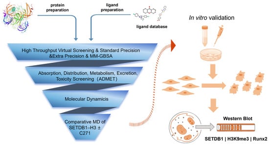
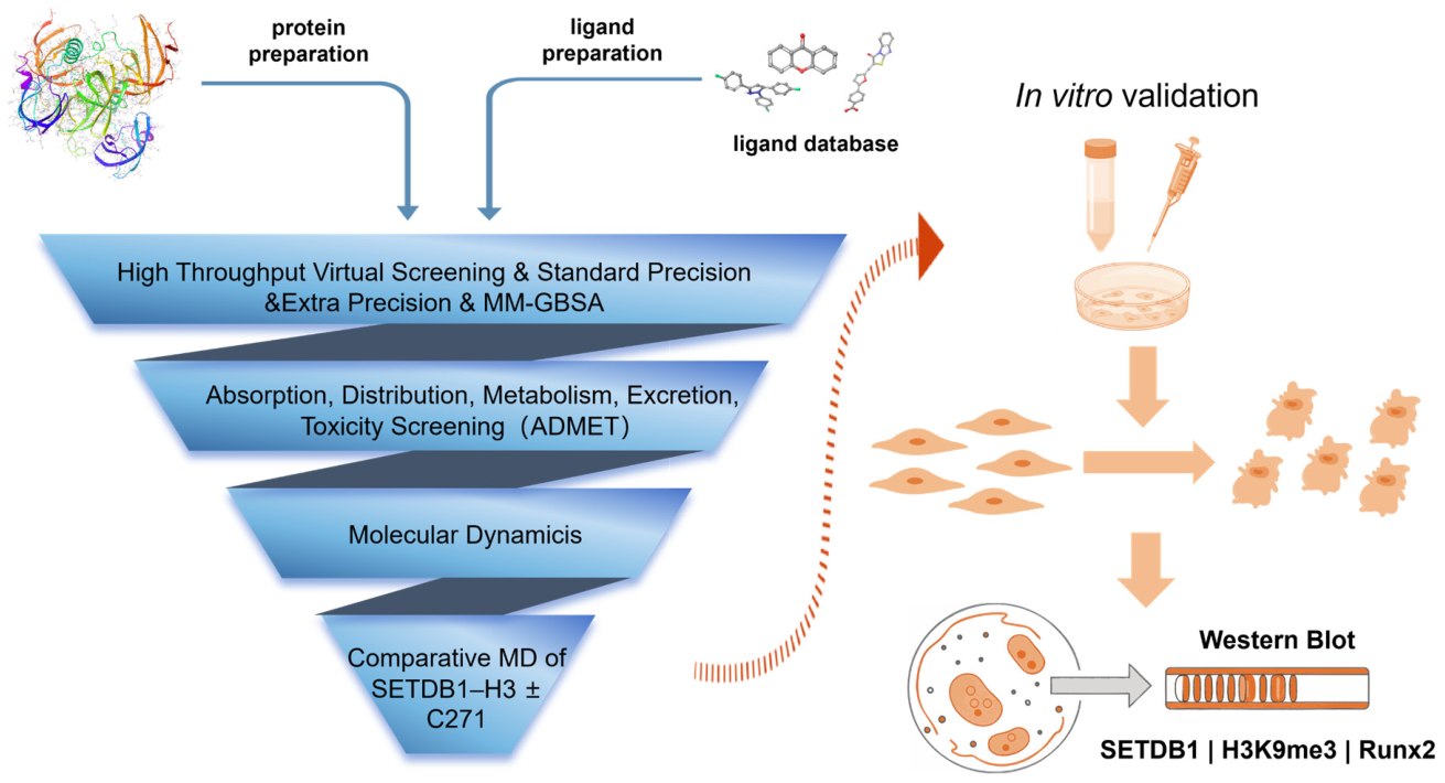
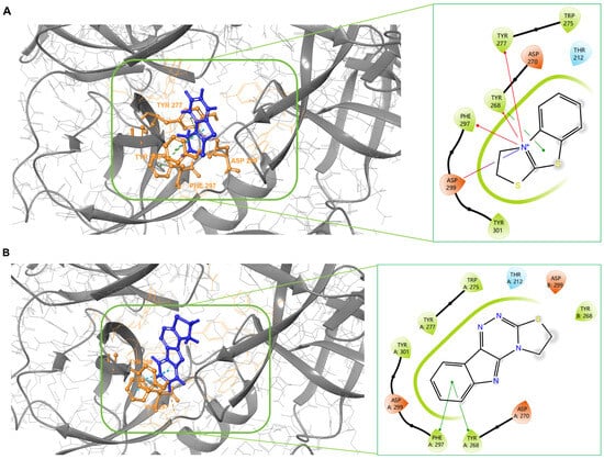
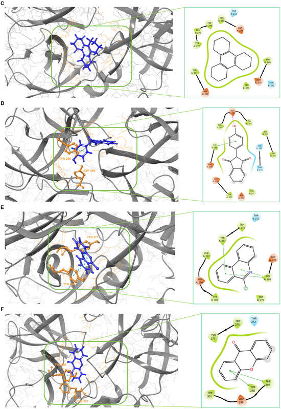
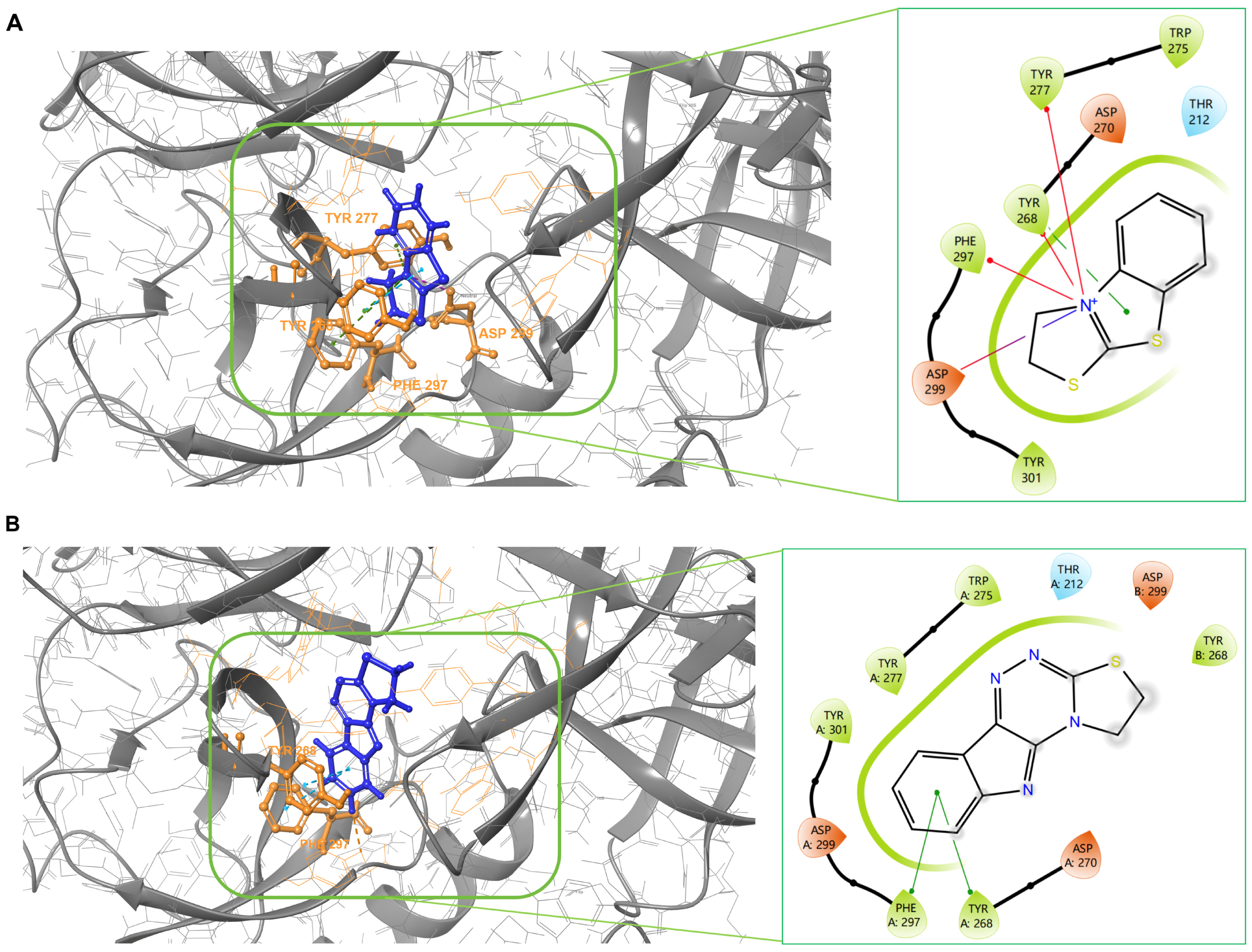
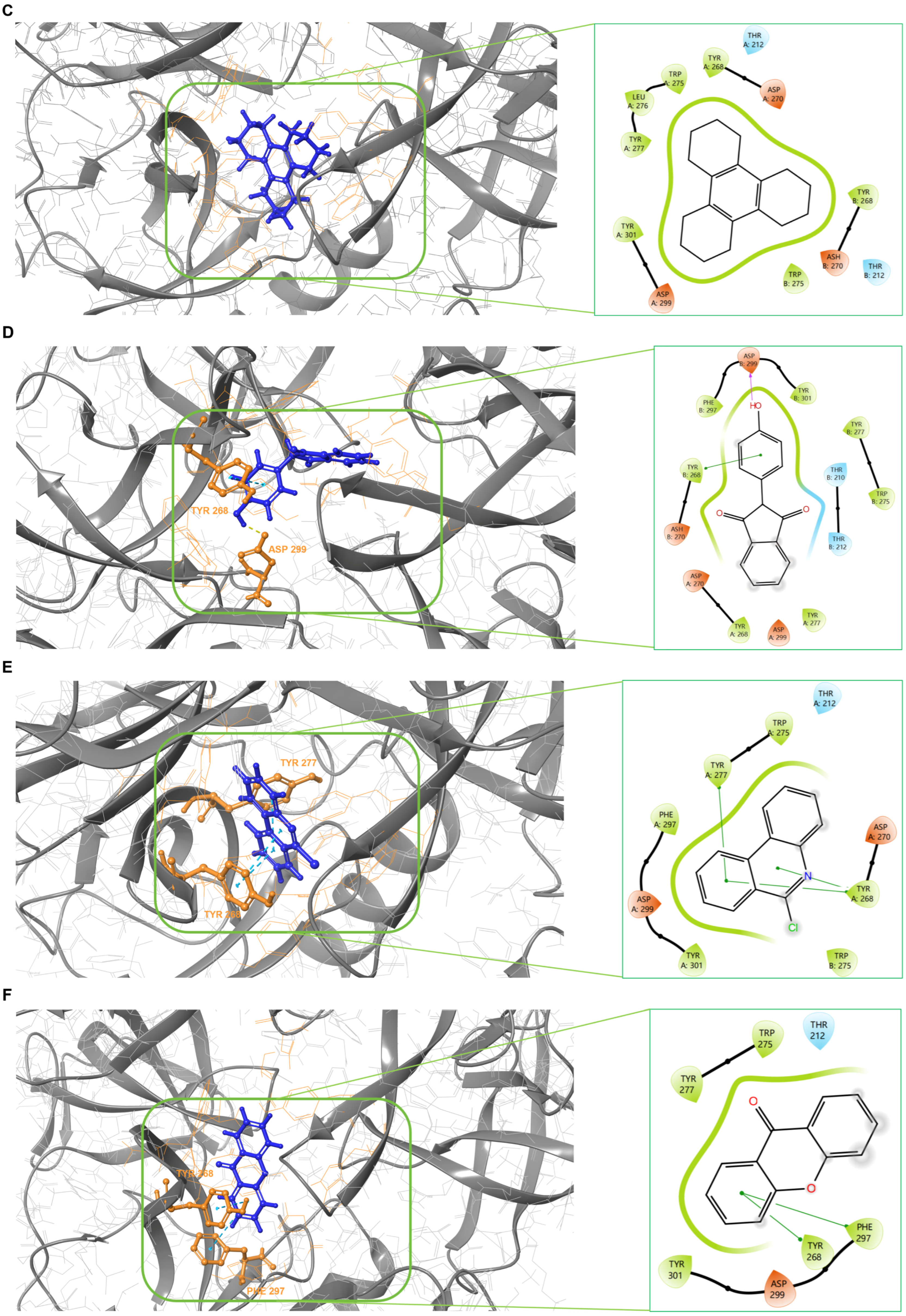
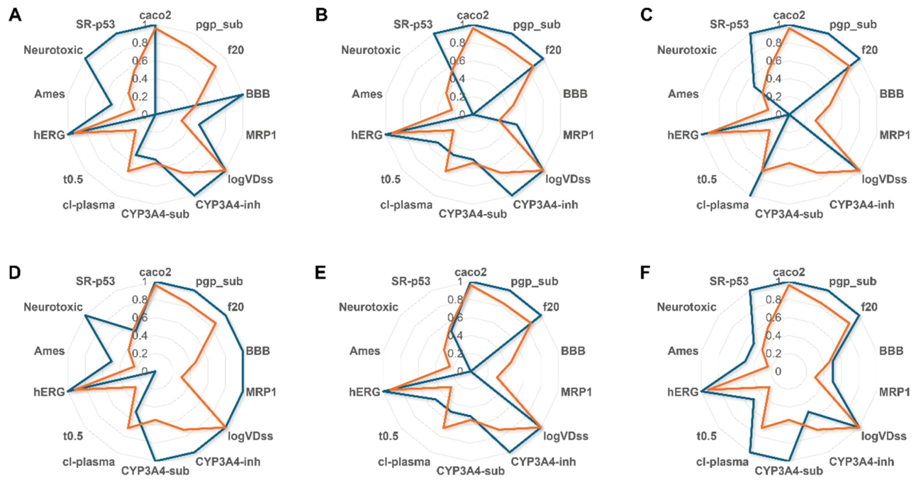
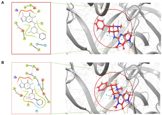
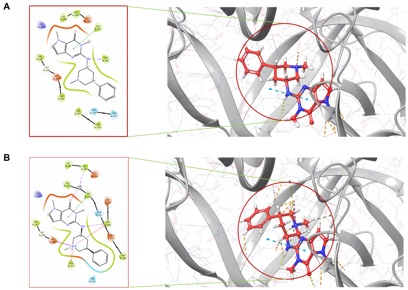
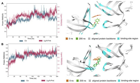
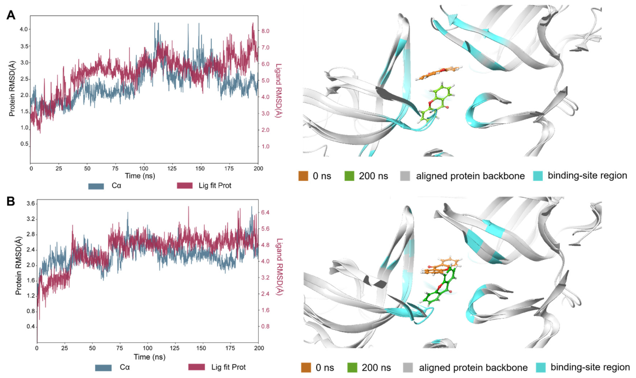
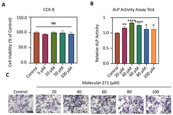
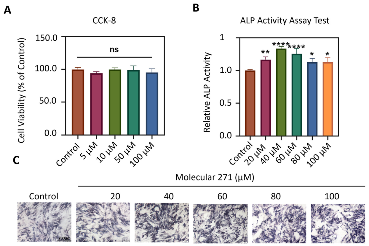
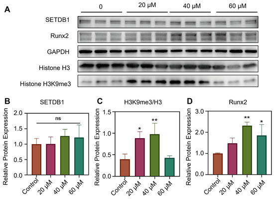
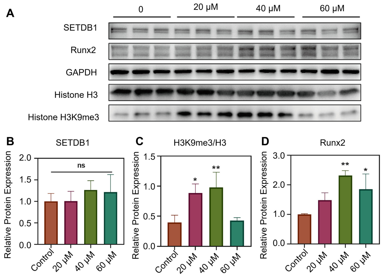

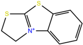
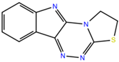

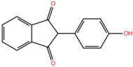
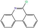
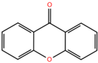
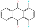
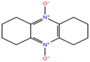
Disclaimer/Publisher’s Note: The statements, opinions and data contained in all publications are solely those of the individual author(s) and contributor(s) and not of MDPI and/or the editor(s). MDPI and/or the editor(s) disclaim responsibility for any injury to people or property resulting from any ideas, methods, instructions or products referred to in the content. |
© 2026 by the authors. Licensee MDPI, Basel, Switzerland. This article is an open access article distributed under the terms and conditions of the Creative Commons Attribution (CC BY) license.