


Abstract
Deposition of fibrous connective tissue within the extracellular matrix is initially an adaptive and reversible wound-healing process. However, persistent dysregulation of fibrotic signaling pathways induces irreversible cellular dysfunction, tissue degeneration and organ failure. Despite the high mortality rate associated with fibrotic diseases, there are very limited approved anti-fibrotic treatments and there is no therapeutic drug effective enough to completely invert the fibrotic process. Accordingly, new therapeutic agents that will attenuate ongoing fibrosis and, at the same time, promote regeneration of injured tissue are urgently needed. The search for new therapies has been revolutionized by recent advances in stem cell biology. Mesenchymal stem cells (MSCs) are promising candidates for the therapy of organ fibrosis because of their differentiation capabilities and immunomodulatory properties. The capacity of MSCs to suppress chronic inflammation and promote tissue repair and regeneration underlies their therapeutic effects in diseases such as liver cirrhosis, idiopathic pulmonary fibrosis, cardiac fibrosis, systemic sclerosis, and renal fibrosis. In this review, we summarize the present understanding of fibrotic disease, highlight promising research avenues, including MSC-based treatment options, and discuss the challenges involved with the clinical application of MSCs.
1. Introduction
Fibrosis is a progressive and dynamic pathophysiological response to a broad range of tissue injuries [1]. Although the deposition of fibrous connective tissue within the extracellular matrix (ECM) is initially an adaptive and reversible wound-healing process, persistent dysregulation of fibrotic signaling pathways induces irreversible cellular dysfunction and tissue degeneration [2]. This devastating disorder can affect almost all adult organs, including the liver, lung, kidney, heart, skin, and gastrointestinal tract [3]. Despite organ-specific clinical manifestations, recent findings have underscored the importance of chronic inflammation orchestrated by sustained production of cytokines and growth factors, and differentiation of myofibroblasts followed by excessive matrix deposition [4]. The uncontrolled fibrotic process results in organ failure, substantially contributing to global morbidity and mortality [5]. It is estimated that severe fibrotic complications contribute to nearly 45% of all deaths in industrialized countries [6]. However, despite the high mortality rate, there are very limited approved anti-fibrotic treatments and there is no therapeutic drug effective enough to completely invert the fibrotic process [7]. Due to a substantial clinical burden on patients and a significant financial burden on society [8], researchers and clinicians all over the world have worked on developing efficient therapeutic strategies to treat this life-threatening disease. The search for new therapies has been revolutionized by recent advances in stem cell (SC) biology, which suggest that SCs might be exploited to repair fibrosis-affected tissues. Mesenchymal stem cells (MSCs) are promising candidates for the therapy of fibrotic diseases because of their differentiation capabilities and immunomodulatory properties [9]. Therefore, in this review, we address the present understanding of fibrotic disease, look at promising research avenues including MSC-based treatment options, and discuss the challenges involved with the clinical use of these cells. An extensive literature review was carried out in January 2026 across several databases (MEDLINE, EMBASE, Google Scholar, and ClinicalTrials.gov), from 1990 to present. Keywords used in the selection were “mesenchymal stem cells”, “organ fibrosis”, “lung fibrosis”, “liver fibrosis”, “cardiac fibrosis”, “renal fibrosis”, and “systemic sclerosis”. The abstracts of all these articles were subsequently reviewed by the authors to check their relevance to the subject of this manuscript.
2. Mesenchymal Stem Cells as New Agents in Cell-Based Therapy of Fibrosis
Fibrosis is not a single-factor process; thus, the identification of cell populations and signaling pathways that contribute to fibrosis in various tissues is essential for understanding the pathogenesis of fibrotic disease. At the first stage of the fibrogenic process, increased release of inflammatory cytokines and mechanical stress following tissue injury induce activation, proliferation, and differentiation of resident fibroblasts into myofibroblasts. Several studies indicated that upon activation, fibroblasts proliferate outside fibrotic foci and migrate via chemotaxis and durotaxis while acquiring a biologically activated phenotype [10]. In addition to resident fibroblasts, a variety of other cell types including vascular pericytes, bone marrow-derived cells, epithelial cells, and subcutaneous adipocytes, can be precursors of myofibroblast-like cells [11,12,13]. Scar-forming myofibroblasts, which are phenotypically between fibroblasts and smooth muscle cells, are usually characterized by the expression of the contractile protein alpha-smooth muscle actin (α-SMA) and by their capacity to produce and secrete ECM components including collagen and fibronectin. Most recently, several studies have suggested Periostin (Postn) and Collagen triple helix repeat containing-1 (Cthrc1) as more specific markers of activated fibroblasts across different organs in fibrotic disease [14,15]. Increased tissue accumulation of myofibroblasts leads to uncontrolled deposition of ECM proteins while an imbalance of matrix metalloproteinases (MMPs), and tissue inhibitors of metalloproteinases (TIMPs), further contributes to improper ECM remodeling and the progression of fibrotic disease [9].
A deeper understanding of the myofibroblast regulatory mechanisms has facilitated the identification of new therapeutic targets, with the aim of optimizing scar formation and preventing progression to organ failure. Transforming growth factor (TGF)-β is considered a main inducer and promoter of fibrotic lesions. TGF-β is synthesized as an inactive precursor and activated through proteolytic cleavage mediated by plasmin, MMP2, MMP9 and thrombospondin 1. Active TGF-β binds to the TGF-β receptor type II, leading to formation and translocation of phosphorylated Smads complexes to the nucleus, which induce the transcription of fibrotic markers α-SMA, collagen I and fibronectin [16]. TGFβ1 further enhances the production of a broad range of bioactive factors, such as connective tissue growth factor (CTGF), plasminogen activator inhibitor 1 (PAI-1), endothelin-1, and NADPH oxidase 4, which are also involved in myofibroblast recruitment, maturation and collagen synthesis [17]. TGFβ signaling is also transduced through non-Smad pathways, including the mitogen-activated protein kinase (MAPK) pathways such as the extracellular signal-regulated kinases (Erks), c-Jun amino terminal kinase (JNK), and p38 MAPK, as well as the IκB kinase (IKK), phosphatidylinositol-3 kinase (PI3K) and Akt, and Rho family GTPases [18].
Besides the aforementioned key regulators of fibrosis, there is a wide range of well-established factors that stimulate fibrogenesis, such as Wnt/β-catenin, Hedgehog, and Notch pathways. Noncanonical Wnt signaling via JNK/c-Jun also contributes to fibroblast activation and myofibroblast differentiation, in addition to or in synergy with canonical Wnt3a/β-catenin signaling [19,20,21]. The Sonic Hedgehog (SHH)-GLI axis plays an important role in fibrogenesis, both directly by inducing the transcription of pro-fibrotic genes, including α-SMA, collagen, and fibronectin, and indirectly through interaction with other pro-fibrotic pathways, particularly TGF-β signaling [22,23]. Chronic inflammation plays a significant role in fibrosis and fibrotic diseases [5]. Upon activation of neutrophils and T cells, macrophages invade injured tissues, and release fibrogenic cytokines, chemokines and ROS [24]. Increased production of pro-inflammatory mediators, including TGF-β1, tumor necrosis factor-alpha (TNF-α), monocyte chemoattractant protein (MCP-1), interleukin-6 (IL-6), and IL-8, amplifies tissue damage and promotes fibrosis in a pathogenic manner [25]. Therefore, the systematic identification of signaling pathways and immune cells highlighted MSCs as a promising resource for the therapy of organ fibrosis on the grounds of their immunomodulatory characteristics and paracrine effects.
MSCs are adult, multipotent stem cells, that reside in almost all postnatal tissues. The most common sources of MSCs are bone marrow, adipose tissue, and the umbilical cord. After tissue harvesting, isolated stem cells are identified according to the standard criteria established by the Mesenchymal and Tissue Stem Cell Committee of the International Society for Cellular Therapy (ISCT). MSCs are characterized by plastic adherence with clonogenicability, expression of static membrane markers (CD73, CD90, and CD105), lack of hematopoietic and endothelial markers, and mesodermal differentiation capacity (osteogenic, adipogenic, and chondrogenic) [26]. The vast majority of preclinical and clinical studies demonstrate that the regenerative effects and use of MSCs in fibrotic disease are based on their immunomodulatory properties. Via direct cell–cell interaction or the release of soluble factors, MSCs can modulate activation, proliferation, and function of immune cells, which act as central regulators of the fibrotic process. Accordingly, the capacity of MSCs to suppress chronic inflammation and promote tissue repair and regeneration underlies their therapeutic effects in the treatment of liver cirrhosis, idiopathic pulmonary fibrosis, cardiac fibrosis, systemic sclerosis, and renal fibrosis. In order to elucidate the therapeutic potential of MSCs in the modulation of organ fibrosis, we present the current findings regarding molecular mechanisms involved in the MSC-dependent regulation of fibrotic processes with a brief overview of the published preclinical and clinical studies that utilized MSCs for the treatment of these diseases.
2.1. MSC-Based Therapy of Liver Fibrosis
Hepatic fibrosis, a pathophysiological process of abnormal hyperplasia of connective tissue, is a result of prolonged liver injury, chronic inflammation and exhaustion of the regenerative capacity of hepatocytes. This serious condition, caused by metabolic factors (alcohol, toxins, and drugs), infections (hepatitis B (HBV) and hepatitis C (HBC)), autoimmune diseases (primary biliary cholangitis), or genetic disorders (increased iron storage) [27], may further progress towards liver cirrhosis and hepatocellular carcinoma. However, independent of the etiology [28], after many rounds of injury and repair, mature hepatocytes reach a senescent state of permanent cell-cycle arrest, while quiescent vitamin A-storing hepatic stellate cells (HSCs) become activated and transdifferentiate into contractile myofibroblast-like cells [28]. Excessive production of reactive oxygen species and pro-inflammatory cytokines (IL-6, IL-1β, tumor necrosis factor-α (TNF-α), and TGF-β) triggered by pathogenic factors, is the main driver of hepatocyte apoptosis and HSCs’ activation [29]. Proliferative α-SMA+ myofibroblast-like cells overproduce ECM components, particularly fibrillar collagen type I, leading to matrix remodeling and increased tissue stiffness. Due to impaired matrix degradation by MMPs, the formation of scar tissue in liver parenchyma is characterized by septal thickening and destruction of the space of Disse [30]. These histopathological changes result in liver failure by impairing normal metabolic exchange between portal blood and hepatocytes, and by promoting the development of portal hypertension [30].
Administration of MSCs exerts therapeutic effects for the liver fibrosis through both the inhibition of HSCs’ activation and the attenuation of chronic liver inflammation. Although several in vitro studies demonstrated that a combination of factors including hepatocyte growth factor (HGF), fibroblast growth factor (FGF) 2/4, and dexamethasone induces MSCs to differentiate into hepatocyte-like cells [31,32,33], preclinical and clinical research suggested that the activities of transplanted MSCs are primarily mediated via paracrine processes and immunomodulation instead of through transdifferentiation (Figure 1).
Figure 1.
Therapeutic potential of MSCs in organ fibrosis. MSC administration attenuates inflammation, inhibits fibroblast differentiation and reduces collagen deposition, leading to increased ECM degradation, and improved function of the affected organ.
Aberrations in G1 regulation following liver injury often lead to the rapid proliferation of HSCs. Several studies aimed to investigate the effects of MSCs on the proliferation-related events of activated HSCs and to clarify the underlying molecular mechanism. MSCs are able to suppress the proliferation and α-SMA expression in HSCs through direct cell–cell contact, particularly by activation of Notch 1 signaling [34]. Increased Notch1 and Hes1 expression in activated HSCs cocultured with MSCs leads to HSC growth inhibition and may be mediated by crosstalk with the PI3K/Akt pathway. Moreover, MSCs induce G0/G1 cell-cycle arrest in HSCs partly by secreting TGF-β3 and HGF, which results in up-regulation of p21Cip1 and p27Kip1 expression and down-regulation of cyclinD1 and phosphorylated-ERK [35]. p21Cip1 and p27Kip1 are involved in the regulation of CDK activity and cell-cycle progression in regenerating liver tissue. In particular, p27Kip1 negatively regulates the activity of protein kinase complex cyclin E-CDK2, preventing the transition of G1 to the S phase. Because production of type I collagen is controlled by the ERK pathway through the phosphorylation of its downstream molecules, the observed decrease in ERK phosphorylation highlights the therapeutic potential of MSCs to target this signaling cascade. Wang et al. demonstrated that HGF and direct MSC contact synergize to remarkably modulate HSC activation in vitro through the TLR4/MyD88/NF-kB signaling pathway [36]. MSCs and their products decrease the level of intracellular MyD88 by down-regulating the expression of TLR4, leading to the attenuation of inflammation, which, in turn, blocks the activation of NF-kB, resulting in the inhibition of HSC activation. These findings suggest that, in addition to direct juxtracrine signaling, the paracrine mechanisms by which MSCs inhibit activated HSCs also play an important mechanistic role in the interplay between these two cell types. HSCs co-cultured with MSCs exhibited the shift of balance of anti- and pro-apoptotic proteins. MSC-derived nerve growth factor (NGF), via activation of the p75 neurotrophin receptor, inhibits the nuclear factor kappa B (NF-κB) pathway, resulting in increased expression of pro-apoptotic Bax and caspase-3, and reduced expression of anti-apoptotic Bcl-xl in activated HSCs [37,38]. Beyond modulating the intrinsic mitochondrial apoptotic pathway, BM-MSCs significantly reduce NADPH oxidase (NOX) activity, a key generator of ROS during liver fibrosis. While BM-MSCs have only a minimal effect on total p47phox expression, they significantly reduce the phosphorylation of the p47phox subunit, thereby restraining NOX activation [37]. In accordance with in vitro results, animal studies confirmed that the administration of MSCs inhibited liver fibrogenesis and promoted partial recovery of liver function in carbon tetrachloride 4 (CCl4)-treated rats by inhibiting the proliferation and promoting the apoptosis of HSCs [36,39]. Reduced liver fibrosis after MSC transplantation was associated with the enhanced proteolytic activity of MMP-9 and MMP-13 and the attenuated activation of TIMP-1 [40]. MMPs degrade the triple-helical domain of fibrillar collagen types I and III, which constitute the hepatic scar tissue, into smaller fragments that are subsequently phagocytosed by macrophages. Reduced expression of TIMPs further enables increased degradation of ECM components and tissue repair. Suppression of TGF-β signaling is an important mechanism of MSC-based reversion of liver fibrosis [9]. By inhibiting the phosphorylation of Smad2 and Smad3, and thereby preventing their translocation into the nucleus, MSCs reduce activation of pro-fibrotic genes, including collagen-1 and α-SMA, thus alleviating liver cirrhosis [41]. Moreover, MSC treatment led to the recovery of liver function through improved microcirculation and the amelioration of the hypoxic microenvironment in cirrhotic livers [42,43]. Improved microcirculation and ameliorated fibrosis correlate with down-regulation of hepatic VEGF expression, suggesting a potential molecular mechanism for the observed therapeutic effect of MSC transplantation [42].
We and others provided the evidence that the therapeutic potential of MSCs in liver fibrosis was systemic and that these effects were based on the production of immunoregulatory molecules. Interestingly, MSC-derived secretomes and exosomes have similar therapeutic functions as stem cell transplantation in liver fibrosis, indicating that secreted soluble factors are mainly responsible for the beneficent effects of MSCs in this disease [44]. MSCs are able to modulate chronic hepatic inflammation and fibrosis by interacting directly with cells of both innate and adaptive immunity. MSCs, in a paracrine manner, suppress liver IL-17 producing Th17 cells and promote expansion of IL-10+ T regulatory cells, which leads to the attenuation of CCl4-induced liver fibrosis [45,46]. The hepatoprotective effects of MSCs are mainly mediated by indoleamine 2,3-dioxygenase (IDO), an enzyme involved in the catabolism of tryptophan to immunosuppressive kynurenine, and IL-10, a well-known anti-inflammatory cytokine [47]. IDO-derived kynurenine inhibits the proliferation of effector T cells, but promotes the expression of Treg lineage-defining transcription factor FoxP3 [48]. Since MSCs, in a paracrine manner, significantly increase the capacity of Tregs to produce immunosuppressive IL-10, it could be proposed that MSCs, in an IDO-dependent manner, induce increased production of IL-10 in Tregs, which, in turn, attenuates liver inflammation through an IL-10-dependent mechanism [48]. Moreover, MSCs ameliorate liver cirrhosis by modulating the macrophage phenotype [49,50]. Although the majority of transplanted cells are trapped in the lungs, MSCs support a systemic anti-inflammatory state and facilitate macrophage polarization to the M2 phenotype [49]. In a pro-inflammatory environment, activated MSCs produce high levels of PGE2, a product of the cyclooxygenase 2 (COX-2) pathway. PGE2 binds to EP2 and EP4 receptors on the macrophage, leading to macrophage polarization toward an anti-inflammatory phenotype [9]. Additionally, MSCs secrete TSG-6, which interacts with the CD44 on macrophages, resulting in suppressed TLR2/NFκ-B signaling and reduced release of pro-inflammatory cytokines [9]. Alternatively activated M2 macrophages produce anti-inflammatory cytokines such as C-C motif chemokine ligand 1 (CCL-1) and IL-10, which further suppress chronic liver inflammation and attenuate liver fibrosis. Luo et al. have demonstrated that MSC transplantation can promote the activation of F4/80 + CD206 + M2 macrophages expressing MMP13 and the inhibition of F4/80 + iNOS+ M1 macrophages, which subsequently inhibit HSCs [50]. MSCs effectively suppressed the expression of profibrogenic α-SMA, ΤGF-β1, and collagen-1 and pro-inflammatory genes IL-12, interferon (IFN)-γ, TNF-α, and IL-6 in the liver, which are positively correlated with fibrosis and induced by CCl4 injection [45,50].
Over the past decade, 16 clinical trials investigating the therapeutic potential of MSCs obtained from bone marrow, umbilical cord tissue, and adipose tissue for treating liver cirrhosis have been completed and the majority of these were registered in Asian countries. These completed clinical studies generally reported that MSC transplantation is safe, well tolerated and without significant adverse events or complications (Table 1). Autologous bone marrow-derived MSCs (BM-MSCs), as well as umbilical cord-derived MSCs (UC-MSCs), improved liver function and clinical features in patients with end-stage liver failure and decompensated liver cirrhosis with different etiologies. The therapeutic effects of MSCs were demonstrated by a reduced Model for End-Stage Liver Disease (MELD) score, decreased fatigue scale and bilirubin, and improvement in coagulation parameters and serum albumin level [51,52,53,54,55,56]. Autologous BM-MSC transplantation was effective in terms of improving liver function in patients with HBV-associated liver cirrhosis, which was mediated, at least in part, through the regulation of Treg/Th17 cell balance, resulting in significantly lower serum concentration of pro-inflammatory IL-17 and TNF-α in the transplantation group compared with the control [57]. In addition, patients with HBV-induced liver cirrhosis had lower expression of fibrotic markers at 48 weeks after MSC injection [55,56]. Similarly, in patients with alcoholic liver cirrhosis, hepatic arterial injection of autologous BM-MSCs safely improved liver function and reduced collagen deposition [58]. Liver regeneration-related factors, such as HGF and IL-6, were elevated one day after adipose tissue-derived stem cell treatment in four cirrhotic patients [59]. Since cirrhosis is considered one of the most important risk factors for hepatocellular carcinoma (HCC) and can lead to a high mortality rate for patients with hepatitis B, Peng and coworkers provided evidence that autologous MSC transplantation might exert protective effects for cirrhosis patients with regard to the occurrence of HCC and mortality [60]. Although the results from many controlled trials support the use of autologous MSCs for liver cirrhosis, some studies [61,62] have not demonstrated significant therapeutic effects being derived from MSC transplantation through the peripheral vein in cirrhotic patients.
2.2. MSCs—A Promising Therapeutic Approach for Idiopathic Pulmonary Fibrosis
Idiopathic pulmonary fibrosis (IPF) is a progressive lung disorder characterized by scarring of the lungs from an unknown cause. Recurrent injury to the alveolar epithelium, and increased apoptosis of alveolar epithelial II (ATII) cells, triggers a cascade of dysregulated immune response leading to the activation of fibroblasts and excessive ECM deposition in the interstitial and perivascular space [63]. The key immune cells that propagate ongoing inflammation often related to exposure to cigarette smoke, air pollution, respiratory infections and radiation therapy are neutrophils, monocytes, Th2-lymphocytes and dendritic cells. The origin of myofibroblasts, a major driver of pulmonary fibrogenesis, is still a matter of debate. The proposed origin of α-SMA+ myofibroblast in lung parenchyma include the proliferation of resident fibroblasts, transdifferentiation of epithelial and endothelial cells by epithelial–mesenchymal transition (EMT) or endothelial–mesenchymal transition (EndoMT), invasion of fibrocytes, and differentiation of pericytes [64]. More recently, Tsukul et al. demonstrated by lineage tracing that Scube2+-alveolar fibroblasts are the dominant source of newly formed Cthrc1+ pro-fibrotic fibroblasts in response to lung injury [65]. The destruction of lung architecture due to scarring impairs gas exchange and progresses to hypoxic respiratory failure, which is clinically characterized by shortness of breath, progressive dyspnea, and a dry, nonproductive cough [66]. Current treatment strategies include supportive measures such as oxygen supplementation, and immunosuppressive and anti-fibrotic medications. Lung transplantation often represents the only remaining therapeutic option for these patients; thus, MSC transplantation stands as a promising curative option for severe cases.
Many investigators using animal models of lung fibrosis demonstrated the promising therapeutic effects of MSCs. After systemic administration, MSCs successfully reach and engraft injured lung tissue, where they exhibit regenerative properties. The production of substances chemotactic for stem cells by injured lung tissue may help to explain the selective homing of these cells to areas of tissue injury. Transplantation of MSCs isolated from different tissues, including bone marrow, adipose tissue and the umbilical cord, significantly reduced bleomycin-induced lung fibrosis and notably decreased the mortality rates of experimental animals by similar molecular mechanisms [67,68]. MSC-attenuated collagen deposition brought about by modulating the activity of MMP-2, MMP-9, and MMP-13 restores the balance between MMP-2 and TIMP 1–4 in lung tissue [67] and downregulates the expression of other pro-fibrotic factors responsible for ECM remodeling, such as bFGF, connective tissue growth factor (CTGF), collagen type I alpha 1 chain (Col1a1) and collagen type III alpha 1 chain (Col1a3) in lung tissue [68]. Mechanistically, reduced collagen synthesis has been a consequence of the simultaneous reduction in Smad2 phosphorylation and down-regulated TGF-β1 activity following MSC treatment [67]. Moreover, MSCs managed to reduce inflammation and pulmonary edema, as demonstrated by decreased production of pro-inflammatory IL-2, IL-1β, TNF-α, and IFN-γ [67,68]. MSC-derived keratinocyte growth factor (KGF), VEGF, and HGF play crucial roles in stimulating regeneration of ATII cells, and stabilizing the endothelial and epithelial cell junctions in the alveolar-epithelial barrier. These paracrine effects are associated with decreased vascular permeability and reduced accumulation of protein-reach fluid in the alveoli. Most recently, it was demonstrated that MSC-secreted KGF upregulates Gab1 protein expression and inhibits the activation of the ERK/NF-kB signaling pathway, thereby upregulating the ENaC protein level in ATII cells and alleviating pulmonary edema [69]. Importantly, histological analysis and high-resolution computed tomography of lungs confirmed substantial improvement in alveolar architecture and thickening of alveolar septa in MSCs + bleomycin-treated animals [68].
The anti-inflammatory effect of MSCs in pulmonal fibrosis is manifested by the reduced influx of neutrophils, macrophages, and lymphocytes as well as by the reduced synthesis and releases of inflammatory cytokines and chemokines in lung-infiltrated immune cells [70,71,72] (Figure 1). During the late stage of immune response due to prolonged injuries, M2 macrophages produce CCL18 and IL-10, which, in conjunction with the PGE2 and TGF-β produced by MSCs, enhance the production of HLA-G5. Moreover, Kotani et al. found significantly lower production of pro-inflammatory cytokines (TNF-α and IL-12) and increased programmed cell death in activated macrophages co-cultured with MSCs [70]. The described cellular crosstalk promotes expansion of Tregs and prevents migration of neutrophils into the lungs, protecting tissue from consequent oxidative damage. This includes phenotypic conversion of CD4+ T lymphocytes into regulatory T lymphocytes, which are cells important in maintaining immune homeostasis and preventing inflammatory damage, including lung fibrosis [70]. Several in vivo studies confirmed that the systemic administration of MSCs effectively abolished neutrophilic lung inflammation [71] and down-regulated the bleomycin-induced increase in levels of IL-2, IL-1β, TGF-β, vascular endothelial growth factor (VEGF), IL-6, TNF-α, and NOS in the lungs [72]. These data suggest that MSC transplantation may be an effective strategy for the treatment of lung fibrosis via modulation of the microenvironment of injured lung tissue. Moreover, when bleomycin-treated animals received stem cells, circulating levels of granulocyte colony-stimulating factor (G-CSF) and granulocyte-macrophage colony-stimulating factor (GM-CSF) were significantly higher [72]. Thus, part of the effect of stem cell transplantation on lung fibrosis may be due to humorally mediated mobilization of endogenous stem cells. However, additional studies are necessary to determine the specific cell source and nature of these soluble mediators, and their effects on the expansion of the endogenous stem cell pool.
Preclinical studies demonstrated that MSCs had the potential to limit pulmonary fibrosis after exposure to ionizing irradiation [73,74]. Interestingly, the capacity of MSCs to differentiate into functional lung epithelial and endothelial cells is controlled by the injured microenvironment and highly dependent on the timing of cell administration [73]. Another study undoubtedly indicated an effect of the donor’s age on the anti-fibrotic properties of MSCs. In contrast to old (22 months) cells, adipose stem cells from young (4 months) mice decreased interstitial and perivascular collagen accumulation, and reduced the expression of lung fibrosis-related markers, including TGFβ, MMP-2, and protein kinase B [75]. A diminished radiation-induced pulmonary fibrosis was primary characterized by attenuated inflammation, impaired proliferation and activation of fibroblasts, and reduced collagen synthesis [73,74]. Dong et al. indicated that treatment with MSCs may suppress the activation of fibroblasts by elevating the release of HGF and prostaglandin E2 (PGE2), while host production of TGF-β1 and TNF-α is simultaneously reduced [74]. The general mechanisms responsible for the HGF- and PGE2-dependent protection of alveolar epithelial cells and inhibition of pulmonary fibrosis is partially understood. HGF induces expression of the negative regulator of TGF-β signaling, Smad7, in a MAPK-dependent manner, and promotes its export from the nucleus to the cytoplasm, thereby inhibiting the α-SMA expression in the alveolar epithelial cells and limiting their transition to a myofibroblast phenotype [76]. PGE2 does not interfere with TGF-β1-induced Smad phosphorylation or its translocation to the nucleus. Rather, PGE2 has dramatic effects on cell shape and cytoskeletal architecture and disrupts the formation of appropriate focal adhesions. This inhibition of focal adhesion kinase signaling by PGE2 is associated with reversing myofibroblast phenotype [77].
Although most clinical studies related to the MSC-based therapy of IPF were not designed to delineate mechanisms of actions or authenticate efficacy, they provide indications that the current therapeutic approach may be of some benefit (Table 1). A single intravenous infusion of human MSCs in patients with mild–moderate IPF was well tolerated and without treatment-emergent serious adverse effects such as nonfatal pulmonary embolism, stroke, hospitalization for worsening dyspnea, and clinically significant laboratory test abnormalities [78].
After six months of follow-up, Chambers et al. concluded that intravenous administration of placenta-derived MSCs is feasible and has a good short-term safety profile in patients with moderately severe IPF [79]. In order to achieve maximum accumulation into sites of ongoing injury and, thus, maximize their therapeutic potential, several authors investigated the feasibility of endobronchial administration of BM-MSCs in patients with mild-to-moderate IPF. Tzouvelekis and coworkers demonstrated an acceptable safety profile of endobronchially administered adipose tissue-derived MSCs (AT-MSCs) in patients with IPF, without short-term infusional toxicities and long-term ectopic tissue formation [80]. The number of patients without functional progression was six (46%) at 3 months and three (23%) at 12 months [81]. In a phase Ib clinical trial using AT-MSCs in IPF patients, the median survival and time to progression were in line with the previously published epidemiological data [82]. However, the first clinical data confirming the therapeutic effect of MSCs in IPF was published in 2019. In this pilot study, the cohorts receiving 1 × 10⁸ BM-MSCs demonstrated slower progression in terms of quantitative lung fibrosis and a smaller decrease in pulmonary function, including the diffusing capacity of the lungs for carbon monoxide (DLCO), than subjects receiving 2 × 10⁷ stem cells [83]. These findings provide a rationale for further investigation of the dose–response relationship of MSCs therapy in IPF, and confirm the safety of high-dose cell administration in regenerative medicine. In accordance, administration of a high cumulative dose of BM-MSCs (1.6 × 10⁹ cells in total) in patients with rapidly progressive IPF provided significant improvements in the 6-min walk distance (in 13 weeks), in DLCO (in 26 weeks), and in forced vital capacity (in 39 weeks) compared with the placebo group. suggesting MSC application is a promising method to reduce IPF progression [84]. These results suggest that determining the ideal stem cell dose may be crucial for effective treatment.
2.3. MSC-Mediated Attenuation of Cardiac Fibrosis
Cardiac fibrosis is a common disorder observed in varying degrees in many kinds of cardiovascular diseases, including myocardial infarction, coronary heart disease, hypertension, myocarditis and cardiomyopathies [85]. Pathological stress such as pressure overload, endothelial loss, or ischemic injury induces death of cardiomyocytes, leading to an inflammatory and fibrotic response [86]. At an early stage of cardiac stress, these changes contribute to an adaptive repair process but eventually lead to adverse cardiac remodeling and progression toward heart failure. In particular, due to persistent myocardial ischemic injury, cardiomyocyte death and replacement fibrosis occur, leading to systolic dysfunction, while in non-ischemic injury, interstitial fibrosis mainly occurs, contributing to diastolic dysfunction [87].
Ca2+/calmodulin-dependent protein kinase II (CaMKII) activation under cardiac stress conditions plays a critical role in the initiation and amplification of inflammatory signaling. In stressed cardiomyocytes, activated CaMKII triggers the inflammasome activation through NF-κB and reactive oxygen species (ROS) generation [88]. This signaling cascade enhances the production of pro-inflammatory cytokines and chemokines, which facilitate immune cell recruitment and infiltration into the myocardium. Activated immune cells produce high levels of inflammatory cytokines that activate ERK1/2 in cardiac fibroblasts, thereby promoting their transformation into activated myofibroblasts with an excessive and continuous production of collagen [89]. Macrophages occupy a central position in cardiac fibrosis and participate in crosstalk with immune cells, mainly through the release of different mediators that affect their chemotaxis and functions [90,91]. Through the secretion of variety of pro-fibrotic mediators, such as TGF-β, platelet-derived growth factor (PDGF), IL-10, VEGF, and amphiregulin (AREG), macrophages directly induce fibroblast proliferation and activation via the engagement of fibroblast receptors (platelet-derived growth factor receptor (PDGFR), transforming growth factor-beta receptor (TGF-βR), and epidermal growth factor receptor (EGFR)), promoting collagen synthesis and cardiac fibrosis [92,93]. Moreover, macrophages secrete TIMPs that inhibit the degradation of the ECM, and facilitate tissue scar formation and myocardial remodeling. Deposition of type I and type III collagen mostly contributes to myocardial stiffness and the development of various forms of cardiac fibrosis, including reactive interstitial, replacement, infiltrative interstitial, or endomyocardial fibrosis [89].
The therapeutic potential of MSCs in cardiac fibrosis and inflammation has been investigated in both preclinical models of ischemic and non-ischemic cardiomyopathies. Several published studies suggest that MSC transplantation is useful for improving heart function after ischemia myocardial infarction (MI) [94,95]. The MSC-mediated anti-fibrotic mechanism is based on secretion of MMP-2, MMP-9 and MMP-14 and inhibition of cardiac fibroblast activity in a HGF-dependent manner [4,95]. BM-MSC transplantation into infarcted myocardium down-regulated expression of extracellular matrix genes including collagen types I and III, TIMP-1 and TGF-β1 [96]. This biological activity of MSCs results in reduced collagen volume in heart tissue and attenuated post-MI ventricular remodeling [94,96]. There is also an increasing body of work supporting the idea that the effect of MSCs on fibrosis may be mediated through reduced inflammation. Du et al. [97] demonstrated that intramyocardial injection of BM-MSCs into infarcted rat hearts after inhibited TNF-α/NF-κB signaling led to reduced levels of pro-inflammatory TNF-α and IL-6, and increased levels of protective IL-10 in the myocardium [97]. Reduced inflammation in injured rat myocardium after BM-MSC transplantation is characterized by lower production of monocyte chemotactic protein-1 (MCP-1) and inhibited infiltration of CD68+ immune cells [98]. Moreover, there is evidence suggesting a direct regenerative potential of MSCs in cardiac fibrosis. In particular, Kudo et al. observed that engrafted bone marrow-derived stem cells had differentiated into cardiomyocytes and vascular cells, leading to reduced infarct size and fibrosis [99]. Research has demonstrated the efficacy of AT-MSCs in reducing infarct size and maintaining cardiac function following a MI [100]. The mechanisms behind these improvements appear to operate through three distinct pathways: the direct differentiation of stem cells into newly formed cardiac tissue, paracrine signaling—particularly the secretion of beneficial cytokines—that promotes vascularization of the infarcted region, and paracrine-mediated effects that enhance tissue survival and attenuate ongoing cell death. Work by Zhu et al. suggests that the paracrine activity of AT-MSCs may actually carry greater therapeutic weight than their capacity for direct tissue regeneration [101]. Through these paracrine mechanisms, AT-MSCs can stimulate angiogenesis, recruit resident stem cells, dampen fibrotic responses, and prevent apoptosis. The growth factors they secrete include endothelial growth factor (EGF), HGF, insulin-like growth factor (IGF-1), and various microRNAs [102]. MSCs represent a promising therapeutic strategy for dilatative cardiomyopathies (DCM) across multiple etiologies, and their benefits derive primarily from anti-inflammatory and anti-apoptotic effects. In rodent DCM models, the beneficial effects of MSCs might be mediated not only by their differentiation into cardiomyocytes and vascular cells but also by their ability to supply large amounts of angiogenic, anti-apoptotic, and mitogenic factors such as vascular endothelial growth factor, hepatocyte growth factor, adrenomedullin, and insulin-like growth factor-1 [103]. Interestingly, BM-MSCs demonstrated stronger anti-fibrotic effects than AT-MSCs [104]. Reduced myocardial collagen network remodeling was mediated by downregulating myocardial TGF-β1, AT1, CYP11B2 (aldosterone synthase) gene expressions [105]. Moreover, Martire et al. demonstrated that MSCs suppressed inflammatory processes in the heart, most likely by inhibition of NFκBp65 signaling via sTNF-RI, leading to improved disease parameters, including extended survival of mice suffering from inflammatory DCM [106]. Umbilical cord-derived MSCs (UC-MSCs) preserve cardiac function after intramyocardial transplantation in a DCM mouse, and this effect may be associated with reductions in cellular apoptosis, inflammation, hypertrophy and myocardial fibrosis, in addition to up-regulation of Akt, VEGF and IGF-1 and enhanced angiogenesis [107]. Treatment with UC-MSCs repressed the increase in myocardial fibrosis and endothelial–mesenchymal transition observed in DCM rats, which correlated with decreased activity in terms of TGF-β1/ERK1/2 signaling [108]. Myocardial interstitial fibrosis is the main histopathological feature of diabetic cardiomyopathy. Transplanted BM-MSCs effectively migrated into heart tissue, and protected the myocardium by upregulating 14-3-3 protein expression. The increased 14-3-3 protein binds to phosphorylated apoptosis signal-regulating kinase 1 (p-ASK1) to inhibit the ASK1-mediated cardiomyocyte apoptosis [109]. Through the production of PGE2, MSCs ameliorated metabolic abnormalities, reduced inflammation and fibrosis, and preserved cardiac structure and function in diabetic cardiomyopathy [110]. MSC-derived soluble factors reduce macrophage recruitment, suppress M1 polarization, and promote M2 polarization to exert anti-inflammatory effects, and hold significant therapeutic implications for treating cardiac fibrosis in diabetic cardiomyopathy [110,111]. Although MSCs show promising results in terms of treating cardiac fibrosis, transplanted cells have a low survival rate in damaged cardiac tissue, and a broad range of methods are being tested in order to increase migration ability, survival, and therapeutic potential of MSCs in vivo. Strategies such as pre-conditioning before transplantation, genetic modifications and biomaterials could compromise their efficacy in clinical studies [112]. When it comes to human clinical trials, results seem to be promising; the APOLLO experiment was a major step forward because it was the first time that AT-MSCs were administered to patients with MI, specifically those with ST elevation [113]. Six months after intracoronary infusion of AT-MSCs, the results were encouraging: cardiac function had improved, as measured by ejection fraction on CT scan, tissue perfusion had increased, and infarct size had shrunk by an average of 50% compared to the placebo group [113]. Rather than relying on intracoronary delivery, the investigators opted for transendocardial injection of AT-MSCs, a more targeted approach to reaching damaged tissue [114]. Patients who received active treatment showed modest but genuine gains in contractility (ejection fraction), scar burden, and myocardial perfusion. The ATHENA program marked AT-MSC therapy’s initial significant entry into the United States, and was organized as a phase II clinical trial featuring two concurrent double-blind studies involving patients with myocardial ischemia [115]. The protocol was refreshingly patient-centered in its design—adipose tissue was obtained from each individual, processed to yield the stem cell fraction, and redelivered directly into the myocardium via intramyocardial injection. Although no significant difference in left ventricular function emerged between those treated and the placebo group, myocardial perfusion improved, 12-month hospitalization rates fell, and functional status advanced meaningfully on both the NYHA and CCS scales. Finally, it is worth noting that a particularly bold phase I safety trial pushed the boundaries of AT-MSC therapy by administering cells to patients without matching donor serotypes [116]. While some patients did develop donor-specific HLA antibodies following treatment, this immunological response carried no discernible clinical consequences—a reassuring finding that speaks to the relative tolerability of allogeneic AT-MSC administration [116]. Numerous clinical trials demonstrated the safety and feasibility and recorded no adverse events for the guided BM-MSC treatment of ischemic heart failure [117,118] (Table 1). A randomized, double-blind, multicenter, placebo-controlled clinical trial confirmed improvements in cardiac performance, left ventricular remodeling, and the functional status of patients with chronic myocardial ischemia [119]. MSC-injected segments of myocardium led to a concordant reduction in scar size, leading to contractile improvement and global left ventricular (LV) function [120,121]. In addition to excellent long-term safety, several clinical trials suggested that administration of autologous MSCs, either alone or in combination with c-kit+ cardiac cells, were associated with reduced hospital admissions and improved quality of life, indicating possible systemic or paracrine cellular mechanisms [122,123]. It is well known that MSCs are ideal candidates for allogeneic cellular therapy because they show minimal major histocompatibility complex class II and intercellular adhesion molecule expression, they lack B-7 costimulatory molecules, and, thus, they have low immunogenicity. Importantly, allogeneic MSCs are as safe and effective as autologous MSCs in patients with ischemic and non-ischemic heart failure [124,125,126]. Transendocardial injection of both, allogeneic and autologous MSCs were associated with low rates of treatment-emergent serious adverse events, including immunologic reactions and also prevented ventricular remodeling [124,125]. Moreover, administration of allogeneic MSCs is a promising approach to treat chemotherapy-induced cardiomyopathy [127] because of the potential concern that autologous cells may lead to dissemination of the original malignancy for which patients were treated. Mesenchymal stem cells isolated from other sources including adipose tissue and umbilical cord tissue were similarly as safe and effective as BM-MSCs in patients with ischemic heart disease [128]. Although MSCs from different tissues share a typical core phenotype (CD105+, CD73+, CD90+, CD44+, or CD29+), their functional behavior and surface marker repertoire vary substantially according to anatomical source and microenvironmental cues [129]. AT-MSCs are typically enriched in CD36+, CD10+, and CD200+, while UC-MSCs commonly express CD29+, CD44+, CD90+, CD105+, or CD54+ [129]. In the context of ischemic cardiomyopathy, both cell types exert their therapeutic effects predominantly through paracrine mechanisms rather than long-term engraftment. Beyond the well-established trilineage potential these cells exhibit, under standard in vitro conditions a substantial body of evidence has since demonstrated that AT-MSCs are equally capable of adopting endothelial and cardiomyogenic phenotypes when exposed to appropriate inductive stimuli [130]. When introduced into the ischemic environment, the overwhelming majority of these cells fail to persist near the site of injury. What AT-MSCs lack in engraftment efficiency, they appear to compensate for through an extraordinarily rich paracrine output. AT-MSCs are characterized by a robust secretion of pro-angiogenic and immunomodulatory factors—including VEGF, bFGF, HGF, PDGF, and IGF-1—which collectively promote neovascularization and modulate inflammation, notably by shifting macrophage polarization from the pro-inflammatory M1 to the reparative M2 phenotype [130]. UC-MSCs demonstrate a comparatively low level of immunogenicity due to their restricted expression of MHC I, and the absence of HLA-A, MHC II molecules, and costimulatory antigens, along with their immunosuppressive secretome and the presence of HLA-G [131]. The predominant factor for in vivo cardiac regeneration obtained from UC-MSCs is commonly recognized as paracrine secretion. Notably, UC-MSCs secrete higher concentrations of immunomodulatory factors, such as IL-10, IL-8, TGF-β2, and HGF, compared to AT-MSCs and BM-MSCs [132]. Besides paracrine actions, UC-MSCs may promote neovascularization, have anti-apoptotic properties, demonstrate anti-fibrotic effects, and possess immunoregulatory functions [131]. The majority of clinical and preclinical evidence supports anti-fibrotic and anti-inflammatory roles for both AT-MSCs and UC-MSCs [133]. However, the risks of fibrosis induction through myofibroblast differentiation, coagulation activation, and tumor support are real, documented, and mechanistically grounded [134,135]. AT-MSCs carry additional, donor-dependent risks that UC-MSCs do not—particularly the vulnerability of adipose tissue to obesity, aging, and metabolic disease, which can fundamentally alter cell phenotype from therapeutic to pro-inflammatory [136]. UC-MSCs, derived from a standardized, immunologically naive neonatal source, tend to exhibit more predictable and stable behavior, though they are not exempt from thrombogenic or immunosuppressive side effects. Nevertheless, larger phase III clinical trials are in progress, and outcomes will be crucial for future development MSC-based therapies in cardiology.
2.4. MSCs for Renal Fibrosis Treatment
Renal fibrosis is defined as the common final pathway of progressive chronic kidney diseases [137]. This condition is characterized by the accumulation of ECM within the renal interstitium and glomeruli, ultimately leading to the loss of nephrons and the development of permanent scarring, which is a key driver in the progression to end-stage renal disease. Chronic renal inflammation promotes tubular epithelial cell stress and death, creating a hostile microenvironment that favors fibrotic over regenerative responses. As fibrosis progresses, the gradual loss of the capillary network develops, resulting in impaired renal perfusion and inadequate oxygen delivery to tubular epithelial cells. This hypoxic environment promotes apoptosis, inflammatory responses, and the release of pro-fibrotic mediators. Changes such as rising blood pressure and increased serum creatinine or albuminuria generally appear when fibrotic damage has become widespread within the kidney. Fibrotic lesions within the kidney are unevenly distributed among regions of normal tissue, leading to the concept of the fibrotic niche, a specialized microenvironment in which injured parenchyma and various non-parenchymal cells interact to promote focal fibroblast activation and ECM deposition [138]. Among ECM proteins, matricellular proteins are the most abundant in the fibrotic kidney and play key roles in regulating cellular processes including migration, apoptosis, ECM organization, inflammation, and fibrogenesis. Many intrinsic kidney cells, including tubular epithelial cells, endothelial cells, mesangial cells, pericytes, and fibroblasts/myofibroblasts, as well as infiltrating inflammatory cells such as macrophages and T cells, actively contribute to the transition from renal inflammation to fibrosis. Interstitial myofibroblasts are the main ECM-producing cells, however, transdifferentiation of renal tubular epithelial cells or macrophages into myofibroblast-like cells has been identified as a crucial component in renal fibrosis as a response to chronic inflammation [139,140]. Overall, these findings highlight renal fibrosis as a complex, self-amplifying condition where persistent damage, improper repair mechanisms, sustained inflammation, and dysregulated molecular pathways interact to produce irreversible fibrotic tissue.
MSCs have emerged as a potential therapeutic intervention in renal fibrosis, due to their immunomodulatory and paracrine effects. In animal models of kidney fibrosis, treatment with human UC-MSCs or their exosomes (MSC-Exos) significantly improved renal function, as shown by reductions in serum creatinine and blood urea nitrogen, reduced proteinuria, and alleviated structural kidney damage [141]. Decreased numbers of CD68+ macrophages and TUNEL+ apoptotic tubular cells, as well as reduced expression of α-SMA and TGF-β1, suggested that human UC-MSCs and MSC-Exos ameliorate tubular apoptosis, inflammation and fibrosis by inhibiting macrophage-to-myofibroblast transition (MMT) [141]. Although further studies are required to shed light on the precise molecular mechanism involved in repairing renal function, it is evident that UC-MSC-derived EGF, FGF, HGF, and VEGF are the key players in the anti-inflammatory and anti-fibrotic effects of UC-MSCs on renal fibrosis in diabetic nephropathy [141]. Moreover, Zu et al. found that human AT-MSCs employed exosomes to activate tubular Sox9 and prevent TGF-β1-induced transformation of tubular epithelial cells (TECs) into pro-fibrotic phenotype [142]. Namely, the transcription factor Sox9 is an early injury response marker within the kidney epithelium and takes part in the intrinsic mechanisms regulating kidney development and repair. AT-MSC treatment promotes the activation of tubular Sox9, which facilitates the regeneration of TECs by promoting proliferation, reducing the proportion of cells in aberrant cell-cycle phase and improving renal hypoxia [142]. High-throughput sequencing analysis revealed that BM-MSC treatment significantly alleviated cisplatin-induced renal fibrosis by upregulating microRNA miR-146a-5p/Tfdp2 in renal tubular epithelial, highlighting the significance of MSC-mediated epigenetic regulation in kidney repair [143].
The therapeutic effects of MSCs in renal fibrosis result from a combination of mechanisms (Figure 1). Regarding their immunomodulatory properties, we and others have demonstrated that BM-MSCs attenuate acute kidney injury by reducing the influx of immune cells, and mitigate subsequent renal fibrosis through downregulation of inflammatory cytokines such as IFN-α/β, TNF-α, IL-1β, IL-6 and IL-17 [144,145]. BM-MSC treatment, in an iNOS-dependent manner, significantly attenuated the influx of leukocytes, macrophages, dendritic cells, neutrophils, CD4+ T helper, and CD8+ cytotoxic T lymphocytes in damaged kidneys and attenuated the capacity of renal-infiltrated inflammatory cells to produce nephrotoxic and inflammatory mediators [145]. Contrary to these findings, Niu X et al. reported that MSCs could have a two-sided nature and, while MSCs could express protective effects due to their regenerative capabilities, in pathological conditions under the stimulation of pro-fibrotic factors such as TGF-β1 they may transform into myofibroblasts, losing their renoprotective role and promoting renal fibrosis [146]. One of the possible explanations could be functional and immunomodulatory heterogeneity among MSCs from various types of tissue and the markers they express, likely influenced by variations in local signaling environments. Therefore, MSC products intended for clinical use must undergo rigorous characterization and identification to guarantee consistent therapeutic efficacy.
Many preclinical studies have demonstrated the efficacy of MSC-derived exosomes as a cell-free therapeutic strategy in kidney disease. Li et al. demonstrated that MSC-derived exosomes normalize related fibrotic genes (α-SMA, Col1a1, fibronectin, and E-cadherin) better than MSCs, proving that MSCs exert their anti-kidney-fibrosis effect through exosomes [147]. The anti-fibrotic activity of MSC-derived exosomes after renal interstitial fibrosis occurs partially by autophagy suppression via excreted exosomes containing mainly miRNA-122a [147]. MSC-Exo treatment improves injured kidney architecture by inhibiting detrimental autophagy in TECs via modulation of the mTOR signaling pathway. MSC-EVs target the fibrotic niche in diabetic kidney disease and alleviate renal fibrosis by restoring the ubiquitin–kinase system’s function and promoting YAP degradation in mesangial cells [148]. Human UC-MSC-derived extracellular vehicles (EVs) overexpressing miR-13474 effectively targeted and inhibited ADAM17, subsequently suppressing Notch1 activation, TGF-β signaling, and renal fibrosis progression [149]. Bearing in mind that metalloprotease 17 (ADAM17) is a transmembrane metalloproteinase that plays a central role in driving renal fibrosis progression by activating Notch 1 and downstream TGF-β signaling, the described mechanisms underscore the potential of MSC-EVs as a promising cell-free therapeutic strategy for renal fibrosis.
Until now, only a few clinical studies investigating the safety and efficacy of MSCs in treating renal fibrosis were conducted, and unfortunately their status is unknown, since the results have not been posted. However, the use of MSCs seems to have potential as a therapeutic approach for renal fibrosis-related diseases (Table 1). The first-in-man dose-escalation study provides evidence that intra-arterial infusion of autologous MSCs in patients with atherosclerotic renovascular disease is safe and is associated with increased renal tissue oxygenation and cortical blood flow [150]. However, after cardiac surgery, intra-aortic administration of allogenic MSCs within 48 h of acute kidney injury did not decrease the time of recovery of kidney function, the need for the provision of dialysis or mortality [151]. Makhlough et al. demonstrated the safety and tolerability of an intravenous transplantation of autologous BM-MSCs in autosomal-dominant polycystic kidney disease (ADPKD) [152]. They have hypothesized that BM-MSCs are renotropic and may improve kidney function via anti-apoptotic, anti-fibrotic, and anti-inflammatory effects; however, MSCs’ efficacy in renal fibrosing disorder patients must be investigated in detail in a randomized placebo-controlled trial with a larger population.
2.5. MSCs as Novel Therapeutic Agents in the Treatment of Systemic Sclerosis
Although the term “scleroderma” derives from the most characteristic manifestation of the disease—skin thickening [153]—systemic sclerosis is a multisystem autoimmune disease in which fibrosis affects not only the skin but also internal organs, leading to progressive dysfunction. Pulmonary involvement commonly manifests as interstitial lung disease and pulmonary hypertension, which are major contributors to mortality. Cardiac complications include pericarditis, arrhythmias, and cardiomyopathy, while renal involvement may result in scleroderma renal crisis and renal failure. The gastrointestinal tract is frequently affected, leading to esophageal dysmotility, reflux, and malabsorption. Musculoskeletal manifestations include arthralgia and restricted mobility, reflecting the widespread impact of fibrosis across multiple organ systems [154]. The development and progression of systemic sclerosis result from a complex interplay between blood vessels, immune cells, and fibroblasts, leading to vasculopathy, inflammation, autoimmunity, and excessive ECM deposition. Early vascular injury causes endothelial cell apoptosis, infiltration of immune cells, and loss of small blood vessels. These events release cytokines, damage-associated molecular patterns (DAMPs), and ROS, which activate innate and adaptive immune cells. Activated immune cells produce pro-inflammatory and pro-fibrotic cytokines, while B cells generate autoantibodies that may contribute to tissue damage. Resident fibroblasts respond to fibrogenic signals, differentiate into myofibroblasts and secrete large amounts of ECM, causing tissue stiffening. This increased rigidity is sensed by integrins on fibroblasts, triggering signaling that further activates myofibroblasts, creating a self-amplifying cycle of ECM accumulation and resistance to apoptosis [155].
Using a well-established bleomycin-induced animal model of systemic sclerosis, it was demonstrated that MSC-treated mice exhibited attenuation of skin fibrosis and accelerated wound healing [156,157]. Umbilical cord-derived MSCs significantly ameliorated collagen synthesis, and reduced expression of pro-fibrotic markers including collagen-1α1, collagen-1α2, fibronectin-1 and α-SMA, primarily by eliminating local inflammation [157]. UC-MSC-mediated inhibition of Th17-cell infiltration and activation in skin was associated with decreased expression of retinoic acid-related orphan receptor γt, a key transcription factor that mediates Th17 cell differentiation and IL-17 production. These data suggest that MSCs may present a promising strategy for suppressing fibrosis in SSc by regulating the Th17/Treg imbalance through inhibition of Th17 differentiation from naive and memory T-cell precursors and, in part, through activation of the Notch1 signaling pathway, which promotes regulatory T-cell induction [158]. Infusions of AT-MSCs significantly decreased levels of TGF-β1 and increased levels of VEGF, resulting in reduced skin thickness and total hydroxyproline content in mice with limited cutaneous scleroderma [159] (Figure 1). The AT-MSC-induced alterations in the expression of TGF-β1 and VEGF, which play a key role in the condition of the skin, prevented hyperplasia of the collagen fibers and may have promoted the formation of blood vessels. The therapeutic effect of MSCs in sclerosis is mainly based on their interplay with immune cells and anti-inflammatory outcome [160]. Macrophages, the most abundant immune cell population in the skin, play a critical role in the development of SSc, probably by substantial infiltration of the dermis and secretion of pro-fibrotic factors such as TGF-β1 [161]. Recently, Gong et al. demonstrated that MSCs reduce the accumulation of pathogenic macrophages by inhibiting the generation of CCL2 from fibroblasts and macrophages [161]. In hypochlorite (HOCl)-induced systemic sclerosis in mice, which mimics the multi-step process of scleroderma development, Maria et al. observed promising therapeutic effects with MSC application [162]. Depending on the phase of fibrogenesis development and the injection time-point, MSCs reduced skin thickness through immunosuppressive, antioxidant, trophic or regenerative mechanisms. In particular, MSCs infused immediately after HOCl injections (d0) ameliorated immune cell infiltration and decreased pro-inflammatory cytokine production through the secretion of various soluble factors [162]. MSCs treatment in the second phase of sclerosis development (d21) was associated with inhibited TGFβ signaling, resulting in the abrogation of myofibroblast activation, and less matrix deposition [162]. In mice with established systemic sclerosis (d42), MSCs enhanced the MMP1/TIMP1 ratio, promoting more rapid clearance of fibrotic lesions and tissue remodeling [162]. Moreover, sera from experimental animals exhibited lower levels of anti–Scl-70 autoantibodies and enhanced antioxidant capacity, confirming the systemic effect of MSCs [162].
Clinical study results regarding MSCs’ potential for treating systemic sclerosis and its complications are still limited (Table 1). Guiducci et al., in 2010, published a case report describing the efficiency of intravenous infusions of autologous MSCs in patients with critical limb ischemia due to systemic sclerosis [163]. The results suggested that MSCs may promote the recovery of the vascular network, restore blood flow, and reduce skin necrosis [163]. Another clinical case reported that transplantation of allogeneic haploidentical-BM-MSCs provides significant clinical improvement in patients with severe progressive systemic sclerosis. MSC therapy promoted revascularization and reduced skin fibrosis, potentially through immunomodulatory mechanisms mediated by the CD137/CD137L signaling pathway [164]. Treatment of severe progressive systemic sclerosis with transplantation of MSCs from allogeneic-related donors did not cause any major adverse events [165]. Injection of 3.61 × 10⁶ AT-MSCs into each finger of systemic sclerosis patients with a hand disability was tolerable and significantly reduced skin fibrosis, hand edema, and improved quality of life [166]. Currently, several ongoing phase I/II clinical trials registered at clinicaltrials.gov aim to evaluate the safety, feasibility, and potential efficacy of allogenic MSCs isolated from various tissues for the treatment of severe systemic sclerosis and digital ulcers. The results of these trials are expected to pave the way for the future clinical application of MSCs in systemic sclerosis therapy.
Table 1.
Summary of clinical trials investigating the beneficial effects of MSCs in organ fibrosis.
3. Challenges Involved with the Clinical Use of MSCs in the Therapy of Organ Fibrosis
Despite tremendous advances in the understanding of the mechanism of organ fibrosis, there is still an urgent need for new treatments to slow the progression to organ failure. Recent advancements in stem cell research have significantly enhanced the understanding of anti-fibrotic therapy. Although the in vitro and in vivo research above is promising, various challenges and issues must be addressed before such a therapy method may be safely applied to clinical practice for fibrotic disease patients. The principal issue associated with the application of MSCs in medicine is their inherent variability [167], which arises from their tissue of origin and persists thereafter. Bone marrow, the umbilical cord, adipose tissue, and other sources produce cells with unique phenotypic and functional characteristics. While these cells may share a common set of surface markers and exhibit similar fundamental traits, they significantly differ in terms of gene expression and functional outcomes when compared directly (Table 2). Maria et al. directly compared BM-MSCs and AT-MSCs in an HOCl-induced SSc mouse model and found that AT-MSCs were “significantly more efficient in alleviating skin fibrosis” [168]. Yet for pulmonary indications, BM-MSCs may hold advantages due to stronger monocyte-to-M2 macrophage polarization [169]. However, no universal “best source” exists—the choice is disease-specific and inadequately standardized [169]. The characteristics of the donor are equally significant; factors such as age, sex, body mass index, and any underlying conditions can subtly influence the MSCs’ morphology, differentiation capacity, and immunomodulatory function in ways that may not be fully anticipated through standard characterization methods alone [167]. Even when accounting for tissue source and donor, the methods of cell culture and preparation exacerbate the issue. The composition of the medium, the presence or absence of certain growth factors, the type of serum supplement used, and the details of the culture method can all affect what should be the same cell population, resulting in very different biological states by the time it is administered to the patient [167].
Table 2.
BM-MSCs, AT-MSCs, and UC-MSCs: Comprehensive Comparison Table.
The ISCT (International Society for Cell & Gene Therapy) acknowledges that MSCs comprise a heterogeneous population, comprising fibroblasts, myofibroblasts, and a minor fraction of stem/progenitor cells, while being devoid of hematopoietic or endothelial cells [174]. Single-cell RNA sequencing (scRNA-seq) of MSCs has identified several potential subpopulations with unique functional attributes. Certain subpopulations exhibit augmented proliferation capacities, whereas others have heightened osteogenic, chondrogenic, or adipogenic differentiation potential and preservation of stemness [175]. Studies demonstrate that the ECM profoundly affects the heterogeneity of MSC populations in a tissue-specific manner [176]. The secretory and immunomodulatory functions associated with clinical benefits in MSC-based therapies are thought to originate from the bulk, heterogeneous stromal cell fraction [174]. Conversely, some researchers contend that distinct MSC populations ought to be separated promptly post-isolation, expanded individually in vitro, and selected based on their specific characteristics for the treatment of various diseases [177,178]. However, research indicates that colonies derived from a single cell will eventually exhibit functional heterogeneity.
Adding to that, the transition of MSCs from laboratory settings to clinical applications has encountered obstacles related to insufficient quality control and inconsistencies in various factors, including stability, immunocompatibility, differentiation, and migratory capacity, as evidenced in clinical trials. The primary challenge for MSC-based therapies is the long-term survival of allogeneic cells post-administration, particularly in the context of treating specific diseases. While most MSCs are generally retained in the lungs and removed from circulation, concerns have been raised about the potential for embolism events during infusion. Clinical studies using radiolabeled MSCs for MI showed a “complete lack of MSCs homing in the infarcted myocardium following intravascular injection,” with only 2.1% of cells remaining at the injection site after one hour, and the remainder migrating to the liver and spleen [179]. Even local injection fares poorly: in some cases, fewer than 5% of administered cells remain in the target tissue in the hours following transplantation. The homing efficiency of MSCs “is not optimistic,” being influenced by donor age, passage number, culture conditions, and the fibrotic disease itself, with primary uncultured MSCs demonstrating significantly better homing capacity than cultured cells. On top of that, the volume of dead cells administered to patients presents a considerable obstacle in MSC-based therapies. One study [180] demonstrated that the release of phosphatidylserine from dead MSCs produced an immunomodulatory effect comparable to that of viable MSCs. The cell-based product invariably includes non-viable cells, which raises concerns regarding their effects on patient health. Likewise, it is worth mentioning that MSCs can differentiate into undesired tissues, for instance, bone and tendon. This process can be particularly dangerous when MSCs are used to treat life-threatening conditions, such as MI or heart failure.
It is well established that MSCs are not constitutively immunosuppressive; their environment dictates their immunomodulatory properties, meaning that when MSCs are administered in the early stage of inflammation, they acquire a pro-inflammatory phenotype, and, vice versa, when MSCs are administered in the late stage of inflammation, they acquire an anti-inflammatory phenotype and produce immunosuppressive cytokines, diminishing the inflammatory response [9]. However, it is critical to carefully evaluate both the advantages and risks of MSC treatments in inflammatory and fibrotic disorders, as well as their context and disease-dependent effects, particularly their environmentally dependent pro- and anti-inflammatory potential. Apart from their immunomodulatory properties, MSCs can facilitate tumorogenesis by undergoing malignant transformation and by promoting the growth and advancement of pre-existing cancers through their influence on the tumor microenvironment [181]. Exceptionally, the timing of MSC administration can negatively impact their immunomodulatory potential, along with their anti-tumor or pro-tumorigenic properties [182]. Consequently, the early administration of MSCs during tumor development enhances anti-tumor properties and inhibits tumor progression, whereas later administration facilitates immune evasion and tumor growth. Adding to that, it must be emphasized that MSCs do not consistently exhibit a beneficial role in inflammatory and fibrotic diseases. Perhaps the most unsettling challenge is that the very cell type employed therapeutically also fuels the pathology it aims to reverse. Cell-lineage-tracing studies have shown that tissue-resident MSCs, particularly Gli1+ perivascular cells, differentiate into myofibroblasts during injury in the kidneys, lungs, liver, and heart. In renal fibrosis, approximately 35% of myofibroblasts originate from circulating BM-MSCs [183], while in pulmonary fibrosis, resident MSCs—rather than epithelial or BM-derived cells—appear to be the main source of lung myofibroblasts [184]. The mechanism governing whether a given MSC will adopt an anti-fibrotic or a myofibroblast-forming phenotype remains poorly understood, and how to distinguish these two functional states at the marker level is still a crucial challenge for researchers.
4. Conclusions
Although the results of the in vitro and in vivo studies mentioned above are promising, several challenges and issues currently remain to be addressed: (a) clearly defining and standardizing the optimal dose, route, time, and course of MSC administration; (b) optimization and refinement of isolation, purification, characterization, cultivation, propagation, differentiation, and pretreatment protocols; (c) considering safety issues, including immunogenic risks, clearly assessing oncogenic transformation risk, and determining the mechanisms of homing and long-term safety of utilizing MSCs; and (d) recognizing the exact anti-fibrotic mechanisms of MSCs in organ fibrosis. MSCs are at the forefront of research owing to their demonstrated safety, feasibility, and promising outcomes in current clinical trials. To make final decisions about safety and effectiveness, we need more data from clinical trials. Recent studies on MSCs suggest that this approach may soon move from the lab to clinical settings. Taking all of these factors into account makes stem cell-based therapy more effective and less likely to cause side effects. This approach improves survival rates and reduces mortality rates for millions of people suffering from organ fibrosis.
Author Contributions
Conceptualization, M.G.J.; methodology, M.G.J., D.P., Z.I. and B.L.; validation, M.G.J., D.P., Z.I. and B.L.; formal analysis, M.G.J., D.P., Z.I. and B.L.; investigation, M.G.J., D.P. and Z.I.; data curation, M.G.J.; writing—original draft preparation, M.G.J., D.P. and Z.I.; writing—review and editing, M.G.J. and B.L.; visualization, D.P.; and supervision, M.G.J. All authors have read and agreed to the published version of the manuscript.
Funding
This study was supported by the Ministry of Education, Science and Technological Development of the Republic of Serbia (Agreement No. 451-03-34/2026-03/200111) and the Faculty of Medical Sciences, University of Kragujevac (22/2025).
Institutional Review Board Statement
Not applicable.
Informed Consent Statement
Not applicable.
Data Availability Statement
All data are contained within the article.
Conflicts of Interest
The authors declare no conflicts of interest.
Abbreviations
| MSCs | Mesenchymal stem cells |
| ECM | Extracellular matrix |
| SC | Stem cell |
| α-SMA | alpha-smooth muscle actin |
| MMPs | matrix metalloproteinases |
| TIMPs | tissue inhibitors of metalloproteinases |
| TGF-β | transforming growth factor |
| MAPK | mitogen-activated protein kinase |
| ERKs | extracellular signal-regulated kinases |
| JNK | c-Jun amino terminal kinase |
| IKK | IκB kinase |
| PI3K | phosphatidylinositol-3 kinase |
| SHH | Sonic Hedgehog |
| TNF-α | tumor necrosis factor-alpha |
| MCP-1 | monocyte chemoattractant protein |
| IL | interleukin |
| HBV | hepatitis B virus |
| HCV | hepatitis C virus |
| HSCs | hepatic stellate cells |
| FGF | fibroblast growth factor |
| HGF | hepatocyte growth factor |
| NGF | nerve growth factor |
| NF-κB | nuclear factor kappa B |
| HCC | hepatocellular carcinoma |
| EMT | epithelial–mesenchymal transition |
| En-doMT | endothelial–mesenchymal transition |
| CTGF | Connective tissue growth factor |
| Col1a1 | collagen type I alpha 1 chain |
| Col1a3 | collagen type III alpha 1 chain |
| VEGF | vascular endothelial growth factor |
| G-CSF | granulocyte colony-stimulating factor |
| GM-CSF | granulocyte-macrophage colony-stimulating factor |
| PGE2 | prostaglandin E2 |
| IPF | idiopathic pulmonary fibrosis |
| BM-MSCs | bone marrow-derived MSCs |
| CaMKII | Ca2+/calmodulin-dependent protein kinase II |
| ROS | reactive oxygen species |
| PDGFR | platelet-derived growth factor receptor |
| MI | myocardial infarction |
| AT-MSCs | adipose tissue-derived MSCs |
| DCM | dilatative cardiomyopathy |
| UC-MSCs | umbilical cord-derived MSCs |
| MMT | macrophage-to-myofibroblast transition |
| EVs | extracellular vesicles |
| ADPKD | autosomal dominant polycystic kidney disease |
| DAMPs | damage-associated molecular patterns |
| CD | cluster of differentiation |
References
- Wynn, T.A. Cellular and molecular mechanisms of fibrosis. J. Pathol. 2008, 214, 199–210. [Google Scholar] [CrossRef] [Scilit]
- Lurje, I.; Gaisa, N.T.; Weiskirchen, R.; Tacke, F. Mechanisms of organ fibrosis: Emerging concepts and implications for novel treatment strategies. Mol. Asp. Med. 2023, 92, 101191. [Google Scholar] [CrossRef] [Scilit]
- Zeisberg, M.; Kalluri, R. Cellular mechanisms of tissue fibrosis. 1. Common and organ-specific mechanisms associated with tissue fibrosis. Am. J. Physiol. Cell Physiol. 2013, 304, C216–C225. [Google Scholar] [CrossRef] [Scilit]
- Wynn, T.A. Fibrotic disease and the T(H)1/T(H)2 paradigm. Nat. Rev. Immunol. 2004, 4, 583–594. [Google Scholar] [CrossRef] [Scilit]
- Antar, S.A.; Ashour, N.A.; Marawan, M.E.; Al-Karmalawy, A.A. Fibrosis: Types, Effects, Markers, Mechanisms for Disease Progression, and Its Relation with Oxidative Stress, Immunity, and Inflammation. Int. J. Mol. Sci. 2023, 24, 4004. [Google Scholar] [CrossRef] [Scilit]
- Henderson, N.C.; Rieder, F.; Wynn, T.A. Fibrosis: From mechanisms to medicines. Nature 2020, 587, 555–566. [Google Scholar] [CrossRef] [Scilit]
- Thannickal, V.J.; Zhou, Y.; Gaggar, A.; Duncan, S.R. Fibrosis: Ultimate and proximate causes. J. Clin. Investig. 2014, 124, 4673–4677. [Google Scholar] [CrossRef] [Scilit] [PubMed]
- Padmanabhan, J.; Maan, Z.N.; Kwon, S.H.; Kosaraju, R.; Bonham, C.A.; Gurtner, G.C. In Vivo Models for the Study of Fibrosis. Adv. Wound Care 2019, 8, 645–654. [Google Scholar] [CrossRef] [Scilit] [PubMed]
- Gazdic, M.; Arsenijevic, A.; Markovic, B.S.; Volarevic, A.; Dimova, I.; Djonov, V.; Arsenijevic, N.; Stojkovic, M.; Volarevic, V. Mesenchymal Stem Cell-Dependent Modulation of Liver Diseases. Int. J. Biol. Sci. 2017, 13, 1109–1117. [Google Scholar] [CrossRef] [Scilit] [PubMed]
- Al-Hilal, T.A.; Chrysovergi, M.A.; Grasberger, P.E.; Liu, F.; Auernheimer, V.; Zhou, Y.; Xiao, Z.; Leon-Duque, M.A.; Santos, A.; Islam, T.; et al. Durotaxis is a driver and potential therapeutic target in lung brosis and metastatic pancreatic cancer. Nat. Cell Biol. 2025, 27, 1543–1554. [Google Scholar] [CrossRef] [Scilit]
- Kalluri, R.; Neilson, E.G. Epithelial-mesenchymal transition and its implications for fibrosis. J. Clin. Investig. 2003, 112, 1776–1784. [Google Scholar] [CrossRef]
- Kim, K.K.; Kugler, M.C.; Wolters, P.J.; Robillard, L.; Galvez, M.G.; Brumwell, A.N.; Sheppard, D.; Chapman, H.A. Alveolar epithelial cell mesenchymal transition develops in vivo during pulmonary fibrosis and is regulated by the extracellular matrix. Proc. Natl. Acad. Sci. USA 2006, 103, 13180–13185. [Google Scholar] [CrossRef] [Scilit]
- Marangoni, R.G.; Korman, B.; Wei, J.; Wood, T.A.; Graham, L.V.; Whitfield, M.L.; Scherer, P.E.; Tourtellotte, W.G.; Varga, J. Myofibroblasts in Murine Cutaneous Fibrosis Originate From Adiponectin-Positive Intradermal Progenitors. Arthritis Rheumatol. 2015, 67, 1062–1073. [Google Scholar] [CrossRef] [Scilit] [PubMed]
- Amrute, J.M.; Luo, X.; Penna, V.; Yang, S.; Yamawaki, T.; Hayat, S.; Bredemeyer, A.; Jung, I.H.; Kadyrov, F.F.; Heo, G.S.; et al. Targeting immune-fibroblast cell communication in heart failure. Nature 2024, 635, 423–433. [Google Scholar] [CrossRef] [Scilit] [PubMed]
- Fang, Y.; Chung, S.S.W.; Xu, L.; Xue, C.; Liu, X.; Jiang, D.; Li, R.; Korogi, Y.; Yuan, K.; Saqi, A.; et al. RUNX2 promotes fibrosis via an alveolar-to-pathological fibroblast transition. Nature 2025, 640, 221–230. [Google Scholar] [CrossRef] [Scilit] [PubMed]
- Verrecchia, F.; Chu, M.L.; Mauviel, A. Identification of novel TGF-beta/Smad gene targets in dermal fibroblasts using a combined cDNA microarray/promoter transactivation approach. J. Biol. Chem. 2001, 276, 17058–17062. [Google Scholar] [CrossRef] [Scilit]
- Lafyatis, R. Transforming growth factor beta- At the centre of systemic sclerosis. Nat. Rev. Rheumatol. 2014, 10, 706–719. [Google Scholar] [CrossRef] [Scilit]
- Zhang, Y.E. Non-Smad Signaling Pathways of the TGF-β Family. Cold Spring Harb. Perspect. Biol. 2017, 9, a022129. [Google Scholar] [CrossRef] [Scilit]
- Akhmetshina, A.; Palumbo, K.; Dees, C.; Bergmann, C.; Venalis, P.; Zerr, P.; Horn, A.; Kireva, T.; Beyer, C.; Zwerina, J.; et al. Activation of canonical Wntsignalling is required for TGF-beta-mediated fibrosis. Nat. Commun. 2012, 3, 735. [Google Scholar] [CrossRef] [Scilit]
- He, W.; Dai, C.; Li, Y.; Zeng, G.; Monga, S.P.; Liu, Y. Wnt/beta-catenin signaling promotes renal interstitial fibrosis. J. Am. Soc. Nephrol. 2009, 20, 765–776. [Google Scholar] [CrossRef] [Scilit]
- Liu, T.; Gonzalez De Los Santos, F.; Hirsch, M.; Wu, Z.; Phan, S.H. Noncanonical Wnt Signaling Promotes Myofibroblast Differentiation in Pulmonary Fibrosis. Am. J. Respir. Cell Mol. Biol. 2021, 65, 489–499. [Google Scholar] [CrossRef] [Scilit]
- Horn, A.; Palumbo, K.; Cordazzo, C.; Dees, C.; Akhmetshina, A.; Tomcik, M.; Zerr, P.; Avouac, J.; Gusinde, J.; Zwerina, J.; et al. Hedgehog signaling controls fibroblast activation and tissue fibrosis in systemic sclerosis. Arthritis Rheum. 2012, 64, 2724–2733. [Google Scholar] [CrossRef] [Scilit]
- Beyer, C.; Huscher, D.; Ramming, A.; Bergmann, C.; Avouac, J.; Guiducci, S.; Meier, F.; Vettori, S.; Siegert, E.; Jaeger, V.K.; et al. Elevated serum levels of sonic hedgehog are associated with fibrotic and vascular manifestations in systemic sclerosis. Ann. Rheum. Dis. 2018, 77, 626–628. [Google Scholar] [CrossRef] [Scilit] [PubMed]
- Binatti, E.; Gerussi, A.; Barisani, D.; Invernizzi, P. The Role of Macrophages in Liver Fibrosis: New Therapeutic Opportunities. Int. J. Mol. Sci. 2022, 23, 6649. [Google Scholar] [CrossRef] [Scilit]
- ElMahdy, M.K.; Antar, S.A.; Elmahallawy, E.K.; Abdo, W.; Hijazy, H.H.A.; Albrakati, A.; Khodir, A.E. A Novel Role of Dapagliflozin in Mitigation of Acetic Acid-Induced Ulcerative Colitis by Modulation of Monocyte Chemoattractant Protein 1(MCP-1)/Nuclear Factor-Kappa B (NF-κB)/Interleukin-18 (IL-18). Biomedicines 2021, 10, 40. [Google Scholar] [CrossRef] [Scilit]
- Dominici, M.; Le Blanc, K.; Mueller, I.; Slaper-Cortenbach, I.; Marini, F.C.; Krause, D.S.; Deans, R.J.; Keating, A.; Prockop, D.J.; Horwitz, E.M. Minimal Criteria for Defining Multipotent Mesenchymal Stromal Cells. The International Society for Cellular Therapy Position Statement. Cytotherapy 2006, 8, 315–317. [Google Scholar]
- Weiskirchen, R.; Sauerbruch, T. (Eds.) Liver Fibrosis: Mechanisms, Targets, Assessment and Treatment; MDPI: Basel, Switzerland, 2025. [Google Scholar] [CrossRef] [Scilit]
- Mederacke, I.; Hsu, C.C.; Troeger, J.S.; Huebener, P.; Mu, X.; Dapito, D.H.; Pradere, J.P.; Schwabe, R.F. Fate tracing reveals hepatic stellate cells as dominant contributors to liver fibrosis independent of its aetiology. Nat. Commun. 2013, 4, 2823. [Google Scholar] [CrossRef] [Scilit]
- Liang, S.; Kisseleva, T.; Brenner, D.A. The role of NADPH oxidases (NOXs) in liver fibrosis and the activation of myofibroblasts. Front. Physiol. 2016, 7, 17. [Google Scholar] [CrossRef] [Scilit] [PubMed]
- Puche, J.E.; Saiman, Y.; Friedman, S.L. Hepatic stellate cells and liver fibrosis. Compr. Physiol. 2013, 3, 1473–1492. [Google Scholar] [CrossRef] [Scilit] [PubMed]
- Chivu, M.; Dima, S.O.; Stancu, C.I.; Dobrea, C.; Uscatescu, V.; Necula, L.G.; Bleotu, C.; Tanase, C.; Albulescu, R.; Ardeleanu, C.; et al. In vitro hepatic differentiation of human bone marrow mesenchymal stem cells under differential exposure to liver-specific factors. Transl. Res. 2009, 154, 122–132. [Google Scholar] [CrossRef] [Scilit]
- Kang, X.Q.; Zang, W.J.; Song, T.S.; Xu, X.L.; Yu, X.J.; Li, D.L.; Meng, K.W.; Wu, S.L.; Zhao, Z.Y. Rat bone marrow mesenchymal stem cells differentiate into hepatocytes in vitro. World J. Gastroenterol. 2005, 11, 3479–3484. [Google Scholar] [CrossRef] [Scilit]
- Lee, K.D.; Kuo, T.K.; Whang-Peng, J.; Chung, Y.F.; Lin, C.T.; Chou, S.H.; Chen, J.R.; Chen, Y.P.; Lee, O.K. In vitro hepatic differentiation of human mesenchymal stem cells. Hepatology 2004, 40, 1275–1284. [Google Scholar] [CrossRef] [Scilit]
- Chen, S.; Xu, L.; Lin, N.; Pan, W.; Hu, K.; Xu, R. Activation of Notch1 signaling by marrow-derived mesenchymal stem cells through cell-cell contact inhibits proliferation of hepatic stellate cells. Life Sci. 2011, 89, 975–981. [Google Scholar] [CrossRef] [Scilit]
- Wang, J.; Bian, C.; Liao, L.; Zhu, Y.; Li, J.; Zeng, L.; Zhao, R.C. Inhibition of hepatic stellate cells proliferation by mesenchymal stem cells and the possible mechanisms. Hepatol. Res. 2009, 39, 1219–1228. [Google Scholar] [CrossRef] [Scilit]
- Wang, P.P.; Xie, D.Y.; Liang, X.J.; Peng, L.; Zhang, G.L.; Ye, Y.N.; Xie, C.; Gao, Z.L. HGF and direct mesenchymal stem cells contact synergize to inhibit hepatic stellate cells activation through TLR4/NF-kB pathway. PLoS ONE 2012, 7, e43408. [Google Scholar] [CrossRef] [Scilit]
- Qiao, H.; Zhou, Y.; Qin, X.; Cheng, J.; He, Y.; Jiang, Y. NADPH Oxidase Signaling Pathway Mediates Mesenchymal Stem Cell-Induced Inhibition of Hepatic Stellate Cell Activation. Stem Cells Int. 2018, 2018, 1239143. [Google Scholar] [CrossRef] [Scilit]
- Lin, N.; Hu, K.; Chen, S.; Xie, S.; Tang, Z.; Lin, J.; Xu, R. Nerve growth factor-mediated paracrine regulation of hepatic stellate cells by multipotent mesenchymal stromal cells. Life Sci. 2009, 85, 291–295. [Google Scholar] [CrossRef] [Scilit] [PubMed]
- Yu, F.; Ji, S.; Su, L.; Wan, L.; Zhang, S.; Dai, C.; Wang, Y.; Fu, J.; Zhang, Q. Adipose-derived mesenchymal stem cells inhibit activation of hepatic stellate cells in vitro and ameliorate rat liver fibrosis In Vivo. J. Formos. Med. Assoc. 2015, 114, 130–138. [Google Scholar] [CrossRef] [Scilit] [PubMed]
- Ali, G.; Mohsin, S.; Khan, M.; Nasir, G.A.; Shams, S.; Khan, S.N.; Riazuddin, S. Nitric oxide augments mesenchymal stem cell ability to repair liver fibrosis. J. Transl. Med. 2012, 10, 75. [Google Scholar] [CrossRef] [Scilit] [PubMed]
- Jang, Y.O.; Kim, M.Y.; Cho, M.Y.; Baik, S.K.; Cho, Y.Z.; Kwon, S.O. Effect of bone marrow-derived mesenchymal stem cells on hepatic fibrosis in a thioacetamide-induced cirrhotic rat model. BMC Gastroenterol. 2014, 14, 198. [Google Scholar] [CrossRef] [Scilit]
- Wang, Y.; Lian, F.; Li, J.; Fan, W.; Xu, H.; Yang, X.; Liang, L.; Chen, W.; Yang, J. Adipose-derived mesenchymal stem cells transplantation via portal vein improves microcirculation and ameliorates liver fibrosis induced by CCl4 in rats. J. Transl. Med. 2012, 10, 133. [Google Scholar] [CrossRef] [Scilit]
- Zhang, L.; Zhou, D.; Li, J.; Yan, X.; Zhu, J.; Xiao, P.; Chen, T.; Xie, X. Effects of bone marrow-derived mesenchymal stem cells on hypoxia and the transforming growth factor beta 1 (TGF-β1) and SMADs pathway in a mouse model of cirrhosis. Med. Sci. Monit. 2019, 25, 7182–7190. [Google Scholar] [CrossRef] [Scilit]
- Lee, C.; Kim, M.; Han, J.; Yoon, M.; Jung, Y. Mesenchymal stem cells influence activation of hepatic stellate cells and constitute a promising therapy for liver fibrosis. Biomedicines 2021, 9, 1598. [Google Scholar] [CrossRef] [Scilit]
- Milosavljevic, N.; Gazdic, M.; Markovic, B.S.; Arsenijevic, A.; Nurkovic, J.; Dolicanin, Z.; Jovicic, N.; Jeftic, I.; Djonov, V.; Arsenijevic, N.; et al. Mesenchymal stem cells attenuate liver fibrosis by suppressing Th17 cells—An experimental study. Transpl. Int. 2018, 31, 102–115. [Google Scholar] [CrossRef] [Scilit]
- Fallowfield, J.A.; Mizuno, M.; Kendall, T.J.; Constandinou, C.M.; Benyon, R.C.; Duffield, J.S.; Iredale, J.P. Scar-associated macrophages are a major source of hepatic matrix metalloproteinase-13 and facilitate the resolution of murine hepatic fibrosis. J. Immunol. 2007, 178, 5288–5295. [Google Scholar] [CrossRef] [Scilit]
- Gazdic, M.; Simovic Markovic, B.; Vucicevic, L.; Nikolic, T.; Djonov, V.; Arsenijevic, N.; Trajkovic, V.; Lukic, M.L.; Volarevic, V. Mesenchymal stem cells protect from acute liver injury by attenuating hepatotoxicity of liver natural killer T cells in an inducible nitric oxide synthase- and indoleamine 2,3-dioxygenase-dependent manner. J. Tissue Eng. Regen. Med. 2018, 12, e1173–e1185. [Google Scholar] [CrossRef] [Scilit]
- Gazdic, M.; Markovic, B.S.; Arsenijevic, A.; Jovicic, N.; Acovic, A.; Harrell, C.R.; Fellabaum, C.; Djonov, V.; Arsenijevic, N.; Lukic, M.L.; et al. Crosstalk between mesenchymal stem cells and T regulatory cells is crucially important for the attenuation of acute liver injury. Liver Transpl. 2018, 24, 687–702. [Google Scholar] [CrossRef] [Scilit] [PubMed]
- Watanabe, Y.; Tsuchiya, A.; Seino, S.; Kawata, Y.; Kojima, Y.; Ikarashi, S.; Starkey Lewis, P.J.; Lu, W.Y.; Kikuta, J.; Kawai, H.; et al. Mesenchymal stem cells and induced bone marrow-derived macrophages synergistically improve liver fibrosis in mice. Stem Cells Transl. Med. 2019, 8, 271–284. [Google Scholar] [CrossRef] [Scilit] [PubMed]
- Luo, X.Y.; Meng, X.J.; Cao, D.C.; Wang, W.; Zhou, K.; Li, L.; Guo, M.; Wang, P. Transplantation of bone marrow mesenchymal stromal cells attenuates liver fibrosis in mice by regulating macrophage subtypes. Stem Cell Res. Ther. 2019, 10, 16. [Google Scholar] [CrossRef] [Scilit] [PubMed]
- Kharaziha, P.; Hellström, P.M.; Noorinayer, B.; Farzaneh, F.; Aghajani, K.; Jafari, F.; Telkabadi, M.; Atashi, A.; Honardoost, M.; Zali, M.R.; et al. Improvement of liver function in liver cirrhosis patients after autologous mesenchymal stem cell injection: A phase I–II clinical trial. Eur. J. Gastroenterol. Hepatol. 2009, 21, 1199–1205. [Google Scholar] [CrossRef] [Scilit]
- El-Ansary, M.; Abdel-Aziz, I.; Mogawer, S.; Abdel-Hamid, S.; Hammam, O.; Teaema, S.; Wahdan, M. Phase II trial: Undifferentiated versus differentiated autologous mesenchymal stem cells transplantation in Egyptian patients with HCV-induced liver cirrhosis. Stem Cell Rev. Rep. 2012, 8, 972–981. [Google Scholar] [CrossRef] [Scilit]
- Amin, M.A.; Sabry, D.; Rashed, L.A.; Aref, W.M.; Ahmed, O.A.; Fahmy, N.; Fouad, H.A.; Youssef, Y.A. Short-term evaluation of autologous transplantation of bone marrow-derived mesenchymal stem cells in patients with cirrhosis: Egyptian study. Clin. Transplant. 2013, 27, 607–612. [Google Scholar] [CrossRef] [Scilit]
- Jang, Y.O.; Kim, Y.J.; Baik, S.K.; Kim, M.Y.; Eom, Y.W.; Cho, M.Y.; Park, H.J.; Park, S.Y.; Kim, B.R.; Kim, J.W.; et al. Histological improvement following administration of autologous bone marrow-derived mesenchymal stem cells for alcoholic cirrhosis: A pilot study. Liver Int. 2014, 34, 33–41. [Google Scholar] [CrossRef] [Scilit] [PubMed]
- Zhang, Z.; Lin, H.; Shi, M.; Xu, R.; Fu, J.; Lv, J.; Chen, L.; Lv, S.; Li, Y.; Yu, S.; et al. Human umbilical cord mesenchymal stem cells improve liver function and ascites in decompensated liver cirrhosis patients. J. Gastroenterol. Hepatol. 2012, 27, 112–120. [Google Scholar] [CrossRef] [Scilit]
- Shi, M.; Zhang, Z.; Xu, R.; Lin, H.; Fu, J.; Zou, Z.; Zhang, A.; Shi, J.; Chen, L.; Lv, S.; et al. Human mesenchymal stem cell transfusion is safe and improves liver function in acute-on-chronic liver failure patients. Stem Cells Transl. Med. 2012, 1, 725–731. [Google Scholar] [CrossRef] [Scilit] [PubMed]
- Xu, L.; Gong, Y.; Wang, B.; Shi, K.; Hou, Y.; Wang, L.; Lin, Z.; Han, Y.; Lu, L.; Chen, D.; et al. Randomized trial of autologous bone marrow mesenchymal stem cells transplantation for hepatitis B virus cirrhosis: Regulation of Treg/Th17 cells. J. Gastroenterol. Hepatol. 2014, 29, 1620–1628. [Google Scholar] [CrossRef] [Scilit]
- Suk, K.T.; Yoon, J.H.; Kim, M.Y.; Kim, C.W.; Kim, J.K.; Park, H.; Hwang, S.G.; Kim, D.J.; Lee, B.S.; Lee, S.H.; et al. Transplantation with autologous bone marrow-derived mesenchymal stem cells for alcoholic cirrhosis: Phase 2 trial. Hepatology 2016, 64, 2185–2197. [Google Scholar] [CrossRef] [Scilit] [PubMed]
- Sakai, Y.; Takamura, M.; Seki, A.; Sunagozaka, H.; Terashima, T.; Komura, T.; Yamato, M.; Miyazawa, M.; Kawaguchi, K.; Nasti, A.; et al. Phase I clinical study of liver regenerative therapy for cirrhosis by intrahepatic arterial infusion of freshly isolated autologous adipose tissue-derived stromal/stem (regenerative) cells. Regen. Ther. 2017, 6, 52–64. [Google Scholar] [CrossRef] [Scilit]
- Peng, L.; Xie, D.Y.; Lin, B.L.; Liu, J.; Zhu, H.P.; Xie, C.; Zheng, Y.B.; Gao, Z.L. Autologous bone marrow mesenchymal stem cell transplantation in liver failure patients caused by hepatitis B: Short-term and long-term outcomes. Hepatology 2011, 54, 820–828. [Google Scholar] [CrossRef] [Scilit]
- Mohamadnejad, M.; Alimoghaddam, K.; Bagheri, M.; Ashrafi, M.; Abdollahzadeh, L.; Akhlaghpoor, S.; Bashtar, M.; Ghavamzadeh, A.; Malekzadeh, R. Randomized placebo-controlled trial of mesenchymal stem cell transplantation in decompensated cirrhosis. Liver Int. 2013, 33, 1490–1496. [Google Scholar] [CrossRef] [Scilit]
- Kantarcioglu, M.; Demirci, H.; Avcu, F.; Karslioglu, Y.; Babayigit, M.A.; Karaman, B.; Ozturk, K.; Gurel, H.; Kayhan, M.A.; Kacar, S.; et al. Efficacy of autologous mesenchymal stem cell transplantation in patients with liver cirrhosis. Turk. J. Gastroenterol. 2015, 26, 244–250. [Google Scholar] [CrossRef] [Scilit]
- Luppi, F.; Kalluri, M.; Faverio, P.; Kreuter, M.; Ferrara, G. Idiopathic pulmonary fibrosis beyond the lung: Understanding disease mechanisms to improve diagnosis and management. Respir. Res. 2021, 22, 109. [Google Scholar] [CrossRef] [Scilit]
- Günther, A.; Korfei, M.; Mahavadi, P.; von der Beck, D.; Ruppert, C.; Markart, P. Unravelling the progressive pathophysiology of idiopathic pulmonary fibrosis. Eur. Respir. Rev. 2012, 21, 152–160. [Google Scholar] [CrossRef] [Scilit]
- Tsukui, T.; Wolters, P.J.; Sheppard, D. Alveolar fibroblast lineage orchestrates lung inflammation and fibrosis. Nature 2024, 631, 627–634. [Google Scholar] [CrossRef] [Scilit] [PubMed]
- Geiger, S.; Hirsch, D.; Hermann, F.G. Cell therapy for lung disease. Eur. Respir. Rev. 2017, 26, 170044. [Google Scholar] [CrossRef] [Scilit]
- Moodley, Y.; Atienza, D.; Manuelpillai, U.; Samuel, C.S.; Tchongue, J.; Ilancheran, S.; Boyd, R.; Trounson, A. Human umbilical cord mesenchymal stem cells reduce fibrosis of bleomycin-induced lung injury. Am. J. Pathol. 2009, 175, 303–313. [Google Scholar] [CrossRef] [Scilit] [PubMed]
- Reddy, M.; Fonseca, L.; Gowda, S.; Chougule, B.; Hari, A.; Totey, S. Human adipose-derived mesenchymal stem cells attenuate early stage of bleomycin-induced pulmonary fibrosis: Comparison with pirfenidone. Int. J. Stem Cells 2016, 9, 192–206. [Google Scholar] [CrossRef] [Scilit] [PubMed]
- Xin, S.; Ding, Y.; Yu, T.; Fu, Y.; Cui, Y.; Nie, H. Mesenchymal stem cell-secreted KGF ameliorates acute lung injury via the Gab1/ERK/NF-κB signaling axis. Cell. Mol. Biol. Lett. 2025, 30, 79. [Google Scholar] [CrossRef] [Scilit]
- Kotani, T.; Masutani, R.; Suzuka, T.; Oda, K.; Makino, S.; Ii, M. Anti-inflammatory and anti-fibrotic effects of intravenous adipose-derived stem cell transplantation in a mouse model of bleomycin-induced interstitial pneumonia. Sci. Rep. 2017, 7, 14608. [Google Scholar] [CrossRef] [Scilit]
- Cargnoni, A.; Gibelli, L.; Tosini, A.; Signoroni, P.B.; Nassuato, C.; Arienti, D.; Lombardi, G.; Albertini, A.; Wengler, G.S.; Parolini, O. Transplantation of allogeneic and xenogeneic placenta-derived cells reduces bleomycin-induced lung fibrosis. Cell Transplant. 2009, 18, 405–422. [Google Scholar] [CrossRef] [Scilit]
- Rojas, M.; Xu, J.; Woods, C.R.; Mora, A.L.; Spears, W.; Roman, J.; Brigham, K.L. Bone marrow-derived mesenchymal stem cells in repair of the injured lung. Am. J. Respir. Cell Mol. Biol. 2005, 33, 145–152. [Google Scholar] [CrossRef] [Scilit] [PubMed]
- Yan, X.; Liu, Y.; Han, Q.; Jia, M.; Liao, L.; Qi, M.; Zhao, R.C. Injured microenvironment directly guides the differentiation of engrafted Flk-1+ mesenchymal stem cells in lung. Exp. Hematol. 2007, 35, 1466–1475. [Google Scholar] [CrossRef] [Scilit]
- Dong, L.H.; Jiang, Y.Y.; Liu, Y.J.; Cui, S.; Xia, C.C.; Qu, C.; Jiang, X.; Qu, Y.Q.; Chang, P.Y.; Liu, F. The antifibrotic effects of mesenchymal stem cells on irradiated lungs via stimulating endogenous secretion of HGF and PGE2. Sci. Rep. 2015, 5, 8713. [Google Scholar] [CrossRef] [Scilit] [PubMed]
- Tashiro, J.; Elliot, S.J.; Gerth, D.J.; Xia, X.; Pereira-Simon, S.; Choi, R.; Catanuto, P.; Shahzeidi, S.; Toonkel, R.L.; Shah, R.H.; et al. Therapeutic benefits of young, but not old, adipose-derived mesenchymal stem cells in a chronic mouse model of bleomycin-induced pulmonary fibrosis. Transl. Res. 2015, 166, 554–567. [Google Scholar] [CrossRef] [Scilit] [PubMed]
- Shukla, M.N.; Rose, J.L.; Ray, R.; Lathrop, K.L.; Ray, A.; Ray, P. Hepatocyte growth factor inhibits epithelial to myofibroblast transition in lung cells via Smad7. Am. J. Respir. Cell Mol. Biol. 2009, 40, 643–653. [Google Scholar] [CrossRef] [Scilit]
- Thomas, P.E.; Peters-Golden, M.; White, E.S.; Thannickal, V.J.; Moore, B.B. PGE(2) inhibition of TGF-beta1-induced myofibroblast differentiation is Smad-independent but involves cell shape and adhesion-dependent signaling. Am. J. Physiol. Lung Cell. Mol. Physiol. 2007, 293, L417–L428. [Google Scholar] [CrossRef] [Scilit]
- Glassberg, M.K.; Minkiewicz, J.; Toonkel, R.L.; Simonet, E.S.; Rubio, G.A.; DiFede, D.; Shafazand, S.; Khan, A.; Pujol, M.V.; LaRussa, V.F.; et al. Allogeneic human mesenchymal stem cells in patients with idiopathic pulmonary fibrosis via intravenous delivery (AETHER): A phase I safety clinical trial. Chest 2017, 151, 971–981. [Google Scholar] [CrossRef] [Scilit]
- Chambers, D.C.; Enever, D.; Ilic, N.; Sparks, L.; Whitelaw, K.; Ayres, J.; Yerkovich, S.T.; Khalil, D.; Atkinson, K.M.; Hopkins, P.M. A phase 1b study of placenta-derived mesenchymal stromal cells in patients with idiopathic pulmonary fibrosis. Respirology 2014, 19, 1013–1018. [Google Scholar] [CrossRef] [Scilit]
- Tzouvelekis, A.; Paspaliaris, V.; Koliakos, G.; Ntolios, P.; Bouros, E.; Oikonomou, A.; Zissimopoulos, A.; Boussios, N.; Dardzinski, B.; Gritzalis, D.; et al. A prospective, non-randomized, no placebo-controlled phase Ib clinical trial to study the safety of adipose-derived stromal cells–stromal vascular fraction in idiopathic pulmonary fibrosis. J. Transl. Med. 2013, 11, 171. [Google Scholar] [CrossRef] [Scilit]
- Campo, A.; Gonzalez-Ruiz, J.M.; Andreu, E.; Alcaide, A.B.; Ocón, M.M.; De-Torres, J.; Pueyo, J.; Cordovilla, R.; Villaron, E.; Sanchez-Guijo, F.; et al. Endobronchial autologous bone marrow mesenchymal stromal cells in idiopathic pulmonary fibrosis: A phase I trial. ERJ Open Res. 2021, 7, 00773–2020. [Google Scholar] [CrossRef] [Scilit]
- Ntolios, P.; Manoloudi, E.; Tzouvelekis, A.; Bouros, E.; Steiropoulos, P.; Anevlavis, S.; Bouros, D.; Froudarakis, M.E. Longitudinal outcomes of patients enrolled in a phase Ib clinical trial of adipose-derived stromal cells–stromal vascular fraction in idiopathic pulmonary fibrosis. Clin. Respir. J. 2018, 12, 2084–2089. [Google Scholar] [CrossRef] [Scilit]
- Fishman, J.E.; Kim, G.J.; Kyeong, N.Y.; Goldin, J.G.; Glassberg, M.K. Intravenous stem cell dose and changes in quantitative lung fibrosis and DLCO in the AETHER trial: A pilot study. Eur. Rev. Med. Pharmacol. Sci. 2019, 23, 7568–7572. [Google Scholar]
- Averyanov, A.; Koroleva, I.; Konoplyannikov, M.; Revkova, V.; Lesnyak, V.; Kalsin, V.; Danilevskaya, O.; Nikitin, A.; Sotnikova, A.; Kotova, S.; et al. First-in-human high-cumulative-dose stem cell therapy in idiopathic pulmonary fibrosis with rapid lung function decline. Stem Cells Transl. Med. 2020, 9, 6–16. [Google Scholar] [CrossRef] [Scilit] [PubMed]
- Jellis, C.; Martin, J.; Narula, J.; Marwick, T.H. Assessment of nonischemic myocardial fibrosis. J. Am. Coll. Cardiol. 2010, 56, 89–97. [Google Scholar] [CrossRef] [Scilit]
- Talman, V.; Ruskoaho, H. Cardiac fibrosis in myocardial infarction—From repair and remodeling to regeneration. Cell Tissue Res. 2016, 365, 563–581. [Google Scholar] [CrossRef] [Scilit] [PubMed]
- López, B.; González, A.; Ravassa, S.; Beaumont, J.; Moreno, M.U.; San José, G.; Querejeta, R.; Díez, J. Diffuse myocardial fibrosis: Mechanisms, diagnosis and therapeutic approaches. Nat. Rev. Cardiol. 2021, 18, 479–498. [Google Scholar] [CrossRef] [Scilit]
- Suetomi, T.; Willeford, A.; Brand, C.S.; Cho, Y.; Ross, R.S.; Miyamoto, S.; Brown, J.H. Inflammation and NLRP3 inflammasome activation initiated in response to pressure overload by Ca2+/calmodulin-dependent protein kinase II delta signaling in cardiomyocytes are essential for adverse cardiac remodeling. Circulation 2018, 138, 2530–2544. [Google Scholar] [CrossRef] [Scilit] [PubMed]
- Naugle, J.E.; Olson, E.R.; Zhang, X.; Mase, S.E.; Pilati, C.F.; Maron, M.B.; Folkesson, H.G.; Horne, W.I.; Doane, K.J.; Meszaros, J.G. Type VI collagen induces cardiac myofibroblast differentiation: Implications for post-infarction remodeling. Am. J. Physiol. Heart Circ. Physiol. 2006, 290, H323–H330. [Google Scholar] [CrossRef] [Scilit]
- Jian, Y.; Zhou, X.; Shan, W.; Chen, C.; Ge, W.; Cui, J.; Yi, W.; Sun, Y. Crosstalk between macrophages and cardiac cells after myocardial infarction. Cell Commun. Signal. 2023, 21, 109. [Google Scholar] [CrossRef] [Scilit]
- Frangogiannis, N.G. Cardiac fibrosis. Cardiovasc. Res. 2021, 117, 1450–1488. [Google Scholar] [CrossRef] [Scilit]
- Buechler, M.B.; Fu, W.; Turley, S.J. Fibroblast–macrophage reciprocal interactions in health, fibrosis, and cancer. Immunity 2021, 54, 903–915. [Google Scholar] [CrossRef] [Scilit] [PubMed]
- Kuppe, C.; Ramirez Flores, R.O.; Li, Z.; Hayat, S.; Levinson, R.T.; Liao, X.; Hannani, M.T.; Tanevski, J.; Wünnemann, F.; Nagai, J.S.; et al. Spatial multi-omic map of human myocardial infarction. Nature 2022, 608, 766–777. [Google Scholar] [CrossRef] [Scilit]
- Mias, C.; Lairez, O.; Trouche, E.; Roncalli, J.; Calise, D.; Seguelas, M.H.; Ordener, C.; Piercecchi-Marti, M.D.; Auge, N.; Salvayre, A.N.; et al. Mesenchymal stem cells promote matrix metalloproteinase secretion by cardiac fibroblasts and reduce cardiac ventricular fibrosis after myocardial infarction. Stem Cells 2009, 27, 2734–2743. [Google Scholar] [CrossRef] [Scilit] [PubMed]
- Huang, W.; Wang, T.; Zhang, D.; Zhao, T.; Dai, B.; Ashraf, A.; Wang, X.; Xu, M.; Millard, R.W.; Fan, G.C.; et al. Mesenchymal stem cells overexpressing CXCR4 attenuate remodeling after myocardial infarction. Stem Cells 2010, 28, 167–178. [Google Scholar]
- Xu, X.; Xu, Z.; Xu, Y.; Cui, G. Selective down-regulation of extracellular matrix gene expression by bone marrow-derived stem cell transplantation into infarcted myocardium. Circ. J. 2005, 69, 1275–1283. [Google Scholar] [CrossRef] [Scilit]
- Du, Y.Y.; Zhou, S.H.; Zhou, T.; Su, H.; Pan, H.W.; Du, W.H.; Liu, B.; Liu, Q.M. Immuno-inflammatory regulation effect of mesenchymal stem cell transplantation in a rat model of myocardial infarction. Cytotherapy 2008, 10, 469–478. [Google Scholar] [CrossRef] [Scilit]
- Ohnishi, S.; Yanagawa, B.; Tanaka, K.; Miyahara, Y.; Obata, H.; Kataoka, M.; Kodama, M.; Ishibashi-Ueda, H.; Kangawa, K.; Kitamura, S.; et al. Transplantation of mesenchymal stem cells attenuates myocardial injury and dysfunction in a rat model of acute myocarditis. J. Mol. Cell. Cardiol. 2007, 42, 88–97. [Google Scholar] [CrossRef] [Scilit]
- Kudo, M.; Wang, Y.; Wani, M.A.; Xu, M.; Ayub, A.; Ashraf, M. Implantation of bone marrow stem cells reduces infarction and fibrosis in ischemic mouse heart. J. Mol. Cell. Cardiol. 2003, 35, 1113–1119. [Google Scholar] [CrossRef] [Scilit]
- Tsai, I.T.; Sun, C.K. Stem Cell Therapy against Ischemic Heart Disease. Int. J. Mol. Sci. 2024, 25, 3778. [Google Scholar] [CrossRef] [Scilit] [PubMed]
- Zhu, X.; Zhang, X.; Xu, L.; Zhong, X.; Ding, Q.; Chen, Y. Transplantation of adipose-derived stem cells overexpressing hHGF into cardiac tissue. Biochem. Biophys. Res. Commun. 2009, 379, 1084–1090. [Google Scholar] [CrossRef] [Scilit]
- Bruun, K.; Schermer, E.; Sivendra, A.; Valaik, E.; Wise, R.B.; Said, R.; Bracht, J.R. Therapeutic applications of adipose-derived stem cells in cardiovascular disease. Am. J. Stem Cells 2018, 7, 94–103. [Google Scholar] [PubMed]
- Nagaya, N.; Kangawa, K.; Itoh, T.; Iwase, T.; Murakami, S.; Miyahara, Y.; Fujii, T.; Uematsu, M.; Ohgushi, H.; Yamagishi, M.; et al. Transplantation of mesenchymal stem cells improves cardiac function in a rat model of dilated cardiomyopathy. Circulation 2005, 112, 1128–1135. [Google Scholar] [CrossRef] [Scilit] [PubMed]
- Carmona, M.D.; Cañadillas, S.; Romero, M.; Blanco, A.; Nogueras, S.; Herrera, C. Intramyocardial bone marrow mononuclear cells versus bone marrow-derived and adipose mesenchymal stem cells in a rat model of dilated cardiomyopathy. Cytotherapy 2017, 19, 947–961. [Google Scholar] [CrossRef] [Scilit]
- Yu, Q.; Li, Q.; Na, R.; Li, X.; Liu, B.; Meng, L.; Liutong, H.; Fang, W.; Zhu, N.; Zheng, X. Impact of repeated intravenous bone marrow mesenchymal stem cell infusion on myocardial collagen network remodeling in a rat model of doxorubicin-induced dilated cardiomyopathy. Mol. Cell. Biochem. 2014, 387, 279–285. [Google Scholar] [CrossRef] [Scilit]
- Martire, A.; Bedada, F.B.; Uchida, S.; Pöling, J.; Krüger, M.; Warnecke, H.; Richter, M.; Kubin, T.; Herold, S.; Braun, T. Mesenchymal stem cells attenuate inflammatory processes in the heart and lung via inhibition of TNF signaling. Basic Res. Cardiol. 2016, 111, 54. [Google Scholar] [CrossRef] [Scilit]
- Gong, X.; Wang, P.; Wu, Q.; Wang, S.; Yu, L.; Wang, G. Human umbilical cord blood-derived mesenchymal stem cells improve cardiac function in cTnT R141W transgenic mouse of dilated cardiomyopathy. Eur. J. Cell Biol. 2016, 95, 57–67. [Google Scholar] [CrossRef] [Scilit]
- Zhang, C.; Zhou, Y.; Lai, X.; Zhou, G.; Wang, H.; Feng, X.; Chen, Y.; Wu, Y.; Wang, T.; Ma, L. Human umbilical cord mesenchymal stem cells alleviate myocardial endothelial-mesenchymal transition in a rat dilated cardiomyopathy model. Transplant. Proc. 2019, 51, 936–941. [Google Scholar] [CrossRef] [Scilit] [PubMed]
- Dong, X.; Zhu, F.; Liu, Q.; Zhang, Y.; Wu, J.; Jiang, W.; Zhang, L.; Dong, S. Transplanted bone marrow mesenchymal stem cells protect myocardium by regulating 14-3-3 protein in a rat model of diabetic cardiomyopathy. Int. J. Clin. Exp. Pathol. 2014, 7, 3714–3723. [Google Scholar]
- Jin, L.; Zhang, J.; Deng, Z.; Liu, J.; Han, W.; Chen, G.; Si, Y.; Ye, P. Mesenchymal stem cells ameliorate myocardial fibrosis in diabetic cardiomyopathy via the secretion of prostaglandin E2. Stem Cell Res. Ther. 2020, 11, 122. [Google Scholar] [CrossRef] [Scilit]
- Jin, L.; Deng, Z.; Zhang, J.; Yang, C.; Liu, J.; Han, W.; Ye, P.; Si, Y.; Chen, G. Mesenchymal stem cells promote type 2 macrophage polarization to ameliorate myocardial injury caused by diabetic cardiomyopathy. J. Transl. Med. 2019, 17, 251. [Google Scholar] [CrossRef] [Scilit] [PubMed]
- Gubert, F.; da Silva, J.S.; Vasques, J.F.; de Jesus Gonçalves, R.G.; Martins, R.S.; de Sá, M.P.L.; Mendez-Otero, R.; Zapata-Sudo, G. Mesenchymal stem cell therapies on fibrotic heart diseases. Int. J. Mol. Sci. 2021, 22, 7447. [Google Scholar] [CrossRef] [Scilit]
- Houtgraaf, J.H.; den Dekker, W.K.; van Dalen, B.M.; Springeling, T.; de Jong, R.; van Geuns, R.J.; Geleijnse, M.L.; Fernandez-Aviles, F.; Zijlsta, F.; Serruys, P.W.; et al. First experience in humans using adipose tissue-derived regenerative cells in the treatment of patients with ST-segment elevation myocardial infarction. J. Am. Coll. Cardiol. 2012, 59, 539–540. [Google Scholar] [CrossRef] [Scilit]
- Perin, E.C.; Sanz-Ruiz, R.; Sánchez, P.L.; Lasso, J.; Pérez-Cano, R.; Alonso-Farto, J.C.; Pérez-David, E.; Fernández-Santos, M.E.; Serruys, P.W.; Duckers, H.J.; et al. Adipose-derived regenerative cells in patients with ischemic cardiomyopathy: The PRECISE Trial. Am. Heart J. 2014, 168, 88–95.e2. [Google Scholar] [CrossRef] [Scilit]
- Henry, T.D.; Pepine, C.J.; Lambert, C.R.; Traverse, J.H.; Schatz, R.; Costa, M.; Povsic, T.J.; David Anderson, R.; Willerson, J.T.; Kesten, S.; et al. The Athena trials: Autologous adipose-derived regenerative cells for refractory chronic myocardial ischemia with left ventricular dysfunction. Catheter. Cardiovasc. Interv. 2017, 89, 169–177. [Google Scholar] [CrossRef] [Scilit]
- Kastrup, J.; Haack-Sørensen, M.; Juhl, M.; Søndergaard, R.H.; Follin, B.; Lund, L.D.; Johansen, E.M.; Qayyum, A.A.; Mathiasen, A.B.; Jørgensen, E.M.; et al. Cryopreserved off-the-shelf allogeneic adipose-derived stromal cells for therapy in patients with ischemic heart disease and heart failure—A safety study. Stem Cells Transl. Med. 2017, 6, 1963–1971. [Google Scholar] [CrossRef] [Scilit]
- Bartunek, J.; Behfar, A.; Dolatabadi, D.; Vanderheyden, M.; Ostojic, M.; Dens, J.; El Nakadi, B.; Banovic, M.; Beleslin, B.; Vrolix, M.; et al. Cardiopoietic stem cell therapy in heart failure: The C-CURE multicenter randomized trial with lineage-specified biologics. J. Am. Coll. Cardiol. 2013, 61, 2329–2338. [Google Scholar] [CrossRef] [Scilit] [PubMed]
- Bartunek, J.; Terzic, A.; Davison, B.A.; Filippatos, G.S.; Radovanovic, S.; Beleslin, B.; Merkely, B.; Musialek, P.; Wojakowski, W.; Andreka, P.; et al. Cardiopoietic cell therapy for advanced ischaemic heart failure: Results at 39 weeks of the prospective, randomized, double-blind, sham-controlled CHART-1 clinical trial. Eur. Heart J. 2017, 38, 648–660. [Google Scholar] [PubMed]
- Guijarro, D.; Lebrin, M.; Lairez, O.; Bourin, P.; Piriou, N.; Pozzo, J.; Lande, G.; Berry, M.; Le Tourneau, T.; Cussac, D.; et al. Intramyocardial transplantation of mesenchymal stromal cells for chronic myocardial ischemia and impaired left ventricular function: Results of the MESAMI 1 pilot trial. Int. J. Cardiol. 2016, 209, 258–265. [Google Scholar] [CrossRef] [Scilit]
- Karantalis, V.; Difede, D.L.; Gerstenblith, G.; Pham, S.; Symes, J.; Zambrano, J.P.; Fishman, J.; Pattany, P.; McNiece, I.; Conte, J.; et al. Autologous mesenchymal stem cells produce concordant improvements in regional function, tissue perfusion, and fibrotic burden when administered to patients undergoing coronary artery bypass grafting—The PROMETHEUS trial. Circ. Res. 2014, 114, 1302–1310. [Google Scholar] [CrossRef] [Scilit]
- Mathiasen, A.B.; Qayyum, A.A.; Jørgensen, E.; Helqvist, S.; Fischer-Nielsen, A.; Kofoed, K.F.; Haack-Sørensen, M.; Ekblond, A.; Kastrup, J. Bone marrow-derived mesenchymal stromal cell treatment in patients with severe ischaemic heart failure: A randomized placebo-controlled trial (MSC-HF trial). Eur. Heart J. 2015, 36, 1744–1753. [Google Scholar] [CrossRef] [Scilit] [PubMed]
- Bolli, R.; Mitrani, R.D.; Hare, J.M.; Pepine, C.J.; Perin, E.C.; Willerson, J.T.; Traverse, J.H.; Henry, T.D.; Yang, P.C.; Murphy, M.P.; et al. A Phase II study of autologous mesenchymal stromal cells and c-kit positive cardiac cells, alone or in combination, in patients with ischaemic heart failure: The CCTRN CONCERT-HF trial. Eur. J. Heart Fail. 2021, 23, 661–674. [Google Scholar] [CrossRef] [Scilit]
- Mathiasen, A.B.; Haack-Sørensen, M.; Jørgensen, E.; Kastrup, J. Autotransplantation of mesenchymal stromal cells from bone marrow to heart in patients with severe stable coronary artery disease and refractory angina—Final 3-year follow-up. Int. J. Cardiol. 2013, 170, 246–251. [Google Scholar] [CrossRef] [Scilit]
- Hare, J.M.; Fishman, J.E.; Gerstenblith, G.; Velazquez, D.L.D.; Zambrano, J.P.; Suncion, V.Y.; Tracy, M.; Ghersin, E.; Johnston, P.V.; Brinker, J.A.; et al. Comparison of allogeneic vs autologous bone marrow–derived mesenchymal stem cells delivered by transendocardial injection in patients with ischemic cardiomyopathy. JAMA 2012, 308, 2369–2379. [Google Scholar] [CrossRef] [Scilit]
- Hare, J.M.; DiFede, D.L.; Castellanos, A.M.; Florea, V.; Landin, A.M.; El-Khorazaty, J.; Khan, A.; Mushtaq, M.; Lowery, M.H.; Byrnes, J.A.; et al. Randomized comparison of allogeneic vs autologous mesenchymal stem cells for non-ischaemic dilated cardiomyopathy: POSEIDON-DCM trial. J. Am. Coll. Cardiol. 2017, 69, 526–537. [Google Scholar] [CrossRef] [Scilit] [PubMed]
- Perin, E.C.; Borow, K.M.; Silva, G.V.; DeMaria, A.N.; Marroquin, O.C.; Huang, P.; Traverse, J.H.; Krum, H.; Skerrett, D.; Zheng, Y.; et al. A Phase II dose-escalation study of allogeneic mesenchymal precursor cells in patients with ischemic or nonischemic heart failure. Circ. Res. 2015, 117, 576–584. [Google Scholar] [CrossRef] [Scilit] [PubMed]
- Bolli, R.; Perin, E.C.; Willerson, J.T.; Yang, P.C.; Traverse, J.H.; Henry, T.D.; Pepine, C.J.; Mitrani, R.D.; Hare, J.M.; Murphy, M.P.; et al. Allogeneic mesenchymal cell therapy in anthracycline-induced cardiomyopathy heart failure patients: The CCTRN SENECA trial. JACC CardioOncol 2020, 2, 581–595. [Google Scholar] [CrossRef] [Scilit] [PubMed]
- Ulus, A.T.; Mungan, C.; Kurtoglu, M.; Celikkan, F.T.; Akyol, M.; Sucu, M.; Toru, M.; Gul, S.S.; Cinar, O.; Can, A. Intramyocardial transplantation of umbilical cord mesenchymal stromal cells in chronic ischemic cardiomyopathy: A controlled, randomized clinical trial (HUC-HEART trial). Int. J. Stem Cells 2020, 13, 364–376. [Google Scholar] [CrossRef] [Scilit]
- Santoro, A.; Grimaldi, M.; Marino, C.; Napolitano, E.; Buonocore, M.; D’Ursi, A.M. Cell Surface Markers of Mesenchymal Stem Cells: Current Knowledge and Advances in Characterization Technologies. Life 2025, 16, 10. [Google Scholar] [CrossRef] [Scilit]
- Xiao, W.; Shi, J. Application of adipose-derived stem cells in ischemic heart disease: Theory, potency, and advantage. Front. Cardiovasc. Med. 2024, 11, 1324447. [Google Scholar] [CrossRef] [Scilit]
- Chen, Y.; Shen, H.; Ding, Y.; Yu, Y.; Shao, L.; Shen, Z. The application of umbilical cord-derived MSCs in cardiovascular diseases. J. Cell. Mol. Med. 2021, 25, 8103–8114. [Google Scholar] [CrossRef] [Scilit]
- Amable, P.R.; Teixeira, M.V.; Carias, R.B.; Granjeiro, J.M.; Borojevic, R. Protein synthesis and secretion in human mesenchymal cells derived from bone marrow, adipose tissue and Wharton’s jelly. Stem Cell Res. Ther. 2014, 5, 53. [Google Scholar] [CrossRef] [Scilit] [PubMed]
- Ni, Y.L.; Shen, H.T.; Lee, C.Y.; Lee, T.P.; Sieber, M.; Tseng, C.C.; Tsao, C.T.; Kuan, Y.H. UC-MSCs exhibit superior antifibrotic and anti-inflammatory effects compared to BM-MSCs in a bleomycin-induced idiopathic pulmonary fibrosis model. Open Life Sci. 2026, 21, 20251293. [Google Scholar] [CrossRef] [Scilit]
- Baranovskii, D.S.; Klabukov, I.D.; Arguchinskaya, N.V.; Yakimova, A.O.; Kisel, A.A.; Yatsenko, E.M.; Ivanov, S.A.; Shegay, P.V.; Kaprin, A.D. Adverse events, side effects and complications in mesenchymal stromal cell-based therapies. Stem Cell Investig. 2022, 9, 7. [Google Scholar] [CrossRef] [Scilit]
- Jung, J.W.; Kwon, M.; Choi, J.C.; Shin, J.W.; Park, I.W.; Choi, B.W.; Kim, J.Y. Familial occurrence of pulmonary embolism after intravenous, adipose tissue-derived stem cell therapy. Yonsei Med. J. 2013, 54, 1293–1296. [Google Scholar] [CrossRef] [Scilit]
- Zhu, X.Y.; Klomjit, N.; Conley, S.M.; Ostlie, M.M.; Jordan, K.L.; Lerman, A.; Lerman, L.O. Impaired immunomodulatory capacity in adipose tissue-derived mesenchymal stem/stromal cells isolated from obese patients. J. Cell. Mol. Med. 2021, 25, 9051–9059. [Google Scholar] [CrossRef] [Scilit] [PubMed]
- Yin, C.X.; Fan, J.R.; Du, X.G. Renal fibrosis: Research progress on mechanisms and therapeutic strategies. Kidney Res. Clin. Pract. 2026, 45, 22–35. [Google Scholar] [CrossRef] [Scilit]
- Li, L.; Fu, H.; Liu, Y. The fibrogenic niche in kidney fibrosis: Components and mechanisms. Nat. Rev. Nephrol. 2022, 18, 545–557. [Google Scholar] [CrossRef] [Scilit]
- Sheng, L.; Zhuang, S. New insights into the role and mechanism of partial epithelial-mesenchymal transition in kidney fibrosis. Front. Physiol. 2020, 11, 569322. [Google Scholar] [CrossRef] [Scilit] [PubMed]
- Wei, J.; Xu, Z.; Yan, X. The role of the macrophage-to-myofibroblast transition in renal fibrosis. Front. Immunol. 2022, 13, 934377. [Google Scholar] [CrossRef] [Scilit]
- Xiang, E.; Han, B.; Zhang, Q.; Rao, W.; Wang, Z.; Chang, C.; Zhang, Y.; Tu, C.; Li, C.; Wu, D. Human umbilical cord-derived mesenchymal stem cells prevent the progression of early diabetic nephropathy through inhibiting inflammation and fibrosis. Stem Cell Res. Ther. 2020, 11, 336. [Google Scholar] [CrossRef] [Scilit]
- Zhu, F.; Shin, O.L.S.C.L.; Pei, G.; Hu, Z.; Yang, J.; Zhu, H.; Wang, M.; Mou, J.; Sun, J.; Wang, Y.; et al. Adipose-derived mesenchymal stem cells employed exosomes to attenuate AKI-CKD transition through tubular epithelial cell dependent Sox9 activation. Oncotarget 2017, 8, 70707–70726. [Google Scholar] [CrossRef] [Scilit] [PubMed]
- Wu, L.; Rong, C.; Zhou, Q.; Zhao, X.; Zhuansun, X.M.; Wan, S.; Sun, M.M.; Wang, S.L. Bone marrow mesenchymal stem cells ameliorate cisplatin-induced renal fibrosis via miR-146a-5p/Tfdp2 axis in renal tubular epithelial cells. Front. Immunol. 2021, 11, 623693. [Google Scholar] [CrossRef] [Scilit]
- Kim, H.S.; Lee, J.S.; Lee, H.K.; Park, E.J.; Jeon, H.W.; Kang, Y.J.; Lee, T.Y.; Kim, K.S.; Bae, S.C.; Park, J.H.; et al. Mesenchymal stem cells ameliorate renal inflammation in Adriamycin-induced nephropathy. Immune Netw. 2019, 19, e36. [Google Scholar] [CrossRef] [Scilit]
- Simovic Markovic, B.; Gazdic, M.; Arsenijevic, A.; Jovicic, N.; Jeremic, J.; Djonov, V.; Arsenijevic, N.; Lukic, M.L.; Volarevic, V. Mesenchymal stem cells attenuate cisplatin-induced nephrotoxicity in iNOS-dependent manner. Stem Cells Int. 2017, 2017, 1315378. [Google Scholar] [CrossRef] [Scilit]
- Niu, X.; Xu, X.; Xu, C.; Cheuk, Y.C.; Rong, R. Recent advances of MSCs in renal IRI: From injury to renal fibrosis. Bioengineering 2024, 11, 432. [Google Scholar] [CrossRef] [Scilit]
- Li, D.; Qu, J.; Yuan, X.; Zhuang, S.; Wu, H.; Chen, R.; Wu, J.; Zhang, M.; Ying, L. Mesenchymal stem cells alleviate renal fibrosis and inhibit autophagy via exosome transfer of miRNA-122a. Stem Cells Int. 2022, 2022, 1981798. [Google Scholar] [CrossRef] [Scilit]
- Zheng, L.; Chen, W.; Yao, K.; Lin, Y.; Liao, C.; Zhou, T. Mechanism of mesenchymal stem cells in treating diabetic kidney disease. Stem Cell Res. Ther. 2025, 16, 629. [Google Scholar] [CrossRef] [Scilit]
- Shi, L.; Hu, Y.; Zeng, H.; Shi, H.; Xu, W.; Sun, Y.; Chu, H.; Ji, C.; Qian, H. Mesenchymal stem cell-derived extracellular vesicles ameliorate renal interstitial fibrosis via the miR-13474/ADAM17 axis. Sci. Rep. 2024, 14, 17703. [Google Scholar] [CrossRef] [Scilit] [PubMed]
- Saad, A.; Dietz, A.B.; Herrmann, S.M.S.; Hickson, L.J.; Glockner, J.F.; McKusick, M.A.; Misra, S.; Bjarnason, H.; Armstrong, A.S.; Gastineau, D.A.; et al. Autologous mesenchymal stem cells increase cortical perfusion in renovascular disease. J. Am. Soc. Nephrol. 2017, 28, 2777–2785. [Google Scholar] [CrossRef] [Scilit]
- Swaminathan, M.; Stafford-Smith, M.; Chertow, G.M.; Warnock, D.G.; Paragamian, V.; Brenner, R.M.; Lellouche, F.; Fox-Robichaud, A.; Atta, M.G.; Melby, S.; et al. Allogeneic mesenchymal stem cells for treatment of AKI after cardiac surgery. J. Am. Soc. Nephrol. 2018, 29, 260–267. [Google Scholar] [CrossRef] [Scilit] [PubMed]
- Makhlough, A.; Shekarchian, S.; Moghadasali, R.; Einollahi, B.; Hosseini, S.E.; Jaroughi, N.; Bolurieh, T.; Baharvand, H.; Aghdami, N. Safety and tolerability of autologous bone marrow mesenchymal stromal cells in ADPKD patients. Stem Cell Res. Ther. 2017, 8, 116. [Google Scholar] [CrossRef] [Scilit]
- Ferreli, C.; Gasparini, G.; Parodi, A.; Cozzani, E.; Rongioletti, F.; Atzori, L. Cutaneous manifestations of scleroderma and scleroderma-like disorders: A comprehensive review. Clin. Rev. Allergy Immunol. 2017, 53, 306–336. [Google Scholar] [CrossRef] [Scilit]
- Sobolewski, P.; Maślińska, M.; Wieczorek, M.; Łagun, Z.; Malewska, A.; Roszkiewicz, M.; Nitskovich, R.; Szymańska, E.; Walecka, I. Systemic sclerosis—Multidisciplinary disease: Clinical features and treatment. Reumatologia 2019, 57, 221–233. [Google Scholar] [CrossRef] [Scilit]
- Cutolo, M.; Soldano, S.; Smith, V. Pathophysiology of systemic sclerosis: Current understanding and new insights. Expert Rev. Clin. Immunol. 2019, 15, 753–764. [Google Scholar] [CrossRef] [Scilit]
- Rubio, G.A.; Elliot, S.J.; Wikramanayake, T.C.; Xia, X.; Pereira-Simon, S.; Thaller, S.R.; Glinos, G.D.; Jozic, I.; Hirt, P.; Pastar, I.; et al. Mesenchymal stromal cells prevent bleomycin-induced lung and skin fibrosis in aged mice and restore wound healing. J. Cell. Physiol. 2018, 233, 5503–5512. [Google Scholar] [CrossRef] [Scilit]
- Yang, Y.; Zhu, S.; Li, Y.; Lu, Q.; Zhang, Q.; Su, L.; Zhang, Q.; Zhao, Y.; Luo, Y.; Liu, Y. Human umbilical cord mesenchymal stem cells ameliorate skin fibrosis development in a mouse model of bleomycin-induced systemic sclerosis. Exp. Ther. Med. 2020, 20, 257. [Google Scholar] [CrossRef] [Scilit] [PubMed]
- Del Papa, B.; Sportoletti, P.; Cecchini, D.; Rosati, E.; Balucani, C.; Baldoni, S.; Fettucciari, K.; Marconi, P.; Martelli, M.F.; Falzetti, F.; et al. Notch1 modulates mesenchymal stem cells mediated regulatory T-cell induction. Eur. J. Immunol. 2013, 43, 182–187. [Google Scholar] [CrossRef] [Scilit] [PubMed]
- Chen, W.; Xia, Z.K.; Zhang, M.H.; Ding, G.C.; Zhang, X.Y.; Wang, Z.X.; Yang, R.Y. Adipose tissue-derived stem cells ameliorate dermal fibrosis in a mouse model of scleroderma. Asian Pac. J. Trop. Med. 2017, 10, 52–56. [Google Scholar] [CrossRef] [Scilit]
- Ganesan, N.; Chang, Y.D.; Hung, S.C.; Lan, J.L.; Liao, J.W.; Fu, S.T.; Lee, C.C. Mesenchymal stem cells suppressed skin and lung inflammation and fibrosis in topoisomerase I-induced systemic sclerosis associated with lung disease mouse model. Cell Tissue Res. 2023, 391, 323–337. [Google Scholar] [CrossRef] [Scilit] [PubMed]
- Gong, P.; Ding, Y.; Sun, R.; Jiang, Z.; Li, W.; Su, X.; Tian, R.; Zhou, Y.; Wang, T.; Jiang, J.; et al. Mesenchymal stem cells alleviate systemic sclerosis by inhibiting the recruitment of pathogenic macrophages. Cell Death Discov. 2022, 8, 466. [Google Scholar] [CrossRef] [Scilit]
- Maria, A.T.J.; Toupet, K.; Maumus, M.; Rozier, P.; Vozenin, M.C.; Le Quellec, A.; Jorgensen, C.; Noël, D.; Guilpain, P. Fibrosis development in HOCl-induced systemic sclerosis: A multistage process hampered by mesenchymal stem cells. Front. Immunol. 2018, 9, 2571. [Google Scholar] [CrossRef] [Scilit]
- Guiducci, S.; Porta, F.; Saccardi, R.; Guidi, S.; Ibba-Manneschi, L.; Manetti, M.; Mazzanti, B.; Dal Pozzo, S.; Milia, A.F.; Bellando-Randone, S.; et al. Autologous mesenchymal stem cells foster revascularization of ischemic limbs in systemic sclerosis: A case report. Ann. Intern. Med. 2010, 153, 650–654. [Google Scholar] [CrossRef] [Scilit] [PubMed]
- Christopeit, M.; Schendel, M.; Föll, J.; Müller, L.P.; Keysser, G.; Behre, G. Marked improvement of severe progressive systemic sclerosis after transplantation of mesenchymal stem cells from an allogeneic haploidentical-related donor mediated by ligation of CD137L. Leukemia 2008, 22, 1062–1064. [Google Scholar] [CrossRef] [Scilit]
- Keyszer, G.; Christopeit, M.; Fick, S.; Schendel, M.; Taute, B.M.; Behre, G.; Müller, L.P.; Schmoll, H.J. Treatment of severe progressive systemic sclerosis with transplantation of mesenchymal stromal cells from allogeneic related donors: Report of five cases. Arthritis Rheum. 2011, 63, 2540–2542. [Google Scholar] [CrossRef] [Scilit]
- Park, Y.; Lee, Y.J.; Koh, J.H.; Lee, J.; Min, H.K.; Kim, M.Y.; Kim, K.J.; Lee, S.J.; Rhie, J.W.; Kim, W.U.; et al. Clinical efficacy and safety of injection of stromal vascular fraction derived from autologous adipose tissues in systemic sclerosis patients with hand disability: A proof-of-concept trial. J. Clin. Med. 2020, 9, 3023. [Google Scholar] [CrossRef] [Scilit]
- Česnik, A.B.; Švajger, U. The issue of heterogeneity of MSC-based advanced therapy medicinal products-a review. Front. Cell Dev. Biol. 2024, 12, 1400347. [Google Scholar] [CrossRef] [Scilit]
- Maria, A.T.; Toupet, K.; Maumus, M.; Fonteneau, G.; Le Quellec, A.; Jorgensen, C.; Guilpain, P.; Noël, D. Human adipose mesenchymal stem cells as potent anti-fibrosis therapy for systemic sclerosis. J. Autoimmun. 2016, 70, 31–39. [Google Scholar] [CrossRef] [Scilit]
- Yang, H.; Cheong, S.; He, Y.; Lu, F. Mesenchymal stem cell-based therapy for autoimmune-related fibrotic skin diseases-systemic sclerosis and sclerodermatous graft-versus-host disease. Stem Cell Res. Ther. 2023, 14, 372. [Google Scholar] [CrossRef] [Scilit] [PubMed]
- Bakinowska, E.; Kiełbowski, K.; Boboryko, D.; Bratborska, A.W.; Olejnik-Wojciechowska, J.; Rusiński, M.; Pawlik, A. The Role of Stem Cells in the Treatment of Cardiovascular Diseases. Int. J. Mol. Sci. 2024, 25, 3901. [Google Scholar] [CrossRef] [Scilit]
- Qin, L.; Liu, N.; Bao, C.L.; Yang, D.Z.; Ma, G.X.; Yi, W.H.; Xiao, G.Z.; Cao, H.L. Mesenchymal stem cells in fibrotic diseases-the two sides of the same coin. Acta Pharmacol. Sin. 2023, 44, 268–287. [Google Scholar] [CrossRef] [Scilit] [PubMed]
- Li, P. Comparative breakthrough: Umbilical cord mesenchymal stem cells vs bone marrow mesenchymal stem cells in heart failure treatment. World J. Cardiol. 2024, 16, 776–780. [Google Scholar] [CrossRef] [Scilit] [PubMed]
- Wang, Y.; Yi, H.; Song, Y. The safety of MSC therapy over the past 15 years: A meta-analysis. Stem Cell Res. Ther. 2021, 12, 545. [Google Scholar] [CrossRef] [Scilit] [PubMed]
- Viswanathan, S.; Shi, Y.; Galipeau, J.; Krampera, M.; Leblanc, K.; Martin, I.; Nolta, J.; Phinney, D.G.; Sensebe, L. Mesenchymal stem versus stromal cells: International Society for Cell & Gene Therapy (ISCT®) Mesenchymal Stromal Cell committee position statement on nomenclature. Cytotherapy 2019, 21, 1019–1024. [Google Scholar] [CrossRef] [Scilit] [PubMed]
- Xie, Z.; Yu, W.; Ye, G.; Li, J.; Zheng, G.; Liu, W.; Lin, J.; Su, Z.; Che, Y.; Ye, F.; et al. Single-cell RNA sequencing analysis of human bone-marrow-derived mesenchymal stem cells and functional subpopulation identification. Exp. Mol. Med. 2022, 54, 483–492. [Google Scholar] [CrossRef] [Scilit]
- Wang, Z.; Chai, C.; Wang, R.; Feng, Y.; Huang, L.; Zhang, Y.; Xiao, X.; Yang, S.; Zhang, Y.; Zhang, X. Single-cell transcriptome atlas of human mesenchymal stem cells exploring cellular heterogeneity. Clin. Transl. Med. 2021, 11, e650. [Google Scholar] [CrossRef] [Scilit]
- Wang, L.; Li, P.; Tian, Y.; Li, Z.; Lian, C.; Ou, Q.; Jin, C.; Gao, F.; Xu, J.Y.; Wang, J.; et al. Human Umbilical Cord Mesenchymal Stem Cells: Subpopulations and Their Difference in Cell Biology and Effects on Retinal Degeneration in RCS Rats. Curr. Mol. Med. 2017, 17, 421–435. [Google Scholar] [CrossRef] [Scilit]
- Zhang, C.; Zhou, L.; Wang, Z.; Gao, W.; Chen, W.; Zhang, H.; Jing, B.; Zhu, X.; Chen, L.; Zheng, C.; et al. Eradication of specific donor-dependent variations of mesenchymal stem cells in immunomodulation to enhance therapeutic values. Cell Death Dis. 2021, 12, 357. [Google Scholar] [CrossRef] [Scilit]
- Hofmann, M.; Wollert, K.C.; Meyer, G.P.; Menke, A.; Arseniev, L.; Hertenstein, B.; Ganser, A.; Knapp, W.H.; Drexler, H. Monitoring of bone marrow cell homing into the infarcted human myocardium. Circulation 2005, 111, 2198–2202. [Google Scholar] [CrossRef] [Scilit]
- He, X.; Hong, W.; Yang, J.; Lei, H.; Lu, T.; He, C.; Bi, Z.; Pan, X.; Liu, Y.; Dai, L.; et al. Spontaneous apoptosis of cells in therapeutic stem cell preparation exert immunomodulatory effects through release of phosphatidylserine. Signal Transduct. Target. Ther. 2021, 6, 270. [Google Scholar] [CrossRef] [Scilit]
- Zhang, L.; Xiang, J.; Zhang, F.; Liu, L.; Hu, C. MSCs can be a double-edged sword in tumorigenesis. Front. Oncol. 2022, 12, 1047907. [Google Scholar] [CrossRef] [Scilit]
- Papic, D.; Pavlovic, D.; Niciforovic, D.; Jurisic, V.; Volarevic, V. Temporal dynamics of mesenchymal stem cell administration influence immune modulation in a 4T1 breast cancer model. Cell Transplant. 2026, 35, 9636897251414744. [Google Scholar] [CrossRef] [Scilit] [PubMed]
- LeBleu, V.S.; Taduri, G.; O’Connell, J.; Teng, Y.; Cooke, V.G.; Woda, C.; Sugimoto, H.; Kalluri, R. Origin and function of myofibroblasts in kidney fibrosis. Nat. Med. 2013, 19, 1047–1053. [Google Scholar] [CrossRef] [Scilit] [PubMed]
- El Agha, E.; Moiseenko, A.; Kheirollahi, V.; De Langhe, S.; Crnkovic, S.; Kwapiszewska, G.; Szibor, M.; Kosanovic, D.; Schwind, F.; Schermuly, R.T.; et al. Two-Way Conversion between Lipogenic and Myogenic Fibroblastic Phenotypes Marks the Progression and Resolution of Lung Fibrosis. Cell Stem Cell 2017, 20, 261–273.e3. [Google Scholar] [CrossRef] [Scilit] [PubMed]
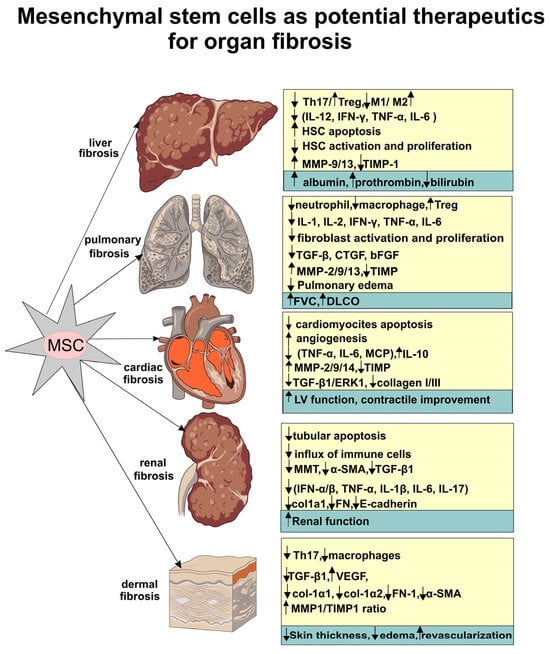
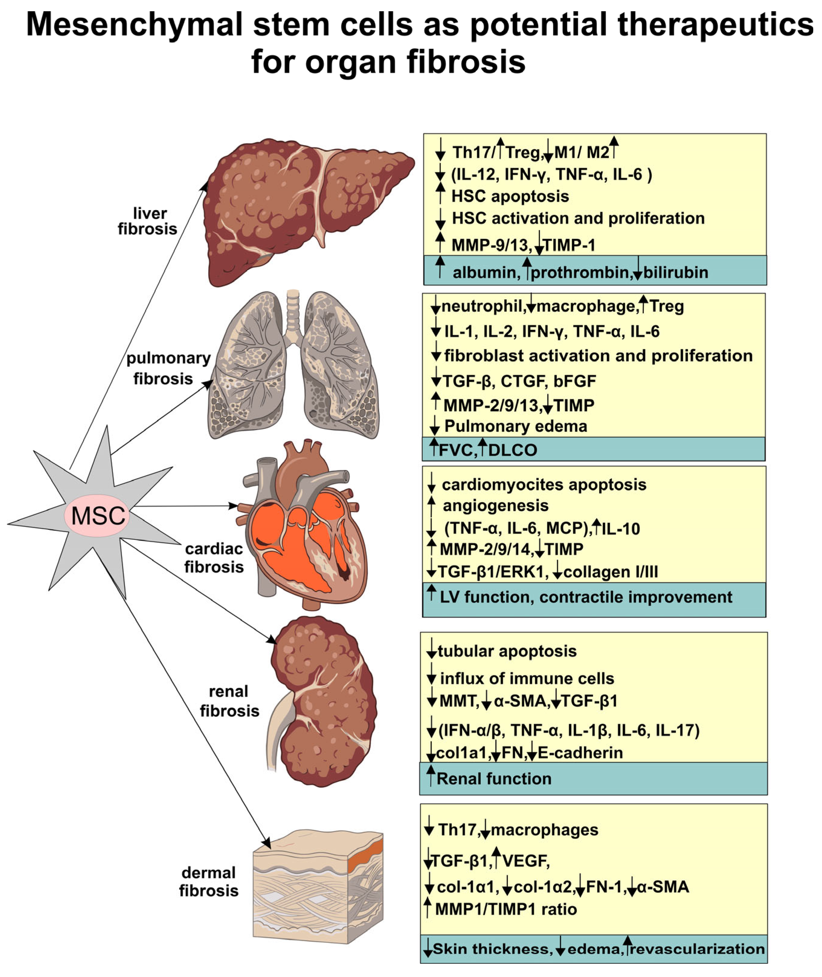

Disclaimer/Publisher’s Note: The statements, opinions and data contained in all publications are solely those of the individual author(s) and contributor(s) and not of MDPI and/or the editor(s). MDPI and/or the editor(s) disclaim responsibility for any injury to people or property resulting from any ideas, methods, instructions or products referred to in the content. |
© 2026 by the authors. Licensee MDPI, Basel, Switzerland. This article is an open access article distributed under the terms and conditions of the Creative Commons Attribution (CC BY) license.