


Abstract
Gamma irradiation is widely used for microbial decontamination and preservation of plant-derived materials intended for the food, pharmaceutical, and cosmetic industries. The effects of gamma irradiation (1, 5, and 10 kGy) on free radical formation, antioxidant activity, total phenolic content, and polyphenolic composition of Prunella vulgaris L. leaves (LS) and fruit spikes with flowers (FS) were investigated. Electron Paramagnetic Resonance (EPR) spectroscopy was used to characterize radiation-induced free radicals and evaluate free radical scavenging activity (FRSA) by the DPPH assay. Total phenolic content (TPC) was determined spectrophotometrically, and individual phenolic compounds were analyzed by HPLC. Gamma irradiation of P. vulgaris induced a singlet signal superimposed on the semiquinone radical signal, while the characteristic cellulose satellite peaks were not detected. The radiation-generated radicals gradually decayed during storage, approaching the natural radical level after approximately 120 days. Irradiation did not significantly affect the FRSA of leaves but induced a dose-dependent response in fruit spikes, with decreased FRSA at 5 kGy and increased activity at 10 kGy. HPLC analysis revealed irradiation-induced changes in the phenolic profile, including degradation of some phenolic compounds and accumulation of others. The results demonstrate that the effects of gamma irradiation on P. vulgaris are dose- and plant organ-dependent and that antioxidant activity is influenced primarily by qualitative changes in phenolic composition.
Keywords:
gamma-irradiation; Prunella vulgaris; leaves; fruit spikes; free radicals; antioxidant activity; EPR; HPLC 1. Introduction
Prunella vulgaris is a perennial medicinal herb belonging to the Lamiaceae (Labiatae) family and is widely distributed throughout Europe, East Asia, and North America. Commonly known as “self-heal”, it has been used in traditional medicine for centuries, particularly for the treatment of wounds, burns, and tissue injuries. Numerous pharmacological studies have demonstrated that P. vulgaris exhibits diverse biological activities, including antioxidant, anti-inflammatory, antibacterial, antiviral, hepatoprotective, and antitumor effects [1,2,3,4]. In recent years, P. vulgaris has attracted increasing scientific interest due to its high content of biologically active compounds and its potential as a valuable source of natural antioxidants. Consequently, the plant has attracted considerable interest as a source of natural antioxidants and as a promising ingredient for functional foods, nutraceuticals, pharmaceutical preparations, and cosmetic formulations.
Phytochemical investigations have demonstrated that P. vulgaris contains a wide range of secondary metabolites, including phenolic acids, flavonoids, triterpenoids, polysaccharides, sterols, tannins, and volatile compounds [3,5]. Among them, rosmarinic acid, caffeic acid, rutin, quercetin derivatives, and ursolic acid are considered the major contributors to its antioxidant and anti-inflammatory activity [6]. Accordingly, extracts from P. vulgaris have been reported to exhibit significant free radical scavenging activity and protective effects against oxidative stress [3,5].
As with many medicinal herbs, preservation of microbiological quality during storage and processing is an important issue. Gamma irradiation is currently one of the most widely used non-thermal technologies for decontamination of herbs and spices because of its effectiveness, reliability and ability to treat products without significant heating. According to the International Atomic Energy Agency, irradiation has been approved for sanitation of herbal materials in many countries, with doses up to 10 kGy being internationally accepted for commercial applications. The method offers several advantages, including reduction in microbial contamination and prolongation of shelf life without substantial heating of the treated material [7].
Despite the practical advantages of gamma treatment, concerns still exist regarding possible radiation-induced alterations in biologically active compounds and antioxidant properties of medicinal plants. Exposure to ionizing radiation generates free radicals through ionization and cleavage of chemical bonds. Some of these radicals recombine rapidly, whereas others remain trapped within the solid plant matrix and can persist for prolonged periods. These stable paramagnetic species can be effectively detected by Electron Paramagnetic Resonance (EPR) spectroscopy, a highly sensitive technique widely used for the investigation of radiation-induced radicals in biological materials. In addition, EPR spectroscopy can also be used to evaluate antioxidant capacity by monitoring the scavenging of stable radicals, such as 2,2-diphenyl-1-picrylhydrazyl (DPPH).
Available studies indicate that gamma irradiation may alter total phenolic content, the composition of individual polyphenolic compounds, and antioxidant activity, depending on the irradiation dose and plant matrix [8,9]. Tang et al. [10] indicated that UV-B pre-treatment in the seedling stage is effective for inducing phenolic acid and flavonoid accumulation in the mature fruiting stage of P. vulgaris ears. However, information regarding the effect of gamma irradiation on free radical generation and antioxidant properties of Prunella vulgaris remains limited. Furthermore, comparative data concerning leaves and fruit spikes with flowers are still scarce.
The present study aimed to evaluate the effects of gamma irradiation at doses of 1, 5, and 10 kGy on free radical formation in leaves and fruit spikes with flowers of Prunella vulgaris using EPR spectroscopy. Furthermore, irradiation-induced changes in antioxidant activity, total phenolic content, and individual polyphenolic compounds were investigated using EPR spectroscopy, spectrophotometric methods, and high-performance liquid chromatography (HPLC).
2. Materials and Methods
2.1. Materials
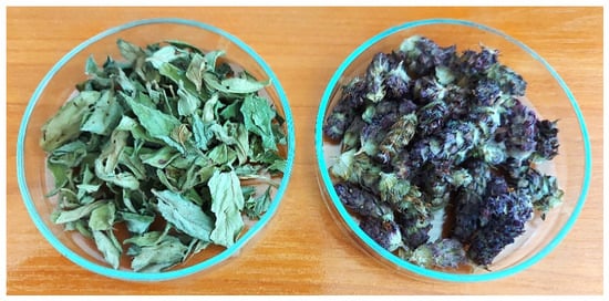
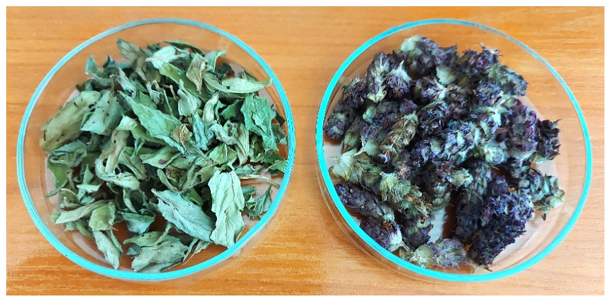
The herb Prunella vulgaris L. was harvested from the Rodopi mountain, Zabardo region, Bulgaria during the month of August. They were dried at room temperature for several days and stored in a dry, cool and dark place until further processing. Leaves (LS) and fruit spikes with flowers (FS) were separated (Scheme 1).
Scheme 1.
Images of the leaves (left) and fruit spikes with flowers (right) of Prunella vulgaris.
The reagents used in this study, such as 1,1-diphenyl-2-picrylhydrazyl (DPPH), Trolox, Folin-Ciocâlteu reagent (FCR), methyl and ethyl alcohol, acetic acid, gallic acid, sodium carbonate, and standards for HPLC analysis (catechin, p-coumaric acid, caffeic acid, 1,2-dibenzoic acid, rosmarinic acid, hesperidin, quercetin and rutin) were purchased from Sigma Aldrich (St. Louis, MA, USA). The water was distilled. All the reagents used in the study were of analytical grade.
2.2. Preparation of Extracts
Extracts for all analyses were prepared by suspending 0.5 g of non-irradiated and irradiated leaves and fruit spikes with flowers in 10 mL of extraction solvent consisting of 7.0 mL ethanol, 2.8 mL distilled water, and 0.2 mL acetic acid. Ethanol was selected as the extraction solvent based on previous studies demonstrating its high efficiency for polyphenol extraction from P. vulgaris [3]. The mixtures were extracted for 17 h at room temperature in the dark without air exposure and subsequently filtered.
Prior to chromatographic analysis, the extracts were additionally filtered through a 0.22 μm syringe membrane filter. For all analyses, 1 mL of leaf extract was diluted with 7 mL of the extraction solvent, whereas 1 mL of fruit spike extract was diluted with 4 mL of the extraction solvent. The supernatants were prepared fresh daily and used for all subsequent analyses.
To minimize variability in the starting plant material, the dried plant material was finely ground and thoroughly homogenized before being divided into equal portions for irradiation. Consequently, all experimental groups originated from the same homogeneous starting material and were subjected to identical extraction procedures and analytical conditions.
2.3. Sample Irradiation
Leaves and fruit spike samples of Prunella vulgaris were packed in polyethylene bags and subjected to gamma irradiation using a cobalt-60 (60Co) source with an activity of 8 200 Ci. Irradiation was carried out at doses of 1, 5, and 10 kGy in a semi-industrial irradiation facility (“NIGU-7”) equipped with a 4 L irradiation chamber. All treatments were conducted at ambient temperature in air at the National Centre of Radiobiology and Radiation Protection, Sofia, Bulgaria. The absorbed dose distribution was assessed using alanine dosimeters (Kodak BioMax, Eastman Kodak Company, Rochester, NY, USA). Dosimeter readings were obtained with an E-scan Bruker ESR spectrometer and expressed as absorbed dose in water following appropriate calibration procedures.
2.4. Electron Paramagnetic Resonance Spectroscopy
The EPR measurements were performed at room temperature using a JEOL JES-FA100 spectrometer (JEOL Ltd., Tokyo, Japan) operating in the X-band (9.3 GHz) and equipped with a standard TE011 cylindrical resonator. Spectra were recorded as the first derivative of the microwave absorption signal. Solid samples were placed in standard quartz EPR tubes (4 mm inner diameter) and positioned in the resonator cavity. Each sample was measured three times as technical repetitions following a repositioning procedure, in which the sample was inserted, removed, and reinserted into the cavity before each measurement. The recorded spectra were averaged, and the variability among the three repeated measurements was used to estimate the experimental error.
2.4.1. Determination of DPPH Free Radical Scavenging Activity (FRSA)
The free radical scavenging activity (FRSA) of non-irradiated and irradiated Prunella vulgaris extracts (leaves and fruit spikes) was evaluated by Electron Paramagnetic Resonance (EPR) spectroscopy using the stable DPPH radical.
Briefly, aliquots of the extracts (0.02–0.13 mL) were added to 1 mL of a 0.002 M ethanolic DPPH solution. After approximately 35 min, allowing the reaction to reach completion, the mixtures were transferred into glass capillary tubes and placed in the resonant cavity of the EPR spectrometer for signal acquisition.
The percentage of scavenged DPPH radicals was calculated according to the following equation:
where I0 represents the intensity of the second peak in the EPR spectrum of the control sample (containing DPPH solution and solvent without extract), and I is the intensity of the corresponding peak after addition of the extract. Control samples were prepared under identical conditions in the absence of plant extract.
Scavenged DPPH radicals (%) = [(I0 − I)/I0] × 100,
2.4.2. Trolox Equivalent Antioxidant Capacity (TEAC) Determination
The Trolox equivalent antioxidant capacity (TEAC) of Prunella vulgaris extracts was determined using a previously reported EPR-DPPH method [11,12]. Briefly, 0.06 mL of Trolox standard solutions at various concentrations were mixed with 0.5 mL of 0.002 M DPPH solution in order to construct a calibration curve. For each P. vulgaris extract, the linear relationship between the percentage of scavenged DPPH radicals and the volume of the extract was determined by regression analysis (the corresponding regression equations are given below). Based on these equations, the percentage of scavenged DPPH radicals corresponding to 1 mL of the investigated extract was calculated. The obtained values were then converted to Trolox equivalents using the regression equation describing the linear relationship between the percentage of scavenged DPPH radicals and Trolox concentration:
where y represents the percentage of scavenged DPPH radicals calculated for each one of the samples. The regression equations obtained for the individual P. vulgaris extracts were as follows: FS, 0 kGy: y = 1090x − 11.28; 1 kGy: y = 1078.5x − 12.91; 5 kGy: y = 959x − 10.98; 10 kGy: y = 1218.5x − 12.50; LS, 0 kGy: y = 730.34x − 20.63; 1 kGy: y = 703.28x − 19.08; 5 kGy: y = 759.66x − 20.95; and 10 kGy: y = 713.28x − 19.60. The antioxidant capacity was expressed as mmol Trolox equivalents per gram of dry herb (mmol TE/g) for both fruit spikes and leaves, taking into account the extraction ratio and dilution factors.
y = 1.87x − 39.73
2.5. Spectrophotometric Determination of Total Phenolic Content
The total phenolic content (TPC) of Prunella vulgaris extracts (fruit spikes and leaves) was determined using the Folin–Ciocalteu method, as described by Valyova et al. [13] with slight modifications. Briefly, 0.5 mL of the extract was mixed with 3.0 mL of distilled water, followed by the addition of 0.25 mL of Folin–Ciocalteu reagent and 0.75 mL of sodium carbonate solution (20% w/v). After incubation, the absorbance of the reaction mixture was measured at 765 nm against a blank sample on a Lybra S22 spectrophotometer (Biochrom Ltd., Cambridge, UK). A calibration curve was constructed using gallic acid (y = 0.0101x − 0.0178, R2 = 0.9991). The results were expressed as mg gallic acid equivalents per g of dry sample (mg GAE/g).
2.6. High-Performance Liquid Chromatography (HPLC) Analysis
The analysis of polyphenolic compounds in Prunella vulgaris extracts (leaves and fruit spikes with flowers, FSF) was performed using an Agilent 1260 Infinity Quaternary LC system (Agilent Technologies ink., Waldbronn, Germany) equipped with a diode-array detector (DAD, λ = 254/8 nm, Ref = 380/20 nm). Separation was achieved on an Agilent Poroshell 120 HPH-C18 column (4.6 × 100 mm, 2.7 µm particle size).
The mobile phases consisted of methanol/ultrapure water/acetic acid (5:93:2, v/v/v) as phase A and (86:12:2, v/v/v) as phase B. The injection volume was 10 µL. The following gradient programme was applied: 0 min—A (100%), B (0); 0.5 min—A (95%), B (5%); 4 min—A (80%), B (20%); 6 min—A (60%), B (40%); 7 min—A (20%), B (80%), 8.5 min—A (97%), B (3%). The flow rate was set at 1.2 mL/min, and the column temperature at 40 °C. All analyses were performed in triplicate.
Polyphenolic compounds were identified by comparing their retention times and UV–Vis spectra with those of commercially available authentic reference standards. Quantification of individual phenolic compounds was performed using external calibration curves prepared from the corresponding authentic standards. The concentrations of the major polyphenolic groups (flavan-3-ols, phenolic acids, flavanones, and quercetin glycosides) were expressed as equivalents of representative standards for each respective group. Peaks eluting before 2.0 min were excluded from quantitative evaluation because they were identified as chromatographic artefacts or low-molecular-weight carboxylic acids rather than target phenolic constituents. Quantitative results were expressed as mg/g dry plant material.
2.7. Statistical Analysis
Statistical analysis was performed separately for leaves and fruit spikes with flowers. For each experimental group, three independent sample preparations and extractions were performed, and the resulting extracts were analyzed independently for EPR-DPPH radical-scavenging activity, total phenolic content (TPC), and HPLC determination of individual phenolic compounds. These three independent preparations were considered independent experimental replicates. The results are expressed as arithmetic means, with the corresponding variability indicated in the tables and figures. Differences among the four irradiation doses (0, 1, 5, and 10 kGy) were evaluated using one-way analysis of variance (ANOVA), followed by Tukey’s HSD post hoc test. Differences were considered statistically significant at p < 0.05.
3. Results
3.1. EPR Study
3.1.1. Features of EPR Spectra of Prunella vulgaris L. Samples
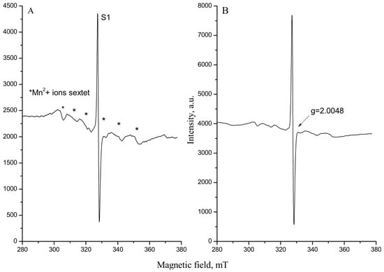
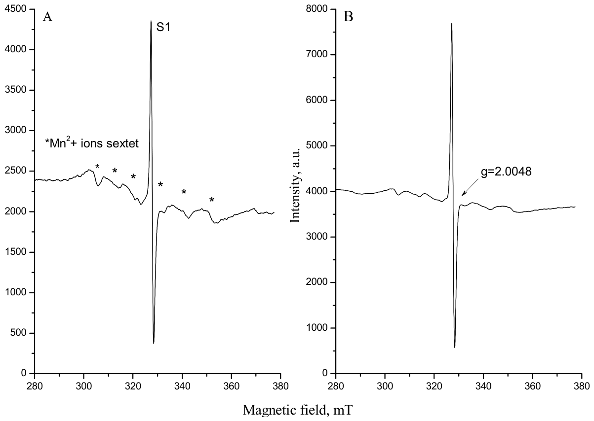
The EPR spectra of non-irradiated and gamma-irradiated Prunella vulgaris L. samples are presented in Figure 1. Two types of paramagnetic species were detected in the non-irradiated samples (Figure 1A). The first signal (S1) appeared as a single line at g = 2.0048 and was attributed to stable semiquinone radicals formed by oxidation of polyphenolic compounds. The second signal (marked with an asterisk) consisted of six hyperfine components with g = 2.0212 and with a resolved huperfine splitting constant (A) gradually increasing from 8.939 mT to 10.693 mT in the upfield direction, with an average value (Aavg) of 9.864 mT. This signal is characteristic of Mn2+ ions and originates from hyperfine interaction with the manganese nucleus (55Mn, nuclear spin I = 5/2), which has 100% natural abundance [14]. The resolved six-line spectrum indicates that the Mn2+ ions are sufficiently isolated within the plant matrix, resulting in minimal magnetic interaction between neighbouring ions. The fourth manganese line overlapped with the endogenous signal of stable organic radicals.
Figure 1.
EPR spectra of Prunella vulgaris L. (A) non-irradiated and (B) irradiated P. vulgaris samples. The central singlet corresponds to organic radicals, whereas the six-line hyperfine signal of naturally occurring Mn2+ ions is marked with asterisks.
After gamma irradiation, an additional singlet signal was generated and superimposed on the natural radical signal (Figure 1B), leading to a significant increase in the intensity of the central EPR line. Similar radiation-induced effects have previously been reported for irradiated plant materials [15]. The increase in signal intensity indicates enhanced formation of stable radical species following irradiation, most likely due to ionization and bond cleavage processes affecting phenolic constituents and other organic molecules in the plant matrix.
3.1.2. Time Stability of Gamma-Induced Radicals in Prunella vulgaris L. Studied by EPR Spectroscopy
Although gamma irradiation induced a significant increase in the intensity of the central EPR signal, the radiation-specific satellite lines associated with cellulose radicals and described in the European Standard EN 1787 [16] were not detected in either leaf or fruit spike samples. Similar observations have been reported for other plant materials, where these satellite signals are often characterized by low intensity, rapid decay after irradiation, or complete absence depending on the composition and physical structure of the sample [17,18,19].
When the specific radiation signal was not detected, the approach proposed by Yordanov and Aleksieva [20] was applied. The EPR spectra of Prunella vulgaris consisted of a dose-dependent singlet arising from radiation-induced radicals superimposed on a radiation-independent sextet originating from naturally occurring Mn2+ ions. Since manganese ions remain unaffected by thermal treatment and storage, their signal was used as an internal reference for monitoring changes in the intensity of the radiation-induced radicals. Thus, the intensity ratio of the radiation-induced singlet/manganese signal decreased significantly in irradiated samples in comparison to unirradiated. In this way, we can clearly distinguish irradiated from non-irradiated samples.
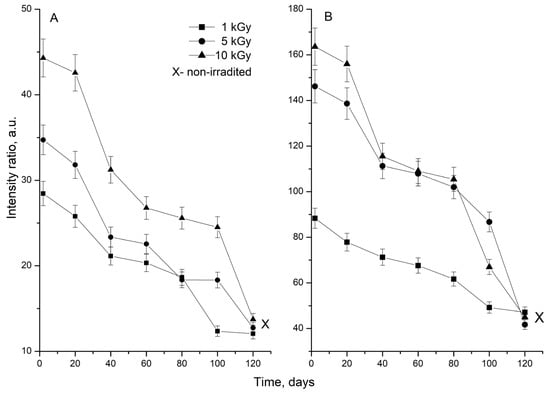
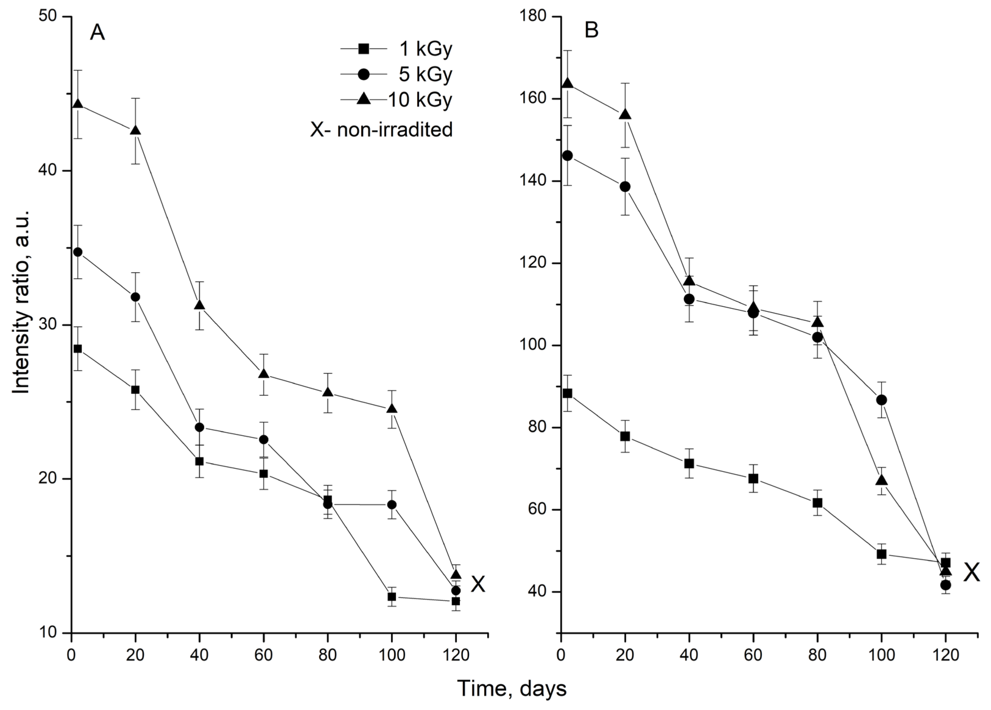
Thermal treatment of irradiated samples (2 h at 60 °C) resulted in an almost complete disappearance of the radiation-induced component, whereas the manganese sextet remained unchanged. This observation indicates that the radicals generated by gamma irradiation are sufficiently stable to be detected by EPR but can gradually recombine when additional energy is supplied. A similar trend was observed during storage, where the intensity of the radiation-induced signal gradually decreased with time (Figure 2). Approximately 120 days after irradiation, the EPR signal approached the intensity of the natural semiquinone radical signal (without thermal treatment) recorded in non-irradiated samples.
Figure 2.
Time-dependent decay of the gamma-induced EPR signal in: (A) fruit spikes with flowers and (B) leaves of Prunella vulgaris. The intensity of the irradiation-induced signal is expressed as a ratio relative to the naturally occurring Mn2+ signal detected in the same sample and used as an internal reference. Values represent the mean intensity ratio of three repeated EPR measurements at each storage time. Error bars represent the experimental measurement error (approximately 5%).
The gradual decay of radiation-induced radicals suggests that both radical-radical recombination and radical scavenging processes contribute to the decrease in the EPR signal during storage. Considering the high antioxidant potential of P. vulgaris, naturally occurring phenolic compounds may participate in radical quenching reactions, while recombination of radiation-induced radicals is also expected to occur. Therefore, although gamma irradiation initially generates additional radical species, these radicals are not permanently accumulated in the plant material but gradually decay during storage.
3.1.3. Gamma Irradiation Effect on Free Radical Scavenging Activity
The free radical scavenging activity of non-irradiated and gamma-irradiated Prunella vulgaris extracts was evaluated by EPR spectroscopy using the stable DPPH• radical. Antioxidant compounds present in the extracts reduce DPPH• through hydrogen atom or electron donation, resulting in a decrease in the intensity of the EPR signal. Therefore, the reduction in the DPPH signal is directly proportional to the radical scavenging capacity of the analyzed samples.
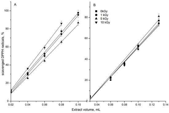
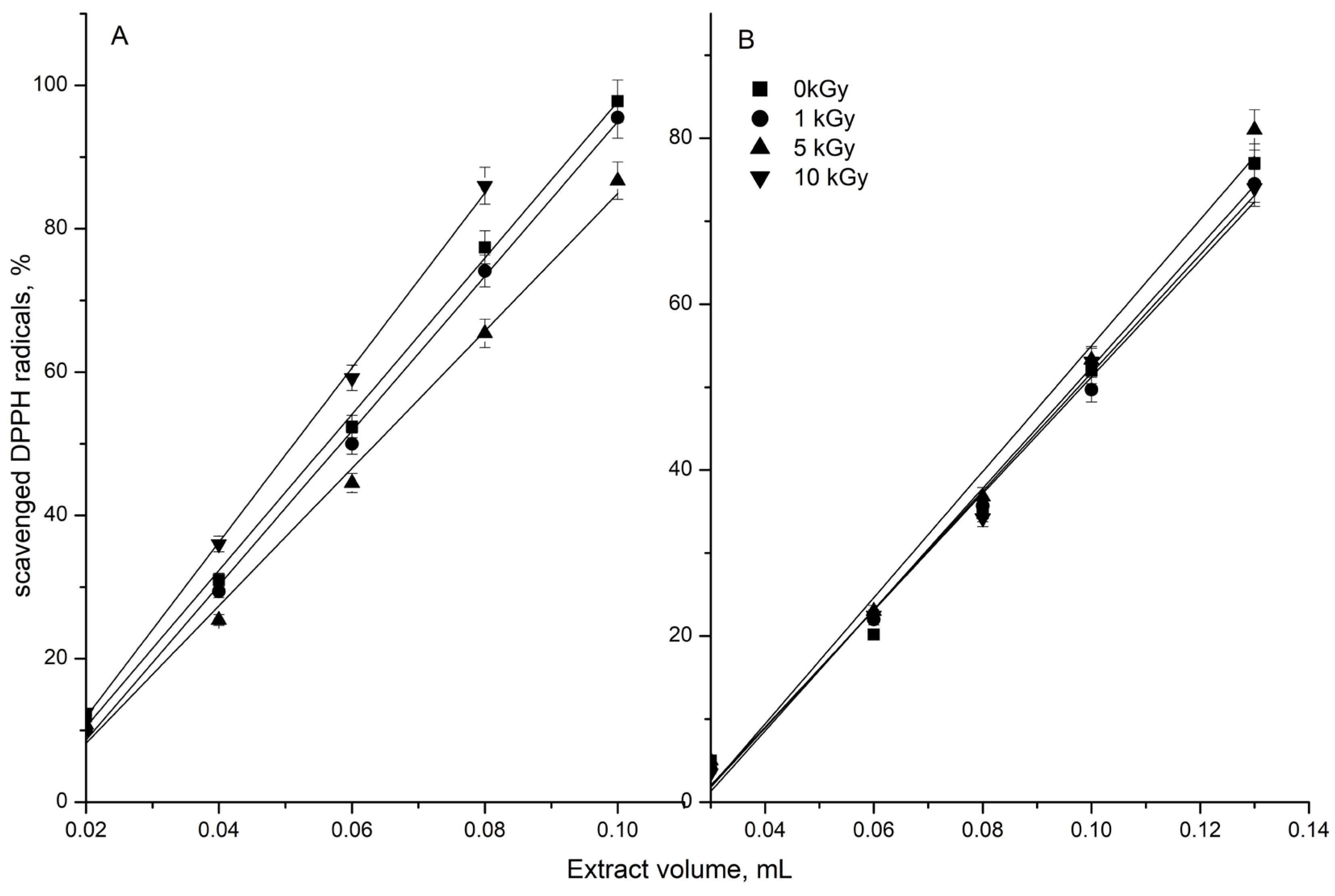
The results revealed a dose-dependent effect of gamma irradiation on the FRSA of fruit spikes with flowers (Figure 3A). Irradiation at 1 kGy did not significantly affect the antiradical activity compared to the control. However, irradiation at 5 and 10 kGy produced opposite effects: FRSA decreased at 5 kGy, whereas a significant increase of approximately 10% was observed at 10 kGy. The calculated Trolox Equivalent Antioxidant Capacity (TEAC) values (Table 1) followed the same trend, confirming the reliability of the observed changes.
Figure 3.
Dependence between percentage of scavenged DPPH radicals and volume (mL) of non-irradiated and irradiated: (A) fruit spikes with flowers and (B) leaf extracts.
Table 1.
Total phenolic content and TEAC values, for non-irradiated and irradiated with 1, 5 and 10 kGy fruit spikes (FS) and leaf samples (LS) of Prunella vulgaris.
In contrast, the antioxidant activity of leaf extracts remained essentially unchanged following gamma irradiation (Figure 3B). Although slightly higher FRSA and TEAC values were observed at 5 kGy, these differences were not statistically significant compared with the non-irradiated control.
It should also be noted that the non-irradiated leaf extracts exhibited higher free radical scavenging activity than the fruit spike extracts. This difference is noticeable considering the fact that leaf extracts were diluted to a greater extent (1:7) than fruit spike extracts (1:4) prior to analysis, indicating a considerably higher antiradical capacity of the leaves.
3.2. Effect of Gamma Irradiation on Total Phenolic Content
Herbs are rich in polyphenolic compounds, which represent one of the major classes of natural antioxidants due to their ability to scavenge free radicals and inhibit oxidative processes [21]. Therefore, evaluation of the effect of gamma irradiation on the total phenolic content (TPC) of Prunella vulgaris was considered essential for understanding the radiation-induced changes in its antioxidant properties.
Gamma irradiation affected the TPC of P. vulgaris differently depending on the plant organ. In fruit spikes with flowers, TPC gradually decreased with increasing irradiation dose, whereas in leaves a statistically significant increase was observed after irradiation at 5 and 10 kGy (Table 1). These contrasting responses most likely reflect differences in the phytochemical composition in the two plant organs.
The irradiation-induced decrease in TPC of FS may be attributed to the degradation of certain phenolic compounds into non-phenolic products or to the breakdown of high-molecular-weight tannins following cleavage of glycosidic bonds by ionizing radiation [8,22]. Previous studies also described the negative effect of irradiation on the total phenolic content of irradiated plant products [8]. Conversely, the increase in TPC observed in LS at 5 and 10 kGy may result from the release of bound phenolic compounds from the plant matrix or from the depolymerization of complex phenolics into lower-molecular-weight compounds that react more readily with the Folin–Ciocalteu reagent. Similar effects have been reported for various irradiated medicinal and aromatic plants, where low or moderate irradiation doses increased the extractable phenolic content [22,23]. The present results demonstrate that the response of P. vulgaris to gamma irradiation depends not only on the absorbed radiation dose but also on the botanical part of the plant. It should also be considered that the phenolic content of medicinal plants is influenced by numerous factors in addition to irradiation. These include the extraction solvent, the plant part analyzed, the physiological stage at harvest, the analytical method employed, as well as genotypic and geographical variations [21].
Non-irradiated fruit spikes contained higher levels of total phenolics than leaves, in agreement with previous reports for P. vulgaris [3]. The higher phenolic content in reproductive plant parts is probably associated with the accumulation of phenolic acids and flavonoids involved in protective and physiological functions.
3.3. Analysis of HPLC Profile Before and After Gamma Irradiation
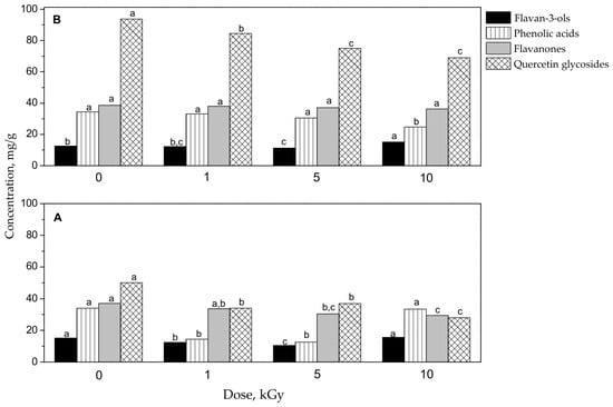
To obtain a more detailed insight into the effect of gamma irradiation on the antioxidant constituents of Prunella vulgaris, the polyphenolic composition of leaves and fruit spikes with flowers was investigated by HPLC analysis. Four major groups of phenolic compounds were identified and quantified: flavan-3-ols (catechin equivalent), phenolic acids (caffeic acid and p-coumaric acid equivalents), flavanones (hesperidin equivalent), and quercetin glycosides (quercetin equivalent). The obtained results are presented in Figure 4 and Table 2.
Figure 4.
The irradiation effect on the concentration of polyphenols identified as flavan-3-ols, phenolic acids, flavanones and quercetin glycosides in non-irradiated and irradiated Prunella vulgaris: (A) fruit spike with flowers and (B) leaves. Different letters (a–c) above the bars indicate statistically significant differences (p < 0.05) among irradiation doses for the same polyphenolic group within each plant organ. Bars sharing identical letters are not significantly different.
Table 2.
Concentration (mg/g) of individual phenolic compounds for non-irradiated and irradiated with 1, 5, and 10 kGy fruit spikes (FS) and leaf samples (LS) of Prunella vulgaris.
The polyphenolic composition differed considerably between the two plant organs. In both non-irradiated samples, quercetin glycosides represented the predominant group of phenolic compounds, reaching 50.10 mg/g in FS and 93.71 mg/g in LS, whereas flavan-3-ols were present in the lowest amounts (15.15 mg/g and 12.45 mg/g, respectively).
Comparison of the control samples revealed that leaves contained significantly higher amounts of flavanones and quercetin glycosides, whereas fruit spikes were slightly richer in flavan-3-ols. These differences are consistent with the organ-specific distribution of secondary metabolites reported for P. vulgaris and likely reflect their distinct physiological functions. Among the individual phenolic compounds, hesperidin was the predominant constituent in the fruit spikes with flowers, followed by 1,2-dibenzoic acid and rosmarinic acid. In contrast, rutin was the most abundant compound in leaves, followed by hesperidin and rosmarinic acid. These findings are in line with published data identifying hesperidin, and rosmarinic acid among the major phenolic constituents of Prunella vulgaris [3].
Gamma irradiation induced substantial modifications in the distribution of these polyphenolic groups. In FS samples, irradiation at 1 and 5 kGy resulted in significant reductions in flavan-3-ols, phenolic acids, quercetin glycosides, and flavanones. These changes indicate that a considerable proportion of the phenolic constituents in reproductive tissues is susceptible to radiation-induced degradation or structural modification. At 10 kGy, the contents of flavan-3-ols and phenolic acids were comparable to those of the control, whereas flavanones and quercetin glycosides remained lower. This response suggests that higher irradiation doses may induce simultaneous degradation of some phenolic compounds together with the formation or improved extractability of others, resulting in partial restoration of certain polyphenolic groups.
Leaves exhibited a different response to irradiation. Among the quantified groups, the most pronounced change was observed for flavan-3-ols, whose concentration increased significantly after treatment with 10 kGy. In contrast, the contents of flavanones, phenolic acids and quercetin glycosides showed only minor variations compared with the control. The increase in flavan-3-ols may be associated with radiation-induced disruption of cellular structures, facilitating the release of phenolic compounds previously bound to the plant matrix. Similar observations have been reported for other irradiated medicinal plants and have been attributed to enhanced extractability of phenolic constituents.
The analysis of individual compounds identified in Prunella vulgaris further demonstrated the complexity of the radiation-induced transformations (Table 2). In FS samples, catechin, p-coumaric acid, hesperidin, and quercetin (after 10 kGy) increased following irradiation, with the most pronounced increase in catechin and hesperidin occurring at 1 kGy and followed by 5 kGy. In contrast, caffeic acid, 1,2-dibenzoic acid, rosmarinic acid, rutin, and quercetin (after 1 and 5 kGy) decreased, the strongest reduction being observed at 1 and 5 kGy. Notably, 1,2-dibenzoic acid was not detected in the FS samples irradiated at 1 and 5 kGy, indicating its high susceptibility to gamma treatment. These changes are in agreement with the reduction in the total phenolic acid fraction observed at the same irradiation doses.
A different pattern was observed in leaves. Catechin concentration increased, particularly after irradiation at 10 kGy, whereas rutin and 1,2-dibenzoic acid showed slight increases after 1 and 5 kGy. Hesperidin also increased after irradiation at 1 and 10 kGy, while caffeic acid remained essentially unchanged throughout the studied dose range. Conversely, rosmarinic acid, quercetin and p-coumaric acid (except after 1 kGy) decreased following irradiation, and hesperidin showed a temporary reduction at 5 kGy.
The HPLC results complement the spectrophotometric determination of total phenolic content by demonstrating that gamma irradiation affects not only the overall amount of phenolic compounds but also their qualitative composition. Since individual phenolics differ substantially in their antioxidant efficiency, these compositional changes provide a plausible explanation for the dose-dependent variations in free radical scavenging activity discussed in the following section.
4. Discussion
Relationship Between Antioxidant Activity and Phenolic Content
Previous studies investigating the effects of gamma irradiation on medicinal and aromatic plants have reported inconsistent results regarding antioxidant activity and phenolic composition. Horváthová et al. [24] observed a significant increase in total phenolic content in oregano following irradiation at doses of 5–30 kGy, whereas the DPPH radical scavenging activity remained insignificantly changed. Similarly, Brandstetter et al. [25] reported that irradiation at 10 kGy had insignificant effect on the antioxidant capacity of sage, thyme and oregano. Studies on other medicinal plants have likewise shown variable responses, ranging from enhanced antioxidant activity and phenolic content to no detectable changes or degradation of antioxidant constituents. Such variability has been attributed to differences in plant matrix composition, moisture content, the extraction procedure and solvent used, irradiation dose, and the stability of individual bioactive compounds [26,27].
The present findings are in agreement with these reports, confirming that the response of Prunella vulgaris to gamma irradiation is dose- and tissue-dependent. Although irradiation induced significant alterations in total phenolic content and in the composition of individual polyphenolic groups; however, these changes were not always reflected by corresponding changes in antioxidant activity. The lack of a direct correspondence between TPC and FRSA/TEAC indicates that the antioxidant response depends not only on the total content of phenolic compounds but also on qualitative changes in their composition, as well as on the contribution of other non-phenolic antioxidants.
Potential matrix-related effects should also be considered when interpreting the analytical results obtained from complex plant extracts. In HPLC analysis co-extracted matrix components may affect chromatographic separation and detector response, and their influence depends on the composition of the particular plant matrix. González-González et al. [28] evaluated the matrix effect in a validated HPLC-DAD method for the determination of phenolic acids in complex medicinal plant extracts and reported matrix effects below 5% for all compounds investigated, indicating only a minor influence of the matrix under their experimental conditions. In the present study, a formal matrix-effect validation was not performed; therefore, the possible influence of the plant matrix cannot be completely excluded. Moreover, its magnitude may vary among irradiation treatments because gamma irradiation can alter the chemical composition of the plant matrix. Therefore, the HPLC results are interpreted primarily as comparative changes in the quantified phenolic profile rather than as fully matrix-independent absolute concentrations.
The EPR-DPPH assay provides a different type of information, as the decrease in DPPH signal reflects the combined radical-scavenging activity of the extract under the applied experimental conditions. The use of EPR for DPPH detection also reduces the optical interference associated with coloured plant extracts that can affect conventional spectrophotometric DPPH assays. Sanna et al. [29] reported that EPR provided more reliable DPPH measurements than UV-Vis across different plant extract matrices, as the EPR response was not dependent on the chemical composition of the extracts. Nevertheless, the complex composition of the extracts may affect the reaction behaviour of their constituents with DPPH, and this possibility cannot be completely excluded in the present study. Therefore, the FRSA and TEAC values are considered comparative indicators of changes in the overall radical-scavenging capacity associated with gamma irradiation.
The non-linear dose–response relationship was particularly evident in the fruit spikes with flowers. The decrease in FRSA observed after irradiation at 5 kGy coincided with a marked reduction in the amount of flavan-3-ols, phenolic acids and flavanones detected by HPLC. At the level of individual compounds, lower concentrations of caffeic acid, rosmarinic acid, rutin, quercetin, and, together with the disappearance of 1,2-dibenzoic acid, are likely to have contributed to the reduced antioxidant activity. These compounds are well known for their high radical scavenging capacity [30,31]. Consistent with these observations, Li et al. [27] identified quercetin, rosmarinic acid, and luteolin as the major contributors to the antioxidant activity of P. vulgaris fruit spikes, while Xia et al. [32] also reported excellent antioxidant properties for caffeic and rosmarinic acids.
Interestingly, despite a similar decline in several phenolic constituents after irradiation at 1 kGy, FRSA remained comparable with the control. This may be explained by the simultaneous increase in catechin and hesperidin, all of which possess strong radical scavenging activity. Their increased abundance may have compensated for the loss of other antioxidant compounds, thereby maintaining the overall antioxidant capacity. Previous studies have likewise demonstrated that p-coumaric acid, rutin, and catechin possess pronounced DPPH radical scavenging activity [30].
In contrast, irradiation at 10 kGy resulted in significantly higher FRSA and TEAC values despite a further decrease in total phenolic content. This response coincided with increased concentrations of p-coumaric acid and quercetin, both recognized as highly effective radical scavengers capable of donating hydrogen atoms or electrons and stabilizing the resulting radicals. Their accumulation therefore appears to have played an important role in the enhanced antioxidant activity of the irradiated FS samples.
In addition to the accumulation of these highly active phenolic compounds, the enhanced antioxidant activity may also be attributed to other radiation-induced changes reported in the literature. Gamma irradiation can promote cleavage of covalent bonds in complex phytochemicals, leading to the formation of lower molecular weight compounds with higher antioxidant potential or improved extractability from the plant matrix. Irradiation has also been associated with increased levels of reducing sugars and structural modifications that may further enhance radical scavenging properties [33,34]. Moreover, synergistic interactions among phenolic compounds, flavonoids, polysaccharides, and other naturally occurring antioxidants may further contribute to the overall antioxidant response. Consequently, the enhanced antioxidant activity observed after irradiation at 10 kGy most likely reflects the combined effects of changes in individual phenolic constituents and radiation-induced modifications of the overall phytochemical profile.
Overall, no clear relationship was found between total phenolic content and antioxidant activity in fruit spikes. Whereas the reduction in FRSA at 5 kGy was associated with lower TPC, irradiation at 10 kGy increased FRSA and TEAC despite a further decline in total phenolics. These findings indicate that antioxidant activity in fruit spikes is governed primarily by changes in the composition and relative abundance of individual phenolic compounds.
A different response was observed in leaves. The statistically significant increase in TPC observed at 5 and 10 kGy was not accompanied by a corresponding statistically significant increase in FRSA or TEAC. This finding suggests that the higher total phenolic content alone was insufficient to produce a measurable enhancement of the antioxidant activity. HPLC analysis revealed only modest changes in the phenolic profile, including slight increases in rutin and 1,2-dibenzoic acid at 1 and 5 kGy, and in catechin at 10 kGy, together with a decrease in quercetin glycosides, while the other major polyphenolic groups remained largely unaffected. These relatively small changes in the composition of individual phenolic compounds are consistent with the absence of a statistically significant change in antioxidant activity. The results therefore suggest that the radiation-induced increase in TPC was not accompanied by corresponding qualitative changes in the quantified phenolic compounds sufficient to modify the radical scavenging capacity. However, it should be noted that the present HPLC analysis was limited to selected groups of polyphenols and selected individual phenolic compounds. Consequently, the possible contribution of other phenolic constituents or non-phenolic antioxidants was not evaluated and cannot be excluded. It is therefore possible that other radiation-induced changes in the overall phytochemical composition, including constituents not determined in the present study, also contributed to the preservation of the antioxidant activity of the leaf extracts.
Ionizing radiation can initiate ionization and excitation processes and generate short-lived reactive species, which may interact with organic constituents of the plant matrix. Depending on the chemical structure and susceptibility of individual compounds, such radiation-induced reactions may lead to oxidation, bond cleavage, structural modification, partial degradation, formation of reaction products, or changes in compound extractability [26,33]. Similar radiation-induced changes in the phenolic composition and antioxidant properties of plant materials have been reported following gamma irradiation [22,23,26,33]. The relative contribution of these processes is likely to differ among phenolic constituents and between plant organs, providing a possible explanation for the non-uniform changes observed in the HPLC profiles.
5. Conclusions
To the best of our knowledge, this study represents the first comprehensive investigation of the effects of gamma irradiation on Prunella vulgaris, combining EPR spectroscopy, spectrophotometric determination of total phenolic content, HPLC analysis of individual phenolic compounds, and evaluation of antioxidant activity in both leaves and fruit spikes with flowers.
Gamma irradiation induced the formation of relatively stable free radicals, which gradually decayed during storage, as demonstrated by EPR spectroscopy. The overall response was strongly dependent on both the irradiation dose and the plant organ. Although irradiation affected total phenolic content and the profile of individual phenolic compounds, these changes were not consistently reflected in antioxidant activity, indicating that the antioxidant response is influenced not only by the overall phenolic content but also by qualitative changes in the phytochemical composition.
Among the investigated doses, irradiation at 10 kGy produced the most favourable overall response. Although irradiation altered the phenolic profile, the antioxidant potential of P. vulgaris was preserved, and in fruit spikes with flowers the radical scavenging activity was even enhanced. The different responses observed in leaves and fruit spikes with flowers emphasize the importance of evaluating individual plant organs separately when assessing the effects of gamma irradiation on medicinal plants.
The findings presented here expand the current knowledge of radiation-induced changes in P. vulgaris and provide experimental evidence that gamma irradiation at 10 kGy preserved the antioxidant potential of dried P. vulgaris under the conditions investigated. This dose therefore shows promise as a post-harvest treatment for dried P. vulgaris intended for herbal products, functional foods, nutraceuticals, and pharmaceutical applications, while providing a basis for further research on the quality and stability of the irradiated plant material.
Author Contributions
Conceptualization, R.M.; methodology, R.M., K.A. and N.S.; investigation, R.M., K.A., N.S. and K.L.; writing—original draft preparation, R.M.; writing—review and editing, R.M. and K.A. All authors have read and agreed to the published version of the manuscript.
Funding
This research was funded by the Bulgarian National Science Fund—Bulgarian Ministry of Education within the framework of Project “KП-06-H99/1”.
Data Availability Statement
The raw data supporting the conclusions of this article will be made available by the authors on request.
Acknowledgments
The authors thank the Bulgarian National Science Fund—Bulgarian Ministry of Education within the framework of Project “KП-06-H99/1” for the financial support. Research equipment from the distributed research infrastructure INFRAMAT (part of the Bulgarian National roadmap for research infrastructures) supported by the Bulgarian Ministry of Education and Science was used in this investigation.
Conflicts of Interest
The authors declare no conflicts of interest.
Abbreviations
The following abbreviations are used in this manuscript:
| EPR | Electron Paramagnetic Resonance |
| HPLC | High-Performance Liquid Chromatography |
| DPPH | 1,1-diphenyl-2-picrylhydrazyl |
| FRSA | Free radical scavenging activity |
| TEAC | Trolox equivalent antioxidant capacity |
| SD | Standard deviation |
| TPC | Total phenolic content |
| LS | Leaves samples |
| FS | Fruit spike with flowers |
References
- Feng, L.; Jia, X.; Zhu, M.-M.; Chen, Y.; Shi, F. Antioxidant Activities of Total Phenols of Prunella vulgaris L. In Vitro and in Tumor-bearing Mice. Molecules 2010, 15, 9145–9156. [Google Scholar] [CrossRef] [Scilit] [PubMed]
- Pan, J.; Wang, H.; Chen, Y. Prunella vulgaris L.—A Review of its Ethnopharmacology, Phytochemistry, Quality Control and Pharmacological Effects. Front. Pharmacol. 2022, 13, 903171. [Google Scholar] [CrossRef] [Scilit] [PubMed]
- Mîrza, C.-M.; Mîrza, T.-V.; Odagiu, A.C.M.; Uifălean, A.; But, A.E.; Pârvu, A.E.; Bulboacă, A.-E. Phytochemical Analysis and Antioxidant Effects of Prunella vulgaris in Experimental Acute Inflammation. Int. J. Mol. Sci. 2024, 25, 4843. [Google Scholar] [CrossRef] [Scilit]
- Leng, L.; Li, P.; Liu, R.; Francis, O.B.; Song, S.; Siu, Y.; Yang, Y.; Wang, Y.; Sun, X.; Miao, R.; et al. The main active components of Prunella vulgaris L. alleviate myocardial ischemia-reperfusion injury by inhibiting oxidative stress and ferroptosis via the NRF2/GPX4 pathway. J. Ethnopharmacol. 2025, 345, 119630. [Google Scholar] [CrossRef] [Scilit]
- Wang, S.J.; Wang, X.H.; Dai, Y.Y.; Ma, M.H.; Rahman, K.; Nian, H.; Zhang, H. Prunella vulgaris: A Comprehensive Review of Chemical Constituents, Pharmacological Effects and Clinical Applications. Curr. Pharm. Des. 2019, 25, 359–369. [Google Scholar] [CrossRef] [Scilit] [PubMed]
- Wei, J.; Leng, L.; Sui, Y.; Song, S.; Owusu, F.B.; Li, X.; Cao, Y.; Li, P.; Wang, H.; Li, R.; et al. Phenolic acids from Prunella vulgaris alleviate cardiac remodeling following myocardial infarction partially by suppressing NLRP3 activation. Phytother. Res. 2024, 38, 384–399. [Google Scholar] [CrossRef] [Scilit] [PubMed]
- Rahmati, M.H.; Khoshtaghasa, M.H.; Banakar, A.; Ebadi, M.-T. Decontamination technologies for medicinal and aromatic plants: A review. Food Sci. Nutr. 2022, 10, 784–799. [Google Scholar] [CrossRef] [Scilit] [PubMed]
- Toledo, T.C.F.; Canniatti-Brazaca, S.G.; Arthur, V.; Piedade, S.M.S. Effects of gamma radiation on total phenolics, trypsin and tannin inhibitors in soybean grains. Rad. Phys. Chem. 2007, 76, 1653–1656. [Google Scholar] [CrossRef] [Scilit]
- Mladenova, R.; Solakov, N.; Loginovska, K.; Karakirova, Y. Analysis of Gamma-Irradiation Effect on Radicals Formation and on Antiradical Capacity of Horse Chestnut (Aesculus hippocastanum L.) Seeds. Appl. Sci. 2025, 15, 3287. [Google Scholar] [CrossRef] [Scilit]
- Tang, H.; Yang, J.; Zhang, X.R.; Cao, L.P.; Qin, Q.; Zhao, M.; Li, C.; Chen, Y.H. Effects of UV-B pre-treatments on physiological properties and accumulation of active ingredients in herbal plant, Prunella vulgaris. J. Environ. Biol. 2020, 41, 556–562. [Google Scholar] [CrossRef] [Scilit]
- Bartoszek, M.; Polak, J. An electron paramagnetic resonance study of antioxidant properties of alcoholic beverages. Food Chem. 2012, 132, 2089–2093. [Google Scholar] [CrossRef] [Scilit]
- Mladenova, R.B.; Aleksieva, K.I.; Nacheva, I.B. Effect of gamma irradiation on antiradical activity of gogi berry fruits (Lycium barbarum) evaluated by EPR spectroscopy. J. Radioanal. Nucl. Chem. 2019, 320, 569–575. [Google Scholar] [CrossRef] [Scilit]
- Valyova, M.; Hadjimitova, V.; Stoyanov, S.; Ganeva, Y.; Traykov, T.; Petkov, I. Radical scavenger and Antioxidant activities of extracts and fractions from Bulgarian ononis spinosa L. and GC-MS analysis of Ethanol extract. Internet J. Altern. Med. 2008, 7, 1–5. [Google Scholar] [CrossRef] [Scilit]
- Chandra, S.; Ruchi. Synthesis, spectroscopic characterization, molecular modeling and antimicrobial activities of Mn(II), Co(II), Ni(II), Cu(II) complexes containing the tetradentate aza Schiff base ligand. Spectrochim. Acta Part A Mol. Biomol. Spectrosc. 2013, 103, 338–348. [Google Scholar] [CrossRef] [Scilit] [PubMed]
- Raffi, J.; Yordanov, N.D.; Chabane, S.; Douifi, L.; Gancheva, V.; Ivanova, S. Identification of irradiation treatment of aromatic herbs, spices and fruits by electron paramagnetic resonance and thermoluminescence. Spectrochim. Acta 2000, 56, 409–416. [Google Scholar] [CrossRef] [Scilit] [PubMed]
- EN 1787; Foodstuffs—Detection of Irradiated Food Containing Cellulose—Method by ESR Spectroscopy. European Committee for Standardization: Brussels, Belgium, 2000.
- Polonia, I.; Esteves, M.P.; Andrade, M.E.; Empis, J. Identification of irradiated peppers by electron spin resonance, thermoluminescence and viscosity. Radiat. Phys. Chem. 1995, 46, 757–760. [Google Scholar] [CrossRef] [Scilit]
- Delincee, H.; Soika, C. Improvement of the ESR detection of irradiated food containing cellulose employing a simple extraction method. Radiat. Phys. Chem. 2002, 63, 437–441. [Google Scholar] [CrossRef] [Scilit]
- Raffi, J. ESR indentification of irradiated foodstuffs: LARQUA research. In Detection Methods for Irradiated Foods; McMurray, C.H., Stewart, E.M., Gray, R., Pearce, J., Eds.; Royal Chemical Society Printing House: Cambridge, UK, 1996; pp. 93–97. [Google Scholar]
- Yordanov, N.D.; Aleksieva, K. X- and Q-band studies on fine powders of irradiated plants. New approach for detection of their radiation history by using Q-band EPR spectrometry. Rad. Phys. Chem. 2004, 69, 59–64. [Google Scholar] [CrossRef] [Scilit]
- Pérez, M.B.; Banek, C.A.; Croci, C.A. Retention of antioxidant activity in gamma irradiated argentinian sage and oregano. Food Chem. 2011, 126, 121–126. [Google Scholar] [CrossRef] [Scilit]
- Taheri, S.; Abdullah, T.L.; Karimi, E.; Oskoueian, E.; Ebrahimi, M. Antioxidant Capacities and Total Phenolic Contents Enhancement with Acute Gamma Irradiation in Curcuma alismatifolia (Zingiberaceae) Leaves. Int. J. Mol. Sci. 2014, 15, 13077–13090. [Google Scholar] [CrossRef] [Scilit] [PubMed]
- Harrison, K.; Were, L.M. Effect of gamma irradiation on total phenolic content yield and antioxidant capacity of Almond skin extracts. Food Chem. 2007, 102, 932–937. [Google Scholar] [CrossRef] [Scilit]
- Horváthová, J.; Suhaj, M.; Polovka, M.; Brezová, V.; Šimko, P. The influence of gamma-irradiation on the formation of free radicals and antioxidant status of oregano (Origanum vulgare L.). Czech J. Food Sci. 2007, 25, 131–143. [Google Scholar] [CrossRef] [Scilit]
- Brandstetter, S.; Berthold, C.; Isnardy, B.; Solar, S.; Elmadfa, I. Impact of gamma-irradiation on the antioxidative properties of sage, thyme, and oregano. Food Chem. Toxicol. 2009, 47, 2230–2235. [Google Scholar] [CrossRef] [Scilit] [PubMed]
- Darfour, B.; Agbenyegah, S.; Ofosu, D.O.; Okyere, A.A.; Asare, I.K. Gamma irradiation of Tetrapleura fruit as a post-harvest technique and its subsequent effect on some phytochemicals, free scavenging activity and physicochemical properties. Radiat. Phys. Chem. 2014, 102, 153–158. [Google Scholar] [CrossRef] [Scilit]
- Pereira, E.; Antonio, A.L.; Barreira, J.C.M.; Barros, L.; Bento, A.; Ferreira, I.C.F.R. Gamma irradiation as a practical alternative to preserve the chemical and bioactive wholesomeness of widely used aromatic plants. Food. Res. Int. 2015, 67, 338–348. [Google Scholar] [CrossRef] [Scilit]
- González-González, R.M.; Barragán-Mendoza, L.; Peraza-Campos, A.L.; Muñiz-Valencia, R.; Ceballos-Magaña, S.G.; Parra-Delgado, H. Validation of an HPLC-DAD method for the determination of plant phenolics. Rev. Bras. Farmacogn. 2019, 29, 689–693. [Google Scholar] [CrossRef] [Scilit]
- Sanna, D.; Delogu, G.; Mulas, M.; Schirra, M.; Fadda, A. Determination of Free Radical Scavenging Activity of Plant Extracts through DPPH Assay: An EPR and UV-VIS Study. Food Anal. Methods 2012, 5, 759–766. [Google Scholar] [CrossRef] [Scilit]
- Teixeira, T.D.; Machado, B.A.S.; Barreto, G.A.; Anjos, J.P.; Leal, I.L.; Nascimento, R.Q.; Hodel, K.V.S.; Umsza-Guez, M.A. Extraction of Antioxidant Compounds from Brazilian Green Propolis Using Ultrasound-Assisted Associated with Low- and High-Pressure Extraction Methods. Molecules 2023, 28, 2338. [Google Scholar] [CrossRef] [Scilit] [PubMed]
- Izuta, H.; Narahara, Y.; Shimazawa, M.; Mishima, S.; Kondo, S.I.; Hara, H. 1,1 Diphenyl-2-picrylhydrazil Radical Scavenging Activity of Bee Products and Their Constituents Determined by ESR. Biol. Pharm. Bull. 2009, 32, 1947–1951. [Google Scholar] [CrossRef] [Scilit] [PubMed]
- Xia, B.H.; Yu, Z.L.; Lu, Y.A.; Liu, S.J.; Li, Y.M.; Xie, M.X.; Lin, L.M. Green and Efficient Extraction of Phenolic Components from Plants with Supramolecular Solvents: Experimental and Theoretical Studies. Molecules 2024, 29, 2067. [Google Scholar] [CrossRef] [Scilit] [PubMed]
- Marathe, S.A.; Deshpande, R.; Khamesra, A.; Ibrahim, G.; Jamdar, S.N. Effect of radiation processing on nutritional, functional, sensory and antioxidant properties of red kidney beans. Radiat. Phys. Chem. 2016, 125, 1–8. [Google Scholar] [CrossRef] [Scilit]
- Sen, M.; Quoc Hien, N.; Van Phu, D.; Quang Luan, L.; Zaman, K.; Abad, L.V.; Relleve, L.S.; Aranilla, C.T.; Racadio, C.D.T.; De La Rosa, A.M.; et al. The Radiation Chemistry of Polysaccharides; Al-Assaf, S., Coqueret, X., Dahlan, K.Z.H.M., Sen, M., Ulanski, P., Eds.; International Atomic Energy Agency: Vienna, Austria, 2016; pp. 257–278. [Google Scholar]

Disclaimer/Publisher’s Note: The statements, opinions and data contained in all publications are solely those of the individual author(s) and contributor(s) and not of MDPI and/or the editor(s). MDPI and/or the editor(s) disclaim responsibility for any injury to people or property resulting from any ideas, methods, instructions or products referred to in the content. |
© 2026 by the authors. Licensee MDPI, Basel, Switzerland. This article is an open access article distributed under the terms and conditions of the Creative Commons Attribution (CC BY) license.