


Abstract
Agastache foeniculum (Pursh) Kuntze, a member of the Lamiaceae family, is a phytochemically rich yet underexplored species with potential biomedical applications. This study aimed to provide an organ-specific chemical characterization of its secondary metabolites and to evaluate the antimicrobial potential of ethanolic extracts derived separately from its leaves and flowers. Using UHPLC-MS/MS, we identified a total of 54 compounds, including phenolic acids and flavonoids. In total, 35 compounds in the flower extract and 38 in the leaf extract were reported here for the first time. The antimicrobial activity of both extracts was tested against six multidrug-resistant (MDR) clinical bacterial isolates (Staphylococcus aureus, Enterococcus faecalis, Escherichia coli, Citrobacter freundii, Achromobacter xylosoxidans, and Acinetobacter baumannii) using seven concentration levels (12.5–87.5%). While the leaf extract showed limited antibacterial effects, the flower extract demonstrated stronger, concentration-dependent inhibitory effects. At concentrations of 62.5% and above, it markedly reduced viable bacterial counts in all tested MDR strains. These findings highlight the importance of organ-specific phytochemical analysis in medicinal plants and suggest that A. foeniculum, particularly its flowers, may serve as a promising source of bioactive compounds for further antimicrobial research and development.
1. Introduction
The Lamiaceae family comprises over 245 genera and more than 7800 species, many of which are well known for their medicinal and culinary uses. Members of this family are typically aromatic shrubs or herbs, characterized by the production of secondary metabolites such as essential oils, flavonoids, and phenolic compounds [1]. Prominent genera include Mentha, Thymus, Origanum, Salvia, Melissa and Lavandula, widely employed in traditional medicine and gastronomy [2,3,4,5,6,7].
Within this family, the genus Agastache, commonly referred to as “giant hyssop”, comprises approximately 22 species, primarily native to North America. Several species, including Agastache foeniculum (Pursh) Kuntze, have been used in ethnomedicine for treating pain, hypertension, and gastrointestinal conditions [8,9]. A. foeniculum, in particular, was valued by Indigenous North American peoples for its cardiovascular, respiratory, and digestive applications, and its leaves were also used as a culinary spice due to their characteristic anise-like flavor [10]. Recent studies further demonstrated its therapeutic potential in dermatology: Abbasi et al. [11] reported that the topical application of A. foeniculum essential oil significantly accelerated wound healing in mice by enhancing tissue regeneration and reducing oxidative stress.
Phytochemical investigations have revealed a broad array of bioactive compounds in A. foeniculum and other Agastache species, including methyl chavicol (estragole), phenolic acids, and flavonoid glycosides, which may contribute to its antibacterial, antifungal, antioxidant, anti-inflammatory, and even anticancer properties [12,13,14,15,16,17,18,19].
Recent chromatographic and spectrophotometric studies have reported high levels of rosmarinic acid, chlorogenic acid, caffeic acid, apigenin, apigenin-7-O-glucoside, rutin, and quercetin in A. foeniculum, with rosmarinic acid often being the dominant compound [20]. According to Lebedeva et al. [21], the leaves and inflorescences are the primary accumulators of the content of flavonols, catechins, tannins, protopectins, saponins, and carotenoids, with the mass flowering stage being the optimal harvesting time due to peak antioxidant capacity. Similarly, Duda et al. [22] recommended collection during early flowering and afternoon hours to maximize polyphenol yield. Phenological evaluations using the BBCH scale by Vârban et al. [23] have further defined the agronomic stages of several Agastache species, supporting optimal harvesting strategies based on developmental phase.
The essential oil (EO) composition of A. foeniculum has been extensively studied. Mazza and Kiehn [24] identified 50 components in the EO, with methyl chavicol (estragole) accounting for up to 98%, alongside camphene, p-cymene, β-pinene, and α-guaiene. These findings have since been expanded by Nechita et al. [8], who reported 42 volatile compounds using GC–MS, including additional constituents such as eucalyptol, menthone, and caryophyllene. Their study further emphasized the antimicrobial and cytotoxic potential of A. foeniculum EO. Similarly, Bălănescu et al. [15] identified 41 EO constituents in the leaves and 36 in the flowers, reinforcing the organ-specific diversity of volatile compounds in the species. Omidbaigi et al. [25] demonstrated that nitrogen fertilization influences EO composition by modulating the relative abundance of major terpenoids. Post-harvest conditions also affect oil content: Mahmoodi Sourestani et al. [26] showed that drying at room temperature resulted in the highest EO yield, while oven drying at 40 °C significantly reduced the oil content.
Although A. foeniculum has been widely studied for its essential oil composition, relatively few publications have assessed its antimicrobial activity against clinically relevant human pathogens. Among the available data, the essential oil has shown inhibitory effects against Gram-positive bacteria such as Staphylococcus aureus and Listeria monocytogenes [27,28,29]. Antifungal activity has also been demonstrated against Candida albicans and Aspergillus flavus [28,30]. In contrast, Gram-negative pathogens, including Escherichia coli, Salmonella Enteritidis, and Salmonella Typhimurium, have shown higher resistance to the essential oil [27,29], indicating a more selective antimicrobial spectrum [31].
While most previous phytochemical studies of A. foeniculum have focused on its essential oil composition [8,13,15,24,32], the analysis of non-volatile polar constituents has remained limited. Among the few studies utilizing methanolic extracts [33], Shanaida et al. [20] applied UHPLC-DAD-MS/MS to identify 9 secondary metabolites in the aerial parts of the plant. Building on this, our study employed high-resolution UHPLC-MS/MS combined with organ-specific sampling (leaves and flowers) to achieve a more comprehensive and differentiated profiling of polar secondary metabolites. This approach extends current knowledge of the species’ phytochemistry beyond its volatile constituents and contributes to the evaluation of A. foeniculum as a pharmacologically valuable resource.
Despite growing evidence for the antimicrobial properties of A. foeniculum, particularly against Gram-positive bacteria and some fungi, studies involving clinically relevant human pathogens remain scarce, and no investigations have yet addressed their efficacy against multidrug-resistant (MDR) bacterial strains, an urgent gap our study aims to fill.
2. Materials and Methods
2.1. Plant Material and Extraction
A. foeniculum plants were cultivated in the Medicinal and Herb Garden of the Institute of Agricultural and Engineering Sciences, University of Nyíregyháza (Hungary). Plant material was collected at full flowering on 20 July. Leaves and flowers were harvested from ten plants growing in the same raised bed and pooled to obtain representative samples for phytochemical and antimicrobial analyses. The plant material is part of the botanical garden collection. Species identification was confirmed by two co-authors, botanists Oleh Kolesnyk and Judit Csabai.
2.2. UHPLC-MS/MS Analysis
Plant tissues (leaves and flowers) were washed with distilled water, air-dried, and ground to a fine powder using a laboratory mill. Samples were stored at −20 °C until analysis.
For phytochemical analysis, 2 g of dried material was extracted in 50 mL of 70% methanol (v/v) at room temperature (~22 °C) for 2 h on a horizontal shaker (150 rpm). Extracts were filtered through 0.22 μm PTFE syringe filters prior to chromatographic injection.
The metabolite profile was analyzed by ultra-high-performance liquid chromatography coupled with tandem mass spectrometry (UHPLC-MS/MS). Analyses were performed using a Dionex Ultimate 3000RS UHPLC system (Thermo Scientific, Waltham, MA, USA) equipped with a Phenomenex Kinetex XB-C18 column (Phenomenex, Torrance, CA, USA; 100 mm × 2.1 mm, 2.6 μm particle size), maintained at 25 ± 1 °C.
The mobile phases consisted of 0.1% formic acid in water (A) and methanol (B). The gradient elution was programmed as follows: 0–3 min, 95% A; 3–43 min, linear decrease to 0% A; 43–61 min, 0% A; 61–62 min, re-equilibration to 95% A; 62–70 min, 95% A. The flow rate was set at 200 μL/min.
Mass detection was conducted using a Q Exactive Orbitrap mass spectrometer (Thermo Scientific, Waltham, MA, USA) equipped with an electrospray ionization (ESI) source, operated in both positive and negative ion modes. Full scan spectra were acquired at 70,000 resolution, and MS/MS spectra at 35,000 resolution with normalized collision energy (NCE) of 30, in the m/z range of 100–1500.
Metabolite identification was based on accurate mass, isotopic pattern, diagnostic fragments, and retention time, using in-house and public spectral databases (METLIN, MassBank of North America, mzCloud). All compounds were confirmed with mass error <5 ppm using TraceFinder 3.1 software (Thermo Scientific, Waltham, MA, USA).
2.3. Antimicrobial Assay
While solvent-related differences may affect the exact composition of the extracts, the use of ethanol instead of methanol in antimicrobial assays was considered more appropriate to avoid potential toxicity-related interference with microbial viability. This approach was chosen to ensure more reliable biological results.
The dried plant material was extracted using 70% ethanol at room temperature, followed by filtration and solvent removal under reduced pressure [34]. The extraction was performed at a plant material-to-solvent ratio of 1:10 (w/v). The antimicrobial activity of ethanolic extracts was evaluated in Mueller–Hinton broth (MHB; HiMedia, Thane, India).
Test cultures. Multidrug-resistant clinical bacterial isolates were obtained from wound swabs of patients at the Uzhhorod City Multidisciplinary Clinical Hospital (Ukraine). Bacterial species included Staphylococcus aureus, Enterococcus faecalis, Escherichia coli, Citrobacter freundii, Achromobacter xylosoxidans, and Acinetobacter baumannii.
The broth dilution method was used to determine antimicrobial activity.
The reconstituted extracts were tested at seven concentrations: 12.5% (18.75 mg/mL), 25% (37.5 mg/mL), 37.5% (56.25 mg/mL), 50% (75.0 mg/mL), 62.5% (93.75 mg/mL), 75% (112.5 mg/mL), and 87.5% (131.25 mg/mL) in sterile 96-well microplates containing Mueller–Hinton broth. Extract concentrations are expressed as percentage dilutions of the stock solution. Each well contained 90 μL of extract dilution and 10 μL of microbial suspension standardized to 0.5 McFarland (1.5 × 10⁸ CFU/mL). Growth control (medium without extract) and solvent control were included. Plates were incubated at 37 °C for 24 h. Following incubation, 10 μL aliquots from each well were plated onto HM Peptone Agar (HiMedia, India), and colony formation was evaluated after an additional 24 h of incubation. Cases where no colonies were observed in the plated aliquot were considered below the detection limit of the assay. At high bacterial densities, samples were diluted as necessary prior to plating to obtain countable colony numbers for CFU determination, following routine microbiological practice. The antibacterial activity of the extracts was assessed by determining the number of surviving colony-forming units (CFU) [35].
3. Results
3.1. Secondary Metabolite Profile
In total, 54 secondary metabolites were identified in the extracts of A. foeniculum leaves and flowers. Of these, 50 compounds were detected in the leaves and 46 in the flowers. Seven metabolites were unique to the leaves, while three occurred exclusively in the flowers. The remaining 43 compounds were common to both plant organs (Table A1 and Table A2).
Specifically, the following compounds were detected in the leaves but not in the flowers: neochlorogenic acid (5-O-caffeoylquinic acid), caftaric acid (2-O-caffeoyltartaric acid), agastenol, naringenin-6,8-di-C-glucoside, eugenol (4-allyl-2-methoxyphenol), methoxy-trihydroxy(iso)flavone isomer 2, and chrysoeriol-O-caffeoylhexoside (Table A1).
Conversely, the following metabolites were found exclusively in the flowers: phloretin (dihydronaringenin), dimethoxy-trihydroxy(iso)flavone, and hydroxy-tetramethoxy(iso)flavone (Table A2).
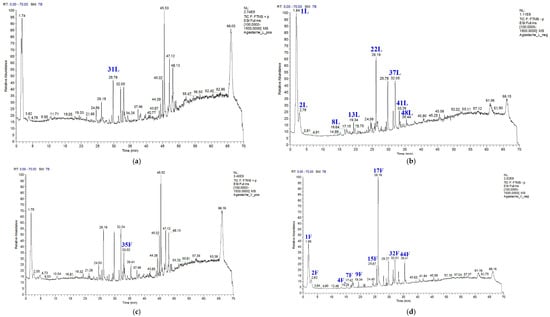
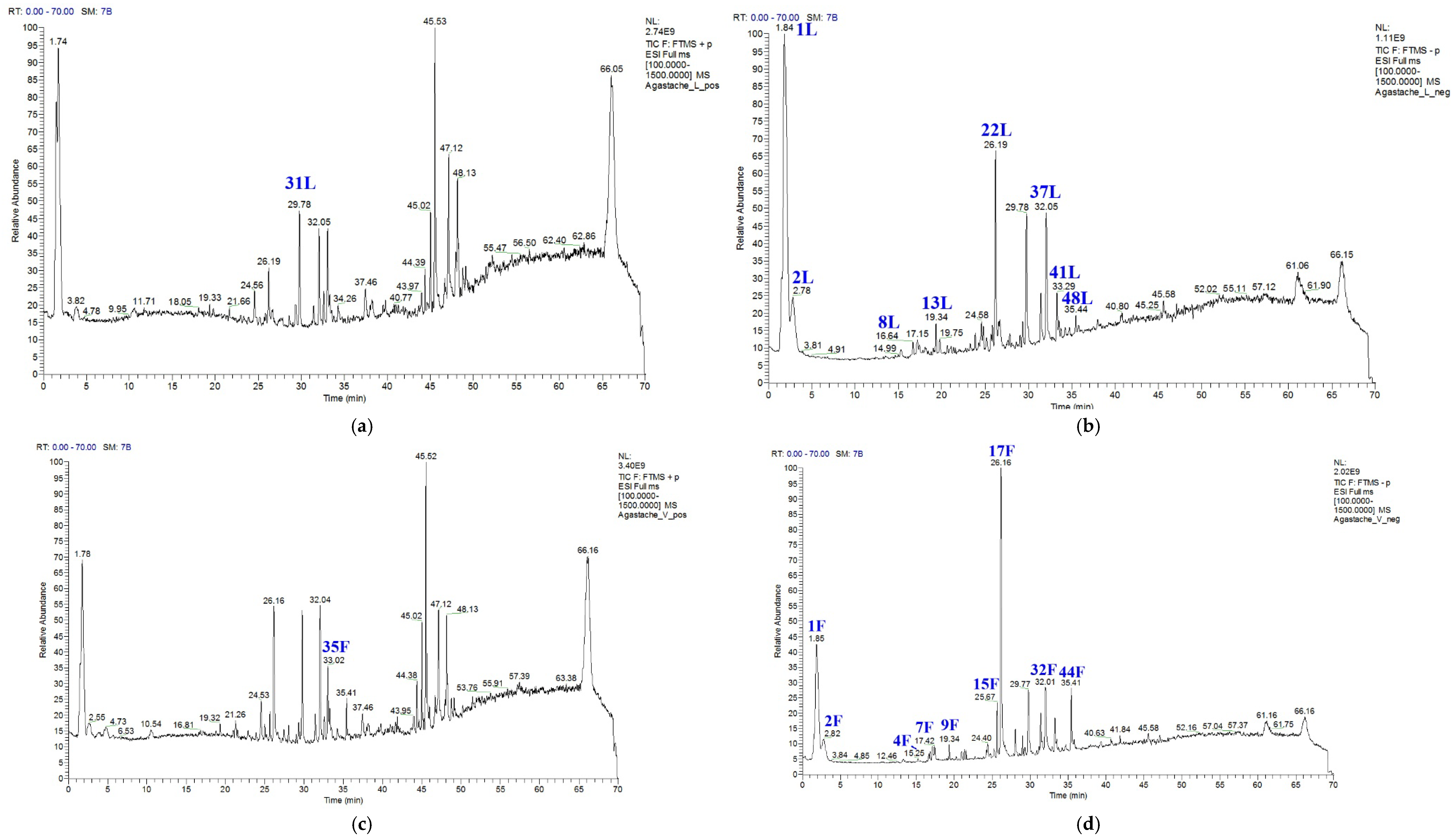
Analysis was performed using total ion chromatograms (TIC), which showed that the highest relative intensity for negative ions was observed for quinic acid in leaves and rosmarinic acid in flowers (Figure 1, Table 1). For positive ions, it was not possible to determine the compound with the highest peak intensity, as the TIC values for these analytes exceeded the upper detection limit of the UHPLC-MS/MS method.
Figure 1.
Total ion chromatograms (TIC) of Agastache foeniculum (Pursh) Kuntze extracts obtained by UHPLC-MS/MS. (a) Leaves—positive ion mode; (b) Leaves—negative ion mode; (c) Flowers—positive ion mode; (d) Flowers—negative ion mode. Blue peaks correspond to identified secondary metabolites; peak numbers refer to compounds listed in Table A1 and Table A2. (“L”—leaves, “F”—flowers).
Table 1.
Relative content of secondary metabolites identified in leaf and flower extracts of Agastache foeniculum (Pursh) Kuntze (“L”—leaves, “F”—flowers).
Within the identified TIC-positive ion range, the compound with the highest relative intensity was tilianin (acacetin-7-O-glucoside) in leaves and pulegone in flowers (Table 1). In contrast, the TIC-negative ion range revealed a significantly greater number of compounds. In the leaf extracts, the following compounds showed the highest relative concentrations (in descending order): quinic acid, dihydroxy-methoxy(iso)flavone isomer 2, dihydroxy-dimethoxy(iso)flavone, citric acid, rosmarinic acid, dihydroxy-methoxy(iso)flavone isomer 3, 12-hydroxyjasmonic acid-12-O-glucoside (or tuberonic acid glucoside), and 4-O-glucosyl-4-hydroxycinnamate (Table 1).
In the flower extracts, the most abundant negative-ion compounds included rosmarinic acid, quinic acid, dihydroxy-methoxy(iso)flavone isomer 2, dihydroxy-methoxy(iso)flavone isomer 3, phlorizin (phloretin-2′-O-glucoside), citric acid, 12-hydroxyjasmonic acid-12-O-glucoside (or tuberonic acid glucoside), caffeic acid, and salicylic acid-2-O-glucoside (Table 1).
As demonstrated by the above-mentioned compounds with the highest relative abundance, the phytochemical profile of A. foeniculum is primarily composed of organic acids and flavonoids. However, both the presence and relative concentration of these metabolites vary between the leaves and flowers. These organ-specific differences should be carefully considered when selecting plant material for nutritional or medicinal purposes.
3.2. Microbial Analysis
The ethanolic leaf extract of A. foeniculum exhibited concentration-dependent antibacterial effects against multidrug-resistant (MDR) clinical isolates, with the most pronounced activity generally observed at the highest tested concentration (87.5%) (Table 2). For S. aureus, bacterial survival decreased markedly from 5.0 ± 0.2 × 10⁸ CFU/mL in the control to below the detection limit at concentrations of 75% and 87.5%, indicating strong antibacterial activity.
Table 2.
Antibacterial activity of Agastache foeniculum (Pursh) Kuntze leaf extract against multidrug-resistant clinical isolates (n = 10 per strain). Values are expressed as colony-forming units (CFU/mL) of surviving bacteria.
For C. freundii, A. xylosoxidans, and A. baumannii, a gradual concentration-dependent reduction in bacterial counts was observed. At the highest tested concentration (87.5%), bacterial counts for all three strains were reduced below the detection limit of the assay. These findings indicate considerable antibacterial activity of the extract against Gram-negative MDR strains, particularly at higher concentrations.
E. faecalis showed moderate susceptibility, with the strongest inhibitory effect observed at 87.5%, where bacterial counts were also reduced below the detection limit of the assay (Table 2).
In the case of E. coli, bacterial counts progressively decreased with increasing extract concentration, reaching levels below the detection limit of the assay at 87.5% (Table 2).
Overall, the leaf extract demonstrated concentration-dependent inhibitory activity against all tested bacterial strains.
The flower extract of A. foeniculum exhibited concentration-dependent antibacterial activity against multidrug-resistant (MDR) clinical isolates, with the strongest inhibitory effects generally observed at 62.5–87.5% concentrations (Table 3).
Table 3.
Antibacterial activity of Agastache foeniculum (Pursh) Kuntze flower extract against multidrug-resistant clinical isolates (n = 10 per strain). Values are expressed as colony-forming units (CFU/mL) of surviving bacteria.
S. aureus and A. baumannii were the most susceptible strains, with no colonies detected in the plated aliquots at higher extract concentrations. C. freundii, E. coli, and A. xylosoxidans also demonstrated progressive reductions in viable bacterial counts with increasing extract concentration.
E. faecalis showed moderate susceptibility, with the greatest reduction observed at the highest tested concentrations. Overall, the flower extract demonstrated substantial antibacterial activity against all tested MDR bacterial strains.
4. Discussion
One of the most significant outcomes of our study is the identification of 35 previously unreported secondary metabolites in the flower extract and 38 in the leaf extract of A. foeniculum. This number substantially exceeds those reported in earlier studies employing either essential oil analysis [8,15,32] or methanolic extractions [20,33]. While previous research has focused primarily on volatile constituents or non-differentiated plant material, our application of high-resolution UHPLC-MS/MS combined with organ-specific sampling enabled a more comprehensive and resolved characterization of polar secondary metabolites. This approach provides new insights into the metabolic specialization of different plant organs, which is often overlooked in phytochemical studies.
The markedly higher number of detected compounds suggests that the phytochemical complexity of A. foeniculum has been substantially underestimated, particularly with regard to its non-volatile constituents. This expanded metabolite coverage is essential for a more accurate evaluation of the species’ biological potential and may contribute to improved standardization and targeted utilization of plant-derived products.
Although several studies have demonstrated the antimicrobial activity of A. foeniculum, the available literature remains limited in scope. Most studies have focused on essential oils and a narrow range of standard laboratory strains, typically using disc diffusion assays [8,27,28,29]. In contrast, the present study employed ethanolic extracts and evaluated antibacterial activity using CFU-based quantification against clinical multidrug-resistant (MDR) isolates. This approach may provide a broader assessment of antimicrobial potential against clinically relevant MDR isolates. Notably, several of the tested species (A. baumannii, A. xylosoxidans, C. freundii and E. faecalis) have not previously been investigated in relation to A. foeniculum, further extending the current knowledge base.
The requirement for relatively high extract concentrations to achieve substantial antibacterial effects may reflect the intrinsic resistance mechanisms of MDR strains, as well as the complex, multi-component nature of plant extracts, where synergistic and antagonistic interactions can influence overall activity [36].
The generally higher antibacterial activity of the flower extract compared to the leaf extract is likely associated with both qualitative and quantitative differences in their phytochemical composition. The presence of unique compounds, such as phloretin [37,38], together with higher relative levels of shared phenolic constituents (e.g., rosmarinic and caffeic acids), may contribute to the enhanced bioactivity.
In contrast to tannin-rich plant extracts, where antibacterial activity is often driven by nonspecific protein precipitation [39], the activity observed here may involve more specific interactions of phenolic compounds with bacterial targets. These findings suggest that organ-specific metabolite composition plays a key role in determining the antibacterial efficacy of A. foeniculum extracts.
It is also noteworthy that certain abundant compounds in the flower extract, such as pulegone, are known constituents of essential oils. Due to their higher solubility in lipophilic matrices, essential oil preparations of A. foeniculum, which concentrate such volatile compounds, may exhibit enhanced antimicrobial activity.
These observations suggest that combining polar and non-polar extraction approaches may provide a broader representation of the plant’s bioactive constituents.
5. Conclusions
Our results demonstrate that A. foeniculum possesses a more complex secondary metabolite profile than previously described, with 35 and 38 compounds not previously reported in the flower and leaf extracts, respectively. These phytochemical insights are essential for understanding the pharmacological potential of the species and lay the foundation for future bioactivity-guided studies. Importantly, the antibacterial activity observed, particularly against multidrug-resistant human pathogens, suggests that A. foeniculum could serve as a valuable reservoir of novel antimicrobial agents. The organ-specific efficacy, especially the superior activity of the flower extract, highlights the importance of targeted extraction strategies in future plant-based antimicrobial research.
Author Contributions
Conceptualization, O.K. (Oleh Kolesnyk), J.C., M.K. and O.K. (Oleksandra Kolesnyk); methodology, O.K. (Oleksandra Kolesnyk), O.K. (Oleh Kolesnyk), M.K., Z.C. and Z.T.H. software, Z.C. validation Z.C., O.K. (Oleh Kolesnyk), M.K. and J.C. formal analysis, J.D. and E.K.K. investigation, O.K. (Oleh Kolesnyk), O.K. (Oleksandra Kolesnyk), J.C., M.K., Z.T.H., Z.C. resources, M.K. and Z.C. data curation, J.D. writing—original draft preparation, O.K. (Oleksandra Kolesnyk), O.K. (Oleh Kolesnyk) and J.C. writing—review and editing, O.K. (Oleksandra Kolesnyk), J.C.; J.D. and M.K. visualization, J.C., E.K.K., B.S. and J.D. supervision, O.K. (Oleh Kolesnyk), M.K., Z.C., J.D. and B.S. project administration, O.K. (Oleksandra Kolesnyk); funding acquisition. All authors have read and agreed to the published version of the manuscript.
Funding
This research received no external funding.
Institutional Review Board Statement
Not applicable.
Informed Consent Statement
Not applicable.
Data Availability Statement
The data supporting the findings of this study are available upon reasonable request. Plant material documentation and UHPLC-MS/MS raw and processed data are archived at the University of Nyíregyháza (Hungary). Microbiological assay data are stored at Uzhhorod National University (Ukraine).
Acknowledgments
This project was supported by the Scientific Council of the University of Nyíregyháza. Generative artificial intelligence tools (ChatGPT (OpenAI, GPT-5.5) and Grammarly) were used exclusively for language editing and stylistic refinement of the manuscript, including grammar, spelling, and clarity. These tools were not used for study design, data generation, data analysis, interpretation of results, or drawing scientific conclusions.
Conflicts of Interest
The authors declare no conflicts of interest.
Abbreviations
The following abbreviations are used in this manuscript:
| BBCH | Biologische Bundesanstalt, Bundessortenamt and Chemical Industry growth scale |
| CFU | Colony-forming unit |
| EO | Essential oil |
| ESI | Electrospray ionization |
| GC–MS | Gas chromatography–mass spectrometry |
| MDR | Multidrug-resistant |
| ME | Methanolic extract |
| MS/MS | Tandem mass spectrometry |
| NCE | Normalized collision energy |
| PTFE | Polytetrafluoroethylene |
| Rt | Retention time |
| SAR | Structure–activity relationship |
| TIC | Total ion chromatogram |
| UHPLC | Ultra-high-performance liquid chromatography |
| UHPLC-MS/MS | Ultra-high-performance liquid chromatography coupled with tandem mass spectrometry |
Appendix A
Table A1.
Secondary metabolite composition of Agastache foeniculum (Pursh) Kuntze leaf extracts (Fragment 1–5 indicate the main MS/MS fragment ions used for compound identification).
Table A2.
Chemical composition of flower extracts from Agastache foeniculum (Pursh) Kuntze (Fragment 1–5 indicate the main MS/MS fragment ions used for compound identification).
References
- Selvi, S.; Polat, R.; Çakilcioğlu, U.; Celep, F.; Dirmençi, T.; Ertuğ, Z.F. An ethnobotanical review on medicinal plants of the Lamiaceae family in Turkey. Turk. J. Bot. 2022, 46, 283–332. [Google Scholar] [CrossRef] [Scilit]
- Uritu, C.M.; Mihai, C.T.; Stanciu, G.D.; Dodi, G.; Alexa-Stratulat, T.; Luca, A.; Tamba, B.I. Medicinal plants of the family Lamiaceae in pain therapy: A review. Pain Res. Manag. 2018, 2018, 7801543. [Google Scholar] [CrossRef] [Scilit] [PubMed]
- Michel, J.; Abd Rani, N.Z.; Husain, K. A review on the potential use of medicinal plants from Asteraceae and Lamiaceae plant family in cardiovascular diseases. Front. Pharmacol. 2020, 11, 852. [Google Scholar] [CrossRef] [Scilit] [PubMed]
- Butaș, A.-A.; Muntean, L.; Costin, A.-D.; Mariș, R.; Grigore, M.-F. Uses of the genus Lavandula for human health: A literature review. Hop Med. Plants 2021, 29, 59–72. [Google Scholar]
- Vârban, D.; Zăhan, M.; Pop, C.R.; Socaci, S.; Ștefan, R.; Crișan, I.; Bota, L.E.; Miclea, I.; Muscă, A.S.; Deac, A.M.; et al. Physicochemical characterization and prospecting biological activity of some authentic Transylvanian essential oils: Lavender, sage and basil. Metabolites 2022, 12, 962. [Google Scholar] [CrossRef] [Scilit] [PubMed]
- Crișan, I.; Ona, A.; Vârban, D.; Muntean, L.; Vârban, R.; Stoie, A.; Mihăiescu, T.; Morea, A. Current Trends for Lavender (Lavandula angustifolia Mill.) Crops and Products with Emphasis on Essential Oil Quality. Plants 2023, 12, 357. [Google Scholar] [CrossRef] [Scilit] [PubMed]
- Pop, C.R.; Zăhan, M.; Crișan, I.; Ștefan, R.; Vârban, D.; Gál, E.; Vârban, R.; Rotar, A.M.; Coldea, Ș.; Olar, L. Recent insight in the phytochemistry and bioactivity of organic Melissa officinalis L. essential oil from Transylvania. Not. Bot. Horti Agrobot. Cluj-Napoca 2025, 53, 14326. [Google Scholar] [CrossRef] [Scilit]
- Nechita, M.A.; Toiu, A.; Benedec, D.; Hanganu, D.; Ielciu, I.; Oniga, O.; Oniga, I. Agastache species: A comprehensive review on phytochemical composition and therapeutic properties. Plants 2023, 12, 2937. [Google Scholar] [CrossRef] [Scilit] [PubMed]
- Hernández-Abreu, O.; Castillo-España, P.; León-Rivera, I.; Ibarra-Barajas, M.; Villalobos-Molina, R.; González-Christen, J.; Vergara-Galicia, J.; Estrada-Soto, S. Antihypertensive and vasorelaxant effects of tilianin isolated from Agastache mexicana are mediated by NO/cGMP pathway and potassium channel opening. Biochem. Pharmacol. 2009, 78, 54–61. [Google Scholar] [CrossRef] [Scilit] [PubMed]
- Matei, C.F.; Duda, M.M.; Ardelean, A.E.; Covaci, A.D.; Madaș, M.N. The importance and usage of the Agastache foeniculum species (Pursh) Kuntze. Hop Med. Plants 2010, 18, 49–52. [Google Scholar]
- Abbasi, F.; Jafarbeglou, A.; Davoodi, F.; Mohammadi, R.; Dezfoulian, O.; Hasani, S.J.; Raisi, A.; Yahoo, A. Effects of Agastache foeniculum essential oil on skin wound healing in mice. Tissue Cell 2025, 96, 102995. [Google Scholar] [CrossRef] [Scilit] [PubMed]
- Santos, B.C.S.; Pires, A.S.; Yamamoto, C.H.; Couri, M.R.C.; Taranto, A.G.; Alves, M.S.; Araújo, A.L.S.M.; Sousa, O.V. Methyl chavicol and its synthetic analogue as possible antioxidant and antilipase agents based on in vitro and in silico assays. Oxidative Med. Cell. Longev. 2018, 2018, 2189348. [Google Scholar] [CrossRef] [Scilit] [PubMed]
- Ivanov, I.G.; Vrancheva, R.Z.; Petkova, N.T.; Tumbarski, Y.; Dincheva, I.N.; Badjakov, I.K. Phytochemical compounds of anise hyssop (Agastache foeniculum) and antibacterial, antioxidant, and acetylcholinesterase inhibitory properties of its essential oil. J. Appl. Pharm. Sci. 2019, 9, 72–78. [Google Scholar] [CrossRef] [Scilit]
- Anand, S.; Deighton, M.; Livanos, G.; Pang, E.C.K.; Mantri, N. Agastache honey has superior antifungal activity in comparison with important commercial honeys. Sci. Rep. 2019, 9, 18197. [Google Scholar] [CrossRef] [Scilit] [PubMed]
- Bălănescu, F.; Botezatu, A.V.; Marques, F.; Busuioc, A.; Marincaș, O.; Vînătoru, C.; Cârâc, G.; Furdui, B.; Dinica, R.M. Bridging the chemical profile and biological activities of a new variety of Agastache foeniculum (Pursh) Kuntze extracts and essential oil. Int. J. Mol. Sci. 2023, 24, 828. [Google Scholar] [CrossRef] [Scilit] [PubMed]
- Ahn, J.; Alford, A.R.; Niemeyer, E.D. Variation in phenolic profiles and antioxidant properties among medicinal and culinary herbs of the Lamiaceae family. J. Food Meas. Charact. 2020, 14, 1720–1732. [Google Scholar] [CrossRef] [Scilit]
- Hernández-Abreu, O.; Torres-Piedra, M.; García-Jiménez, S.; Ibarra-Barajas, M.; Villalobos-Molina, R.; Montes, S.; Rembao, D.; Estrada-Soto, S. Dose-dependent antihypertensive determination and toxicological studies of tilianin isolated from Agastache mexicana. J. Ethnopharmacol. 2013, 146, 187–191. [Google Scholar] [CrossRef] [Scilit] [PubMed]
- Kim, M.H.; Chung, W.T.; Kim, Y.K.; Lee, J.H.; Lee, H.Y.; Hwang, B.; Kim, J.H. The effect of the oil of Agastache rugosa O. Kuntze and three of its components on human cancer cell lines. J. Essent. Oil Res. 2001, 13, 214–218. [Google Scholar] [CrossRef] [Scilit]
- Lashkari, A.; Najafi, F.; Kavoosi, G.; Niazi, S. Evaluating the in vitro anticancer potential of estragole from the essential oil of Agastache foeniculum (Pursh) Kuntze. Biocatal. Agric. Biotechnol. 2020, 27, 101727. [Google Scholar] [CrossRef] [Scilit]
- Shanaida, M.; Palamar, O.; Holembiovska, O. Chromatographic profile of polyphenols in the Agastache foeniculum (Pursh) Kuntze herb: Evaluation of optimal extraction efficiency. Biomed. Pharmacol. J. 2024, 17, 63–69. [Google Scholar] [CrossRef] [Scilit]
- Lebedeva, M.A.; Kukushkina, T.A.; Vorob’eva, T.A.; Shaldaeva, T.M.; Khramova, E.P. Content of biologically active substances and antioxidant properties of some species of the genus Agastache Clayton ex Gron. during the growing season. Russ. J. Bioorg. Chem. 2025, 51, 3010–3019. [Google Scholar] [CrossRef] [Scilit]
- Duda, S.C.; Mărghitaș, L.A.; Dezmirean, D.; Duda, M.; Mărgăoan, R.; Bobiș, O. Changes in major bioactive compounds with antioxidant activity of Agastache foeniculum, Lavandula angustifolia, Melissa officinalis and Nepeta cataria: Effect of harvest time and plant species. Ind. Crops Prod. 2015, 77, 499–507. [Google Scholar] [CrossRef] [Scilit]
- Vârban, R.; Ona, A.; Stoie, A.; Vârban, D.; Crișan, I. Phenological assessment for agronomic suitability of some Agastache species based on standardized BBCH scale. Agronomy 2021, 11, 2280. [Google Scholar] [CrossRef] [Scilit]
- Mazza, G.; Kiehn, F.A. Essential oil of Agastache foeniculum, a potential source of methyl chavicol. J. Essent. Oil Res. 1992, 4, 295–299. [Google Scholar] [CrossRef] [Scilit]
- Omidbaigi, R.; Kabudani, M.; Khoorang, M. Nitrogen fertilizer affecting herb yield, essential oil content and composition of Agastache foeniculum (Pursh). J. Essent. Oil Bear. Plants 2008, 11, 261–266. [Google Scholar] [CrossRef] [Scilit]
- Mahmoodi Sourestani, M.; Malekzadeh, M.; Tava, A. Influence of drying, storage and distillation times on essential oil yield and composition of anise hyssop [Agastache foeniculum (Pursh) Kuntze]. J. Essent. Oil Res. 2014, 26, 177–184. [Google Scholar] [CrossRef] [Scilit]
- Nechita, M.-A.; Pralea, I.-E.; Țigu, A.-B.; Iuga, C.-A.; Pop, C.R.; Gál, E.; Vârban, R.; Nechita, V.-I.; Oniga, O.; Toiu, A.; et al. Agastache species (Lamiaceae) as a valuable source of volatile compounds: GC–MS profiling and investigation of in vitro antibacterial and cytotoxic activities. Int. J. Mol. Sci. 2024, 25, 5366. [Google Scholar] [CrossRef] [Scilit] [PubMed]
- Mollova, S.; Stanev, S.; Bojilov, D.; Manolov, S.; Kostova, I.; Damianova, S.; Fidan, H.; Stoyanova, A.; Ercisli, S.; Assouguem, A.; et al. Chemical composition and biological activity of essential oil from anise hyssop. Biotechnol. Biotechnol. Equip. 2024, 38, 2358995. [Google Scholar] [CrossRef] [Scilit]
- Hashemi, M.; Ehsani, A.; Hassani, A.; Afshari, A.; Aminzare, M.; Sahranavard, T.; Azimzadeh, Z. Phytochemical, antibacterial, antifungal and antioxidant properties of Agastache foeniculum essential oil. J. Chem. Health Risks 2017, 7, 95–104. [Google Scholar] [CrossRef] [Scilit]
- Ownagh, A.O.; Hasani, A.; Mardani, K.; Ebrahimzadeh, S. Antifungal effects of thyme, agastache and satureja essential oils on Aspergillus fumigatus, Aspergillus flavus and Fusarium solani. Vet. Res. Forum 2010, 1, 99–105. [Google Scholar]
- Nistor, A.L.; Pop, C.R.; Mitrea, L.; Cătunescu, G.M.; Vârban, R.; Lipșa, F.D.; Rusu, C.C.; Rotar, A.M. Antimicrobial potential of Hyssopus officinalis L. and Agastache foeniculum (Pursh) Kuntze essential oils for food applications: A review of their chemical compositions and antimicrobial efficacy. Appl. Sci. 2025, 15, 4772. [Google Scholar] [CrossRef] [Scilit]
- Svoboda, K.P.; Gough, J.; Hampson, J.; Galambosi, B. Analysis of the essential oils of some Agastache species grown in Scotland from various seed sources. Flavour Fragr. J. 1995, 10, 139–145. [Google Scholar] [CrossRef] [Scilit]
- Park, W.T.; Kim, H.H.; Chae, S.C.; Cho, J.W.; Park, S.U. Phenylpropanoids in Agastache foeniculum and its cultivar A. foeniculum ‘Golden Jubilee’. Asian J. Chem. 2014, 26, 4599. [Google Scholar] [CrossRef] [Scilit]
- Azmir, J.; Zaidul, I.S.M.; Rahman, M.M.; Sharif, K.M.; Mohamed, A.; Sahena, F.; Jahurul, M.H.A.; Ghafoor, K.; Norulaini, N.A.N.; Omar, A.K.M. Techniques for extraction of bioactive compounds from plant materials: A review. J. Food Eng. 2013, 117, 426–436. [Google Scholar] [CrossRef] [Scilit]
- Balouiri, M.; Sadiki, M.; Ibnsouda, S.K. Methods for in vitro evaluating antimicrobial activity: A review. J. Pharm. Anal. 2016, 6, 71–79. [Google Scholar] [CrossRef] [Scilit] [PubMed]
- Li, J.; Xie, S.; Ahmed, S.; Wang, F.; Gu, Y.; Zhang, C.; Chai, X.; Wu, Y.; Cai, J.; Cheng, G. Antimicrobial activity and resistance: Influencing factors. Front. Pharmacol. 2017, 8, 364. [Google Scholar] [CrossRef] [Scilit] [PubMed]
- Nakhate, K.T.; Badwaik, H.; Choudhary, R.; Sakure, K.; Agrawal, Y.O.; Sharma, C.; Ojha, S.; Goyal, S.N. Therapeutic potential and pharmaceutical development of a multitargeted flavonoid phloretin. Nutrients 2022, 14, 3638. [Google Scholar] [CrossRef] [Scilit] [PubMed]
- Wu, D.; Hao, L.; Liu, X.; Li, X.; Zhao, G. The anti-biofilm properties of phloretin and its analogs against Porphyromonas gingivalis and its complex flora. Foods 2024, 13, 1994. [Google Scholar] [CrossRef] [Scilit] [PubMed]
- Kolodziej, H.; Kayser, O.; Latté, K.P.; Ferreira, D. Evaluation of the antimicrobial potency of tannins and related compounds using the microdilution broth method. Planta Med. 1999, 65, 444–446. [Google Scholar] [CrossRef] [Scilit]

Disclaimer/Publisher’s Note: The statements, opinions and data contained in all publications are solely those of the individual author(s) and contributor(s) and not of MDPI and/or the editor(s). MDPI and/or the editor(s) disclaim responsibility for any injury to people or property resulting from any ideas, methods, instructions or products referred to in the content. |
© 2026 by the authors. Licensee MDPI, Basel, Switzerland. This article is an open access article distributed under the terms and conditions of the Creative Commons Attribution (CC BY) license.