1. Introduction
Cyanobacteriota (Cyanobacteria) are considered to be among the earliest photosynthetic organisms on Earth, which induced the oxygenation of water and atmosphere on the planet approximately 2.4 billion years ago [
1]. Presently, representatives of this group constitute a major component of the phytoplankton in numerous freshwater and marine ecosystems [
2] and thus, they hold a main position as the primary producers on Earth. To date, approximately 860 genera and more than 5700 species have been identified [
3]. A significant number of cyanobacterial species have been shown to produce biologically active metabolites with various beneficial properties. Based on structural characteristics, biologically active compounds derived from cyanoprokaryotes are classified as pigments, lipids, polysaccharides, nucleosides, proteins and peptides, lipopeptides, phenolic compounds, alkaloids and terpenoids [
4,
5,
6]. Generally, they exert diverse biological activities. A summarized instance includes different types of pigments (phycobiliproteins, chlorophylls, carotenoids, mycosporine amino acids (MAA)) that have demonstrated antioxidant, antitumor, antigenotoxic, antimutagenic, cardio protective and antiobesity activities; lipopeptides with antimalarial, anticancer, antiproliferative and/or antifungal properties; cyanobacterial nucleosides that have exerted antitumor, antifungal, antiviral activities; exopolysaccharides that have shown antioxidant, anti-inflammatory, or immunomodulatory properties, while sulfated polysaccharides have been shown to exhibit antiviral activity; phenolic compounds that have demonstrated antibacterial, antifungal, antitumor, and anti-inflammatory activities; lipids, mainly different types of saturated and unsaturated fatty acids, have exerted immunomodulatory and antitumor activities; and peptides with antimicrobial, antifungal and antitumor activity have been described, as well as alkaloids and terpenoids with anti-inflammatory, antitumor, antibacterial, antioxidant and UV-protective effects [
4,
5,
6]. Recent advances in the methods for isolation of cyanobacterial metabolites, combined with molecular modeling and docking analyses, provide new opportunities for a more detailed investigation of cyanobacterial compounds and the development of products with potential applications in pharmacology, food industry, cosmetics, and biotechnology [
7].
Currently, despite extensive research on biologically active substances produced by cyanobacteria, only 10–20% of the identified cyanobacterial metabolites have been comprehensively characterized with respect to their chemical structures and effects on living organisms [
8]. The majority of detected compounds are commonly determined by preliminary bioassays or inferred through genomic and metabolomic analyses without complete elucidation of activity and structure. This considerable knowledge gap highlights the immense, largely untapped potential of cyanobacteria as a source of novel natural products with various beneficial effects. Continued efforts in compound isolation and bioactivity screening are therefore essential to fully explore the functional roles of extracts derived from different cyanobacterial species.
The genus
Tolypothrix includes heterocytous filamentous cyanobacteria belonging to the order Nostocales. A significant number of representatives of this genus have not been examined in detail with regard to their potential use as sources of biologically active natural compounds [
6].
Tolypothrix species are found in stagnant, slow-flowing or subaerial freshwater environments [
9]. They typically form fasciculated colonies that appear as floating tufts or are attached to aquatic plants, rocks, and sediments and reproduce via hormogonia. The filaments are heteropolar, characterized by false branching and presence of a mucilaginous sheath, which contribute to their ecological adaptability. Heterocysts are present singly or in pairs, located intercalary and often at the base of false branches [
10]. Akinetes are less commonly observed.
Tolypothrix species are known to participate in nitrogen fixation and play an important role in the primary productivity and nutrient cycling of freshwater ecosystems [
2]. Despite their ecological importance, systematic studies of biological activity focusing on
Tolypothrix extracts remain limited compared with those on other genera within Nostocales, such as
Nostoc,
Anabaena, and
Calothrix [
4]. Several
Tolypothrix strains were shown to be capable of producing secondary metabolites with antibacterial, antioxidant, antitumor properties; however, only a few of these bioactive compounds (for example, hassallidins and tjipanazoles) have been structurally characterized [
6,
11,
12]. Certain species of the genus
Tolypothrix have been shown to produce compounds with anti-inflammatory properties, which makes them interesting for the development of new agents including nutraceutics for the treatment of inflammatory diseases. An example of such a substance is tolypodiol isolated from
T. nodosa [
13]. Other examples are toyocamycin and tubercidin—nucleoside chemicals with anti-fungal properties isolated from
T. tenuis [
14]. Additionally, a small number of biosynthetic gene clusters determining the production of phosphonate, ectoine, phenazine, β-lactone and tetrapyrrole macrocycles were detected [
4,
15]. A screening study reported the antibacterial and radical-scavenging (antioxidant) activity of extracts derived from environmental isolates of
Tolypothrix spp. [
16]. All these findings highlight
Tolypothrix species as a promising source of novel natural products with potential pharmacological applications. Still, the biological activity of compounds produced by
Tolypothrix species and strains have not been evaluated which leaves much of the biosynthetic potential of this genus unexplored.
Our research group has recently shown that non-polar fractions of extracts derived from five
Tolypothrix strains demonstrate important biological activities in vitro, including antitumor potential and other properties [
17]. Experiments with a mouse macrophage model demonstrated that all extracts significantly decreased the production of proinflammatory cytokines by lipopolysaccharide-stimulated RAW264.7 cells. These findings indicated the anti-inflammatory activity of natural products obtained from the studied
Tolypothrix strains. Chemical analysis showed the presence of 26 different fatty acids distributed differently in the individual strains [
18]. Therefore, to support the pharmacological importance of these cyanobacterial samples, we proceeded our research by evaluating their effects on human immunocompetent cells and also by performing morphological analyses for strain characterization and determination of potential strain-specific features. The present study reports the morphometric characteristics and specific thylakoid arrangement in the cells of five
Tolypothrix strains (
Tolypothrix sp. PACC 5501,
T. tenuis PACC 5497,
T. tenuis PACC 8648,
T. distorta SAG 1482-2,
T. distorta CCALA 194), which were not previously described. Our research demonstrates for the first time the effects of non-polar
Tolypothrix extract fractions on human peripheral blood leukocytes, based on analyses of the proportion of the main immune cell populations and the production of proinflammatory and anti-inflammatory cytokines.
2. Materials and Methods
2.1. Selected Cyanobacterial Strains
Five
Tolypothrix strains (Nostocales, Cyanobacteriota) were included in the present study (
Table 1). Four of them were identified to species level as
Tolypothrix tenuis Kützing ex Bornet & Flahault 1886 (PACC 5497 and PACC 8648) and
Tolypothrix distorta Kützing ex Bornet & Flahault 1886 (CCALA 194 and SAG 1482-2). One strain (
Tolypothrix sp. PACC 5501) was not determined to species level. The strains were obtained from the following culture collections: PACC—Plovdiv Algal Culture Collection (Plovdiv, Bulgaria); CCALA—Culture Collection of Autotrophic Organisms (Třeboň, Czech Republic); and SAG—Sammlung von Algenkulturen (Göttingen, Germany).
2.2. Cultivation of Cyanobacteria and Preparation of Extracts
Z medium (Zehnder Medium for Cyanobacteria) [
19] with pH modification in the alkaline range (pH 8.5–9.0) and ES (“Erddekokt + Salze”) medium (Basal medium) [
20] were used for the cultivation and long-term maintenance of the cyanobacterial strains. Media requirements and the origin of the studied
Tolypothrix strains are indicated in
Table 1.
The cyanobacteria were inoculated in 25 cm2 culture flasks (TPP, Trasadingen, Switzerland)—1 mL of the delivered actively growing cultures was added to 9 mL culture medium. The expanded cultures were subsequently transferred to 75 cm2 flasks (3 mL inoculum and 27 mL culture medium). The Tolypothrix strains were grown in liquid alkaline Z-medium under sterile conditions. Cultivation was performed in a growth chamber equipped with a light source providing constant intensity and stable spectral composition, with automatic photoperiod control. A 12 h:12 h (light:dark) photoperiod was maintained using 40 W cool-white fluorescent lamps with an intensity of 10 µmol m−2 s−1.
The strains were cultured for six weeks under sterile conditions to accumulate the cyanobacterial biomass necessary for the study (morphological analysis, transmission electron microscopy (TEM), and extract preparation), to culture stage corresponding to the stationary phase under the applied conditions. Samples for classical morphological and TEM analyses were taken from actively growing cultures during the third week after inoculation.
To obtain cyanobacterial biomass, expanded cultures were harvested after the cultivation period, transferred to 50 mL centrifuge tubes and centrifuged for 15 min at 4000 rpm. Then, the cell pellet was frozen and lyophilized. Extracts of each
Tolypothrix strain were derived from a total of 500 mg lyophilized cyanobacterial biomass. The material was mixed with 3 mL of methanol and vortexed for 1 min. This was followed by extraction for 20 min in an ultrasonic bath (Branson 5510R-DTH, Wilmington, NC, USA) at 40 °C, 25 kHz frequency. During this step, vortexing was performed every 3–5 min. After that, 6 mL of chloroform were added to the resulting suspension and mixed, which was followed by shaking for 20 min. Then, 3 mL of deionized water were added and the mixture was vortexed for 1 min. The extracts were centrifuged at 4000 rpm for 20 min. The methanol/chloroform (non-polar) and water/methanol (polar) fractions were separated and filtered through a 0.20 μm hydrophobic polytetrafluoroethylene (PTFE) membrane filter (Merck KGaA, Darmstadt, Germany). The organic solvents were removed from the samples by vacuum evaporation at 37 °C using Savant SpeedVac Concentrator (SAVANT Instruments Inc., Farmingdale, NY, USA). The dried extracts were dissolved in DMSO/water (1:1) and solutions with a final concentration of 5 mg/mL (
w/
v) were obtained. To achieve a final DMSO concentration of <1% for in vitro assays, working solutions were prepared with sterile Dulbecco’s modified Eagle medium (DMEM) or Dulbecco’s phosphate-buffered saline (DPBS) (both purchased from Gibco
®, Life Technologies™, Paisley, Scotland, UK). The fatty acid composition of the obtained extract fractions was determined by gas chromatography in a previous study [
17] and is discussed in the following sections in relation to the results reported in this article.
2.3. Microscopy Analyses
2.3.1. Morphometric Evaluations
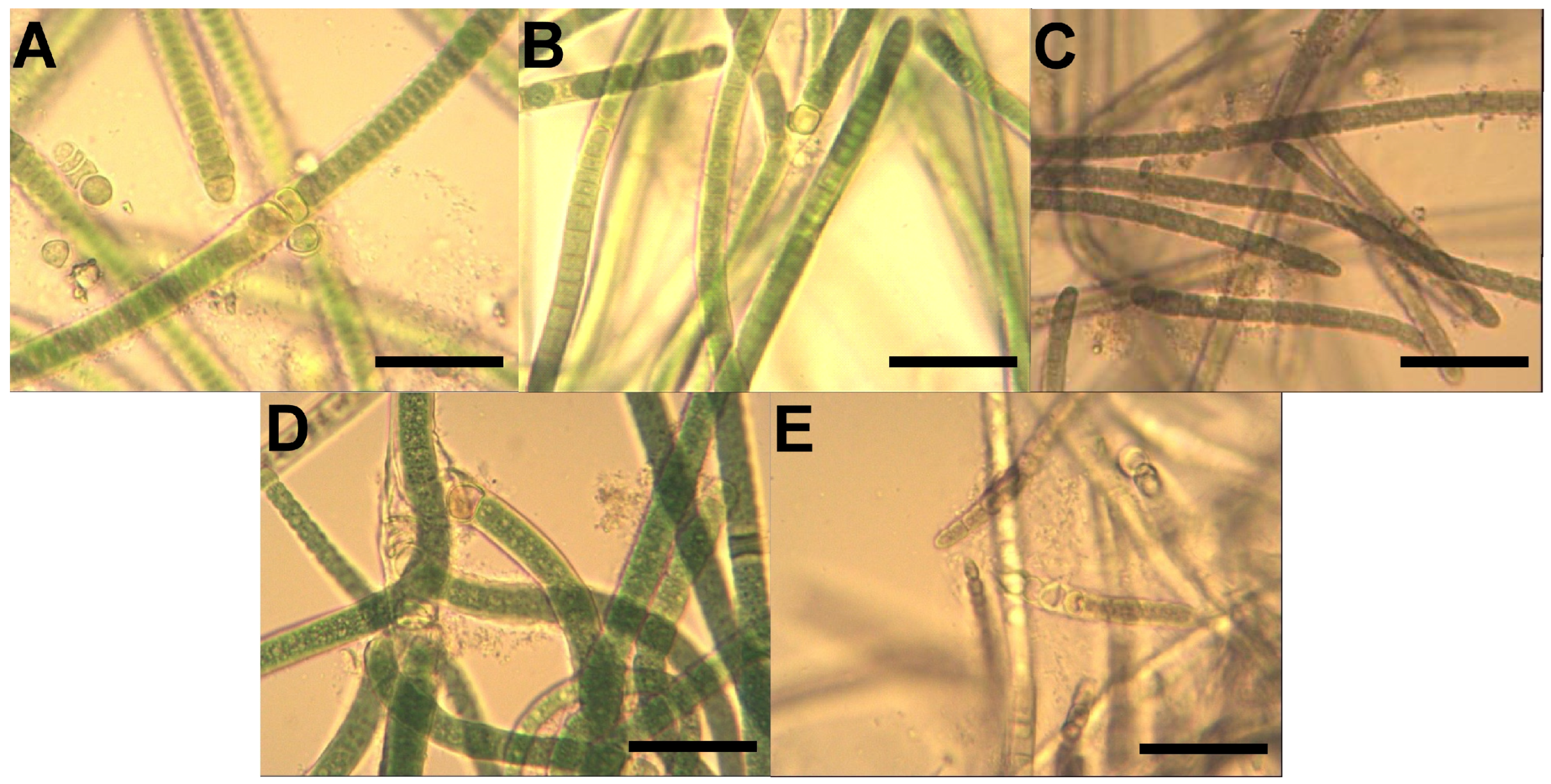
Morphological analyses of the Tolypothrix strains were implemented with a Magnum-T microscope (Medline Scientific Ltd., Chalgrove, UK) and a high-resolution Si-3000 XLiCap digital camera (Medline Scientific Ltd., Chalgrove, UK). Pictures of all analyzed cultures were obtained at a magnification of 100–400×. The following valuable phenotypic features were monitored during the exponential growth phase of the strains: thallus (macroscopic view: color, growth), filaments (width), sheaths (width, color, structure), trichomes (shape, constrictions), cells (shape, dimensions), terminal cells (shape), heterocysts (arrangement, shape, size). Length (L) and width (W) measurements were performed on a minimum of 50 cells (measurement unit—µm).
2.3.2. Analyses of Thylakoid Arrangement
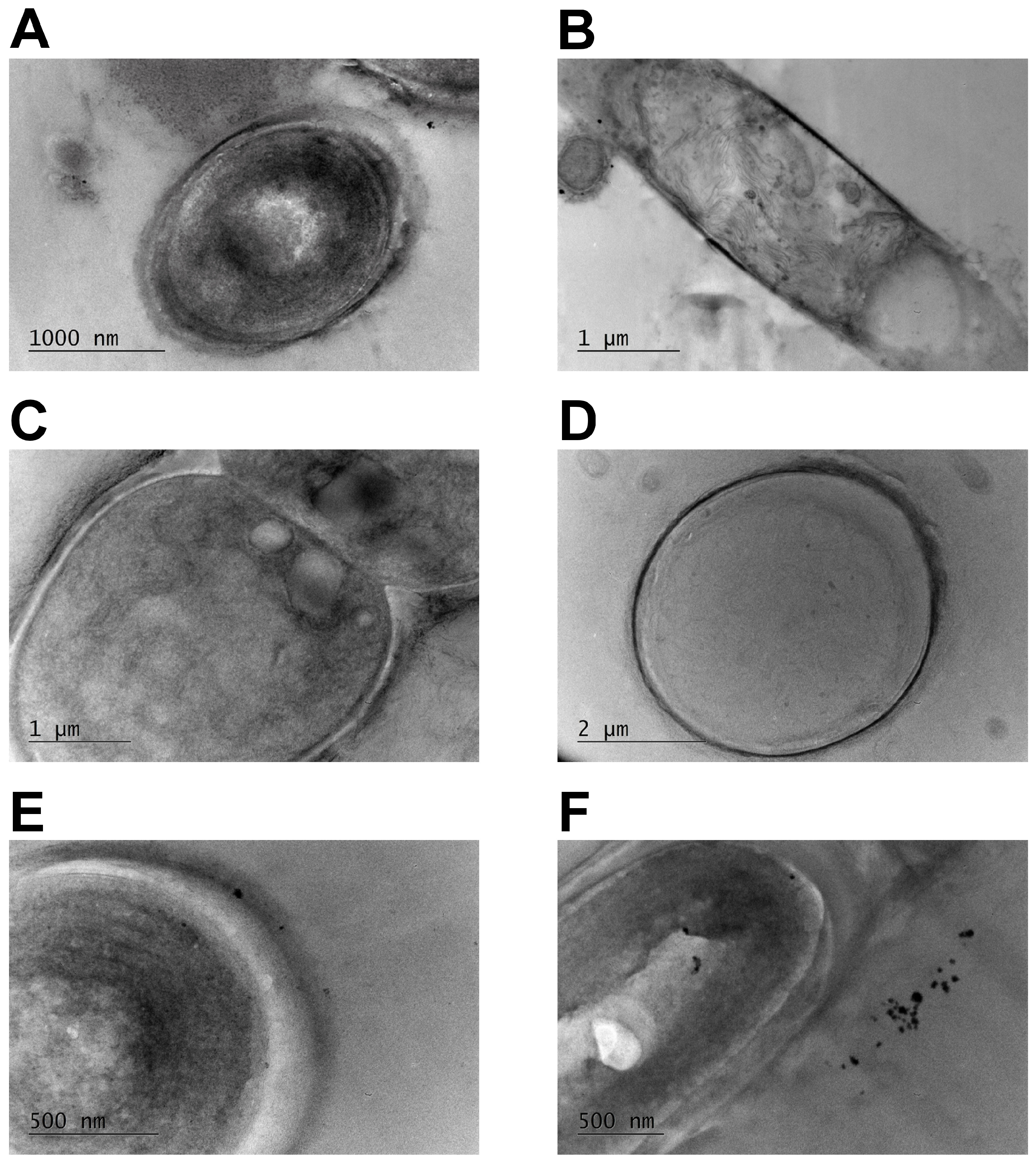
Cyanobacterial cultures were harvested and centrifuged at 4000 rpm for 5 min. The pelleted filaments were washed with 0.1 M cacodylate buffer and after that, they were fixed with 4% glutaraldehyde solution in 0.1 M cacodylate buffer at pH 7.2 for 4 h at 4 °C. The samples were further processed as previously described [
21] and analyzed with a high-resolution transmission electron microscope HR STEM JEOL JEM 2100 (JEOL Ltd., Tokyo, Japan) operating at 200 kV, equipped with a CCD camera GATAN Orius 832 SC1000 (Gatan GmbH, Munich, Germany).
2.4. Isolation of Leukocytes and Cell Culture Conditions
In the present study, peripheral blood was obtained from 3 patients with inflammatory conditions (5 mL sample per patient; C-reactive protein/CRP/concentration in blood plasma > 10 mg/L). Prior to the sample collection, a written consent form has been signed by all participants. The experiment was performed in accordance with the Declaration of Helsinki and approved by the Local Ethical Committee at the University of Plovdiv “Paisii Hilendarski”, Bulgaria (protocol No. 7 from 10 June 2024).
Blood samples were collected from the cubital vein of the patients into BD Vacutainer® K2 EDTA tubes (Becton, Dickinson and Company, Oakville, ON, Canada). Then, the samples were centrifuged at 1000 rpm for 20 min at room temperature and the plasma was discarded. Red blood cells were lysed using 0.84% NH4Cl solution and the samples were washed twice with sterile DPBS (Gibco®, Life Technologies™, Paisley, Scotland, UK). The isolated leukocytes were centrifuged at 1000 rpm for 10 min and then, the cells were resuspended in DMEM supplemented with 10% heat-inactivated fetal bovine serum and a stabilized solution of antibiotic and antifungal agent (all purchased from Sigma Aldrich Chemie GmbH, Steinheim, Germany). This medium is denoted as supplemented DMEM. The isolated leukocytes (1 × 10⁶ cells/mL) were plated in 12-well plates (TPP, Trasadingen, Switzerland) and treated for 48 h with 100 μg/mL of cyanobacterial extract, which was added to the culture medium (1 mL/well). Cells cultured for the same period (48 h) in supplemented DMEM without addition of the extract sample served as a negative non-treated control. Cells incubated for 48 h with 1 μg/mL phytohemagglutinin-L (PHA-L) (Sigma-Aldrich, Saint Louis, MO, USA) were used as a positive control. Leukocytes were cultured at 37 °C, 5% CO2, 95% ambient air, in a high-humidity incubator.
2.5. Immunophenotyping
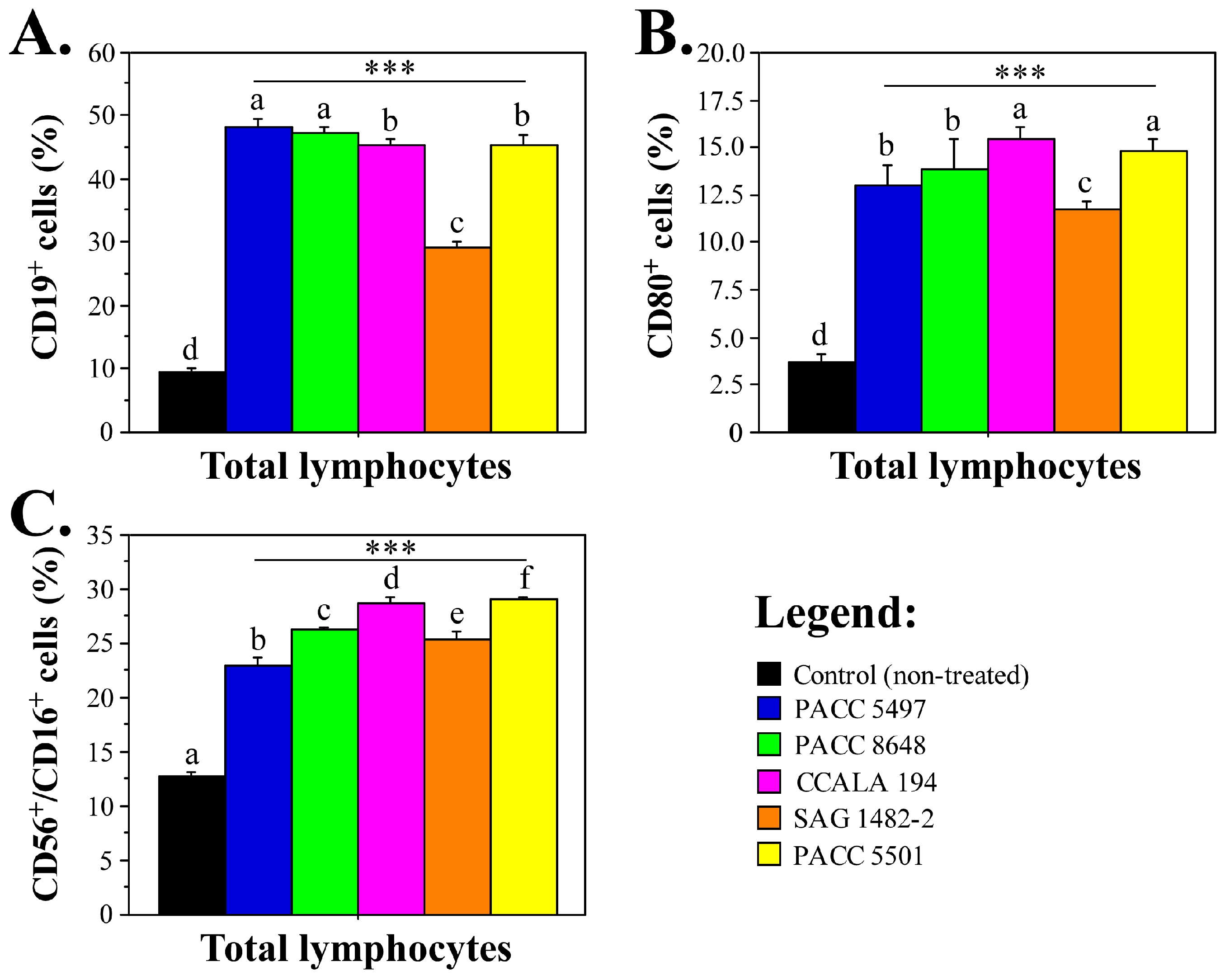
At the end of the 48 h incubation period, treated and control cells were harvested by centrifugation at 1000 rpm for 10 min and were resuspended in FACS buffer (DPBS supplemented with 5% fetal calf serum and 0.05% NaN3). The cells were stained for 20 min at room temperature, with fluorochrome-conjugated anti-human antibodies grouped into the following 3 panels: (I) CD3 PE-Dazzle™ 594, CD4 FITC, CD8 PE, CD25 PE-Cy5 and CD279 PE-Cy7; (II) HLADR/DP FITC, CD19 PE, CD80 PE-Dazzle™ 594, CD11b PE-Cy5, and CD152 PE-Cy7; and III) CD3 PE-Dazzle™ 594, CD56 FITC, CD80 PE-Cy7 and CD16 PE (all from BioLegend®, San Diego, CA, USA). After that, the cells were washed twice and resuspended in 300 μL FACS buffer. The cells were analyzed by flow cytometry using a Cytomics FC500 instrument (Beckman Coulter Inc., Life Sciences, Indianapolis, IN, USA). All samples were analyzed in duplicate.
2.6. Analyses of Cytokine Levels
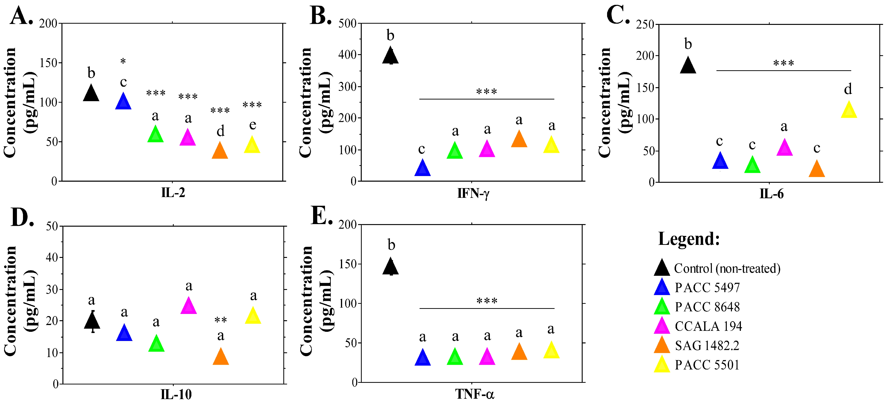
The concentration of interleukin (IL)-2, IL-6, IL-10, interferon (IFN)-γ, and tumor-necrosis factor (TNF)-α in the culture medium of non-treated cells and cells treated with Tolypothrix extracts after 48 h incubation was assayed with LEGEND MAX™ Kits Human IL-2, IL-6 IL-10, IFN-γ and TNF-α ELISA kits (BioLegend Inc., San Diego, CA, USA), according to the instructions of the manufacturer. All samples were analyzed in duplicate.
2.7. Statistics
Statistically significant differences between the test samples and the controls were determined by analysis of variance (ANOVA) and Fisher’s PLSD test for the ELISA and flow cytometry data, using StatView software version 5.0 (SAS Institute Inc., Cary NC, USA). In addition, to compare the effects between the extracts tested, we applied one-way analysis of variance (ANOVA), followed by Tukey’s post hoc test (IBM SPSS Statistics, version 28.0. Armonk, NY, USA). Prior to ANOVA, we checked assumptions for the normality of residuals by Shapiro–Wilk test and the homogeneity of variances by Levene’s test. All analyses used significance level α = 0.05. When ANOVA was significant, we conducted post hoc pairwise comparisons with Tukey’s Honest Significant Difference (Tukey HSD test). Adjusted p-values, confidence intervals, and mean differences were recorded for all group pairs. Group letters (a, b, c, d, e) were assigned for homogeneous subsets (groups were not significantly different), according to each test.
4. Discussion
Certain members of the genus
Tolypothrix have been shown to produce compounds with anti-inflammatory properties, which makes them interesting for the development of new drugs providing effective treatment of inflammatory conditions, as well as nutraceuticals [
5,
12,
22]. However, little is known about the biological activities, particularly about the immunomodulatory properties of substances derived from other
Tolypothrix species. Thus, we have focused our research on five strains belonging to this genus—
T. tenuis PACC 5497,
T. tenuis PACC 8648,
T. distorta CCALA 194,
T. distorta SAG 1482-2 and
Tolypothrix sp. PACC 5501. They were chosen based on recently reported data for their antitumor, antioxidant and anti-inflammatory potential [
17]. The present study started with a morphological and ultrastructural assessment of material from expanded cultures of the selected strains aiming at detailed strain-specific description, which was not previously reported. These evaluations are an important step in strain validation and provide information for specific morphological and ultrastructural characteristics. The observed morphological variability among the studied strains may also reflect adaptive responses to environmental conditions; however, in the present study, these features are primarily considered in the context of strain identification and characterization.
Microscopy observations and morphometric measurements confirmed that the strains PACC 5497 and PACC 8648 belong to the species
T. tenuis, while SAG 1482-2 and CCALA 194 belong to the species
T. distorta. Morphometric results are consistent with the previously reported data for the cellular length and width of
T. distorta (2.5–8.5 µm length, 6–12 µm width) and
T. tenuis (2.3–6 µm length, 3–12 µm width) and correspond to the morphological description of both species [
9].
In cultures of
T. distorta, we observed the following variability of important morphological characters: thalli olive- to dark blue-green, and in fascicles or in a cushion-like formation on the bottom and surface of the culture flasks. The filaments were falsely branched and 10–14 μm wide. The sheaths were firm, up to ±2 μm wide, and colorless to yellowish. The trichomes were cylindrical, distinctly or unclearly constricted at cross-walls, and not widened or narrowed towards ends. The cells were blue-green or olive-green, cylindrical, and usually shorter (up to 1/2) than their width (2.0–7.0 × 6.0–10.0 μm); terminal cells ± were rounded to spherical. The heterocysts were usually at the base of the branches, single, less frequently in pairs, and spherical (8–10 μm) or short and cylindrical (5.0–10 × 7–10.0 μm). TEM analysis: parietal (peripheral) and fascicular arrangement of thylakoids [
23].
Variations in thallus coloration likely reflect differences in pigment composition and physiological state, rather than stable taxonomic traits, as reported for other heterocytous cyanobacteria.
In cultures of
T. tenuis, the thalli was olive-green to brownish, in the form of fascicles on the walls of the cultivation vessel and aerophilic above the liquid medium. The filaments were up to 1 cm long, ±straight or slightly curved, and up to 12 μm wide, with false branches. The sheaths were distinct, thin or thickened, colorless or yellowish, and sometimes mucilaginous. The trichomes were cylindrical and slightly constricted at cross-wall. The cells were bright blue-green to olive-green, cylindrical, ± isodiametric or slightly shorter than wide (2.0–8.0 × 5.0–7.0 μm). The terminal cells were rounded-conical or rounded to almost spherical. The heterocysts were not or were very rarely observed, and single spherical (9.0 μm). TEM analysis: parietal (peripheral) arrangement of thylakoids [
23].
The strain
Tolypothrix sp. PACC 5501 was not identified to the species level and, therefore, could not be included in the species-level comparison. We identified the following valuable morphological features: the filaments were brownish-green, ±straight, and up to 8 μm wide, with false branching. The sheaths were thin, often mucilaginous, and colorless. The trichomes were yellowish green, cylindrical, and clearly constricted at cross-walls. The cells were barrel-shaped to cylindrical, ±isodiametric, and slightly shorter or longer than the width (2.0–7.0 × 4.0–7.0 μm). The terminal cells were rounded and conical. Heterocysts were rare, solitary or in a row of up to three, and almost spherical (6.0 μm). TEM analysis: parietal (peripheral) and fascicular arrangement of thylakoids [
23].
Although no direct relationship can be established within the scope of this study, the structural variability among strains may be associated with differences in metabolic profiles, which is consistent with the observed slight variation in the immunomodulatory activity.
Our studies proceeded with analyses of the potential immunomodulatory properties of extracts obtained from the selected
Tolypothrix strains. A generally accepted strategy for evaluating the potential of cyanobacteria as producers of biologically active substances involves the study of “crude” extracts and fractions. Particularly, the non-polar extract fractions were used, due to their superior biological effects in terms of antitumor activity compared to polar fractions [
17]. In vitro and/or in vivo test systems are used to evaluate different bioactive properties, including antitumor, antioxidant, anti-inflammatory and immunoregulatory activity. Specifically, anti-inflammatory effects are determined based on reduction in the levels of pro-inflammatory cytokines such as TNF-α, IL-1β and IL-6. Per example, low levels of TNF-α, IL-1β, and IL-6 in the culture medium were reported for LPS-stimulated RAW264.7 macrophages and mouse bone marrow-derived macrophages after treatment with lipid fractions from
Nostoc commune var.
spheroides and
Spirulina platensis [
24]. Reduced levels of TNF-α and NF-κB expression were determined in LPS-stimulated THP-1 cells after treatment with the two digalactosyldiacylglycerols isolated from
Nodularia harveyana [
25]. Similar to this effect, other authors reported the anti-inflammatory effect of nonpolar compounds, such as 7(E)-9-keto-octadec-7-enoic acid (C18 acid), isolated from cyanobacteria [
26]. The compound suppressed iNOS expression and nitric oxide (NO) production in LPS-stimulated RAW264.7 cells—a frequently used in vitro model for studying inflammatory processes. Other cyanobacterial species have also been shown to be a source of bioactive compounds with anti-inflammatory activity. An example is
Aphanothece halophytica, which produce mycosporine-2-glycine, a rare mycosporine-like amino acid that induced anti-inflammatory effects in LPS-stimulated RAW 264.7 macrophages [
27]. Different genera of aquatic and terrestrial cyanobacteria produce scytonemin—a pigment that exerts anti-inflammatory activity in vitro by the down-regulation of NF-κB activity and inhibits skin inflammation by blocking the expression of inflammatory mediators [
28]. Several research groups have reported the anti-inflammatory activity of cyanobacterial extracts or lipidic compounds, amino acids and peptides, polysaccharides and pigments based on the observed reduction/downregulation of proinflammatory cytokine production in vitro, using cell lines like RAW264.7, THP-1, human epidermal keratinocytes and others, or by demonstrating a reduction in edema, skin inflammation, lesions and itching using rodent models [
29].
In our previous studies, non-polar fractions of
Tolypothrix extracts significantly reduced the production of proinflammatory cytokines IL-6 and TNF-α by RAW264.7 mouse macrophages [
17]. These findings paved the way for further evaluations of potential immunomodulatory effects of the
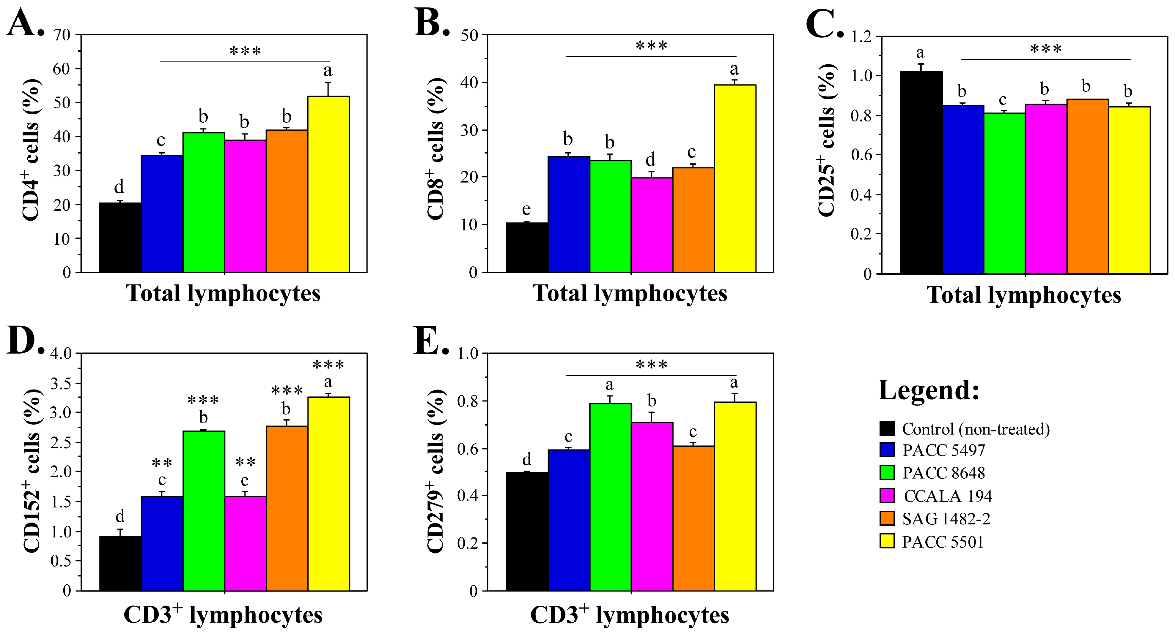
Tolypothrix strains. The present research supported the data for anti-inflammatory activity and showed the important effects of the samples on human immune cells. Our results demonstrated significantly elevated levels of CD4
+ T cells, CD8
+ T cells, CD80
+ lymphocytes, B cells, and NK cell populations in leukocyte cultures treated for 48 h with
Tolypothrix non-polar extract fractions. The uniform enhancement of the percentages of the main leukocyte populations could be a sign for overactivation in response to the ex vivo cultivation in a medium containing cyanobacterial extracts. However, the percentage of CD25
+ lymphocytes was significantly decreased in the
Tolypothrix-treated cell cultures. CD25 is a subunit of the IL-2 receptor—an activation marker [
18] expressed by immune and some non-immune cell types—but high levels of CD25 are specific for activated T lymphocytes and regulatory T cells [
30]. The reduction in CD25
+ lymphocytes together with significantly reduced IL-2 concentration in the cell culture medium following treatment with
Tolypothrix extracts indicates that the test samples did not induce strong activation of the lymphocytes, which could eventually lead to immune reactions with negative effects. In support of this suggestion, we observed increased percentages of CD152
+ and CD279
+ T cells. CD152 (cytotoxic T-lymphocyte-associated antigen-4/CTLA-4/) is a main immune checkpoint regulator of T cell homeostasis and self-tolerance [
31]. Increased transcriptomic expression of CD152 has been shown to be a predictive marker for a positive outcome for patients undergoing immunotherapy [
32]. Therefore, the elevated numbers of CD152
+ T cells after treatment with
Tolypothrix extracts could be considered an indicator of immunomodulatory activity of the cyanobacterial samples. Similar indication has been provided by the detected enhanced proportion of T cells expressing CD279
+—a programmed death 1 (PD-1) receptor that has been shown to exert immunoregulatory roles in T cell activation and tolerance [
33]. Our findings for decreased levels of CD25
+ lymphocytes and increased levels of CD152
+ and CD279
+ T cells, together with the decreased production of cytokines with proinflammatory activity (IL-6, IFN-γ and TNF-α) provide evidence for potential immunoregulatory activity of the selected
Tolypothrix strains. Combined with the data for the elevated levels of the main immune cell populations, increased T cell expression of immune checkpoint regulators (CTLA-4 and PD-1) and sustained production of the anti-inflammatory cytokine IL-10, which was not reduced by treatment with four of the cyanobacterial extracts, our results prove potential to maintain anti-inflammatory responses and exclude the assumption for the induction of long-term immunosuppression with eventual severe consequences like recurrent opportunistic infections, de novo malignancies, renal failure, hypertension and autoimmunity [
34]. The only extract that reduced IL-10 production was
T. distorta SAG 1482.2. A possible reason for this different result could be the lack of stearidonic acid in this sample, which was determined in our previous studies [
17]. Stearidonic acid (SDA) inhibits the action of cyclooxogenase-2 (COX-2) and arachidonate 5-lypoxigenase (ALOX5), which leads to a reduction in prostaglandins (e.g., PGE2) and leukotrienes that are important mediators of inflammatory responses [
35]. In addition, SDA and its metabolites can inhibit nuclear factor kappa B (NF-kB), which reduces the expression of inflammatory cytokines (such as IL-6 and TNF-α) [
36]. Previously, we identified long-chain fatty acids (ω-3 linolenic acid, ω-3 docosahexaenoic acid, and ω-9 oleic acid) as dominant nonpolar metabolites of
Tolypothrix extracts [
17]. These specific structures are known to exert anti-inflammatory effects. Oleic acid modulates membrane fluidity and NF-κB signaling, while ALA (alpha-linolenic acid) and DHA (docosahexaenoic acid) have been reported to downregulate pro-inflammatory genes and oxidative stress [
17]. Thus, the fatty-acid profile aligns with our data for the cytokine levels. Strains that are richer in these unsaturated acids showed stronger inhibition of IL-6 and TNF-α.
Both our previous results [
17] and those observed in this study indicate that
Tolypothrix nonpolar extracts interrupt the inflammatory signaling cascade at multiple points. In LPS-stimulated RAW264.7 macrophages, extracts caused a clear dose-dependent scavenge of free radicals and decreased cytokine production (IL-6 and TNF-α). In parallel, ex vivo human peripheral leukocytes treated with the same extracts showed significantly lower IL-6, TNF-α, IFN-γ and IL-2, which was consistent with an overall suppression of pro-inflammatory signaling. This broad cytokine suppression suggests an upstream block of the NF-κB (Nuclear factor kappa B) and MAPK (Mitogen-activated protein kinase) pathways. Nonpolar compounds can inhibit NF-κB activation (suppressing iNOS and NO in LPS-stimulated cells) and can promote Nrf2/ARE antioxidant responses [
17].
The limitations of the current data include the ex vivo/in vitro nature of the models. Our human leukocytes were from donors with existing inflammation, so baseline cytokines were high and the relative changes were modest. We did not directly measure NF-κB, MAPK phosphorylation or ROS/Nrf2 activation, so the proposed cascade is inferred. Future experiments should track NF-κB p65 nuclear translocation, MAPK activation, and Nrf2 target gene expression in treated cells, and test these extracts (or purified lipids) in animal models of inflammation to validate the pathway. In summary, Tolypothrix extracts appear to inhibit the LPS/TLR4→NF-κB/MAPK axis and boost antioxidant defenses, thereby reducing TNF-α, IL-6 and related cytokines.
The previously defined fatty acid composition of the studied
Tolypothrix extracts [
17] supports the observed inflammation-inhibitory potential. The fatty acids with highest levels in the samples were palmitic, oleic and linoleic acid. Palmitic acid is the most common saturated fatty acid. It has been shown to interact with Toll-like receptors and activate signaling pathways that promote inflammation [
37]. However, in our experiments, we have observed opposite effects of
Tolypothrix extracts on human leukocytes. This could be due to the complex composition of the samples that include a significant amount of unsaturated fatty acids, like oleic and linoleic acids, with proven inflammation inhibitory activity and mechanism of action involving activation of anti-inflammatory responses [
38,
39]. Higher levels of stearidonic acid were also present in the extracts obtained from
T. tennuis PACC5497 and
Tolypothrix sp. PACC5501. SDA contributes to this activity by inhibiting inflammatory cytokine production [
36]. The extracts obtained from the two
T. distorta strains also contained higher levels of palmitoleic acid. The ability of this fatty acid to counteract the proinflammatory activity of palmitic acid has been demonstrated [
40]. Collectively, these findings support our hypothesis that unsaturated fatty acids with anti-inflammatory activity could inhibit the proinflammatory action of palmitic acid present in the samples and collectively contribute to the inflammation inhibitory potential of the
Tolypothrix extracts. Further research will define the activities of purified individual compounds, possible synergistic effects of these compounds and their potential to serve as new candidate nutraceuticals.





 ,
,