


Simple Summary
This study examined whether the level of miR-31-5p in fresh semen from Holstein bulls is linked to the ability of sperm to survive freezing and thawing. Bull semen samples were collected, and sperm motility, viability, mitochondrial function, and cell damage was evaluated before and after freezing. The results showed that freezing significantly reduced sperm quality and increased signs of cellular stress and damage. However, ejaculates with higher levels of miR-31-5p in fresh semen maintained better sperm movement, higher viability, healthier mitochondrial activity, and lower levels of apoptotic damage after thawing. In addition, higher miR-31-5p levels were positively associated with all major indicators of post-thaw sperm quality. These findings suggest that miR-31-5p could serve as a useful biomarker for predicting sperm freezability and selecting higher-quality semen for breeding and artificial insemination programs.
Abstract
This study investigated whether miR-31-5p abundance in fresh Holstein bull semen is associated with post-thaw sperm resilience. The initial cohort comprised 36 Holstein bulls, from which 16 ejaculates representing the highest- and lowest-ranking post-thaw freezers were selected for epigenetic and molecular analyses. Fresh semen was analyzed for miR-31-5p, while BAX and BCL2 were evaluated at transcript and protein levels before and after cryopreservation. Post-thaw sperm quality was assessed using total motility, viability, JC-1 mitochondrial membrane potential, Annexin-negative spermatozoa, reactive oxygen species (ROS) and sperm DNA fragmentation. Cryopreservation significantly impaired sperm quality, increased ROS and DNA fragmentation, and shifted the molecular profile toward increased BAX and reduced BCL2. Ejaculates with superior post-thaw motility showed higher miR-31-5p levels, accompanied by better preservation of viability, mitochondrial function, non-apoptotic membrane status, lower ROS burden and DNA fragmentation after thawing. Fresh miR-31-5p was positively associated with post-thaw motility, viability, mitochondrial function, and Annexin-negative spermatozoa and negatively associated with post-thaw ROS. Our data support miR-31-5p as a potential candidate biomarker of sperm freezability in Holstein bulls.
1. Introduction
In animals, sperm cryopreservation is widely used to accelerate the rate of genetic improvement while minimizing the risk of horizontal or vertical transmission of venereal diseases, leading to significant cost reductions in the breeding process [1,2]. Semen storage is also a valuable tool for protecting the genetic resources and biodiversity of protected or endangered animal species [3]. Unfortunately, the full potential of semen cryopreservation has not yet been fully exploited, as a complete understanding of sperm physiology before or during freezing and thawing is mandatory to ensure maximal success and is still lacking [4,5]. Cryopreservation exposes sperm to oxidative, osmotic and thermal stress, all of which are known to compromise sperm function. Cryodamage causes structural and molecular alterations in spermatozoa, including changes in plasma membrane fluidity, motility, viability, and DNA integrity [6]. During fertilization, the paternal genome is transferred together with a variety of epigenetic information, including small non-coding RNAs. Increasing evidence on microRNAs, short single-stranded RNA molecules consisting of 22–24 nucleotides, and their association with fertility status suggests that they may play a role in sperm quality and tolerance to freezing (freezability) [7].
Because conventional semen evaluation does not fully explain why some ejaculates survive cryopreservation better than others, increasing attention has been directed toward molecular markers of sperm quality and freezability [8]. In this context, sperm-borne RNAs, including small non-coding RNAs such as microRNAs (miRNAs), have emerged as particularly promising candidates [6]. Although mature spermatozoa are transcriptionally silent, they carry a complex RNA cargo that reflects earlier spermatogenic and post-testicular events and may be associated with male fertility, sperm function, and embryo development [9]. In cattle, several studies have shown that bull sperm miRNA profiles differ according to fertility status, motility characteristics, or cryopreservation outcome, supporting their use as candidate biomarkers of semen quality [10].
miR-31-5p is of particular interest because of its reported association with proliferation- and apoptosis-related pathways in male germ-cell biology. Although direct evidence concerning miR-31-5p in mature bull sperm and cryobiology remains limited, studies in spermatogonial stem cells and reproductive biomarker research suggest that it may be linked to cellular resilience and stress-related regulation [11]. This makes miR-31-5p a plausible candidate marker of intrinsic sperm quality, particularly in the context of cryopreservation-induced apoptosis-like and mitochondrial injury.
As apoptotic pathways are activated in spermatozoa during cryopreservation, the present study further investigated the expression of the apoptosis-regulating genes BCL2 and BAX at both mRNA and protein levels using western blot analysis. The balance between the pro-apoptotic BAX and anti-apoptotic BCL2 proteins is a well-established indicator of sperm susceptibility to stress-induced cell death, and its relationship with miR-31-5p may help clarify the molecular mechanisms through which this miRNA influences sperm cryosurvival [12].
We hypothesized that the abundance of miR-31-5p in fresh Holstein bull semen could be associated with subsequent post-thaw sperm resilience. Therefore, miR-31-5p levels in fresh semen, together with apoptosis-related markers at both transcript and protein levels, were evaluated and related to post-thaw sperm motility, viability, mitochondrial membrane potential, and the proportion of Annexin-negative spermatozoa. The aim of the study was to determine whether miR-31-5p measured prior to cryopreservation could serve as a candidate biomarker of individual variation in bull sperm freezability.
2. Materials and Methods
Semen samples were collected from an initial cohort of 36 sexually mature, clinically healthy Holstein breeding bulls housed at the Breeding Centre of the Slovak Biological Services (Nitra, Slovakia), using a sterilized artificial vagina (one ejaculate per animal) in the fall of 2025. The bulls were 4–6 years old, routinely maintained for semen production under uniform management conditions, and showed normal reproductive status at the time of collection. The animals were fed a standard diet based on green and cereal fodder, berseem, straw, and concentrated mixtures, and water was available ad libitum. Since the present work was designed as a pilot study and the epigenetic/molecular analyses were limited by assay capacity, ejaculates were first ranked according to post-thaw total motility, and 16 samples representing good and poor freezers were selected for subsequent miRNA, gene-expression, and protein analyses.
An aliquot of each pre-selected semen sample was cryopreserved in a semen extender (Triladyl, Minitüb GmbH, Tiefenbach, Germany) containing 20% egg yolk (w/v) (Sigma-Aldrich, St. Louis, MO, USA) and antibiotics. The diluted semen was automatically filled into 0.25 mL French straws using an automated filling system (Minitüb GmbH, Tiefenbach, Germany). The straws were subsequently equilibrated at 4 °C for 2 h and cryopreserved using a digital freezer (Digitcool Model 5300 ZB 250; IMV, L’Aigle, France) under a controlled freezing protocol consisting of cooling rates of −3 °C/min from +4 °C to −10 °C, −40 °C/min from −10 °C to −100 °C, and −20 °C/min from −100 °C to −140 °C. Following cryopreservation, the straws were transferred into liquid nitrogen and stored at −196 °C for one month. Another aliquot was immediately transferred to the laboratory for sperm quality assessment, gene expression analysis, miRNA quantification, and Western blot analysis.
For mRNA and miRNA extraction and cDNA synthesis, samples (750 µL) were centrifuged at 10,000× g for 2 min and the supernatant (seminal plasma) was discarded. Both types of nucleic acids were extracted in a single procedure using Hybrid-R™ miRNA (GeneAll®, Seoul, Republic of Korea) according to the manufacturer’s instructions, with a modification in the lysis step. The original lysis buffer was replaced with 1 mL TRIzol (Sigma-Aldrich, St. Louis, MO, USA), followed by sonication (10% amplitude, 15 s) with cooling. Subsequently, 200 µL chloroform was added, and the samples were incubated for 2 min at room temperature. The lysed samples were centrifuged at 10,000× g for 10 min, and upper aqueous phase was transferred to new tubes. The protocol was continued according to the original instructions by adding one volume of 50% ethanol. Concentration and purity of mRNAs and miRNAs were estimated using a NanoPhotometer® P-360 (Implen, Munich, Germany) at an absorbance ratio of A260/280, with a criterion of purity values between 1.8–2.0, and of minimum quantity for miRNA of 5 ng/µL.
An amount of 400 ng of total mRNA was transcribed into cDNA using Maxima First Strand cDNA Synthesis Kit for RT-qPCR (Thermo Scientific™, Waltham, MA, USA) with mix of random hexamers and oligo(dT) primers. Minus-RT control was also transcribed with the use of all samples mix mRNA template to ensure DNA absence. The reverse transcription for miRNA was carried out using the miRCURY LNA RT Kit (Qiagen, Hilden, Germany). The supplier’s instructions were adhered to regarding the protocol and the quantity of miRNA used in the transcription process.
Gene expression analysis of the BAX and BCL2 genes was conducted using RT-qPCR. GAPDH (glyceraldehyde-3-phosphate dehydrogenase) served as the reference gene for these gene-based RT-qPCR assays, as outlined by Chowdhury et al. [13]. The primers were designed based on the mRNA accession numbers showed in Table 1, obtained from the NCBI (National Center for Biotechnology Information) database by its Primer-BLAST tool (version 2.5.0). Relative mRNA expressions were analysed using the Real-Time PCR Thermal Cycler qTOWER3 (Analytik Jena GmbH+Co. KG, Jena, Germany), with the cycling conditions for each gene listed in Table 1. The amplifications were performed in a total volume of 10 µL, utilising HOT FIREPol® EvaGreen® qPCR Mix Plus (Solis BioDyne, Tartu, Estonia) for BAX and BCL2 with cDNA diluted 1:10. The primer and exact cDNA concentrations are listed in Table 1. The reactions were conducted in 96-well plates (Starlab, Hamburg, Germany), and the samples were repeated in technical triplicate. For the RT-qPCR amplicon specificity, a melt curve analysis was conducted, and it was also checked on 1.5% agarose gels for each reaction. Relative expression levels were determined using the 2−ΔΔCt method.
Table 1.
Primers used for RT-qPCR and corresponding amplification conditions.
For gene expression analyses, all assays were performed in technical triplicate together with a no-template control (NTC), which showed no amplification, confirming the absence of reagent contamination. In addition, the reference gene and target genes were evaluated in the -RT control, which was negative in all cases, confirming the absence of genomic DNA contamination. RT-qPCR efficiencies were within the optimal range of 90–110% (GAPDH: 97.96%; BAX: 103.09%; BCL2: 95.30%). The standard-curve slopes were −3.37 for GAPDH, −3.25 for BAX, and −3.44 for BCL2, while the corresponding coefficients of determination (R2) were 0.9938, 0.9858, and 0.9983, respectively. Specificity of the PCR products was further verified by melt curve analysis, which showed a single amplicon for each assay (GAPDH: 85 °C; BAX: 89 °C; BCL2: 87 °C).
RT-qPCR analysis was conducted using the 2x miRCURY SYBR Green Master Mix (Qiagen, Hilden, Germany) along with specific miRCURY LNA PCR Assays (Qiagen, Hilden, Germany). Manufacturer-recommended protocols were adhered to. The cDNA was diluted to a ratio of 1:60 immediately prior to use. The reactions were carried out in a total volume of 10 µL, utilizing 3 µL of the diluted cDNA and 1 µL of the miRCURY LNA PCR Assay primer mix. The PCR assays for both miRNAs were conducted under the following cycling conditions: an initial heat activation at 95 °C for two minutes, followed by 45 cycles of a two-step process. This included denaturation at 95 °C for 10 s and a combined annealing/extension phase at 56 °C for 60 s, concluded with a melt curve analysis. All samples were conducted in technical triplicate. The miRNA targets, their corresponding sequences, and RT-qPCR conditions are summarized in Table 2.
Table 2.
List of miRNAs and conditions used for RT-qPCR analysis.
Sperm motility was evaluated using computer-assisted semen analysis (CASA; Version 14.0 TOX IVOS II.; Hamilton-Thorne Biosciences, Beverly, MA, USA) with the manufacturer’s system setup designed for bulls. Ten microliters of diluted semen (1:40 with Dulbecco’s Phosphate Buffer Saline; Sigma-Aldrich, St. Louis, MO, USA) were placed onto a Makler counting chamber, and at least 300 cells were objectively assessed on ten microscopic fields. Sperm motility was evaluated and expressed as the percentage of motile spermatozoa (≥5 µm/s).
Sperm membrane integrity was evaluated using a multi-fluorescent staining approach [14]. Carboxyfluorescein diacetate (CFDA; 0.75 mg/mL in DMSO; Sigma-Aldrich, St. Louis, MO, USA) serves as an indicator of cellular esterase activity and overall cell viability. Cells with damaged membrane were identified using propidium iodide (PI; 5 μg/mL in PBS; Sigma-Aldrich, St. Louis, MO, USA), while total spermatozoa were enumerated with the nucleic acid dye DAPI (4′,6-diamidino-2-phenylindole; 1 μmol/L in PBS; Sigma-Aldrich, St. Louis, MO, USA). Fluorescence signals were measured using a Glomax Multi+ spectro-fluoro-luminometer (Promega, Madison, WI, USA).
The JC-1 assay kit (Cayman Chemical, Ann Arbor, MI, USA) was used to measure the mitochondrial membrane potential. In short, 5 μL of JC-1 working solution was used to stain 100 μL of the material, which was then incubated for 30 min at 37 °C. Following a 5-min centrifugation at 150× g at 25 °C, the samples were washed twice using the commercial kit’s washing buffer. Glomax Multi+ (Promega, Madison, WI, USA) was used to evaluate the samples after they were transferred to a dark 96-chamber plate. The ratio of JC-1 complexes to JC-1 monomers (green/red ratio) was used to express the resulting ∆Ψm.
Reactive oxygen species (ROS) were quantified in fresh and frozen-thawed spermatozoa using a chemiluminescence assay with luminol (5-amino-2,3-dihydro-1,4-phthalazinedione; Sigma-Aldrich, St. Louis, MO, USA) as the probe. The reaction mixture consisted of 10 μL luminol (5 mM) and 400 μL of the control or experimental sample. The negative control was prepared by replacing the sperm suspension with 400 μL Dulbecco’s phosphate-buffered saline (DPBS), while the positive control consisted of 400 μL DPBS supplemented with 50 μL H2O2 (30%; 8.8 M; Sigma-Aldrich, St. Louis, MO, USA). Chemiluminescence was measured for 15 min in triplicate using the Glomax Multi+ combined spectro-fluoro-luminometer (Promega, Madison, WI, USA). ROS levels were expressed as relative light units per second per 106 spermatozoa (RLU/s/106 sperm).
The proportion of apoptotic spermatozoa (%) was determined using an Annexin-V assay combined with propidium iodide (PI) and analyzed with a Glomax Multi+ spectro-fluoro-luminometer (Promega Corporation, Madison, WI, USA). Washed sperm samples were first incubated with 100 µL Annexin-V solution supplemented with 10 µL PI for 20 min at 37 °C in the dark. Prior to analysis, 10 µL DAPI were added to each sample. Based on fluorescence labeling, spermatozoa were classified into three populations: viable cells (AV−/PI−), apoptotic cells (AV+/PI−), and necrotic cells (AV−/PI+) [15].
Sperm DNA fragmentation was assessed in fresh and frozen-thawed spermatozoa using the Halomax kit (Halotech DNA, Madrid, Spain). Tubes containing low-melting point agarose were incubated in a water bath at 100 °C for 5 min to melt the agarose and then transferred to 37 °C. After 5 min of incubation, 20 μL of each sperm sample was mixed with the agarose. Subsequently, 10 μL of the mixture was pipetted onto agarose-precoated slides and covered with 20 × 20 mm coverslips. The slides were placed at 4 °C for 5 min to allow microgel formation. After gentle coverslip removal, the slides were immersed horizontally in lysis solution for 5 min, washed in distilled water for 5 min, dehydrated in 70% and 100% ethanol for 2 min each, and air-dried. Finally, all slides were stained with SYBR Green (2 μg/mL; Sigma-Aldrich, St. Louis, MO, USA) in Vectashield mounting medium (Vector Laboratories, Burlingame, CA, USA). A minimum of 300 spermatozoa per sample was evaluated under an epifluorescence microscope using a ×40 objective (Leica Microsystems, Wetzlar, Germany). Spermatozoa were classified according to the presence or absence of a characteristic DNA halo, and the proportion of sperm with fragmented DNA was expressed as a percentage of the total counted cells.
For thawing, the straws were placed in a water bath prewarmed to 37 °C for 2 min and subsequently transferred into freshly labeled 0.5 mL tubes for assessment of sperm motility using a computer-assisted sperm analysis (CASA) system. Residual egg-yolk extender was then removed by washing the samples three times with 500 μL phosphate-buffered saline (Sigma-Aldrich, St. Louis, MO, USA), followed by centrifugation at 300× g for 10 min after each washing step.
Proteins for the western blot analysis were isolated from washed sperm samples lysed in 1 mL RIPA buffer (Sigma-Aldrich, St. Louis, MO, USA) supplemented with a protease inhibitor cocktail (Sigma-Aldrich, St. Louis, MO, USA) to prevent protein degradation. The lysates were stored overnight at 4 °C. On the following day, samples were mixed thoroughly and centrifuged at 11,828× g for 10 min at 4 °C, after which the supernatants were collected for total protein quantification. Protein concentration was determined using a commercial Total Protein Kit (DiaSys, Holzheim, Germany) and measured with an RX Monza analyzer (Randox, Crumlin, UK) [16].
Equalized protein samples containing 25 μg total protein were mixed with 4× Laemmli buffer (BioRad, Hercules, CA, USA) supplemented with β-mercaptoethanol at a final concentration of 2.5% (v/v) and denatured at 95 °C for 10 min. Subsequently, 20 μL of each prepared sample together with 7 μL Precision Plus Protein marker (BioRad, Hercules, CA, USA) were loaded onto Mini-PROTEAN TGX Stain-Free polyacrylamide gels (BioRad, Hercules, CA, USA). Electrophoresis was carried out at 90 V for 2 h. Protein uniformity within the gels was verified prior to transfer using the ChemiDoc Imaging System (BioRad, Hercules, CA, USA).
Proteins were then transferred onto PVDF membranes (Trans-Blot Turbo Pack; BioRad, Hercules, CA, USA) using the Trans-Blot Turbo Transfer System (BioRad, Hercules, CA, USA) at 25 V/2.5 A for 7 min. After transfer, membranes were washed three times in Tris-buffered saline (TBS) for 10 min and blocked for 2 h at room temperature using 5% milk (Sigma-Aldrich, St. Louis, MO, USA) prepared in TBS containing 0.1% Tween-20 [17]. Finally, membranes were incubated overnight with the respective primary antibodies:
- Rabbit anti-Bax antibody (BCL2-Associated X Protein) N-term, 1:1000 in 5% milk/TBS/0.1% Tween-20 (Antibodies Online; Dunwoody, GA, USA);
- Rabbit anti-BCL2 antibody (B-Cell CLL/lymphoma 2) N-term, 1:1000 in 5% milk/TBS/0.1% Tween-20 (Antibodies Online; Dunwoody, GA, USA).
The membranes were washed in 1% milk in TBS/0.2% Tween-20 for 5 × 10 min and treated with a secondary anti-rabbit antibody (GE Healthcare, Chicago, IL, USA) diluted 1:15,000 in 1% milk in TBS/0.2% Tween-20 for 1 h. The membranes were then washed in TBS/0.2% Tween-20 for 5 × 10 min. Finally, the membranes were treated with the ECL substrate (GE Healthcare, Chicago, IL, USA) and visualized with the ChemiDoc Imaging System [17]. Band intensity was quantified by densitometric analysis using Image Lab software (Version 3.01, BioRad, Hercules, CA, USA) and normalized to total protein signal obtained from stain-free gels.
Data were analyzed using GraphPad Prism (version 11.0.1; GraphPad Software, Boston, MA, USA) and are presented as mean ± standard deviation (SD). Differences between fresh and frozen semen samples were evaluated using the Wilcoxon signed-rank test. Based on post-thaw total motility, the 16 selected ejaculates were stratified into good freezers (top 8 ejaculates) and poor freezers (bottom 8 ejaculates), and differences between these groups were evaluated using the Mann–Whitney U test. Associations between fresh semen miR-31-5p abundance and post-thaw sperm quality parameters, oxidative stress, DNA fragmentation, as well as cryopreservation-induced changes in sperm traits, were assessed using Spearman’s rank correlation coefficient. To control for multiple testing, p-values were adjusted using the Benjamini–Hochberg false discovery rate (FDR) procedure within each family of comparisons, and adjusted q-values < 0.05 were considered statistically significant.
3. Results
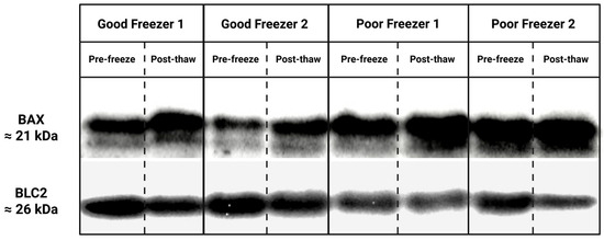
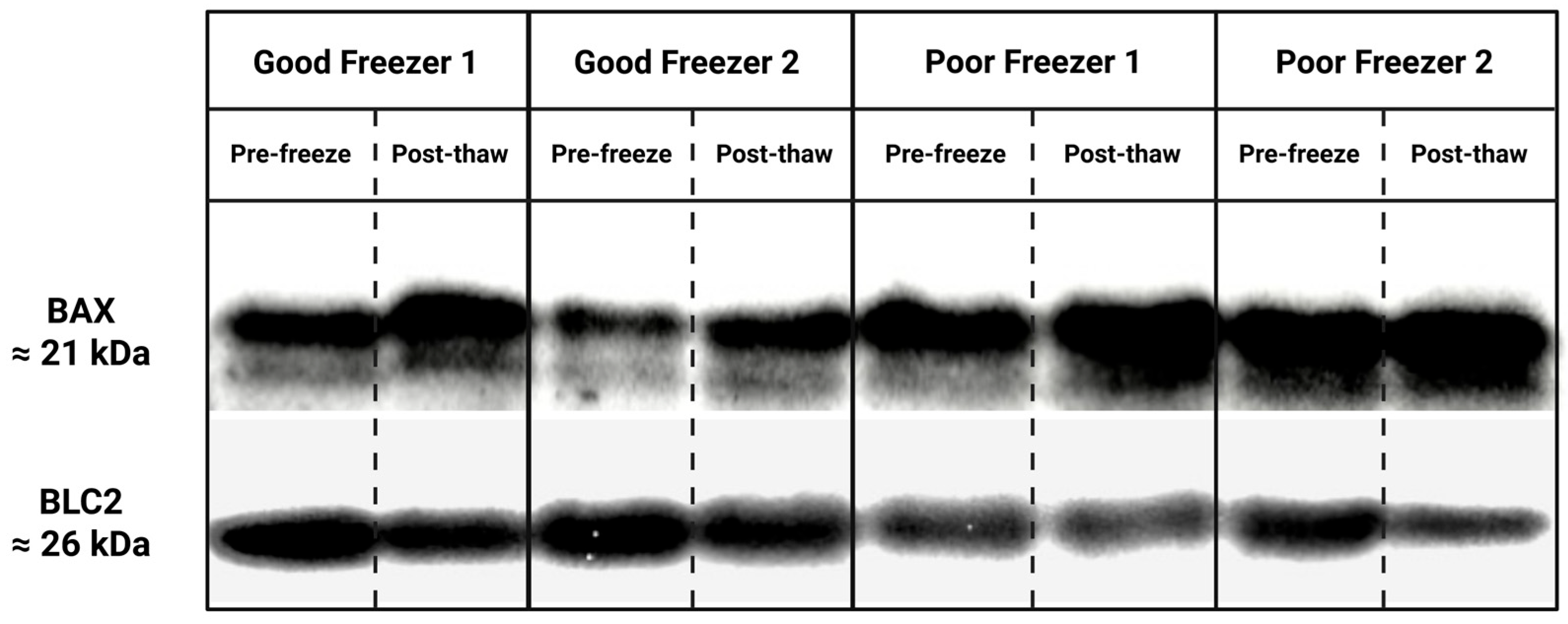
Comparing fresh and frozen semen samples (Table 3; Figure 1) revealed increased BAX gene expression levels and significantly increased BAX protein abundance (both q < 0.05), whereas BCL2 gene expression levels as well as protein abundance were significantly reduced after cryopreservation (both q < 0.05). In parallel, the proportion of non-apoptotic cells significantly decreased after thawing, while ROS levels and sperm DNA fragmentation significantly increased (all q < 0.05). All analyzed functional sperm quality parameters, including JC-1, total motility, and CFDA-assessed viability, were significantly decreased in the frozen samples (all q < 0.05) compared with the fresh control.
Table 3.
Fresh versus frozen sperm quality and apoptosis-related markers in Holstein bull semen.
Figure 1.
Representative western blot showing BAX and BCL2 protein levels in fresh and frozen-thawed bovine spermatozoa from Holstein bulls. Original photographs of the gels and blots are available in the Supplementary Materials. Created with BioRender.com (accessed on 19 May 2026).
Based on post-thaw total motility, ejaculates were classified (Table 4) into good freezers (top 8 ejaculates) and poor freezers (bottom 8 ejaculates). Mean post-thaw total motility of good freezers reached 64.88%, which was significantly higher (q < 0.05) than total motility in poor freezers (38.25%). Levels of miR-31-5p in fresh semen differed significantly (q < 0.05) between ejaculates categorized as poor and good freezers. The superior post-thaw performance of good freezers was accompanied by a more favorable sperm functional phenotype. Compared with poor freezers, good freezers showed significantly higher CFDA viability, significantly higher JC-1 mitochondrial membrane potential, significantly higher proportions of Annexin-negative spermatozoa, significantly lower ROS levels, and significantly lower sperm DNA fragmentation after thawing (all q < 0.05). Post-thaw BAX and BCL2 values did not differ significantly between the freezer groups.
Table 4.
Comparison of good and poor freezers based on post-thaw total motility distribution in Holstein bull semen.
Correlation analysis (Table 5) further supported the role of miR-31-5p as a candidate pre-cryopreservation biomarker of sperm resilience. Fresh semen miR-31-5p showed significant positive Spearman correlations with post-thaw CFDA viability (ρ = 0.755, q = 0.003), post-thaw JC-1 (ρ = 0.662, q = 0.010), post-thaw total motility (ρ = 0.785, q = 0.003), and post-thaw Annexin-negative spermatozoa (ρ = 0.752, q = 0.003). A significant negative correlation was also observed between fresh miR-31-5p and post-thaw ROS levels (ρ = −0.688, q = 0.008). No significant associations were identified between fresh miR-31-5p and post-thaw BAX, BCL2 protein abundance, or DNA fragmentation after FDR correction.
Table 5.
Correlations between fresh semen miR-31-5p and post-thaw sperm quality in Holstein bull semen.
In addition, higher fresh miR-31-5p was associated with a smaller cryopreservation-induced decline in total motility, CFDA viability, JC-1 and Annexin-negative cells, as well as with a smaller cryopreservation-induced increase in ROS (all q < 0.01). Correlations between fresh semen miR-31-5p and cryopreservation-induced change in sperm quality are displayed in Table 6.
Table 6.
Correlations between fresh semen miR-31-5p and cryopreservation-induced change in sperm quality traits.
4. Discussion
Cryopreservation caused a clear deterioration in Holstein bull sperm quality, as shown by lower post-thaw motility, reduced CFDA viability, decreased JC-1 mitochondrial membrane potential, a smaller proportion of Annexin-negative spermatozoa, higher ROS burden and greater sperm DNA fragmentation. This pattern is fully consistent with the current view that freezing injury in spermatozoa is not limited to simple membrane rupture, but instead reflects a broader cascade involving membrane destabilization, mitochondrial dysfunction, oxidative imbalance, chromatin damage and apoptosis-like changes [1]. In bull sperm specifically, cryopreservation has long been associated with phosphatidylserine redistribution, the loss of mitochondrial function and a reduced viability, while more recent reviews continue to describe regulated cell-death signaling and bioenergetic disruption as central features of sperm cryodamage [2]. Within this general cryodamage framework, the most important finding of the present study is that fresh semen miR-31-5p separated semen with good and poor post-thaw outcomes. Since miR-31-5p was quantified only before cryopreservation, the present data should not be interpreted as showing a freezing-induced change in this miRNA. Instead, they suggest that baseline miR-31-5p abundance is linked to the intrinsic capacity of a given ejaculate to withstand freezing and thawing. That distinction is important because, from a practical standpoint, the most useful biomarker is not necessarily one that changes after damage has already occurred, but rather one that can help identify resilient ejaculates before they enter the cryopreservation pipeline. The broader bull sperm literature supports this concept: both coding and non-coding sperm RNA signatures have been associated with fertility-related phenotypes, and cryopreservation itself has been shown to alter the bovine sperm small-RNA landscape [3,4,5].
Our results indicate that the difference in miR-31-5p was not an isolated molecular observation. Semen with higher fresh miR-31-5p retained better post-thaw motility, higher CFDA viability, stronger JC-1 mitochondrial signal, and more Annexin-negative spermatozoa. We observed that higher miR-31-5p baseline was associated with a broadly more favorable post-thaw phenotype rather than with only one isolated sperm quality parameter, suggesting that miR-31-5p may reflect a more stable underlying sperm condition which supports maintenance of cell viability, membrane homeostasis and energy competence during freezing stress. Recent reviews and experimental studies increasingly support the idea that sperm-borne miRNAs are not merely residual products of spermatogenesis but may function as informative markers of sperm quality and male fertility status [6,18,19].
Mitochondrial membrane potential is one of the most informative functional indicators in bull sperm because motility, redox balance and survival all depend on preserved mitochondrial competence [20,21]. Likewise, CFDA reflects an intact and metabolically functional sperm population. When both parameters deteriorate after thawing, they usually indicate that cryostress has exceeded the cell’s ability to preserve energetic and structural homeostasis [22]. In the present study, ejaculates with higher fresh miR-31-5p retained stronger post-thaw JC-1 and CFDA values, suggesting that this miRNA may be linked to an intrinsically more potent bioenergetic state. This does not demonstrate a direct causal role for miR-31-5p in sperm mitochondrial function, but it does support its potential value as a marker of the broader physiological condition that determines cryosurvival.
Because the present study evaluated Annexin-negative spermatozoa, higher values indicate a larger proportion of cells that retained a non-apoptotic membrane state after thawing. This is important because sperm cryopreservation is frequently associated with phosphatidylserine externalization and membrane asymmetry loss, both of which are widely interpreted as part of an apoptosis-like response in sperm [20,21]. The observation that higher fresh miR-31-5p was associated with a larger post-thaw Annexin-negative population suggests that semen rich in this miRNA may be less prone to membrane destabilization during freezing. The most likely interpretation is biomarker-based rather than causal: miR-31-5p appears to identify spermatozoa in which apoptosis-like membrane damage is less pronounced after thawing.
In the present study, cryopreservation shifted the molecular profile toward increased BAX and reduced BCL2, both at the transcript and protein levels. This direction aligns with the commonly used interpretation of BAX as a pro-damage, pro-apoptotic marker and BCL2 as a protective, anti-apoptotic counterpart [23]. In sperm cryobiology, these markers are best viewed not as a proof of apoptosis in a strict somatic-cell sense, but as indicators of an apoptosis-like stress response associated with loss of sperm function and membrane integrity [20]. The dataset further shows that poor freezers were characterized by significantly higher post-thaw ROS levels and greater sperm DNA fragmentation, supporting the view that oxidative imbalance and chromatin instability contribute to inferior cryotolerance. The clearest group differences in our study were therefore observed for miR-31-5p, motility, JC-1, CFDA, Annexin-negative cells, ROS and DNA fragmentation, suggesting that BAX and BCL2 are most valuable as supporting pathway-related evidence.
Although the literature on miR-31-5p, specifically in mature bull sperm cryobiology, is still limited, its broader biological context is highly relevant. miR-31-5p has been implicated in the regulation of proliferation and apoptosis-related pathways in human spermatogonial stem cells [11] and reproductive miRNA reviews continue to place it within apoptosis-linked regulatory networks in the male germline [24]. That does not mean the same mechanism operates directly in mature spermatozoa, which are transcriptionally silent and biologically very different from germline precursor cells [6]. However, it does support the plausibility that miR-31-5p abundance in spermatozoa may reflect aspects of earlier spermatogenic programming or epididymal maturation that later influence cryoresilience. This is especially important for interpretation: in the present study, miR-31-5p should be discussed primarily as a candidate biomarker of intrinsic sperm quality, not as a proven active regulator of BAX or BCL2 in thawed spermatozoa.
The present results suggest that miR-31-5p could be explored as part of a pre-freeze screening panel. It would be premature to propose it as a stand-alone decision tool, especially from a cohort of 16 ejaculates. However, the strength of its association with post-thaw motility, viability, mitochondrial potential and Annexin-negative cell proportion is promising. A more realistic future application is likely to involve a combined panel integrating conventional semen quality assessment with selected molecular biomarkers, including sperm-borne miRNAs, proteins and transcriptomic signatures. Such a multi-parameter strategy is increasingly recognized in bull fertility research, as no single marker is sufficient to fully capture the complexity of sperm function and cryotolerance.
Since miR-31-5p was analyzed only in fresh semen samples, the current experimental setup cannot establish whether its abundance is influenced by cryopreservation. Among the other limitations, although the observed correlations are strong, they remain associative and do not demonstrate a direct regulatory relationship between miR-31-5p and the post-thaw apoptotic, oxidative or mitochondrial phenotypes. The study was conceived as a pilot investigation: the initial cohort comprised 36 bulls, but epigenetic and associated molecular analyses were limited to 16 ejaculates representing the highest- and lowest-ranking post-thaw freezers. Although this extreme-group design increased contrast between poor and good freezers, future studies on larger, independent, and less selectively sampled cohorts are needed. In addition, residual extender was removed by repeated washing after thawing; although all frozen samples were processed identically, the independent effect of these centrifugation steps was not evaluated separately and should be considered when interpreting the magnitude of post-thaw damage. Future studies should validate these findings in larger, independent bull cohorts and integrate miRNA profiles with proteomic, transcriptomic, or extracellular vesicle-associated data to assess whether miR-31-5p maintains predictive utility within a multi-omics framework. Existing evidence from bovine sperm small RNA and cryopreservation-associated RNA dynamics supports the feasibility and relevance of such integrative approaches [10,25,26].
Overall, the present study supports the view that fresh semen miR-31-5p is associated with post-thaw sperm resilience in Holstein bulls. Ejaculates with higher baseline miR-31-5p showed better preservation of motility, viability, mitochondrial function and non-apoptotic membrane status after thawing, together with lower post-thaw ROS levels, while cryopreservation overall shifted the molecular profile toward a more damage-associated BAX/BCL2 balance. Altogether, these findings identify miR-31-5p as a promising pre-cryopreservation biomarker of bull sperm freezability and provide a basis for future validation studies involving larger and more diverse cattle populations.
5. Conclusions
The present study demonstrates that miR-31-5p abundance in fresh Holstein bull semen is associated with post-thaw sperm resilience. Ejaculates with higher baseline miR-31-5p levels exhibited improved preservation of motility, viability, mitochondrial membrane potential and membrane integrity following cryopreservation, together with lower post-thaw ROS levels and lower DNA fragmentation in the good-freezer group. These results support miR-31-5p as a promising pre-cryopreservation biomarker of sperm freezability in Holstein bulls. Further validation in larger cohorts and additional breeds is required to confirm its predictive performance and to define its utility within multi-parameter semen quality assessment frameworks.
Supplementary Materials
The following supporting information can be downloaded at: https://www.mdpi.com/article/10.3390/ruminants6030060/s1, Figure S1: Representative stain-free gel used to verify protein loading uniformity prior to transfer; Figure S2: Full-length annotated Western blot membrane showing BAX immunoreactivity in fresh (pre-freeze) and frozen-thawed (post-thaw) sperm samples from representative Holstein bull ejaculates classified as good and poor freezers according to post-thaw total motility. The expected BAX band is located at approximately 21 kDa. Molecular weight markers are shown on the left; Figure S3: Full-length annotated Western blot membrane showing BCL2 immunoreactivity in fresh (pre-freeze) and frozen-thawed (post-thaw) sperm samples from representative Holstein bull ejaculates classified as good and poor freezers according to post-thaw total motility. The expected BCL2 band is located at approximately 26 kDa. Molecular weight markers are shown on the left.
Author Contributions
Conceptualization, M.Ď.; methodology, M.Ď., L.U., S.F.; validation, E.T.; formal analysis, E.T.; investigation, M.Ď., F.B., L.U., S.F., S.A.-N.; resources, M.Ď., E.T.; data curation, L.U., S.F., E.T.; writing—original draft preparation, M.Ď., L.U., S.F., E.T.; writing—review and editing, M.Ď., E.T.; supervision, M.Ď., E.T.; project administration, M.Ď., E.T.; funding acquisition, M.Ď., E.T. All authors have read and agreed to the published version of the manuscript.
Funding
This research was funded by the EU NextGenerationEU through the Recovery and Resilience Plan for Slovakia under the projects No. 09I03-03-V04-00483, 09I03-03-V05-00018 and with the support of the Grant Agency of the Slovak University of Agriculture in Nitra, project no. 04-GA-SPU-2024 and the Slovak Research and Development Agency (APVV-21-0095).
Institutional Review Board Statement
Ethical review and approval were not required for this study because semen samples were collected from clinically healthy breeding bulls during routine semen collection procedures at a commercial breeding center, and no additional invasive procedures or experimental interventions were performed on the animals specifically for the purposes of this study.
Informed Consent Statement
Not applicable.
Data Availability Statement
The original contributions presented in this study are included in the article/supplementary material. Further inquiries can be directed to the corresponding author.
Acknowledgments
During the preparation of this manuscript, the authors used ChatGPT (GPT-5.5, OpenAI) for language refinement and structural optimization of selected text passages. All generated output was critically reviewed, edited, and verified by the authors, who take full responsibility for the final content of the manuscript.
Conflicts of Interest
The authors declare no conflicts of interest.
References
- Curry, M.R. Cryopreservation of Semen from Domestic Livestock. Rev. Reprod. 2000, 5, 46–52. [Google Scholar] [CrossRef] [PubMed]
- Peris-Frau, P.; Soler, A.J.; Iniesta-Cuerda, M.; Martín-Maestro, A.; Sánchez-Ajofrín, I.; Medina-Chávez, D.A.; Fernández-Santos, M.R.; García-Álvarez, O.; Maroto-Morales, A.; Montoro, V.; et al. Sperm Cryodamage in Ruminants: Understanding the Molecular Changes Induced by the Cryopreservation Process to Optimize Sperm Quality. Int. J. Mol. Sci. 2020, 21, 2781. [Google Scholar] [CrossRef] [Scilit] [PubMed]
- Fickel, J.; Wagener, A.; Ludwig, A. Semen Cryopreservation and the Conservation of Endangered Species. Eur. J. Wildl. Res. 2007, 53, 81–89. [Google Scholar] [CrossRef] [Scilit]
- Nagy, Z.P.; Varghese, A.C.; Agarwal, A. Cryopreservation of Mammalian Gametes and Embryos: Methods and Protocols, 1st ed.; Springer: New York, NY, USA, 2017; pp. 1–369. [Google Scholar]
- Riva, N.S.; Ruhlmann, C.; Iaizzo, R.S.; Marcial López, C.A.; Martínez, A.G. Comparative Analysis between Slow Freezing and Ultra-Rapid Freezing for Human Sperm Cryopreservation. JBRA Assist. Reprod. 2018, 22, 331–337. [Google Scholar] [CrossRef] [Scilit] [PubMed]
- Ozimic, S.; Ban-Frangez, H.; Stimpfel, M. Sperm Cryopreservation Today: Approaches, Efficiency, and Pitfalls. Curr. Issues Mol. Biol. 2023, 45, 4716–4734. [Google Scholar] [CrossRef] [Scilit] [PubMed]
- Joshi, M.; Sethi, S.; Mehta, P.; Kumari, A.; Rajender, S. Small RNAs, Spermatogenesis, and Male Infertility: A Decade of Retrospect. Reprod. Biol. Endocrinol. 2023, 21, 106. [Google Scholar] [CrossRef] [Scilit] [PubMed]
- Ďuračka, M.; Benko, F.; Tvrdá, E. Molecular Markers: A New Paradigm in the Prediction of Sperm Freezability. Int. J. Mol. Sci. 2023, 24, 3379. [Google Scholar] [CrossRef] [Scilit] [PubMed]
- Chen, W. Advances in Sperm Transcription and Translation Activities: From Traditional Views to New Discoveries. Mol. Asp. Med. 2026, 108, 101463. [Google Scholar] [CrossRef] [Scilit] [PubMed]
- Capra, E.; Turri, F.; Lazzari, B.; Cremonesi, P.; Gliozzi, T.M.; Fojadelli, I.; Stella, A.; Pizzi, F. Small RNA Sequencing of Cryopreserved Semen from Single Bull Revealed Altered miRNAs and piRNAs Expression between High- and Low-Motile Sperm Populations. BMC Genom. 2017, 18, 14. [Google Scholar] [CrossRef] [Scilit] [PubMed]
- Fu, H.; Zhou, F.; Yuan, Q.; Zhang, W.; Qiu, Q.; Yu, X.; He, Z. miRNA-31-5p Mediates the Proliferation and Apoptosis of Human Spermatogonial Stem Cells via Targeting JAZF1 and Cyclin A2. Mol. Ther. Nucleic Acids 2019, 14, 90–100. [Google Scholar] [CrossRef] [Scilit] [PubMed]
- Adelakun, S.A.; Akintunde, O.W.; Ogunlade, B.; Peter, A.B.; Siyanbade, J.A. BCL-2 and BAX Expression and Germ Cell Apoptosis Following the Intervention of 1-Isothiocyanato-4-Methylsulfinylbutane in Cisplatin-Induced Testicular Toxicity and Sperm DNA Fragmentation in Sprague-Dawley Rat. Asp. Mol. Med. 2024, 4, 100055. [Google Scholar] [CrossRef] [Scilit]
- Chowdhury, A.S.; Reich, L.A.; Liby, K.T.; Yeh, E.S.; Leal, A.S. RXR Agonist V-125 Induces Distinct Transcriptional and Immunomodulatory Programs in Mammary Tumors of MMTV-Neu Mice Compared to Bexarotene. Biomedicines 2026, 14, 80. [Google Scholar] [CrossRef] [Scilit] [PubMed]
- Baňas, Š.; Benko, F.; Ďuračka, M.; Lukáč, N.; Tvrdá, E. Epicatechin Prevents Cryocapacitation of Bovine Spermatozoa through Antioxidant Activity and Stabilization of Transmembrane Ion Channels. Int. J. Mol. Sci. 2023, 24, 2510. [Google Scholar] [CrossRef] [Scilit] [PubMed]
- Benko, F.; Slanina, T.; Ďuračka, M.; Kačániová, M.; Tvrdá, E. Cryopreservative and Antimicrobial Properties of Kaempferol on the Post-Thaw Quality of Turkey Spermatozoa. Czech J. Anim. Sci. 2025, 70, 404–413. [Google Scholar] [CrossRef] [Scilit]
- Ďuračka, M.; Belić, L.; Tokárová, K.; Žiarovská, J.; Kačániová, M.; Lukáč, N.; Tvrdá, E. Bacterial Communities in Bovine Ejaculates and Their Impact on the Semen Quality. Syst. Biol. Reprod. Med. 2021, 67, 438–449. [Google Scholar] [CrossRef] [Scilit] [PubMed]
- Benko, F.; Baňas, Š.; Ďuračka, M.; Kačániová, M.; Tvrdá, E. Cryoprotective Potential of Theobromine in the Improvement of the Post-Thaw Quality of Bovine Spermatozoa. Cells 2024, 13, 1710. [Google Scholar] [CrossRef] [Scilit] [PubMed]
- Barbu, M.G.; Thompson, D.C.; Suciu, N.; Voinea, S.C.; Cretoiu, D.; Predescu, D.V. The Roles of MicroRNAs in Male Infertility. Int. J. Mol. Sci. 2021, 22, 2910. [Google Scholar] [CrossRef] [Scilit] [PubMed]
- Werry, N.; Russell, S.J.; Gillis, D.J.; Miller, S.; Hickey, K.; Larmer, S.; Lohuis, M.; Librach, C.; LaMarre, J. Characteristics of miRNAs Present in Bovine Sperm and Associations with Differences in Fertility. Front. Endocrinol. 2022, 13, 874371. [Google Scholar] [CrossRef] [Scilit] [PubMed]
- Martin, G.; Sabido, O.; Durand, P.; Levy, R. Cryopreservation Induces an Apoptosis-Like Mechanism in Bull Sperm. Biol. Reprod. 2004, 71, 28–37. [Google Scholar] [CrossRef] [Scilit] [PubMed]
- Ugur, M.R.; Saber Abdelrahman, A.; Evans, H.C.; Gilmore, A.A.; Hitit, M.; Arifiantini, R.I.; Purwantara, B.; Kaya, A.; Memili, E. Advances in Cryopreservation of Bull Sperm. Front. Vet. Sci. 2019, 6, 268. [Google Scholar] [CrossRef] [Scilit] [PubMed]
- Bollwein, H.; Malama, E. Review: Evaluation of Bull Fertility. Functional and Molecular Approaches. Animal 2023, 17, 100795. [Google Scholar] [CrossRef] [Scilit] [PubMed]
- Dalal, J.; Kumar, A.; Honparkhe, M.; Deka, D.; Singh, N. Minimization of Apoptosis-like Changes in Cryopreserved Buffalo Bull Sperm by Supplementing Extender with Bcl-2 Protein. Vet. World 2016, 9, 432–436. [Google Scholar] [CrossRef] [Scilit] [PubMed]
- Yu, P.; Zhao, X.; Zhou, D.; Wang, S.; Hu, Z.; Lian, K.; Zhang, N.; Duan, P. The microRNA-Mediated Apoptotic Signaling Axis in Male Reproduction: A Possible and Targetable Culprit in Male Infertility. Cell Biol. Toxicol. 2025, 41, 54. [Google Scholar] [CrossRef] [Scilit] [PubMed]
- Shangguan, A.; Zhou, H.; Sun, W.; Ding, R.; Li, X.; Liu, J.; Zhou, Y.; Chen, X.; Ding, F.; Yang, L.; et al. Cryopreservation Induces Alterations of miRNA and mRNA Fragment Profiles of Bull Sperm. Front. Genet. 2020, 11, 419. [Google Scholar] [CrossRef] [Scilit] [PubMed]
- Keles, E.; Malama, E.; Bozukova, S.; Siuda, M.; Wyck, S.; Witschi, U.; Bauersachs, S.; Bollwein, H. The micro-RNA content of unsorted cryopreserved bovine sperm and its relation to the fertility of sperm after sex-sorting. BMC Genom. 2021, 22, 30. [Google Scholar] [CrossRef] [Scilit] [PubMed]

Disclaimer/Publisher’s Note: The statements, opinions and data contained in all publications are solely those of the individual author(s) and contributor(s) and not of MDPI and/or the editor(s). MDPI and/or the editor(s) disclaim responsibility for any injury to people or property resulting from any ideas, methods, instructions or products referred to in the content. |
© 2026 by the authors. Licensee MDPI, Basel, Switzerland. This article is an open access article distributed under the terms and conditions of the Creative Commons Attribution (CC BY) license.