


Simple Summary
Dairy cows are susceptible to hypocalcemia around parturition, which impairs their health and reduces milk yield. Calcium lost for fetal skeletal mineralization and lactation is mainly replenished in dairy cows through intestinal calcium absorption and bone calcium mobilization. During evolution, mammals generally prioritize the utilization of skeletal calcium to prevent adverse effects on fetal skeletal development caused by short-term food shortages, insufficient sunlight, and other factors. At present, systematic research on the processes of bone resorption and formation in dairy cows is lacking. Therefore, this review integrates cross-species evidence and current findings to systematically revisit the mechanisms of bone resorption and formation, and compare the roles of parathyroid hormone-related protein and parathyroid hormone in the signaling pathways of bone calcium mobilization.
Abstract
Some dairy cows develop postpartum hypocalcemia because they cannot adapt to the drastic loss of blood calcium during the periparturient period. Parathyroid hormone (PTH) maintains calcium homeostasis by regulating bone calcium mobilization, gastric and intestinal calcium absorption, and renal calcium reabsorption. Traditionally, dysregulation of PTH has been considered the main cause of this disorder. Recent studies have confirmed that parathyroid hormone-related protein (PTHrP) secreted by the mammary gland is also involved in calcium homeostasis by promoting bone resorption. Based on existing research evidence and cross-species comparisons, this paper analyzes the effects of differences in mammary calcium output and compensatory mechanisms among various mammals on the risk of postpartum hypocalcemia, with a particular focus on processes related to bone resorption and bone formation. It further explains the pathogenesis of postpartum hypocalcemia in dairy cows by examining the roles of the mammary–brain–bone axis, PTH, and PTHrP in bone resorption and bone formation in these animals.
1. Introduction
Pregnancy and lactation represent two distinctive physiological stages in the life cycle of mammals, during which significant alterations occur in the regulatory mechanisms of systemic calcium homeostasis [1]. During gestation, to ensure that mammals under diverse latitudinal and sunlight conditions acquire sufficient calcium for fetal skeletal development, intestinal calcium absorption becomes partially independent of the regulation of calcitriol [2]. Upon the onset of lactation, to meet the steady calcium demand for milk synthesis, the body preferentially mobilizes calcium reserves in the skeleton to support offspring growth even when dietary calcium supply is adequate. Consequently, nearly all mammals experience varying degrees of bone calcium loss during early lactation [3].
Compared with other mammals, dairy cows exhibit more drastic fluctuations in blood calcium concentration around parturition. During the final week prepartum, dry matter intake in dairy cows decreases by approximately 20% due to fetal compression, hormonal changes, and systemic inflammation (SI), reaching the lowest peripartal level on the day of parturition [4]. Calcium homeostasis fluctuates in multiparous cows at 2–3 days prepartum, with blood calcium concentration beginning to decline at approximately 9 h before calving when parturition is initiated [5], and reaching the nadir at 24 h postpartum [6]. Traditionally, the regulatory mechanism of calcium homeostasis in periparturient dairy cows has been regarded as largely consistent with that in non-pregnant and non-lactating states, relying mainly on PTH. In contrast, extensive studies in humans and rodents have demonstrated that PTHrP gradually replaces the role of PTH during pregnancy and becomes the dominant hormone regulating calcium homeostasis. During lactation, PTHrP mobilizes calcium from the skeleton into the bloodstream and acts as the primary factor responsible for bone calcium loss during lactation [7]. Recent investigations in dairy cows have also revealed that PTHrP secreted by the mammary gland promotes bone calcium mobilization and participates in maintaining calcium homeostasis after parturition. Integrated with findings from human and rodent models, some researchers have proposed that PTHrP may also serve as the principal hormone governing bone calcium mobilization and calcium homeostasis in periparturient dairy cows [8].
Although research findings on calcium metabolism in humans and rodents cannot be directly extrapolated to dairy cows, current understanding of the regulatory mechanisms underlying calcium metabolism in dairy cows is largely inferred from cross-species studies. Accordingly, this review compares changes in calcium demand and adaptive mechanisms across species during the periparturient period, with a focus on the role of lactational bone calcium mobilization in maintaining calcium homeostasis and its association with postpartum hypocalcemia. By integrating consensus findings from human and rodent studies and relevant advances in dairy cow research, we systematically analyze the mechanisms regulating calcium homeostasis and bone resorption in periparturient dairy cows, as well as the potential functions of PTH and PTHrP in bone calcium mobilization, aiming to provide a theoretical basis for further elucidating the pathogenesis of postpartum hypocalcemia in dairy cows.
2. Adaptation of Calcium Metabolism Around Parturition in Different Mammals
2.1. Differences in Calcium Demand and Blood Calcium Concentrations
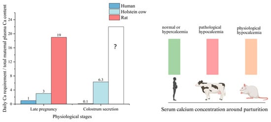
Calcium is an essential macromineral in the body, participating in critical physiological processes including nerve conduction, muscle contraction, blood coagulation, cellular signal transduction, and skeletal mineralization. Late gestation and lactation represent the periods with the greatest metabolic burden for calcium homeostasis in mammals. Taking a 600 kg Holstein dairy cow as an example, the plasma calcium pool contains approximately 3–3.5 g of calcium. During late gestation, the daily fetal calcium requirement is about 10.3 g, equivalent to three times the total plasma calcium content of the dam [9]. In pregnant women weighing approximately 60 kg, the daily fetal calcium demand is roughly equal to the maternal plasma calcium pool, whereas this ratio reaches as high as 19-fold in rats [7].
Upon the onset of lactation, milk secretion becomes the primary route of maternal calcium loss. Colostrum of dairy cows contains 2.3 g calcium/L, with a colostrum yield of 6–8 L; the daily calcium required for colostrum production is approximately 6.3 times the maternal plasma calcium pool. Human colostrum contains 0.26 g calcium/L with a daily secretion volume of 150 mL, and calcium used for colostrum synthesis accounts for only 8–10% of the plasma calcium pool [10]. Colostrum volume in rats is extremely low and secreted intermittently, making precise estimation difficult; however, during peak lactation, daily milk calcium output in rats is approximately 144 times their plasma calcium pool [11]. Differences in calcium demand among species directly lead to distinctly different patterns of blood calcium concentration changes. Around parturition, blood ionized calcium levels remain normal or slightly elevated in humans, and slightly decreased in rats, presenting as controllable physiological hypocalcemia; however, approximately 50% of multiparous dairy cows develop obvious hypocalcemia [12]. The interspecies differences in calcium demand and serum calcium concentration around parturition are summarized in Figure 1.
Figure 1.
Changes in relative calcium demand and serum calcium concentrations in humans, dairy cows, and rats during the late gestation and colostrum secretion period (Created in BioRender. PY, H. (2026) https://BioRender.com/v3af08g (accessed on 26 May 2026)).
2.2. Causes of Postpartum Hypocalcemia
A major factor contributing to postpartum hypocalcemia in dairy cows is the rapid skeletal mineralization of the fetus near term and the sharp surge in calcium demand at the onset of lactation [13]. Maternal calcium loss during late gestation is primarily related to litter size. Ewes rarely develop postpartum hypocalcemia, but twin or triplet-bearing individuals are prone to prenatal hypocalcemia [14]. Although dairy cows and humans typically produce singletons, the relative calcium demand of the bovine fetus compared with maternal plasma calcium during late gestation is nearly three times higher than that in humans. Dairy cows initiate colostrum secretion several days to weeks prepartum, which further increases calcium loss; in contrast, mastectomized cows rarely develop postpartum hypocalcemia [15]. During early lactation, the calcium required for human colostrum production is only one-tenth of calcium demand in late gestation, whereas calcium demand in dairy cows increases manifold.
In the long term, lactational calcium metabolic stress across species can be roughly evaluated by daily milk calcium output per unit body weight (mg/kg BW·d) as summarized in Table 1. Humans exhibit the lowest value, at approximately 3.58 mg/kg BW·d. Among other ruminants, dairy goats and sheep show values of 26.06 and 22.59, respectively, whereas dairy cows reach as high as 64.17 mg/kg BW·d. Moreover, the lactation period in dairy cows is longer than in most mammals. Consequently, humans do not develop postpartum hypocalcemia; instead, some individuals even experience hypercalcemia in late gestation and lactation due to excessive PTHrP secretion [16]. The risk and severity of postpartum hypocalcemia in dairy goats and sheep are lower than in dairy cows, with cases mostly occurring during 1–3 weeks postpartum when milk yield increases [17].
Table 1.
Comparison of calcium output-related parameters during lactation in different mammals.
Another important cause of postpartum hypocalcemia in dairy cows is that the body’s calcium replenishment mechanism fails to adapt to the rapid loss of blood calcium induced by lactation. Offspring calcium requirements provided by mother’s milk can be evolutionarily co-adapted to the maternal calcium compensatory capacity. Lactational calcium metabolic stress is relatively low in humans, and the elevated 1,25(OH)2D3 during gestation declines to baseline levels during lactation [23]. Intestinal calcium absorption does not need to be markedly enhanced, and offspring calcium requirements can be met mainly through bone calcium mobilization. In contrast, rats and dairy cows maintain persistently high 1,25(OH)2D3 levels to meet high calcium demands by strengthening intestinal calcium absorption [24,25,26], accompanied by pronounced bone resorption. Bone mineral content decreases by approximately 1–3% in women 1 month postpartum, by 9–13% in dairy cows within 1 month postpartum, and by up to 33% in rats over the 21-day lactation period, which is associated with the highest calcium metabolic stress [11,27,28]. Rats and dairy cows sustain lactation during pregnancy, while milk production of dairy cows far exceeds the actual nutritional needs of their offspring. Under commercial farming conditions, a dairy calf consumes approximately 330–450 kg of milk from birth to weaning [29], whereas a dairy cow can produce over 10,000 kg of milk in a single lactation cycle [30]. Compared with primiparous cows, multiparous cows produce more milk and have greater calcium demand, yet show reduced gastrointestinal calcium absorption and bone calcium mobilization, making them more susceptible to postpartum hypocalcemia [31].
Rats experience much higher calcium metabolic stress around parturition than dairy cows but only develop mild, physiological hypocalcemia. This phenomenon indicates that calcium metabolic stress is not the decisive factor for the occurrence of hypocalcemia, and hypocalcemia can be avoided if the mother adapts well to high calcium load. The extremely high calcium demand during lactation in rats is mainly attributed to their multiple-birth reproductive characteristics and the altricial nature of rat pups, whose skeletons are incompletely calcified at birth, requiring the mother to secrete high-calcium milk to support skeletal growth. Around parturition, the adaptive and compensatory capacity of rats to high calcium load mainly relies on markedly enhanced bone resorption, compensatory hypertrophy of the gastrointestinal tract and liver, and a nearly threefold increase in food intake. In addition, rat colostrum has low calcium content and small secretion volume, and milk calcium concentration gradually increases within 10 days postpartum, which helps to distribute the calcium metabolic stress during early lactation.
Cross-species comparisons reveal that although mammals all increase calcium consumption during pregnancy and lactation, calcium demand varies considerably among species. Postpartum hypocalcemia in periparturient dairy cows is a direct consequence of disrupted calcium homeostasis, which essentially stems from a sharp increase in calcium metabolic stress combined with insufficient compensatory capacity.
3. Regulatory Role of the Brain in Bone Resorption
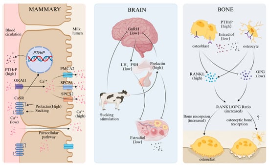
Studies in humans and rodents have demonstrated that the mammary–brain–bone axis serves as the core regulator of systemic calcium homeostasis during lactation [32]. Under conditions of high prolactin, low estrogen, continuous suckling stimulation, and hypocalcemia, the calcium-sensing receptor (CaSR) located on the basolateral membrane of mammary epithelial cells is inhibited, which promotes the synthesis and release of PTHrP in mammary tissue. Meanwhile, suckling stimulation and elevated prolactin secretion during lactation suppress the hypothalamic–pituitary–gonadal axis, leading to reduced secretion of luteinizing hormone (LH) and follicle-stimulating hormone (FSH), thereby markedly lowering estrogen (E2) secretion by the ovaries [33]. High levels of PTHrP primarily act on osteoblasts and osteocytes, whereas low estrogen mainly targets osteoblasts, T cells, and B cells. Together, these two factors enhance the secretion of receptor activator of nuclear factor-κB ligand (RANKL) and inhibit osteoprotegerin (OPG), thereby promoting osteoclastogenesis and the release of bone calcium to meet the calcium demand of the mammary gland during lactation [32]. Either elevated PTHrP or reduced estrogen alone can increase bone loss, but PTHrP represents the dominant driver of lactational bone loss.
In dairy cows, estrogen concentrations surge at the onset of parturition and decline rapidly postpartum. The decrease in estrogen postpartum is closely associated with enhanced activity of the bone resorption marker C-terminal telopeptide of type I collagen (CTX) and the osteoclast marker enzyme tartrate-resistant acid phosphatase isoform 5b (TRAP5b) [34,35]. Furthermore, Filipović et al. reported that plasma PTHrP concentrations in dairy cows were significantly higher on day 10 postpartum than on day 14 prepartum (p < 0.005), and the bone resorption marker CTX was markedly increased on both days 10 and 30 postpartum (p < 0.0001). PTHrP was significantly negatively correlated with estrogen concentration (r = −0.29, p < 0.05), whereas CTX showed an even stronger negative correlation with estrogen (r = −0.54, p < 0.0001), further supporting their synergistic roles during lactation [36]. These correlations tentatively imply that a similar integrated mammary–brain–bone axis may operate in dairy cows to mediate the extensive calcium mobilization required for lactation (Figure 2).
Figure 2.
Schematic of the integrated mammary–brain–bone axis regulating calcium mobilization during lactation in dairy cows. The question mark indicates undefined mechanisms. (Created in BioRender. PY, H. (2026) https://BioRender.com/qvymeds (accessed on 26 May 2026)).
4. Regulatory Role of the Mammary Gland in Bone Resorption
During lactation, the mammary gland is not only the core organ for milk synthesis and secretion, but also participates in the regulation of systemic calcium homeostasis in the mother through local sensing and remote signaling. It ensures a stable supply of calcium in milk while coordinating bone calcium mobilization to meet lactational demands. Numerous studies have shown that to guarantee normal offspring development, milk calcium concentration is highly conserved among lactating mammals and is largely unaffected by short-term maternal calcium nutritional status. Reducing dietary calcium in mice alone or inhibiting bone resorption and calcium mobilization via OPG does not significantly alter milk calcium concentration; only when both intestinal calcium absorption and skeletal calcium mobilization are blocked simultaneously does milk calcium content decrease markedly [37]. The mammary gland can positively regulate the intensity of bone resorption according to lactation demand. Studies in lambs, pigs, and mice have found that milk composition remains relatively constant among individuals with different litter sizes within the same lactation stage, whereas milk yield and bone mineral loss increase proportionally with the number of offspring [7]. High-yielding dairy cows show stronger bone resorption activity postpartum, with significantly higher serum concentrations of type I collagen C-terminal telopeptide (ICTP) [38].
The CaSR located on the basolateral membrane of mammary epithelial cells can precisely sense changes in local blood calcium concentration and promote the secretion of PTHrP when blood calcium levels decline. Calcium ions released from bone into the blood are transported into mammary epithelial cells via ORAI1 (Ca2+ release-activated Ca2+ channel 1) on the basolateral side, and are then secreted into milk through transporters on the apical membrane, including PMCA2 (plasma membrane Ca2+ ATPase 2 pump), SPCA1, and SPCA2 (secretory pathway Ca2+ ATPase 1 and 2) [39].
5. Bone Resorption and Formation
5.1. Bone Tissue Structure and Changes in Bone Mass
Bone consists of approximately 70% inorganic minerals and 30% organic matrix. The inorganic fraction is mainly composed of hydroxyapatite crystals [Ca10(PO4)6(OH)2], containing calcium, phosphorus, and small amounts of sodium, magnesium, fluorine, strontium, and other ions. The organic fraction comprises collagen fibers, glycoproteins, and proteoglycans. Cortical bone is dense and rigid, primarily providing structural support. In contrast, cancellous bone in the epiphyses of long bones, flat bones, and vertebrae is metabolically active and serves as the main site for bone calcium mobilization. Bone remodeling occurs mainly on the surfaces of cortical and trabecular bone and is the primary process maintaining the dynamic balance of bone mineral content. Bone modeling, by contrast, is more commonly observed on periosteal and endosteal surfaces and mainly participates in bone growth and morphological reconstruction [40].
Changes in bone mass rely on the function of bone multicellular units (BMUs) formed by osteoblasts and osteoclasts. BMUs are distributed across the entire bone surface and operate at asynchronous stages. Following activation, a BMU undergoes an initiation phase lasting several hours to days, enters a bone resorption phase of approximately 3 weeks, proceeds to a reversal phase of 4–5 weeks, and finally enters an osteogenic phase lasting 3–4 months [41]. Postpartum hypocalcemia in dairy cows typically occurs within 0–4 days after calving and is less frequent during peak lactation [42,43]. This timing coincides with the period required for bone remodeling units to become activated and initiate bone resorption, suggesting that postpartum hypocalcemia in dairy cows most likely results from insufficiently activated bone calcium mobilization, which cannot promptly meet the sharply increased calcium demand during early lactation.
5.2. Osteoclast-Mediated Bone Resorption
Bone resorption in lactating dairy cows is primarily regulated by the OPG–RANKL–RANK signaling axis. Osteoclasts originate from granulocyte–macrophage progenitor cells (CFU-GM) derived from hematopoietic stem cells. Under the influence of macrophage colony-stimulating factor (M-CSF) and RANKL, they gradually differentiate into preosteoclasts. These precursors further fuse under RANKL and interleukin-1 (IL-1) signaling to form quiescent multinucleated osteoclasts [44]. Upon activation, mature osteoclasts develop characteristic structures including a sealing zone and a ruffled border, enabling them to secrete acids and bone-resorptive enzymes such as tartrate-resistant acid phosphatase (TRAP) and cathepsin K to degrade bone matrix. OPG secreted by osteoblasts and osteocytes acts as a soluble decoy receptor that binds RANKL and prevents its interaction with RANK, thereby inhibiting osteoclast differentiation and activation, preventing excessive bone loss, and maintaining bone metabolic homeostasis [45,46].
5.3. Osteocytic Osteolysis
In addition to osteoclasts, osteocytic osteolysis also plays an important role in bone resorption in rodents [47]. Osteocytes are the most abundant cell type in bone, distributed within the lacuno-canalicular network. During lactation, osteocytes resorb the mineralized matrix surrounding their lacunae and express cathepsin K, TRAP, and matrix metalloproteinase 13 (MMP13) [48]. Evidence indicates that osteoclasts are not the sole source of lactational bone loss. Treatment with bisphosphonates to inhibit osteoclast activity reduces bone mineral density loss by only approximately 60%, whereas osteoprotegerin (OPG-Fc) nearly completely suppresses bone loss [37,49]. Furthermore, selective knockout of the cathepsin K gene in osteocytes significantly attenuates lacunar resorption, prevents increases in osteoclast number and reductions in trabecular volume during lactation, and thereby decreases skeletal mineral loss [50]. These findings suggest that osteocytes participate in lactational bone calcium mobilization both by directly mediating bone matrix degradation and indirectly regulating osteoclast activity. However, relevant studies in dairy cows remain limited.
5.4. Regulatory Role of the Immune System in Bone Resorption
Bone resorption and formation depend not only on interactions within the osteogenic lineage but also on extensive regulation by the immune system. Studies in rodents have demonstrated that T lymphocytes in bone marrow regulate stromal cell differentiation and responsiveness to PTH by secreting RANKL, tumor necrosis factor-α (TNF-α), and surface costimulatory molecules [51].Wnt10b produced by T cells and interleukin-17 (IL-17) secreted by Th17 cells have been confirmed to affect bone resorption and formation. RANKL expressed by B cells also promotes osteoclast differentiation and activation [52]. In addition, cytokines secreted by immune cells in the bone marrow microenvironment, including interleukins (ILs), tumor necrosis factors (TNFs), chemokines (CCLs), and interferons (IFNs), significantly influence bone metabolic balance [53]. Accordingly, some researchers have proposed that dynamic changes in circulating cytokines may represent an important driver of postpartum hypocalcemia in dairy cows [54]. A study by Tarar et al. confirmed that plasma concentrations of the pro-resorptive factors interleukin-6 (IL-6) and vascular endothelial growth factor A (VEGF-A) are significantly elevated in dairy cows during late gestation, with patterns consistent with the bone resorption marker CTX-I. In contrast, the anti-resorptive factor IL-4 increases during late lactation, and IFN-γ remains relatively high during both late gestation and late lactation [55].
5.5. Recovery Pattern of Lactational Bone Loss
Unlike irreversible bone loss caused by osteoporosis, lactational bone loss is reversible. In women, bone mineral lost during lactation is usually fully restored within months after weaning. However, dairy cows that have reached breeding age will continuously undergo the gestation–lactation overlap cycle to maintain persistent lactation. Multiparous cows are usually bred and conceive at 60–90 days postpartum to initiate the next pregnancy, and cease lactation only during the dry period (late gestation) approximately 8 weeks before parturition. This suggests that the bone metabolic recovery pattern in dairy cows may differ from that in humans.
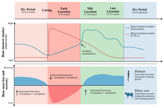
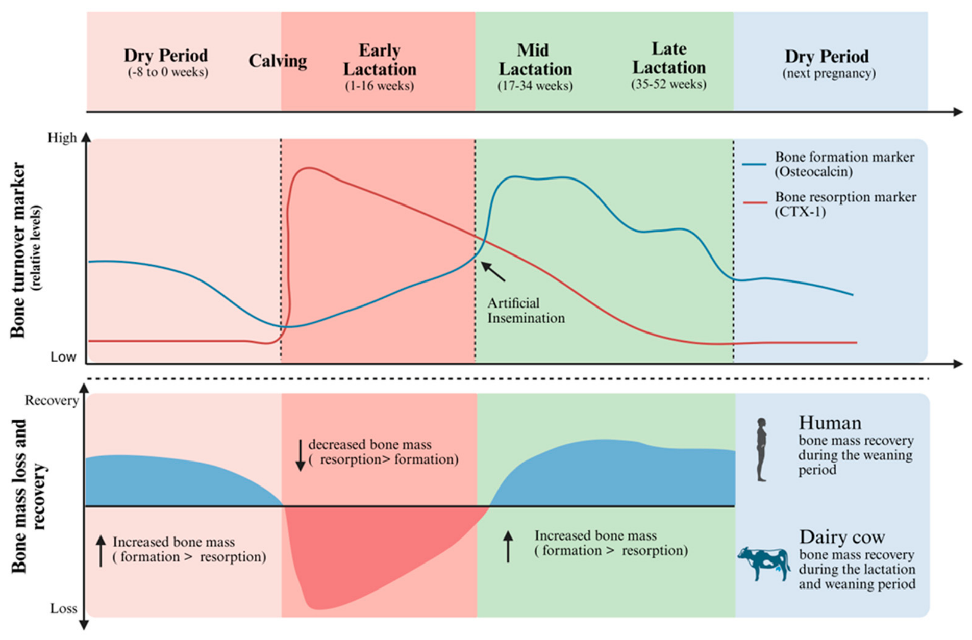
Systematic studies on the recovery pattern of lactational bone loss in dairy cows are scarce. Available evidence indicates that dairy cows may undergo a “reversal” during mid-to-late lactation and the subsequent dry period, during which bone formation exceeds bone resorption, allowing partial bone mass recovery. Holtenius et al. reported that the bone resorption marker CTX remains low during the dry period several weeks before calving, increases rapidly postpartum, and gradually declines to dry-period levels over the subsequent 33-week lactation, maintaining this level until the next parturition. Meanwhile, the bone formation marker osteocalcin decreases continuously during the dry period, reaches its lowest level around parturition, increases steadily postpartum, rises further after breeding (approximately 12 weeks postpartum), peaks around 20 weeks postpartum, shows a temporary decline at 25 weeks, and falls to a minimum near the next calving [56]. Tarar et al. reported similar results: CTX-I increases sharply during early lactation (4–8 weeks postpartum), decreases gradually during mid-lactation (18–22 weeks) and late lactation (35–39 weeks). Osteocalcin levels are moderate during late gestation (1–9 weeks prepartum), rise slightly in early lactation, peak in mid-lactation, and decline in late lactation to levels similar to those 1–9 weeks prepartum [55]. That study also found relatively high CTX-I during late gestation, consistent with Holtenius’ view that reduced dry matter intake and inadequate dietary calcium and phosphorus during late gestation limit maternal calcium and phosphorus intake, thereby triggering enhanced bone resorption. Taken together, these temporal fluctuations in bone turnover markers reflect a unique metabolic adaptation in dairy cows, where bone mass recovery characteristically initiates during mid-to-late lactation to accommodate the continuous gestation–lactation cycle (Figure 3).
Figure 3.
Pattern of bone loss and recovery during the gestation–lactation cycle in dairy cows. Adapted from [56] (Created in BioRender. PY, H. (2026) https://BioRender.com/g247cbd (accessed on 26 May 2026)).
In summary, bone formation gradually surpasses bone resorption in dairy cows during mid-to-late lactation and the subsequent dry period, enabling partial recovery of bone mass. The detailed dynamics of this process require further investigation.
6. Dominant Hormone Regulating Lactational Calcium Homeostasis: PTH or PTHrP?
6.1. Key Hormones Regulating Lactational Bone Loss
Before the discovery of PTHrP, PTH was considered the most likely hormone responsible for lactational bone loss due to the absence of other plausible candidates. Based on the classic role of PTH in maintaining blood calcium balance, researchers naturally attributed lactational bone calcium loss to the dominant action of PTH. Similarly, PTH was regarded as the key hormone regulating bone calcium mobilization in lactating dairy cows [57,58].
In 1983, a factor with biological activity similar to PTH was first identified in patients with humoral hypercalcemia of malignancy (HHM). Its complete amino acid sequence was successfully characterized in 1987. Owing to its similar biological effects to PTH, the new hormone was named PTHrP. In 1990, Garner et al. refuted the hypothesis that PTH induces lactational bone loss [59]. Parathyroidectomized (PTX) rats during lactation exhibited significant bone loss despite varying dietary phosphorus levels (0.8%, 0.6%, 0.4%), even though serum calcium and phosphorus concentrations differed among groups. At a dietary phosphorus level of 0.4%, femoral weight on day 20 of lactation was nearly identical between PTX and sham-operated rats, with both showing approximately a 20% reduction compared with baseline values on day 5. Subsequent studies confirmed PTHrP as the key driver of lactational bone loss, and its status as a central regulatory hormone has been widely accepted. Mice with mammary gland-specific PTHrP knockout during lactation showed preserved bone mass [60]. In contrast, PTH-knockout mice exhibited no significant differences in the intensity of bone resorption or magnitude of bone loss during lactation compared with wild-type mice, and maintained normal serum calcium and phosphorus levels. Lost bone mineral was restored after weaning, followed by reappearance of hypocalcemia and hyperphosphatemia [61]. Meanwhile, plasma PTHrP concentration in lactating mice was positively correlated with bone resorption marker levels and negatively correlated with bone loss [49].
It is therefore hypothesized that the primary hormone regulating calcium homeostasis in lactating dairy cows is PTHrP rather than PTH [8]. Direct evidence comes from lactating cows undergoing thyroparathyroidectomy (TPTX). When fed a diet containing 120 g calcium and 80 g phosphorus daily, these cows showed significantly decreased total blood calcium and increased blood phosphorus (p < 0.05), yet milk calcium and phosphorus composition did not differ significantly from the sham-operated group [62], indicating that milk calcium supply can be maintained independent of PTH. Peripartum feeding of diets containing zeolite or anionic salts can improve postpartum blood calcium levels in dairy cows [63]. However, Frizzarini et al. reported no significant treatment effects on plasma PTH levels among three groups during either the prepartum period (14 days before calving to parturition) or postpartum period (1–21 days after calving). In contrast, serum serotonin concentrations in the anionic salt and zeolite groups were significantly higher than in the control group 3 days before calving [64]. Previous studies have confirmed that serotonin improves postpartum blood calcium concentrations in dairy cows by promoting PTHrP secretion [65]. Cows injected with a serotonin precursor prepartum exhibited enhanced PTHrP secretion, no typical PTH peak at 24 h postpartum, but higher bone resorption marker and blood calcium concentrations [66]. These findings suggest that although PTH participates in blood calcium regulation in lactating dairy cows to some extent, its role in bone resorption may be replaced by PTHrP.
6.2. Limitations of PTHrP Detection and Differences in PTH Concentrations Among Species During Pregnancy
PTHrP has long been overlooked in dairy cows, partly because plasma PTHrP concentrations detected in the few available studies are extremely low, usually just above the lower limit of detection, whereas PTHrP is present at high concentrations in milk [67,68]. One reason for the difficulty in detecting PTHrP in bovine plasma is its inherent instability and rapid degradation in the bloodstream. In human studies, approximately 50% of PTHrP immunoreactivity was lost within 4 h of storage at 20 °C in EDTA-anticoagulated blood samples, whereas PTH loss was only 5–10% [69]. PTHrP circulates in multiple fragment forms with markedly different stabilities and half-lives. Intact full-length PTHrP is present at very low concentrations in plasma and mainly acts locally in a paracrine manner [70]. Second, current immunoassays recognize only certain fragments, leading to poor comparability among results. Systematic studies on the profiles and concentrations of PTHrP fragments in the circulation of dairy cows remain lacking. Multiple PTHrP fragments, including 1–72, 1–108, and 1–141, have been detected only in bovine milk [68,71]. Immunoassay results for PTHrP are highly dependent on the amino acid region (epitope) recognized by the detection antibody and its sequence consistency with the assay standard. Moreover, antibodies targeting human or murine-specific fragments may exhibit markedly reduced sensitivity or complete failure in cattle if corresponding amino acid sequences differ.
Another important reason for the neglect of PTHrP is that PTH levels remain relatively high in dairy cows during gestation and lactation, whereas PTH secretion is suppressed and remains low in women during late pregnancy and lactation [7]. This difference may arise from species-specific variations in calcium metabolic stress. Periparturient dairy cows face extremely high calcium demand, and PTH can be upregulated compensatory to maintain blood calcium stability when calcium intake is insufficient or bone calcium mobilization is restricted. Similarly, PTH levels are normal or elevated during late gestation and lactation in humans with insufficient calcium intake or high phytate consumption (which binds calcium and reduces absorption) [72,73]. Thus, in dairy cows, circulating PTH does not decrease during late gestation and lactation as it does in humans, which may reflect compensatory regulation under high calcium metabolic pressure.
6.3. Interspecies Comparison of PTHrP Homology
From an evolutionary perspective, PTHrP is highly conserved among mammals. Bovine PTHrP shares more than 90% amino acid sequence homology with human and canine PTHrP, and over 85% with mouse and rat PTHrP, with most amino acid variations concentrated in the C-terminal region (residues 112–141) [74]. The N-terminal 1–34 amino acid sequence of PTHrP is identical across cattle, humans, dogs, and rodents [75]. This region represents the key domain determining binding activity to the PTH1 receptor, implying functional consistency of PTHrP among species. This high degree of evolutionary conservation further confirms the role of PTHrP in regulating calcium homeostasis during lactation.
7. Roles of PTH in Calcium-Phosphorus Homeostasis and Bone Metabolism
7.1. Regulatory Role of PTH in Calcium-Phosphorus Homeostasis
PTH is an 84-amino acid hormone secreted by chief cells of the parathyroid gland, which regulates systemic calcium and phosphorus homeostasis. The CaSR expressed on parathyroid chief cells senses changes in circulating ionized calcium levels. When blood calcium concentration decreases, CaSR shifts to an inactive conformation, releasing its inhibitory effect on PTH and thereby increasing PTH secretion. Conversely, elevated blood calcium activates CaSR and suppresses PTH secretion. PTH acts on renal tubules to enhance calcium reabsorption and reduces urinary calcium excretion. It inhibits phosphate reabsorption from renal tubular fluid into the blood by downregulating the expression and activity of phosphate transporters NPT2A and NPT2C located on the brush-border membrane of renal proximal tubules [2]. In the proximal convoluted tubule, PTH upregulates the activity of 1α-hydroxylase (CYP27B1), stimulating the conversion of 25-hydroxyvitamin D3 to 1,25-dihydroxyvitamin D3, which in turn enhances intestinal absorption of calcium and phosphorus. PTH also promotes the secretion of fibroblast growth factor 23 (FGF23) by mature osteoblasts and osteocytes. FGF23 increases renal phosphorus excretion, enhances 24-hydroxylase activity, and accelerates the degradation of 1,25-dihydroxyvitamin D3 to prevent its excessive accumulation [76]. Meanwhile, PTH stimulates bone lining cells to translocate calcium from calcium pools into the bloodstream, indirectly promoting osteoclastogenesis and bone resorption, thereby releasing skeletal calcium and phosphorus into the circulation.
7.2. Dual Effects of PTH on Bone Metabolism
Most studies of PTH in dairy cows have focused on its bone-resorptive actions; however, under specific conditions, PTH can also stimulate bone formation. In humans, therapeutic agonists targeting the PTH1 receptor, such as teriparatide [PTH (1–34)], full-length PTH (1–84), and abaloparatide (an analog with partial amino acid substitutions based on PTHrP), have been approved in multiple countries for the treatment of osteoporosis. PTH is rapidly metabolized in vivo. Sustained high levels of PTH lead to bone loss, whereas intermittent and transient PTH elevation activates osteogenic pathways and increases bone mass. To achieve a comparable increase in blood calcium in dairy cows, the intramuscular dose of PTH required is approximately 20-fold higher than that administered via intravenous infusion [77].
Studies in rodents indicate that the opposing skeletal effects of PTH are related to the selective activation of signaling pathways and gene expression programs under different PTH stimulation patterns [78]. PTH1R is widely distributed in bone tissue, including osteoblasts, osteocytes, bone marrow stromal stem cells, adipocytes, and endothelial cells [79]. When PTH is applied in intermittent pulses, it enhances bone formation by prolonging the lifespan of mature osteoblasts and promoting the osteogenic differentiation of bone marrow stromal stem cells [80]. In contrast, sustained elevation of circulating PTH upregulates RANKL expression and downregulates OPG production via the cAMP/PKA–CREB pathway, markedly increasing the RANKL/OPG ratio. This shift promotes osteoclast differentiation and activation, resulting in enhanced bone resorption [81].
8. Role of PTHrP in Calcium Homeostasis
8.1. Physiological Functions of PTHrP
PTHrP is expressed in nearly all tissues, including the skin, mammary gland, bone, gastrointestinal tract, uterus, and bladder, and plays essential roles in embryonic development. In contrast to PTH, which acts primarily as an endocrine hormone, PTHrP mostly functions in an autocrine and paracrine manner. PTHrP secreted by chondrocytes and periosteal cells acts on adjacent osteoblasts and osteoclasts to regulate bone development. Uterine smooth muscle cells secrete PTHrP under mechanical stretch to inhibit excessive contractions and maintain pregnancy stability. PTHrP secreted by mammary epithelial cells, especially myoepithelial cells, acts on surrounding stromal and vascular smooth muscle cells to regulate mammary development and blood flow; during lactation, it increases mammary blood supply by relaxing vascular smooth muscle [82].
During lactation, PTHrP secreted by the mammary gland exerts effects similar to those of PTH. It reaches bone tissue through the circulation, binds to the PTH1 receptor (PTH1R) on osteoblasts and osteocytes, and promotes bone calcium mobilization via transmembrane signal transduction or receptor complex internalization. However, PTHrP cannot regulate gastrointestinal calcium and phosphorus absorption by stimulating the production of 1,25-dihydroxyvitamin D3 [83]. Milk loss triggers PTHrP secretion, and mammary venous concentrations of PTHrP (1–34) increase significantly shortly after milking in dairy cows and remain elevated for approximately 30 min [62]. Although both PTHrP and PTH activate the same PTH1R, their potencies differ across tissues. In vitro studies show that PTHrP (1–34) and PTH (1–34) exhibit similar abilities to activate adenylate cyclase. However, in anabolic bone responses in rats in vivo, the potency of PTHrP (1–34) is only approximately 25% that of PTH (1–34) [84]. PTHrP contains multiple proteolytically sensitive sites, and its mRNA is readily degraded, conferring rapid metabolic turnover and local action suitable for fine autocrine and paracrine regulation [83].
8.2. Different Fragments and Functions of PTHrP
The gene structure of PTHrP varies somewhat among species. The human PTHrP gene undergoes alternative splicing to generate three major mature peptides: PTHrP (1–139), PTHrP (1–141), and PTHrP (1–173), whereas mice predominantly express PTHrP (1–139). The C-terminal extension at residues 141–173 is unique to humans and encoded by the fifth exon [85]. Cloning and sequencing of bovine PTHrP reveal that the mature peptide corresponds to the 1–141 fragment with no alternatively spliced mRNA isoforms [74].
Mature PTHrP can be post-translationally processed into multiple biologically active fragments and is regarded as a multifunctional hormone [70]. These fragments can be roughly divided into three categories, although the actual profile is far more complex. The N-terminal fragments (1–34 or 1–36) share 8 identical residues with PTH within the first 13 amino acids and exhibit similar three-dimensional structures, enabling PTH1R activation and bone resorption [86]. The middle fragments (38–94 or 67–86) are involved in maternal–fetal calcium transport and fetal bone development [87,88], although their specific receptors remain unidentified. The C-terminal region, including PTHrP (107–111) and longer fragments such as 107–119 and 107–139, inhibits osteoclast activity, stimulates osteoblast growth, and reduces bone resorption [70]. Among these, PTHrP (107–111), also known as osteostatin, is highly conserved across species. PTHrP (107–139) binds to vascular endothelial growth factor receptor 2 (VEGFR-2), activates Runx2, and promotes osteoblast proliferation, differentiation, and survival. PTHrP (67–94) and PTHrP (84–107) are thought to contain nuclear localization sequences (NLS), although the precise NLS region remains controversial.
8.3. Role of Serotonin in the Mammary Gland–PTHrP–Calcium Metabolic Axis
5-hydroxytryptamine (5-HT) is an important signaling molecule and neurotransmitter in mammals, synthesized by enterochromaffin cells, immune cells, and osteocytes [39]. During lactation, mammary epithelial cells produce approximately 50% of circulating serotonin in the blood [89]. Studies in dairy cows have shown that serotonin improves calcium homeostasis in periparturient cows by increasing mammary serotonin concentration through autocrine action and promoting the mRNA expression of PTHrP [90]. Specifically, 5-HT upregulates the expression of calcium transporters in mammary epithelial cells, promotes the movement of calcium from the blood into milk, causes transient hypocalcemia, inhibits CaSR activity in mammary epithelial cells, and thereby stimulates the secretion of PTHrP into the blood [91]. In addition, studies in mice have revealed that 5-HT also epigenetically activates the Hedgehog (Hh) signaling pathway by altering DNA methylation at the Sonic Hedgehog (Shh) gene promoter, upregulating Shh expression, which then triggers signal transduction through Smoothened (Smo) and Glioma-Associated Oncogene Homolog (Gli) transcription factors to increase PTHrP gene expression [92]. Meanwhile, 5-HT partially regulates PTHrP expression in mouse mammary epithelial HC11 cells via transglutaminase (TG)-mediated serotonylation [93].
9. Conclusions
Cross-species evidence indicates that the fundamental cause of postpartum hypocalcemia in dairy cows lies in their failure to adapt to the drastic calcium loss during the periparturient period. Bone calcium mobilization during lactation is indispensable for maintaining calcium homeostasis, and effective bone resorption is dependent on the rapid activation of BMUs. Premature or accelerated activation of BMUs may serve as a promising strategy to improve calcium homeostasis in periparturient dairy cows. The mammary gland not only determines the magnitude of milk calcium efflux but also directly participates in bone calcium mobilization via the secretion of PTHrP. Although direct dynamic measurement data in dairy cows remain scarce, existing evidence, combined with findings from rodent and human studies, strongly suggests that PTHrP is similarly involved in regulating calcium homeostasis during lactation in dairy cows. As the primary calcium reservoir in the body, bone mobilization involves not only osteoclast-mediated bone resorption—a process modulated by immune-related signals—but also likely osteocytic osteolysis. Currently, direct experimental evidence in dairy cows is still insufficient. Therefore, future studies are warranted to address key issues, including the development of reliable PTHrP detection methods in cattle, clarification of its molecular mechanisms of action, and comparative analyses of the regulatory roles of PTH and PTHrP in calcium homeostasis. These efforts will facilitate the establishment of a regulatory framework for lactational calcium homeostasis that more accurately reflects the physiological characteristics of dairy cows.
Author Contributions
Conceptualization, J.C.; data curation, P.H. and X.S.; writing—original draft preparation, P.H. and X.S.; writing—review and editing, P.H. and J.C.; funding acquisition, J.C. All authors have read and agreed to the published version of the manuscript.
Funding
This work was supported by the National Key R&D Program of China (grant No. 2023YFD1801100).
Institutional Review Board Statement
Not applicable.
Informed Consent Statement
Not applicable.
Data Availability Statement
No new data were created or analyzed in this study. Data sharing is not applicable to this article.
Acknowledgments
This review received support from the College of Veterinary Medicine, China Agricultural University.
Conflicts of Interest
The authors declare no conflicts of interest.
Abbreviations
The following abbreviations are used in this manuscript:
| SI | Systemic Inflammation |
| PTH | Parathyroid Hormone |
| PTHrP | Parathyroid Hormone-Related Protein |
| BW | Body Weight |
| DMC-BW | Daily Milk Calcium Output per Unit Body Weight |
| CaSR | Calcium-Sensing Receptor |
| LH | Luteinizing Hormone |
| FSH | Follicle-Stimulating Hormone |
| E2 | Estrogen |
| RANKL | Receptor Activator of Nuclear Factor-κB Ligand |
| BMU | Bone Multicellular Unit |
| OPG | Osteoprotegerin |
| CTX | C-Terminal Telopeptide of Type I Collagen |
| TRAP5b | Tartrate-Resistant Acid Phosphatase Isoform 5b |
| ICTP | Carboxy-Terminal Telopeptide of Type I Collagen |
| ORAI1 | Ca2+ Release-Activated Ca2+ Channel 1 |
| PMCA2 | Plasma Membrane Ca2+ ATPase 2 |
| SPCA1 | Secretory Pathway Ca2+ ATPase 1 |
| SPCA2 | Secretory Pathway Ca2+ ATPase 2 |
| CFU-GM | Granulocyte–Macrophage Progenitor Cells |
| M-CSF | Macrophage Colony-Stimulating Factor |
| IL-1 | Interleukin-1 |
| TRAP | Tartrate-Resistant Acid Phosphatase |
| MMP13 | Matrix Metalloproteinase 13 |
| TNF-α | Tumor Necrosis Factor-α |
| IL-17 | Interleukin-17 |
| IL-6 | Interleukin-6 |
| VEGF-A | Vascular Endothelial Growth Factor A |
| IL-4 | Interleukin-4 |
| IFN-γ | Interferon-γ |
| HHM | Humoral Hypercalcemia of Malignancy |
| PTX | Parathyroidectomized |
| TPTX | Thyroparathyroidectomy |
| FGF23 | Fibroblast Growth Factor 23 |
| PTH1R | Parathyroid Hormone 1 Receptor |
| VEGFR-2 | Vascular Endothelial Growth Factor Receptor 2 |
| Runx2 | Runt-Related Transcription Factor 2 |
| NLS | Nuclear Localization Sequence |
| 5-HT | 5-Hydroxytryptamine |
| TPH1 | Tryptophan Hydroxylase 1 |
| Shh | Sonic Hedgehog |
| Smo | Smoothened |
| GLI | Glioma-Associated Oncogene Homolog |
| TG | Transglutaminase |
References
- Hernández-Castellano, L.E.; Hernandez, L.L.; Bruckmaier, R.M. Review: Endocrine Pathways to Regulate Calcium Homeostasis around Parturition and the Prevention of Hypocalcemia in Periparturient Dairy Cows. Animal 2020, 14, 330–338. [Google Scholar] [CrossRef] [Scilit] [PubMed]
- Arnold, A.; Dennison, E.; Kovacs, C.S.; Mannstadt, M.; Rizzoli, R.; Brandi, M.L.; Clarke, B.; Thakker, R.V. Hormonal Regulation of Biomineralization. Nat. Rev. Endocrinol. 2021, 17, 261–275. [Google Scholar] [CrossRef] [Scilit]
- Wysolmerski, J.J. Parathyroid Hormone-Related Protein: An Update. J. Clin. Endocrinol. Metab. 2012, 97, 2947–2956. [Google Scholar] [CrossRef] [Scilit]
- Kuhla, B. Review: Pro-Inflammatory Cytokines and Hypothalamic Inflammation: Implications for Insufficient Feed Intake of Transition Dairy Cows. Anim. Int. J. Anim. Biosci. 2020, 14, s65–s77. [Google Scholar] [CrossRef] [Scilit]
- Megahed, A.; Hiew, M.; Badawy, S.A.E.; Constable, P. Plasma Calcium Concentrations Are Decreased at Least 9 Hours before Parturition in Multiparous Holstein-Friesian Cattle in a Herd Fed an Acidogenic Diet during Late Gestation. J. Dairy Sci. 2018, 101, 1365–1378. [Google Scholar] [CrossRef] [Scilit]
- Martinez, N.; Risco, C.; Lima, F.; Bisinotto, R.; Greco, L.; Ribeiro, E.; Maunsell, F.; Galvão, K.; Santos, J. Evaluation of Peripartal Calcium Status, Energetic Profile, and Neutrophil Function in Dairy Cows at Low or High Risk of Developing Uterine Disease. J. Dairy Sci. 2012, 95, 7158–7172. [Google Scholar] [CrossRef] [Scilit]
- Kovacs, C.S. Maternal Mineral and Bone Metabolism during Pregnancy, Lactation, and Post-Weaning Recovery. Physiol. Rev. 2016, 96, 449–547. [Google Scholar] [CrossRef] [Scilit]
- Hernandez, L.L. Serotonin and the Regulation of Calcium Transport in Dairy Cows. J. Anim. Sci. 2017, 95, 5711–5719. [Google Scholar] [CrossRef] [Scilit] [PubMed]
- House, W.A.; Bell, A.W. Mineral Accretion in the Fetus and Adnexa during Late Gestation in Holstein Cows. J. Dairy Sci. 1993, 76, 2999–3010. [Google Scholar] [CrossRef] [Scilit]
- Atkinson, S.; Alston-Mills, B.; Lönnerdal, B.; Neville, M.C. Major Minerals and Ionic Constituents of Human and Bovine Milks. In Handbook of Milk Composition; Elsevier: Amsterdam, The Netherlands, 1995; pp. 593–622. ISBN 978-0-12-384430-9. [Google Scholar]
- Brommage, R. Measurement of Calcium and Phosphorus Fluxes during Lactation in the Rat. J. Nutr. 1989, 119, 428–438. [Google Scholar] [CrossRef] [Scilit] [PubMed]
- Reinhardt, T.A.; Lippolis, J.D.; McCluskey, B.J.; Goff, J.P.; Horst, R.L. Prevalence of Subclinical Hypocalcemia in Dairy Herds. Vet. J. 2011, 188, 122–124. [Google Scholar] [CrossRef] [Scilit]
- Wilkens, M.R.; Nelson, C.D.; Hernandez, L.L.; McArt, J.A.A. Symposium Review: Transition Cow Calcium Homeostasis-Health Effects of Hypocalcemia and Strategies for Prevention. J. Dairy Sci. 2020, 103, 2909–2927. [Google Scholar] [CrossRef] [Scilit] [PubMed]
- Simões, J.; Margatho, G. Metabolic Periparturient Diseases in Small Ruminants: An Update. Appl. Sci. 2024, 14, 10073. [Google Scholar] [CrossRef] [Scilit]
- Goff, J.P.; Kimura, K.; Horst, R.L. Effect of Mastectomy on Milk Fever, Energy, and Vitamins a, E, and Beta-Carotene Status at Parturition. J. Dairy Sci. 2002, 85, 1427–1436. [Google Scholar] [CrossRef] [Scilit] [PubMed]
- Sato, K. Hypercalcemia during Pregnancy, Puerperium, and Lactation: Review and a Case Report of Hypercalcemic Crisis after Delivery Due to Excessive Production of PTH-Related Protein (PTHrP) without Malignancy (Humoral Hypercalcemia of Pregnancy). Endocr. J. 2008, 55, 959–966. [Google Scholar] [CrossRef] [Scilit]
- Brugger, D.; Liesegang, A. Antepartum High Dietary Supply of Calcium Affects Bone Homeostasis and Offspring Growth in Dairy Sheep and Dairy Goats. J. Dairy Sci. 2025, 108, 5786–5802. [Google Scholar] [CrossRef] [Scilit] [PubMed]
- Currò, S.; De Marchi, M.; Claps, S.; Salzano, A.; De Palo, P.; Manuelian, C.; Neglia, G. Differences in the Detailed Milk Mineral Composition of Italian Local and Saanen Goat Breeds. Animals 2019, 9, 412. [Google Scholar] [CrossRef] [Scilit]
- Ivanova, T.; Pacinovski, N.; Raicheva, E.; Abadjieva, D. Mineral Content of Milk from Dairy Sheep Breeds. Maced. J. Anim. Sci. 2011, 1, 67–71. [Google Scholar] [CrossRef] [Scilit]
- Ferro, M.M.; Tedeschi, L.O.; Atzori, A.S. The Comparison of the Lactation and Milk Yield and Composition of Selected Breeds of Sheep and Goats. Transl. Anim. Sci. 2017, 1, 498–506. [Google Scholar] [CrossRef] [Scilit]
- Jenness, R. The Composition of Human Milk. Semin. Perinatol. 1979, 3, 225–239. [Google Scholar]
- Roznowski, D.M.; Wagner, E.A.; Riddle, S.W.; Nommsen-Rivers, L.A. Validity of a 3-Hour Breast Milk Expression Protocol in Estimating Current Maternal Milk Production Capacity and Infant Breast Milk Intake in Exclusively Breastfeeding Dyads. Breastfeed. Med. 2020, 15, 630–638. [Google Scholar] [CrossRef] [Scilit]
- Reddy, G.S.; Norman, A.W.; Willis, D.M.; Goltzman, D.; Guyda, H.; Solomon, S.; Philips, D.R.; Bishop, J.E.; Mayer, E. Regulation of Vitamin D Metabolism in Normal Human Pregnancy. J. Clin. Endocrinol. Metab. 1983, 56, 363–370. [Google Scholar] [CrossRef] [Scilit]
- Lobaugh, B.; Boass, A.; Garner, S.C.; Toverud, S.U. Intensity of Lactation Modulates Renal 1 Alpha-Hydroxylase and Serum 1,25(OH)2D in Rats. Am. J. Physiol. 1992, 262, E840–E844. [Google Scholar] [CrossRef] [Scilit] [PubMed]
- Barton, B.A.; Horst, R.L.; Jorgensen, N.A.; DeLuca, H.F. Concentration of Calcium, Phosphorus, and 1,25-Dihydroxyvitamin D in Plasma of Dairy Cows during the Lactation Cycle. J. Dairy Sci. 1981, 64, 850–852. [Google Scholar] [CrossRef] [Scilit]
- Horst, R.L.; Goff, J.P.; Reinhardt, T.A. Calcium and Vitamin D Metabolism in the Dairy Cow. J. Dairy Sci. 1994, 77, 1936–1951. [Google Scholar] [CrossRef] [Scilit] [PubMed]
- Kovacs, C.S. Calcium and Bone Metabolism Disorders during Pregnancy and Lactation. Endocrinol. Metab. Clin. N. Am. 2011, 40, 795–826. [Google Scholar] [CrossRef] [Scilit] [PubMed]
- Goff, J.P. The Monitoring, Prevention, and Treatment of Milk Fever and Subclinical Hypocalcemia in Dairy Cows. Vet. J. Lond. Engl. 2008, 176, 50–57. [Google Scholar] [CrossRef] [Scilit]
- Welk, A.; Neave, H.W.; Jensen, M.B. Invited Review: The Effect of Weaning Practices on Dairy Calf Performance, Behavior, and Health-a Systematic Review. J. Dairy Sci. 2024, 107, 5237–5258. [Google Scholar] [CrossRef] [Scilit]
- Martens, H. Invited Review: Increasing Milk Yield and Negative Energy Balance: A Gordian Knot for Dairy Cows? Animals 2023, 13, 3097. [Google Scholar] [CrossRef] [Scilit]
- Walter, L.L.; Gärtner, T.; Gernand, E.; Wehrend, A.; Donat, K. Effects of Parity and Stage of Lactation on Trend and Variability of Metabolic Markers in Dairy Cows. Animals 2022, 12, 1008. [Google Scholar] [CrossRef] [Scilit]
- Athonvarangkul, D.; Wysolmerski, J.J. Crosstalk within a Brain-Breast-Bone Axis Regulates Mineral and Skeletal Metabolism during Lactation. Front. Physiol. 2023, 14, 1121579. [Google Scholar] [CrossRef] [Scilit]
- Kovacs, C.S. Complex Clinical Encounter Series: Osteoporosis Presenting During Pregnancy and Lactation: Wait and Reassess. J. Bone Miner. Res. 2024, 39, 197–201. [Google Scholar] [CrossRef] [Scilit] [PubMed]
- Devkota, B.; Itagaki, K.; Kim, D.; Sasaki, K.; Osawa, T.; Furuhama, K.; Yamagishi, N. Relationship between Sex Hormone Fluctuations and Biomarkers of Bone Resorption in Bovine Plasma during the Oestrous Cycle. Vet. J. 2012, 194, 256–258. [Google Scholar] [CrossRef] [Scilit] [PubMed]
- Starič, J.; Hodnik, J.J. Biochemical Bone Markers During the Transition Period Are Not Influenced by Parenteral Treatment with a High Dose of Cholecalciferol but Can Predict Milk Fever in Dairy Cows. Front. Vet. Sci. 2020, 7, 591324. [Google Scholar] [CrossRef] [Scilit]
- Filipović, N.; Stojević, Z.; Zdelar-Tuk, M.; Kusec, V. Plasma Parathyroid Hormone-Related Peptide and Bone Metabolism in Periparturient Dairy Cows. Acta Vet. Hung. 2008, 56, 235–244. [Google Scholar] [CrossRef] [Scilit]
- Ardeshirpour, L.; Dumitru, C.; Dann, P.; Sterpka, J.; VanHouten, J.; Kim, W.; Kostenuik, P.; Wysolmerski, J. OPG Treatment Prevents Bone Loss during Lactation but Does Not Affect Milk Production or Maternal Calcium Metabolism. Endocrinology 2015, 156, 2762–2773. [Google Scholar] [CrossRef] [Scilit]
- Liesegang, A.; Eicher, R.; Sassi, M.L.; Risteli, J.; Kraenzlin, M.; Riond, J.L.; Wanner, M. Biochemical Markers of Bone Formation and Resorption around Parturition and during Lactation in Dairy Cows with High and Low Standard Milk Yields. J. Dairy Sci. 2000, 83, 1773–1781. [Google Scholar] [CrossRef] [Scilit]
- Connelly, M.K.; Cheng, A.A.; Hernandez, L.L. Graduate Student Literature Review: Serotonin and Calcium Metabolism: A Story Unfolding. J. Dairy Sci. 2021, 104, 13008–13019. [Google Scholar] [CrossRef] [Scilit] [PubMed]
- Parfitt, A.M. Chapter 30—Skeletal Heterogeneity and the Purposes of Bone Remodeling: Implications for the Understanding of Osteoporosis. In Marcus and Feldman’s Osteoporosis, 5th ed.; Dempster, D.W., Cauley, J.A., Bouxsein, M.L., Cosman, F., Eds.; Academic Press: Cambridge, MA, USA, 2021; pp. 707–725. ISBN 978-0-12-813073-5. [Google Scholar]
- Martin, T.J.; Seeman, E. Abaloparatide Is an Anabolic, but Does It Spare Resorption? J. Bone Miner. Res. 2017, 32, 11–16. [Google Scholar] [CrossRef] [Scilit] [PubMed]
- McArt, J.; Neves, R. Association of Transient, Persistent, or Delayed Subclinical Hypocalcemia with Early Lactation Disease, Removal, and Milk Yield in Holstein Cows. J. Dairy Sci. 2020, 103, 690–701. [Google Scholar] [CrossRef] [Scilit]
- Neves, R.C.; Leno, B.M.; Bach, K.D.; McArt, J.A.A. Epidemiology of Subclinical Hypocalcemia in Early-Lactation Holstein Dairy Cows: The Temporal Associations of Plasma Calcium Concentration in the First 4 Days in Milk with Disease and Milk Production. J. Dairy Sci. 2018, 101, 9321–9331. [Google Scholar] [CrossRef] [Scilit] [PubMed]
- Chapuis, A.F.; Alfituri, O.A.; Hope, J.C.; Stevens, J.; Moore, J.; Mclean, K.; Androscuk, D.; Dry, I. Expression and Functional Characterization of Bovine Receptor Activator of NF-κB Ligand (RANKL). Vet. Immunol. Immunopathol. 2024, 268, 110705. [Google Scholar] [CrossRef] [Scilit] [PubMed]
- Hatate, K.; Kawashima, C.; Hanada, M.; Kayano, M.; Yamagishi, N. Short Communication: Serum Osteoprotegerin Concentrations in Periparturient Dairy Cows. J. Dairy Sci. 2018, 101, 6622–6626. [Google Scholar] [CrossRef] [Scilit]
- Tsukasaki, M.; Asano, T.; Muro, R.; Huynh, N.C.-N.; Komatsu, N.; Okamoto, K.; Nakano, K.; Okamura, T.; Nitta, T.; Takayanagi, H. OPG Production Matters Where It Happened. Cell Rep. 2020, 32, 108124. [Google Scholar] [CrossRef] [Scilit]
- Bonewald, L.F. The Amazing Osteocyte. J. Bone Miner. Res. 2011, 26, 229–238. [Google Scholar] [CrossRef] [Scilit]
- Ryan, B.A.; Kovacs, C.S. The Puzzle of Lactational Bone Physiology: Osteocytes Masquerade as Osteoclasts and Osteoblasts. J. Clin. Investig. 2019, 129, 3041–3044. [Google Scholar] [CrossRef] [Scilit]
- VanHouten, J.N.; Wysolmerski, J.J. Low Estrogen and High Parathyroid Hormone-Related Peptide Levels Contribute to Accelerated Bone Resorption and Bone Loss in Lactating Mice. Endocrinology 2003, 144, 5521–5529. [Google Scholar] [CrossRef] [Scilit]
- Lotinun, S.; Ishihara, Y.; Nagano, K.; Kiviranta, R.; Carpentier, V.T.; Neff, L.; Parkman, V.; Ide, N.; Hu, D.; Dann, P.; et al. Cathepsin K–Deficient Osteocytes Prevent Lactation-Induced Bone Loss and Parathyroid Hormone Suppression. J. Clin. Investig. 2019, 129, 3058–3071. [Google Scholar] [CrossRef] [Scilit]
- Pacifici, R. T Cells, Osteoblasts, and Osteocytes: Interacting Lineages Key for the Bone Anabolic and Catabolic Activities of Parathyroid Hormone. Ann. N. Y. Acad. Sci. 2016, 1364, 11–24. [Google Scholar] [CrossRef] [Scilit]
- Manilay, J.O.; Zouali, M. Tight Relationships between B Lymphocytes and the Skeletal System. Trends Mol. Med. 2014, 20, 405–412. [Google Scholar] [CrossRef] [Scilit] [PubMed]
- Xu, J.; Yu, L.; Liu, F.; Wan, L.; Deng, Z. The Effect of Cytokines on Osteoblasts and Osteoclasts in Bone Remodeling in Osteoporosis: A Review. Front. Immunol. 2023, 14, 1222129. [Google Scholar] [CrossRef] [Scilit]
- Gray, C.P.; St George, T.D.; Jonsson, N.N. Milk Fever in Dairy Cattle: A Novel Hypothesis for an Immune Mediated Aetiology. CATTLE Pract. 2007, 15, 277–282. [Google Scholar]
- Tarar, A.; Sanaei, R.; Ayodele, B.A.; DiGiacomo, K.; Leury, B.J.; Mackie, E.J.; Woodward, A.P.; Pagel, C.N. Changes in Concentrations of Cytokines and Markers of Bone Turnover in Dairy Cows during Different Stages of a Production Cycle. J. Dairy Sci. 2025, 108, 4448–4461. [Google Scholar] [CrossRef] [Scilit] [PubMed]
- Holtenius, K.; Ekelund, A. Biochemical Markers of Bone Turnover in the Dairy Cow during Lactation and the Dry Period. Res. Vet. Sci. 2005, 78, 17–19. [Google Scholar] [CrossRef] [Scilit]
- Goff, J.P. Calcium and Magnesium Disorders. Vet. Clin. N. Am. Food Anim. Pract. 2014, 30, 359–381. [Google Scholar] [CrossRef] [Scilit]
- Vieira-Neto, A.; Lean, I.J.; Santos, J.E.P. Periparturient Mineral Metabolism: Implications to Health and Productivity. Animals 2024, 14, 1232. [Google Scholar] [CrossRef] [Scilit]
- Garner, S.C.; Boass, A.; Toverud, S.U. Parathyroid Hormone Is Not Required for Normal Milk Composition or Secretion or Lactation-Associated Bone Loss in Normocalcemic Rats. J. Bone Miner. Res. 1990, 5, 69–75. [Google Scholar] [CrossRef] [Scilit]
- VanHouten, J.N.; Dann, P.; Stewart, A.F.; Watson, C.J.; Pollak, M.; Karaplis, A.C.; Wysolmerski, J.J. Mammary-Specific Deletion of Parathyroid Hormone–Related Protein Preserves Bone Mass during Lactation. J. Clin. Investig. 2003, 112, 1429–1436. [Google Scholar] [CrossRef]
- Kirby, B.J.; Ma, Y.; Martin, H.M.; Buckle Favaro, K.L.; Karaplis, A.C.; Kovacs, C.S. Upregulation of Calcitriol during Pregnancy and Skeletal Recovery after Lactation Do Not Require Parathyroid Hormone. J. Bone Miner. Res. 2013, 28, 1987–2000. [Google Scholar] [CrossRef] [Scilit]
- Barlet, J.-P.; Abbas, S.K.; Care, A.D.; Davicco, M.-J.; Rouffet, J. Parathyroid Hormone-Related Peptide and Milking-Induced Phosphaturia in Dairy Cows. Acta Endocrinol. 1993, 129, 332–336. [Google Scholar] [CrossRef] [Scilit] [PubMed]
- Kerwin, A.L.; Ryan, C.M.; Leno, B.M.; Jakobsen, M.; Theilgaard, P.; Barbano, D.M.; Overton, T.R. Effects of Feeding Synthetic Zeolite a during the Prepartum Period on Serum Mineral Concentration, Oxidant Status, and Performance of Multiparous Holstein Cows. J. Dairy Sci. 2019, 102, 5191–5207. [Google Scholar] [CrossRef] [Scilit] [PubMed]
- Frizzarini, W.S.; Monteiro, P.L.J.; Campolina, J.P.; Vang, A.L.; Soudah, O.; Lewandowski, L.R.; Connelly, M.K.; Arriola Apelo, S.I.; Hernandez, L.L. Mechanisms by Which Feeding Synthetic Zeolite A and Dietary Cation-Anion Difference Diets Affect Mineral Metabolism in Multiparous Holstein Cows: Part I. J. Dairy Sci. 2024, 107, 5204–5221. [Google Scholar] [CrossRef] [Scilit]
- Su, X.; Huang, P.; Guo, Y.; Cao, J. Zeolite for Preventing Periparturient Hypocalcemia in Dairy Cows: Mechanisms and Application Strategies. Front. Vet. Sci. 2025, 12, 1635245. [Google Scholar] [CrossRef] [Scilit]
- Hernández-Castellano, L.E.; Hernandez, L.L.; Sauerwein, H.; Bruckmaier, R.M. Endocrine and Metabolic Changes in Transition Dairy Cows Are Affected by Prepartum Infusions of a Serotonin Precursor. J. Dairy Sci. 2017, 100, 5050–5057. [Google Scholar] [CrossRef] [Scilit] [PubMed]
- Kazama, K.; Nishio, T.; Nagane, M.; Arai, S.; Onda, K. Relationship of Parathyroid Hormone-Related Protein and Neonatal Mineral Metabolism in Dairy Cow Placentas. Anim. Sci. J. Nihon Chikusan Gakkaiho 2022, 93, e13723. [Google Scholar] [CrossRef] [Scilit]
- Onda, K.; Sato, A.; Yamaguchi, M.; Matsuki, N.; Ono, K.; Wada, Y. Parathyroid Hormone-Related Protein (PTHrP) and ca Levels in the Milk of Lactating Cows. J. Vet. Med. Sci. 2006, 68, 709–713. [Google Scholar] [CrossRef] [Scilit]
- Hutchesson, A.C.J.; Hughes, S.V.; Bowden, S.J.; Ratcliffe, W.A. In Vitro Stability of Endogenous Parathyroid Hormone-Related Protein in Blood and Plasma. Ann. Clin. Biochem. Int. J. Lab. Med. 1994, 31, 35–39. [Google Scholar] [CrossRef] [Scilit]
- Martin, T.J. Parathyroid Hormone-Related Protein, Its Regulation of Cartilage and Bone Development, and Role in Treating Bone Diseases. Physiol. Rev. 2016, 96, 831–871. [Google Scholar] [CrossRef] [Scilit]
- Seki, K.; Kato, T.; Sekiya, S.; Makimura, N.; Kudoh, K.; Furuya, K.; Nagata, I. Parathyroid-Hormone-Related Protein in Human Milk and Its Relation to Milk Calcium. Gynecol. Obstet. Investig. 1997, 44, 102–106. [Google Scholar] [CrossRef] [Scilit]
- Chrisostomo, K.R.; Skare, T.L.; Kulak, J.; Urbanetz, A.A.; Chrisostomo, E.R.; Nisihara, R. The Prevalence and Clinical Associations of Hypovitaminosis D in Pregnant Women from Brazil. Int. J. Gynecol. Obstet. 2018, 143, 66–70. [Google Scholar] [CrossRef] [Scilit] [PubMed]
- Pratumvinit, B.; Wongkrajang, P.; Wataganara, T.; Hanyongyuth, S.; Nimmannit, A.; Chatsiricharoenkul, S.; Manonukul, K.; Reesukumal, K. Maternal Vitamin D Status and Its Related Factors in Pregnant Women in Bangkok, Thailand. PLoS ONE 2015, 10, e0131126. [Google Scholar] [CrossRef] [Scilit] [PubMed]
- Wojcik, S.; Schanbacher, F.; McCauley, L.; Zhou, H.; Kartsogiannis, V.; Capen, C.; Rosol, T. Cloning of Bovine Parathyroid Hormone-Related Protein (PTHrP) cDNA and Expression of PTHrP mRNA in the Bovine Mammary Gland. J. Mol. Endocrinol. 1998, 20, 271–280. [Google Scholar] [CrossRef] [Scilit]
- Potts, J.T.; Gardella, T.J. Progress, Paradox, and Potential: Parathyroid Hormone Research over Five Decades. Ann. N. Y. Acad. Sci. 2007, 1117, 196–208. [Google Scholar] [CrossRef] [Scilit] [PubMed]
- Portales-Castillo, I.; Simic, P. PTH, FGF-23, Klotho and Vitamin D as Regulators of Calcium and Phosphorus: Genetics, Epigenetics and Beyond. Front. Endocrinol. 2022, 13, 992666. [Google Scholar] [CrossRef] [Scilit]
- Goff, J.P.; Kehrli, M.E.; Horst, R.L. Periparturient Hypocalcemia in Cows: Prevention Using Intramuscular Parathyroid Hormone. J. Dairy Sci. 1989, 72, 1182–1187. [Google Scholar] [CrossRef] [Scilit]
- Liu, H.; Liu, L.; Rosen, C.J. PTH and the Regulation of Mesenchymal Cells within the Bone Marrow Niche. Cells 2024, 13, 406. [Google Scholar] [CrossRef] [Scilit]
- Chen, T.; Wang, Y.; Hao, Z.; Hu, Y.; Li, J. Parathyroid Hormone and Its Related Peptides in Bone Metabolism. Biochem. Pharmacol. 2021, 192, 114669. [Google Scholar] [CrossRef] [Scilit]
- Rickard, D.J.; Wang, F.-L.; Rodriguez-Rojas, A.-M.; Wu, Z.; Trice, W.J.; Hoffman, S.J.; Votta, B.; Stroup, G.B.; Kumar, S.; Nuttall, M.E. Intermittent Treatment with Parathyroid Hormone (PTH) as Well as a Non-Peptide Small Molecule Agonist of the PTH1 Receptor Inhibits Adipocyte Differentiation in Human Bone Marrow Stromal Cells. Bone 2006, 39, 1361–1372. [Google Scholar] [CrossRef] [Scilit]
- Park, J.H.; Lee, N.K.; Lee, S.Y. Current Understanding of RANK Signaling in Osteoclast Differentiation and Maturation. Mol. Cells 2017, 40, 706–713. [Google Scholar] [CrossRef] [Scilit] [PubMed]
- Srisuwarn, P.; Disthabanchong, S. Role of Parathyroid Hormone and Parathyroid Hormone-Related Protein in Protein-Energy Malnutrition. Front. Biosci. 2023, 28, 167. [Google Scholar] [CrossRef] [Scilit] [PubMed]
- Martin, T.J.; Sims, N.A.; Seeman, E. Physiological and Pharmacological Roles of PTH and PTHrP in Bone Using Their Shared Receptor, PTH1R. Endocr. Rev. 2021, 42, 383–406. [Google Scholar] [CrossRef] [Scilit]
- Hock, J.M.; Fonseca, J.; Hey, M.G.; Kemp, B.E.; Martin, T.J. Comparison of the Anabolic Effects of Synthetic Parathyroid Hormone-Related Protein (PTHrP) 1–34 and PTH 1–34 on Bone in Rats. Endocrinology 1989, 125, 2022–2027. [Google Scholar] [CrossRef] [Scilit]
- Orloff, J.J.; Reddy, D.; de Papp, A.E.; Yang, K.H.; Soifer, N.E.; Stewart, A.F. Parathyroid Hormone-Related Protein as a Prohormone: Posttranslational Processing and Receptor Interactions. Endocr. Rev. 1994, 15, 40–60. [Google Scholar] [CrossRef] [Scilit]
- Orloff, J.J.; Wu, T.L.; Stewart, A.F. Parathyroid Hormone-like Proteins: Biochemical Responses and Receptor Interactions. Endocr. Rev. 1989, 10, 476–495. [Google Scholar] [CrossRef] [Scilit]
- Care, A.D.; Abbas, S.K.; Pickard, D.W.; Barri, M.; Drinkhill, M.; Findlay, J.B.; White, I.R.; Caple, I.W. Stimulation of Ovine Placental Transport of Calcium and Magnesium by Mid-Molecule Fragments of Human Parathyroid Hormone-Related Protein. Exp. Physiol. 1990, 75, 605–608. [Google Scholar] [CrossRef] [Scilit]
- Wu, T.L.; Vasavada, R.C.; Yang, K.; Massfelder, T.; Ganz, M.; Abbas, S.K.; Care, A.D.; Stewart, A.F. Structural and Physiologic Characterization of the Mid-Region Secretory Species of Parathyroid Hormone-Related Protein. J. Biol. Chem. 1996, 271, 24371–24381. [Google Scholar] [CrossRef] [Scilit]
- Weaver, S.R.; Jury, N.J.; Gregerson, K.A.; Horseman, N.D.; Hernandez, L.L. Characterization of Mammary-Specific Disruptions for Tph1 and Lrp5 during Murine Lactation. Sci. Rep. 2017, 7, 15155. [Google Scholar] [CrossRef] [Scilit]
- Connelly, M.K.; Weaver, S.R. Elevated Serotonin Coordinates Mammary Metabolism in Dairy Cows. Physiol. Rep. 2021, 9, e14798. [Google Scholar] [CrossRef] [Scilit]
- Laporta, J.; Moore, S.A.E.; Weaver, S.R.; Cronick, C.M.; Olsen, M.; Prichard, A.P.; Schnell, B.P.; Crenshaw, T.D.; Peñagaricano, F.; Bruckmaier, R.M.; et al. Increasing Serotonin Concentrations Alter Calcium and Energy Metabolism in Dairy Cows. J. Endocrinol. 2015, 226, 43–55. [Google Scholar] [CrossRef] [Scilit]
- Laporta, J.; Keil, K.P.; Vezina, C.M.; Hernandez, L.L. Peripheral Serotonin Regulates Maternal Calcium Trafficking in Mammary Epithelial Cells during Lactation in Mice. PLoS ONE 2014, 9, e110190. [Google Scholar] [CrossRef] [Scilit]
- Sheftel, C.M.; Hernandez, L.L. Serotonin Stimulated Parathyroid Hormone Related Protein Induction in the Mammary Epithelia by Transglutaminase-Dependent Serotonylation. PLoS ONE 2020, 15, e0241192. [Google Scholar] [CrossRef] [Scilit]

Disclaimer/Publisher’s Note: The statements, opinions and data contained in all publications are solely those of the individual author(s) and contributor(s) and not of MDPI and/or the editor(s). MDPI and/or the editor(s) disclaim responsibility for any injury to people or property resulting from any ideas, methods, instructions or products referred to in the content. |
© 2026 by the authors. Licensee MDPI, Basel, Switzerland. This article is an open access article distributed under the terms and conditions of the Creative Commons Attribution (CC BY) license.