


Simple Summary
Spermatozoa contain only a trace amount of RNA, and a significant portion of this RNA is naturally fragmented and densely packed within the cells. This has created difficulties in extracting sufficient genetic material for high-throughput sequencing analyses. Currently, no commercial extraction kit exists specifically for sperm, and most studies on bovine sperm cells have used existing somatic cell extraction protocols and general-purpose extraction kits. The study tested eight different protocols of RNA extraction, and the results showed that a streamlined modified protocol improves the lysis of sperm cells while reducing impurities from non-sperm cells. Importantly, the protocol worked for both fresh and cryopreserved sperm. Initial sequencing assessments indicate that the method works well, as fresh sperm produces an adequate amount of sequencing data for reliable analysis, and cryopreserved sperm produces even more. This indicates that cryopreserved sperm can still serve as a good-quality genetic source for molecular studies. In particular, establishing an RNA-seq-compatible extraction workflow supports exploratory work on fertility- and resilience-related transcriptomic markers, provides a means to examine cryo-associated molecular alterations, and offers evidence that can inform incremental improvements in cryopreservation and handling protocols, which may help improve breeding decisions and long-term conservation of valuable livestock genetics.
Abstract
The highly compact chromatin and naturally fragmented RNA content of bovine sperm make it difficult to obtain sufficient total RNA for transcriptome sequencing. Bovine sperm studies have mostly adapted the somatic-cell protocol and used generic kits. To date, no commercial extraction kit is available for total RNA from sperm. These limitations prompted optimization of a sperm total RNA isolation protocol, tested using both fresh and cryopreserved sperm. Ejaculates were collected from two Girolando bulls (Bos taurus × Bos indicus) and were subsequently processed for cryopreservation. In total, eight RNA extraction protocols were tested, namely four TRIzol®-based (GP) protocols and four spin-column (SC) methods. Across both fresh and cryopreserved sperm, Protocol D of SC protocols (SC-D) was the most suitable choice for total RNA sequencing. For fresh sperm, ~25–29 million filtered reads were obtained, and cryopreserved sperm yielded ~83–125 million filtered reads, indicating that cryopreserved sperm is also a good source of sperm RNA based on initial sequencing metrics. By pinpointing SC-D as the optimal balance in terms of yield and sequencing performance, and by identifying practical solutions for DNA carryover and storage-related duplication, the study’s results provide a streamlined protocol and quality control framework for total RNA sequencing of Girolando sperm.
1. Introduction
Mature spermatozoa have minimal cytoplasm, low amounts of full-length RNAs alongside naturally fragmented RNAs, and highly condensed DNA bound by protamine [1]. Therefore, total RNA isolation requires complete disruption of both the sperm plasma membrane and the nucleoprotamine complex. Because sperm structure and composition vary between species, streamlined RNA extraction protocols specific to cattle sperm are needed [2]. Although previous studies provide useful guidance, reports remain limited on the optimal sperm concentration, the most effective isolation and lysis steps, and quality assessments that are appropriate for total RNA sequencing of both fresh and cryopreserved sperm. Establishing an RNA-seq-compatible extraction protocol will help enable molecular assessment of sperm in its fresh and cryopreserved state. Thereby supporting exploratory works on fertility- and resilience-related transcriptomic markers, provide a means to examine the molecular alterations potentially caused by cryopreservation, and offers evidence that may further improve the handling and cryopreservation protocols. In order to address these gaps, the study should first define the main challenges.
The first concern is sperm cell input. One bovine sperm cell contains ~1.8 × 10⁻⁴ pg of total RNA compared with ~0.45 pg per spermatid, which makes total RNA extraction difficult and limits RNA sequencing where higher yields are required [3]. The second and most critical concern is selecting lysis and reducing agents. Many studies use guanidinium thiocyanate–phenol–chloroform (AGPC) extraction with β-mercaptoethanol (βME), dithiothreitol (DTT), or tris(2-carboxyethyl) phosphine (TCEP) because sperm resist chaotropic lysis [4,5]. However, widely established AGPC protocols that consistently yield high RNA remain lacking, even with reducing agents [6]. Commercial kits and combinations of AGPC with commercial kits have been reported to improve outcomes [5].
Somatic cell removal before extraction is also critical. This is commonly performed using density gradient centrifugation or somatic cell lysis buffer treatment [4]. Somatic cell lysis buffer (SCLB) can remove somatic cells but may damage sperm membranes and reduce mitochondrial sequences, which can deplete midpiece transcripts [7]. Swim-up or gradient centrifugation is less damaging and enriches motile, mature spermatozoa, but it can reduce sperm recovery, especially for cryopreserved sperm [7]. Therefore, sperm purification should align with study objectives.
Homogenization is another key step because RNA within the nucleus and perinuclear theca must be released from highly condensed chromatin [4]. Reported approaches include repeated pipetting, syringe shearing, vortexing, and bead-based homogenization under varied time and temperature conditions [2,5,6,8,9,10]. Insufficient disruption can reduce yield and increase genomic DNA contamination, while overly harsh disruption can fragment RNA and compromise integrity.
Finally, sperm RNA requires a sperm-appropriate quality assessment strategy. Integrity cannot rely on intact 18S/28S rRNA because mature spermatozoa carry a naturally fragmented RNA repertoire. As a result, RNA integrity (RIN or RINe) values are often low even when RNA is suitable for downstream analyses. Animal sperm studies report that RIN values < ~2.6 are typical for high-purity sperm RNA, whereas RIN values > ~4 may indicate somatic rRNA contamination [8]. Because spectrophotometry cannot assess fragmentation or rRNA profile, it should be paired with other analysis, such as electrophoresis, to confirm transcriptomic suitability.
This study evaluated the sperm cell input that produced optimum yields, identified an efficient lysis mixture combining commercial lysis buffers with reducing agents, optimized sperm purification techniques, and applied mechanical homogenization to enhance sperm head disruption. The optimal protocol for fresh sperm was then tested on cryopreserved sperm to evaluate protocol performance after sperm cryopreservation. Finally, RNA from fresh and cryopreserved samples underwent library preparation and sequencing to assess suitability for transcriptome analysis. In interpreting RNA integrity measures, the discussion considers the sperm-specific context, where RNA is often naturally fragmented and RIN values may not represent usability in the same way as in somatic tissues. Overall, the study identified an optimized total RNA extraction protocol for fresh sperm and demonstrated its applicability to cryopreserved sperm through RNA-seq library preparation and sequencing performance metrics. The study also transparently reports practical issues encountered during protocol optimization and outlines recommended procedural adjustments that helped mitigate these challenges.
2. Materials and Methods
2.1. Semen Collection and Cryopreservation
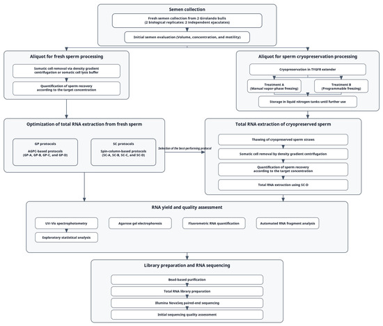
Fresh semen was collected by trained technicians at an accredited government bull farm during routine semen processing from two sexually mature Girolando bulls. Ejaculates were maintained at 35 °C until semen volume, sperm concentration, and initial motility were assessed. Motility was evaluated using a computer-assisted sperm analyzer (CASA) (Microptics, Barcelona, Spain). For clarity, the overall workflow of the study is summarized in Figure 1.
Figure 1.
Overview of the experimental workflow for total RNA extraction and sequencing from fresh and cryopreserved Girolando bull sperm. The workflow identifies the major steps of the study, from semen collection, sperm processing, optimization of total RNA extraction, selection of methods for cryopreserved sperm, quality assessment of RNA, and library construction for sequencing.
Following routine on-site evaluation in the center laboratory, an aliquot from each ejaculate was provided to the research team for downstream processing and analysis. For downstream analyses, two biological replicates were included, consisting of two independent ejaculates, one collected from each bull. Somatic cells were removed from the fresh ejaculate either by density gradient centrifugation with PureSperm® (NidaCon International AB, Mölndal, Sweden) or by SCLB. In the case of SCLB, the target sperm cell count of 5–6 × 10⁸ was treated with 0.1% SDS and 0.5% Triton X-100 (Merck, Darmstadt, Germany), incubated in a cold-water bath, and then centrifuged thereafter [7]. For the density gradient, a similar target sperm cell count was used. The density gradient was prepared using 40% PureSperm® underlaid with 80% PureSperm® in a pre-warmed 37 °C tube, where semen was placed on top, then centrifuged. The increase in input sperm cells was due to an estimated 10–15% loss of sperm cell counts after the purification step [7]. After washing with 1× PBS for SCLB or PureSperm® wash for density gradient, the sperm pellet was resuspended in 1 mL 1× PBS, and sperm concentration was verified using SDM 6 photometer (Minitüb GmbH, Tiefenbach, Germany) to ensure recovery of 3–5 × 10⁸ sperm cells prior to lysis.
Remaining fresh samples were subjected to total RNA extraction, following the cryopreservation protocol of the study. Fresh bull semen was cryopreserved using a two-step Tris–egg yolk extender supplemented with fructose and glycerol (TYGFR), based on the USDA Agricultural Research Service National Animal Germplasm Program (USDA ARS NAGP) bull semen cryopreservation protocol for both manual vapor-phase freezing and programmable freezing, with modifications [11]. Thus, the study comprised two treatments for cryopreserved sperm: Treatment A (TYGFR + manual vapor-phase freezing) and Treatment B (TYGFR + programmable freezing).
Cryopreserved sperm straws were thawed at 37 °C for RNA extraction. A 20 μL aliquot was diluted in the original extender, and a 10 μL aliquot was mounted on a pre-warmed 37 °C glass slide. Sperm motility was reassessed by CASA to confirm post-thaw viability. After motility assessment, somatic cell removal was performed using PureSperm®, followed by total RNA extraction. Similarly, after washing with PureSperm® wash, the sperm pellet was resuspended in 1 mL 1× PBS, and sperm concentration was verified using SDM 6 photometer to ensure recovery of 3–5 × 10⁸ sperm cells prior to lysis.
2.2. Total RNA Extraction from Sperm Cells
RNA extractions were carried out from sperm cells using four guanidinium phenol-based (GP) protocols and four spin column (SC) protocols. Sperm concentration was analyzed using a photometer, and the sperm cell count before lysis was checked to be within the recommended 3–5 × 10⁸ cells. For bead-beating homogenization, 1.4 mm ceramic beads (zirconium oxide) were used in a Precellys 24 Tissue Homogenizer (Bertin Technologies, Montigny-le-Bretonneux, France). GP-C, GP-D, SC-A, and SC-B adapted a parameter with slight modifications, composed of 3 cycles of 20 s at 2500 rpm with a 10 s pause in between, whereas SC-C and SC-D used a modified 9 cycles [12]. For all SC protocols, total RNA isolation also included the removal of genomic DNA in the column according to the manufacturer’s instructions, and two eluates were obtained from each sample. Step-by-step procedures for all eight protocols are detailed below.
2.3. GITC-Phenol (GP) Solution
2.3.1. GP-A
Total RNA isolation from sperm cells was carried out by the acidic guanidinium phenol chloroform method as per the TRIzol® Reagent manual (Invitrogen, Carlsbad, CA, USA). The cell input for the procedure was adjusted to 1 × 10⁷, 1 × 10⁸, 3 × 10⁸, 5 × 10⁸, and 1 × 10⁹ cells. The sperm cells were washed thrice in 1× PBS, resuspended in 1 mL of 1× PBS, adjusted to the required cell input, washed twice with NH4Cl solution, and centrifuged at 14,000 rpm for 5 min at 25 °C. The pellet was processed for TRIzol® extraction following the manufacturer’s instructions.
2.3.2. GP-B
The recovered sperm cell concentration that resulted in the highest RNA quantity and quality from GP-A was applied. The sample was overlaid with 1× PBS and chilled at 4 °C. After two PBS and NH4Cl washings as in GP-A, the pellet was subjected to cell lysis using 1 mL of preheated TRIzol®, homogenized by pipetting, and then spun. The rest of the protocol followed the manufacturer’s instructions.
2.3.3. GP-C
GP-C followed GP-B, with the addition of using SCLB for the purification of the sperm cells. The mixture was homogenized by pipetting and pulse vortexing, followed by incubation on ice for 10 min and centrifugation at 14,000 rpm for another 10 min at 4 °C [13]. The resulting pellet was washed with 1 mL of 1× PBS, then subjected to cell lysis using 1 mL of TRIzol®, incubated at 65 °C for 15 min and then at room temperature for 15 min, and homogenized by pipetting. The lysate was placed in a 1.5 mL lysing matrix tube, bead homogenized, and transferred to nuclease-free 2 mL tubes, and then processed for phase separation following the manufacturer’s instructions.
2.3.4. GP-D
GP-D was a modification of GP-C with added density gradient centrifugation, treatment with NH4Cl solution, and supplementation with βME solution during lysis. Sperm purification was performed using a PureSperm® 80%/40% density gradient centrifugation medium by layering 2 mL of 40% PureSperm® over 2 mL of 80% PureSperm® in a pre-warmed 15 mL tube, followed by incubation for 5 min at 37 °C. The mixture was then subjected to centrifugation at 3000 rpm for 20 min at room temperature. The pellet was washed with 1× PBS, then centrifuged at 4500 rpm for 10 min at room temperature, followed by resuspension of the pellet in 1 mL of 1× PBS, and then another centrifugation at 14,000 rpm for 1 min. The pellet was then lysed with 1 mL of TRIzol® with βME, incubated at 65 °C for 15 min, incubated again at room temperature for 15 min, and then subjected to bead homogenization. Subsequently, 200 µL of chloroform was added. The mixture was left to incubate for 5 min at room temperature. Centrifugation was performed at 14,000 rpm for 15 min at 4 °C. The aqueous fraction was mixed with an equal volume of isopropanol, which was twice the volume of the aqueous fraction, then inverted 10 times. The mixture was left to incubate for 15 min at room temperature. Centrifugation was performed at 14,000 rpm for 20 min at 4 °C. The pellet was washed twice with 1.5 mL of chilled 75% ethanol, air-dried for 10 min, then resuspended in 10–30 µL of nuclease-free water.
2.4. Spin-Column (SC) Methods
2.4.1. SC-A
For total RNA from fresh sperm, the Quick RNA Miniprep Plus Kit (Zymo Research, Irvine, CA, USA) was used according to the manufacturer’s instructions for tough-to-lyse cells, with modifications. Sperm cells were washed and purified using 1 mL SCLB, following the steps described in GP-C. The pellet was mixed with 800 µL of 1× DNA/RNA Shield™, then homogenized by pipetting. The mixture was then transferred to lysing matrix tubes for bead homogenization. The homogenate was centrifuged briefly, and the cleared lysate was transferred to a new 2 mL tube, then mixed 1:1 with RNA Lysis Buffer supplemented with βME at 1 µL per 100 µL buffer. The clear supernatant was transferred to a new 2 mL tube. An equal volume of RNA lysis buffer was added, then homogenized by bead beating. The subsequent purification steps followed the manufacturer’s protocol.
2.4.2. SC-B
SC-B used the protocol for cell cultures of the PrimeWay Total RNA Extraction Kit (1st BASE, Singapore), with modifications. Purification was performed using the same SCLB method and washing steps as described in GP-C. The sperm pellet was lysed in 700 µL RT buffer with 7 µL βME, then homogenized with beads. The subsequent purification steps followed the manufacturer’s protocol.
2.4.3. SC-C
SC-C was also based on the tough-to-lyse cell protocol of the Quick RNA Miniprep Plus Kit, but with density gradient purification and modified lysis. A PureSperm® 80/40 density gradient was prepared, and the subsequent purification and washing steps were performed according to the described PureSperm® protocol in GP-D. Similar to SC-A, the pellet was mixed with 800 µL of 1× DNA/RNA Shield™ (Zymo Research, Irvine, CA, USA), then homogenized by pipetting. The mixture was then transferred to lysing matrix tubes for bead homogenization. The homogenate was centrifuged briefly, and the cleared lysate was transferred to a new 2 mL tube, then mixed 1:1 with RNA Lysis Buffer supplemented with βME at 1 µL per 100 µL buffer. After 5 min at room temperature and 10 min at 65 °C, the mixture was homogenized by pipetting and another round of bead beating. The subsequent purification steps followed the manufacturer’s protocol.
2.4.4. SC-D
SC-D also used the PrimeWay Total RNA Extraction Kit, similar to SC-B, but sperm purification by density gradient centrifugation and heating during lysis was modified. Sperm cells were purified and washed through density gradient centrifugation following the described PureSperm® protocol in GP-D. Sperm cells were carefully removed with a Pasteur pipette and washed with the same steps in SC-C. After centrifugation, the sperm pellet was lysed by adding 700 µL of TR buffer with 7 µL of βME. The sample was mixed by pipetting and vortexing, incubated at room temperature for 5 min, and then incubated at 65 °C for 10 min, followed by bead homogenization. The subsequent purification steps followed the manufacturer’s protocol.
2.5. Estimation of Total RNA Yield via Spectrophotometer and Quality via Agarose Gel Electrophoresis
The total RNA quantity and quality were assessed using the NanoDrop® Spectrophotometer (Thermo Fisher Scientific, Wilmington, DE, USA). The integrity of isolated RNA was assessed by running RNA samples directly into a 1% agarose gel stained with GelRed™ (Biotium, Hayward, CA, USA) and using 0.5X Tris-Borate-EDTA (TBE) as buffer.
2.6. Data Analysis
Data analyses for the yield and purity values were performed in GraphPad Prism version 9 (GraphPad Software, Boston, MA, USA). For each endpoint, values from the two biological sources were summarized across extraction protocols as mean ± SD. Normality of residuals was assessed visually via normal probability (Q–Q) plots, while variance homogeneity was evaluated using the Brown–Forsythe test. When ANOVA assumptions were met, one-way ANOVA followed by Tukey’s HSD was used as an exploratory comparison of protocols, and two-sided α = 0.05 was used. Because the study included only two biological sources, these inferential comparisons were interpreted cautiously and were used to support, rather than solely determine, protocol evaluation.
2.7. Library Preparation and RNA Sequencing
The total RNA samples were sent to the Philippine Genome Center—DNA Sequencing Core Facility (PGC-DSCF), University of the Philippines Diliman, Quezon City, Philippines for quantification and quality assessment after initial quality checks by spectrophotometry and agarose gel electrophoresis (AGE). RNA concentration was measured using a Qubit RNA HS assay on 2 µL RNA (Invitrogen, Eugene, OR, USA). Assessment of RNA integrity and fragment size was performed on a TapeStation 2200 with RNA ScreenTape (Agilent Technologies, Santa Clara, CA, USA) by mixing 1 µL RNA with 5 µL RNA Sample Buffer and processing it according to manufacturer instructions.
At PGC-DSCF, the two RNA eluates obtained for each sample were pooled, then purified and concentrated with AMPure XP beads (1:1.8 sample-to-bead) (Beckman Coulter, Brea, CA, USA) and eluted in 20 µL nuclease-free water. Eluates were quantified with the Qubit RNA BR assay (Invitrogen, Eugene, OR, USA). Libraries were prepared using the TruSeq Stranded Total RNA kit (Illumina, San Diego, CA, USA) according to the manufacturer’s protocol.
Sequencing was performed on an Illumina NovaSeq (Illumina, San Diego, CA, USA), paired-end reads, at PGC-DSCF, after which the FASTQs were forwarded to the PGC-Core Facility of Bioinformatics (PGC-CFB). Reads were quality-filtered and adapter-trimmed using fastp, summarized using MultiQC, aligned using HISAT2, and quantified against the reference annotation using HTSeq. Quality control steps included checking library size and PCA before differential expression analysis.
3. Results and Discussion
3.1. Effect of Sperm Cell Input, Sperm Purification, Reducing Agent Supplementation, and Homogenization on RNA Yield
Of the sperm input levels examined, the highest mean was achieved with GP-A at 5 × 10⁸ cells (63.03 ± 3.84) (Table 1). This input level is presumably optimal in providing a balance between the need to provide sufficient cellular material for efficient lysis. At a higher input of 1 × 10⁹ cells, the total RNA isolated was lower (23.41 ± 3.10), as expected due to sample overload or reagent limitations. When the sample is overloaded, the lysate can become overly viscous due to accumulated unlysed cellular debris, which can overwhelm the extraction chemistry. This, in return, may limit mixing and reduce effective contact between the lysis reagents and sperm heads, ultimately lowering recoverable RNA. In contrast, very low input at 1 × 10⁶ cells resulted in minimal recovery (1.70 ± 0.71), reflecting insufficient starting material for efficient RNA extraction, as expected given the low RNA content of bovine spermatozoa [3]. Based on the total RNA mean, ~5 × 10⁸ sperm cells were the most efficient and served as the basis of sperm cell input for the subsequent protocol optimization.
Table 1.
RNA concentration from Girolando sperm cells (n = 2) at various sperm concentrations, extracted using GP-A and estimated from A260 spectrophotometric absorbance (mean ± SD).
RNA concentration differed across the eight protocols. Overall, there were three (3) that had the highest RNA means. Specifically, GP-C yielded the highest RNA concentration and was comparable only to SC-C, while SC-D consistently produced a higher mean than the unmodified SC protocols. GP-D was mid-range, higher than unmodified SC-A or SC-B, but below the mean of GP-C. The lowest tier included SC-A, SC-B, GP-A, and GP-B, which showed consistently lower means than the modified protocols (Table 2).
Table 2.
Spectrophotometric RNA concentration in fresh sperm across eight extraction protocols (mean ± SD).
These patterns are consistent with the resistance of the protamine-condensed sperm nucleus to chaotropic lysis, where effective reduction via reducing agents and mechanical disruption are often required to release RNA [1]. A similar study using bovine samples reported an improved and more consistent RNA recovery using membrane-based columns combined with TRIzol® and βME, with SCLB treatment or sperm purification beforehand, which gave higher and more consistent recoveries compared to unmodified kits. This aligns with the superiority of SC-C and SC-D over SC-A and SC-B [5].
Thiol-based reducing agents can facilitate lysis and phenol extraction with heat assistance; however, βME has pH dependence and might be less effective in AGPC protocols using acidic TRIzol® alone. TCEP is less affected by the acidic conditions and can facilitate sperm RNA isolation in AGPC protocols, thus making it an alternative [6]. In this study, adding βME with heating and bead-assisted homogenization increased RNA recovery in GP-C and GP-D relative to the unmodified protocols. This result suggests that strong disruption, accompanied by heating, partially mitigated βME’s pH-dependent limitations. Following the same modifications also resulted in an improved performance, where SC-C and SC-D produced higher recovery than unmodified SC protocols. This is consistent with the reports that βME performs better with guanidinium-based column lysis buffers than with TRIzol® alone [5,6]. Intense protamination tightens sperm chromatin to protect paternal DNA during transit in the female tract, increasing tolerance to damage [14]. Therefore, combining heat, mechanical homogenization, and a suitable reducing agent enhanced the lysis and disrupted the nucleus, thus bolstering recovery from tough-to-lyse sperm [1,4,15].
Second, somatic cell removal by density gradients generally yields cleaner sperm fractions than chemical lysis with SCLB, which can incompletely deplete leukocytes and epithelial cells and can also disturb sperm membranes. In bovine, BoviPure® and PureSperm®-type gradients reduce non-sperm contamination and maintain better integrity of RNA profiles, whereas SCLB can alter cellular components, and may not fully eliminate somatic RNA [7].
Among the protocols tested, GP-D showed a higher mean than the unmodified GP and SC protocols by adding gradient purification and βME in a heated and bead-assisted protocol, but did not reach the mean of GP-C. One plausible explanation is that gradient centrifugation reduces absolute RNA recovery by removing somatic remnants and other non-sperm cells that can contribute to measured RNA [7]. In contrast, GP-C had a higher concentration than GP-D. This result plausibly reflects the somatic cell or non-sperm cell contamination left even after SCLB that can inflate RNA. Somatic cells contain a higher amount of RNA, about 10–20 pg, compared to 20–30 fg in spermatozoa [15]. However, this interpretation remains speculative, as somatic contamination was not directly quantified.
The unmodified SC protocols paired with SCLB produced minimal RNA, suggesting that the lysis was insufficient under the unmodified conditions. When lysis was intensified in the modified SC protocols, RNA recovery increased and exceeded the best-performing modified GP protocol under the same purification conditions. This improvement indicates that optimized sperm head lysis is a dominant driver of recovery in these protocol comparisons, and also makes it difficult to isolate the independent contribution of purification alone. Overall, the data support a combined protocol that integrates effective sperm purification with robust sperm head lysis.
For GP protocols, GP-C and GP-D showed the most favorable overall concentration patterns based on means. For column-based with DNase, SC-C and SC-D also showed favorable performance. However, concentration alone was necessary but not sufficient for protocol selection. Comparison of purity ratios and gel-based profiles is needed to ensure that higher yields were not due to contamination or degraded RNA, since phenol carryover or residual salts can inflate A260 and compromise downstream assays [16].
3.2. RNA Purity Metrics Across Protocols
Purity metrics refined protocol ranking beyond concentration alone (Table 3). Based on A260/A280, which reflects protein carryover, was highest in SC-C, indicating the lowest protein contamination among protocols and surpassing the SC-D protocol. GP protocols and unmodified SC protocols had lower A260/A280 values, but the difference between groups was statistically insignificant, likely due to the distribution of the data and the limited biological replications. Overall, SC-C showed the highest mean A260/A280 in this dataset [17].
Table 3.
Spectrophotometric RNA purity ratios in fresh sperm across eight extraction protocols (mean ± SD).
As a representation of organic and chaotropic carryover, A260/A230 had the lowest mean values in GP protocols and higher values in the SC protocols. This result is consistent with the pattern among AGPC protocols that tends to retain phenol or guanidium residuals even when A260/A280 appears to be acceptable [18]. Although the differences between groups were deemed to be statistically insignificant, likely due to limited sample size, column-based methods had higher mean A260/A230 values. The GP-C had mid-range A260/A230 with high variability, matching well with potential fluctuating organic carryover in AGPC protocol, as well as potential variability in somatic depletion [7,18]. However, it is also important to note that since sperm RNA yields are low, spectrophotometric ratios can also become less stable at low A260, which may contribute to the observed variability.
Operationally, GP extraction was simple and cost-effective, but RNA quality and purity were highly dependent on operator technique and sample handling. In contrast, SC protocols were more streamlined and generally produced cleaner RNA, which is consistent with selective nucleic acid binding to silica and wash-based removal of contaminants [6]. Similar patterns in purity have been seen with bovine sperm RNA extraction protocols. Membrane-column protocols combined with TRIzol® and βME, together with sperm purification, showed higher and more consistent A260/280 and A260/230 compared to unmodified columns or phenol-only protocols [5]. In this study, gradient centrifugation likely reduced somatic contamination but did not fully address the organic or chaotropic carryover in GP protocols, consistent with the low A260/A230 observed in GP-C and GP-D. In addition, acidic GP conditions may reduce the effectiveness of pH-dependent thiol reducers, which may have contributed to the variable purity when a pH-compatible reductant is not used [6].
In modified SC protocols, the modifications likely enhanced sperm head disruption and aid reagent exchange without introducing organic carryover, consistent with the higher A260/280 mean in SC-C and better A260/230 in SC-D than phenol-only methods [5,6]. Overall, SC-C has the most favorable performance by A260/280 and A260/230. Next best for A260/230 with lower variability is SC-D. GP-D remained acceptable by A260/A280 but was limited by low A260/A230. Whereas, GP-C showed the highest A260/A230 mean among GP protocols, but with high variability. Thus, it is recommended to consider SC-C and SC-D protocols, keeping GP-D for higher A260/280 despite lower A260/230, and consider GP-C for the highest but variable A260/230. However, spectrophotometry alone cannot assess fragmentation or rRNA contamination, and mature sperm do not show canonical 18S/28S peaks, so it is necessary to interpret purity alongside agarose-based gel or electropherogram profiles to check integrity and ensure non-sperm contamination [6].
3.3. RNA Integrity Across Protocols
As spermatogenesis progresses, round spermatids lose cytoplasm, and Sertoli cells phagocytose the droplet, reducing RNA content and translational machinery from the residual body of a mature spermatozoon. Thus, sperm RNAs are interpreted as residual transcripts from spermatogenesis that persist in mature sperm, rather than being selectively retained. Consequently, sperm RNA is typically fragmented, and RIN/RINe values are often low yet still suitable for downstream analysis. An appropriate sperm electropherogram shows the absence of prominent 18S/28S peaks and a continuous fragment distribution above ~25 bp extending into mid-high ranges. Whereas higher RIN/RINe values with clear 18S/28S peaks often indicate somatic rRNA contamination [4]. Typical high-purity sperm RNA has RIN < ~2.5, whereas RIN > ~4 may indicate somatic rRNA contamination [8]. In bovine sperm, low RIN with broad smears and peaks near ~75 bp without canonical somatic rRNA has been reported as a typical high-purity pattern [3,19].
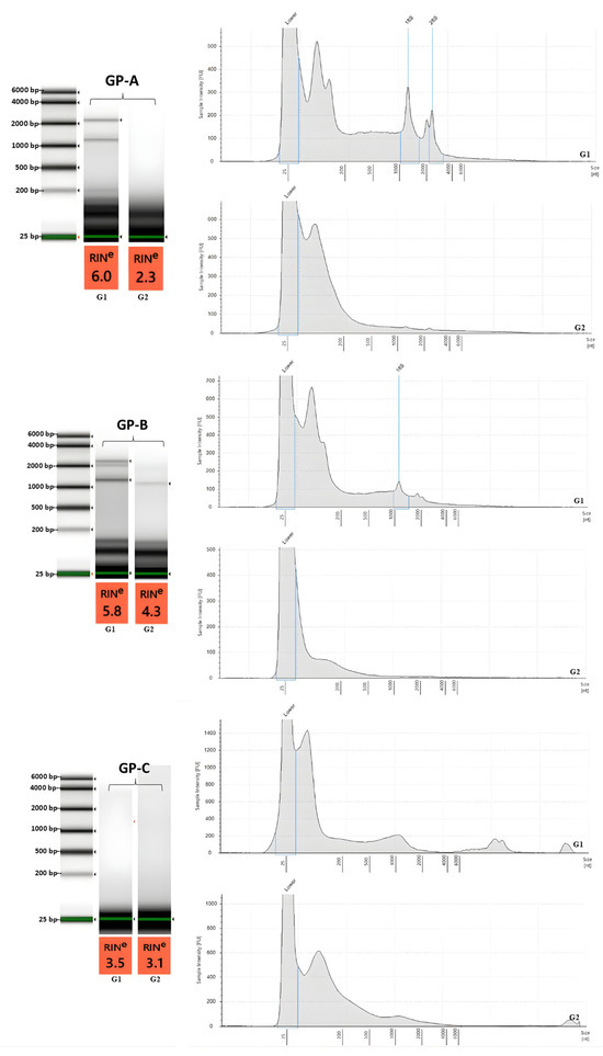
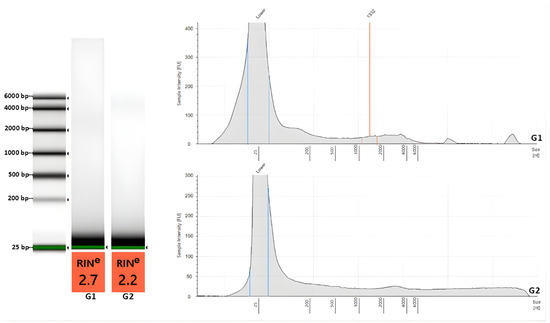
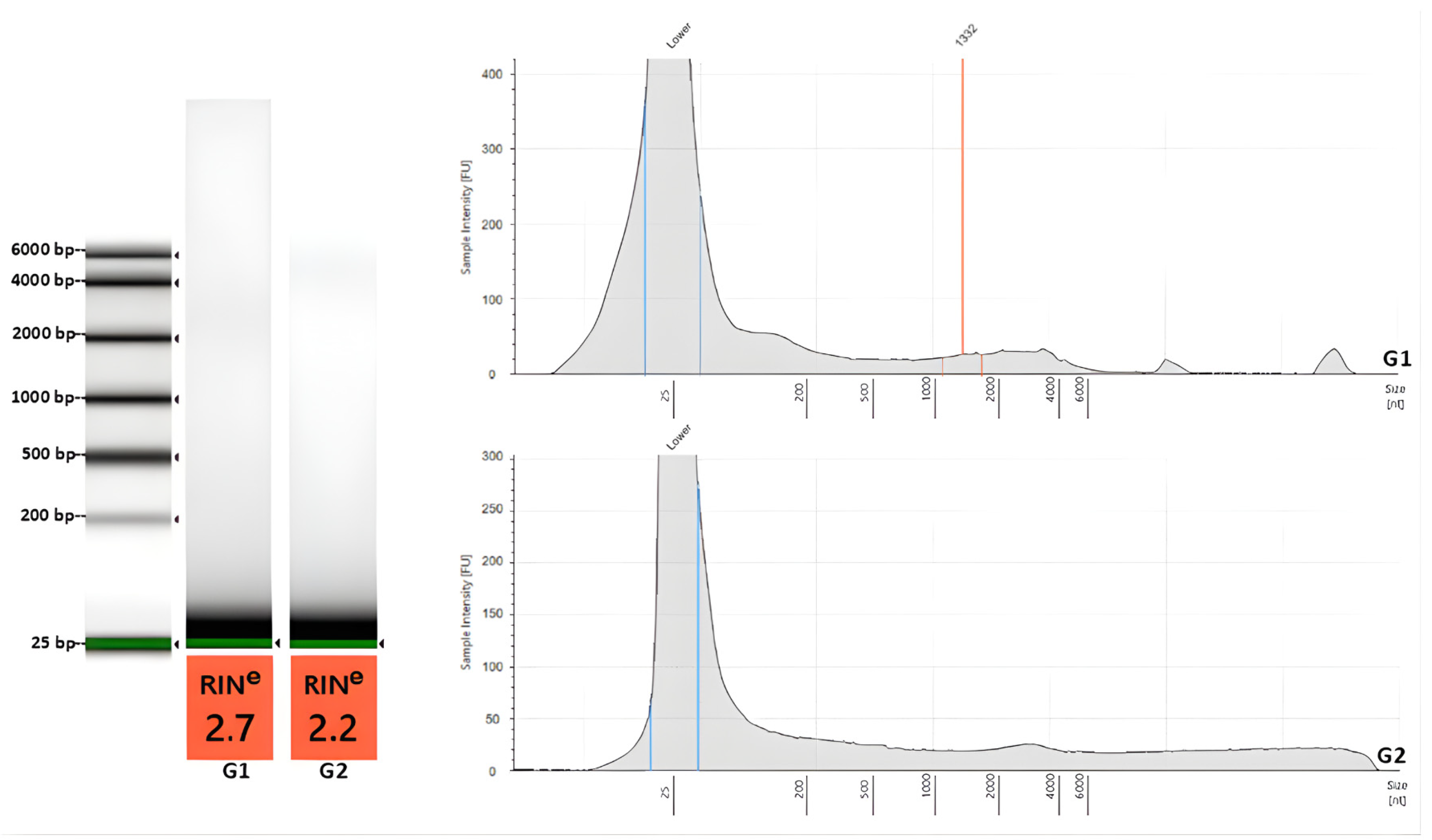
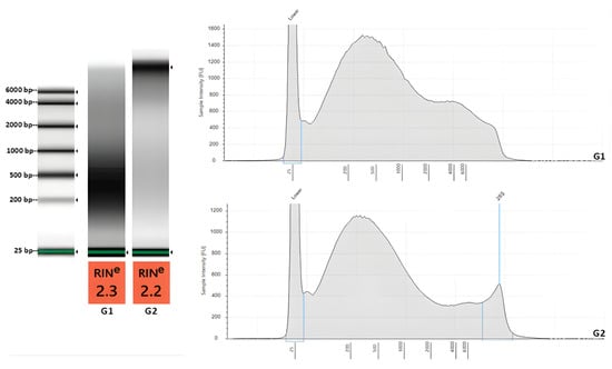
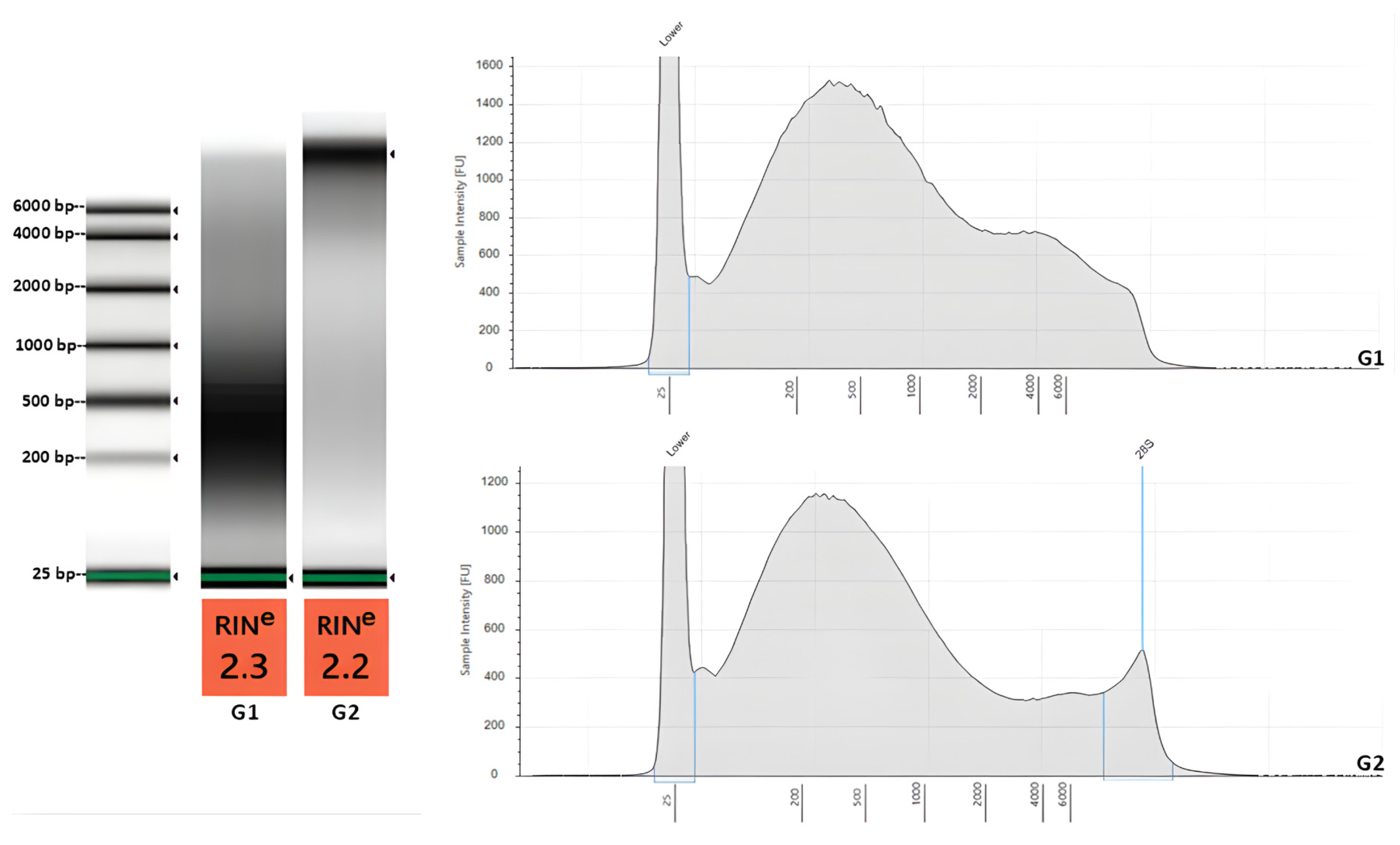
Among the protocols, GP-A and GP-B exhibited distinct 18S/28S peaks with higher RINe (6.0, 5.8) (Figure 2). GP-C exhibits an rRNA-region signal with intermediate RINe (3.5, 3.1). Since intact 18S/28S peaks are not expected in mature sperm, these profiles most likely reflect co-purified somatic or non-sperm cells [8,19] (Figure 2). GP-C was incorporated with SCLB yet still retained the signal of the rRNA-region, consistent with incomplete leukocyte or epithelial removal reported when using the SCLB approach for the sperm purification [7]. In contrast, the GP-D, which used density gradient purification, demonstrated an absence of rRNA peaks with ~2.5 RINe, which denotes minimal somatic contamination compared to other GP protocols [8] (Figure 3).
Figure 2.
Electropherogram and RIN profiles for RNA from fresh sperm using GP protocols. Fresh sperm RNA extracted using GP protocol variants (GP-A to GP-C) is shown with representative virtual gel images, electropherogram traces, and RINe values.Blue vertical lines in the electropherograms denote software-assigned marker positions, including the lower marker and rRNA marker(s), whereas the green band in the virtual gel image denotes the lower marker. Within each protocol, G1 and G2 were obtained from the same electrophoresis run; however, the lane order (G1 and G2) was rearranged for presentation. Panels for GP-A, GP-B, and GP-C were generated from separate runs and were assembled into one figure in the order shown.
Figure 3.
Electropherogram and RIN profiles for RNA from fresh sperm using GP protocols. Fresh sperm RNA extracted using GP-D is shown with representative virtual gel images, electropherogram traces, and RINe values. Blue vertical lines in the electropherograms denote software-assigned marker positions including the lower marker, red vertical line indicates a software-identified peak or selected RNA fragment region with the corresponding estimated fragment size shown in nucleotides, whereas the green band in the virtual gel image denotes the lower marker. Within protocol, G1 and G2 were obtained from the same electrophoresis run; however, the lane order (G1 and G2) was rearranged for presentation.
All GP electropherograms displayed predominantly short fragments. In GP-D, signals cluster below ~200 nt, matching sperm RNA patterns for bovine [3] (Figure 3). This pattern consistently aligns with the findings that AGPC workflows combined with effective reduction and a strong homogenization method improve sperm head lysis without artifactual rRNA peaks [6]. Taken together with spectrophotometry, GP-C had the highest concentration but showed low A260/230 and rRNA peaks, whereas GP-D showed a lower concentration but cleaner A260/230 and the absence of rRNA peaks. GP-C may have a higher RNA concentration that potentially reflects somatic contamination, further inflating yield and purity. While GP-D showed purer sperm RNA but with lower yield and purity in the absence of somatic or non-sperm cell contamination [7,18].
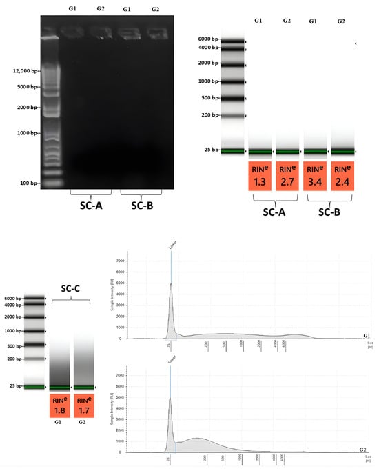
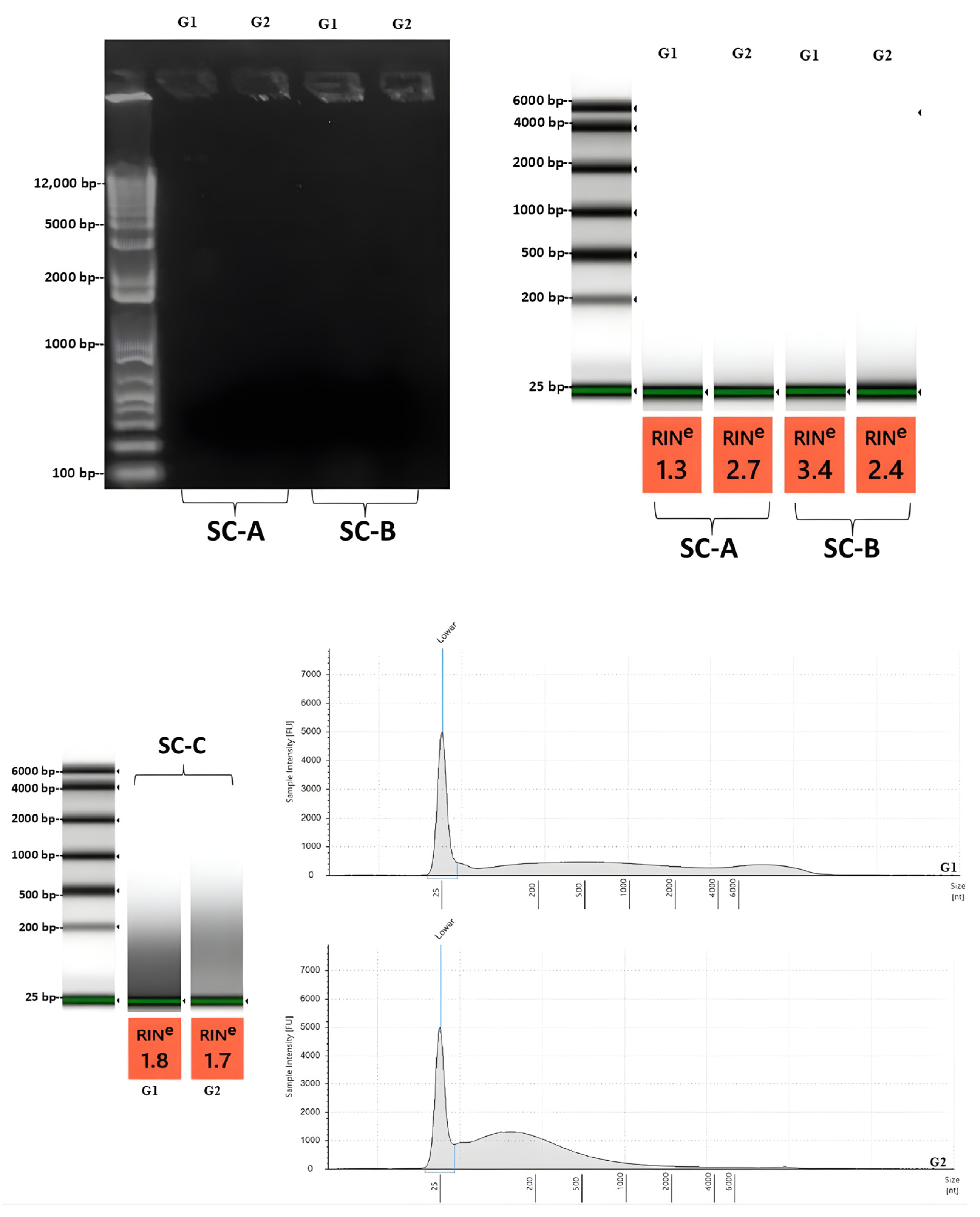
In SC protocols, SC-A and SC-B showed faint smears, very low fluorescence, and RINe ~1.3–3.4 (Figure 4). Although low RINe alone is not a reliable indicator of poor sperm RNA, a near-background fluorescence signal suggests inadequate RNA recovery. This likely reflects insufficient lysis under the unmodified protocols. The unmodified SC protocols may not have effectively opened the protamine-crosslinked heads, as a more effective reducing agent and stronger homogenization method are necessary to break disulfide bonds [4,6,20]. Accordingly, SC-A and SC-B were not suitable for RNA-seq in their current form. The SC-C protocol showed a typical sperm-consistent profile of negligible 18S/28S, fluorescence above ~25 bp, and most signal <200 bp (Figure 4). This indicates the removal of somatic rRNA and the recovery of short or fragmented RNAs. However, strong sub-200 bp nucleotides may underrepresent longer transcripts in total RNA libraries, depending on library preparation and size-selection behavior. SC-D showed the broadest fluorescence distribution, extending to ~4 kb with minimal rRNA peaks, which better supports total RNA-seq coverage across small RNAs and fragmented longer RNAs (Figure 5). This is consistent with the applied modifications, which enhanced access to the sperm nucleus and the release of RNA [4,7,19]. However, a high-molecular-weight signal above ~6 kb suggested potential genomic DNA carryover.
Figure 4.
Electropherogram and RIN profiles for RNA from fresh sperm using SC protocols. Fresh sperm RNA extracted using SC protocol variants (SC-A to SC-C) is shown with representative virtual gel images, electropherogram traces, and RINe values. Blue vertical lines in the electropherograms denote software-assigned marker positions, including the lower marker and rRNA marker(s), whereas the green band in the virtual gel image denotes the lower marker. Within each protocol, G1 and G2 were obtained from the same electrophoresis run; however, the lane order (G1 and G2) was rearranged for presentation. Panels for SC-A, SC-B, and SC-C were generated from separate runs and were assembled into one figure in the order shown.
Figure 5.
Electropherogram and RIN profiles for RNA from fresh sperm using SC protocols. Fresh sperm RNA extracted using SC-D with representative virtual gel images, electropherogram traces, and RINe values. Blue vertical lines in the electropherograms denote software-assigned marker positions, including the lower marker and rRNA marker(s), whereas the green band in the virtual gel image denotes the lower marker. Within protocol, G1 and G2 were obtained from the same electrophoresis run; however, the lane order (G1 and G2) was rearranged for presentation.
When combined with spectrophotometry, SC-C showed the cleanest protein profile and a sperm-specific signal, though with a short-fragment bias. SC-D also yielded higher A260/A230 ratios than the phenol-based workflows; however, a ~4 kb fluorescence signal was observed. Overall, SC-D appears to be the most practical option for total RNA-seq, provided that an additional DNase cleanup step or size selection is included before library preparation. Alternatively, a more vigorous but still controlled lysis step during extraction, such as using a refrigerated homogenizer to avoid excessive heat, may also help reduce gDNA contamination [15].
The results obtained align with bovine data, where density gradient centrifugation results in motile mature sperm, and RNA shifts away from non-sperm or immature sperm signals [15]. Modified column-based workflows combining sperm purification, effective reduction, and strong head lysis are most reliable for reproducible sperm-appropriate RNA [1,5,6].
3.4. Identification of Optimal RNA Extraction Protocol for Fresh Sperm Cells
Using GP-A as a baseline with 5 × 10⁸ sperm cells, the input produced measurable RNA feasible for downstream protocol optimization. Across protocols, SC-D provided the best overall performance based on three converging indicators, specifically, the combination of high spectrophotometric purity, strong RNA mass measured by fluorescence, and an electropherogram profile suitable for sperm. Although GP-C produced the highest RNA yield, it also showed very low A260/230 ratios and clear rRNA peaks. Incorporating sperm density gradient centrifugation in GP-D reduced rRNA-associated signals, but with a lower RNA concentration relative to GP-C. SC-C produced very clean RNA by purity ratios, but the profile was enriched for fragments < 200 bp. In contrast, SC-D captured both small RNAs and fragmented long RNAs, while maintaining minimal rRNA peaks. For these reasons, SC-D was selected as the primary protocol for extracting fresh Girolando sperm RNA for sequencing, with the remaining limitation of potential genomic DNA carryover addressed through DNase treatment and cleanup. SC-D was subsequently tested to assess its performance in extracting total RNA from cryopreserved sperm of Girolando bulls.
3.5. RNA Extraction from Frozen Sperm Using the Best-Performing Protocol
Using SC-D on cryopreserved sperm, workable RNA amounts and ~3.3–3.6 × 10⁸ purified sperm were recovered per extraction (Table 4). Because only a limited number of cryopreserved sperm straws were available, the sperm cell input for extraction was constrained. Extractions were therefore delayed to allow accumulation of additional straws, targeting a minimum input of approximately 3.0 × 10⁸ sperm cells. Thus, two sequential elutions per straw were performed to maximize RNA recovery, using sperm from both cryopreservation treatments for each Girolando bull.
Table 4.
Input sperm cell concentration, and spectrophotometric RNA concentration and purity, in cryopreserved Girolando sperm from Treatments A and B using two elutions (mean ± SD).
A260/A280 values remained within the expected range for RNA, indicating minimal protein carryover, whereas A260/A230 values were lower and more variable, suggesting residual chaotropic salts and column-buffer components (Table 3). Both fresh and cryopreserved samples were subjected to density gradient purification; however, cryopreserved sperm samples are expected to have higher debris due to the freeze–thaw process and the presence of extender components. This plausibly impaired the effectiveness of silica-column washes and increased carryover of contaminants that absorb at 230 nm, such as guanidinium salts or residues of wash buffers. This could be the reason that the A260/A230 ratios of cryopreserved sperm were preferentially reduced compared to the fresh sperm, whereas A260/A280 ratios in both sperm conditions remained unaffected. These contaminants may be reduced by an additional ethanol wash or a brief post-extraction cleanup.
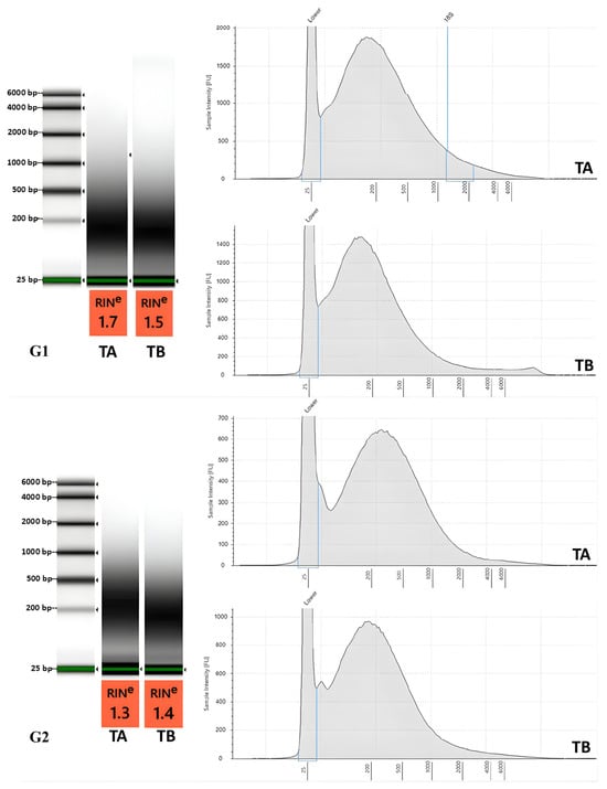
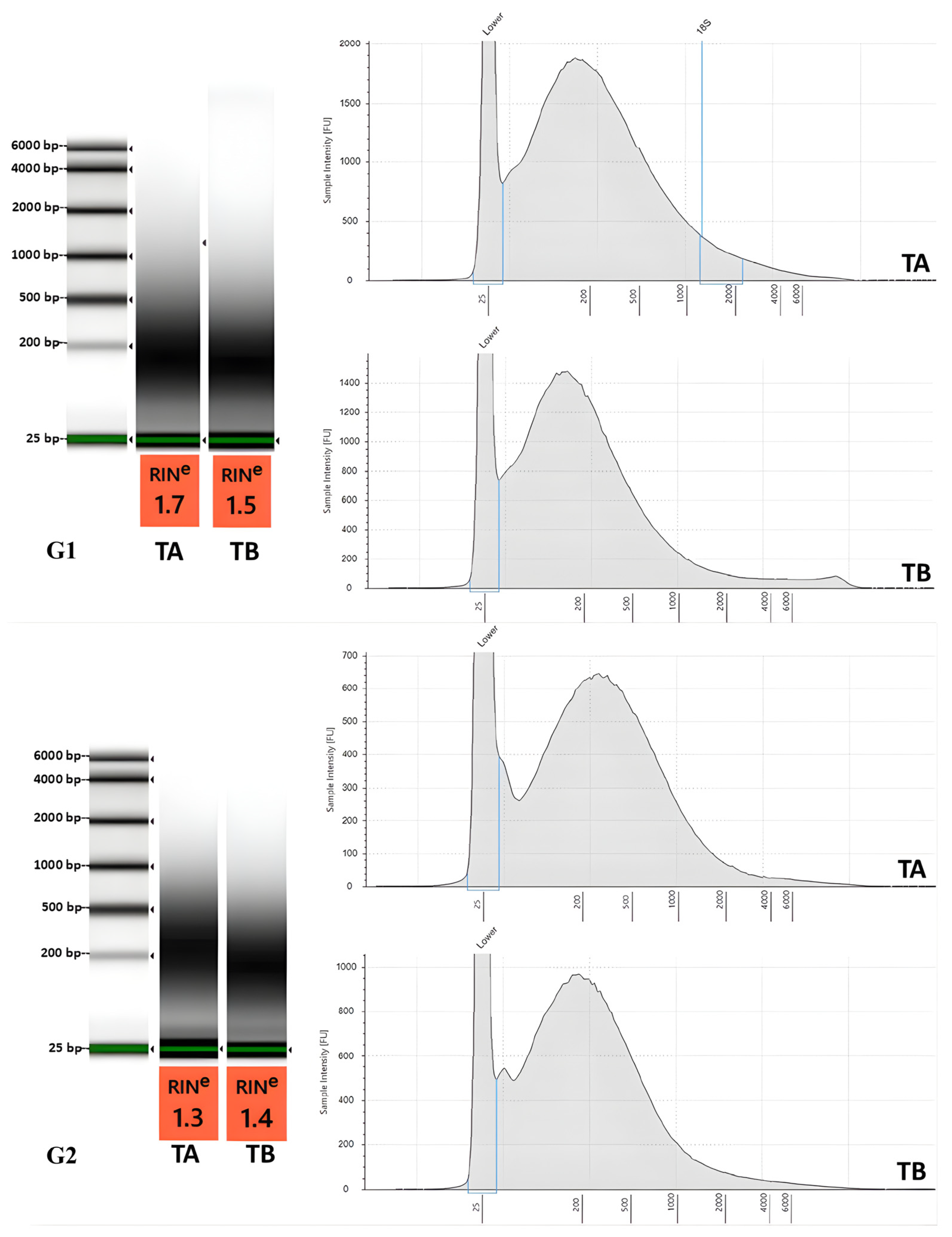
Total RNA extracted from cryopreserved sperm using SC-D demonstrates primarily < 1000 bp fragments, with broad peaks at <500–800 bp, and absence of rRNA peaks (Figure 6). The shift toward shorter fragments after cryopreservation is biologically plausible, since bull sperm cryopreservation has been reported to alter small RNA and mRNA fragment landscapes. Evidence indicates that cryopreservation can shorten the length distribution of mapped reads and change the abundance of miRNA and mRNA fragments [21].
Figure 6.
Electropherogram and RIN profiles for RNA from cryopreserved sperm extracted using SC-D. Cryopreserved sperm RNA extracted using SC-D is shown with representative virtual gel images, electropherogram traces, and RINe values. Within protocol, TA and TB were obtained from the same electrophoresis run; however, the lane order (TA and TB) was rearranged for presentation. Panels for G1 and G2 were generated from separate runs and were assembled into one figure in the order shown.
In addition, the less prominent high-molecular-weight gDNA signal in cryopreserved samples can be interpreted as a consequence of freeze–thaw associated DNA damage and chromatin destabilization. The process of post-thawing may increase DNA fragmentation and reduce the visibility of intact, long genomic DNA species on the electropherogram [21]. In general, the SC-D yielded acceptable protein purity but needs an additional cleanup or purification to stabilize purity ratios and control residual DNA-related signal before sequencing.
3.6. RNA Library Preparation and Sequencing Metrics
Using a fixed 100 ng RNA input per library, frozen sperm RNA produced larger average library sizes (277–320 bp) compared to fresh sperm RNA (152–158 bp) of the same animals (Table 5). Short library inserts are commonly associated with fragmented or degraded input RNA and can increase adapter-related products during library construction [22]. When inserts are shorter than the sequencing read length, reads extend into the adapter sequence, increasing trimming and filtering losses and reducing the fraction of reads that pass quality filters.
Table 5.
Fluorometric RNA yield, library preparation input, and mean library size for fresh and cryopreserved cattle sperm under cryopreservation Treatments A and B.
Across both animals, cryopreserved sperm libraries retained more filtered reads with moderate duplication, whereas fresh sperm libraries showed fewer filtered reads and higher duplication (Table 6).
Table 6.
Sequencing read counts and duplication rates for sperm RNA from fresh and cryopreserved sperm under cryopreservation Treatments A and B.
The larger inserts in the frozen group suggest cryopreserved sperm RNA had a broader nucleotide distribution compared to fresh sperm RNA. The difference may be due to frozen samples that were prepared as libraries sooner after collection, whereas fresh sperm RNA had been kept at −80 °C for at least three months before library preparation. Although −80 °C slows degradation, duration, handling, and buffers can still affect RNA integrity and impact the results of library preparation [22,23]. RNA integrity can be affected by handling factors such as freeze–thaw exposure and processing variability, which are known to compromise RNA quality and downstream measurements [24]. Higher duplication is also consistent with reduced effective library complexity and amplification effects. This is because PCR duplicate rates increase as library complexity decreases and as PCR cycles increase [25]. Furthermore, high duplication in sperm RNA-seq is expected, as a few highly abundant transcripts will dominate libraries and inflate duplicates even in the absence of technical issues [26]. This reflects the low-complexity, small-RNA-rich nature of bovine sperm RNA, where a limited set of RNAs dominates.
Overall, the library metrics based on the initial sequencing assessments show that informative sperm RNA-seq data can be generated from both fresh and cryopreserved sperm. Nonetheless, it should be noted that short-insert and reduced library complexity can lower read retention and elevate duplication even at matched RNA input, similar to the fresh and cryopreserved RNA sperm in this dataset. Thus, careful handling of the extracted RNA is crucial to preserve integrity and producing a more informative sequencing library. This helps minimize additional degradation beyond the inherent fragmentation of sperm RNA, allowing the generation of informative RNA-seq data.
4. Conclusions
Although statistical comparisons were performed for concentration and purity metrics, protocol selection was not based on these values alone. Greater weight was given to the combined quality profile of each protocol, including RNA recovery, spectrophotometric purity, electropherogram pattern, evidence of rRNA carryover, and downstream library performance. On this basis, SC-D provided the most balanced overall performance across fresh and cryopreserved sperm.
Libraries from cryopreserved sperm showed better run performance, with larger insert sizes and better read retention. In contrast, libraries from fresh sperm RNA showed smaller inserts and higher duplication, which likely reflects a difference in processing time. Fresh sperm RNA was stored at −80 °C for ~3 months before library preparation, whereas cryopreserved RNA proceeded in library preparation within a week while stored at −80 °C. Longer storage and handling can increase the chances of having shorter insert sizes and subsequently result in fewer usable reads.
Overall, the SC protocols performed better than GP protocols due to their capability to standardize lysis chemistry and wash steps. These procedures in SC protocols improved access to the condensed, disulfide-stabilized sperm head and reduced carryover contaminants. For GP protocols, only βME was tested. Thus, it is recommended to consider other pH-compatible reductants such as TCEP or DTT matched with appropriate conditions, such as longer incubation, and heat with bead-assisted disruption. Additional cleanup or purification steps can also help reduce residual salts and improve A260/A230, particularly for cryopreserved sperm.
gDNA carryover remained an important consideration; therefore, routine DNase treatment followed by an RNA cleanup step prior to library preparation was necessary. In addition, bead-assisted homogenization parameters could be optimized to help minimize gDNA contamination, such as extending the lysis duration or increasing the homogenization speed in rpm. If more aggressive settings are applied, using a refrigerated homogenizer or performing homogenization under cold conditions can also be considered to reduce heat buildup and mechanical stress that may accelerate RNA degradation. With these measures in place, SC-D remains the recommended protocol for total RNA-seq of Girolando sperm from both fresh and cryopreserved sperm, supporting gene-level profiling and providing substantial margins for detection of low-abundance transcripts.
Overall, this study provides an initial optimization of sperm RNA extraction under fresh and cryopreserved conditions and offers a preliminary assessment of sequencing quality. While the selected protocol showed the most favorable overall performance across multiple quality parameters, these findings should be interpreted as preliminary and warrant validation in a larger set of biological sources. Further validation using sperm samples from additional bulls and targeted RT-qPCR analysis is also recommended to confirm the present findings. Moreover, future studies should include a more comprehensive bioinformatic assessment of RNA-seq data, including mapping rate, percentage of rRNA reads, and transcript coverage, to further validate the suitability of extracted sperm RNA for downstream transcriptomic applications.
Author Contributions
Conceptualization, M.A.M.B. and L.P.V.; Methodology, S.M.D.C.G., A.J.E.C., T.P.C.C., D.C.C.C., J.A.F., L.P.B., M.A.M.B. and L.P.V.; Validation, S.M.D.C.G., A.J.E.C., T.P.C.C. and L.P.V.; Formal Analysis, S.M.D.C.G.; Investigation, S.M.D.C.G., A.J.E.C., T.P.C.C., J.A.F., D.C.C.C., L.P.B., M.A.M.B. and L.P.V.; Resources, S.M.D.C.G., A.J.E.C., T.P.C.C., L.P.B., M.A.M.B. and L.P.V.; Data Curation, S.M.D.C.G. and J.A.F.; Writing—Original Draft Preparation, S.M.D.C.G.; Writing—Review and Editing, L.P.B., M.A.M.B. and L.P.V.; Visualization, S.M.D.C.G. and J.A.F.; Supervision, L.P.B., M.A.M.B. and L.P.V.; Project Administration, A.J.E.C., T.P.C.C. and L.P.V.; Funding Acquisition, A.J.E.C., T.P.C.C. and L.P.V. All authors have read and agreed to the published version of the manuscript.
Funding
The study was supported by the Department of Agriculture under grant number DA-BAR-2023-0008. The funding agency did not take part in the design of the study, data gathering, data analysis, or the decision to submit the manuscript for publication.
Institutional Review Board Statement
The study was reviewed and approved by the Philippine Carabao Center Ethics Committee (BG23001-RC, 3 April 2023).
Informed Consent Statement
Not applicable.
Data Availability Statement
The original contributions presented in this study are included in the article. Further inquiries can be directed to the corresponding author.
Acknowledgments
Grateful acknowledgment is extended to the implementing agency, the Philippine Carabao Center (PCC), for spearheading the study and supporting its implementation. Special thanks to the National Bull Farm of PCC at Central Luzon State University for assistance and support during semen collection and cryopreservation. Recognition is likewise extended to the DNA Sequencing Core Facility and the Core Facility for Bioinformatics of the Philippine Genome Center, University of the Philippines Diliman, for technical support in RNA sequencing and downstream data processing. Finally, sincere thanks are expressed to the project staff and project research assistants, as well as the staff of the Cryobank Section/National Livestock Cryobank, Alfredo C. Costales III, John Paul R. Orevillo, and John Exequiel D. Hilario, for technical support throughout the conduct of the research.
Conflicts of Interest
The authors declare no conflict of interest.
References
- Johnson, G.D.; Lalancette, C.; Linnemann, A.K.; Leduc, F.; Boissonneault, G.; Krawetz, S.A. The sperm nucleus: Chromatin, RNA, and the nuclear matrix. Reproduction 2011, 141, 21–36. [Google Scholar] [CrossRef] [Scilit]
- Parthipan, S.; Selvaraju, S.; Somashekar, L.; Kolte, A.P.; Arangasamy, A.; Ravindra, J.P. Spermatozoa input concentrations and RNA isolation methods on RNA yield and quality in bull (Bos taurus). Anal. Biochem. 2015, 482, 32–39. [Google Scholar] [CrossRef] [Scilit]
- Gilbert, I.; Bissonnette, N.; Boissonneault, G.; Vallée, M.; Robert, C. A molecular analysis of the population of mRNA in bovine spermatozoa. Reproduction 2007, 133, 1073–1086. [Google Scholar] [CrossRef] [Scilit]
- Tiwari, S.; Shahat, A.; Kastelic, J.; Thakor, N.; Thundathil, J. Optimized total RNA isolation from bovine sperm with enhanced sperm head lysis. Biochem. Cell Biol. 2024, 102, 194–205. [Google Scholar] [CrossRef] [Scilit] [PubMed]
- Dewry, R.K.; Mohanty, T.K.; Nath, S.; Bhakat, M.; Yadav, H.P.; Baithalu, R.K. Comparative RNA isolation methods from fresh ejaculated spermatozoa in Sahiwal cattle (Bos indicus) and Murrah buffalo (Bubalus bubalis) bulls for high quality and enhanced RNA yield. Anim. Biotechnol. 2023, 34, 5180–5191. [Google Scholar] [CrossRef] [Scilit]
- Roszkowski, M.; Mansuy, I.M. High Efficiency RNA Extraction from Sperm Cells Using Guanidinium Thiocyanate Supplemented with Tris(2-carboxyethyl)phosphine. Front. Cell Dev. Biol. 2021, 9, 648274. [Google Scholar] [CrossRef] [Scilit] [PubMed]
- Mao, S.; Goodrich, R.J.; Hauser, R.; Schrader, S.M.; Chen, Z.; Krawetz, S.A. Evaluation of the effectiveness of semen storage and sperm purification methods for spermatozoa transcript profiling. Syst. Biol. Reprod. Med. 2013, 59, 287–295. [Google Scholar] [CrossRef] [Scilit] [PubMed]
- Gòdia, M.; Estill, M.; Castelló, A.; Balasch, S.; Rodríguez-Gil, J.E.; Krawetz, S.A.; Sánchez, A.; Clop, A. A RNA-Seq Analysis to Describe the Boar Sperm Transcriptome and Its Seasonal Changes. Front. Genet. 2019, 10, 299. [Google Scholar] [CrossRef] [Scilit]
- Ibrahim, S.; Mahmoud, K.G.M.; Sosa, A.S.A.; Sakr, A.A.M.; El-Naby, A.A.H.; Nawito, M.F. Establishment a protocol for total RNA isolation from buffalo fresh and frozen semen for molecular applications. Andrologia 2020, 52, e13526. [Google Scholar] [CrossRef] [Scilit]
- Shtratnikova, V.; Naumov, V.; Bezuglov, V.; Zheludkevich, A.; Smigulina, L.; Dikov, Y.; Denisova, T.; Suvorov, A.; Pilsner, J.R.; Hauser, R.; et al. Optimization of small RNA extraction and comparative study of NGS library preparation from low count sperm samples. Syst. Biol. Reprod. Med. 2021, 67, 230–243. [Google Scholar] [CrossRef] [Scilit]
- USDA ARS National Animal Germplasm Program. Bull Semen Collection, Transportation, Processing and Cryopreservation Protocol. 2020. Available online: https://agrin.ars.usda.gov/protocol_documents/Bull%20Semen%20Collection%20Processing%20and%20Cryopreservation%202020.pdf (accessed on 25 July 2025).
- Schuster, A.; Tang, C.; Xie, Y.; Ortogero, N.; Yuan, S.; Yan, W. SpermBase: A Database for Sperm-Borne RNA Contents. Biol. Reprod. 2016, 95, 99. [Google Scholar] [CrossRef] [Scilit]
- Goodrich, R.; Johnson, G.; Krawetz, S.A. The Preparation of Human Spermatozoal RNA for Clinical Analysis. Arch. Androl. 2007, 53, 161–167. [Google Scholar] [CrossRef] [Scilit]
- Rathke, C.; Barckmann, B.; Burkhard, S.; Jayaramaiah-Raja, S.; Roote, J.; Renkawitz-Pohl, R. Distinct functions of Mst77F and protamines in nuclear shaping and chromatin condensation during Drosophila spermiogenesis. Eur. J. Cell Biol. 2010, 89, 326–338. [Google Scholar] [CrossRef] [Scilit] [PubMed]
- Vasisth, R.; Gurao, A.; Kumari, N.; Kumar, G.; Kumar, A.; Sriranga, K.R.; Dige, M.S.; Mukesh, M.; Aggarwal, R.A.K.; Singh, P.; et al. Development and validation of most efficient RNA isolation method from buffalo bull spermatozoa. Mol. Biol. Rep. 2023, 50, 6717–6727. [Google Scholar] [CrossRef] [Scilit]
- El Fekih, S.; Nguyen, M.-H.; Perrin, A.; Beauvillard, D.; Morel, F.; Saad, A.; Ben Ali, H.; De Braekeleer, M. Sperm RNA preparation for transcriptomic analysis: Review of the techniques and personal experience. Andrologia 2017, 49, e12767. [Google Scholar] [CrossRef] [Scilit] [PubMed]
- Dabrowski, P.; Rasmus, M.; Jundzill, A.; Drewa, T.; Pokrywczynska, M. A comparison of five methods to maximize RNA and DNA isolation yield from adipose tissue. PeerJ 2024, 12, e17071. [Google Scholar] [CrossRef] [Scilit] [PubMed]
- Vikhe Patil, K.; Canlon, B.; Cederroth, C.R. High quality RNA extraction of the mammalian cochlea for qRT-PCR and transcriptome analyses. Hear. Res. 2015, 325, 42–48. [Google Scholar] [CrossRef] [Scilit]
- Selvaraju, S.; Parthipan, S.; Somashekar, L.; Kolte, A.P.; Binsila, B.K.; Arangasamy, A.; Ravindra, J.P. Occurrence and functional significance of the transcriptome in bovine (Bos taurus) spermatozoa. Sci. Rep. 2017, 7, 42392. [Google Scholar] [CrossRef] [Scilit]
- Balhorn, R. The protamine family of sperm nuclear proteins. Genome Biol. 2007, 8, 227. [Google Scholar] [CrossRef] [Scilit]
- Shangguan, A.; Zhou, H.; Sun, W.; Ding, R.; Li, X.; Liu, J.; Zhou, Y.; Chen, X.; Ding, F.; Yang, L.; et al. Frontiers|Cryopreservation Induces Alterations of miRNA and mRNA Fragment Profiles of Bull Sperm. Front. Genet. 2020, 11, 419. [Google Scholar] [CrossRef] [Scilit]
- Adiconis, X.; Borges-Rivera, D.; Satija, R.; DeLuca, D.S.; Busby, M.A.; Berlin, A.M.; Sivachenko, A.; Thompson, D.A.; Wysoker, A.; Fennell, T.; et al. Comparative analysis of RNA sequencing methods for degraded or low-input samples. Nat. Methods 2013, 10, 623–629. [Google Scholar] [CrossRef] [Scilit]
- Olivieri, E.H.R.; Franco, L.d.A.; Pereira, R.G.; Mota, L.D.C.; Campos, A.H.J.F.M.; Carraro, D.M. Biobanking Practice: RNA Storage at Low Concentration Affects Integrity. Biopreserv. Biobank. 2014, 12, 46–52. [Google Scholar] [CrossRef] [Scilit]
- Ji, X.; Wang, M.; Li, L.; Chen, F.; Zhang, Y.; Li, Q.; Zhou, J. The Impact of Repeated Freeze-Thaw Cycles on the Quality of Biomolecules in Four Different Tissues. Biopreserv. Biobank. 2017, 15, 475–483. [Google Scholar] [CrossRef] [Scilit]
- Rochette, N.C.; Rivera-Colón, A.G.; Walsh, J.; Sanger, T.J.; Campbell-Staton, S.C.; Catchen, J.M. On the causes, consequences, and avoidance of PCR duplicates: Towards a theory of library complexity. Mol. Ecol. Resour. 2023, 23, 1299–1318. [Google Scholar] [CrossRef] [Scilit]
- Prakash, M.A.; Kumaresan, A.; Sinha, M.K.; Kamaraj, E.; Mohanty, T.K.; Datta, T.K.; Morrell, J.M. RNA-Seq analysis reveals functionally relevant coding and non-coding RNAs in crossbred bull spermatozoa. Anim. Reprod. Sci. 2020, 222, 106621. [Google Scholar] [CrossRef] [Scilit]

Disclaimer/Publisher’s Note: The statements, opinions and data contained in all publications are solely those of the individual author(s) and contributor(s) and not of MDPI and/or the editor(s). MDPI and/or the editor(s) disclaim responsibility for any injury to people or property resulting from any ideas, methods, instructions or products referred to in the content. |
© 2026 by the authors. Licensee MDPI, Basel, Switzerland. This article is an open access article distributed under the terms and conditions of the Creative Commons Attribution (CC BY) license.