


Abstract
Foodborne bacterial infections remain an important public health concern, creating a need for effective antimicrobial materials produced through safer and more sustainable methods. Silver nanoparticles (AgNPs) are promising candidates because of their broad antimicrobial activity; however, conventional synthesis commonly involves multistep procedures and potentially hazardous reducing or stabilizing agents. This study presents a rapid, single-step synthesis of AgNPs using D-fructose as a carbohydrate-based reducing and stabilizing agent. The nanoparticles were produced within 3 min by heating an aqueous mixture of AgNO3 and D-fructose under controlled conditions. UV–Vis spectroscopy showed a surface plasmon resonance band centered at 427 nm, consistent with AgNP formation. X-ray diffraction revealed the characteristic (111), (200), (220), and (311) reflections of face-centered cubic metallic silver, with an estimated crystallite size of 24.7 ± 6.3 nm. Scanning electron microscopy showed nanometric structures with predominantly rounded and irregular morphologies organized into larger aggregates, while energy-dispersive X-ray spectroscopy confirmed Ag as the principal component of the analyzed material (92.92 mass% and 63.61 atom%). Carbon and oxygen were also detected, consistent with the presence of fructose-derived organic species. FTIR spectroscopy further supported this interpretation through the retention and modification of O–H- and C–O-associated bands in the AgNP spectrum. The synthesized AgNPs exhibited bactericidal activity against Escherichia coli ATCC 25922 and Salmonella typhimurium ATCC 14028. MIC/MBC values were 375/750 ppm for E. coli and 4000/5500 ppm for S. typhimurium, demonstrating greater susceptibility of E. coli under the tested conditions. These findings demonstrate a rapid, aqueous, fructose-mediated route for producing crystalline and silver-rich nanostructures with antimicrobial activity, warranting further studies of coating performance, silver migration, activity in food matrices, and cytotoxicity before any food-contact application can be considered.
1. Introduction
Foodborne illnesses remain a major public health concern worldwide and in Puerto Rico, posing significant risks to human health and food security while increasingly being complicated by antimicrobial resistance (AMR). According to the Centers for Disease Control and Prevention (CDC), approximately forty-eight million people in the United States contract foodborne illnesses annually, resulting in more than 128,000 hospitalizations and nearly 3000 deaths. Among the most relevant pathogens, nontyphoidal Salmonella alone accounts for an estimated 1.35 million infections each year in the United States, including over 200,000 antibiotic-resistant cases, and represents approximately 47.1% of reported foodborne illness cases in Puerto Rico [1]. National surveillance data indicate that major foodborne pathogens are responsible for millions of infections, tens of thousands of hospitalizations, and hundreds of deaths annually, with Salmonella, Escherichia coli, Listeria monocytogenes, and norovirus consistently identified among the leading causes of outbreaks [2]. Locally, analyses of retail meats in Puerto Rico (2022–2023) revealed the presence of nontyphoidal Salmonella in 11% of samples, with high resistance to tetracycline, aminoglycosides, and sulfonamides, and approximately 25% of isolates classified as multidrug-resistant [1]. Similarly, Campylobacter strains from Puerto Rico retail meats exhibit resistance to multiple clinically relevant antibiotics [3]. These findings highlight a significant regional AMR burden within a food system that imports approximately 85% of its food supply, increasing vulnerability to contamination events and reinforcing the need for improved food safety strategies.
Conventional food preservation and antimicrobial approaches, while effective in extending shelf life, are often limited by nutrient degradation, incomplete microbial control, and the growing ineffectiveness of antibiotics against resistant strains. As a result, the development of alternative antimicrobial materials capable of preventing bacterial growth at the source, such as on food-contact surfaces and in packaging, has become an urgent research priority. Nanotechnology provides a promising framework to address these challenges, as metal-based nanoparticles exhibit unique physicochemical properties, including high surface-to-volume ratios and tunable surface chemistry, which enhance their interaction with microbial cells. Among these, silver nanoparticles (AgNPs) have demonstrated potent broad-spectrum antimicrobial activity through multiple mechanisms, including membrane disruption, generation of reactive oxygen species, and interference with intracellular processes [4]. Previous work demonstrated a rapid, single-step microwave-assisted synthesis of Ag, Cu, and Ag–Cu nanoparticles using a modified polyol method, producing crystalline nanoparticles of approximately 10–15 nm within two minutes and exhibiting bactericidal activity against Gram-negative and Gram-positive bacteria and yeast [5]. This approach highlights the potential of fast, efficient nanoparticle synthesis platforms that can be further adapted for more sustainable, application-specific systems.
Despite these advantages, conventional synthesis methods for silver nanoparticles often rely on toxic reducing agents, organic solvents, and energy-intensive conditions, generating hazardous byproducts that limit their environmental compatibility and practical application in food-related systems. To overcome these limitations, green synthesis strategies have emerged as environmentally friendly alternatives, replacing harsh reagents with renewable, biodegradable materials such as carbohydrates. Sugars such as maltodextrin and sucrose have been shown to reduce Ag+ to Ag0 in aqueous systems, particularly under alkaline conditions, where non-reducing sugars can convert into reducing forms, accelerating nanoparticle formation [6]. Additionally, carbohydrate-mediated synthesis systems show that glucose can act as both a reducing and capping agent, while fructose, although a weaker reducing agent, plays a critical role in nanoparticle stabilization and surface functionalization, enabling the formation of monodispersed nanoparticles with enhanced stability [7]. Computational studies further support that saccharides interact differently with silver clusters, influencing reduction kinetics and nanoparticle structure [8]. These findings provide a strong rationale for designing fructose-functionalized AgNPs, where fructose primarily contributes to surface stabilization while a green reduction system enables the formation of bioactive silver cores.
Green-synthesized silver nanoparticles have demonstrated significant antimicrobial performance against key foodborne pathogens relevant to Puerto Rico, including Salmonella, E. coli, and Listeria [9]. For example, Ivanova et al. recently reported the biogenic synthesis of silver nanoparticles using Chlorella vulgaris extract and evaluated their antimicrobial activity against several microorganisms, including E. coli and Salmonella enteritidis [10]. Various systems have reported rapid bacterial inactivation and low minimum inhibitory concentrations, with mechanisms involving membrane damage and leakage of intracellular contents [11,12]. These results highlight the potential of AgNP-based materials as effective antimicrobial agents for food safety applications. Building on this evidence and the established regional burden of antimicrobial resistance, the present study proposes a green, single-step synthesis of fructose-functionalized silver nanoparticles as a sustainable extension of rapid nanoparticle synthesis platforms. This work aims to develop and characterize these nanomaterials and evaluate their antimicrobial activity against high-priority foodborne pathogens, providing a foundation for their potential application in food preservation systems and antimicrobial coatings in Puerto Rico and the broader global AMR crisis.
2. Materials and Methods
2.1. Materials
Silver nitrate (AgNO3, ACS reagent, 99.9% min trace metals basis, MW 169.87 g/mol, BeanTown Chemical, Hudson, NH, USA), D-fructose (C6H12O6, Chemical Fructose, Granular, USP, ≥98%, MW 180.16 g/mol, Spectrum Chemical, New Brunswick, NJ, USA), 2-propanol ((CH3)2CHOH, 99.8%, MW 60.1 g/mol, VWR Chemicals, Radnor, PA, USA) were used as precursors for the synthesis of the Ag nanoparticles. The antibacterial activity of the synthesized nanoparticles was evaluated against Salmonella typhimurium (ATCC 14028) and Escherichia coli (ATCC 25922). Tryptic Soy Broth and Mueller-Hinton Broth (Hardy Diagnostics, Santa Maria, CA, USA) were used for bacterial cultivation and antimicrobial susceptibility testing. MacConkey Agar (Sigma-Aldrich, St. Louis, MO, USA) and Salmonella-Shigella Agar (Hardy Diagnostics, Santa Maria, CA, USA) were used as selective media. All reagents were of analytical grade and were used as received without further purification.
2.2. Production of Ag Nanoparticles
Silver nanoparticles (Ag) were synthesized through a green, single-step aqueous method using D-fructose as both a reducing and functionalizing agent. We used an AgNO3:D-fructose molar ratio of 1:2.5 in deionized water. We evaluated heating times of 1, 2, 3, and 4 min at 100 °C with stirring, keeping the reagent composition constant. We selected the 3 min condition for subsequent characterization based on the observed SPR band profile. The resulting Ag nanoparticles were coagulated using 2-propanol, centrifuged at 5000 rpm for 30 min, dried in an oven at 100 °C, and stored in airtight containers until further characterization.
2.3. Characterization of Nanoparticles
The optical properties of Ag nanoparticles were evaluated by ultraviolet–visible (UV–Vis) spectroscopy using a UV-2700i spectrophotometer (Shimadzu, Columbia, MD, USA). Absorption spectra were recorded over 200–800 nm to assess the optical response of the synthesized materials. We determined the AgNP stock concentration gravimetrically using the dry-weight method. A known-volume aliquot of the stock suspension was removed and sacrificed for this determination. The aliquot was dried, and the recovered dry material was weighed. The concentration was calculated by dividing the dry mass (mg) by the aliquot volume (L), yielding a stock concentration of 5200 mg/L. This value represents the mass concentration of the recovered nanoparticle preparation, including associated nonvolatile species, rather than an independently measured elemental silver concentration. For UV–Vis analysis, we diluted 0.15 mL of the remaining stock suspension with deionized water to a final volume of 3.00 mL, yielding a concentration of 260 mg/L.
Fourier-transform infrared (FTIR) spectra were acquired using an IRSpirit spectrometer (Shimadzu, Columbia, MD, USA) to identify characteristic vibrational bands associated with surface functional groups associated with fructose-derived species.
The crystalline structure of the nanoparticles was investigated by X-ray diffraction (XRD) using a SmartLab diffractometer (Rigaku Corporation, Tokyo, Japan). The diffraction patterns were analyzed to evaluate the phase composition and crystallographic characteristics of the Ag nanoparticle samples.
The surface morphology of the AgNPs was examined using a JSM-IT500HR/LA scanning electron microscope (JEOL Ltd., Tokyo, Japan) operated at an accelerating voltage of 20.0 kV under high-vacuum conditions. Secondary-electron images were acquired at magnifications of ×25,000 and ×75,000, using working distances between 10.7 and 11.1 mm, to examine particle morphology, apparent nanometric dimensions, and aggregation. Elemental analysis was performed by energy-dispersive X-ray spectroscopy (EDS) using a DRY SD30 detector (Thermo Fisher Scientific, Hillsboro, OR, USA). The EDS spectrum and corresponding semiquantitative mass and atomic percentages were used to determine the elemental composition of the analyzed region.
2.4. Antimicrobial Assay Assessment
The antimicrobial activity of Ag nanoparticles was evaluated against Salmonella typhimurium (ATCC 14028) and Escherichia coli (ATCC 25922) using a colorimetric broth microdilution assay. Bacterial cultures were grown overnight in Tryptic Soy Broth (TSB) at 37 °C. The resulting suspensions were adjusted to approximately 1 × 10⁸ CFU mL−1 using a 0.5 McFarland standard. Nanoparticle suspensions were prepared in Mueller–Hinton Broth (MHB) and serially diluted to obtain concentrations ranging from 93.8 to 8000 ppm. Antimicrobial assays were conducted in sterile 96-well microplates by combining 50 μL of the standardized bacterial suspension with 50 μL of the corresponding nanoparticle dilution, resulting in a final volume of 100 μL per well and an approximate final bacterial concentration of 5 × 10⁷ CFU mL−1. The microplates were incubated at 37 °C with continuous agitation at 150 rpm for 24 h.
Following incubation, a 0.02% (w/v) resazurin solution was added to each well as an indicator of bacterial metabolic activity. Wells that remained blue were interpreted as showing no detectable resazurin reduction, whereas a color change from blue to pink indicated the presence of metabolically active bacteria. The resazurin-based inhibitory endpoint was defined as the lowest AgNP concentration at which no color change was observed. Antimicrobial susceptibility assays were performed in technical triplicate for each nanoparticle formulation and bacterial strain to ensure reproducibility of the measurements.
The minimum bactericidal concentration (MBC) was determined by subculturing aliquots from wells showing no visible bacterial growth onto selective agar media. Samples from S. typhimurium assays were streaked onto Salmonella–Shigella (SS) agar, whereas samples from E. coli assays were streaked onto MacConkey agar using a sterile 1 μL inoculating loop. The agar plates were incubated at 37 °C for 24 h and subsequently examined for bacterial colony formation. The MBC was defined as the lowest nanoparticle concentration that resulted in the complete absence of visible colonies on the agar surface [5].
3. Results and Discussion
3.1. Morphological, Compositional, and Optical Characterization
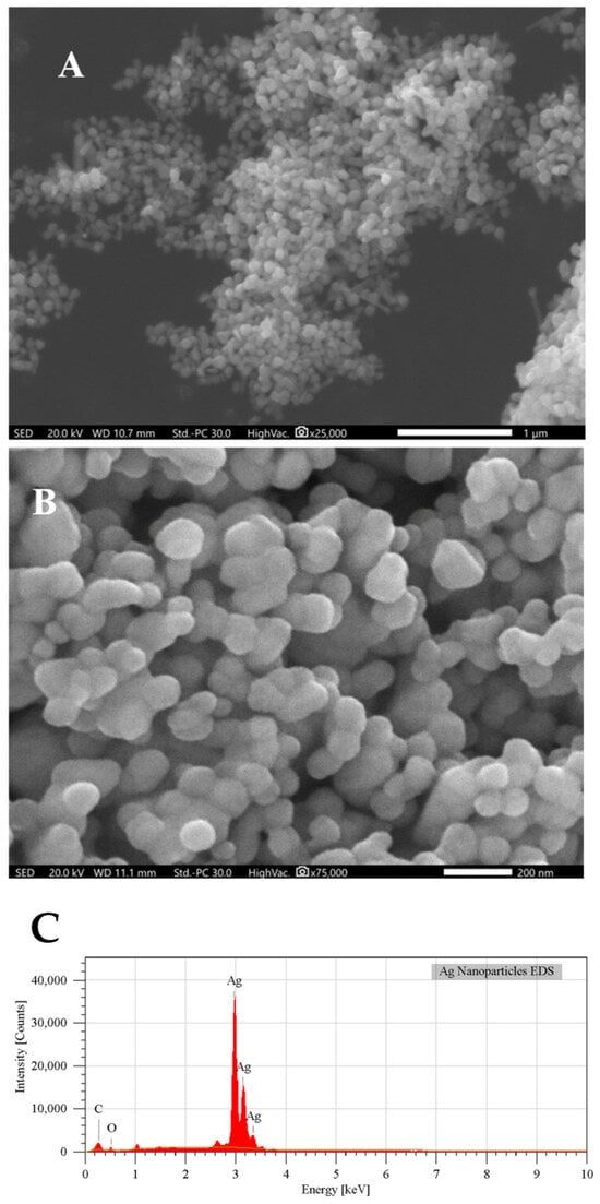
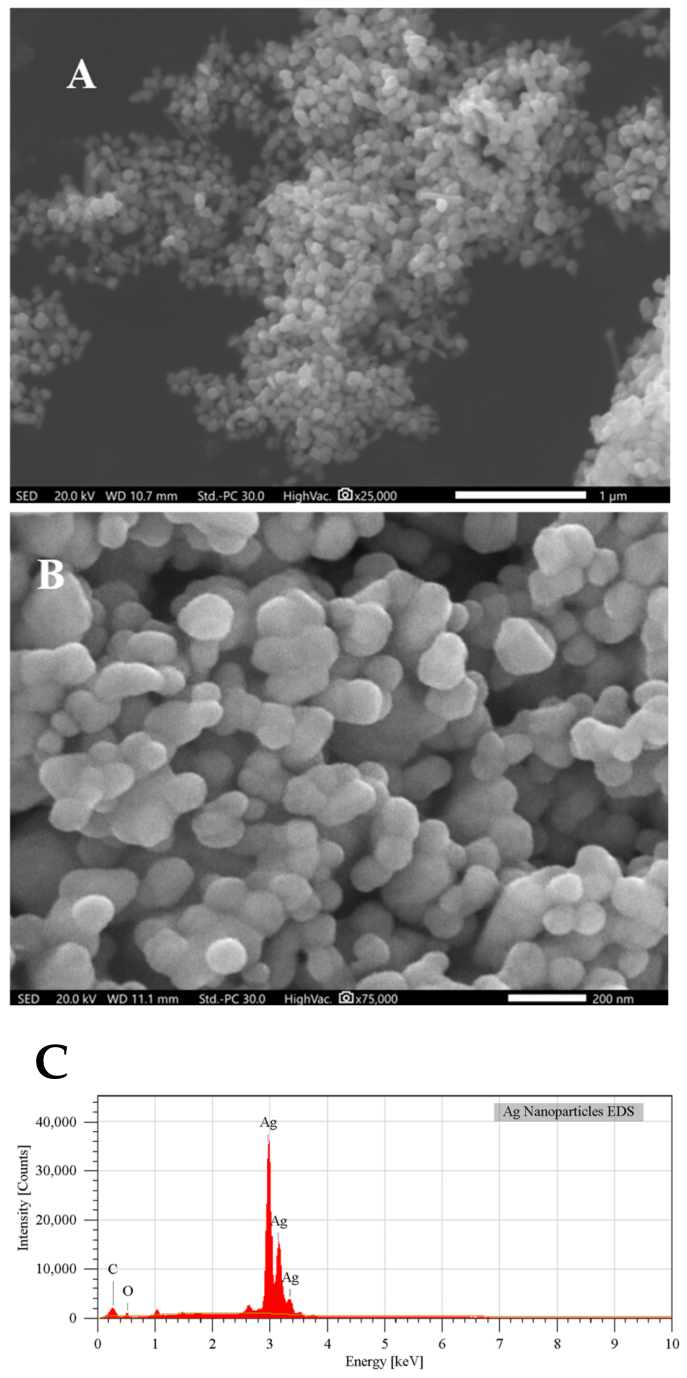
The morphology and elemental composition of the fructose-mediated AgNPs were evaluated by scanning electron microscopy coupled with energy-dispersive X-ray spectroscopy (SEM–EDS), as shown in Figure 1. The lower-magnification SEM image (Figure 1A) reveals the formation of irregular clusters composed of numerous smaller particles. These aggregates range from submicrometer to micrometer scales and are distributed nonuniformly across the analyzed area. The aggregation observed in the dried sample may result from attractive interactions among the nanoparticles during synthesis or solvent evaporation during preparation for SEM analysis. Because we did not perform DLS, zeta potential, or time-dependent stability measurements, the hydrodynamic size, surface charge, and colloidal stability of the AgNPs under the assay conditions remain undetermined. Accordingly, the aggregation observed by SEM should not be taken as a quantitative measure of aggregation in suspension.
Figure 1.
Morphological and elemental characterization of fructose-mediated silver nanoparticles. SEM images acquired at (A) ×25,000 magnification, showing the formation of aggregated particle clusters, and (B) ×75,000 magnification, showing the morphology and extensive interparticle association of the constituent nanostructures. (C) Representative EDS spectrum confirming Ag as the predominant element, with additional signals corresponding to C and O.
At higher magnification (Figure 1B), the aggregates are resolved into individual nanostructured domains with predominantly rounded, quasi-spherical, and irregular morphologies. The particles display a heterogeneous apparent size distribution and extensive interparticle contact, with relatively few completely isolated structures. Some larger features may represent associations of multiple primary particles rather than individual nanoparticles. Although the scale bar confirms the nanometric dimensions of the constituent structures, the substantial overlap and aggregation prevent reliable determination of the primary particle-size distribution from these SEM images alone. Quantitative size measurements would require analysis of a sufficient number of clearly distinguishable particles or complementary high-resolution microscopy.
The EDS spectrum of the synthesized material (Figure 1C) was dominated by the characteristic Ag signals, particularly the intense Ag L-series peaks located near 3 keV. Quantitative analysis indicated a Ag content of 92.92 ± 0.15 mass% and 63.61 ± 0.10 atom%, confirming that silver was the predominant component of the analyzed region. Carbon and oxygen were also detected at 2.41 ± 0.01 and 4.67 ± 0.06 mass%, respectively. Because of their lower atomic masses, their corresponding atomic percentages were comparatively higher, reaching 14.81 ± 0.08 atom% for C and 21.57 ± 0.28 atom% for O.
The simultaneous detection of carbon and oxygen is consistent with oxygen-containing organic species derived from fructose in the AgNP preparation. This interpretation agrees with the FTIR results, which showed the retention of O–H- and C–O-associated vibrational bands in the synthesized material. Collectively, the FTIR and EDS findings support the association of fructose-derived compounds with the nanoparticles, potentially contributing to their surface stabilization. Nevertheless, EDS does not provide information about chemical bonding or molecular identity; consequently, the C and O signals cannot independently confirm the formation of a fructose-derived coating. Carbon contributions from the specimen support and oxygen associated with adsorbed water or surface oxygen-containing species should also be considered.
No additional elements were detected at appreciable levels within the EDS sensitivity, suggesting the absence of major inorganic impurities in the analyzed region. Furthermore, the oxygen signal should not be interpreted by itself as evidence of silver oxide formation. In conjunction with the XRD pattern, in which the characteristic reflections of face-centered cubic Ag0 constituted the predominant crystalline phase, the SEM–EDS results confirm the production of silver-rich nanostructured material containing carbon- and oxygen-bearing surface-associated species.
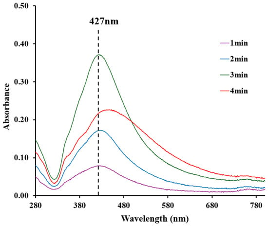
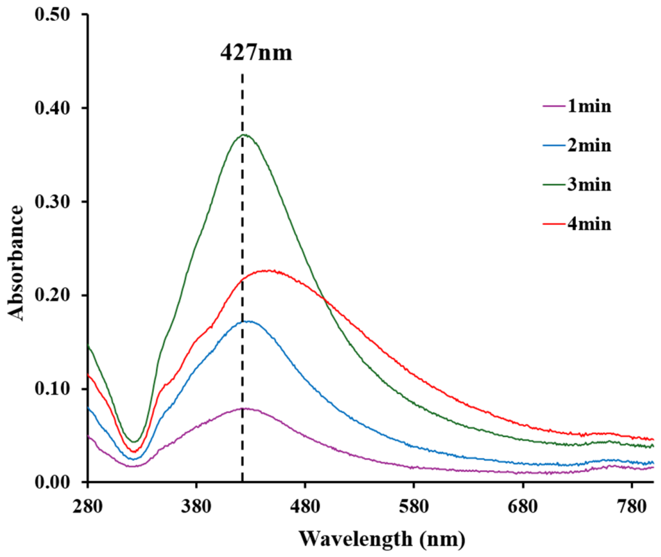
The UV–Vis absorption spectra of Ag nanoparticles synthesized using a 1:2.5 molar ratio of AgNO3 to D-fructose are presented in Figure 2. The spectra show a distinct, well-defined absorption band centered at approximately 427 nm, corresponding to the characteristic surface plasmon resonance (SPR) of silver nanoparticles. This optical feature, typically observed within the 380–450 nm region, is widely associated with the collective oscillation of conduction electrons at the nanoparticle surface and serves as a primary indicator of AgNP formation and nanoscale size distribution [6,7]. The presence of this peak confirms the successful reduction of Ag+ ions and the formation of metallic silver nanoparticles under the applied synthesis conditions.
Figure 2.
UV-Vis spectra of Ag nanoparticles.
Among the evaluated conditions, a heating time of three minutes produced the narrowest and most symmetric SPR band, possibly indicating the formation of small, spherical, and relatively monodispersed nanoparticles. Narrow SPR bands are commonly associated with uniform particle size distributions and effective surface stabilization, suggesting that the reduction of silver ions by D-fructose was most efficient under these conditions [7]. This result further implies that the chosen synthesis parameters enable a homogeneous nanoparticle population with well-defined optical properties.
In contrast, samples subjected to longer heating times (4 min or more) exhibit slight spectral broadening of the SPR band, indicating changes in nanoparticle size distribution and possible interparticle interactions. Broadening and minor red-shifting of plasmon peaks have been widely attributed to particle agglomeration and increased size heterogeneity, as observed in polyol-synthesized silver nanoparticles under extended reaction conditions [5]. These effects suggest that prolonged heating may promote secondary growth processes or partial aggregation, reducing the uniformity achieved after 3 min of heating under the conditions tested.
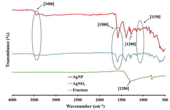
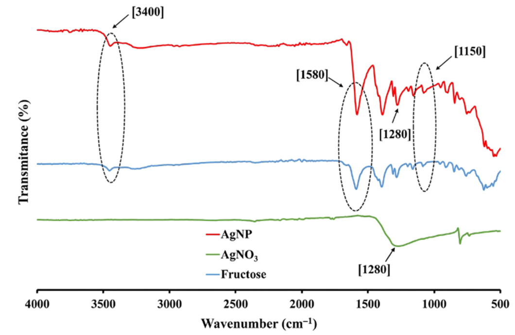
The interaction of the synthesized AgNPs with fructose-derived functional groups was evaluated by FTIR spectroscopy through comparison with the spectra of the D-fructose and AgNO3 precursors (Figure 3). The spectrum of D-fructose displayed a broad band centered at approximately 3400 cm−1, attributed to O–H stretching vibrations, together with several signals in the fingerprint region. In particular, the bands between approximately 1450 and 1000 cm−1 can be associated with C–H and O–H deformation modes and C–O and C–C stretching vibrations characteristic of carbohydrate structures [13,14,15]. In contrast, the AgNO3 spectrum was dominated by a broad absorption in the approximately 1450–1250 cm−1 region, consistent with nitrate vibrations, with an additional feature at lower wavenumbers.
Figure 3.
FTIR spectra of Ag nanoparticles, AgNO3, and D-fructose.
The AgNP spectrum retained the broad O–H absorption and several signals within the 1600–1000 cm−1 region. The persistence of these fructose-related features indicates that oxygen-containing organic species remained associated with the recovered nanoparticles after synthesis. Nevertheless, noticeable changes in the positions and relative profiles of the bands near approximately 1580, 1400, 1280, 1150, and 1070 cm−1 were observed compared with the fructose precursor. These spectral changes suggest modification of the chemical environment of the hydroxyl and C–O-containing groups during nanoparticle formation. Similar retention and modification of O–H and C–O vibrational bands have been reported for AgNPs synthesized using carbohydrate-based reducing agents and plant-derived compounds, where surface-associated organic species contribute to nanoparticle stabilization [16,17,18]. Accordingly, the present FTIR results support the association of fructose-derived compounds with the AgNP surface and their possible contribution as stabilizing or capping species.
The features observed in the approximately 1660–1580 cm−1 region may include contributions from adsorbed water and carbonyl- or carboxylate-containing species formed during the reaction. Likewise, the band near 1400 cm−1 may contain overlapping contributions from fructose-derived groups and residual nitrate. Because these vibrational modes overlap, the chemical transformation of fructose and the extent of precursor removal cannot be established conclusively from the present spectra. In particular, the absence of a clearly resolved new carboxyl-related band does not exclude partial oxidation of fructose, since low-abundance oxidation products may overlap with other bands in this region [15]. Overall, the FTIR analysis supports the presence of fructose-derived oxygen-containing species associated with the synthesized AgNPs, whereas the formation of crystalline metallic Ag0 is more directly demonstrated by the XRD results.
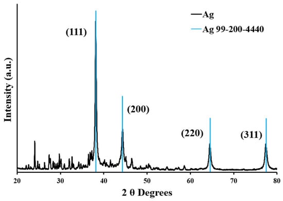
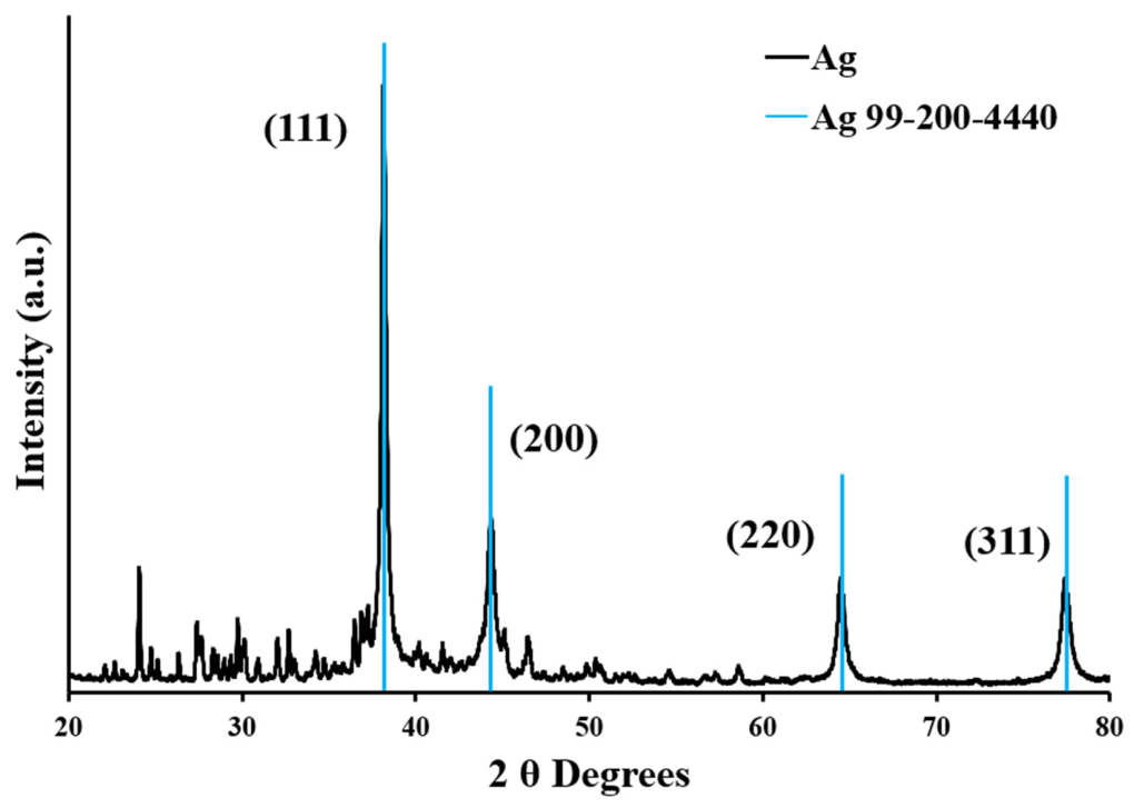
The X-ray diffraction (XRD) pattern of the fructose-mediated AgNPs is presented in Figure 4. The major diffraction peaks observed at approximately 38.1°, 44.3°, 64.4°, and 77.4° (2θ) were indexed to the (111), (200), (220), and (311) crystallographic planes, respectively. These reflections are consistent with the face-centered cubic (fcc) structure of metallic silver and agree with the reference pattern PDF No. 99-200-4440 and previously reported data for green-synthesized AgNPs [19,20,21]. Therefore, the XRD results provide clear evidence of crystalline Ag0 nanoparticle formation under the applied synthesis conditions.
Figure 4.
X-ray diffraction pattern of silver nanoparticles synthesized using fructose.
The (111) reflection exhibited the highest intensity, in agreement with the characteristic diffraction pattern of fcc silver. The broadening of the principal Ag reflections is also consistent with nanoscale crystalline domains. Several weak reflections were observed below approximately 35° (2θ). Their origin could not be established from the available XRD data; minor contributions from silver oxide or residual crystalline salts cannot be excluded. Thus, while fcc metallic Ag is the predominant identified crystalline phase, the presence of minor additional phases remains unresolved.
The average crystallite size was estimated as 24.7 ± 6.3 nm using the Debye–Scherrer equation. This value represents the approximate size of the coherently diffracting crystalline domains rather than necessarily the physical diameter of the nanoparticles. Because this estimate was obtained by XRD analysis of the dried sample, it does not establish the hydrodynamic diameter or aggregation state of the AgNPs in suspension. Dynamic light scattering (DLS) measurements were not performed; therefore, the particle size distribution in suspension could not be determined or compared with the XRD-derived crystallite size. This is a limitation of the present study. The estimated size falls within the range commonly reported for green-synthesized silver nanoparticles, for which crystallite dimensions can vary considerably depending on the reducing agent, synthesis conditions, and surface-stabilizing components [21,22].
3.2. Antimicrobial Properties
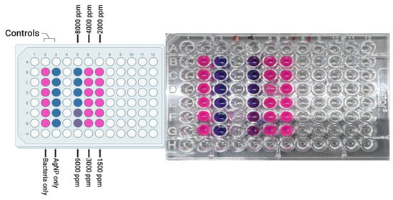
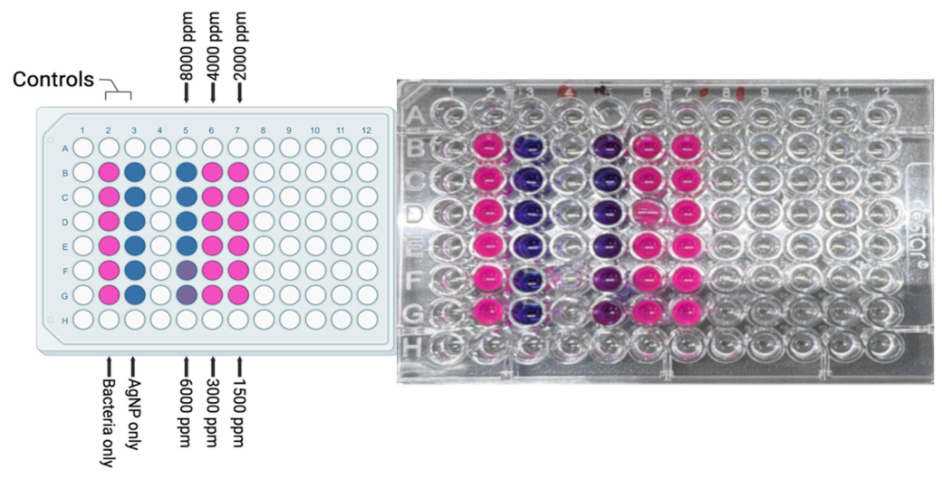
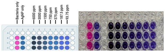
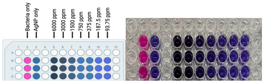
The antimicrobial activity of the fructose-mediated AgNPs was evaluated against Escherichia coli ATCC 25922 and Salmonella typhimurium ATCC 14028 using a resazurin-based broth microdilution assay. Resazurin serves as a metabolic indicator that changes from blue to pink when metabolically active bacterial cells reduce it. Accordingly, retention of the blue or purple coloration was interpreted as inhibition of bacterial metabolic activity, whereas development of a pink coloration indicated bacterial growth. Because AgNP suspensions may affect the color and appearance of the wells, we included bacteria-only and nanoparticle-only controls and evaluated the inhibitory results by agar subculture. The antimicrobial activity was assessed in technical triplicates for each nanoparticle concentration and bacterial strain.
The resazurin assay showed a concentration-dependent inhibitory response for both bacterial strains (Figure 5 and Figure 6). E. coli exhibited a minimum inhibitory concentration (MIC) of 375 ppm, whereas Salmonella typhimurium showed an MIC of 4000 ppm (Table 1). Thus, under the conditions evaluated, a substantially lower AgNP concentration was required to inhibit the metabolic activity of E. coli than that of Salmonella typhimurium.
Figure 5.
Resazurin-based broth microdilution assay of fructose-mediated AgNPs against Salmonella typhimurium. AgNP concentrations ranging from 1500 to 8000 ppm were evaluated. Bacteria-only and AgNP-only wells were included as controls. Pink coloration indicates bacterial metabolic activity, whereas retention of blue or purple coloration indicates inhibition of resazurin reduction.
Figure 6.
Resazurin-based broth microdilution assay of fructose-mediated AgNPs against Escherichia coli. AgNP concentrations ranging from 93.75 to 6000 ppm were evaluated using twofold serial dilutions. Bacteria-only and AgNP-only wells were included as controls. Pink coloration indicates bacterial metabolic activity, whereas retention of blue or purple coloration indicates inhibition of resazurin reduction.
Table 1.
Minimum inhibitory concentration (MIC), minimum bactericidal concentration (MBC), and MBC/MIC ratio of fructose-mediated AgNPs against Escherichia coli and Salmonella typhimurium.
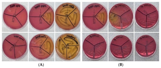
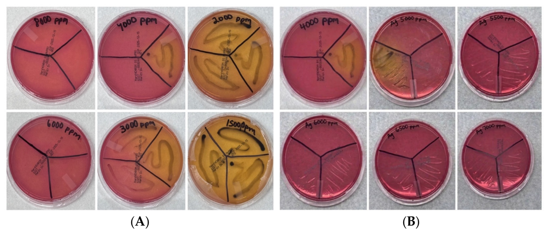
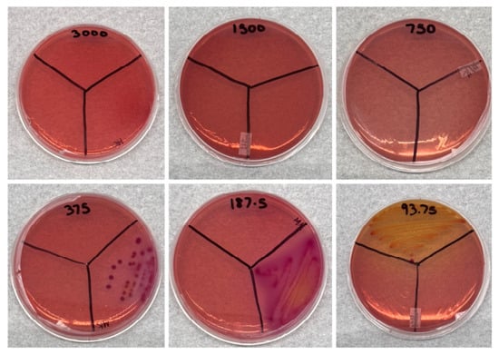
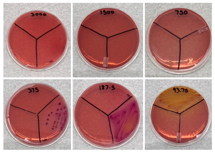
The minimum bactericidal concentration (MBC) was determined by subculturing samples from the microdilution assay onto selective agar. For Salmonella typhimurium, the initial assessment on Salmonella–Shigella agar showed bacterial growth at the lower AgNP concentrations and no detectable growth at the highest concentrations tested (Figure 7A). A subsequent evaluation using intermediate concentrations between 4000 and 7000 ppm more precisely defined the bactericidal endpoint (Figure 7B), resulting in an MBC of 5500 ppm. For E. coli, growth was observed on MacConkey agar following exposure to 375 ppm and lower concentrations, whereas no visible colonies were detected following exposure to 750 ppm (Figure 8). Therefore, the MBC for E. coli was established at 750 ppm.
Figure 7.
Determination of the minimum bactericidal concentration of fructose-mediated AgNPs against Salmonella typhimurium by subculture on Salmonella–Shigella agar. (A) Initial evaluation of AgNP concentrations ranging from 1500 to 8000 ppm. (B) Subsequent evaluation of intermediate concentrations ranging from 4000 to 7000 ppm. The MBC was established as the lowest tested concentration producing no visible colony growth after incubation.
Figure 8.
Determination of the minimum bactericidal concentration of fructose-mediated AgNPs against Escherichia coli by subculture on MacConkey agar. AgNP concentrations ranging from 93.75 to 3000 ppm were evaluated. The MBC was established as the lowest tested concentration producing no visible colony growth after incubation.
The resulting MBC/MIC ratios were 2.0 for E. coli and 1.4 for Salmonella typhimurium (Table 1). Because both ratios were below 4, the activity of the fructose-mediated AgNPs was classified as bactericidal under the experimental conditions. Nevertheless, E. coli was more susceptible to the AgNP preparation, displaying MIC and MBC values approximately 10.7- and 7.3-fold lower, respectively, than those obtained for Salmonella typhimurium.
Both microorganisms are Gram-negative bacteria and possess a cell envelope comprising an inner membrane, a thin peptidoglycan layer, and an outer membrane containing lipopolysaccharides [22]. This outer membrane acts as a selective permeability barrier and can influence bacterial susceptibility to antimicrobial materials. AgNPs have been reported to exert antimicrobial effects through multiple, potentially concurrent processes, including interaction with the bacterial surface, alteration of membrane integrity, release of Ag+ ions, induction of oxidative stress, and interaction with intracellular proteins and nucleic acids [5,22,23].
The different susceptibility observed between the two bacterial strains may reflect species-dependent differences in cell-envelope composition, surface properties, silver-ion homeostasis, or cellular stress-response mechanisms. These factors could influence AgNP association with the bacterial surface and the cellular response to nanoparticle-associated silver. However, because the present study did not directly evaluate membrane disruption, Ag+ release, reactive oxygen species generation, or intracellular interactions, the specific mechanism responsible for the observed difference cannot be established. The results therefore demonstrate differential susceptibility under the tested conditions, with E. coli showing greater sensitivity to the fructose-mediated AgNP preparation than Salmonella typhimurium.
The lower MIC and MBC values observed for E. coli in this study suggest that its outer membrane structure may allow more effective interaction with, or penetration by, Ag nanoparticles and Ag+ ions than in S. typhimurium. This would result in a higher effective intracellular dose at lower external concentrations, leading to enhanced membrane damage and bactericidal activity. In contrast, S. typhimurium may possess slightly more protective envelope characteristics, such as differences in LPS structure, membrane proteins, or stress-response mechanisms, requiring higher nanoparticle concentrations to achieve similar levels of cellular damage. Similar trends have been reported in nanoparticle studies, where even among Gram-negative bacteria, species-specific variations in cell envelope composition result in differences in antimicrobial susceptibility [22].
Because the study did not include an AgNO3 control matched to the silver content of the AgNP preparation or measurements of dissolved silver, the relative contributions of particulate silver and released Ag+ to the observed antimicrobial activity cannot be determined. In addition, the antimicrobial assays were performed in technical triplicate without independent biological replicates. Therefore, the results demonstrate within-assay repeatability but do not establish reproducibility across independently prepared bacterial cultures.
3.3. Experimental Considerations
The antimicrobial assay used an approximate final bacterial concentration of 5 × 10⁷ CFU mL−1, which is higher than the inoculum typically used in standardized broth microdilution susceptibility testing. Therefore, the antimicrobial concentrations reported in this study should be interpreted as resazurin-based inhibitory endpoints under the experimental conditions evaluated rather than as directly comparable CLSI-standardized MIC values. The resazurin assay provides a qualitative indication of detectable bacterial metabolic activity and does not directly quantify the viable bacterial population.
Antimicrobial activity was evaluated using technical triplicates for each nanoparticle concentration and bacterial strain without independent biological replicates. Therefore, the results reflect within-assay repeatability rather than biological variability. The consistent response among technical replicates supports the reproducibility of the observed resazurin-based endpoints within the assay but does not constitute independent biological confirmation. Future studies incorporating independent experiments, standardized inoculum conditions, and quantitative measurements of bacterial viability will be required to further validate the antimicrobial activity of the fructose-mediated AgNPs.
4. Conclusions
This study demonstrated the rapid, single-step synthesis of silver nanoparticles using D-fructose in an aqueous medium. The AgNPs formed within 3 min in an aqueous medium, offering a simple alternative to conventional synthesis methods that require more complex procedures or potentially hazardous reagents. UV–Vis spectroscopy showed a characteristic surface plasmon resonance band at 427 nm, supporting nanoparticle formation. XRD analysis confirmed that face-centered cubic metallic silver was the predominant crystalline phase, with an apparent crystallite size of approximately 24.7 nm. Minor unassigned reflections were also observed and may originate from residual crystalline components of the synthesis medium, although their identity could not be established from the available pattern.
SEM analysis revealed nanometric structures with predominantly rounded and irregular morphologies assembled into larger aggregates. EDS confirmed that Ag was the principal component of the analyzed material, accounting for 92.92 mass% and 63.61 atom%. The additional detection of C and O, together with the retention and modification of O–H- and C–O-associated bands in the FTIR spectrum, supports the presence of fructose-derived organic species associated with the synthesized nanoparticles. These species may contribute to surface stabilization; however, their chemical identity and specific interaction with the AgNP surface require further investigation.
The fructose-mediated AgNPs exhibited bactericidal activity against both tested Gram-negative bacteria. Escherichia coli ATCC 25922 displayed MIC and MBC values of 375 and 750 ppm, respectively, whereas Salmonella typhimurium ATCC 14028 exhibited corresponding values of 4000 and 5500 ppm. The resulting MBC/MIC ratios of 2.0 and 1.4 were consistent with bactericidal activity under the experimental conditions. The lower MIC and MBC values for E. coli demonstrate greater susceptibility to the AgNP preparation than Salmonella typhimurium.
Overall, the findings establish a rapid, aqueous, and fructose-mediated approach for producing crystalline, silver-rich nanostructures with antimicrobial activity. Further studies addressing colloidal stability, primary particle-size distribution, silver-ion release, antimicrobial mechanisms, cytotoxicity, coating performance, silver migration, and performance in food-relevant matrices are needed to determine the suitability of these materials for antimicrobial coatings and food-safety applications.
Author Contributions
Conceptualization, M.R.-B.; Methodology, M.R.-B. and D.C.-L.; Validation, M.R.-B.; Formal analysis, M.R.-B., D.C.-L. and S.J.B.-R.; Investigation, D.C.-L.; Resources, M.R.-B.; Writing—original draft, M.R.-B. and S.J.B.-R.; Writing—review and editing, M.R.-B. and S.J.B.-R.; Visualization, D.C.-L.; Supervision, M.R.-B. and S.J.B.-R.; Project administration, M.R.-B. and S.J.B.-R.; Funding acquisition, M.R.-B. and S.J.B.-R. All authors have read and agreed to the published version of the manuscript.
Funding
This research received external funding (NIFA/USDA 2026-67012-45562 Award).
Data Availability Statement
The data presented in this study are available in this article.
Acknowledgments
The authors gratefully acknowledge the Department of Chemistry and Physics at the University of Puerto Rico at Ponce for its support of this work. The authors also thank the Department of Mathematics and Physics at the University of Puerto Rico at Cayey, particularly Danilo Barrionuevo-Diestra, for his assistance with X-ray diffraction characterization (NSF grant 2215247).
Conflicts of Interest
The authors declare no conflicts of interest.
References
- Vega Quiñones, E.M.; Quiñones Justiniano, G.; Dudley, E.G.; M’ikanatha, N.M.; Malavez, Y. Prevalence and antimicrobial resistance of nontyphoidal Salmonella in retail meat in Puerto Rico: A two-year study (2022–2023). J. Food Prot. 2025, 88, 100589. [Google Scholar] [CrossRef] [Scilit] [PubMed]
- Walter, E.J.S.; Cui, Z.; Tierney, R.; Griffin, P.M.; Hoekstra, R.M.; Payne, D.C.; Rose, E.B.; Devine, C.; Namwase, A.S.; Mirza, S.A.; et al. Foodborne Illness Acquired in the United States—Major Pathogens, 2019. Emerg. Infect. Dis. 2025, 31, 669–677. [Google Scholar] [CrossRef] [Scilit] [PubMed]
- Quiñones Justiniano, G.; Vega Quiñones, E.M.; Dudley, E.G.; M’ikanatha, N.M.; Malavez, Y. Antimicrobial resistance profiles of Campylobacter spp. isolated from retail meat in Puerto Rico. Microbiol. Resour. Announc. 2025, 14, e00821-25. [Google Scholar] [CrossRef] [Scilit] [PubMed]
- Helmy, Y.A.; Taha-Abdelaziz, K.; Hawwas, H.A.E.-H.; Ghosh, S.; AlKafaas, S.S.; Moawad, M.M.M.; Saied, E.M.; Kassem, I.I.; Mawad, A.M.M. Antimicrobial Resistance and Recent Alternatives to Antibiotics for the Control of Bacterial Pathogens with an Emphasis on Foodborne Pathogens. Antibiotics 2023, 12, 274. [Google Scholar] [CrossRef] [Scilit] [PubMed]
- Reyes-Blas, G.-M.; Maldonado-Luna, N.M.; Rivera-Quiñones, C.M.; Vega-Avila, A.L.; Roman-Velázquez, F.R.; Perales-Perez, O.J. Single Step Microwave Assisted Synthesis and Antimicrobial Activity of Silver, Copper and Silver-Copper Nanoparticles. J. Mater. Sci. Chem. Eng. 2020, 8, 13–29. [Google Scholar] [CrossRef]
- Hemmati, S.; Retzlaff-Roberts, E.; Scott, C.; Harris, M.T. Artificial sweeteners and sugar ingredients as reducing agent for green synthesis of silver nanoparticles. J. Nanomater. 2019, 2019, 9641860. [Google Scholar] [CrossRef] [Scilit]
- González Fá, A.J.; Juan, A.; Di Nezio, M.S. Synthesis and Characterization of Silver Nanoparticles Prepared with Honey: The Role of Carbohydrates. Anal. Lett. 2017, 50, 877–888. [Google Scholar] [CrossRef] [Scilit]
- Gallegos, F.E.; Meneses, L.M.; Cuesta, S.A.; Santos, J.C.; Arias, J.; Carrillo, P.; Pilaquinga, F. Computational Modeling of the Interaction of Silver Clusters with Carbohydrates. ACS Omega 2022, 7, 4750–4756. [Google Scholar] [CrossRef] [Scilit] [PubMed]
- Loo, Y.Y.; Rukayadi, Y.; Nor-Khaizura, M.-A.; Kuan, C.H.; Chieng, B.W.; Nishibuchi, M.; Radu, S. In Vitro antimicrobial activity of green synthesized silver nanoparticles against selected Gram-negative foodborne pathogens. Front. Microbiol. 2018, 9, 1555. [Google Scholar] [CrossRef] [Scilit] [PubMed]
- Ivanova, A.; Todorova, M.; Petrov, D.; Gledacheva, V.; Stefanova, I.; Milusheva, M.; Slavchev, V.; Kostadinova, G.; Petkova, Z.; Teneva, O.; et al. Biogenic Synthesis, Structural Characterization, and Biological Evaluation of Nanoparticles Derived from Chlorella vulgaris Ethanolic Extract. Nanomaterials 2026, 16, 177. [Google Scholar] [CrossRef] [Scilit] [PubMed]
- Alsammarraie, F.K.; Wang, W.; Zhou, P.; Mustapha, A.; Lin, M. Green synthesis of silver nanoparticles using turmeric extracts and investigation of their antibacterial activities. Colloids Surf. B Biointerfaces 2018, 171, 398–405. [Google Scholar] [CrossRef] [Scilit] [PubMed]
- Soto, K.M.; Quezada-Cervantes, C.T.; Hernández-Iturriaga, M.; Luna-Bárcenas, G.; Vazquez-Duhalt, R.; Mendoza, S. Fruit peels waste for the green synthesis of silver nanoparticles with antimicrobial activity against foodborne pathogens. LWT 2019, 103, 293–300. [Google Scholar] [CrossRef] [Scilit]
- Arshadi, E.; Sedaghat, S.; Moradi, O. Green synthesis and characterization of silver nanoparticles using fructose. Asian J. Green Chem. 2018, 2, 41–50. [Google Scholar] [CrossRef] [Scilit]
- Mehtab, S.; Zaidi, M.G.H.; Siddiqi, T.I. Designing Fructose Stabilized Silver Nanoparticles for Mercury(II) Detection and Potential Antibacterial Agents. Mater. Sci. Res. India 2018, 15, 241–249. [Google Scholar] [CrossRef] [Scilit]
- Pasieczna-Patkowska, S.; Cichy, M.; Flieger, J. Application of Fourier transform infrared (FTIR) spectroscopy in characterization of green synthesized nanoparticles. Molecules 2025, 30, 684. [Google Scholar] [CrossRef] [Scilit] [PubMed]
- Ullah, S.; Khalid, R.; Rehman, M.F.; Irfan, M.I.; Abbas, A.; Alhoshani, A.; Anwar, F.; Amin, H.M.A. Biosynthesis of phyto-functionalized silver nanoparticles using olive fruit extract and evaluation of their antibacterial and antioxidant properties. Front. Chem. 2023, 11, 1202252. [Google Scholar] [CrossRef] [Scilit] [PubMed]
- Moldovan, B.; Sincari, V.; Perde-Schrepler, M.; David, L. Biosynthesis of silver nanoparticles using Ligustrum ovalifolium fruits and their cytotoxic effects. Nanomaterials 2018, 8, 627. [Google Scholar] [CrossRef] [Scilit] [PubMed]
- Mustapha, T.; Ithnin, N.R.; Othman, H.; Abu Hasan, Z.I.; Misni, N. Bio-fabrication of silver nanoparticles using Citrus aurantifolia fruit peel extract (CAFPE) and the role of plant extract in the synthesis. Plants 2023, 12, 1648. [Google Scholar] [CrossRef] [Scilit] [PubMed]
- Ali, M.H.; Azad, M.A.K.; Khan, K.A.; Rahman, M.O.; Chakma, U.; Kumer, A. Analysis of crystallographic structures and properties of silver nanoparticles synthesized using PKL extract and nanoscale characterization techniques. ACS Omega 2023, 8, 28133–28142. [Google Scholar] [CrossRef] [Scilit] [PubMed]
- Ohiduzzaman, M.; Khan, M.N.I.; Khan, K.A.; Paul, B. Biosynthesis and characterizations of silver nanoparticles by using green banana peel extract: Evaluation of their antibacterial and electrical performances. Heliyon 2024, 10, e31140. [Google Scholar] [CrossRef] [Scilit] [PubMed]
- Asif, M.; Yasmin, R.; Asif, R.; Ambreen, A.; Mustafa, M.; Umbreen, S. Green synthesis of silver nanoparticles (AgNPs), structural characterization, and their antibacterial potential. Dose-Response 2022, 20, 15593258221088709. [Google Scholar] [CrossRef] [Scilit] [PubMed]
- Das, B.; Dash, S.K.; Mandal, D.; Ghosh, T.; Chattopadhyay, S.; Tripathy, S.; Das, S.; Dey, S.K.; Das, D.; Roy, S. Green synthesized silver nanoparticles destroy multidrug resistant bacteria via reactive oxygen species mediated membrane damage. Arab. J. Chem. 2017, 10, 862–876. [Google Scholar] [CrossRef] [Scilit]
- Quaranta, D.; Krans, T.; Santo, C.E.; Elowsky, C.G.; Domaille, D.W.; Chang, C.J.; Grass, G. Mechanisms of contact-mediated killing of yeast cells on dry metallic copper surfaces. Appl. Environ. Microbiol. 2011, 77, 416–426. [Google Scholar] [CrossRef] [Scilit] [PubMed]

Disclaimer/Publisher’s Note: The statements, opinions and data contained in all publications are solely those of the individual author(s) and contributor(s) and not of MDPI and/or the editor(s). MDPI and/or the editor(s) disclaim responsibility for any injury to people or property resulting from any ideas, methods, instructions or products referred to in the content. |
© 2026 by the authors. Licensee MDPI, Basel, Switzerland. This article is an open access article distributed under the terms and conditions of the Creative Commons Attribution (CC BY) license.