


Abstract
This pilot study aimed to identify and characterize particles of microplastics (MPs) and other potentially harmful compounds excreted in the urine of children undergoing surgery at a free-standing children’s hospital in the United States. Procedural blank controls of molecular-grade water were collected in an identical manner to the urine samples to account for background environmental contamination. Specimens underwent digestion to remove organic components, followed by vacuum filtration through quartz microfiber filters with 1 micron pore size. Particles captured on the filters were characterized with Raman microspectroscopy. A total of 11 urine samples from 11 patients were analyzed, with a total of 112 particles detected, including MPs polymers and non-polymers (minerals/metal oxides, pigments, and natural fibers). Urine sample MP particle concentrations were significantly elevated after correction for background contamination with procedural blank controls (median 80 particles/L, p = 0.0078). Of the 112 particles characterized, 38 (33.9%) were synthetic plastic polymers (i.e., MPs), 57 (50.9%) were non-polymers, and 17 (15.2%) were complexes of polymers with non-polymers. The most common polymer was polyethylene/polypropylene co-polymer (14/38, 37%) and the non-polymer was hematite (23/57, 40%). Distribution of specific polymers differed in urine samples versus controls, with polyethylene being significantly more prevalent in samples (p = 0.05). Pigments identified across multiple samples included carbon black, cromophtal blue/violet, and irisol blue. Nearly one in six urine particles consisted of complexes of polymers with industrial additives and pigments, raising further concern for MPs acting as pathologic carriers of other harmful compounds. Future investigations should explore the association of MPs and additives with clinical diseases in children.
1. Introduction
Over 350 million tons of plastic is produced globally every year; however, less than ten percent of that plastic is recycled [1]. Plastic waste in the environment breaks down into small particles, with plastic particles between 1 micron and 0.5 cm typically termed microplastics (MPs), although there are varying definitions based on particle size [2]. First described in marine settings, MPs are now known to be ubiquitous in Earth’s environments [3].
Humans ingest substantial quantities of MPs on an annual basis [4,5] and MPs may be an emerging threat to human health. Microplastics have recently been associated with negative clinical outcomes, including cardiovascular events and dementia in adults [6,7] and behavioral problems in children [8]. Studies of MPs in children have been limited by potential environmental cross-contamination and unclear generalizability [9]. Microplastics have been shown to penetrate cell membranes and have been detected in several human organs and body fluids, including human kidney and urine [10,11]. However, most studies of MPs to date have focused on adults. Children may also be exposed to a variety of other toxic substances, including industrial additives, from their environments [12,13]. The way in which these other substances may interact with MPs has not been well-defined in children.
To elucidate the potential impact of MPs on child health, MPs must first be rigorously identified and characterized in the pediatric population. This pilot study aimed to detect and characterize MPs and other additives in the urine of pediatric surgical patients, as a preliminary step toward larger pediatric studies.
2. Materials and Methods
2.1. Subject Inclusion/Exclusion Criteria
The study recruited children of any age who received elective pediatric surgical consultation in the outpatient setting at a free-standing children’s hospital in the U.S. state of Florida, between October 2024 and May 2025, under an IRB-approved protocol (IRB00393797). Exclusion criteria were weight less than 2 kg or inability to provide a urine sample.
2.2. Clinical Data Collection
Clinical data were collected on study subjects through a chart review and questionnaire. Data points included demographics, dietary restrictions, and two geographic social determinants of health (SDOH), the Social Vulnerability Index (SVI) and Environmental Justice Index (EJI). The SVI and EJI have been utilized in multiple prior studies of child health [14,15]. These variables were collected for descriptive purposes only, recognizing that the small sample size of this analytical pilot study would be insufficient for formal statistical comparisons. Therefore, our pilot study does not provide enough exposure data to explain possible sources of microplastics in the children.
2.3. Sample Collection and Preparation
A strict procedure of sample and control collection was followed to account for any environmental contaminants being shed from the air or patient clothing. Study subjects underwent urine sample collection in either the outpatient clinic/pre-op short stay unit (potty-trained patients) or the operating room (non-potty-trained patients). Samples were collected prior to the performance of any surgical procedure. For potty-trained patients, a plastic-free collection system, consisting of a fenestrated metal bowl with an affixed glass jar, was used. The collection system was first rinsed with deionized water that had been pre-filtered through a 25 mm quartz microfiber filter with 1 micron pore size, then set up on the toilet in the patient’s restroom. The patient then entered the restroom, closed the door, and provided the urine sample. The resulting urine sample of up to 50 mL was then immediately sealed with a metal lid and labeled by the research team. For non-potty-trained patients, the urine sample was collected in the operating room after the patient had undergone induction of anesthesia. The Crede maneuver was used to express a urine sample directly into a glass jar that had been pre-rinsed with filtered deionized molecular-grade water (dH2O).
2.4. Quality Assurance and Procedural Blank Controls
Deionized water was filtered through a 1 micron quartz microfiber filter (Whatman QMA, Cytiva Life Sciences, Danaher Global, Marlborough, MA, USA) using vacuum filter apparatus. The filtered dH2O was stored in pre-rinsed glass containers. After initial filtration, a random 50 mL aliquot of the dH2O was re-filtered and evaluated using Raman microspectroscopy (RMS) as described in the subsequent section to ensure it was free of MPs. For each patient, samples consisting of dH2O were collected in the exact same manner as the patient urine samples to be used as procedural blank controls. For potty-trained patients, a new collection system was rinsed with filtered dH2O in the same restroom environment. With the patient still present and standing close by, the research team poured a volume of dH2O equal to the urine sample into another glass jar. The procedural blank control was immediately sealed with a metal lid and labeled. For non-potty-trained patients, the research team poured an equal volume of dH2O into another glass jar, in the same operating room environment and at the same time. The procedural blank controls described above were used for monitoring the contamination at all steps of the protocol including reagent preparation, digestion, vacuum filtration, filter drying, transport and micro-Raman analysis. The actual sample MP concentrations were obtained by subtracting the procedural control background as described below. Prior to the pilot study, the RMS recovery experiments were performed using 30 µm PS spheres (SigmaAldrich, Milwaukee, WI, USA), which showed 100% detection efficiency. The reagent and filter blanks did not show any detectable MPs.
2.5. Digestion and Filtration
The protocol for specimen processing was modified from prior work [16]. Sealed and labeled samples and controls were immediately delivered to the lab for digestion. Processing of patient and control specimens was performed inside a laminar flow biological safety cabinet to minimize laboratory contamination [17]. A volume of filtered 3% hydrogen peroxide equal to the amount of sample (1:1 solution: sample) was added to the sample and control jars. Samples and controls were incubated in a 40 °C incubator for a minimum of 24 h. After digestion, samples and controls were filtered through 1 micron quartz microfiber filter (Whatman QMA, Cytiva Life Sciences, Danaher Global, Marlborough, MA, USA) using vacuum filter apparatus. The resulting filter was placed in a glass Petri dish, covered, and allowed to dry. Dried filters were transported in glass containers to another lab for RMS analysis.
2.6. Raman Microspectroscopy
Raman data collection was performed using a confocal microscope (XPloRa microscope, Horiba Scientific, Piscataway, NJ, USA) with 532 nm cw laser focused using an objective lens (NA = 0.9). The laser was focused directly on the sample in the confocal Raman measurements. Our system had a ×50 objective and ~0.5 µm spatial resolution. Using a backscattering configuration with a 532 nm edge filter, the backscattered optical signals were collected and detected by a spectrometer with 600 gr/mm grating coupled to an electrically cooled CCD camera. This corresponds to the spectral range of 100–5000 cm−1 and the spectral resolution of ~5 cm−1. The experimental parameters were optimized and adjusted when needed to obtain the high-quality Raman spectra. Typical laser acquisition time was in the range of 1–100 s and the laser power range was 1–10 mW. We used low power for polymers and high power for minerals, as the latter have high thermal stability. We used the averaging of 5–10 spectral acquisitions. Spectral intensity and wavelength calibrations were performed using the built-in calibration protocol of Horiba Scientific based on the Si substrate. Baseline correction was performed by subtracting the 10th order polynomial fitting using a fitting procedure built in the LabSpec6 software version 6.7 (Horiba Scientific, Piscataway, NJ, USA). The Raman analysis procedure was validated using 10 micrometer polystyrene (PS) spheres. All experiments were performed under ambient conditions at room temperature. We checked the sample for thermal damage by optical imaging and by repeated measurements of the Raman signals. No significant sample degradation was observed. Particle polymer and additive composition were identified by comparison with a library of ~10,000 Raman spectra using IDFinder software version 6.7 (Horiba Scientific, Piscataway, NJ, USA), which includes the SLoPP-E spectral library (University of Toronto, Toronto, ON, Canada), and a HQI match rate of >70% was used. Manual verification was additionally performed for ambiguous matches by visually comparing the spectral overlaps of the data with the database spectra. Particle identification was performed visually using optical images.
2.7. Categorization of Particles
Particles of MPs and other anthropogenic materials detected on RMS were categorized into the following three broad categories: (1) polymer (i.e., microplastic particle alone), (2) non-polymer, and (3) polymer + non-polymer complex (microplastic particle complexed with a non-polymer particle). Polymer particles were subcategorized by specific type. Non-polymer particles were subcategorized as mineral/metal oxides, industrial pigments, and natural fibers. The terms “polymer” and “microplastics (MPs)” are used interchangeably in this manuscript. Particles were characterized by shape as either fragments (length:width ratio ≤ 3:1) or fibers (length:width ratio > 3:1) and by size in microns.
2.8. Statistical Analysis
Descriptive statistics were calculated to summarize the study population and particle characteristics. Concentrations of polymer particles were calculated as the number of polymer particles per liter of urine. Sample and control polymer particle concentrations were compared using Wilcoxon rank-sum tests. Because each urine sample had a corresponding procedural blank (paired design), a corrected polymer concentration was calculated for each patient by subtracting the number of procedural blank polymers from the total number of patient sample polymers. The Wilcoxon signed rank test was used to test the hypothesis that the differences between patient sample and procedural blank polymer concentrations were equally distributed around zero (i.e., no difference). Given the focus of this pilot study on microplastics, formal statistical comparisons were limited to polymer particles; other particle types were compared in descriptive fashion only. Limit of detection/limit of quantification (LOD/LOQ) was calculated for each particle type. LOD and LOQ were defined as 3 times and 10 times, respectively, the standard deviation of the particle concentration in procedural blank controls collected in parallel. The distributions of overall particles and specific polymer types were compared for samples versus controls using chi-square or Fisher’s exact tests. For the comparison of specific polymer types in samples versus controls, both individual polymers and polymers complexed with other particles were included. The overall and polymer type distributions were also represented graphically with bar charts and pie charts, respectively. Statistical analysis was performed using SAS 9.4 (SAS Corporation, Cary, NC, USA). Generative artificial intelligence (GenAI) was not used in this paper.
3. Results
3.1. Description of Study Cohort
Twelve children (ages 4–15 years) were enrolled in the study, and one patient’s family withdrew from the study prior to sample collection, citing inconvenience. The characteristics of the eleven patients who had urine samples collected are provided in Table 1.
Table 1.
Description of study patients (n = 11).
All patients had at least one polymer particle detected in their urine sample. Concentration of polymers in the patient samples (median 100 particles/L, interquartile range 80–120) exceeded procedural blank controls (median 40 particles/L, IQR 20–40), with the difference being statistically significant (p = 0.0035, Wilcoxon rank-sum test). When the patient concentrations were corrected for paired procedural blanks, the corrected polymer concentration was 80 particles/L. The corrected polymer concentration differed significantly from zero on the Wilcoxon signed-rank test (p = 0.0078). LOD/LOQ in particles/L was calculated as 68/226 for polymers, 245/817 for additives, 302/1007 for pigments, and 152/505 for complexes of different particle types. Six patients (6/11, 55%) had polymer particle concentrations greater than the LOD in the urine sample.
3.2. Description of Particles
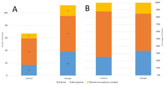
Table 2 provides details regarding all 112 particles identified in pediatric urine samples. Non-polymers were the most common type of particle identified (n = 57, 50.9%), followed by plastic polymers (i.e., MPs, n = 38, 33.9%) and polymer-additive complexes (n = 17, 15.2%). A total of 15 particles contained industrial pigments, either alone (n = 9, 8.0%) or complexed with a polymer (n = 6, 5.4%). The most common particle overall was the additive hematite, followed by polyethylene/polypropylene co-polymer. Most particles (91%) had a fragment shape; nine (8%) had a fiber shape and one (<1%) was considered non-classifiable due to shape irregularity. Most particles were in the 10–100 μm size range. The smallest particle detected was a 2 μm polypropylene particle and the largest was an 838 μm polyethylene–polypropylene–corundum fiber.
Table 2.
Particles (n = 112) identified in pediatric urine samples.
3.3. Photographs, Raman Spectra, and Comparison of Particle Types
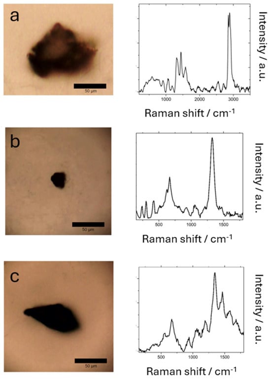
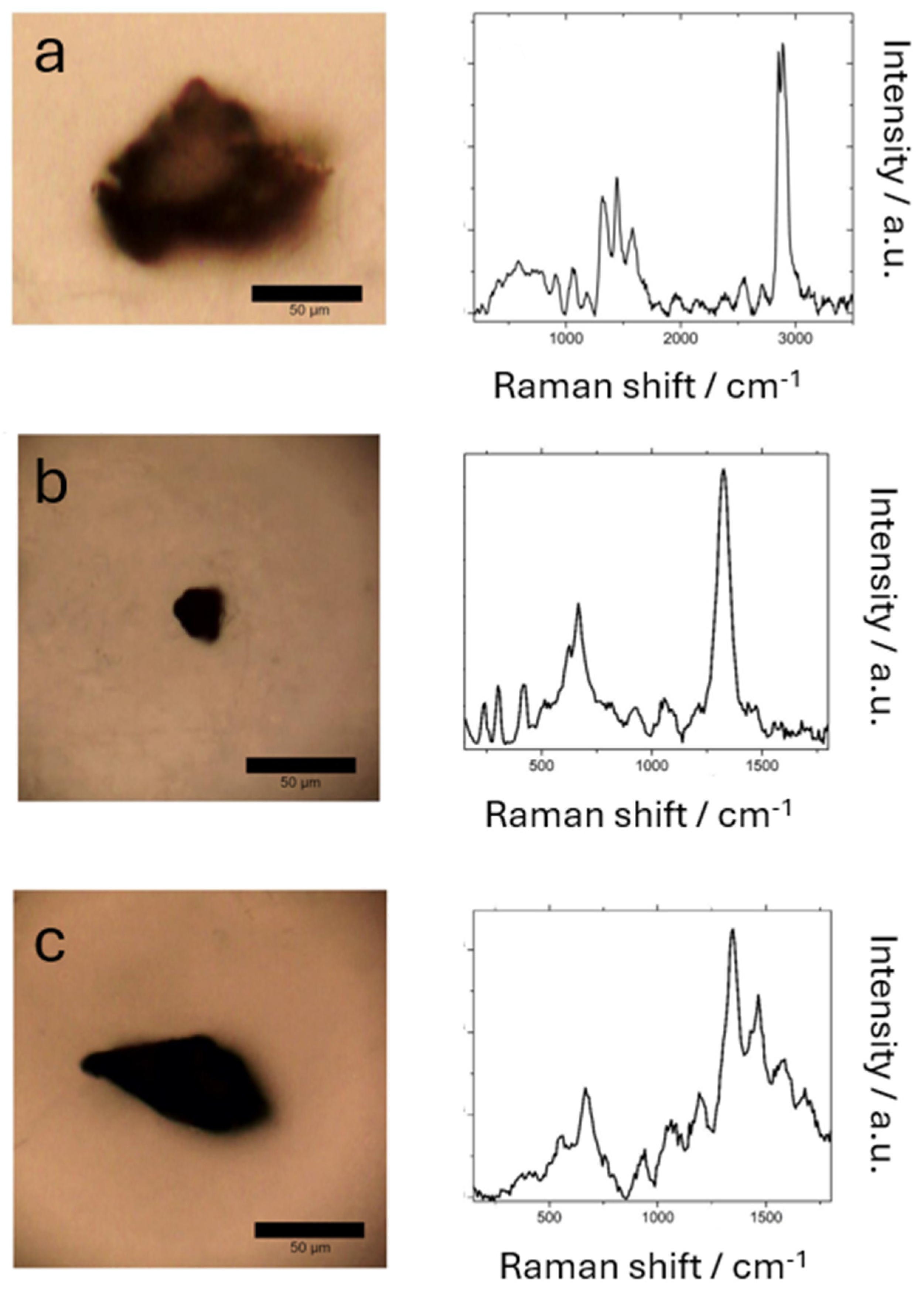
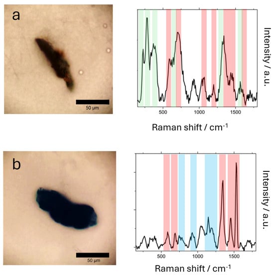
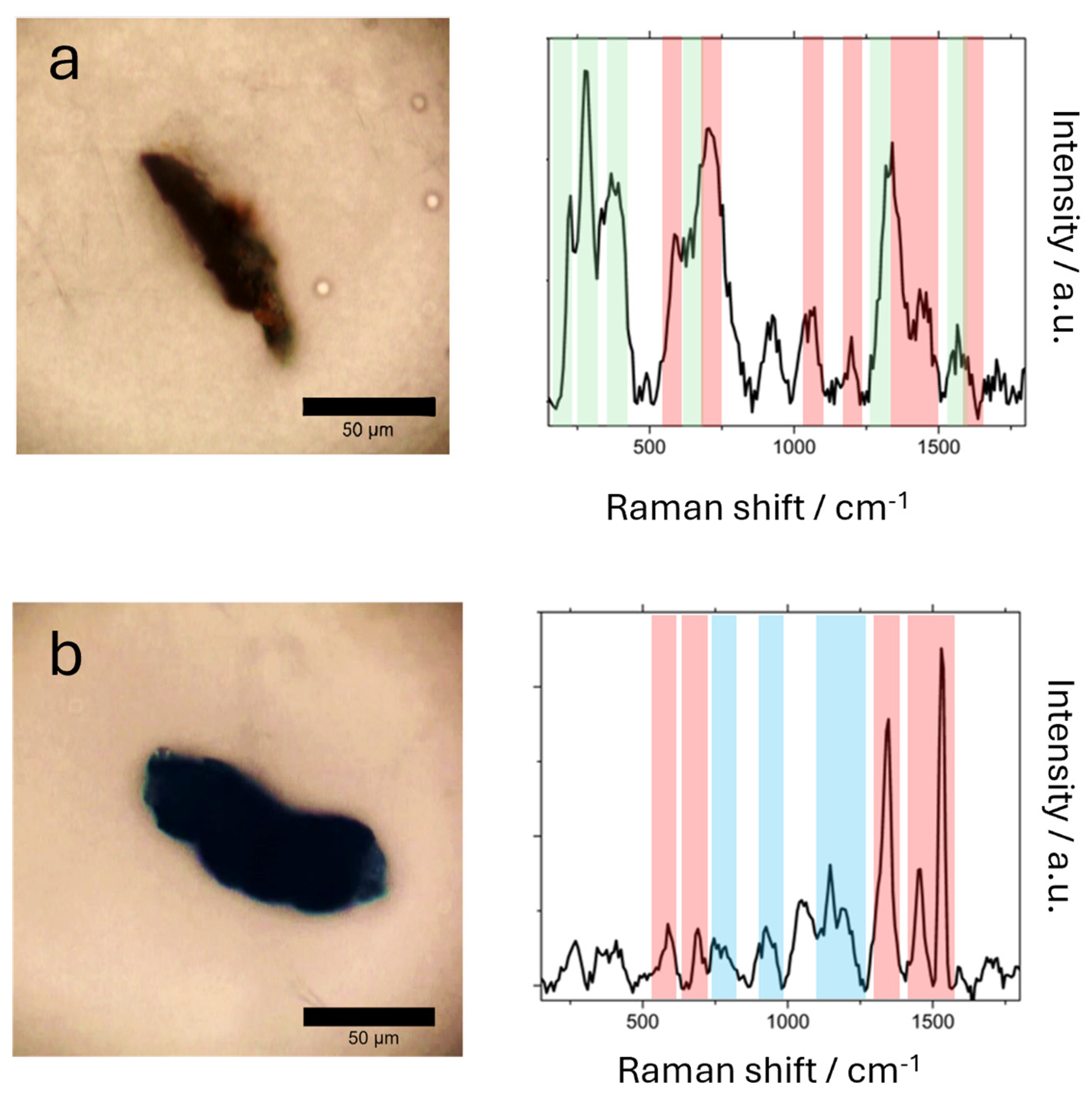
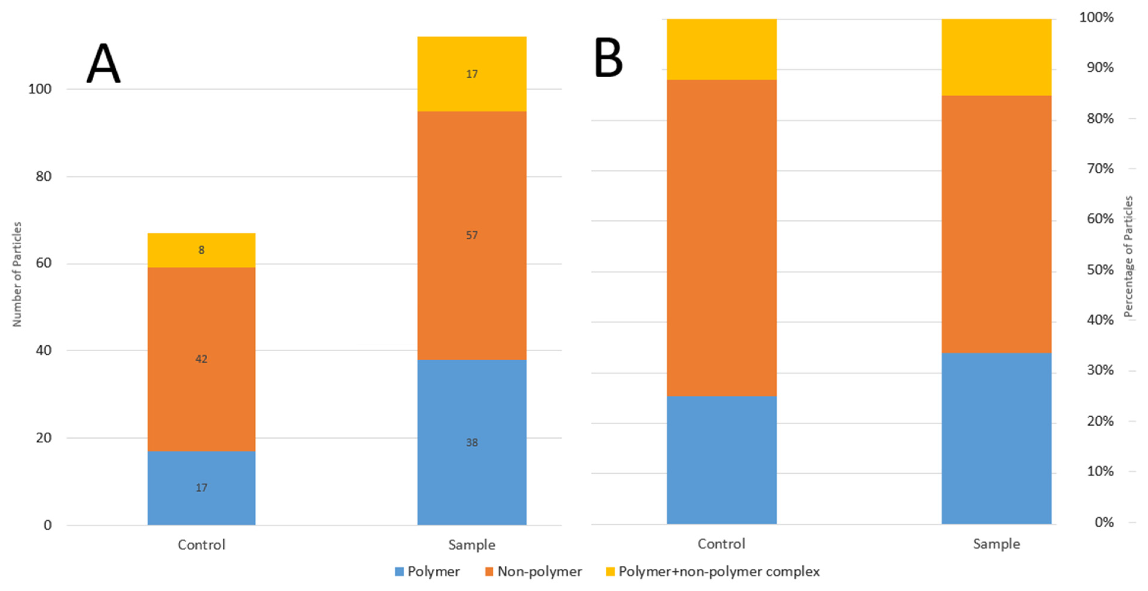
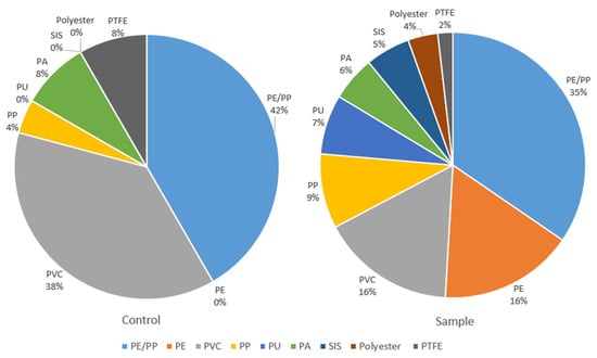
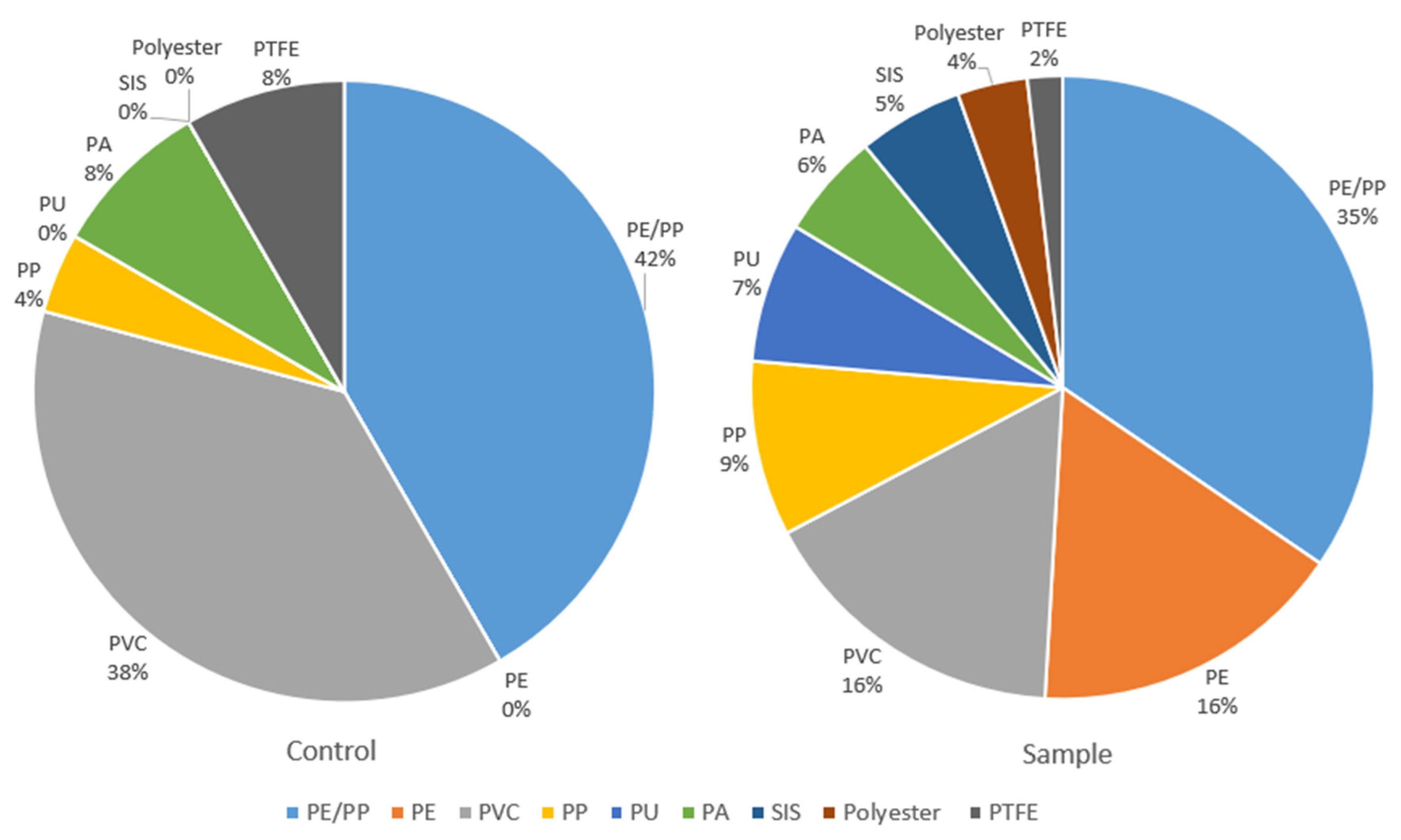
Representative examples of particles from each category are provided in Figure 1 and Figure 2. A photomicrograph of the particle is shown, along with its respective Raman spectrum. Figure 3 shows a graphical comparison of overall particle types in samples versus controls. The overall quantity of particles was higher for samples (n = 112) than controls (n = 67), but the distribution of overall particle categories did not differ significantly between samples and controls (p = 0.22, Fisher’s exact test). However, differences between samples and controls were seen in the distribution of polymer types (Figure 4). Samples had a total of 55 polymer-containing particles (38 individual plus 17 complexed), while controls had a total of 24 polymer-containing particles. Samples contained polyethylene, polyurethane, styrene/isoprene, and polyester particles, while controls contained none of these polymer types. Polyethylene/polypropylene, polyvinyl chloride, polypropylene, polyamide, and polytetrafluoroethylene particles were found in both samples and controls. On formal statistical comparison of polymer distributions, polyethylene was significantly more prevalent in samples versus controls (9/55 vs. 0/24 particles, p = 0.05, Fisher’s exact test), while other polymer types did not reach statistical significance.
Figure 1.
Representative photomicrographs and Raman spectra for individual particle types: (a) plastic polymer; (b) non-polymer mineral additive; (c) non-polymer pigment.
Figure 2.
Representative photomicrographs and Raman spectra for particle complexes: (a) polymer + mineral additive; (b) polymer + pigment. The Raman peaks of MPs, pigments and mineral additives are highlighted by red, blue and green shaded areas, respectively.
Figure 3.
Stacked bar charts comparing particle types for urine samples and procedural blank controls based on (A) particle count and (B) particle percentage.
Figure 4.
Pie charts comparing polymer types for urine samples and procedural blank controls. PE/PP, polyethylene/polypropylene co-polymer. PE, polyethylene. PVC, polyvinyl chloride. PP, polypropylene. PU, polyurethane. PA, polyamide. SIS, styrene/isoprene. PTFE, polytetrafluoroethylene.
The characteristic Raman peaks of pigments are in the following spectral range for carbon black (D-band ~ 1350 cm−1, G-band ~ 1580–1600 cm−1), phthalocyanines (C-N-C and C-H bending ~1100–1150 cm−1, pyrrole ring ~ 1420–1450 cm−1, C=C and C=N stretching ~ 1500–1550 cm−1), and methylene blue (C-H ~ 1150 cm−1, 1300 cm−1, C-N ~ 1400 cm−1, and C-C stretching ~ 1620 cm−1). The characteristic Raman peaks of polymers are in the following spectral range: ring breathing ~ 1000 cm−1 (SIS); CH2 ~ 800 cm−1 (PP); 1300 cm−1 (PE); 1430 cm−1 (PVC); C-F stretching ~ 1380 cm−1 (PTFE); C=O stretching ~ 1640 cm−1 (PA); ~ 1720 cm−1 (PU).
4. Discussion
4.1. Summary and Significance of Study Findings
This pilot study characterized a variety of MPs, additives, and pigments in the urine of children. Levels of these substances were present in the urine at significantly higher levels than rigorously collected blank controls. The levels of urinary microplastics exceeded the LOD in over half of the patients. Polyethylene comprised 16% of the polymer particles in patient urine samples, whereas it was not identified in any controls. These findings suggest that polymer particles, particularly the polyethylene particles, were excreted from patient’s bodies and did not originate from environmental sample contamination. Other non-MPs substances were found in the urine, including industrial mineral/metal oxide additives and pigments. These substances were found both alone and complexed with MPs. Over a third of children had MPs complexed with industrial dyes/pigments, some of which (e.g., carbon black) are recognized carcinogens [18]. This finding increases concerns for pathologic roles of MPs as “carriers” of other chemicals that may have carcinogenic, endocrine-disrupting, or other toxic effects [19]. Though small and preliminary in nature, this pilot study is novel and significant because it is the first to rigorously identify and describe microplastics in the urine of children in the United States.
4.2. Potential Mechanism for Urinary Excretion of Microplastics and Other Small Particles
The size of the urinary MPs, most of which were greater than 10 μm, suggests excretion mechanisms other than glomerular filtration of blood. Since filtration across the intact glomerulus is only thought to be possible for particles < 10 nm, MPs are thought to be taken up by the epithelial cells of the proximal convoluted tubules through endo- or micropinocytosis, then secreted into the tubular lumen for eventual excretion in the urine [11]. Other mechanisms for renal filtration of larger particles based on blood vessel transport via lower urinary tract system have also been proposed in our previous work [16]. The mean particle size of ~ 100 µm is an order of magnitude larger than the size limits of any other previously studied transport mechanisms and requires alternative explanation. Such alternative pathways and mechanisms have recently been reviewed but until now mostly remain speculative [20]. Aggregation of smaller particles into large clusters within the urinary system or during sample processing might offer another alternative explanation. In this regard, it is important to investigate the kinetics of urinary excretion of microplastics by considering the particle accumulation periods, for example, prior fasting (12–24 h) before urine collection. This may provide insight into the nature of MPs from recent exposure during hospitalization or longer time accumulation in urban areas. Other potential future directions for investigation of urinary excretion mechanisms include temporal correlation of levels in the blood and urine, and utilization of microparticles labeled with an identifiable marker (e.g., radiolabeling, fluorescence, etc.).
4.3. Comparison with Previous Literature
A group in Italy recently characterized MPs and other particles in 10 urine samples and 10 kidney samples from healthy adults using RMS [10]. Their findings resembled ours in that hematite was the most common additive, and polyethylene was the most common polymer. They also identified an industrial pigment, copper phthalocyanine blue, in the urine. Their study did not specifically identify complexes of pigments or additives with MPs, as our study did. Their particle sizes also tended to be slightly smaller than ours, ranging from 3 to 13 μm. Previous studies have identified MPs in the urine of children in China. A study of urine samples from 12 patients from the Chongqing region, that included 6 children with median age 8.3 years, showed that urine MPs were present in children at median abundance of 1 mg/kg [9]. In that study, polyethylene was the most common polymer detected, and most particles were 50 μm or smaller in size. This is congruent with our study findings that polyethylene was significantly more prevalent in patient urine samples compared to blank controls. Their study did not evaluate additives or pigments, nor the complexes of these substances with MPs. Their small sample size also limited the power of comparison by age, sex, urban/rural region of residence, and other exposure factors. Another study of school-age children (mean age 8.5 years) in China using RMS demonstrated MPs in urine at a median concentration of 9 particles/100 mL [8]. Polyamide, polypropylene, and polyvinyl chloride were all found in similar concentrations. The amount of urinary MPs was associated with higher risk of behavioral problems; however, the study lacked rigorous controls and did not evaluate non-polymer particles or complexes of polymers with other additives/pigments [8].
The finding of MPs in the present study’s procedural blanks, which were collected and processed in the exact same manner as samples, confirms the considerable background environmental MPs contamination rate and corroborates findings from other studies. For example, in a recent study using µ-FTIR spectroscopy and RMS to identify MPs in the blood of Korean adults, mean MPs concentration was 4.2 particles/mL in samples and 1.67 particles/mL (range 0–5 particles/mL) in ultrapure water procedural blanks [21].
4.4. Study Limitations
The relatively small sample size (n = 11) of this pilot study limits the generalizability of the conclusions. Although, to the best of our knowledge, this is the first study to document MPs in the urine of children in the United States. MPs accumulation has been shown to vary by geographic region at the U.S. state level [22]. However, our pilot study was limited to a single US hospital with restricted patient enrollment. Therefore, large-scale analysis of demographic and geographic exposure risk factors is beyond the scope of our pilot study and will be the focus of future work. The use of RMS for identification and characterization of particles was consistent with the aims of this study to identify and characterize MPs and other additives; however, undetected nanoplastics may have also been present in the samples. Most samples showed fluorescence background at 532 nm illumination, which was subtracted by baseline correction described in Section 2.6. Finally, while MPs and other particles were clearly identified at concentrations above the limit of detection defined by procedural blank controls, most concentrations fell below the limit of quantification, indicating that the precise particle concentrations must be interpreted with caution.
4.5. Potential Mechanisms of Toxicity
The mechanisms of toxicity of microplastics in children are currently poorly understood and require further studies. The main effects are due to physical tissue damage and chemical toxicity of microplastics acting as carriers of environmental contaminants (e.g., heavy metals (e.g., lead, cadmium), persistent organic pollutants, endocrine disruptors) [23]. The latter may potentiate the biological effects beyond those of the particles themselves. For example, polymer type determines buoyancy, surface chemistry, and susceptibility to degradation while polymer crystallinity influences rigidity and the potential for additive leaching. Size, shape, and surface roughness are critical for physical toxicity [24]. There is currently a critical gap between lab-based and real-world studies that needs to be addressed, facilitating the translation of scientific insights into practical strategies for mitigating risks to human health and, especially, children.
5. Conclusions
The current pilot study found that a variety of MPs were present in the urine of children undergoing surgical procedures at a U.S. children’s hospital. Polyethylene polymers were particularly prevalent in the urine compared to blank controls. Microplastics were frequently found complexed with additives and pigments, some of which are classed as carcinogens. Considerable background contamination was found in the procedural controls, limiting the assignment of the particle origin. Larger studies are needed to confirm these findings and explore the relationship between particle levels in blood and urine, as well as the association of MPs and health disorders in children.
Author Contributions
Conceptualization, C.W.S., W.S., and D.V.V.; methodology, C.W.S., W.S., J.M.R., V.I. and D.V.V.; software, A.C., T.D. and D.V.V.; validation, A.C., T.D. and D.V.V.; formal analysis, C.W.S., W.S., R.H. and D.V.V.; investigation, J.M.R. and D.V.V.; data curation, J.D. and A.F.; writing—original draft preparation, C.W.S.; writing—review and editing, W.S., R.H., J.D., A.F., S.B., A.C., T.D., J.M.R., V.I. and D.V.V.; visualization, R.H., S.B., A.C. and T.D.; supervision, J.M.R. and V.I.; project administration, J.D. and A.F.; funding acquisition, C.W.S., W.S. and V.I. All authors have read and agreed to the published version of the manuscript.
Funding
This research was funded by the Johns Hopkins All Children’s Foundation, pilot grant number 90110942.
Institutional Review Board Statement
The study was conducted in accordance with the Declaration of Helsinki and approved by the Institutional Review Board of Johns Hopkins All Children’s Hospital (number IRB00393797, date of approval 7 February 2024).
Informed Consent Statement
Informed consent was obtained from all subjects involved in the study.
Data Availability Statement
The datasets presented in this article are not readily available because the data are part of an ongoing study.
Acknowledgments
The authors acknowledge Alyssa Green, for assistance with project administration.
Conflicts of Interest
The authors declare no conflicts of interest. The funders had no role in the design of the study; in the collection, analyses, or interpretation of data; in the writing of the manuscript; or in the decision to publish the results.
Abbreviations
The following abbreviations are used in this manuscript:
| MPs | Microplastics |
| IRB | Institutional Review Board |
| SDOH | Social determinants of health |
| SVI | Social Vulnerability Index |
| EJI | Environmental Justice Index |
| dH2O | Deionized molecular-grade water |
| RMS | Raman microspectroscopy |
| CCD | Charge-coupled device |
| LOD | Limit of detection |
| LOQ | Limit of quantification |
| µ-FTIR | Micro-Fourier transform infrared spectroscopy |
References
- Geyer, R.; Jambeck, J.R.; Law, K.L. Production, Use, and Fate of All Plastics Ever Made. Sci. Adv. 2017, 3, e1700782. [Google Scholar] [CrossRef] [Scilit] [PubMed]
- Gruber, E.S.; Stadlbauer, V.; Pichler, V.; Resch-Fauster, K.; Todorovic, A.; Meisel, T.C.; Trawoeger, S.; Hollóczki, O.; Turner, S.D.; Wadsak, W.; et al. To Waste or Not to Waste: Questioning Potential Health Risks of Micro- and Nanoplastics with a Focus on Their Ingestion and Potential Carcinogenicity. Expo. Health 2023, 15, 33–51. [Google Scholar] [CrossRef] [Scilit] [PubMed]
- Kasmuri, N.; Tarmizi, N.A.A.; Mojiri, A. Occurrence, Impact, Toxicity, and Degradation Methods of Microplastics in Environment—A Review. Environ. Sci. Pollut. Res. 2022, 29, 30820–30836. [Google Scholar] [CrossRef] [Scilit] [PubMed]
- Roslan, N.S.; Lee, Y.Y.; Ibrahim, Y.S.; Tuan Anuar, S.; Yusof, K.M.K.K.; Lai, L.A.; Brentnall, T. Detection of Microplastics in Human Tissues and Organs: A Scoping Review. J. Glob. Health 2024, 14, 04179. [Google Scholar] [CrossRef] [Scilit] [PubMed]
- Ziani, K.; Ioniță-Mîndrican, C.-B.; Mititelu, M.; Neacșu, S.M.; Negrei, C.; Moroșan, E.; Drăgănescu, D.; Preda, O.-T. Microplastics: A Real Global Threat for Environment and Food Safety: A State of the Art Review. Nutrients 2023, 15, 617. [Google Scholar] [CrossRef] [Scilit] [PubMed]
- Marfella, R.; Prattichizzo, F.; Sardu, C.; Fulgenzi, G.; Graciotti, L.; Spadoni, T.; D’Onofrio, N.; Scisciola, L.; La Grotta, R.; Frigé, C.; et al. Microplastics and Nanoplastics in Atheromas and Cardiovascular Events. N. Engl. J. Med. 2024, 390, 900–910. [Google Scholar] [CrossRef] [Scilit] [PubMed]
- Nihart, A.J.; Garcia, M.A.; El Hayek, E.; Liu, R.; Olewine, M.; Kingston, J.D.; Castillo, E.F.; Gullapalli, R.R.; Howard, T.; Bleske, B.; et al. Bioaccumulation of Microplastics in Decedent Human Brains. Nat. Med. 2025, 31, 1114–1119. [Google Scholar] [CrossRef] [Scilit] [PubMed]
- Dong, L.; Li, X.; Zhang, Y.; Liu, B.; Zhang, X.; Yang, L. Urinary Microplastic Contaminants in Primary School Children: Associations with Behavioral Development. Ecotoxicol. Environ. Saf. 2025, 295, 118097. [Google Scholar] [CrossRef] [Scilit] [PubMed]
- Song, X.; Chen, T.; Chen, Z.; Du, L.; Qiu, X.; Zhang, Y.; Li, Y.; Zhu, Y.; Tan, Z.; Mo, Y.; et al. Micro(Nano)Plastics in Human Urine: A Surprising Contrast between Chongqing’s Urban and Rural Regions. Sci. Total Environ. 2024, 917, 170455. [Google Scholar] [CrossRef] [Scilit] [PubMed]
- Massardo, S.; Verzola, D.; Alberti, S.; Caboni, C.; Santostefano, M.; Eugenio Verrina, E.; Angeletti, A.; Lugani, F.; Ghiggeri, G.M.; Bruschi, M.; et al. MicroRaman Spectroscopy Detects the Presence of Microplastics in Human Urine and Kidney Tissue. Environ. Int. 2024, 184, 108444. [Google Scholar] [CrossRef] [Scilit] [PubMed]
- Pironti, C.; Notarstefano, V.; Ricciardi, M.; Motta, O.; Giorgini, E.; Montano, L. First Evidence of Microplastics in Human Urine, a Preliminary Study of Intake in the Human Body. Toxics 2022, 11, 40. [Google Scholar] [CrossRef] [Scilit] [PubMed]
- Bole, A.; Bernstein, A.; White, M.J.; Council on Environmental Health and Climate Change; Bole, A.; Balk, S.J.; Byron, L.G.; Huerta-Montañez, G.M.; Landrigan, P.J.; Marcus, S.M.; et al. The Built Environment and Pediatric Health. Pediatrics 2024, 153, e2023064773. [Google Scholar] [CrossRef] [Scilit] [PubMed]
- Zajac, L.; Landrigan, P.J.; Council on Environmental Health and Climate Change; Zajac, L.; Balk, S.J.; Byron, L.G.; Maria Huerta-Montañez, G.; Landrigan, P.J.; Marcus, S.M.; Nerlinger, A.L.; et al. Environmental Issues in Global Pediatric Health: Technical Report. Pediatrics 2025, 155, e2024070076. [Google Scholar] [CrossRef] [Scilit] [PubMed]
- Szeto, J.J.; Radack, J.K.; DeMauro, S.B.; Jensen, E.A.; Gibbs, K.; Novick, N.P.; Scott, K.A.; Murosko, D.C.; Burris, H.H.; Nelin, T.D. Environmental Determinants of Post-Discharge Acute Respiratory Illness among Preterm Infants with Bronchopulmonary Dysplasia. Int. J. Environ. Res. Public Health 2024, 21, 648. [Google Scholar] [CrossRef] [Scilit] [PubMed]
- Tipirneni, R.; Schmidt, H.; Lantz, P.M.; Karmakar, M. Associations of 4 Geographic Social Vulnerability Indices with US COVID-19 Incidence and Mortality. Am. J. Public Health 2022, 112, 1584–1588. [Google Scholar] [CrossRef] [Scilit] [PubMed]
- Rotchell, J.M.; Austin, C.; Chapman, E.; Atherall, C.A.; Liddle, C.R.; Dunstan, T.S.; Blackburn, B.; Mead, A.; Filart, K.; Beeby, E.; et al. Microplastics in Human Urine: Characterisation Using μFTIR and Sampling Challenges Using Healthy Donors and Endometriosis Participants. Ecotoxicol. Environ. Saf. 2024, 274, 116208. [Google Scholar] [CrossRef] [Scilit] [PubMed]
- Noonan, M.J.; Grechi, N.; Mills, C.L.; de A. M. M. Ferraz, M. Microplastics Analytics: Why We Should Not Underestimate the Importance of Blank Controls. Microplast. Nanoplast. 2023, 3, 17. [Google Scholar] [CrossRef] [Scilit] [PubMed]
- Zhang, J.; Li, X.; Cheng, W.; Li, Y.; Shi, T.; Jiang, Y.; Wang, T.; Wang, H.; Ren, D.; Zhang, R.; et al. Chronic Carbon Black Nanoparticles Exposure Increases Lung Cancer Risk by Affecting the Cell Cycle via Circulatory Inflammation. Environ. Pollut. 2022, 305, 119293. [Google Scholar] [CrossRef] [Scilit] [PubMed]
- Rafa, N.; Ahmed, B.; Zohora, F.; Bakya, J.; Ahmed, S.; Ahmed, S.F.; Mofijur, M.; Chowdhury, A.A.; Almomani, F. Microplastics as Carriers of Toxic Pollutants: Source, Transport, and Toxicological Effects. Environ. Pollut. 2024, 343, 123190. [Google Scholar] [CrossRef] [Scilit] [PubMed]
- La Porta, E.; Exacoustos, O.; Lugani, F.; Angeletti, A.; Chiarenza, D.S.; Bigatti, C.; Spinelli, S.; Kajana, X.; Garbarino, A.; Bruschi, M.; et al. Microplastics and Kidneys: An Update on the Evidence for Deposition of Plastic Microparticles in Human Organs, Tissues and Fluids and Renal Toxicity Concern. Int. J. Mol. Sci. 2023, 24, 14391. [Google Scholar] [CrossRef] [Scilit] [PubMed]
- Lee, D.-W.; Jung, J.; Park, S.-A.; Lee, Y.; Kim, J.; Han, C.; Kim, H.-C.; Lee, J.H.; Hong, Y.-C. Microplastic Particles in Human Blood and Their Association with Coagulation Markers. Sci. Rep. 2024, 14, 30419. [Google Scholar] [CrossRef] [Scilit] [PubMed]
- Aschner, M. Mind over Microplastics—What We Still Don’t Know. Nat. Rev. Neurol. 2025, 21, 351–352. [Google Scholar] [CrossRef] [Scilit] [PubMed]
- Zhang, Y.; Ren, J.; Zheng, B.; Sun, J.; Zhang, J.; Niu, Y.; Shao, B.; Jin, Y. Microplastic toxicity: Mechanisms, assessment methods, and future research directions. Front. Toxicol. 2026, 8, 1766103. [Google Scholar] [CrossRef] [Scilit] [PubMed]
- Choi, D.; Hwang, J.; Bang, J.; Han, S.; Kim, T.; Oh, Y.; Hwang, Y.; Choi, J.; Hong, J. In vitro toxicity from a physical perspective of polyethylene microplastics based on statistical curvature change analysis. Sci. Total Environ. 2021, 752, 142242. [Google Scholar] [CrossRef] [Scilit] [PubMed]

Disclaimer/Publisher’s Note: The statements, opinions and data contained in all publications are solely those of the individual author(s) and contributor(s) and not of MDPI and/or the editor(s). MDPI and/or the editor(s) disclaim responsibility for any injury to people or property resulting from any ideas, methods, instructions or products referred to in the content. |
© 2026 by the authors. Licensee MDPI, Basel, Switzerland. This article is an open access article distributed under the terms and conditions of the Creative Commons Attribution (CC BY) license.