


Abstract
Previous studies have indicated that polyethylene terephthalate (PET) micro/nanoplastics (MNPs) may coexist with antibiotics in environmental and biological matrices; however, their combined behavior in blood remains insufficiently characterized. This study examined PET MNPs prepared from water bottles at 0.5, 2.5, and 7.5 mg/mL together with tetracycline (2–50 µg/mL) in defibrinated horse blood. After 24 h exposure and particle removal, UV–visible absorbance, intrinsic fluorescence, redox indicators, and subsequent Escherichia coli responses were evaluated. The blood biochemical results showed condition-dependent changes in hemoglobin-associated absorbance, tryptophan-dominated fluorescence, reactive oxygen species, reduced glutathione, superoxide dismutase, and lipid peroxidation. When treated blood supernatants were applied to Escherichia coli, tetracycline alone reduced bacterial OD600, whereas selected PET MNP–tetracycline co-exposures partially restored bacterial proliferation and modified oxidative-stress responses. ATR–FTIR analysis of Escherichia coli pellets showed dose-dependent modulation of phosphate-, lipid-, and protein-associated bands, indicating changes in bacterial biochemical fingerprints under specific exposure combinations. Overall, the findings suggest that PET MNPs can modify tetracycline-associated spectral, redox, and bacterial response patterns in a blood matrix. Future studies incorporating adsorption assays, free tetracycline quantification, protein-corona profiling, time-course exposure designs, and antibiotic susceptibility testing would further clarify the mechanistic basis and biological relevance of these matrix-dependent interaction effects.
1. Introduction
Micro- and nanoplastics (MNPs) have become ubiquitous contaminants across aquatic, terrestrial, and atmospheric environments, where they frequently co-occur with pharmaceutically active compounds, including antibiotics [1,2]. Among these compounds, tetracyclines (Tet) represent one of the most widely used antimicrobial classes in human and veterinary medicine, leading to their widespread detection in environmental matrices and increasing concern regarding their ecological and biological impacts [3,4,5,6]. Recent studies have shown that MNP–Tet interactions can involve electrostatic forces, hydrophobic interactions, hydrogen bonding, and π–π interactions, while adsorption behavior is strongly influenced by particle size, surface properties, polymer type, and surrounding chemical conditions, including metals and dissolved organic matter.
Although most MNP–Tet interaction studies have focused on aquatic and soil environments, emerging evidence indicates that plastic particles can also enter biological systems [6]. The detection of plastic particles in human blood has raised new questions regarding their potential interactions with blood components and co-occurring contaminants [7]. Blood is a chemically complex matrix containing proteins, redox-active species, metabolites, and cells; therefore, MNPs may interact with antibiotics and blood components in ways that alter protein-associated spectral signals, redox balance, and downstream biological responses [8]. Previous studies have also shown that MNPs can interact with blood-relevant proteins, including hemoglobin and serum albumin, leading to conformational or functional changes detectable by spectroscopic techniques [9,10,11,12].
Oxidative stress is considered an important response pathway associated with both MNPs and antibiotics. Exposure to MNPs has been linked to changes in reactive oxygen species (ROS), lipid peroxidation (LPO), and antioxidant enzyme activities in various biological systems [8,13,14]. Similarly, Tet exposure can influence redox balance through metal chelation, redox cycling, and protein-associated interactions. Importantly, co-exposure scenarios may not simply produce additive responses. Interactions between Tet, plastic particles, and blood constituents may modify the apparent availability of Tet and alter oxidative-stress-related outcomes. However, experimental data addressing such combined effects in blood matrices remain limited.
Beyond host-related responses, MNP–antibiotic interactions may also be relevant to studies examining changes in antibiotic bioavailability, bacterial growth, and short-term stress responses [15]. However, direct AMR endpoints such as resistance-gene expression, MIC shifts, or stable resistance acquisition require dedicated assays and should be distinguished from short-term bacterial growth or oxidative-stress responses. Plastic particles can provide surfaces for bacterial attachment, interact with antibiotics, and create heterogeneous exposure microenvironments that may influence bacterial survival under antibiotic stress [7,16,17,18]. Escherichia coli (E. coli), a widely used indicator organism across environmental, veterinary, and clinical contexts, has been reported to show changes in growth, oxidative status, biofilm formation, and stress-related traits following exposure to MNPs, antibiotics, or their combinations. However, direct AMR endpoints such as resistance-gene expression, MIC shifts, or stable resistance acquisition require dedicated assays and should be distinguished from short-term bacterial growth or oxidative-stress responses.
The simultaneous presence of MNPs and Tet in blood may therefore represent an important interface linking environmental contamination, host-systemic responses, and bacterial physiology [15]. Blood-mediated changes in oxidative balance and antibiotic-associated response patterns may indirectly influence bacterial growth and stress adaptation across environmental, animal, and human health contexts. In this framework, evaluating how blood pre-exposed to PET MNPs and Tet affects E. coli growth, oxidative indicators, and biochemical fingerprints can provide useful preliminary information for mechanistic risk assessment.
Although PS and PVC may receive high scores in chemical-composition-based polymer hazard indices, PET was selected on the basis of exposure and matrix relevance rather than as the polymer with the highest theoretical intrinsic hazard. PET is extensively used in food and beverage packaging, particularly drinking-water bottles, and PET-derived polymer material has been identified in human blood [7,19,20]. In addition, previous experimental studies have demonstrated the adsorption and desorption of tetracycline on PET microplastics [20]. Therefore, the use of a previously characterized, bottle-derived PET particle batch provided an exposure-relevant and analytically defined model for examining micro/nanoplastic–tetracycline interactions in blood. Nevertheless, because polymer chemistry can influence contaminant adsorption, protein-corona formation, and biological responses, direct comparison of PET with PS, PVC, and other polymers is warranted.
Accordingly, this study evaluated the 24 h responses of defibrinated horse blood exposed to previously characterized PET MNPs and Tet, followed by an exploratory assessment of E. coli growth, redox markers, and ATR–FTIR fingerprints. The aim was to identify condition-dependent spectral, redox, and bacterial response patterns in a blood matrix and to provide a basis for future studies incorporating direct Tet adsorption measurements, free-drug quantification, time-course exposure designs, and antibiotic susceptibility testing.
2. Materials and Methods
2.1. Experimental Preparation and Exposure Design
Defibrinated horse blood was commercially purchased from Oxoid (Thermo Scientific, Leicestershire, UK) and used as a protein-rich experimental matrix. No animals were handled directly in this study. Before exposure, the blood was equilibrated to 37 °C under gentle mixing.
PET MNPs were prepared from PET water bottles using the previously reported in-house procedure [21]. The tested particles were not subjected to artificial UV, thermal, or chemical aging before exposure. They were therefore used as a non-artificially weathered, bottle-derived PET model relevant to the direct release of particles from beverage-contact materials. This designation does not imply a completely virgin surface, because manufacturing, storage, and mechanical processing may have modified the original bottle material. The particle-size range was corrected to 300–500 nm. The same batch was previously characterized by particle-size distribution/TEM (Hitachi High-Tech HT7700, Minato, Japan), EDX (FEI, Thermo Fisher Scientific, Waltham, MA, USA), BET sorption–desorption analysis (Micromeritics Gemini VII 2390 T, Norcross, GA, USA), FTIR (Bruker Invenio S, Billerica, MA, USA), XRD (PANalytical Xpert Pro X-ray Diffractometer, Malvern, UK), 1H-NMR (Agilent, Santa Clara, CA, USA), and zeta-potential measurements (Zetasizer Nano ZS, Malvern Instruments, Malvern, UK), with reported particle-size and surface/structural descriptors supporting its identification as PET MNPs [21,22]. Studies simultaneously characterizing the abundance, polymer identity, and particle-size distribution of micro- and nanoplastics in human blood remain very limited. Leslie et al. [7] quantified the mass of plastic polymer in blood after filtration through a 0.7 µm glass-fibre filter; however, the Py-GC/MS method used in that study did not permit particle-by-particle size determination. In subsequent size-resolved studies, Leonard et al. [23] reported particle lengths ranging from 7 to 3000 µm, whereas Lee et al. [24] found that most microplastic particles detected in human blood were within the 20–50 µm range. More recently, Li et al. [25] reported a mean blood microplastic size of 64.50 ± 38.20 µm, with measured microplastic sizes ranging from 9.79 to 193.29 µm and separately quantified a nanoplastic fraction operationally defined as <1 µm using a polymer-mass-based approach. These findings indicate that plastic particles considerably larger than traditionally expected may be detected in the bloodstream. At the same time, methodological limitations continue to restrict the direct particle-resolved identification and sizing of submicrometre and nanoplastic fractions in complex biological matrices. Accordingly, the 300–500 nm PET particles used in the present study were employed as an experimental submicrometre model representing a particle-size fraction that may be biologically relevant to blood exposure but remains difficult to characterize directly at the individual-particle level in human blood. Additionally, PET MNPs of 300–500 nm were used because this size class coincides with three biologically relevant thresholds documented in prior work: (i) it lies well below the 5–8 µm capillary inner diameter that constitutes the theoretical mechanical barrier for circulating rigid microplastics [23]; (ii) it brackets the 50–400 nm window in which polystyrene most potently activates platelets and the intrinsic coagulation cascade [26] and at which 500 nm spheres damage endothelial membranes rather than being fully endocytosed [2]; and (iii) it is small enough for sub-capillary transit from sites of intestinal uptake, including M-cell translocation and persorption [27], while remaining within the size band that interacts directly with endothelial monolayers—in contrast to 1 µm particles, which show only ≈3.80% binding to HUVECs at 25 µg/mL [28]. In the present blood experiment, three PET MNP masses were added to 4.0 mL of blood: 2, 10, and 30 mg, corresponding to 0.5, 2.5, and 7.5 mg/mL, and designated as MNP1x, MNP5x, and MNP15x, respectively. The exposure range of 0.5–7.5 mg/mL (500–7500 µg/mL) was selected to span the full biologically active dose–response envelope identified by meta-regression of 17 in vitro human cell studies, in which adverse effects on cell viability emerge at 10 µg/mL and cytokine release at 20 µg/mL, scaling with concentration up to ≥1000 µg/mL [29]. The lowest dose (500 µg/mL) matches the concentration at which prior work established ≈4% hemolysis from sub-5 µm polystyrene [30] and PS-induced cytokine release in mast cells and PBMCs [30]. Although the upper bound exceeds mean circulating concentrations reported in human biomonitoring (1.6 µg/mL [7]; 268 ng/mL [22]; 0.66 ± 0.87 µg/mL [23]), it is consistent with tissue-level exposure documented at sites of microplastic accumulation, where symptomatic carotid plaques reach 2888 µg/g (≈2.9 mg/mL) [31] and atherosclerotic plaques contain 13.99 ± 3.00 µg/mg plastic [32]. Nominal concentrations were further adjusted upward to compensate for protein-corona sequestration in serum-containing media [2] and for non-uniform particle settling, both of which reduce bioavailable dose in static culture. The selected range therefore captures the lowest biologically active dose while extending sufficiently high to identify the inflection between mechanistic and overt cytotoxic responses. Because particle number and effective surface area in blood were not remeasured in this experiment, the dose is reported as mass per volume.
Tetracycline hydrochloride was prepared as a light-protected aqueous stock solution. Final concentrations in blood were 2, 10, and 50 µg/mL, designated as Tet1x, Tet5x, and Tet25x, respectively. These concentrations were selected as an experimental range and should not be interpreted as a pharmacokinetic representation of systemic exposure. Additional volumes were kept constant across all groups.
Blood was added into experimental tubes with a final volume of 4.0 mL and assigned to untreated blood control, PBS-added blood vehicle control, Tet-only, MNP-only, or combined MNP–tetracycline treatment groups. PBS was used as the solvent/vehicle for tetracycline preparation; therefore, a PBS-added blood control was included to verify whether the vehicle itself affected the measured endpoints. Because no significant difference was observed between untreated blood and PBS-added blood, only the blood control was presented in the figures for clarity. Similar controls were applied as MNP, including PBS in blood, and no significant changes were found between MNP-blood and MNP including PBS-blood. Tet-free/MNP-added and MNP-free/Tet-added groups were included as single-exposure controls. No surfactant or dispersing agent was added to the PET MNP suspensions or blood samples, because such additives could alter the PET surface, protein-corona formation, tetracycline adsorption, blood-component integrity, and the measured optical and redox endpoints. Samples were incubated at 37 °C for 24 h under gentle agitation to maintain particle–matrix contact and reduce bulk settling. However, gentle agitation was not expected to prevent particle association, and aggregation or agglomeration may have occurred in the protein-rich and ionic blood matrix.
After exposure, samples were centrifuged at 800× g for 10 min at 4 °C to remove the cellular fraction. The supernatant was then centrifuged at 1500× g for 5 min at 4 °C to reduce residual PET MNPs and larger particle agglomerates. This procedure was intended to obtain a particle-depleted supernatant but does not demonstrate complete removal of all individual or nanoscale particles. The resulting particle-depleted supernatant was analyzed immediately. No Tet mass balance was performed in the supernatant, pellet, or vessel-wall fractions.
All controls were included from the initial exposure step. The experimental design included untreated blood as the matrix control, blood containing tetracycline without PET MNPs as the particle-free matrix-matched control, and blood containing PET MNPs without tetracycline as the drug-free matrix-matched control. In addition, tetracycline and PET MNPs prepared separately in PBS were analyzed as matrix-free optical blanks to assess the intrinsic optical contribution of tetracycline and the absorbance or light-scattering contribution of PET MNPs, respectively. The PBS-based blanks were not considered substitutes for the corresponding blood-matrix controls.
2.2. Particle Characterization
The PET MNP batch was not newly characterized after blood exposure in the present experiment; however, the same batch had previously been characterized in detail and is cited here as the particle-characterization source [21,22]. These prior data include particle-size distribution/TEM, elemental composition by EDX, BET sorption–desorption behavior, FTIR functional-group features, XRD crystallinity, 1H-NMR structural information, and zeta-potential/particle-size changes after protein exposure [21,22]. These data support the identity and baseline physicochemical description of the particles used in the present study. Some characteristics are presented in Supplementary Figure S1. Nevertheless, exposure-specific particle number, effective surface area in blood, post-exposure corona composition, leachate composition in blood, and soluble/particle-associated tetracycline mass balance were not determined in this study.
2.3. UV–Visible Spectroscopy
UV–visible spectra of the particle-depleted blood supernatants were recorded from 300 to 800 nm using a SkyHigh microplate reader (Thermo Scientific, Multiskan SkyHigh, Cleveland, OH, USA). The measurements were used to compare heme-associated spectral features across treatments. Because total hemoglobin and individual hemoglobin species were not independently quantified, changes in absorbance were not assigned uniquely to oxidation, degradation, adsorption, or loss from the measured supernatant.
Matched wells containing the corresponding blank were used. Samples were diluted when required to avoid signal saturation, with the same dilution factor applied within each comparison. Full spectra were collected, and selected wavelengths were extracted for descriptive comparison.
The Soret region is near 414 nm, and the oxyhemoglobin-associated features near 540 and 576 nm were evaluated. Absorbance near 630 nm was treated as an oxidation-related spectral feature but not as a quantitative measurement of methemoglobin. Absorbance at 750 nm was monitored as a scattering/turbidity check after particle removal.
2.4. Fluorescence Spectroscopy
Intrinsic fluorescence spectra of particle-depleted blood supernatants were recorded using a Varioskan LUX microplate spectrofluorometer (Thermo Scientific, Cleveland, OH, USA) [21]. The measurements were used to compare treatment-related changes in tryptophan-dominated emission.
Samples were excited at 280 nm, and emission was collected from 300 to 450 nm using identical instrumental settings. Samples were diluted so that the excitation wavelength remained below 0.1, and the same dilution factor was used across the relevant treatment set.
Changes in fluorescence intensity were interpreted as composite spectral responses. These responses may include protein-environment changes, Tet absorption and inner-filter effects, oxidation, or removal of protein from the measured supernatant. Fluorescence changes alone were not used to calculate a binding constant or to establish direct tetracycline–MNP or protein-corona interactions.
2.5. Oxidative Stress Biomarkers
After the 24 h exposure, particle-depleted blood supernatants were analyzed for ROS, GSH, SOD, and LPO using the referenced spectrophotometric or fluorometric procedures [21,22]. Untreated blood served as a normalization control. These assays were used as comparative biochemical endpoints; particle-free assay controls and tetracycline-specific interference controls were not available for every method.
ROS was measured using DCFH-DA at an excitation wavelength of 488 nm and an emission wavelength of 530 nm. LPO was assessed using the TBARS method at 532 nm. SOD activity was evaluated by nitroblue tetrazolium reduction at 560 nm, and GSH-related activity was monitored using an NADPH-dependent procedure at 340 nm. Results were expressed relative to the untreated blood control [21,22].
2.6. Bacterial Exposure and ATR–FTIR Fingerprinting
E. coli ATCC 35218 was incubated for 24 h with aliquots of particle-depleted supernatants from untreated blood, Tet-only, MNP-only, and combined-treatment groups. A bacterial control without blood exposure was also included [33]. The study used one strain and did not determine the Tet MIC in the test matrix. Consequently, the assay was designed to compare growth and stress-related endpoints, not to assess antibiotic susceptibility or changes in resistance.
Bacterial growth was estimated by optical density at 600 nm. OD600 is a turbidity-based growth proxy and was not interpreted as a direct viability or colony-count measurement. Following the OD600 measurements, the same bacterial samples and controls were used for the oxidative indicators. Intracellular ROS was measured using DCFH-DA at 488/530 nm. GSH-related activity was measured according to Arora et al. [34] using sodium azide, glutathione reductase, NADPH, and H2O2, with absorbance monitored at 340 nm. LPO was measured using the TBARS assay at 532 nm [34]. SOD activity was evaluated using the nitroblue tetrazolium method at 560 nm [34].
After centrifugation, E. coli pellets were analyzed using a Bruker InvenioS FTIR spectrometer equipped with an ATR module (FTIR-ATR, Billerica, MA, USA). Spectra were collected from 4000 to 400 cm−1 at 4 cm−1 resolution with 32 scans per sample [35]. After spectral measurements, the 1800–1000 cm−1 fingerprint region was processed in Python (version 3.13.5) using baseline-offset correction, vector normalization, and a Savitzky–Golay second derivative transformation with a window length of 21 and polynomial order of 3. This procedure improves visualization of overlapping bands but does not identify individual metabolites and should not be considered metabolism. Band assignments were therefore used to describe relative ATR–FTIR spectral features rather than to infer specific molecular pathways or direct structural damage. Chat GPT-Plus (OpenAI; web-based platform, no fixed version number; https://chatgpt.com/, accessed on 23 July 2026) was used for the graphics.
2.7. Statistical Analysis
Each condition included four replicate results (n = 4), and analytical measurements were averaged within each result before statistical evaluation. Data are presented as mean ± standard deviation. ANOVA was performed using the analytical results (p < 0.05).
3. Results
3.1. Fluorescence Spectroscopy
Figure 1 shows that the intrinsic protein fluorescence, primarily capturing tryptophan signals, was used to evaluate conformational changes in blood proteins. Tet-only treatment reduced normalized tryptophan-dominated fluorescence by 21.6%, 17.5%, and 14.2% at Tet1x, Tet5x, and Tet25x, respectively. MNP-only treatment reduced fluorescence by 20.4%, 22.1%, and 10.9% at MNP1x, MNP5x, and MNP15x, respectively. Combined treatments showed condition-dependent differences rather than a monotonic dose response. At Tet1x, fluorescence was 11.1% below control with MNP1x, 3.8% above control with MNP5x, and 20.8% below control with MNP15x. At Tet5x, the reductions were 21.1%, 18.9%, and 20.8%; at Tet25x, they were 16.7%, 11.9%, and 20.0%, respectively.
Figure 1.
Normalized fluorescence intensity of blood supernatants after treatment with Tet and PET MNPs. MNP1x, MNP5x, and MNP15x correspond to 0.5, 2.5, and 7.5 mg/mL, respectively. Final concentrations of Tet in blood were 2, 10, and 50 µg/mL, designated as Tet1x, Tet5x, and Tet25x, respectively. Bars show mean ± SD for n = 4 biological replicates; ‘*’ indicates significant difference with their control (p < 0.05).
3.2. Heme-Associated UV–Visible Features
The Soret band near 414 nm was stable at Tet1x, increased by approximately 13.9% at Tet5x, and decreased by 30.5% at Tet25x (Figure 2A). MNP-only treatment changed Soret absorbance by +3.9%, −15.2%, and +8.8% at MNP1x, MNP5x, and MNP15x, respectively. For combined treatment, Soret absorbance was 8.1% and 16.4% below control at Tet1x with MNP1x and MNP5x, whereas the Tet1x + MNP15x value was close to control (0.4% higher). At Tet5x, the corresponding changes were −12.1%, −18.1%, and +6.5%. At Tet25x, Soret absorbance was reduced by 33.6%, 8.2%, and 13.3% with MNP1x, MNP5x, and MNP15x.
Figure 2.
Normalized UV–visible absorbance of blood: (A) Soret region at 414 nm, (B) oxidation-related feature at 630 nm, and oxyhemoglobin-associated bands at (C) 540 nm and (D) 576 nm. MNP1x, MNP5x, and MNP15x correspond to 0.5, 2.5, and 7.5 mg/mL PET MNPs. Final concentrations of Tet in blood were 2, 10, and 50 µg/mL, designated as Tet1x, Tet5x, and Tet25x, respectively. Bars show mean ± SD for n = 4 biological replicates; ‘*’ indicates significant difference from their control.
As shown in Figure 2B–D, the 630 nm and 540/576 nm bands generally changed in the same direction as Soret.
3.3. Oxidative Indicators in Blood Supernatants
Oxidative indicators of blood supernatants were tested using ROS, GSH, SOD, and LPO responses (Figure 3). Relative to untreated blood, ROS increased by 112.3%, 108.9%, and 187.6% with Tet1x, Tet5x, and Tet25x, and by 140.2%, 83.3%, and 106.1% with MNP1x, MNP5x, and MNP15x, respectively (Figure 3A). In the combined groups, ROS remained above control but was lower than in the corresponding Tet-only group for several conditions. At Tet1x, the increases were 110.0%, 62.5%, and 38.8%; at Tet5x, 52.0%, 43.0%, and 44.3%; and at Tet25x, 0.2%, 45.6%, and 20.8% with MNP1x, MNP5x, and MNP15x.
Figure 3.
Normalized ROS (A), GSH (B), SOD (C), and LPO (D) responses in blood after tetracycline and PET MNP treatment. MNP1x, MNP5x, and MNP15x correspond to 0.5, 2.5, and 7.5 mg/mL PET MNPs. Final concentrations of Tet in blood were 2, 10, and 50 µg/mL, designated as Tet1x, Tet5x, and Tet25x, respectively. Bars show mean ± SD for n = 4 biological replicates; ‘*’ indicates a significant difference with their control.
For GSH, MNP-only treatment decreased GSH-related activity by 9.5%, 26.9%, and 35.8% at MNP1x, MNP5x, and MNP15x (Figure 3B). Tet-only treatment increased the endpoint by 12.6% and 59.5% at Tet1x and Tet5x, whereas Tet25x produced a 34.6% decrease (Figure 3B). In combined treatments, GSH-related activity decreased by 32.3%, 5.2%, and 27.2% at Tet1x with MNP1x, MNP5x, and MNP15x. At Tet5x, the changes were −22.9%, +57.8%, and +33.2%; at Tet25x, they were −5.2%, +11.8%, and +33.5%.
SOD activity increased with a low concentration of Tet (an increase of 2.0% at Tet1x) (Figure 3C). However, SOD levels then decreased at the medium and highest Tet concentrations (a 16.6% reduction at Tet5x and a 15.9% reduction at Tet25x). A similar trend was observed with MNP exposure alone; however, higher SOD levels were determined with 1x and 5x MNP exposure (13.8% and 12.9%, respectively), before decreasing to 6.6%. In the presence of both, SOD responses were observed at Tet1x: a reduction at 1x MNP (0.2%) and an increase at 5x and 15x MNP (1.2% and 13.2%, respectively). At Tet5x, SOD levels declined in the presence of MNPs, e.g., a 4.3%, 13.7%, and 7.2% reduction with 1x, 5x, and 15x MNPs, respectively.
In all conditions, LPO levels were higher than in the control group (Figure 3D). When MNP was present alone, LPO increased with MNP dose; however, when only Tet was present, LPO decreased with increased Tet concentration. When they were present together, slightly higher LPO levels were detected. Specifically, the lowest LPO levels were obtained with the co-exposure of Tet and 5x MNP, compared to 1x and 15x MNP.
3.4. E. coli Growth, Redox Endpoints, and ATR-FTIR Fingerprints
As shown in Figure 4, Tet-only blood supernatants produced a limited concentration-dependent pattern in E. coli OD600. The Tet1x and Tet5x groups remained close to the blood control, with normalized OD600 values of approximately 1.02 and 1.00, respectively, whereas Tet25x decreased OD600 to approximately 0.88, corresponding to about a 12% reduction relative to the untreated blood control. MNP-only supernatants produced a non-monotonic response: MNP1x and MNP5x increased normalized OD600 to approximately 1.19 and 1.09, respectively, while MNP15x decreased OD600 to approximately 0.74. In the combined treatments, several MNP–Tet conditions produced higher OD600 than the corresponding Tet-only treatment. For Tet1x, co-exposure with MNP1x and MNP5x increased OD600 to approximately 1.17 and 1.11, respectively, whereas MNP15x remained slightly below the Tet1x-only group at approximately 0.97. For Tet5x, OD600 increased to approximately 1.08, 1.02, and 1.19 in the MNP1x, MNP5x, and MNP15x co-exposure groups, respectively. The most pronounced restoration was observed at Tet25x, where OD600 increased from approximately 0.88 in the Tet25x-only group to approximately 1.18, 1.10, and 1.20 in the presence of MNP1x, MNP5x, and MNP15x, respectively.
Figure 4.
Normalized E. coli OD600 after exposure to treated blood supernatants. OD600 is reported as a growth/turbidity proxy. MNP1x, MNP5x, and MNP15x correspond to 0.5, 2.5, and 7.5 mg/mL PET MNPs. Final concentrations of Tet in blood were 2, 10, and 50 µg/mL, designated as Tet1x, Tet5x, and Tet25x, respectively. Bars show mean ± SD for n = 4 biological replicates; ‘*’ indicates a significant difference with their control.
The oxidative-stress responses of E. coli exposed to treated blood supernatants are shown in Figure 5. ROS showed a treatment-dependent and non-monotonic pattern (Figure 5A). In the absence of Tet, MNP-only supernatants increased ROS from the control level to approximately 1.07, 1.51, and 1.08 for MNP1x, MNP5x, and MNP15x, respectively, with the strongest increase observed at MNP5x. Tet-only exposure increased ROS at Tet1x and Tet5x, reaching approximately 1.31 and 1.33, respectively, whereas Tet25x reduced ROS to approximately 0.78. In the combined treatments, ROS was generally lower than the corresponding Tet-only group at Tet1x and Tet5x. At Tet1x, ROS decreased from approximately 1.31 in the Tet-only group to 0.57, 1.22, and 0.80 in the presence of MNP1x, MNP5x, and MNP15x, respectively. Similarly, at Tet5x, ROS decreased from approximately 1.33 to 0.96, 1.06, and 0.88 with MNP1x, MNP5x, and MNP15x, respectively. At Tet25x, the combined treatments remained close to or slightly below the control range, with values of approximately 0.96, 0.76, and 0.82 for MNP1x, MNP5x, and MNP15x, respectively.
Figure 5.
Normalized E. coli ROS (A), GSH (B), SOD (C), and LPO (D) responses exposed to blood after tetracycline and PET MNP treatment. MNP1x, MNP5x, and MNP15x correspond to 0.5, 2.5, and 7.5 mg/mL PET MNPs. Final concentrations of Tet in blood were 2, 10, and 50 µg/mL, designated as Tet1x, Tet5x, and Tet25x, respectively. Bars show mean ± SD for n = 4 biological replicates; ‘*’ indicates a significant difference with their control.
GSH-related activity showed a stronger induction than ROS and was markedly affected by both MNPs and Tet (Figure 5B). MNP-only treatments increased GSH-related activity from 1.00 in the control to approximately 2.72, 2.49, and 2.38 for MNP1x, MNP5x, and MNP15x, respectively. Tet-only exposure also increased GSH-related activity, with values of approximately 2.98, 2.35, and 1.98 for Tet1x, Tet5x, and Tet25x, respectively. In the combined treatments, Tet1x co-exposure maintained high GSH-related activity, with values of approximately 3.22, 2.96, and 2.89 for MNP1x, MNP5x, and MNP15x, respectively. At Tet5x, GSH-related activity increased further in the presence of MNPs, particularly with MNP5x, where the value reached approximately 3.56 compared with 2.35 in the Tet5x-only group. In contrast, Tet25x co-exposure reduced GSH-related activity compared with Tet25x alone, with values decreasing to approximately 1.32, 1.16, and 1.37 for MNP1x, MNP5x, and MNP15x, respectively. This indicates that the combined response differed strongly according to Tet level, with enhancement at Tet5x but suppression at Tet25x.
SOD activity showed only minor variation for the treatments (Figure 5C). Values remained within a range of approximately 0.97–1.07, indicating that SOD was less responsive than ROS, GSH, or LPO under the tested conditions. MNP-only groups ranged from approximately 0.97 to 1.05, while Tet-only groups remained close to control at approximately 1.02–1.04. In the combined treatments, SOD values were similarly clustered, ranging from approximately 0.99 to 1.07. The highest SOD response was observed in the Tet25x + MNP5x group, reaching approximately 1.07, but the overall magnitude of change remained small.
LPO showed the most pronounced increase among the E. coli oxidative-stress endpoints (Figure 5D). MNP-only supernatants increased LPO from the control value of 1.00 to approximately 4.02, 3.85, and 4.35 for MNP1x, MNP5x, and MNP15x, respectively. Tetracycline-only exposure also strongly increased LPO, with values of approximately 3.80, 3.96, and 4.30 for Tet1x, Tet5x, and Tet25x, respectively. In the combined treatments, Tet1x and Tet5x generally maintained high LPO levels. At Tet1x, LPO was approximately 3.82, 3.96, and 4.35 with MNP1x, MNP5x, and MNP15x, respectively. At Tet5x, LPO increased from approximately 3.96 in the Tet-only group to 3.95, 4.20, and 4.55 in the MNP1x, MNP5x, and MNP15x groups, respectively. By contrast, Tet25x co-exposure markedly reduced LPO relative to Tet25x alone. LPO decreased from approximately 4.30 in the Tet25x-only group to approximately 1.96, 2.10, and 2.24 with MNP1x, MNP5x, and MNP15x, respectively. Thus, the combined treatments produced divergent responses: low and medium Tet co-exposures maintained or slightly increased lipid peroxidation, whereas high Tet co-exposure with MNPs substantially attenuated LPO.
ATR-FTIR was used to compare whole-cell biochemical fingerprints (Table 1 and Figure 6). Relative differences were observed near 1082 cm−1 (polysaccharide/phosphate region), 1240 cm−1 (phosphodiester region), 1406 cm−1 (carboxylate region), and 1540 cm−1 (amide II region). Selected normalized ATR–FTIR fingerprint values of E. coli exposed to treated blood supernatants are summarized in Table 1. Because the spectra were vector-normalized and second-derivative processed, the values were interpreted as relative fingerprint changes rather than absolute biochemical concentrations. The phosphodiester/polysaccharide-associated band at 1082 cm−1 showed clear treatment-dependent modulation. Under Tet1x, the value was 0.148 in the Tet-only blood group, decreased to 0.101 with MNP1x and to 0.115 with MNP15x, but was maintained or slightly increased with MNP5x at 0.154. Under Tet5x, the 1082 cm−1 value increased from 0.126 in the Tet5x-only group to 0.149 and 0.164 in the MNP1x and MNP5x co-exposure groups, respectively, while MNP15x remained close to the Tet5x-only level at 0.127. At Tet25x, the response was distinct: MNP1x and MNP15x remained close to the Tet25x-only value of 0.152, with values of 0.147 and 0.150, respectively, whereas the Tet25x + MNP5x group showed a marked reduction to 0.058.
Table 1.
Selected normalized ATR-FTIR fingerprint values for E. coli exposed to treated blood supernatants (reported analytical SD < 3.6%).
Figure 6.
Vector-normalized second-derivative ATR-FTIR fingerprints of E. coli exposed to blood supernatants treated with tetracycline and PET MNPs. Panels: (A) Tet1x, (B) Tet5x, and (C) Tet25x.
A similar pattern was observed for the PO2− asymmetric stretching band at 1240 cm−1. At Tet1x, the value decreased from 0.093 in the Tet1x-only group to 0.061 and 0.068 with MNP1x and MNP15x, respectively, while MNP5x increased the value to 0.105. At Tet5x, MNP5x again produced the highest value, increasing the 1240 cm−1 band from 0.093 to 0.114, whereas MNP1x and MNP15x gave values of 0.097 and 0.090, respectively. At Tet25x, MNP1x and MNP15x showed values of 0.101 and 0.105, slightly higher than the Tet25x-only value of 0.094, while the Tet25x + MNP5x group decreased sharply to 0.035. Thus, both phosphate-associated bands showed attenuation at Tet1x with MNP1x/MNP15x, enhancement or preservation at Tet5x, especially with MNP5x, and a pronounced reduction specifically at Tet25x + MNP5x.
The COO− symmetric stretching band at 1406 cm−1, associated with proteins and fatty acids, followed the same general trend. At Tet1x, the value decreased from 0.072 to 0.044 with MNP1x and to 0.047 with MNP15x, but increased to 0.087 with MNP5x. At Tet5x, the value increased from 0.083 in the Tet5x-only group to 0.087 and 0.100 with MNP1x and MNP5x, respectively, while MNP15x slightly reduced the signal to 0.076. At Tet25x, MNP1x and MNP15x increased the band from 0.073 to 0.090 and 0.094, respectively, whereas MNP5x reduced it markedly to 0.022. This selective decrease at Tet25x + MNP5x was also evident in the aromatic/protein-associated band at 1515 cm−1, which shifted from 0.001 in the Tet25x-only group to −0.019 with MNP5x, while MNP1x and MNP15x increased the value to 0.041 and 0.046, respectively.
The Amide II region at 1540 cm−1 also showed dose-specific combined responses. At Tet1x, the Tet-only value of 0.034 decreased to 0.016 with both MNP1x and MNP15x, whereas MNP5x increased the value to 0.053. At Tet5x, the values remained relatively close across treatments, ranging from 0.051 to 0.065, with the highest value again observed in the Tet5x + MNP5x group. At Tet25x, the Tet-only value of 0.034 increased to 0.059 and 0.063 with MNP1x and MNP15x, respectively, but decreased strongly to 0.005 with MNP5x. Overall, the ATR–FTIR data indicate that the combined treatments did not produce a uniform MNP-dose response. Instead, MNP5x tended to increase or maintain phosphate-, COO−-, and Amide II-associated signals at Tet1x and Tet5x, whereas the same MNP5x condition produced a broad attenuation of the selected fingerprint bands at Tet25x. In contrast, MNP1x and MNP15x under Tet25x generally preserved or increased the selected band intensities relative to Tet25x alone. These findings support a ratio-dependent modulation of the bacterial ATR–FTIR fingerprint after exposure to treated blood supernatants.
Vector-normalized second-derivative ATR–FTIR spectra in the 1800–1000 cm−1 region are shown in Figure 6. Compared with the E. coli-only control, all blood-supernatant-exposed groups showed more pronounced second-derivative features, particularly in the protein-rich amide region between approximately 1700 and 1500 cm−1. The E. coli-only spectrum remained relatively close to the baseline across most of the fingerprint region, whereas the blood-exposed groups displayed strong negative minima around ~1650–1660 cm−1 and ~1530–1545 cm−1, together with positive features around ~1600 cm−1 and ~1480–1510 cm−1. These changes indicate that exposure to blood-derived supernatants markedly altered the whole-cell ATR–FTIR fingerprint relative to untreated E. coli.
In the Tet1x panel (Figure 6A), the blood-only and Tet1x-containing spectra showed broadly similar band positions, but the intensity of the second-derivative features varied with MNP addition. The most evident differences occurred in the amide-related region, where the minima near ~1650–1660 cm−1 and ~1535–1540 cm−1 were more pronounced in the Tet1x blood-exposed groups than in the E. coli-only control. In the lower fingerprint region, treatment-related changes were also visible around ~1400 cm−1, ~1240 cm−1, and ~1080–1100 cm−1. These regions correspond to carboxylate-, phosphodiester-, polysaccharide-, and phosphate-associated spectral contributions. Consistent with Table 1, Tet1x + MNP5x retained or increased several selected normalized fingerprint values, including 1082 cm−1 and 1540 cm−1, whereas Tet1x + MNP1x and Tet1x + MNP15x generally showed lower selected values in the phosphate/carboxylate/amide regions.
In the Tet5x panel (Figure 6B), the spectra again showed strong amide-region features and relatively consistent band positions across treatments, indicating that the main biochemical fingerprint regions were preserved. However, the magnitude of the bands differed according to MNP dose. The Tet5x + MNP5x group showed relatively enhanced features in several fingerprint regions, which agrees with the selected normalized values in Table 1. For example, Tet5x + MNP5x produced the highest values at 1082 cm−1, 1240 cm−1, 1406 cm−1, and 1540 cm−1 among the Tet5x groups. In contrast, Tet5x + MNP15x remained closer to, or slightly below, the Tet5x-only group for several selected bands. Thus, at the intermediate tetracycline level, MNP5x produced the most evident increase in selected phosphate-, carboxylate-, and amide-associated fingerprint values.
In the Tet25x panel (Figure 6C), the spectra showed the strongest divergence among the combined-treatment groups. The Tet25x + MNP5x spectrum displayed a more pronounced second-derivative response in the amide region, especially around ~1650–1660 cm−1 and ~1530–1545 cm−1, and also differed from the MNP1x and MNP15x combinations in the lower fingerprint region. This group also showed the broadest decrease in the selected normalized values listed in Table 1, including 1082 cm−1, 1240 cm−1, 1406 cm−1, 1515 cm−1, and 1540 cm−1. By contrast, Tet25x + MNP1x and Tet25x + MNP15x generally preserved or increased the selected band values relative to Tet25x-only exposure. Therefore, the Tet25x + MNP5x condition represented the most distinct ATR–FTIR fingerprint pattern among the high-tetracycline co-exposures.
Overall, Figure 6 confirms that the largest spectral differences occurred in the amide-rich region and in the phosphate/carbohydrate-rich fingerprint region. The response was not a simple linear function of MNP dose. Instead, the spectra and selected values in Table 1 suggest ratio-dependent modulation: MNP5x tended to maintain or increase selected fingerprint features at Tet1x and Tet5x, whereas the same MNP dose produced a broad attenuation of selected fingerprint values at Tet25x. These findings support treatment-specific changes in the bacterial whole-cell ATR–FTIR fingerprint after exposure to blood supernatants, rather than a uniform MNP- or Tet-dose response.
4. Discussion
4.1. Fluorescence Spectroscopy
Intrinsic protein fluorescence, mainly associated with tryptophan residues in blood proteins, provides a sensitive indicator of changes in the local protein microenvironment. In blood protein systems, especially serum albumins, tryptophan residues such as Trp-214 in HSA and Trp-212 in BSA dominate the intrinsic fluorescence response and are highly responsive to ligand binding, conformational rearrangement, oxidative modification, and spectral interference. Therefore, changes in fluorescence intensity following Tet exposure can be interpreted as evidence of an altered protein microenvironment rather than as a direct structural endpoint alone (Figure 1). The quenching behavior observed under Tet exposure is consistent with previous reports showing that Tet-class antibiotics can interact with serum proteins and reduce tryptophan-dominated emission through drug–protein complexation, inner-filter effects, and/or local conformational changes [36,37,38,39,40,41,42].
Exposure to MNPs may also influence intrinsic protein fluorescence through adsorption of blood proteins onto plastic surfaces, formation of a protein–plastic corona, and reorganization of aromatic amino acid environments. Such interactions can bring tryptophan residues into environments with different polarity, solvent accessibility, or proximity to quenching groups. Previous spectroscopic studies have similarly reported that micro- and nanoplastics can alter protein fluorescence, which is commonly attributed to protein adsorption, corona formation, and conformational perturbation around aromatic residues [8,43]. Thus, fluorescence changes in the MNP-treated blood matrix can be interpreted as an indirect marker of protein–particle interaction and alteration of the protein microenvironment.
The combined presence of Tet and MNPs suggests a more complex interaction scenario than a simple additive response. In co-exposure systems, Tet may bind to circulating proteins, interact with MNP surfaces, or become incorporated into a mixed protein–plastic corona. These simultaneous processes can modify the accessibility and microenvironment of tryptophan residues in different directions depending on the relative abundance of protein, antibiotic, and particle surfaces. At some ratios, MNP-induced conformational rearrangement may partially reduce local quenching around tryptophan residues, whereas at higher interaction loads, ground-state complex formation, adsorption-related shielding, and structural perturbation may dominate [43,44]. This type of non-linear fluorescence behavior is consistent with the broader concept of multipollutant corona formation, where proteins, antibiotics, and plastic particles compete and reorganize dynamically at the biointerface. In such systems, the fluorescence response does not necessarily reflect only one mechanism; rather, it may represent the combined contribution of Tet–protein binding, Tet–MNP association, protein adsorption to MNPs, and conformational changes in the protein corona. However, because formal correction for primary and secondary inner-filter effects was not applied, the observed fluorescence decreases cannot be attributed uniquely to molecular quenching, drug–protein complexation, or conformational alteration. The blood + Tet and blood + PET MNP controls permit comparison of the individual matrix-associated responses with the co-exposure groups, while the Tet-only and PET MNP-only PBS blanks provide information on their intrinsic optical contributions. Nevertheless, these controls do not isolate a single fluorescence mechanism. Accordingly, the fluorescence data are interpreted as treatment-dependent composite intensity changes that may include contributions from Tet absorption, matrix attenuation, protein redistribution, and interaction-related effects [3,38,41,45,46,47].
4.2. Heme-Associated UV–Visible Features
The Soret band, also known as the B band, originates from electronic transitions within the porphyrin ring of the heme group, which is embedded within the hydrophobic pocket of the hemoglobin structure [48]. Because of this localization, the Soret band is highly sensitive to changes in the physical and chemical state of heme, including oxidation, ligand binding, coordination changes, and structural perturbation of the surrounding globin environment [48,49,50]. Therefore, alterations in this region can be interpreted as heme-associated spectral responses rather than as nonspecific absorbance changes alone.
As shown in Figure 2A, Tet-associated changes in the Soret region can be discussed in relation to drug–protein interaction, heme microenvironment alteration, and possible oxidative or spectral effects. At lower interaction levels, Tet binding may occur without major disruption of the electronic environment of the heme group or its native coordination state [48,49,50]. However, with increasing interaction intensity, conformational expansion, coordination shifts, oxidative stress, inner-filter effects, or partial destabilization of the heme pocket may contribute to altered Soret absorbance [48,50,51,52,53,54,55,56,57,58]. Since Tet-class antibiotics are capable of interacting with protein matrices and metal-containing biomolecular sites, the Soret response provides useful evidence of hemoglobin-related microenvironment changes, but it should not be interpreted as direct proof of a single mechanism.
MNP-associated changes in the Soret band can be explained by the interaction of blood proteins with plastic surfaces and the formation of a protein corona. Hemoglobin and other blood proteins may adsorb onto MNP surfaces through hydrophobic, electrostatic, and van der Waals interactions, leading to redistribution of protein species between the particle surface and the bulk phase [58,59,60]. Such adsorption may reduce the amount of freely dispersed hemoglobin in the supernatant and may also induce conformational rearrangement of surface-bound proteins. In addition, plastic-induced changes in secondary structure and heme-pocket organization may modify the electronic transition intensity of the porphyrin group [8,43,48,52,61]. Therefore, the Soret response in MNP-treated blood can be evaluated as a combined indicator of protein adsorption, corona formation, and heme-environment perturbation.
In Tet–MNP co-exposure systems, the Soret band reflects a more complex biointerface than either Tet or MNP exposure alone. MNPs may act as carriers by adsorbing Tet and increasing its local availability near protein surfaces, thereby facilitating drug–protein interaction at the particle–protein interface [3,45,46,47]. At the same time, MNPs may also act as scavenging or buffering surfaces by sequestering freely dissolved Tet, which can reduce the fraction of antibiotic directly available to interact with hemoglobin [6,47]. These opposing processes may occur simultaneously, producing non-linear spectral behavior depending on the relative abundance of Tet, MNP surfaces, and blood proteins. Such behavior is consistent with a mixed corona concept, in which proteins, antibiotics, and particles compete, reorganize, and redistribute dynamically at the biointerface.
The absorbance region around 630 nm is commonly associated with methemoglobin, the oxidized ferric form of hemoglobin [62,63]. Under oxidative conditions, conversion of oxyhemoglobin to methemoglobin may generate a broad absorption feature near this wavelength, while the characteristic oxyhemoglobin bands decrease [64,65]. However, changes at 630 nm should be interpreted carefully in particle-containing blood systems (Figure 2B). If absorbance changes at 630 nm occur together with parallel changes in the Soret and oxyhemoglobin regions, this may indicate not only redox conversion but also a broader decrease or redistribution of hemoglobin species in the measured supernatant. MNP-mediated adsorption can remove both native and oxidized hemoglobin fractions from the bulk phase, leading to apparent attenuation across multiple hemoglobin-related bands [3,59,60]. Thus, the 630 nm response should not be discussed solely as methemoglobin formation. Instead, it likely represents the combined contribution of oxidative transformation, hemoglobin adsorption onto MNPs, protein-corona formation, and particle-removal effects before spectrophotometric measurement. In co-exposure systems, low particle availability may enhance localized Tet–protein contact, whereas higher particle surface availability may sequester part of the antibiotic and limit direct interaction with heme [6,45,46,47]. This dual role of MNPs as both carriers and buffers is important for interpreting oxidation-related hemoglobin bands in complex biological matrices.
The visible oxyhemoglobin bands near 540 and 576 nm are characteristic of the ferrous oxygenated hemoglobin state and arise from electronic transitions within the heme–globin complex [66,67,68]. Their presence and intensity are widely used as indicators of oxyhemoglobin integrity and oxygenation-related spectral behavior in blood samples [69,70]. Because the Soret band and visible oxyhemoglobin bands originate from the same heme system, they are expected to respond collectively to perturbations affecting the porphyrin ring, heme coordination state, or globin microenvironment [48,66]. A coordinated alteration of the Soret, 540 nm, and 576 nm bands may therefore reflect a shared heme-associated response rather than independent wavelength-specific effects (Figure 2C,D). Potential mechanisms include oxidative conversion of ferrous hemoglobin, destabilization of the heme–oxygen bond, displacement or perturbation of the heme pocket, protein adsorption onto MNP surfaces, and surface-induced conformational changes [50,56,59,61,64,66,71,72]. In MNP-containing systems, proportional changes across these hemoglobin-related bands may also result from physical removal of hemoglobin from the supernatant through particle-associated corona formation.
Overall, the matrix-matched controls permit comparison of untreated blood, blood + Tet, blood + PET MNPs, and the combined exposure groups; however, the UV–Vis changes cannot be assigned to a single process. Changes at 414, 540, and 576 nm may reflect alterations in the heme environment, while hemoglobin adsorption onto PET MNP surfaces and its subsequent sedimentation or removal during centrifugation may also reduce the amount of hemoglobin remaining in the analyzed supernatant. Because no validated multiwavelength hemoglobin-speciation analysis was performed, the response near 630 nm is interpreted only as an oxidation-related spectral feature and not as quantitative evidence of methemoglobin formation. Thus, the spectra demonstrate treatment-dependent heme-associated optical changes and redistribution within the analyzed phase but do not distinguish conclusively among oxidation, adsorption, corona formation, and phase-removal effects [3,6,45,46,47].
4.3. Oxidative Indicators in Blood Supernatants
Environmental weathering may substantially modify the interactions of PET particles with tetracycline and blood constituents. Oxidative aging can increase surface roughness and available surface area, introduce oxygen-containing functional groups, and alter particle charge, aggregation, and adsorption behavior. UV- and H2O2-aged PET microplastics have been reported to show enhanced tetracycline adsorption compared with non-aged particles [73]. Therefore, oxidized PET could sequester a larger fraction of tetracycline while simultaneously modifying protein-corona formation and particle-associated biological responses. Nevertheless, prolonged environmental weathering is not the only relevant human-exposure scenario. Micro- and nanoplastics may also be released directly from food- and beverage-contact materials during manufacture, storage, transportation, and routine use. In addition to environmental weathering, intrinsic stresses introduced during plastic manufacturing and product shaping may contribute to the generation of polymer-derived micropollutants. Li et al. demonstrated that processing-induced compressive residual stress in semicrystalline polypropylene and polyethylene promoted the segregation of low-molecular-weight amorphous polymer domains onto plastic surfaces and their subsequent release into water as amorphous polymer micropollutants. Particularly high release was associated with highly stressed bottle neck and mouth regions. Although this mechanism was demonstrated for PP and PE rather than PET, it broadens the current understanding of packaging-derived pollutant dynamics by showing that released polymer particles may result not only from external weathering and fragmentation but also from intrinsic manufacturing-related phase separation [74]. PET nanoplastics with an average size of approximately 88.2 nm have been identified in commercially bottled drinking water [75]. Qian et al. further reported approximately 2.4 ± 1.3 × 10⁵ micro- and nanoplastic particles per liter of bottled water, approximately 90% of which occurred in the nanoplastic fraction [19]. These observations support direct consumer exposure to packaging-associated particles that may not have undergone prolonged environmental aging, although their surfaces may have been partially modified during manufacture, storage, or handling. Accordingly, the non-artificially weathered, bottle-derived PET used in the present study represents a relevant baseline model for direct packaging-related exposure. Future studies should compare this material with UV-aged and chemically oxidized PET under matched particle-size, particle-number, and surface-area conditions.
These weathering- and source-dependent differences in PET surface properties are also likely to influence the oxidative responses generated in blood. Changes in Tet adsorption, protein-corona composition, particle aggregation, and surface reactivity may alter both the availability of Tet and the extent of particle-mediated interaction with redox-sensitive blood components. Oxidative measurements, therefore, provide an important approach for assessing how such physicochemical changes may translate into biological effects, because ROS are key mediators of damage to proteins, lipids, and other macromolecules [76,77]. Antibiotics such as Tet can disturb biochemical and redox balance and may promote ROS formation through altered oxygen handling, protein interaction, and oxidative stress-related pathways [6,78]. In blood-related systems, Tet-associated perturbation of hemoglobin and its oxygen-binding environment may contribute to the generation of reactive intermediates [67]. MNPs may also induce oxidative stress through several mechanisms, including membrane perturbation, disruption of protein microenvironments, mitochondrial-related dysfunction in cellular systems, and interaction with heme-containing proteins [8,76,79]. Such processes can contribute to superoxide and hydrogen peroxide formation, which may further react with hemoglobin-associated iron through Fenton-like chemistry and generate highly reactive hydroxyl radicals [67].
In Tet–MNP co-exposure systems, the ROS response should be interpreted as the outcome of competing carrier and sequestration effects (Figure 3A). MNPs can adsorb Tet and increase its local concentration near biological interfaces, potentially enhancing oxidative interactions [3,46]. However, when sufficient particle surface is available, MNPs may also sequester Tet and reduce the freely dissolved fraction of the antibiotic, thereby lowering its direct interaction with hemoglobin and other redox-sensitive targets [6,47,80,81]. This dual behavior can produce antagonistic or mitigating effects in some co-exposure systems rather than a purely additive oxidative response [6,14].
GSH is a key non-enzymatic antioxidant involved in maintaining cellular and extracellular redox balance. It participates in the detoxification of ROS and electrophilic species and helps protect proteins and lipids from oxidative damage [82,83,84]. Therefore, changes in GSH activity can reflect both antioxidant consumption and compensatory redox adaptation (Figure 3B). MNP-associated oxidative pressure may consume reduced GSH as part of the antioxidant defense response. Plastic particles can perturb protein and membrane environments and promote ROS formation, which increases the demand for GSH-mediated neutralization [72,81,82,83,84]. In contrast, mild chemical stress caused by Tet may stimulate compensatory antioxidant responses, including GSH maintenance or upregulation [6,84,85,86]. However, when the oxidative burden becomes excessive, antioxidant demand may exceed the capacity for GSH regeneration, leading to depletion [6,77,87].
In co-exposure conditions, GSH behavior may reflect the balance between MNP-driven oxidative stress, Tet-associated redox disturbance, and MNP-mediated sequestration of Tet. At lower particle surface availability, MNPs may act as carriers and increase Tet localization near proteins or membranes, intensifying antioxidant demand [14,88]. At higher particle surface availability, adsorption of Tet onto MNPs may reduce the free antibiotic fraction and partially preserve the GSH pool [6,45,79,80]. Thus, GSH responses in Tet–MNP systems should be interpreted as dynamic indicators of redox compensation, antioxidant consumption, and pollutant redistribution.
SOD is a primary enzymatic antioxidant defense that catalyzes the dismutation of superoxide radicals into hydrogen peroxide and oxygen [72,86]. Its activity is therefore closely linked to the early response against superoxide-driven oxidative stress. Under mild stress conditions, SOD activity may increase as part of an adaptive or hormetic antioxidant response [46,85]. However, stronger or prolonged oxidative stress may suppress SOD activity through enzyme inhibition, oxidative modification, or exhaustion of antioxidant capacity [6,14,77].
Tet and MNPs may affect SOD activity through different but overlapping mechanisms. Tet can contribute to oxidative imbalance and interfere with protein and heme-associated redox processes, while MNPs can induce particle-mediated ROS formation and disrupt protein or membrane environments [6,8,72,76,77,78]. These mechanisms may either stimulate antioxidant defense or impair enzymatic function depending on exposure intensity and interaction context. In Tet–MNP co-exposure systems, SOD activity should be interpreted through the interaction between vector effects and buffering effects (Figure 3C). MNPs may facilitate Tet localization at biological interfaces, increasing oxidative pressure and suppressing SOD function [3,46]. Conversely, when MNP surface availability is sufficient, Tet adsorption onto particles may reduce the freely available antibiotic fraction and partially alleviate chemical stress [6,47]. This explains why SOD responses in combined systems may not follow a simple dose-dependent or additive pattern, but instead reflect a shifting balance between oxidative induction, enzymatic inhibition, and pollutant sequestration.
LPO is a widely used marker of oxidative damage to lipid-containing structures and is commonly associated with the formation of lipid peroxidation products such as malondialdehyde [76,83,89]. It reflects oxidative attack on membrane lipids and is therefore useful for evaluating downstream damage following ROS generation. MNPs can contribute to LPO through direct interaction with lipid bilayers, physical membrane perturbation, and stimulation of ROS formation [60,76,77,83]. These processes may promote oxidation of polyunsaturated fatty acids and increase lipid damage. Tet-related effects on LPO may be more complex, because Tet-class compounds can participate in oxidative stress pathways but may also show concentration- and context-dependent radical-scavenging or antioxidant-like behavior under some conditions. As shown in Figure 3D, in Tet–MNP co-exposure systems, LPO can be discussed as the outcome of both chemical and particle-mediated processes. MNPs may adsorb Tet and reduce its direct interaction with lipid structures, thereby acting as a chemical sink [6,46,47]. However, when the particle burden becomes dominant, the intrinsic oxidative and membrane-perturbing effects of MNPs may outweigh any protective sequestration effect [6,14,79,80]. Therefore, lipid peroxidation responses in combined Tet–MNP systems likely reflect a balance between Tet sequestration, particle-induced membrane stress, ROS formation, and downstream oxidative damage.
4.4. E. coli Growth, Redox Endpoints, and ATR-FTIR Fingerprints
The co-exposure of MNPs and Tet may produce complex bacterial responses through changes in antibiotic availability, oxidative stress, and particle–cell interactions. In the present study, these effects were evaluated using short-term OD600, oxidative indicators, and ATR–FTIR biochemical fingerprints. Therefore, the results describe treatment-dependent bacterial growth and stress responses rather than antimicrobial resistance, resistance acquisition, or resistance-gene transmission [15,80,81,90,91].
The growth response of E. coli after exposure to Tet–MNP-treated blood supernatants may reflect the combined influence of tetracycline-associated growth inhibition, particle-related stress, and matrix-dependent changes in effective antibiotic exposure (Figure 4). Tet25x-only exposure reduced OD600 relative to the untreated blood control, whereas several MNP–Tet co-exposure conditions produced higher OD600 values than the corresponding Tet-only groups. This pattern indicates a partial attenuation of the apparent Tet-associated growth-inhibitory effect under selected co-exposure conditions. However, because freely dissolved, protein-bound, and particle-associated Tet fractions were not quantified, the observed response cannot be attributed conclusively to Tet adsorption or sequestration by PET MNPs [6,16,76]. Protein-corona formation, redistribution of Tet among blood components and particle surfaces, residual matrix effects, and particle-associated changes in bacterial turbidity may also have contributed [6,46,47,92]. Accordingly, the higher OD600 values in the combined groups are interpreted as an endpoint-specific apparent antagonistic pattern in short-term growth/turbidity response, rather than as evidence of reduced antimicrobial susceptibility, acquired resistance, or resistance-gene transmission. Confirmation of a sequestration-mediated reduction in effective Tet exposure would require direct Tet mass-balance analysis together with MIC, CFU, and viability-based measurements.
Oxidative stress is an important mechanism underlying bacterial responses to Tet, MNPs, and their co-exposure. ROS formation can increase membrane permeability, alter cellular redox balance, and facilitate the uptake of pollutants or genetic material [80,81]. Tet may promote oxidative stress by disturbing bacterial metabolism and interacting with redox-active components in the blood matrix, including hemoglobin-associated iron centers [68]. MNPs may also promote ROS generation through membrane perturbation, surface-associated chemical interactions, and release of leachable substances [8,61]. As illustrated in Figure 5A, in co-exposure systems, ROS responses should not be interpreted as a simple additive outcome. MNPs may increase Tet localization near bacterial or protein interfaces and thereby enhance oxidative stress under some conditions [3,47]. Conversely, when sufficient particle surface area is available, MNPs may adsorb Tet and reduce the freely available antibiotic fraction, limiting its interaction with bacterial cells and blood proteins [6,46,47,92]. This creates the possibility of antagonistic toxicity, where the combined exposure produces lower oxidative pressure than expected from the individual stressors [14,92].
GSH is a central non-enzymatic antioxidant in E. coli and plays a major role in maintaining intracellular redox homeostasis [93,94,95,96,97]. Under mild oxidative stress, bacteria may increase GSH-related responses as an adaptive defense mechanism [86,95]. However, under stronger or prolonged stress, the GSH pool may become depleted if consumption exceeds regeneration or if protein synthesis and redox-cycle enzymes are impaired (Figure 5B) [6,95,98,99]. In Tet–MNP co-exposure, GSH behavior may reflect the transition between adaptive defense and antioxidant exhaustion. At lower combined stress levels, GSH induction may indicate successful redox compensation. In contrast, stronger chemical–particle stress may consume reduced GSH and impair its regeneration, particularly because Tet can interfere with bacterial protein synthesis [6,98,99]. Therefore, GSH changes provide useful evidence for evaluating whether E. coli remains in an adaptive state or shifts toward redox-system collapse.
SOD represents the primary enzymatic defense against superoxide radicals, converting superoxide into hydrogen peroxide and oxygen [80,81]. Compared with GSH, SOD may behave as a more stable antioxidant marker because it depends on enzymatic regulation rather than rapid thiol consumption [94,95,98]. Limited modulation of SOD can suggest that the oxidative burden is being handled primarily through thiol-based pathways rather than requiring strong induction of enzymatic superoxide defense (Figure 5C). In co-exposure systems, the relative stability of SOD may also support the sequestration concept. By adsorbing Tet, MNPs may reduce the freely reactive antibiotic fraction and prevent stronger superoxide-driven enzymatic disruption [6,46,47]. This does not exclude oxidative stress, but it suggests that the major redox response may occur through GSH-related buffering and lipid damage pathways rather than through extensive SOD activation.
LPO is an important downstream marker of oxidative damage to bacterial membrane lipids [73,83]. MNPs can promote lipid peroxidation through direct interaction with the bacterial envelope, membrane permeability changes, and ROS generation [16,73,80,81]. Tet may also contribute to membrane stress through oxidative imbalance and metabolic disruption [6,16]. The LPO response in Tet–MNP co-exposure should be interpreted as a balance between membrane-damaging and mitigating processes (Figure 5D). At lower or moderate interaction levels, MNPs may act as antibiotic carriers and enhance Tet delivery to the bacterial surface, thereby supporting membrane damage [14,16,47,90,100,101,102]. However, at higher Tet or particle interaction levels, two mitigating mechanisms may become relevant [101,102]. First, tetracycline-class compounds can show radical-scavenging activity due to phenolic structural features [103,104,105,106]. Second, MNPs may adsorb Tet and act as a chemical sink, reducing the free antibiotic fraction available to interact with bacterial membranes [6,46,47]. Together, these mechanisms may shift the system toward lower net lipid oxidation under selected co-exposure conditions.
ATR–FTIR spectroscopy provides a composite whole-cell biochemical fingerprint arising from overlapping contributions of proteins, lipids, polysaccharides, phosphates, nucleic-acid-associated groups, and other cellular constituents [23]. In the present study, the regions near 1082, 1240, 1406, and 1540 cm−1 were therefore assigned cautiously as phosphate/polysaccharide-, phosphodiester-, carboxylate-, and amide-associated spectral regions, respectively. Because the spectra were vector-normalized and second-derivative processed, the observed differences represent relative changes in band shape and intensity within the overall fingerprint rather than absolute changes in individual biochemical components [3,6,47,48].
The treatment-dependent differences observed in these regions indicate remodeling of the overall E. coli ATR–FTIR fingerprint after exposure to treated blood supernatants. However, these data do not identify specific metabolites, metabolic pathways, membrane-damage mechanisms, protein denaturation, or nucleic-acid disruption. Such assignments would require complementary targeted metabolomics, proteomics, lipidomics, or molecular assays. Accordingly, the ATR–FTIR results are interpreted only as evidence of dose- and ratio-dependent differences in the bacterial whole-cell biochemical fingerprint.
Vector-normalized second-derivative processing improved the resolution of overlapping features within the 1800–1000 cm−1 fingerprint region and facilitated comparative evaluation among treatment groups (Figure 6). Differences in the amide-, phosphate-, lipid-, and polysaccharide-associated regions were treated as relative spectral variations only and were not assigned to specific metabolites, metabolic pathways, or definitive structural damage.
Overall, the E. coli responses to Tet–MNP-treated blood are best explained by a multiprocess mechanism involving antibiotic sequestration, particle-mediated vector effects, blood-protein corona formation, oxidative stress modulation, membrane perturbation, and bacterial biochemical adaptation. The combined exposure should therefore not be interpreted as purely synergistic or purely antagonistic. Instead, the biological outcome likely depends on the relative dominance of Tet bioavailability, MNP surface adsorption capacity, protein-corona effects, and bacterial antioxidant defense capacity. Since the present design cannot determine whether the observed blood responses were specific to PET or represented more general particle-mediated effects. Materials with a similar size and interfacial surface area may also induce protein adsorption, corona formation, aggregation, optical changes, and oxidative imbalance. Nevertheless, particle size alone does not determine biological behavior. Polymer chemistry, density, hydrophobicity, surface charge, functional groups, additives, leachates, and the resulting protein-corona composition may substantially modify particle interactions with blood components and tetracycline. Comparative studies have shown polymer- and surface-dependent responses in human whole blood exposed to PET, PLA, and differently functionalized PS nanoplastics [107], while PET and PVC nanoplastics produced greater cytotoxic effects than PS nanoplastics in HepG2 cells [108]. Therefore, the current findings should be interpreted as PET-associated responses under the tested experimental conditions rather than as effects proven to be unique to PET.
5. Conclusions, Limitations, and Future Perspectives
This study provides exploratory evidence that PET MNPs can modify Tet-associated spectral, redox, and bacterial response patterns in a defibrinated blood matrix. After 24 h exposure, Tet and PET MNPs individually altered hemoglobin-associated UV–visible features, tryptophan-dominated fluorescence, and oxidative-stress indicators, suggesting changes in blood protein/heme microenvironments and redox balance. Under co-exposure, the responses were not simply additive, indicating that PET MNPs may influence Tet behavior through concentration-dependent interactions involving adsorption, altered bioavailability, and matrix-mediated redistribution.
The bacterial assays further showed that blood supernatants pre-exposed to Tet and PET MNPs produced condition-dependent changes in E. coli growth and oxidative endpoints. Selected co-exposure conditions partially restored bacterial OD600 relative to Tet-only exposure and modified ROS, GSH, SOD, and LPO responses, supporting the possibility that PET MNPs can reduce the effective antibacterial pressure of Tet under some matrix conditions. However, ATR–FTIR fingerprints also showed dose- and ratio-dependent modulation of phosphate-, lipid-, carboxylate-, and protein-associated bands, indicating that the mitigating effect of PET MNPs was not universal.
Overall, the findings support a nonlinear interaction model in which PET MNPs may act either as carriers that enhance local Tet–protein or Tet–bacteria interactions, or as buffering surfaces that sequester Tet and reduce its apparent bioavailability. These interaction regimes are likely governed by the relative concentrations of Tet, PET MNP surface availability, blood-matrix composition, and bacterial stress-response capacity. From a One-Health perspective, such matrix-dependent interactions may be relevant because they can alter effective antibiotic activity and bacterial stress physiology. Therefore, future co-exposure and antibiotic-effect assessments should consider not only the simultaneous presence of MNPs and antibiotics but also their concentration ratios, biological matrix effects, and threshold-dependent response patterns. These short-term responses should not be interpreted as evidence of altered antibiotic susceptibility, acquired resistance, or resistance-gene dissemination.
This study has several limitations that should be considered when interpreting the findings. First, the work was designed as an in vitro exploratory blood-matrix model using commercially obtained defibrinated horse blood. Therefore, the results reflect controlled matrix-mediated interactions rather than direct in vivo exposure. The experimental system does not reproduce absorption, distribution, metabolism, immune clearance, vascular flow, tissue partitioning, or host-level inflammatory regulation. In addition, the selected Tet and PET MNP concentrations were used to establish an experimental interaction range and should not be interpreted as direct pharmacokinetic or environmental exposure equivalents.
Second, the study focused on one PET MNP batch prepared from water bottles and one antibiotic, Tet. Although the PET MNP batch had been previously characterized, particle number, effective surface area, agglomeration state, surface charge, and corona composition were not remeasured after blood exposure. Therefore, the observed responses cannot yet be generalized to all PET particles, environmentally aged plastics, other polymer types, or other antibiotic classes. Additionally, because no equivalent-size polymeric or non-plastic particle controls were included, the study cannot separate general size- and surface-area-dependent particulate effects from PET-specific effects. Future studies should compare pristine and weathered PET with other polymers such as PE, PP, PS, and PVC, while including particle-number-based dosing, post-exposure DLS/zeta-potential analysis, microscopy, and surface-area normalization.
Third, post-exposure DLS, zeta-potential analysis, microscopy, or particle-number measurements were not performed in the blood matrix. Therefore, the extent of PET aggregation/agglomeration, protein-corona-induced size changes, sedimentation, and the residual nanoscale particle fraction after centrifugation could not be determined. Future studies should monitor particle size and surface charge before and after blood exposure and quantify particles in both the recovered supernatant and pellet fractions.
Fourth, the proposed carrier and buffering mechanisms were inferred indirectly from spectroscopic, redox, bacterial growth, and ATR–FTIR responses. Direct quantification of freely dissolved Tet, MNP-bound Tet, protein-bound Tet, and pellet-associated Tet was not performed. Therefore, the sequestration-based interpretation should be considered mechanistic but not directly confirmed. Future work should include HPLC or LC–MS/MS-based Tet mass balance, adsorption/desorption isotherms, time-dependent release tests, and fraction-specific analysis of supernatant, pellet, and particle-bound phases. These measurements would clarify whether PET MNPs truly reduce the freely bioavailable Tet fraction in blood-containing systems.
Fifth, UV–visible and fluorescence spectroscopy provided sensitive indicators of blood protein and heme-associated changes, but these optical responses cannot distinguish unambiguously among oxidation, adsorption, conformational rearrangement, inner-filter effects, hemoglobin redistribution, or particle-removal effects. Similarly, the 630 nm absorbance feature was treated as oxidation-related but not as a quantitative methemoglobin measurement. Future studies should include total hemoglobin, oxyhemoglobin, methemoglobin, and free hemoglobin quantification, together with protein-corona profiling, SDS-PAGE, proteomics, and targeted assays for hemolysis and heme oxidation.
Sixth, oxidative endpoints were used as comparative indicators, but additional redox endpoints such as catalase, total antioxidant capacity, protein carbonyls, and thiol/disulfide balance would also strengthen mechanistic interpretation.
Seventh, the bacterial part of the study used a single E. coli strain and OD600 as a growth/turbidity proxy. Therefore, the results should not be interpreted as direct evidence of altered antibiotic susceptibility, resistance acquisition, or antimicrobial resistance gene dissemination. The study did not include MIC testing, colony-forming unit enumeration, live/dead staining, membrane permeability assays, resistance-gene expression, efflux-pump analysis, plasmid-transfer assays, or long-term adaptation experiments. Future studies should include antibiotic susceptibility testing, CFU-based viability, membrane integrity assays, qPCR of resistance and stress-response genes, transcriptomics, and conjugation assays to determine whether the observed short-term growth and redox responses translate into resistance-relevant outcomes.
Finally, the current design used a single 24 h blood pre-exposure followed by bacterial testing. Because PET MNP–Tet interactions are likely time-dependent, future work should include time-course experiments, repeated exposure cycles, and recovery phases. Testing additional biological and environmental matrices, including plasma, serum, artificial gastrointestinal fluid, wastewater, and protein-free buffer systems, would help determine whether the observed carrier–buffering behavior is specific to blood or generalizable across exposure environments. Such studies would provide a stronger basis for incorporating MNP–antibiotic concentration ratios, matrix-dependent bioavailability, and threshold-driven responses into One-Health-oriented antimicrobial risk assessment.
Because no equivalent-size polymeric or non-plastic particle controls were included, the study cannot separate general size- and surface-area-dependent particulate effects from PET-specific effects. Future experiments should compare PET with other polymers and non-plastic reference particles using matched size, morphology, surface charge, particle number, and effective surface area.
Supplementary Materials
The following supporting information can be downloaded at https://www.mdpi.com/article/10.3390/microplastics5030160/s1, Figure S1: Physicochemical parameters of tested PET MNPs MNPs [22].
Author Contributions
Conceptualization, H.S.; methodology, H.S. and A.B.; formal analysis, H.S.; investigation, E.A.; resources, A.B. and H.S.; data curation, H.S. and A.B.; writing—original draft preparation, H.S. and A.B.; visualization, A.B. and H.S.; supervision, H.S. and A.B. All authors have read and agreed to the published version of the manuscript.
Funding
This research received no external funding.
Institutional Review Board Statement
Not applicable. The horse blood used throughout the study is commercially available material, and no animals were handled directly for this study. Therefore, there was no need for any ethical approval.
Informed Consent Statement
Not applicable.
Data Availability Statement
Data is contained within the article.
Acknowledgments
Figures were generated using Microsoft Excel. ChatGPT Plus (OpenAI; web-based platform, no fixed version number; https://chatgpt.com/, accessed on 23 July 2026) was used to enhance the resolution of the finalized figure files; it was not used for data analysis, modification of the scientific content.
Conflicts of Interest
The authors declare no conflicts of interest.
References
- Bastante-Rabadán, M.; Boltes, K. Mixtures of micro and nanoplastics and contaminants of emerging concern in environment: What we know about their toxicological effects. Toxics 2024, 12, 589. [Google Scholar] [CrossRef] [Scilit]
- Zhu, X.; Wang, C.; Duan, X.; Liang, B.; Xu, E.G.; Huang, Z. Micro- and nanoplastics: A new cardiovascular risk factor? Environ. Int. 2022, 171, 107662. [Google Scholar] [CrossRef] [Scilit] [PubMed]
- Dick, L.; Batista, P.R.; Zaby, P.; Manhart, G.; Kopatz, V.; Kogler, L.; Pichler, V.; Grebien, F.; Bakos, V.; Plósz, B.G.; et al. The adsorption of drugs on nanoplastics has severe biological impact. Sci. Rep. 2024, 14, 25853. [Google Scholar] [CrossRef] [Scilit] [PubMed]
- Perez-Sanchez, E.; Martínez, A. Interaction of nanoplastics with metronidazole and ciprofloxacin: The Trojan Horse effect. PLoS ONE 2025, 20, e0330708. [Google Scholar] [CrossRef] [Scilit] [PubMed]
- Wang, K.; Guo, C.; Jiang, L.; Wang, K.; Liang, S.; Wang, W.; Wang, J. A critical review of the adsorption-desorption characteristics of antibiotics on microplastics and their combined toxic effects. Environ. Technol. Innov. 2024, 35, 103729. [Google Scholar] [CrossRef] [Scilit]
- Zhang, C.; Wang, Q.; Zuo, Z.; Ding, J.; Xu, G.; Zou, J. Interactive Effects of Microplastics and Tetracycline on Bioaccumulation and Biochemical Status in Jian Carp (Cyprinus carpio var. Jian). Front. Environ. Sci. 2021, 9, 764344. [Google Scholar] [CrossRef] [Scilit]
- Leslie, H.A.; van Velzen, M.J.M.; Brandsma, S.H.; Vethaak, D.; García-Vallejo, J.J.; Lamoree, M.H. Discovery and quantification of plastic particle pollution in human blood. Environ. Int. 2022, 163, 107199. [Google Scholar] [CrossRef] [Scilit] [PubMed]
- Rajendran, D.; Chandrasekaran, N. Journey of micronanoplastics with blood components. RSC Adv. 2023, 13, 31435. [Google Scholar] [CrossRef] [Scilit] [PubMed]
- Abdel-Zaher, S.; Mohamed, M.S.; Sayed, A.E.H. Hemotoxic effects of polyethylene microplastics on mice. Front. Physiol. 2023, 14, 1072797. [Google Scholar] [CrossRef] [Scilit] [PubMed]
- Ammar, E.E.; Hamed, M.; Mohamed, M.S.; Sayed, A.E.H. The synergetic effects of 4-nonylphenol and polyethylene microplastics in Cyprinus carpio juveniles using blood biomarkers. Sci. Rep. 2023, 13, 11635. [Google Scholar] [CrossRef] [Scilit] [PubMed]
- Sayed, A.E.H.; Emeish, W.F.A.; Bakry, K.A.; Al-Amgad, Z.; Lee, J.; Mansour, S.F. Polystyrene nanoplastic and engine oil synergistically intensify toxicity in Nile tilapia, Oreochromis niloticus. BMC Vet. Res. 2024, 20, 143. [Google Scholar] [CrossRef] [Scilit] [PubMed]
- Yahaya, T.; Ibrahim, A.B.; Kalgo, A.S.; Adewale, M.K.; Emmanuela, C.C.; Abdulkadir, B.; Fari, A.Z.; Attahiru, A.K.; Saadatu, A.; Wanda, J.D. Microplastics exposure altered hematological and lipid profiles as well as liver and kidney function parameters in albino rats (Rattus norvegicus). Environ. Anal. Health Toxicol. 2024, 39, e2024021. [Google Scholar] [CrossRef] [Scilit] [PubMed]
- Soliman, H.A.M.; Salaah, S.M.; Hamed, M.; Sayed, A.E.H. Toxicity of co-exposure of microplastics and lead in African catfish (Clarias gariepinus). Front. Vet. Sci. 2023, 10, 1279382. [Google Scholar] [CrossRef] [Scilit] [PubMed]
- Tang, K.H.D. Combined Toxicity of Microplastics and Antimicrobials on Animals: A Review. Antibiotics 2025, 14, 896. [Google Scholar] [CrossRef] [Scilit] [PubMed]
- Gross, N.; Muhvich, J.; Ching, C.; Gomez, B.; Horváth, E.; Nahum, Y.; Zaman, M.H. Effects of microplastic concentration, composition, and size on Escherichia coli biofilm-associated antimicrobial resistance. Appl. Environ. Microbiol. 2025, 91, e0228224. [Google Scholar] [CrossRef] [Scilit] [PubMed]
- Du, J.; Huang, W.; Pan, Y.; Xu, S.; Li, H.; Jin, M.; Liu, Q. Ecotoxicological Effects of Microplastics Combined with Antibiotics in the Aquatic Environment: Recent Developments and Prospects. Environ. Toxicol. Chem. 2024, 43, 1950–1961. [Google Scholar] [CrossRef] [Scilit] [PubMed]
- Nath, J.; Banerjee, G.; De, J.; Dsouza, N.; Sur, S.; Scott, J.W.; Banerjee, P. Nanoplastics-mediated physiologic and genomic responses in pathogenic Escherichia coli O157:H7. J. Nanobiotechnology 2025, 23, 304. [Google Scholar] [CrossRef] [Scilit] [PubMed]
- Kim, S.Y.; Lee, E. Toxic Effects of Tetracycline-Adsorbed Polystyrene Microplastics on E. coli. In Proceedings of the European Geosciences Union (EGU) General Assembly 2025, Vienna, Austria, 27 April–2 May 2025; Copernicus Gesellschaft mbH: Göttingen, Germany, 2025; p. EGU25-16212. [Google Scholar] [CrossRef] [Scilit]
- Qian, N.; Gao, X.; Lang, X.; Deng, H.; Bratu, T.M.; Chen, Q.; Stapleton, P.; Yan, B.; Min, W. Rapid single-particle chemical imaging of nanoplastics by SRS microscopy. Proc. Natl. Acad. Sci. USA 2024, 121, e2300582121. [Google Scholar] [CrossRef] [Scilit] [PubMed]
- Brits, M.; Van Velzen, M.J.M.; Sefiloglu, F.Ö.; Scibetta, L.; Groenewoud, Q.; Garcia-Vallejo, J.J.; Vethaak, A.D.; Brandsma, S.H.; Lamoree, M.H. Quantitation of micro and nanoplastics in human blood by pyrolysis-gas chromatography–mass spectrometry. Microplastics Nanoplastics 2024, 4, 12. [Google Scholar] [CrossRef] [Scilit]
- Saygin, H.; Baysal, A.; Tilkili, B.; Apaydin, E.; Ozbek, P. Understanding the PET micro/nanoplastics as an environmental stressor on pancreatin enzyme: Leaching and binding characterization by multi-spectroscopic and molecular docking examination, and the resulting impact on Escherichia coli. Nanotoxicology 2025, 19, 508–527. [Google Scholar] [CrossRef] [Scilit] [PubMed]
- Baysal, A.; Saygin, H.; Soyocak, A. A Comparative Study on the Interaction Between Protein and PET Micro/Nanoplastics: Structural and Surface Characteristics of Particles and Impacts on Lung Carcinoma Cells (A549) and Staphylococcus aureus. Environ. Toxicol. 2024, 39, 4899–4926. [Google Scholar] [CrossRef] [Scilit] [PubMed]
- Leonard, S.V.L.; Liddle, C.R.; Atherall, C.A.; Chapman, E.; Watkins, M.; Calaminus, S.D.J.; Rotchell, J.M. Microplastics in human blood: Polymer types, concentrations and characterisation using μFTIR. Environ. Int. 2024, 188, 108751. [Google Scholar] [CrossRef] [Scilit] [PubMed]
- Lee, D.; Jung, J.; Park, S.; Lee, Y.; Kim, J.; Han, C.; Kim, H.; Lee, J.H.; Hong, Y. Microplastic particles in human blood and their association with coagulation markers. Sci. Rep. 2024, 14, 30419. [Google Scholar] [CrossRef] [Scilit] [PubMed]
- Li, X.; Wang, Y.; Feng, Z.; Zhang, J.; Xing, B.; Ding, T. Microplastics and nanoplastics in matched human blood, bone, and intervertebral discs: Accumulation patterns and risks. npj Emerg. Contam. 2026, 2, 22. [Google Scholar] [CrossRef] [Scilit]
- Fröhlich, E. Action of Nanoparticles on Platelet Activation and Plasmatic Coagulation. Curr. Med. Chem. 2016, 23, 408–430. [Google Scholar] [CrossRef] [Scilit] [PubMed]
- Hirt, N.; Body–Malapel, M. Immunotoxicity and intestinal effects of nano- and microplastics: A review of the literature. Part. Fibre Toxicol. 2020, 17, 57. [Google Scholar] [CrossRef] [Scilit] [PubMed]
- Lu, Y.-Y.; Cao, M.; Tian, M.; Huang, Q. Internalization and cytotoxicity of polystyrene microplastics in human umbilical vein endothelial cells. J. Appl. Toxicol. 2022, 43, 262–271. [Google Scholar] [CrossRef] [Scilit] [PubMed]
- Danopoulos, E.; Twiddy, M.; West, R.; Rotchell, J.M. A rapid review and meta-regression analyses of the toxicological impacts of microplastic exposure in human cells. J. Hazard. Mater. 2021, 427, 127861. [Google Scholar] [CrossRef] [Scilit] [PubMed]
- Hwang, J.; Choi, D.; Han, S.; Jung, S.Y.; Choi, J.; Hong, J. Potential toxicity of polystyrene microplastic particles. Sci. Rep. 2020, 10, 7391. [Google Scholar] [CrossRef] [Scilit] [PubMed]
- Clark, R.; Massie, P.; Garcia, M.; Decker, A.M.; Liu, R.; MazloumiBakhshayesh, M.; Kulkarni, D.K.; Rana, A.; Pace, C.; Campen, M.J. Abstract Tu0030: Carotid Plaque Microplastics are Associated with Clinical Symptoms and Mesenchymal Stem Cell-Macrophage Immunomodulatory Effects. Arterioscler. Thromb. Vasc. Biol. 2025, 45. [Google Scholar] [CrossRef] [Scilit]
- Sciarrillo, R.; Gallicchio, V.; Lallo, A.; Carrella, F.; Barbarisi, D.; Falzarano, A.; De Falco, M.; Flora, F.; Peluso, A.; Piccolo, S.; et al. P69|Microplastics and atherosclerosis: Evidence of microplastics in carotid atherosclerotic plaques. Eur. J. Histochem. 2025, 69. [Google Scholar] [CrossRef] [Scilit]
- Saygin, H.; Baysal, A. Single and combined effects of antibiotics and nanoplastics from surgical masks and plastic bottles on pathogens. Comp. Biochem. Physiol. Part C Toxicol. Pharmacol. 2022, 257, 109340. [Google Scholar] [CrossRef] [Scilit] [PubMed]
- Arora, S.; Jain, J.; Rajwade, J.M.; Paknikar, K.M. Cellular Responses Induced by Silver Nanoparticles: In Vitro Studies. Toxicol. Lett. 2008, 179, 93–100. [Google Scholar] [CrossRef] [Scilit] [PubMed]
- Saygin, H.; Baysal, A. Micro/Nanoplastic-Enhanced Oxidative Potential, Antioxidant Depletion, Inflammation in PM2.5 and Cytologic and Metabolomic Shifts. Microplastics 2026, 5, 43. [Google Scholar] [CrossRef] [Scilit]
- Agudelo, D.; Bourassa, P.; Bruneau, J.; Bérubé, G.; Asselin, É.; Tajmir-Riahi, H. Probing the binding sites of antibiotic drugs doxorubicin and n-(trifluoroacetyl) doxorubicin with human and bovine serum albumins. PLoS ONE 2012, 7, e43814. [Google Scholar] [CrossRef] [Scilit] [PubMed]
- Azimirad, M.; Javaheri-Ghezeldizaj, F.; Soleymani, J.; Dolatabadi, J.E.N.; Torbati, M. Spectroscopic aspects on the interaction of nisin with serum albumin: Thermodynamic and kinetic studies. Bioimpacts 2023, 13, 467. [Google Scholar] [CrossRef] [Scilit] [PubMed]
- Tong, X.; Mao, M.; Xie, J.; Zhang, K.; Xu, D. Insights into the interactions between tetracycline, its degradation products and bovine serum albumin. SpringerPlus 2016, 5, 1073. [Google Scholar] [CrossRef] [Scilit] [PubMed]
- Zhang, Q.; Ni, Y. Comparative studies on the interaction of nitrofuran antibiotics with bovine serum albumin. RSC Adv. 2017, 7, 39833. [Google Scholar] [CrossRef] [Scilit]
- Anand, U.; Jash, C.; Boddepalli, R.K.; Shrivastava, A.; Mukherjee, S. Exploring the Mechanism of Fluorescence Quenching in Proteins Induced by Tetracycline. J. Phys. Chem. B 2011, 115, 6312–6320. [Google Scholar] [CrossRef] [Scilit] [PubMed]
- Vayá, I.; Lhiaubet-Vallet, V.; Jiménez, M.C.; Miranda, M.A. Photoactive assemblies of organic compounds and biomolecules: Drug–protein supramolecular systems. Chem. Soc. Rev. 2014, 43, 4102. [Google Scholar] [CrossRef] [Scilit] [PubMed]
- Jalali, E.; Sargolzaei, J. Protocol for evaluating drug-protein interactions based on fluorescence spectroscopy. STAR Protoc. 2024, 5, 103429. [Google Scholar] [CrossRef] [Scilit] [PubMed]
- Rajendran, D.; Chandrasekaran, N.; Waychal, Y.; Mukherjee, A. Nanoplastics alter the conformation and activity of human serum albumin. NanoImpact 2022, 27, 100412. [Google Scholar] [CrossRef] [Scilit] [PubMed]
- Karim, A.; Yadav, A.; Sweety, U.H.; Kumar, J.; Delgado, S.A.; Hernández, J.A.; White, J.C.; Vuković, L.; Narayan, M. Interfacial interactions between nanoplastics and biological systems: Toward an atomic and molecular understanding of plastics-driven biological dyshomeostasis. ACS Appl. Mater. Interfaces 2024, 16, 25740. [Google Scholar] [CrossRef] [Scilit] [PubMed]
- Gopinath, P.M.; Saranya, V.; Vijayakumar, S.; Meera, M.M.; Ruprekha, S.; Kunal, R.; Agarwal, P.; Thomas, J.; Mukherjee, A.; Chandrasekaran, N. Assessment on interactive prospectives of nanoplastics with plasma proteins and the toxicological impacts of virgin, coronated and environmentally released-nanoplastics. Sci. Rep. 2019, 9, 8860. [Google Scholar] [CrossRef] [Scilit] [PubMed]
- Atugoda, T.; Vithanage, M.; Wijesekara, H.; Bolan, N.; Sarmah, A.K.; Bank, M.S.; You, S.; Ok, Y.S. Interactions between microplastics, pharmaceuticals and personal care products: Implications for vector transport. Environ. Int. 2021, 149, 106367. [Google Scholar] [CrossRef] [Scilit] [PubMed]
- Lu, S.; Liu, L.; Yang, Q.; Demissie, H.; Jiao, R.; An, G.; Wang, D. Removal characteristics and mechanism of microplastics and tetracycline composite pollutants by coagulation process. Sci. Total Environ. 2021, 786, 147508. [Google Scholar] [CrossRef] [Scilit]
- Babu, E.P.; Ariraman, S.; Arumugam, S.; Premkumar, K.; Sujatha, V.; Aristatile, B.; Alshammari, G.M.; Dharuman, V.; Thirunavukkarasu, C. Size dependent uptake and hemolytic effect of zinc oxide nanoparticles on erythrocytes and biomedical potential of ZnO-Ferulic acid conjugates. Sci. Rep. 2017, 7, 4203. [Google Scholar] [CrossRef] [Scilit] [PubMed]
- Kamnaksh, A.; Puhakka, N.; Ali, I.; Smith, G.S.; Aniceto, R.; McCullough, J.; Gupta, S.D.; Ndode-Ekane, X.E.; Brady, R.D.; Casillas-Espinosa, P.M.; et al. Harmonization of pipeline for preclinical multicenter plasma protein and miRNA biomarker discovery in a rat model of post-traumatic epileptogenesis. Epilepsy Res. 2018, 149, 92. [Google Scholar] [CrossRef] [Scilit] [PubMed]
- Möller, C.; Davis, W.C.; Thompson, V.R.; Marí, F.; DeCaprio, A.P. Proteomic Analysis of Thiol Modifications and Assessment of Structural Changes in Hemoglobin Induced by the Aniline Metabolites N-Phenylhydroxylamine and Nitrosobenzene. Sci. Rep. 2017, 7, 14794. [Google Scholar] [CrossRef] [Scilit] [PubMed]
- Wani, T.A.; Zargar, S.; Hussain, A. Spectroscopic, Thermodynamic and Molecular Docking Studies on Molecular Mechanisms of Drug Binding to Proteins. Molecules 2022, 27, 8405. [Google Scholar] [CrossRef] [Scilit] [PubMed]
- McDonald, M.J.; Turci, S.M.; Mrabet, N.T.; Himelstein, B.P.; Bunn, H.F. The kinetics of assembly of normal and variant human oxyhemoglobins. J. Biol. Chem. 1987, 262, 5951. [Google Scholar] [CrossRef] [Scilit]
- Al-Hashimi, B.R.; Omer, K.M.; Rahman, H.S. Inner filter effect (IFE) as a simple and selective sensing platform for detection of tetracycline using milk-based nitrogen-doped carbon nanodots as fluorescence probe. Arab. J. Chem. 2020, 13, 5151. [Google Scholar] [CrossRef] [Scilit]
- Fakharian, P.; Taghavi, F.; Kianmehr, Z.; Atashian, M. Inhibitory effects of thymol and carvacrol on heme degradation and oxidative products due to tartrazine: In silico and in vitro studies. Heliyon 2024, 10, e24576. [Google Scholar] [CrossRef] [Scilit] [PubMed]
- Ioannidis, L.A.; Nikolaou, P.; Panagiotopoulos, A.; Vassi, A.; Topoglidis, E. Microperoxidase-11 modified mesoporous SnO2 film electrodes for the detection of antimalarial drug artemisinin. Anal. Methods 2019, 11, 3117. [Google Scholar] [CrossRef] [Scilit]
- Iolascon, A.; Bianchi, P.; Andolfo, I.; Russo, R.; Barcellini, W.; Fermo, E.; Toldi, G.; Ghirardello, S.; Rees, D.; van Wijk, R.; et al. Recommendations for diagnosis and treatment of methemoglobinemia. Am. J. Hematol. 2021, 96, 1666. [Google Scholar] [CrossRef] [Scilit] [PubMed]
- Yong, H.I.; Han, M.; Kim, H.; Suh, J.; Jo, C. Mechanism Underlying Green Discolouration of Myoglobin Induced by Atmospheric Pressure Plasma. Sci. Rep. 2018, 8, 9790. [Google Scholar] [CrossRef] [Scilit] [PubMed]
- Kozlova, E.; Chernysh, A.; Kozlov, A.; Sergunova, V.; Sherstyukova, E. Assessment of carboxyhemoglobin content in the blood with high accuracy: Wavelength range optimization for nonlinear curve fitting of optical spectra. Heliyon 2020, 6, e04622. [Google Scholar] [CrossRef] [Scilit] [PubMed]
- Saudrais, F. Physico-Chemistry of Interactions Between Microstructured Plastics and Proteins or Model Peptides by Experimental and In Silico Approaches. Ph.D. Thesis, Université Paris-Saclay, Paris, France, 2023. Available online: https://theses.hal.science/tel-04119636 (accessed on 23 July 2026).
- Saudrais, F.; Schvartz, M.; Renault, J.; Vieira, J.M.; Devineau, S.; Leroy, J.; Taché, O.; Boulard, Y.; Pin, S. The impact of virgin and aged microstructured plastics on proteins: The case of hemoglobin adsorption and oxygenation. Int. J. Mol. Sci. 2024, 25, 7047. [Google Scholar] [CrossRef] [Scilit] [PubMed]
- Hollóczki, O.; Gehrke, S. Nanoplastics can change the secondary structure of proteins. Sci. Rep. 2019, 9, 16013. [Google Scholar] [CrossRef] [Scilit] [PubMed]
- Sunil, Z.; Thomas, J.W.; Shivashankar, M.; Chandrasekaran, N. The impact of nano-polystyrene on human serum albumin–paracetamol interactions: Understanding the impact on therapeutic development and safety. Environ. Sci. Nano 2023, 11, 1012. [Google Scholar] [CrossRef] [Scilit]
- Bickler, M.P.; Rhodes, L.J. Accuracy of detection of carboxyhemoglobin and methemoglobin in human and bovine blood with an inexpensive, pocket-size infrared scanner. PLoS ONE 2018, 13, e0193891. [Google Scholar] [CrossRef] [Scilit] [PubMed]
- Siggaard-Andersen, O.; Nørgaard-Pedersen, B.; Rem, J. Hemoglobin pigments. Spectrophotometric determination of oxy-, carboxy-, met-, and sulfhemoglobin in capillary blood. Clin. Chim. Acta 1972, 42, 85. [Google Scholar] [CrossRef] [Scilit] [PubMed]
- Heckl, C.; Lang, A.; Rühm, A.; Sroka, R.; Duffield, T.; Vogeser, M.; Paal, M. Spectrophotometric evaluation of hemolysis in plasma by quantification of free oxyhemoglobin, methemoglobin, and methemalbumin in presence of bilirubin. J. Biophotonics 2021, 14, e202000461. [Google Scholar] [CrossRef] [Scilit] [PubMed]
- do Nascimento, T.S.; Pereira, R.O.L.; de Mello, H.L.D.; Costa, J. Metemoglobinemia: Do diagnóstico ao tratamento. Braz. J. Anesthesiol. 2008, 58, 651. [Google Scholar] [CrossRef] [Scilit] [PubMed]
- Meng, F.; Alayash, A.I. Determination of extinction coefficients of human hemoglobin in various redox states. Anal. Biochem. 2017, 521, 11. [Google Scholar] [CrossRef] [Scilit] [PubMed]
- Alayash, A.I. Oxidation reactions of cellular and acellular hemoglobins: Implications for human health. Front. Med. Technol. 2022, 4, 1068972. [Google Scholar] [CrossRef] [Scilit] [PubMed]
- Sabino, C.P.; Deana, A.M.; Yoshimura, T.M.; da Silva, D.d.F.T.; França, C.M.; Hamblin, M.R.; Ribeiro, M.S. The optical properties of mouse skin in the visible and near infrared spectral regions. J. Photochem. Photobiol. B Biol. 2016, 160, 72. [Google Scholar] [CrossRef] [Scilit] [PubMed]
- Batool, S.; Nisar, M.; Mangini, F.; Frezza, F.; Fazio, E. Scattering of light from the systemic circulatory system. Diagnostics 2020, 10, 1026. [Google Scholar] [CrossRef] [Scilit] [PubMed]
- Robles, F.E.; Chowdhury, S.; Wax, A. Assessing hemoglobin concentration using spectroscopic optical coherence tomography for feasibility of tissue diagnostics. Biomed. Opt. Express 2010, 1, 310–317. [Google Scholar] [CrossRef] [Scilit] [PubMed]
- Elmer-Dixon, M.M.; Fawcett, L.P.; Sorensen, E.N.; Maurer-Jones, M.A. Bovine serum albumin bends over backward to interact with aged plastics: A model for understanding protein attachment to plastic debris. Environ. Sci. Technol. 2024, 58, 10207. [Google Scholar] [CrossRef] [Scilit] [PubMed]
- Wang, H.; Qiu, C.; Song, Y.; Bian, S.; Wang, Q.; Chen, Y.; Fang, C. Adsorption of tetracycline and Cd(II) on polystyrene and polyethylene terephthalate microplastics with ultraviolet and hydrogen peroxide aging treatment. Sci. Total Environ. 2022, 845, 157109. [Google Scholar] [CrossRef] [Scilit] [PubMed]
- Li, D.; Li, P.; Shi, Y.; Sheerin, E.D.; Zhang, Z.; Yang, L.; Xiao, L.; Hill, C.; Gordon, C.; Ruether, M.; et al. Stress-induced phase separation in plastics drives the release of amorphous polymer micropollutants into water. Nat. Commun. 2025, 16, 3814. [Google Scholar] [CrossRef] [Scilit] [PubMed]
- Zhang, J.; Peng, M.; Lian, E.; Xia, L.; Asimakopoulos, A.G.; Luo, S.; Wang, L. Identification of poly(ethylene terephthalate) nanoplastics in commercially bottled drinking water using surface-enhanced Raman Spectroscopy. Environ. Sci. Technol. 2023, 57, 8365–8372. [Google Scholar] [CrossRef] [Scilit] [PubMed]
- Kadac-Czapska, K.; Ośko, J.; Knez, E.; Grembecka, M. Microplastics and oxidative stress—Current problems and prospects. Antioxidants 2024, 13, 579. [Google Scholar] [CrossRef] [Scilit] [PubMed]
- Yang, Y.; Chen, Y.; Gao, Y.; Jin, Y.; Fang, X. Mapping research frontiers in microplastics-induced oxidative stress: A bibliometric analysis (2010–2024). Front. Environ. Sci. 2025, 13, 1555341. [Google Scholar] [CrossRef] [Scilit]
- Zhou, W.; Shi, W.; Du, X.; Han, Y.; Tang, Y.; Ri, S.; Ju, K.; Kim, T.; Huang, L.; Zhang, W.; et al. Assessment of nonalcoholic fatty liver disease symptoms and gut–liver axis status in zebrafish after exposure to polystyrene microplastics and oxytetracycline, alone and in combination. Environ. Health Perspect. 2023, 131, 47006. [Google Scholar] [CrossRef] [Scilit] [PubMed]
- Yang, Z.; DeLoid, G.M.; Zarbl, H.; Baw, J.; Demokritou, P. Micro- and nanoplastics (MNPs) and their potential toxicological outcomes: State of science, knowledge gaps and research needs. NanoImpact 2023, 32, 100481. [Google Scholar] [CrossRef] [Scilit] [PubMed]
- Liu, J.; Cheng, Z.; Xie, Y.; Zhang, Y.; Jiang, W.; Jiang, Z.; Zhang, L. Ecological toxicity of microplastics, aluminum and their combination to ectomycorrhizal fungi (Lactarius delicious). Glob. NEST J. 2023, 25, 156–166. [Google Scholar] [CrossRef] [Scilit]
- Liu, X.; Xu, R.; Wu, H.; Xu, K.-W.; Zhang, W.; Wang, Z.; Gong, W. Nanoplastics promote the dissemination of antibiotic resistance through conjugative gene transfer: Implications from oxidative stress and gene expression. Environ. Sci. Nano 2023, 10, 1329. [Google Scholar] [CrossRef] [Scilit]
- Čapek, J.; Roušar, T. Detection of Oxidative Stress Induced by Nanomaterials in Cells—The Roles of Reactive Oxygen Species and Glutathione. Molecules 2021, 26, 4710. [Google Scholar] [CrossRef] [Scilit] [PubMed]
- Farag, A.A.; Youssef, H.S.; Sliem, R.E.; Gazzar, W.B.E.; Nabil, N.; Mokhtar, M.M.; Marei, Y.; Ismail, N.S.; Radwaan, S.E.; Badr, A.M.; et al. Hematological consequences of polyethylene microplastics toxicity in male rats: Oxidative stress, genetic, and epigenetic links. Toxicology 2023, 492, 153545. [Google Scholar] [CrossRef] [Scilit] [PubMed]
- Forman, H.J.; Zhang, H.; Rinna, A. Glutathione: Overview of its protective roles, measurement, and biosynthesis. Mol. Asp. Med. 2008, 30, 1–12. [Google Scholar] [CrossRef] [Scilit] [PubMed]
- Alisik, M.; Neselioglu, S.; Erel, O. A colorimetric method to measure oxidized, reduced and total glutathione levels in erythrocytes. J. Lab. Med. 2019, 43, 269. [Google Scholar] [CrossRef] [Scilit]
- Leela, M.; Pushpalatha, V.; Swetha, S.; Anusuya, S.; Madhavan, S.; Eswari, S.; Thilagam, H.; Thiagarajan, R.; Krishnamurthy, R. Ingestion of weathered high density polyethylene microplastics-induced oxidative stress and modulation of antioxidant responses in post larval stages of Litopenaeus vannamei. Front. Mar. Sci. 2025, 12, 1617070. [Google Scholar] [CrossRef] [Scilit]
- Benedetti, M.; Giuliani, M.E.; Mezzelani, M.; Nardi, A.; Pittura, L.; Gorbi, S.; Regoli, F. Emerging environmental stressors and oxidative pathways in marine organisms: Current knowledge on regulation mechanisms and functional effects. Biocell 2021, 46, 37. [Google Scholar] [CrossRef] [Scilit]
- Zhong, X.; Zhu, Y.; Wang, Y.; Zhao, Q.; Huang, H. Effects of three antibiotics on growth and antioxidant response of Chlorella pyrenoidosa and Anabaena cylindrica. Ecotoxicol. Environ. Saf. 2021, 211, 111954. [Google Scholar] [CrossRef] [Scilit] [PubMed]
- Li, J.; You, L.; Xu, Z.; Gin, K.Y.; He, Y. Nano-scale and micron-scale plastics amplify the bioaccumulation of benzophenone-3 and ciprofloxacin, as well as their co-exposure effect on disturbing the antioxidant defense system in mussels, Perna viridis. Environ. Pollut. 2024, 346, 123547. [Google Scholar] [CrossRef] [Scilit] [PubMed]
- Fard, N.J.H.; Ahmadi, B.; Jahedi, F.; Abolfazli, P.; Maniati, M. Microplastics as vectors for antibiotic resistance genes and their implications for gut health. Discov. Med. 2025, 2, 6. [Google Scholar] [CrossRef] [Scilit]
- Khan, T.; Khanem, A.; Batool, I.; Ullah, I.; Younas, F. Microplastics: Disseminators of Antibiotic Resistance Genes and Pathogenic Bacteria. Aquat. Toxicol. 2025, 289, 107591. [Google Scholar] [CrossRef] [Scilit] [PubMed]
- Tang, K.H.D. Microplastics and Antibiotics in Aquatic Environments: A Review of Their Interactions and Ecotoxicological Implications. Trop. Aquat. Soil Pollut. 2024, 4, 60. [Google Scholar] [CrossRef] [Scilit]
- Murakami, Y.; Kawata, A.; Suzuki, S.; Fujisawa, S. Radical-scavenging and pro-/anti-inflammatory activity of tetracycline and related phenolic compounds with or without visible light irradiation. In Vivo 2019, 34, 81. [Google Scholar] [CrossRef] [Scilit] [PubMed]
- Matthews, S.; Mai, L.; Jeong, C.; Lee, J.; Zeng, E.Y.; Xu, E.G. Key mechanisms of micro- and nanoplastic (MNP) toxicity across taxonomic groups. Comp. Biochem. Physiol. Part C Toxicol. Pharmacol. 2021, 247, 109056. [Google Scholar] [CrossRef] [Scilit] [PubMed]
- Sales, S.F.; da Silva, E.O.; Araújo, G.d.F.; Soares, L.O.S.; Parente, C.E.T.; Malm, O.; Saggioro, E.M.; Correia, F.V. Antioxidant system alterations and biological health status of earthworms following long-term exposure to antibiotic-contaminated poultry litter. Environ. Sci. Pollut. Res. 2021, 29, 23607. [Google Scholar] [CrossRef] [Scilit] [PubMed]
- Acker, H.V.; Gielis, J.F.; Acke, M.; Cools, F.; Cos, P.; Coenye, T. The role of reactive oxygen species in antibiotic-induced cell death in Burkholderia Cepacia complex bacteria. PLoS ONE 2016, 11, e0159837. [Google Scholar] [CrossRef] [Scilit] [PubMed]
- Liu, X.; Wang, J.; Chen, M.; Che, R.; Ding, W.; Yu, F.; Zhou, Y.; Cui, W.; Xing, X.; Bello-Onaghise, G.; et al. Comparative proteomic analysis reveals drug resistance of Staphylococcus xylosus ATCC700404 under tylosin stress. BMC Vet. Res. 2019, 15, 224. [Google Scholar] [CrossRef] [Scilit] [PubMed]
- Ding, N.; Jiang, L.; Wang, X.; Wang, C.; Geng, Y.; Zhang, J.; Sun, Y.; Zhang, Y.; Yuan, Q.; Liu, H. Polyethylene microplastic exposure and concurrent effect with Aeromonas hydrophila infection on zebrafish. Environ. Sci. Pollut. Res. Int. 2022, 29, 63964–63972. [Google Scholar] [CrossRef] [Scilit] [PubMed]
- Seregina, T.A.; Petrushanko, I.Y.; Zaripov, P.I.; Kuleshova, Y.D.; Lobanov, K.V.; Shakulov, R.S.; Mitkevich, V.A.; Makarov, A.A.; Mironov, A.S. Low-Molecular thiols as a factor improving the sensitivity of Escherichia coli mutants with impaired ADP–Heptose synthesis to antibiotics. Mol. Biol. 2023, 57, 993–1003. [Google Scholar] [CrossRef] [Scilit]
- Dawson, A.L.; Bose, U.; Ni, D.; Nelis, J.L.D. Unravelling protein corona formation on pristine and leached microplastics. Microplastics Nanoplastics 2024, 4, 9. [Google Scholar] [CrossRef] [Scilit]
- Likpalimor, O.N.; Zhang, S.; Adagba, F.I.; Fatoye, S.; Adega, K.J.; Njue, J. Mechanisms of microplastics sorption of antibiotics and impacts on aquatic ecosystems for sustainable development goals. Discov. Environ. 2025, 3, 215. [Google Scholar] [CrossRef] [Scilit]
- Angelucci, D.M.; Piergiacomo, F.; Donati, E.; Pagani, L.; Minuti, E.; Brusetti, L.; Tomei, M.C. Combined effects of ciprofloxacin and microplastics on alpine spring water microbiota: Evidence from glacier-fed microcosm experiments. Front. Microbiol. 2025, 16, 1654589. [Google Scholar] [CrossRef] [Scilit] [PubMed]
- Soory, M. A Role for Non-Antimicrobial Actions of Tetracyclines in Combating Oxidative Stress in Periodontal and Metabolic Diseases: A Literature Review. Open Dent. J. 2008, 2, 5. [Google Scholar] [CrossRef] [Scilit] [PubMed]
- Tilakaratne, A.; Soory, M. Anti-inflammatory Actions of Adjunctive Tetracyclines and Other Agents in Periodontitis and Associated Comorbidities. Open Dent. J. 2014, 8, 109. [Google Scholar] [CrossRef] [Scilit] [PubMed]
- Mohamed, N.E.; Gassar, E.S.; Abdulla, S.; Elfakhri, M.; Patel, A.; Haider, S. Doxycycline: An antibiotic with brain protective function in vanadium-intoxicated rats. Libyan Int. Med. Univ. J. 2020, 5, 37. [Google Scholar] [CrossRef] [Scilit] [PubMed]
- Romero-Perez, D.; Fricovsky, E.; Yamasaki, K.G.; Griffin, M.O.; Barraza-Hidalgo, M.; Dillmann, W.; Villarreal, F. Cardiac Uptake of minocycline and mechanisms for in vivo cardioprotection. J. Am. Coll. Cardiol. 2008, 52, 1086. [Google Scholar] [CrossRef] [Scilit] [PubMed]
- Arribas Arranz, J.; Villacorta, A.; Rubio, L.; García-Rodríguez, A.; Sánchez, G.; Llorca, M.; Farré, M.; Ferrer, J.F.; Marcos, R.; Hernández, A. Kinetics and toxicity of nanoplastics in ex vivo exposed human whole blood as a model to understand their impact on human health. Sci. Total Environ. 2024, 948, 174725. [Google Scholar] [CrossRef] [Scilit] [PubMed]
- Ma, L.; Wu, Z.; Lu, Z.; Yan, L.; Dong, X.; Dai, Z.; Sun, R.; Hong, P.; Zhou, C.; Li, C. Differences in toxicity induced by the various polymer types of nanoplastics on HepG2 cells. Sci. Total Environ. 2024, 918, 170664. [Google Scholar] [CrossRef] [Scilit] [PubMed]
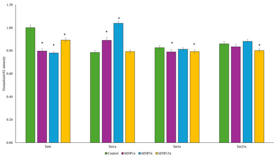
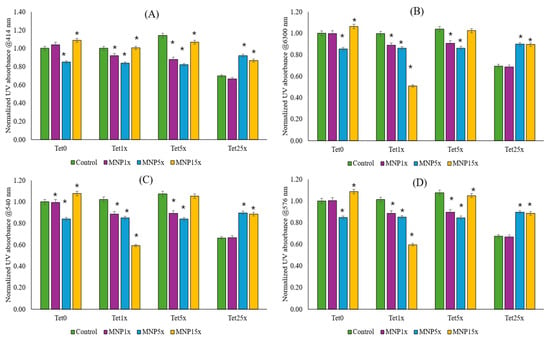
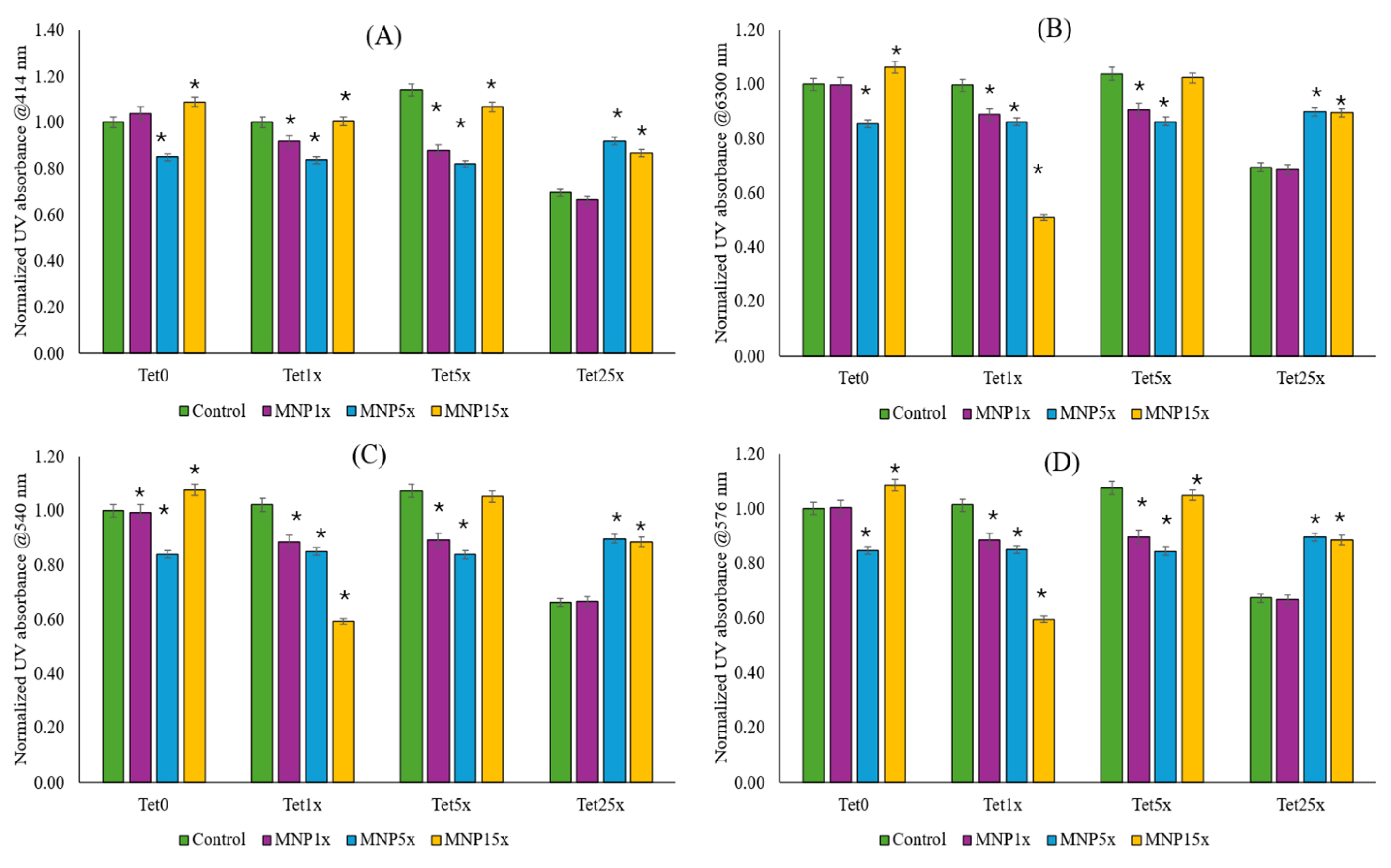
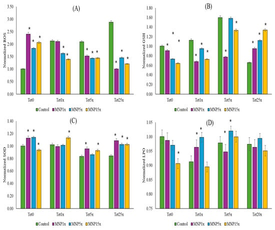
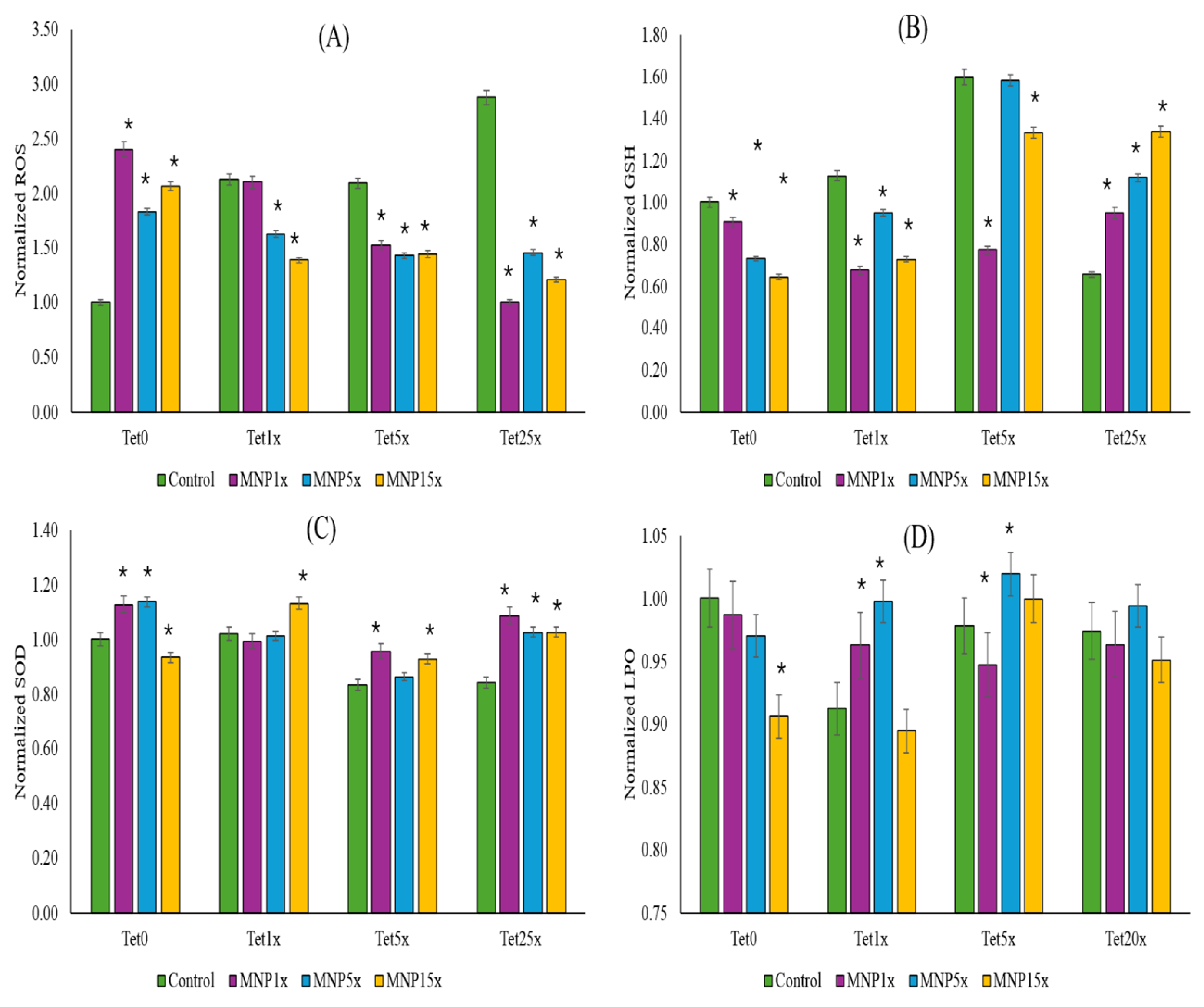
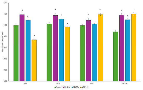
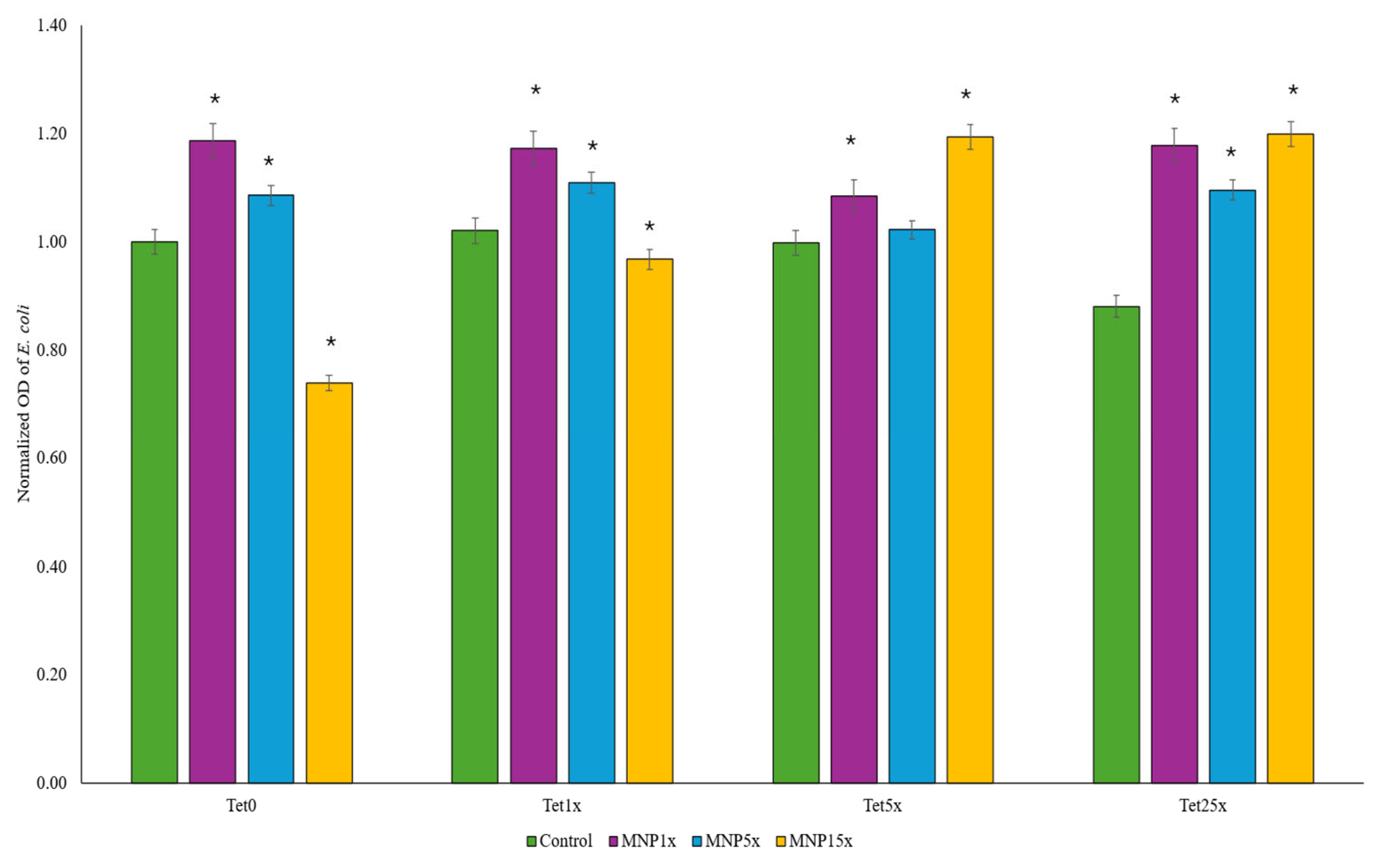
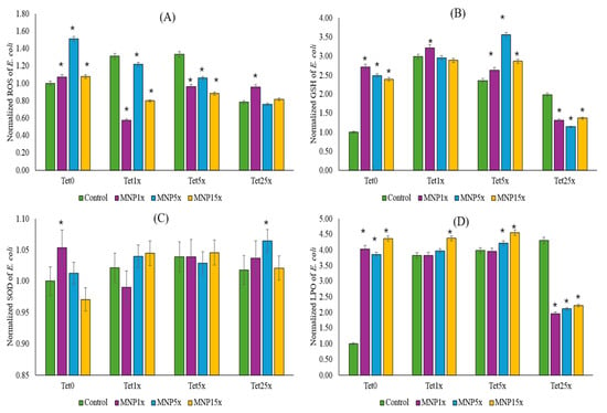
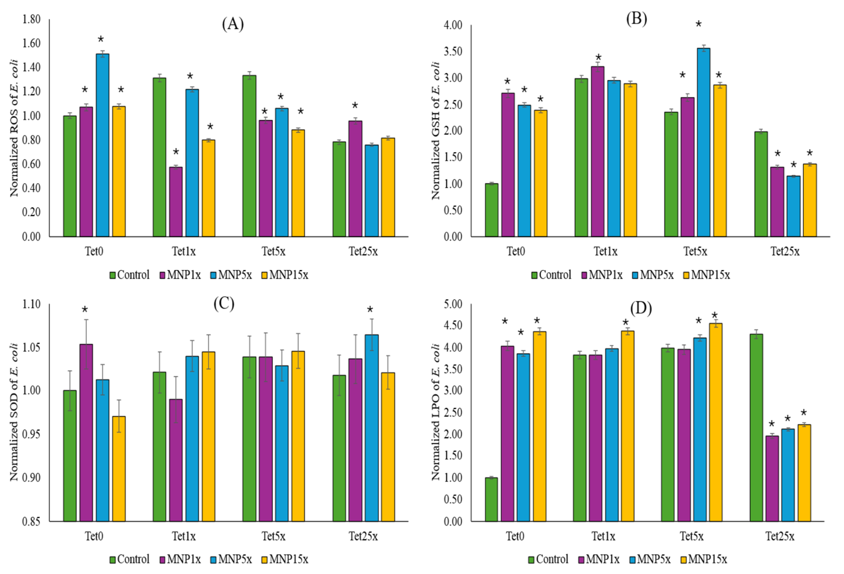
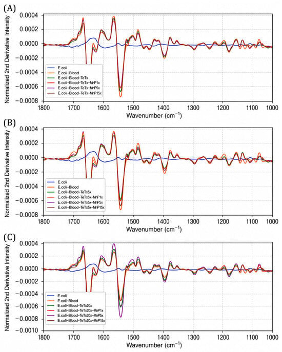
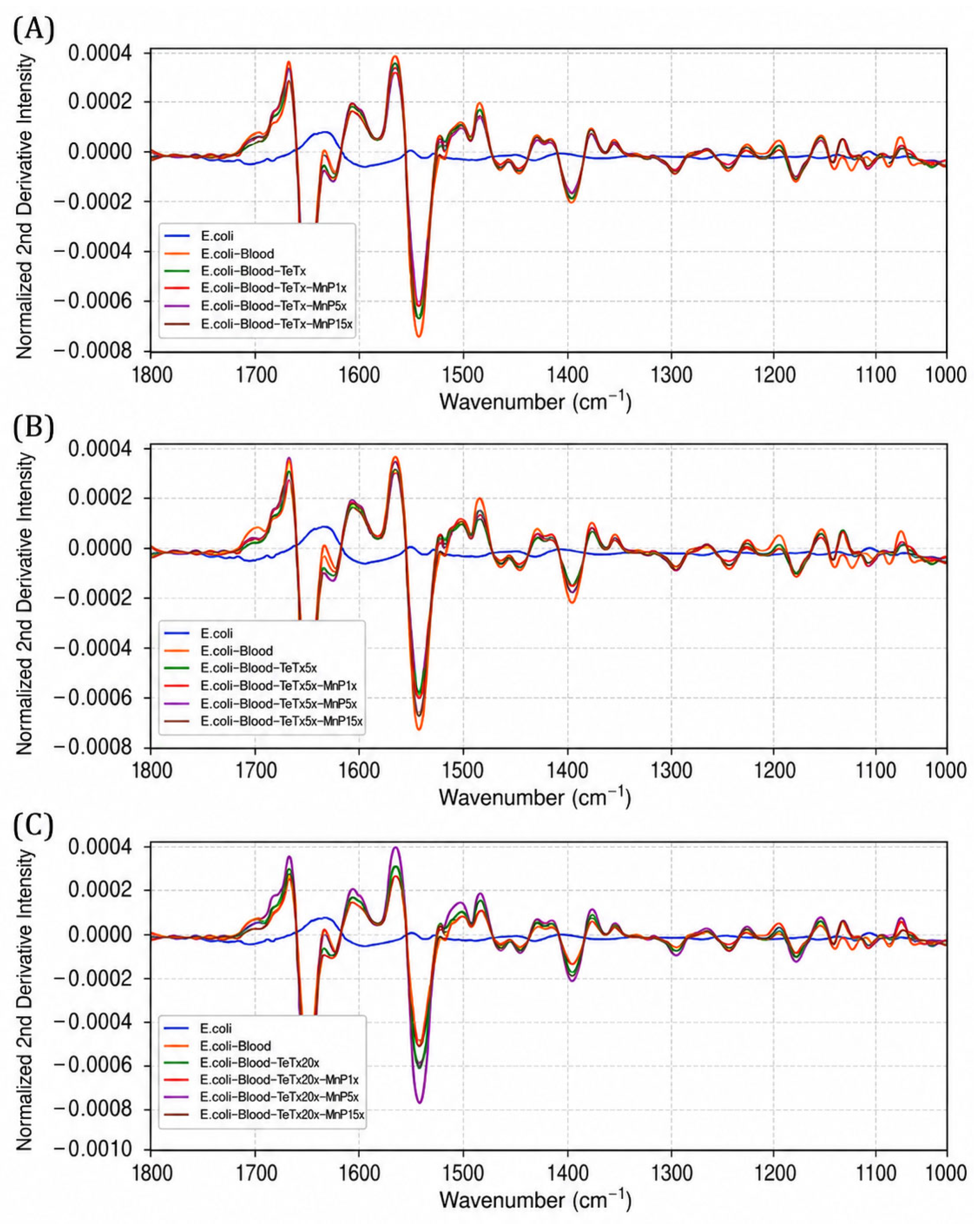

Disclaimer/Publisher’s Note: The statements, opinions and data contained in all publications are solely those of the individual author(s) and contributor(s) and not of MDPI and/or the editor(s). MDPI and/or the editor(s) disclaim responsibility for any injury to people or property resulting from any ideas, methods, instructions or products referred to in the content. |
© 2026 by the authors. Licensee MDPI, Basel, Switzerland. This article is an open access article distributed under the terms and conditions of the Creative Commons Attribution (CC BY) license.