


Abstract
Microplastics (MPs) are widely distributed among humans and companion animals. Fecal samples from members of the university community at the Pontifical Javeriana University in Bogotá, Colombia, and their companion animals (pets) were analyzed to investigate a possible association between MP loads in humans and their dogs and/or cats. Structures compatible with microplastics were detected in the feces of 97.78% of humans and 100% of companion animals. No significant correlation was found between MP loads in humans and their companion animals (p = 0.6846), suggesting that exposure is not determined solely by the shared household environment. If the home were the dominant and exclusive source of exposure, a positive correlation between owners and their companion animals would be expected. Instead, the results indicate the existence of species-specific exposure routes. This exploratory study provides the first report of MP-like structures in fecal samples from humans and their companion animals in Colombia.
1. Introduction
Plastic waste pollution in aquatic and terrestrial environments is currently one of the fastest-growing environmental problems worldwide. Prolonged exposure of plastic materials to physical abrasion and ultraviolet (UV) radiation causes their fragmentation into particles smaller than 5 mm, known as microplastics (MPs), and into even smaller particles (<1 µm), referred to as nanoplastics (NPs) [1,2]. These particles pose a significant threat to ecosystems because they can be ingested by aquatic organisms such as zooplankton and fish, which often mistake them for food, thereby facilitating their transfer through the food web.
In humans, MPs may enter the body through the consumption of contaminated food and beverages, including fish and salt [3], by inhalation [4], or through direct contact [5]. Increasing evidence indicates that MPs may induce adverse health effects, including oxidative stress and inflammation [6], intestinal damage [7], apoptosis, tissue necrosis, and fibrosis [8], as well as potential associations with infertility, obesity, and cancer [9]. Nevertheless, the long-term health consequences of MP exposure remain poorly understood.
Experimental studies in mice have demonstrated that ingested MPs can accumulate in organs such as the liver, kidneys, and intestine, leading to alterations in energy and lipid metabolism, oxidative stress, and impaired immune cell function. MPs have also been associated with intestinal damage extending from the proximal small intestine to the colon, together with significant changes in key intestinal biomarkers [10]. In addition to their direct biological effects, MPs may act as vectors for pathogenic microorganisms [11]. This is related to the ability of microorganisms to adhere to plastic surfaces and develop biofilms [12]. The microbial communities colonizing plastic debris have been collectively termed the plastisphere [13]. Furthermore, the physicochemical properties of MPs may facilitate the attachment of protozoa to pre-existing biofilms, potentially enhancing the persistence and dissemination of pathogenic microorganisms [14].
The detection of MPs in both humans and animals has raised concerns about their potential role in transporting biological contaminants [15,16], thereby increasing the risk of pathogen transmission, including possible zoonotic and anthroponotic pathways between humans and their companion animals.
One approach for assessing MP exposure in humans and animals is the analysis of fecal samples. A pilot study conducted by Schwabl et al. (2019) detected approximately 20 microplastic particles per 10 g of human feces, with polyethylene (PE), polyethylene terephthalate (PET), and polystyrene (PS) identified as the predominant polymers [17]. Similarly, PE was detected in 50% of stool samples collected from Chinese students, with particle sizes ranging from 20 to 800 µm [18]. Despite these findings, the burden of MPs in the human gastrointestinal tract remains poorly understood [17].
Studies on fecal samples from companion animals have likewise reported the presence of MPs. In cats, polyethylene terephthalate (PET) concentrations ranged from <2300 to 340,000 ng/g, while polycarbonate (PC) concentrations ranged from <32 to 13,000 ng/g. In dogs, PET concentrations ranged from 7700 to 190,000 ng/g, whereas PC concentrations ranged from <32 to 26,000 ng/g [19].
To date, most studies have investigated the occurrence of MPs independently in different environmental compartments or living organisms. A review of twelve studies reported that MPs were detected most frequently in human feces (75%), whereas substantially lower detection rates were observed in dogs and cats (8% each) [20]. However, no information is currently available regarding the occurrence of MPs in fecal samples from companion animals and their owners in Colombia. Therefore, as an initial step toward strengthening microplastics research in the country, the present study aimed to determine the presence of MPs, or structures compatible with MPs, in fecal samples collected from humans and their companion animals (dogs and cats) within the community of the Faculty of Science at the Pontificia Universidad Javeriana (PUJ), Bogotá, Colombia.
2. Materials and Methods
2.1. Study Population and Ethical Approval
This observational study used a convenience sampling approach based on the voluntary participation of members of the Faculty of Science at the Pontificia Universidad Javeriana (PUJ), Bogotá, Colombia, and their companion animals (dogs and/or cats). Participants were recruited through a collective invitation distributed within the faculty community.
The study was conducted in accordance with ethical principles and was approved by the Institutional Research and Ethics Committee (ICD Act No. 7/2024) and the Institutional Committee for the Care and Use of Laboratory Animals (CICUAL No. C-734-24; FUA No. 161-2). All participants provided written informed consent authorizing the analysis of both their own fecal samples and those of their companion animals.
Before sample collection, participants received an information sheet describing the objectives of the study and an instructional video explaining the sample collection procedure, including measures to minimize cross-contamination and the correct submission of samples to the laboratory.
Participants also completed a structured questionnaire that collected information on sociodemographic characteristics, dietary habits, deworming practices, hygiene behaviors, selected physiological conditions, bowel movement frequency, and potential sources of microplastic exposure. Equivalent information was collected for companion animals whenever applicable.
2.2. Sample Collection and Laboratory Procedures
Each participant received a sample collection kit containing sterile glass containers and wooden spatulas. Fecal samples were collected immediately after spontaneous defecation following the procedures described in the instructional video (https://www.youtube.com/watch?v=4LR0QSaFND0) (accessed on 21 July 2026). Sample collection was conducted between August 2024 and September 2025 at the Faculty of Science, Pontificia Universidad Javeriana, Bogotá, Colombia.
To minimize external contamination by MPs or MP-like structures, all laboratory procedures were performed on work surfaces previously cleaned with 70% ethanol followed by 5% hydrochloric acid. Only sterilized glassware (sterilized within the previous 24 h) was used throughout the analysis. Cotton laboratory clothing was worn to reduce the release of synthetic fibers, nitrile gloves were avoided, and paper bags were used whenever possible instead of plastic materials.
2.3. Detection and Quantification of MP-like Structures
MP-like structures were isolated by digesting 3 g of fecal material with 10 mL of 30% (v/v) hydrogen peroxide (H2O2) at 60 °C for 12 h to remove organic matter, followed by filtration through a nitrocellulose membrane. As procedural contamination control, an unused nitrocellulose membrane was exposed to the laboratory environment for the same period and processed under identical conditions.
The retained particles were examined and counted using a Zeiss® Stemi 350 stereomicroscope (Carl Zeiss, Jena, Germany) equipped with 8×–40× magnification objectives [21,22]. Particles exhibiting morphological characteristics consistent with microplastics, including symmetrical and unbranched fibers with uniform coloration and no visible organic tissue, were classified as MP-like structures [21,23].
All detected structures were categorized according to their morphology (e.g., fiber or fragment) and color. The total number of MP-like structures retained on the entire membrane surface was recorded, and particle abundance was expressed as the number of particles recovered from 3 g of fecal material.
2.4. Data Analysis
All data were compiled in Microsoft Excel and analyzed using descriptive statistics.
The distribution of MP-like structure counts was evaluated using the Shapiro–Wilk test. Because the data were not normally distributed and the study followed a matched-pair design (44 human–companion animal pairs), differences in MP-like structure abundance between humans and their companion animals were assessed using a two-tailed Wilcoxon signed-rank test.
To estimate the magnitude of these differences, the Hodges–Lehmann median difference estimator and the matched-pair rank-biserial correlation (rrbs) were calculated, together with their corresponding 95% confidence intervals (95% CIs). Effect sizes were interpreted according to the guidelines proposed by Funder and Ozer (2019) [24].
The association between MP-like structure abundance in owners and their companion animals was evaluated using Spearman’s rank correlation coefficient (ρ). Confidence intervals (95% CIs) were estimated using a nonparametric bootstrap procedure with 2000 replicates.
Finally, the effects of household plastic-use habits on MP-like structure abundance were evaluated using a generalized linear model (GLM) with a Poisson error distribution and a log-link function (n = 88 observations). The model included species (human, dog, or cat) and five explanatory variables related to household plastic exposure: consumption of beverages packaged in plastic containers, heating food in plastic containers, the use of plastic kitchen utensils, frequency of food delivery orders, and the use of plastic food wrap (cling film). The significance of the explanatory variables was assessed using Type III analysis of deviance based on likelihood-ratio chi-square tests.
All statistical analyses were performed using R version 4.4.1 (R Core Team, 2024), and statistical significance was established at α = 0.05.2.
3. Results
3.1. Detection of MP-like Structures
A total of 88 fecal samples were analyzed, including 44 from humans and 44 from companion animals (17 dogs and 27 cats). MP-like structures were detected in 43 of the 44 human samples (97.7%) and in all companion animal samples (100%).
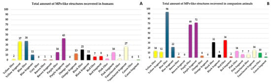
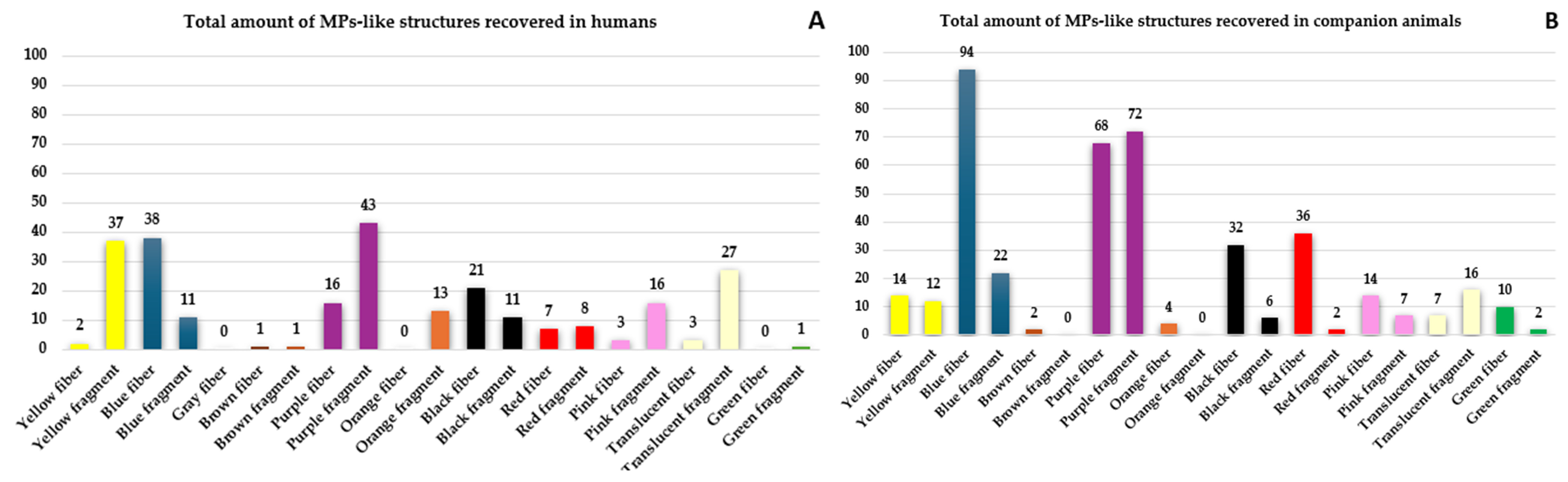
Overall, 680 MP-like structures were recovered. Of these, 250 particles (36.8%) were detected in human feces, 269 (39.6%) in cats, and 161 (23.7%) in dogs. Fibers accounted for 54.8% of all recovered MP-like structures, whereas fragments represented the remaining 45.2% (Figure 1).
Figure 1.
Distribution of MP-like structures according to particle morphology and color in (A) humans and (B) companion animals. Purple fragments and blue fibers were the most frequently observed particle types.
Table 1 summarizes the abundance of fragments and fibers recovered from human and companion animal fecal samples. Although fragments predominated in human samples (51.8%), fibers represented the dominant morphology in companion animals (75.6%).
Table 1.
Number and percentage of MP-like structures recovered from human and companion animal fecal samples.
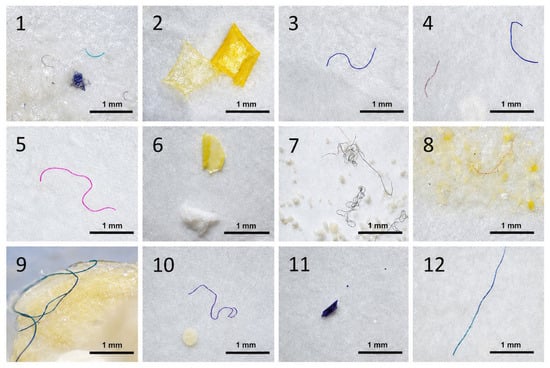
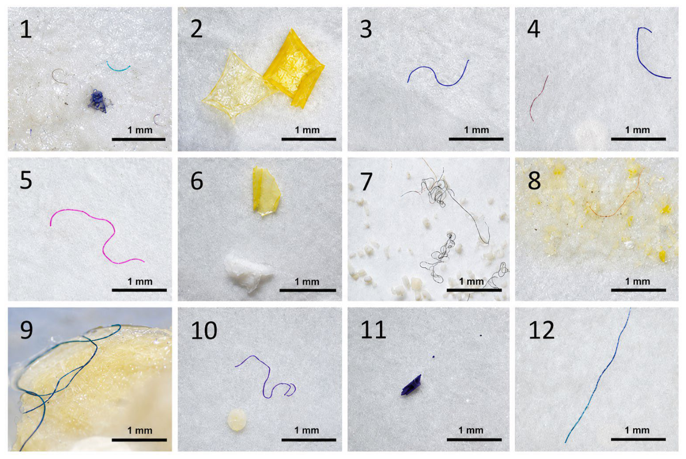
Figure 2 illustrates representative examples of the MP-like structures identified under the stereomicroscope, including both fragments and fibers recovered from human, dog, and cat fecal samples.
Figure 2.
Representative MP-like structures observed under a Zeiss® Stemi 350 stereomicroscope (8×–40× magnification). Images (1,3–5,7,9,10) correspond to cats; images (2,8,12) to dogs; and images (6,11) to humans.
3.2. Structured Survey
Most participants were undergraduate or graduate students (72.7%), whereas professors and administrative staff accounted for the remaining 27.3%. Women represented 86.4% of the study population, and men represented 13.6%.
Approximately 80% of participants reported consuming beverages, storing food, or eating meals from plastic containers. A similar proportion indicated that their companion animals consumed food stored or served in plastic containers.
Regarding bowel movement frequency, 38.6% of participants reported one bowel movement per day, 29.5% one every two to three days, 25.0% two per day, 4.5% more than two per day, and 2.3% fewer than one every three days. Among companion animals, the most common frequency was two bowel movements per day (45.5%), followed by more than two per day (31.8%), one per day (18.2%), and one every two to three days (4.5%).
According to the Bristol Stool Scale, type 3 stool was the most frequently reported category in both humans and companion animals, accounting for 34.1% of observations [25].
3.3. Statistical Analysis
The Wilcoxon signed-rank test showed significant differences in MP-like structure abundance between humans and their companion animals (p = 0.0231). The Hodges–Lehmann estimator indicated that companion animals had a median of 0.75 more particles than their owners (95% CI: 0.125–1.500). This difference corresponded to a moderate effect size (matched-pair rank-biserial correlation, rrbs = 0.39; 95% CI: 0.08–0.63) [24].
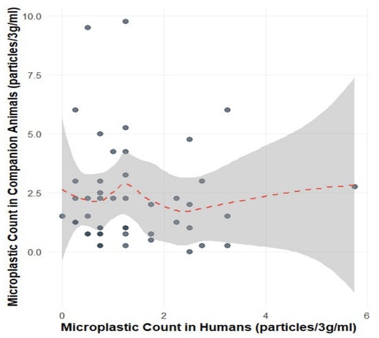
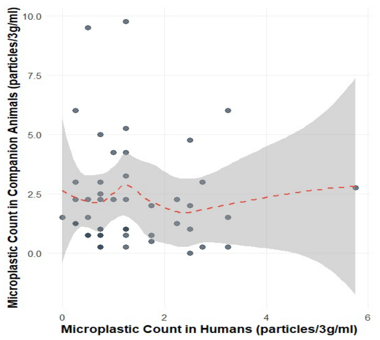
No significant correlation was found between the abundance of MP-like structures in humans and their companion animals (Spearman’s ρ = −0.060, 95% CI: −0.3177 to 0.2279, p = 0.6846), indicating that particle counts in owners were not associated with those observed in their corresponding pets (Figure 3).
Figure 3.
Relationship between MP-like structure counts in paired human and companion animal samples. Each point represents one owner–pet pair. The dashed red line represents the LOESS regression, and the shaded area indicates the corresponding 95% confidence interval.
The generalized linear model (Poisson distribution) identified species as a significant predictor of MP-like structure abundance (likelihood-ratio χ2 = 38.03, p < 0.001). Among the household exposure variables, the use of plastic food wrap (likelihood-ratio χ2 = 28.14, p < 0.001), heating food in plastic containers (χ2 = 24.07, p < 0.001), the use of plastic kitchen utensils (χ2 = 22.60, p < 0.001), and frequent consumption of delivered food (χ2 = 16.38, p < 0.001) were significantly associated with higher MP-like structure counts. In contrast, consumption of beverages packaged in plastic was not significantly associated with particle abundance (χ2 = 3.26, p = 0.354).
Descriptive statistics are presented in Table 2. Companion animals showed higher MP-like structure counts than humans, with a mean (± SD) of 8.91 ± 8.74 particles (median = 6, IQR = 8.25) compared with 5.41 ± 4.45 particles in humans (median = 4, IQR = 4.75).
Table 2.
Descriptive statistics for MP-like structures recovered from human and companion animal fecal samples.
4. Discussion
Quantification of MP-like structures in fecal samples provides a useful, non-invasive approach for assessing recent exposure by estimating both the abundance and morphology of particles excreted [26,27]. Because ingestion is considered one of the principal routes of microplastic exposure, fecal samples have become an increasingly valuable biomonitoring matrix for evaluating gastrointestinal exposure. Consequently, fecal analysis may provide insights into recent dietary and environmental exposure while serving as an early indicator of contamination. Previous studies have shown that factors such as diet, age, geographic location, and the consumption of plastic-packaged foods may influence the abundance of MPs detected in feces [28,29,30,31].
In the present study, companion animals exhibited a higher number of MP-like structures than humans. Although the underlying mechanisms were not investigated, this difference may reflect distinct exposure pathways between the two groups. Companion animals spend more time in close contact with floors and household dust, which has been recognized as an important environmental reservoir of microplastics. In addition, species-specific behaviors such as grooming, paw licking, and chewing polymer-based toys may increase the likelihood of ingesting plastic particles. Dietary factors may also contribute, as commercial pet foods are commonly stored in plastic packaging for extended periods. Nevertheless, these potential sources of exposure were not evaluated in the present study and should be investigated in future research.
Fragments and fibers were the predominant MP-like structures identified in fecal samples, whereas spherical particles were not observed. Similar particle profiles have been reported in previous studies. For example, Elamparuthi et al. found that fibers and fragments, particularly transparent, white, and black particles, predominated in fecal samples from wild animals [32]. Likewise, Gómez-Méndez et al. reported that blue fibers, purple fibers and fragments, and black fibers were the most abundant MPs detected in bees and bee-derived products [21]. Although differences in sampling matrices limit direct comparisons, these findings suggest that fibers and fragments are consistently the dominant particle morphologies across diverse biological systems.
Because the recovered particles were identified exclusively by their morphological characteristics, their polymer composition could not be confirmed. This represents one of the main limitations of the present study. Previous investigations using spectroscopic techniques have shown that polyethylene terephthalate (PET), polypropylene (PP), polyethylene (PE), and polystyrene (PS) are among the polymers most frequently detected in human feces [17,20,28,33]. In companion animals, PET and polycarbonate (PC) have been commonly reported, while PET, PE, PP, polytetrafluoroethylene (PTFE), and butyl benzyl phthalate (BBP) have also been detected in commercial pet foods [34]. Although these findings provide useful context, the present study cannot infer the polymer composition of the recovered MP-like structures because no chemical characterization was performed.
The use of fecal samples as a biomonitoring matrix also presents several methodological challenges. MPs are generally present at relatively low concentrations and are embedded within complex organic matrices, requiring efficient digestion procedures and rigorous contamination-control measures to obtain reliable results. Moreover, differences in analytical protocols among studies, including sample processing, particle identification, and reporting criteria, continue to hinder direct comparisons and emphasize the need for standardized methodologies for fecal MP biomonitoring [35].
Beyond their occurrence, the potential biological implications of MP-like structures deserve consideration. Experimental and epidemiological studies have shown that microplastics may induce physical, chemical, and biological effects, including oxidative stress, inflammation, and alterations in the gut microbiota. Furthermore, MPs can act as carriers of toxic chemicals, proteins, pathogenic microorganisms, antimicrobial compounds, and antimicrobial resistance genes, potentially contributing to the environmental dissemination of antimicrobial resistance and other health hazards [2,36,37]. Although the present study did not evaluate these effects, the widespread occurrence of MP-like structures in both humans and companion animals highlights the need to investigate their possible biological consequences.
Despite the growing body of evidence, important knowledge gaps remain regarding human and animal exposure to MPs and their potential health effects [26,29,38]. Additional uncertainty exists concerning whether the close coexistence between people and their companion animals may facilitate shared exposure to MPs or promote the transfer of microorganisms associated with plastic particles under a One Health framework. Future studies should therefore include physicochemical characterization of recovered particles using spectroscopic techniques, together with the identification of contamination sources and the evaluation of associated microbial communities. To our knowledge, this study represents the first report of MP-like structures in fecal samples from humans and their companion animals in Colombia, providing a baseline for future investigations on exposure pathways, contamination sources, and potential health implications from a One Health perspective.
5. Conclusions
The detection of MP-like structures in 97.7% of human fecal samples and in all samples collected from companion animals indicates widespread exposure within the study population. These findings suggest that contact with MP-like structures is common among both humans and their companion animals living in the same urban environment.
Companion animals exhibited a significantly higher number of MP-like structures than their owners. Both the Wilcoxon signed-rank test and the Poisson generalized linear model identified species as a significant factor associated with particle abundance. The estimated median difference of 0.75 particles and the moderate effect size indicate that this difference was not only statistically significant but also quantitatively meaningful.
Fibers represented the predominant particle morphology (54.8%), followed by fragments (45.2%). This pattern is consistent with previous studies reporting fibers as the most frequently detected microplastic morphology in biological samples, likely reflecting the widespread release of synthetic fibers from textiles and other household sources.
Among the household variables evaluated, the use of plastic food wrap, heating food in plastic containers, the use of plastic kitchen utensils, and frequent consumption of delivered food were significantly associated with higher abundances of MP-like structures. In contrast, consumption of beverages packaged in plastic was not significantly associated with particle counts after adjustment for the remaining variables.
Although humans and their companion animals shared the same household environment, no significant correlation was observed between the numbers of MP-like structures detected in paired fecal samples. This finding suggests that, in addition to shared environmental exposure, species-specific physiological, dietary, and behavioral factors may also influence the ingestion and excretion of MP-like structures.
Companion animals also exhibited greater interindividual variability than humans, as reflected by their higher mean, median, and dispersion of particle counts. This variability may indicate differences in environmental exposure, feeding behavior, or gastrointestinal processing among individual animals, although these factors were not specifically investigated in the present study.
The demographic characteristics of the participants should also be considered when interpreting the findings. The study population consisted predominantly of women and university students, and the widespread use of plastic materials for food storage and consumption was reported for both participants and their companion animals. Consequently, caution should be exercised when extrapolating these results to populations with different demographic or socioeconomic characteristics.
Overall, these findings suggest that the household environment may represent an important setting for exposure to MP-like structures in both humans and companion animals. The identification of household practices associated with increased particle abundance highlights potential exposure pathways that warrant further investigation.
From a One Health perspective, this study demonstrates that MP-like structures are widely detected in both humans and their companion animals, although the magnitude of particle abundance differs between species. These results provide baseline information for Colombia and support the need for integrated studies combining environmental, animal, and human health approaches, including physicochemical characterization of recovered particles and evaluation of their potential biological effects.
Author Contributions
Conceptualization, A.P.-V. and L.D.G.-M.; methodology, A.P.-V., L.D.G.-M., V.C.-A., V.A.-P. and S.J.G.-G.; investigation, A.P.-V., L.D.G.-M., V.C.-A., V.A.-P. and S.J.G.-G.; writing—original draft preparation, L.D.G.-M., V.A.-P. and A.P.-V.; data analysis, A.P.-V., M.A.-S. and L.D.G.-M.; writing—review and editing, L.D.G.-M., V.A.-P. and A.P.-V.; project administration, A.P.-V. and L.D.G.-M.; funding acquisition, A.P.-V. All authors have read and agreed to the published version of the manuscript.
Funding
This research was funded by Pontificia Universidad Javeriana, grant number VRI-ID20762. In addition, the publication of this article was supported by the call for proposals “Support for the publication of high-quality articles (VRI01-2026): ID PPTA 22456”.
Institutional Review Board Statement
The study was conducted in accordance with ethical principles and was approved by the Institutional Research and Ethics Committee (ICD Act No. 7/2024) and the Institutional Committee for the Care and Use of Laboratory Animals (CICUAL No. C-734-24; FUA No. 161-2). All participants provided written informed consent authorizing the analysis of both their own fecal samples and those of their companion animals.
Informed Consent Statement
Informed consent was obtained from all subjects involved in the study.
Data Availability Statement
The datasets presented in this article are not readily available because the primary data involve members of a university community and are protected under Colombian data protection regulations (Habeas Data Law, Ley 1581 de 2012), with the Pontificia Universidad Javeriana acting as the data custodian to ensure participant privacy and data integrity. Requests to access the datasets should be directed to the corresponding authors (A.P.-V.: adriana.pulido@javeriana.edu.co; L.D.G.-M.: luis.gomez@javeriana.edu.co).
Acknowledgments
Thanks are extended to the community of the Faculty of Sciences of the PUJ, who participated voluntarily; to the Management Support team of the same faculty for their support, willingness to participate, and facilitating the use of the teaching laboratories; and to the research groups of “Enfermedades Infecciosas Veterinarias y Zoonosis” and “Degradación de polímeros plásticos contaminantes”.
Conflicts of Interest
The authors declare no conflicts of interest.
References
- Vethaak, A.D.; Legler, J. Microplastics and human health. Knowledge gaps should be addressed to ascertain the health risks of microplastics. Science 2021, 371, 672–674. [Google Scholar] [CrossRef] [Scilit] [PubMed]
- Gross, N.; Muhvich, J.; Ching, C.; Gomez, B.; Horvath, E.; Nahum, Y.; Zaman, M.H. Effects of microplastic concentration, composition, and size on Escherichia coli biofilm-associated antimicrobial resistance. Appl. Environ. Microbiol. 2025, 91, e02282-24. [Google Scholar] [CrossRef] [Scilit] [PubMed]
- Karbalaei, S.; Hanachi, P.; Walker, T.R.; Cole, M. Occurrence, sources, human health impacts and mitigation of microplastic pollution. Environ. Sci. Pollut. Res. 2018, 25, 36046–36063. [Google Scholar] [CrossRef] [Scilit] [PubMed]
- Jenner, L.C.; Rotchell, J.M.; Bennett, R.T.; Cowen, M.; Tentzeris, V.; Sadofsky, L.R. Detection of microplastics in human lung tissue using μFTIR spectroscopy. Sci. Total Environ. 2022, 831, 154907. [Google Scholar] [CrossRef] [Scilit] [PubMed]
- Kannan, K.; Vimalkumar, K. A review of human exposure to microplastics and insights into microplastics as obesogens. Front. Endocrinol. 2021, 12, 724989. [Google Scholar] [CrossRef] [Scilit] [PubMed]
- Hu, M.; Palić, D. Micro-and nano-plastics activation of oxidative and inflammatory adverse outcome pathways. Redox Biol. 2020, 37, 101620. [Google Scholar] [CrossRef] [Scilit] [PubMed]
- Prata, J.C.; da Costa, J.; Lopes, I.; Duarte, A.C.; Rocha-Santos, T. Environmental exposure to microplastics: An overview on possible human health effects. Sci. Total Environ. 2020, 702, 134455. [Google Scholar] [CrossRef] [Scilit] [PubMed]
- Bhagat, J.; Zang, L.; Nishimura, N.; Shimada, Y. Zebrafish: An emerging model to study microplastic and nanoplastic toxicity. Sci. Total Environ. 2020, 728, 138707. [Google Scholar] [CrossRef] [Scilit] [PubMed]
- Prüst, M.; Meijer, J.; Westerink, R.H. The plastic brain: Neurotoxicity of micro-and nanoplastics. Part. Fibre Toxicol. 2020, 17, 24. [Google Scholar] [CrossRef] [Scilit] [PubMed]
- Pastor-Belda, M.; Campillo, N.; Arroyo-Manzanares, N.; Torres, C.; Pérez-Cárceles, M.D.; Hernández-Córdoba, M.; Viñas, P. Bioaccumulation of Polycyclic Aromatic Hydrocarbons for Forensic Assessment Using Gas Chromatography–Mass Spectrometry. Chem. Res. Toxicol. 2019, 32, 1680–1688. [Google Scholar] [CrossRef] [Scilit] [PubMed]
- Djouina, M.; Vignal, C.; Dehaut, A.; Caboche, S.; Hirt, N.; Waxin, C.; Himber, C.; Beury, D.; Hot, D.; Dubuquoy, L.; et al. Oral exposure to polyethylene microplastics alters gut morphology, immune response, and microbiota composition in mice. Environ. Res. 2022, 212, 113230. [Google Scholar] [CrossRef] [Scilit] [PubMed]
- Ye, G.; Li, M.; Huang, H.; Avellán-Llaguno, R.D.; Chen, J.; Chen, G.; Huang, Q. Polystyrene microplastic exposure induces selective accumulation of antibiotic resistance genes in gut microbiota and its potential health risks. Int. J. Biol. Macromol. 2025, 309, 142983. [Google Scholar] [CrossRef] [Scilit] [PubMed]
- Zettler, E.R.; Mincer, T.J.; Amaral-Zettler, L.A. Life in the “plastisphere”: Microbial communities on plastic marine debris. Environ. Sci. Technol. 2013, 47, 7137–7146. [Google Scholar] [CrossRef] [Scilit] [PubMed]
- He, Y.; Wei, G.; Tang, B.; Salam, M.; Shen, A.; Wei, Y.; Zhou, X.; Liu, M.; Yang, Y.; Li, H.; et al. Microplastics benefit bacteria colonization and induce microcystin degradation. J. Hazard. Mater. 2022, 431, 128524. [Google Scholar] [CrossRef] [Scilit] [PubMed]
- Cox, K.D.; Covernton, G.A.; Davies, H.L.; Dower, J.F.; Juanes, F.; Dudas, S.E. Human Consumption of Microplastics. Environ. Sci. Technol. 2019, 53, 7068–7074. [Google Scholar] [CrossRef] [Scilit] [PubMed]
- Pennino, M.G.; Bachiller, E.; Lloret-Lloret, E.; Albo-Puigserver, M.; Esteban, A.; Jadaud, A.; Bellido, J.M.; Coll, M. Ingestion of microplastics and occurrence of parasite association in Mediterranean anchovy and sardine. Mar. Pollut. Bull. 2020, 158, 111399. [Google Scholar] [CrossRef] [Scilit] [PubMed]
- Schwabl, P.; Koppel, S.; Konigshofer, P.; Bucsics, T.; Trauner, M.; Reiberger, T.; Liebmann, B. Detection of various microplastics in human stool: A prospective case series. Ann. Intern. Med. 2019, 171, 453–457. [Google Scholar] [CrossRef] [Scilit] [PubMed]
- Zhang, N.; Li, Y.B.; He, L.H.; Zhang, J.F.; Ma, G.S. You are what you eat: Microplastics in the feces of young men living in Beijing. Sci. Total Environ. 2021, 767, 144345. [Google Scholar] [CrossRef] [Scilit] [PubMed]
- Sangkham, S.; Phairuang, W.; Sakunkoo, P.; Ta, A.T. A review on microplastics in mammalian feces: Monitoring techniques and associated challenges. Environ. Chall. 2025, 20, 101217. [Google Scholar] [CrossRef] [Scilit]
- Mollova, E.; Ivanona, E.; Turmanova, S.C.H.; Dimirtov, A. Microplastics–Ecosystem pollutant. J. Serb. Chem. Soc. 2024, 89, 939–980. [Google Scholar] [CrossRef] [Scilit]
- Gómez-Méndez, L.D.; Robles-Camargo, J.E.; Vera-Bravo, R.; Moncaleano-Niño, A.M.; Devia Castillo, C.A.; Ospina-Torres, R.; Escobar-Cortés, Y.; Camacho-Erazo, M.; Amarillo-Suárez, A.R. Characterization of Microplastics in Bees and Their Products in Urban and Rural Areas of the Sabana De Bogotá, Colombia. Microplastics 2024, 3, 589–598. [Google Scholar] [CrossRef] [Scilit]
- Porras-Rojas, M.A.; Charry-Vargas, C.; Muñoz-Yustres, J.L.; Martínez-Silva, P.; Gómez-Méndez, L.D. Characterization of microplastics and mesoplastics and presence of biofilms, collected in the Gualí Wetland Cundinamarca, Colombia. Microplastics 2023, 2, 255–267. [Google Scholar] [CrossRef] [Scilit]
- Hidalgo-Ruz, V.; Gutow, L.; Thompson, R.C.; Thiel, M. Microplastics in the marine environment: A review of the methods used for identification and quantification. Environ. Sci. Technol. J. 2012, 46, 3060–3075. [Google Scholar] [CrossRef] [Scilit] [PubMed]
- Funder, D.C.; Ozer, D.J. Evaluating Effect Size in Psychological Research: Sense and Nonsense. Adv. Methods Pract. Psychol. Sci. 2019, 2, 156–168. [Google Scholar] [CrossRef] [Scilit]
- Riegler, G.; Esposito, I. Bristol Scale Stool Form. A still valid help in medical practice and clinical research. Tech. Coloproctol. 2001, 5, 163–164. [Google Scholar] [CrossRef] [Scilit] [PubMed]
- Alvarez-Andrade, A.; Wakida, F.; Piñon-Collin, T.; Wakida-Kusunoki, A.T.; Castillo-Quiñonez, J.E.; García-Florez, E. Microplastics abundance of lagomorphs in relation to urbanization. Sci. Total. Environ. 2023, 864, 161025. [Google Scholar] [CrossRef] [Scilit] [PubMed]
- Li, Z.M.; Kannan, K. Urinary and fecal excretion of 121 environmental chemicals in pet dogs and cats: Significance of feces in biomonitoring of exposures. Environ. Sci. Technol. 2024, 58, 14169–14179. [Google Scholar] [CrossRef] [Scilit] [PubMed]
- Ali, W.; Ali, H.; Gillani, S.; Zinck, P.; Souissi, S. Polylactic acid synthesis, biodegradability, conversion to microplastics and toxicity: A review. Environ. Chem. Lett. 2023, 21, 1761–1786. [Google Scholar] [CrossRef] [Scilit]
- Hartmann, C.; Lomako, I.; Schachner, C.; El Said, E.; Abert, J.; Satrapa, V.; Kaiser, A.-M.; Walch, H.; Köppel, S. Assessment of microplastics in human stool: A pilot study investigating the potential impact of diet-associated scenarios on oral microplastics exposure. Sci. Total Environ. 2024, 951, 175825. [Google Scholar] [CrossRef] [Scilit] [PubMed]
- Casella, C.; Cornelli, U.; Ballaz, S.; Luque, R.; Porta, A.; Zanoni, G. Occurrence and characterization of microplastics in dry pet food: Investigating geographical variations between European and South American markets. Environ. Pollut. 2026, 406, 128610. [Google Scholar] [CrossRef] [Scilit] [PubMed]
- Zhang, S.; Li, Y.; Chen, X.; Jiang, X.; Li, J.; Yang, L.; Yin, X.; Zhang, X. Occurrence and distribution of microplastics in organic fertilizers in China. Sci. Total Environ. 2022, 844, 157061. [Google Scholar] [CrossRef] [Scilit] [PubMed]
- Elamparuthi, A.; Muniyandi, M. A Case Study of Feces Evidence of Macro and Microplastic Pollution in Western Ghats, India. Int. J. Environ. Res. 2026, 20, 111. [Google Scholar] [CrossRef] [Scilit]
- Lam, T.W.L.; Chow, A.S.Y.; Fok, L. Human exposure to microplastics via the consumption of nonalcoholic beverages in various packaging materials: The case of Hong Kong. J. Hazard. Mater. 2024, 472, 134575. [Google Scholar] [CrossRef] [Scilit] [PubMed]
- Sobhani, Z.; Lei, Y.; Tang, Y.; Wu, L.; Zhang, X.; Naidu, R.; Megharaj, M.; Fang, C. Microplastics generated when opening plastic packaging. Sci. Rep. 2020, 10, 4841. [Google Scholar] [CrossRef] [Scilit] [PubMed]
- Vibhatabandhu, P.; Srithongouthai, S. Abundance and Characteristics of Microplastics Contaminating the Surface Water of the Inner Gulf of Thailand. Water Air Soil Pollut. 2022, 233, 50. [Google Scholar] [CrossRef] [Scilit]
- Yan, Z.; Zhao, H.; Zhao, Y.; Zhu, Q.; Qiao, R.; Ren, H.; Zhang, Y. An efficient method for extracting microplastics from feces of different species. J. Hazard. Mater. 2020, 384, 121489. [Google Scholar] [CrossRef] [Scilit] [PubMed]
- Luqman, A.; Nugrahapraja, H.; Wahyuono, R.A.; Islami, I.; Haekal, M.H.; Fardiansyah, Y.; Putri, B.Q.; Amalludin, F.I.; Rofiqa, E.A.; Götz, F.; et al. Microplastic contamination in human stools, foods, and drinking water associated with Indonesian coastal population. Environments 2021, 8, 138. [Google Scholar] [CrossRef] [Scilit]
- Martínez-Cepeda, G.; Ramos-Sarmiento, D.; Sailema-Rosado, G.; Sangache-Guamán, A.; Bustos-Chávez, N.; Salcán-Guamán, H.; Fernández-Morales, M.; Díaz-Cevallos, L. Microplásticos en ambientes urbanos: El perro como bioindicador de exposición y riesgo gastrointestinal humano. Biomédica 2026, 46, 2–22. [Google Scholar]

Disclaimer/Publisher’s Note: The statements, opinions and data contained in all publications are solely those of the individual author(s) and contributor(s) and not of MDPI and/or the editor(s). MDPI and/or the editor(s) disclaim responsibility for any injury to people or property resulting from any ideas, methods, instructions or products referred to in the content. |
© 2026 by the authors. Licensee MDPI, Basel, Switzerland. This article is an open access article distributed under the terms and conditions of the Creative Commons Attribution (CC BY) license.