


Abstract
Background/Objectives: Extended-spectrum β-lactamase (ESBL)-producing Enterobacterales represent a major concern in neonatal intensive care units (NICUs), where colonization may precede infection and contribute to the spread of antimicrobial resistance. This study aimed to investigate the distribution of β-lactamase-encoding genes among Enterobacterales colonizing neonates in a Brazilian NICU. Methods: Rectal swab samples (n = 427) were collected over a one-year period. Forty-six isolates recovered on ESBL-selective chromogenic medium were subsequently phenotypically confirmed as ESBL producers by the disk approximation method. Bacterial identification was performed by MALDI-TOF MS, and the presence of blaCTX-M, blaSHV, and blaTEM genes was determined by PCR. Multidrug resistance (MDR) and Multiple Antibiotic Resistance (MAR) Index were determined according to international criteria. Results: Overall, 41.3% of isolates were classified as MDR, and the mean MAR Index was 0.234 ± 0.130. The blaCTX-M gene was the most frequently detected (33%), followed by blaSHV (13%) and blaTEM (11%). Simultaneous occurrence of multiple β-lactamase genes was observed in a small number of isolates (17.4%), mainly associated with Escherichia coli and Klebsiella pneumoniae. MDR was significantly associated with the detection of at least one of the investigated β-lactamase genes (83.3% vs. 14.3%; Fisher’s exact test, p < 0.001; OR = 30.0). Conclusions: Our findings show that β-lactamase genes, particularly the ESBL gene blaCTX-M, are present among colonizing Enterobacterales in neonates and highlight the importance of monitoring these genetic determinants in NICU settings.
1. Introduction
Infections caused by MDR Enterobacterales have become an important public health challenge, profoundly affecting the management of infected patients, high treatment costs, reduced quality of life, and high mortality rates [1,2]. Extended-spectrum β-lactamase-producing Enterobacterales (ESBL-E) infections are serious, difficult to treat, and have a high mortality rate, especially in critically ill and susceptible patients, such as newborns [3,4].
Newborns admitted to neonatal intensive care units (NICUs) are at high risk of infection due to a combination of factors, including an immature immune system, prolonged hospitalization, and frequent use of invasive devices [3,4,5]. Newborns in NICUs are usually colonized by ESBL-E during the hospital stay where they are exposed to invasive medical devices and long-term use of antibiotics [5,6,7]. Studies have shown a relationship between gut colonization and sepsis among newborns [6,8]. Treatment of ESBL-E infections in neonates and children is particularly challenging due to the limited number of available effective antimicrobials [9]. Surveillance studies in Rio de Janeiro have shown that ESBL-producing Klebsiella sp. and E. coli were the most frequently reported as associated with colonization agents in NICUs (36.9%) [10]. ESBL of the types CTX-M, TEM, and SHV and CMY-2, produced by Gram-negative bacteria, have been associated with the persistence of colonization by the same pathogen in the newborn [11].
ESBL CTX-M exhibits an emerging prevalence in Latin America, as it is the most widely disseminated and successful enzyme in various epidemiological scenarios worldwide [12,13]. Patients colonized by bacteria carrying ESBL-encoding genes are one of the main reservoirs of resistance mechanisms. Therefore, surveillance cultures are increasingly important. Screening is essential in NICUs where premature infants are admitted, as these are high-prevalence areas for ESBL-producing microorganisms that are capable of surviving in the environment, on the hands of healthcare professionals, and colonizing pregnant patients [14]. In this context, the aim of this study was to investigate the presence of ESBL-encoding genes carried by enterobacteria colonizing hospitalized newborns at Antônio Pedro University Hospital in Brazil, from February 2021 to February 2022.
2. Materials and Methods
This study was conducted with rectal swab samples collected from newborns from the NICU at Antonio Pedro University Hospital, Federal Fluminense University—Niterói—RJ/Brazil (HUAP—UFF), for one year (2021–2022). As a public hospital, it provides free medical care through the Unified Health System (UHS) and covers seven municipalities. The hospital has 175 beds, of which 16 are allocated to the NICU.
Rectal swabs were originally collected from newborns as part of the hospital’s routine infection prevention and control program, independently of the present research. The current study involved only bacterial isolates recovered from these surveillance cultures and subsequently preserved in the hospital’s microbiological collection. No additional biological samples were collected from newborns for research purposes, and no direct contact with patients occurred during the study. The microbiological and molecular analyses were performed exclusively on the previously cultured bacterial isolates. The original rectal swabs were not used in the research.
This study was approved by the Ethics and Research Committee of Antonio Pedro University Hospital/Medical School, UFF, under registration number: 95984018.6.0000.5243.
2.1. Bacterial Isolation
Bacterial isolates were collected from surveillance rectal swabs, according to the protocol instructions of the Hospital Infection Control Committee (HICC), which requires that surveillance cultures be collected shortly after the newborn’s birth and repeated weekly during their stay in the hospital. Analysis of bacterial samples was performed as part of the routine procedures in the Microbiology Laboratory of HUAP-UFF. Samples were selected based on growth observed on the selective and differential medium ChromAgar ESBL (Plastlabor, Rio de Janeiro, Brazil). Samples were initially screened based on growth on CHROMagar ESBL (Plastlabor, Rio de Janeiro, Brazil). Isolates recovered from the selective medium were subsequently subjected to phenotypic confirmation of ESBL production using the disk approximation method, as described below.
2.2. Bacterial Identification
Bacterial identification was carried out using Matrix Assisted Laser Desorption/Ionization Time of Flight Mass Spectrometry (MALDI-TOF-MS) technology. After incubation, isolated colonies, with at least 105 CFU/mL bacterial cells, were deposited in each sample onto the metal plate of the MALDI-TOF. This plate with the deposited samples was kept in a laminar flow for the drying of the deposited material. Then, 1 μL of a matrix composed of α-cyano-4-hydroxycinnamic acid (Sigma, St. Louis, MO, USA) prepared in 50% acetonitrile and 2.5% trifluoroacetic acid (Sigma, St. Louis, MO, USA) was added to each well of the plate containing a sample. Standard strain E. coli DH5α was used as a calibrant for the analyses and control. After the mixture (sample + matrix) dried and crystals formed, the metal plate was inserted into the equipment for sample reading. Isolates were run in duplicate on a ground steel MALDI-TOF target.
The obtained spectra were processed using Biotyper software version 3.1 (Bruker Daltonics, Billerica, MA, USA). As a result, a table containing the identification of microorganisms that showed spectra most similar to the test sample was presented. Analyses by Biotyper were classified using score values: a score between 2.300 and 3.000 indicates reliable species identification; a score between 2.000 and 2.299 indicates reliable genus identification and probable species identification; a score between 1.700 and 1.999 indicates probable genus identification; and a score below 1.700 indicates no reliable identification.
Species frequencies were expressed as absolute numbers and percentages. Because these data represent categorical proportions rather than repeated continuous measurements, variability was assessed using 95% confidence intervals (95% CIs) for proportions calculated using the Wilson score method.
2.3. Antimicrobial Susceptibility Testing and Phenotypic Confirmation of ESBL Production
Antimicrobial susceptibility testing (AST) was performed with the BD PhoenixTM M50 automated system (BD Diagnostics, Sparks, MD, USA) based on the manufacturer’s instructions. Susceptibility results were interpreted according to the Clinical and Laboratory Standards Institute (CLSI) M100 guidelines, 32nd edition (2022) [15].
All isolates were subjected to the disk approximation method as a confirmatory method for ESBL production, according to BrCAST recommendations (2018) [16].
2.4. Multidrug Resistance (MDR) Classification and Multiple Antibiotic Resistance (MAR) Index
MDR was defined according to the international criteria proposed by Magiorakos et al. [17]. Resistance traits considered intrinsic for each bacterial species were excluded from the MDR classification to avoid overestimation of acquired MDR. For the purposes of MDR classification, isolates categorized as Resistant (R) or Intermediate (I) (currently defined by CLSI as Susceptible, Increased Exposure) were considered non-susceptible. The association between β-lactamase gene detection profiles and the MDR phenotype was evaluated using the Fisher–Freeman–Halton exact test because of the small sample sizes and low expected frequencies in several categories. The strength of the association was quantified using Cramér’s V. In addition, isolates were grouped according to the detection of at least one of the investigated β-lactamase genes (blaCTX-M, blaSHV, and blaTEM) versus no detection of these three targets, and the association with MDR was assessed using Fisher’s exact test. The corresponding odds ratio (OR) was calculated as a measure of effect size. All tests were two-sided, and p < 0.05 was considered statistically significant.
The Multiple Antibiotic Resistance (MAR) Index was calculated for each isolate using the following equation: MAR Index = a/b, where a represents the number of antimicrobial agents for which the isolate was classified as resistant (R), excluding intrinsic resistance (expected resistant phenotype), and b represents the total number of eligible antimicrobial agents tested for that isolate after exclusion of intrinsically ineffective drugs. Species-specific intrinsic resistance and expected resistant phenotypes were assessed according to the recommendations of the Brazilian Committee on Antimicrobial Susceptibility Testing (BrCAST) [18] and the Clinical and Laboratory Standards Institute (CLSI) [15]. Antimicrobial agents with missing susceptibility results were not included in the denominator. A MAR Index value greater than 0.20 was interpreted as indicative of isolates originating from environments under high antimicrobial selective pressure, as originally proposed by Krumperman (1983) [19]. Descriptive statistics, including mean and standard deviation, were calculated according to bacterial species and β-lactamase gene detection profile. Because of the small and highly unequal sample sizes across β-lactamase gene detection profiles, MAR Index values were compared descriptively, and no formal inferential testing among molecular profiles was performed.
2.5. Genotypic Detection of β-Lactamase Genes
For the detection of resistance determinants, bacterial DNA was extracted through mechanical lysis. From a recent growth on agar, a bacterial loop was taken and transferred to Eppendorf tubes containing 500 µL of ultrapure water. Subsequently, the bacterial suspension was homogenized and subjected to sonication using an ultrasonic bath, twice for 30 s with a 10 s interval between these time cycles. After sonication, the suspension underwent centrifugation (16,000× g for 10 min) and was stored at −20 °C until further use.
PCR reactions were performed for the detection of β-lactamase-encoding genes (blaCTX-M, blaSHV, and blaTEM). Reactions were performed individually for each target gene, using a commercial kit REDTaq® ReadyMix™ Reaction Mix (Sigma), according to the manufacturer’s instructions. Extracted DNA was added to each tube for PCR amplification of the target gene. Primers were selected from the conventional PCR primers reported by Roschanski et al. [20]. The blaTEM and blaSHV genes were simultaneously amplified by multiplex PCR. PCR reactions were performed in a final volume of 50 µL containing 1× PCR buffer, 2 mM MgCl2, 0.2 mM dNTPs, 0.4 µM of each primer, 1 U of Taq DNA polymerase, and 25 ng of template DNA. The amplification conditions consisted of an initial denaturation at 94 °C for 10 min, followed by 30 cycles of denaturation at 94 °C for 40 s, annealing at 54 °C for 40 s, and extension at 72 °C for 1 min. A final extension step was performed at 72 °C for 7 min, followed by a hold at 4 °C. For blaCTX-M gene amplification, reactions were performed in a final volume of 25 µL containing 1× PCR buffer, 2 mM MgCl2, 0.2 mM dNTPs, 0.4 µM of each primer, 0.5 U of Taq DNA polymerase, and 12.5 ng of template DNA. Amplification consisted of an initial denaturation at 94 °C for 7 min, followed by 35 cycles of denaturation at 94 °C for 50 s, annealing at 50 °C for 40 s, and extension at 72 °C for 60 s. A final extension was carried out at 72 °C for 5 min, followed by a hold at 4 °C.
Previously characterized isolates carrying blaCTX-M, blaSHV, and blaTEM were included as positive controls, and a no-template control containing nuclease-free water instead of DNA was included in each PCR run. Amplification products were separated by electrophoresis on 1.5% agarose gels in 0.4× TBE buffer, stained with ethidium bromide, and visualized by gel documentation. Amplicons of the expected sizes were interpreted as positive.
3. Results
3.1. Bacterial Isolation and Identification
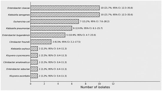
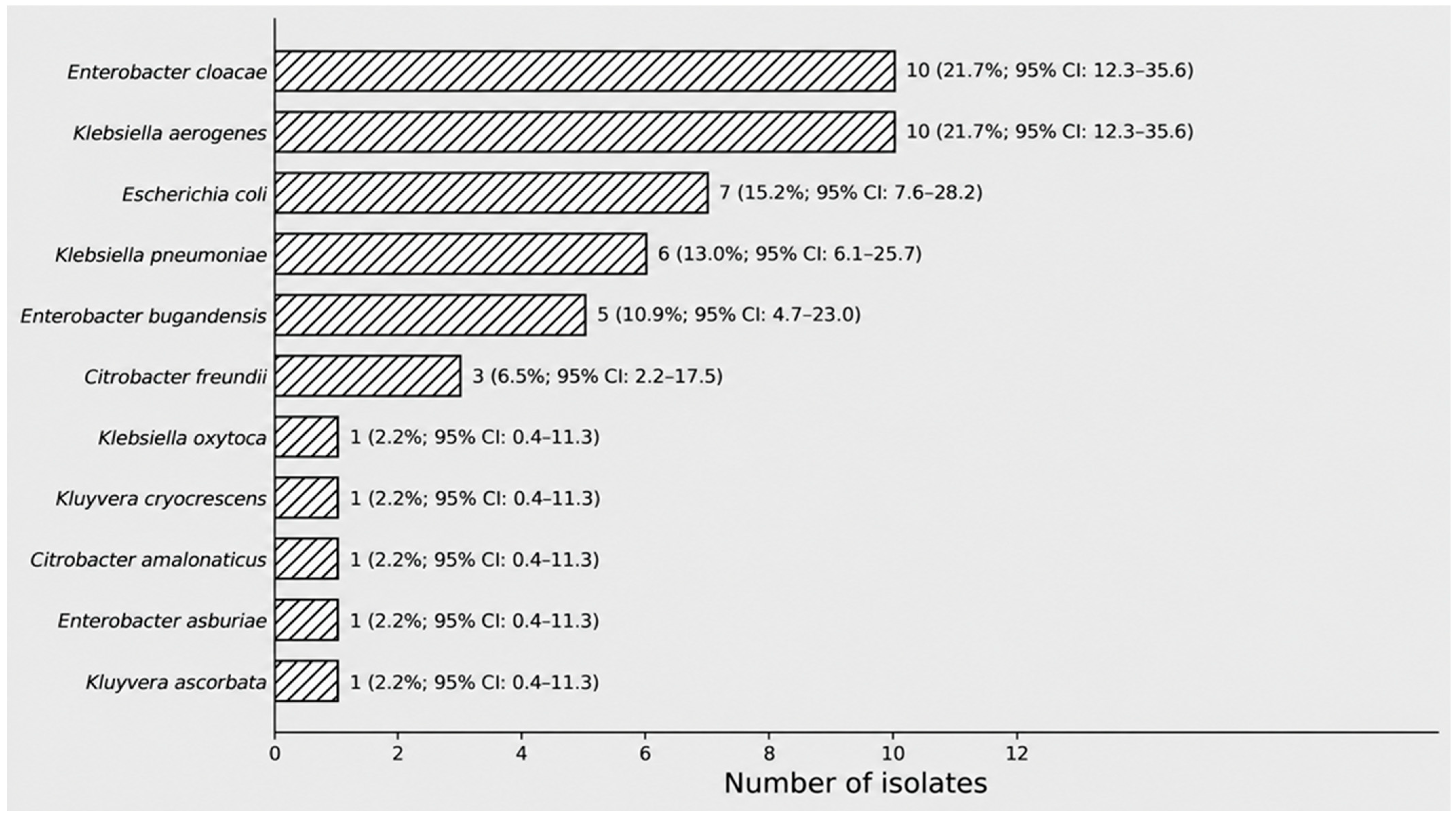
A total of 427 colonization samples recovered from rectal swabs of 142 newborns in a HUAP NICU were screened for ESBL production. Among these, 46 (10.8% of samples) grew on CHROMagar ESBL and were subsequently subjected to phenotypic confirmatory testing. These isolates, recovered from 39 unique neonates, were subsequently identified at the species level by MALDI-TOF MS. The most frequent bacterial species were K. aerogenes and E. cloacae (n = 10/46; 21.7%), followed by E. coli (n = 7/46; 15.2%), K. pneumoniae (n = 6/46; 13%), E. bugandensis (n = 5/46; 10.9%) and C. freundii (n = 3/46; 6.5%) (Figure 1).
Figure 1.
Distribution of bacterial species identified by matrix-assisted laser desorption/ionization time-of-flight mass spectrometry (MALDI-TOF MS). Bars represent the number of isolates identified for each species. Values beside the bars indicate the absolute frequency, corresponding percentage, and 95% confidence interval for the proportion of each species among the 46 isolates analyzed.
3.2. Antimicrobial Susceptibility
Table 1 below shows the susceptibility profile observed among the 46 bacterial isolates in the study. Higher resistance rates were observed against Ampicillin/Sulbactam (84.8%), Ceftriaxone (82.6%), Piperacillin/Tazobactam (50%), Cefepime (39.1%), and Ceftazidime (65.2%). On the other hand, lower resistance rates were found against the tested aminoglycosides: Amikacin (0%) and Gentamicin (11%). Regarding carbapenems, 15% of the samples showed resistance to Ertapenem; however, none of the 46 samples showed resistance to Imipenem or Meropenem. MDR was observed in 19 of the 46 colonization isolates (41.3%).
Table 1.
Antimicrobial susceptibility profile of the 46 bacterial isolates included in the study.
3.3. MDR Classification and MAR Index
Among the 46 Enterobacterales isolates analyzed, 19 (41.3%) fulfilled the criteria for MDR after exclusion of intrinsic resistance mechanisms (Table 2). The frequency of MDR varied substantially among bacterial species. The highest proportions were observed in K. oxytoca (100%; 1/1), E. coli (85.7%; 6/7), and K. pneumoniae (83.3%; 5/6). No MDR isolates were identified among K. aerogenes, C. freundii, E. asburiae, K. cryocrescens, or C. amalonaticus.
Table 2.
Distribution of multidrug resistance and MAR Index according to bacterial species.
The overall mean MAR Index was 0.234 ± 0.130. The highest mean MAR Index values were observed for K. oxytoca (0.467), followed by K. pneumoniae (0.337 ± 0.043), E. coli (0.312 ± 0.169), and E. cloacae (0.220 ± 0.147). In contrast, lower MAR Index values were found for C. freundii (0.119 ± 0.109), K. cryocrescens (0.062), and C. amalonaticus (0.062) (Table 2).
3.4. Genotypic Detection of β-Lactamases-Encoding Genes
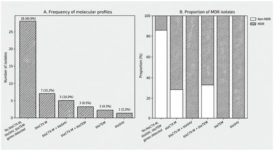
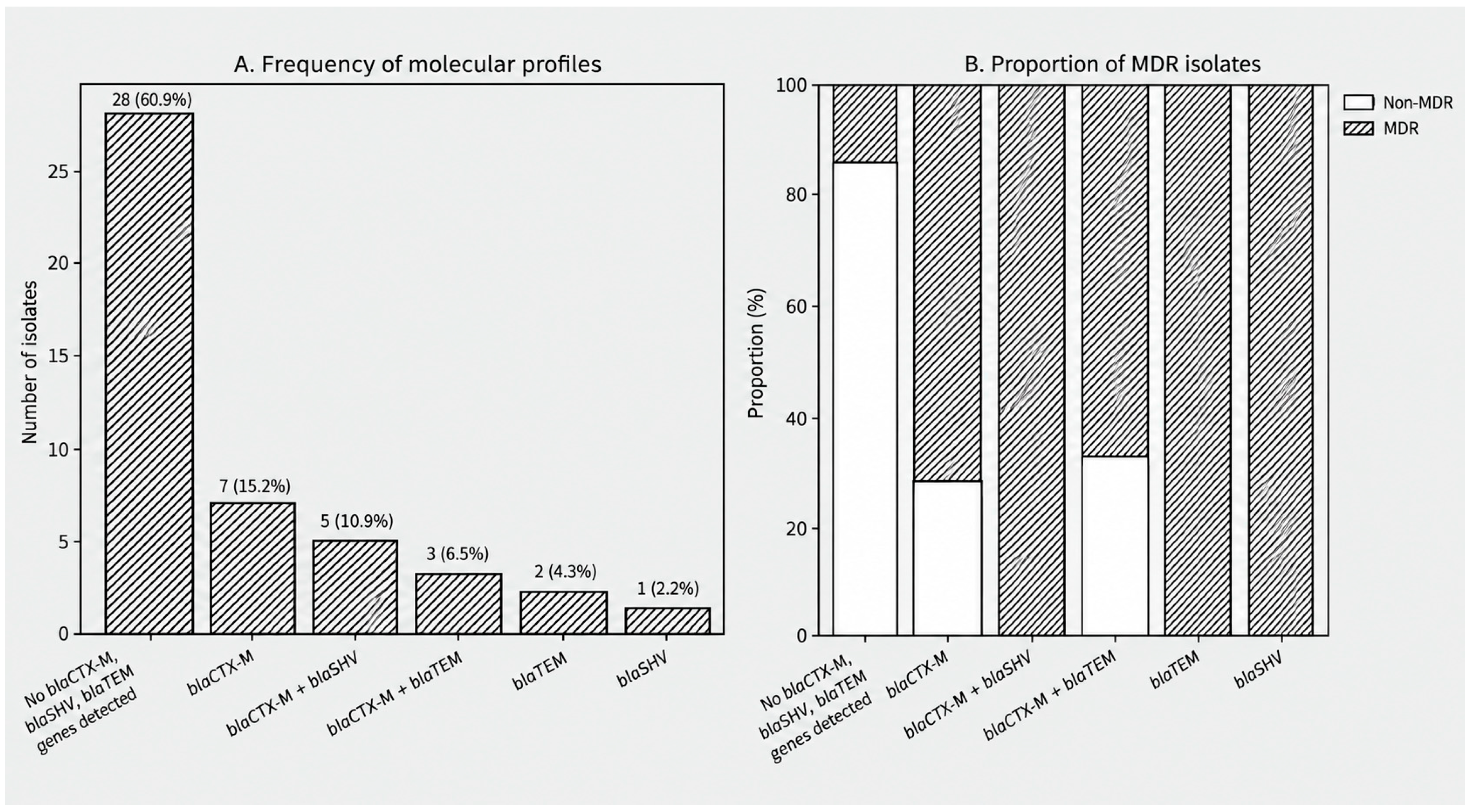
The β-lactamase genes results are summarized in Table 3. The most prevalent gene was blaCTX-M, present in 15 out of 46 (32.6%) isolates. The second most frequently detected gene was blaSHV, present in six bacterial isolates (13%), and the gene blaTEM was present in 5 samples (10.9%). Co-occurrence of the genes blaCTX-M and blaSHV was observed in 5 isolates, and blaCTX-M and blaTEM in three of the analyzed bacterial isolates (Figure 2A). The β-lactamases genes were mostly found among E. coli and K. pneumoniae isolates.
Table 3.
Distribution of β-lactamase-encoding genes among bacterial isolates recovered from neonatal rectal swabs (n = 46).
Figure 2.
Frequency of β-lactamase gene detection profiles and distribution of multidrug resistance (MDR) among Enterobacterales isolates. (A) Frequency of molecular profiles based on detection of blaCTX-M, blaSHV, and blaTEM. (B) Proportion of MDR and non-MDR isolates within each molecular profile. MDR distribution differed significantly among profiles (Fisher–Freeman–Halton exact test, p < 0.001; Cramér’s V = 0.710).
Among the 46 isolates, 28 (60.9%) were negative for all three β-lactamase targets investigated (blaCTX-M, blaSHV, and blaTEM). Notably, this group included isolates belonging to species with a recognized potential for inducible chromosomal AmpC production, particularly K. aerogenes, E. cloacae and C. freundii. Therefore, absence of the three PCR targets should not be interpreted as absence of a β-lactamase-mediated resistance mechanism.
The distribution of MDR differed significantly among the β-lactamase gene detection profiles (Fisher–Freeman–Halton exact test, p < 0.001; Cramér’s V = 0.710). All isolates carrying the combined blaCTX-M + blaSHV profile, as well as those carrying blaTEM or blaSHV alone, were classified as MDR (100%). Similarly, isolates harboring blaCTX-M alone exhibited a high MDR frequency (71.4%), whereas the blaCTX-M + blaTEM profile showed an MDR frequency of 66.7%. In contrast, only 14.3% of isolates without detectable β-lactamases genes were classified as MDR (Figure 2B). When isolates were grouped according to detection of at least one of the investigated β-lactamase genes, MDR was observed in 15/18 (83.3%) gene-positive isolates compared with 4/28 (14.3%) isolates negative for all three targets. This association was statistically significant (Fisher’s exact test, p < 0.001; OR = 30.0).
Descriptive analysis of the MAR Index showed variation across β-lactamase gene detection profiles. Isolates in which none of the three investigated genes were detected had a mean MAR Index of 0.180 ± 0.111, whereas higher mean values were observed in several gene-positive profiles, including blaCTX-M + blaTEM (0.429 ± 0.124), blaSHV alone (0.467), blaTEM alone (0.321 ± 0.051), and blaCTX-M + blaSHV (0.317 ± 0.041). Because of the small and highly unequal subgroup sizes, including profiles represented by only one or two isolates, these MAR Index differences were interpreted descriptively and no formal inferential comparison among molecular profiles was performed (Table 4).
Table 4.
MAR Index according to beta-lactamase gene detection profiles defined by PCR detection of blaCTX-M, blaSHV, and blaTEM.
4. Discussion
The present study provides a comprehensive molecular and phenotypic characterization of ESBL-producing Enterobacterales colonizing neonates admitted to a Brazilian NICU. Our main findings were (i) a predominance of CTX-M enzymes among ESBL-positive isolates; (ii) a strong association between β-lactamase gene carriage and multidrug resistance; (iii) preservation of susceptibility to carbapenems and aminoglycosides despite extensive resistance to β-lactams; and (iv) the identification of Enterobacterales species by MALDI-TOF, highlighting the value of high-resolution laboratory surveillance.
Here, we described the use of MALDI-TOF for identifying microorganisms cultured from colonization samples isolated from neonatal rectal swabs. The method of microorganism identification through mass spectrometry tends to yield significantly better results than automated biochemical methods. Saffert et al. (2011) conducted a study with 427 samples of Gram-negative bacilli, previously identified through sequencing, to compare MALDI-TOF and Phoenix techniques. They found that MALDI-TOF exhibited better performance in identifying samples at the genus and species levels (93% and 82% respectively) compared to Phoenix (83% and 75% respectively) [21]. This difference may be associated with MALDI-TOF superior performance in identifying samples that are rarely isolated in the clinical laboratory. Additionally, BD Phoenix™ M50 has a technical limitation in identifying certain species, such as those of the genus Enterobacter, which were present in the sample collection of this study [22]. The use of MALDI-TOF MS was particularly relevant because it enabled the reliable identification of uncommon Enterobacterales species, including E. bugandensis, which are frequently overlooked or misidentified by conventional biochemical methods. Accurate species identification is increasingly important because members of the E. cloacae complex differ in epidemiology, virulence, and antimicrobial resistance mechanisms, making species-level identification relevant for both clinical management and epidemiological surveillance [23,24].
The overall prevalence of ESBL-producing Enterobacterales colonization observed in the present study (11%) was lower than that reported in several neonatal intensive care units (NICUs) from Africa and Asia, where colonization rates frequently exceed 30–50%. In Tanzania, Silago et al. (2021) reported that approximately 43% of hospitalized neonates were colonized by ESBL-producing Enterobacterales, with K. pneumoniae and E. coli predominating and widespread carriage of blaCTX-M, blaSHV, and blaTEM genes [25]. Similarly, studies from India have demonstrated high intestinal colonization rates by ESBL-producing Enterobacterales among hospitalized patients, reflecting the intense selective pressure imposed by antimicrobial exposure and the persistence of resistant clones in healthcare settings [25,26]. In contrast, surveillance studies conducted in Latin America have reported more heterogeneous prevalence rates [3,27].
The prevalence of colonization by ESBL-producing Enterobacterales observed in this study is lower than that reported by Bomfim et al. (2023), who found a rate of 58% among adult patients in an ICU in the city of Natal, northeastern Brazil. This variation may be attributed to intrinsic differences between neonatal and adult populations, as well as local epidemiological profiles. However, both studies converge in identifying the blaCTX-M gene as one of the main determinants of resistance, which corroborates the trend of dissemination of this enzyme in Latin America. Furthermore, Bomfim et al. (2023) discuss the presence and persistence of high-risk Enterobacterales clones, which facilitate cross-transmission in the critical environment of ICUs, and although not investigated in our study, deserves significant attention [27].
In a study conducted in Serbia with samples from rectal swabs of 103 neonates in a tertiary hospital, 60% of the patients were colonized by MDR bacteria, including ESBL-producing K. pneumoniae and E. coli. As expected, most of these strains were resistant to 3rd and 4th generation cephalosporins (ceftazidime 76% to 83% and cefepime 73% to 87%), penicillins (piperacillin/tazobactam 65%), as well as aminoglycosides (gentamicin 75% to 100% and amikacin 50% to 94%). Similarly, in the present study, high resistance rates were observed for ceftriaxone, ampicillin, and ampicillin–sulbactam, whereas resistance to aminoglycosides was markedly lower, particularly for gentamicin and amikacin (Table 1). This contrasts with the higher aminoglycoside resistance rates reported among colonizing multidrug-resistant bacteria in hospitalized preterm neonates in Serbia [28].
The preservation of susceptibility to other cephalosporins in our study, despite the marked resistance to ceftriaxone, may be explained by the biochemical characteristics of the prevalent enzymes and current clinical management guidelines. The functional diversity of β-lactamases, especially within the CTX-M family, explains why some isolates exhibit high hydrolytic affinity for ceftriaxone but may not achieve the same level of catalytic efficiency against ceftazidime or cefepime, depending on specific mutations in the enzyme’s active site. This enzymatic selectivity suggests that local selective pressure may be favoring ESBL variants with a less broad spectrum of action [29,30]. Besides that, Tamma and colleagues (2023) suggest that ceftriaxone may induce resistance through the presence of AmpC in isolates of Enterobacter spp., K. aerogenes, and Citrobacter spp., which represent a significant quantity in this study, while cefepime, a fourth-generation cephalosporin, exhibits low affinity for AmpC-mediated hydrolysis. Furthermore, the predominance of AmpC-producing species may also help explain the phenotypic pattern observed in some isolates showing resistance to ertapenem while remaining susceptible to meropenem and imipenem [31]. The Brazilian National Health Surveillance Agency (ANVISA) reported that, among bloodstream infections caused by Gram-negative bacilli in neonatal intensive care units (NICUs), the most frequent isolates resistant to third- and/or fourth-generation cephalosporins were K. pneumoniae (43.8%), Enterobacter spp. (33.0%), and E. coli (23.4%). These findings indicate the widespread circulation of ESBL-producing Enterobacterales in Brazilian NICUs and reinforce the need for continuous microbiological surveillance and antimicrobial stewardship [32].
Although high resistance to third-generation cephalosporins was expected because all isolates were recovered using ESBL-selective media, the complete preservation of susceptibility to imipenem and meropenem, together with the excellent activity of amikacin, is clinically relevant. Although carbapenemase genes were not investigated in the present study, the absence of phenotypic resistance indicates that there is still an opportunity to prevent the emergence and dissemination of carbapenem-resistant clones through continuous surveillance and infection prevention measures [31,33,34]. Current Infectious Diseases Society of America (IDSA) guidance recommends carbapenems as the preferred therapy for serious infections caused by ESBL-producing Enterobacterales and highlights antimicrobial stewardship as a key strategy to preserve the efficacy of these last-line agents [35]. In the present study, the absence of carbapenem resistance indicates that carbapenem-resistant Enterobacterales have not yet emerged among the ESBL-producing isolates recovered from this NICU, although carbapenemase genes were not investigated. This susceptibility profile provides an important opportunity to strengthen infection prevention measures and optimize antimicrobial stewardship before additional resistance mechanisms become established [31,33].
We detected three β-lactamase genes (blaCTX-M, blaSHV, and blaTEM). The most prevalent gene was blaCTX-M, present in 15 samples (32.6%), followed by blaSHV. Similar results were described in Southern Chile in 2019, in a study including 137 enterobacteria isolates from clinical samples of critically ill patients, there was a high prevalence of the encoding genes of CTX-M (84.7% of the isolates), SHV (81%) and TEM (73%) among the isolates, suggesting the dissemination of these resistance mechanisms, especially CTX-M, that appears to be an emerging problem in Latin America [3].
The predominance of blaCTX-M observed in the present study reflects the global replacement of older TEM- and SHV-derived ESBLs by CTX-M enzymes over the last two decades. This transition has been driven by the successful dissemination of conjugative plasmids carrying blaCTX-M genes, frequently associated with insertion sequences such as ISEcp1, together with the global expansion of high-risk bacterial lineages, including Escherichia coli ST131 and epidemic Klebsiella pneumoniae clones. These mechanisms have enabled the spread of CTX-M enzymes across hospitals, community settings, food-producing animals, wildlife, and environmental reservoirs, exemplifying the interconnected nature of antimicrobial resistance within a One Health framework [13,36,37,38,39].
An important finding was that 28 of the 46 isolates (60.9%) were negative for all three β-lactamase targets investigated (blaCTX-M, blaSHV, and blaTEM). This finding should not be interpreted as evidence of the absence of β-lactamase-mediated resistance. A substantial proportion of these isolates belonged to species recognized as being at increased risk for clinically significant inducible chromosomal AmpC production, particularly K. aerogenes, E. cloacae complex, and C. freundii. In these organisms, induction or derepression of chromosomal ampC may result in increased AmpC production and reduced susceptibility or resistance to third-generation cephalosporins [40,41]. AmpC hyperproduction may also contribute to growth on ESBL-selective chromogenic media and to the observed cephalosporin resistance profiles [42,43]. Nevertheless, all isolates included in the present study exhibited a positive phenotypic ESBL confirmatory test. Therefore, among isolates negative for blaCTX-M, blaSHV, and blaTEM, the presence of ESBL determinants outside the PCR panel cannot be excluded. Other β-lactamase-mediated mechanisms, including plasmid-mediated AmpC enzymes and additional β-lactamase families, may also have contributed to the observed resistance phenotypes [41,44]. Because neither ampC genes and their expression nor a broader β-lactamase repertoire were investigated, the specific contribution of these mechanisms could not be determined. Accordingly, isolates negative for the three investigated targets should be interpreted as blaCTX-M, blaSHV, and blaTEM-negative rather than as lacking β-lactamase-mediated resistance mechanisms.
One of the most relevant findings was the strong association between β-lactamase genes and MDR. All isolates carrying blaSHV or blaTEM alone, as well as those harboring the combined blaCTX-M + blaSHV profile, exhibited an MDR phenotype. Conversely, only a small proportion of isolates lacking detectable ESBL genes were classified as MDR. This finding is consistent with previous studies demonstrating that ESBL genes are commonly located on conjugative plasmids together with additional resistance determinants, including genes conferring resistance to aminoglycosides, fluoroquinolones, sulfonamides, and tetracyclines. Such genetic linkage promotes co-selection under antimicrobial pressure, thereby favoring the persistence and dissemination of MDR strains in clinical settings [45,46,47,48,49].
The MAR Index further supports this interpretation. Overall, the MAR index exceeded the threshold of 0.20 in most of the clinically relevant species recovered from colonized patients, particularly K. pneumoniae, E. coli, K. oxytoca, and E. cloacae. According to Krumperman, MAR values above 0.20 are indicative of bacterial populations originating from environments subjected to intense antimicrobial exposure, reflecting sustained selective pressure. The coexistence of elevated MAR indices and high MDR frequencies observed in the predominant Enterobacterales therefore supports the circulation of bacterial populations carrying multiple acquired resistance determinants within the neonatal hospital environment, where antimicrobial use and cross-transmission facilitate the persistence and dissemination of resistant clones [17,19].
Finally, the detection of ESBL-producing Enterobacterales in the NICU highlights the strategic relevance of active surveillance and molecular monitoring. As gut colonization acts as a critical reservoir for resistance mechanisms, these measures are essential to mitigate hospital transmission and prevent severe infections in vulnerable neonates. Molecular surveillance of ESBL-producing Enterobacterales should be considered an essential component of infection prevention strategies in neonatal intensive care units, particularly in regions where multidrug-resistant Gram-negative bacteria continue to emerge [5,6,50,51]. Early identification of intestinal carriage allows implementation of targeted infection prevention measures, including reinforcement of hand hygiene, patient cohorting, contact precautions, and optimization of antimicrobial stewardship programs before resistant organisms become established or cause invasive disease.
This study has some limitations that should be acknowledged. First, it was conducted in a single neonatal intensive care unit and included a relatively small number of bacterial isolates, which may limit the generalizability of the findings to other neonatal populations and healthcare settings. Second, the molecular analysis was restricted to blaCTX-M, blaSHV, and blaTEM. Therefore, the absence of these targets in 28 isolates does not exclude other β-lactamase-mediated resistance mechanisms. In particular, ampC genes and AmpC expression were not investigated, which is relevant because several isolates belonged to species with inducible chromosomal AmpC β-lactamases, including K. aerogenes, E. cloacae, and C. freundii. Other ESBL families, plasmid-mediated AmpC enzymes, and additional β-lactamases were also not assessed. In addition, the detected genes were not further characterized at the variant level. Finally, the study focused on bacterial colonization and did not evaluate the subsequent development of infection among colonized neonates. Despite these limitations, the study provides relevant data on the occurrence of ESBL-associated resistance determinants among Enterobacterales colonizing neonates and contributes to the understanding of antimicrobial resistance in the NICU setting.
5. Conclusions
Overall, our findings demonstrate that blaCTX-M-harboring Enterobacterales may already be established among colonizing neonatal isolates in this Brazilian NICU, although carbapenem resistance remains uncommon. The coexistence of β-lactamase genes, MDR, and the elevated MAR Index indicates the circulation of bacterial populations with substantial acquired resistance and highlights the importance of continuous molecular surveillance, antimicrobial stewardship, and strict infection prevention practices to prevent the transition from asymptomatic colonization to invasive infection and to preserve the effectiveness of currently available antimicrobial agents.
Author Contributions
Conceptualization, S.d.S.V., C.R.V.d.M.S. and T.P.G.C.; methodology, S.d.S.V., L.B.M., V.Z., C.R.V.d.M.S., A.R.X. and T.P.G.C.; formal analysis, S.d.S.V., L.B.M. and T.P.G.C.; investigation, L.B.M., L.C.B., N.d.A.C.C.R., A.L.d.S. and D.G.F.; resources, N.d.A.C.C.R., D.G.F. and V.Z.; data curation, S.d.S.V. and T.P.G.C.; writing—original draft preparation, S.d.S.V., L.B.M., L.C.B., N.d.A.C.C.R., A.L.d.S., D.G.F., C.A.M.A., F.L.P.C.P., V.Z., C.R.V.d.M.S., A.R.X. and T.P.G.C.; writing—review and editing, S.d.S.V., L.B.M., C.A.M.A., F.L.P.C.P., V.Z., C.R.V.d.M.S., A.R.X. and T.P.G.C.; visualization, S.d.S.V. and T.P.G.C.; supervision, N.d.A.C.C.R., D.G.F., C.A.M.A., F.L.P.C.P., V.Z., C.R.V.d.M.S. and A.R.X.; project administration, C.R.V.d.M.S., V.Z., A.R.X. and T.P.G.C.; funding acquisition, V.Z. All authors have read and agreed to the published version of the manuscript.
Funding
This research was supported by FAPERJ—Postgraduate Support—Course and Postgraduate studies (E-26/210.982/2021) and CNPq/MCTI/CT-Saúde Edital 52/2022 (Proc. 408882/2022-0).
Institutional Review Board Statement
This study was approved by the Ethics and Research Committee of Antonio Pedro University Hospital/Medical School, UFF, under registration number: 95984018.6.0000.5243 approved on 26 September 2018.
Informed Consent Statement
This study involved only bacterial isolates obtained from routine laboratory procedures, along with anonymized data regarding the date and sector of the isolates. Therefore, the study was not required to include informed consent (ICF).
Data Availability Statement
The data supporting the findings of this study are available from the corresponding author upon reasonable request, subject to approval by the relevant Ethics Committee and in accordance with institutional policies regarding confidential research data.
Acknowledgments
The authors thank the Antônio Pedro University Hospital (EBSERH) for technical support and assistance with isolate collection.
Conflicts of Interest
The authors declare no conflicts of interest.
Abbreviations
The following abbreviations are used in this manuscript:
| AST | Antimicrobial Susceptibility Testing |
| CLSI | Clinical and Laboratory Standards Institute |
| DNA | Deoxyribonucleic Acid |
| ESBL | Extended-Spectrum β-Lactamase |
| ESBL-E | Extended-Spectrum β-Lactamase-Producing Enterobacterales |
| HICC | Hospital Infection Control Committee |
| HUAP | Antônio Pedro University Hospital |
| UFF | Fluminense Federal University |
| MALDI-TOF MS | Matrix-Assisted Laser Desorption/Ionization Time-of-Flight Mass Spectrometry |
| MAR | Multiple Antibiotic Resistance |
| MDR | Multidrug Resistance |
| NICU | Neonatal Intensive Care Unit |
| PCR | Polymerase Chain Reaction |
| TBE | Tris-Borate-EDTA |
| UHS | Unified Health System |
| blaCTX-M | β-lactamase CTX-M gene |
| blaSHV | Sulfhydryl Variable beta-lactamase gene |
| blaTEM | Temoneira beta-lactamase gene |
References
- Noster, J.; Thelen, P.; Hamprecht, A. Detection of multidrug-resistant Enterobacterales—From ESBLs to carbapenemases. Antibiotics 2021, 10, 1140. [Google Scholar] [CrossRef] [Scilit] [PubMed]
- Sy, C.L.; Chen, P.Y.; Cheng, C.W.; Huang, L.J.; Wang, C.H.; Chang, T.H.; Chang, Y.C.; Chang, C.J.; Hii, I.M.; Hsu, Y.L.; et al. Recommendations and guidelines for the treatment of infections due to multidrug resistant organisms. J. Microbiol. Immunol. Infect. 2022, 55, 359–386. [Google Scholar] [CrossRef] [Scilit] [PubMed]
- Pavez, M.; Troncoso, C.; Osses, I.; Salazar, R.; Illesca, V.; Reydet, P.; Rodríguez, C.; Chahin, C.; Concha, C.; Barrientos, L. High prevalence of CTX-M-1 group in ESBL-producing Enterobacteriaceae infection in intensive care units in southern Chile. Braz. J. Infect. Dis. 2019, 23, 102–110. [Google Scholar] [CrossRef] [Scilit] [PubMed]
- Vurayai, M.; Strysko, J.; Kgomanyane, K.; Bayani, O.; Mokomane, M.; Machiya, T.; Arscott-Mills, T.; Goldfarb, D.M.; Steenhoff, A.P.; McGann, C.; et al. Characterizing the bioburden of ESBL-producing organisms in a neonatal unit using chromogenic culture media: A feasible and efficient environmental sampling method. Antimicrob. Resist. Infect. Control 2022, 11, 14. [Google Scholar] [CrossRef] [Scilit] [PubMed]
- Flannery, D.D.; Chiotos, K.; Gerber, J.S.; Puopolo, K.M. Neonatal multidrug-resistant Gram-negative infection: Epidemiology, mechanisms of resistance, and management. Pediatr. Res. 2022, 91, 380–391. [Google Scholar] [CrossRef] [Scilit] [PubMed]
- Folgori, L.; Tersigni, C.; Hsia, Y.; Kortsalioudaki, C.; Heath, P.; Sharland, M.; Bielicki, J. The relationship between Gram-negative colonization and bloodstream infections in neonates: A systematic review and meta-analysis. Clin. Microbiol. Infect. 2018, 24, 251–257. [Google Scholar] [CrossRef] [Scilit] [PubMed]
- Yap, P.S.; Kamar, A.A.; Chong, C.W.; Yap, I.K.S.; Thong, K.L.; Choo, Y.M.; Yusof, M.Y.M.; Teh, C.S.J. Intestinal carriage of multidrug-resistant Gram-negative bacteria in preterm infants during hospitalization in neonatal intensive care unit (NICU). Pathog. Glob. Health 2016, 110, 238–246. [Google Scholar] [CrossRef] [Scilit] [PubMed]
- Das, P.; Singh, A.K.; Pal, T.; Dasgupta, S.; Ramamurthy, T.; Basu, S. Colonization of the gut with Gram-negative bacilli, its association with neonatal sepsis and its clinical relevance in a developing country. J. Med. Microbiol. 2011, 60, 1651–1660. [Google Scholar] [CrossRef] [Scilit] [PubMed]
- Folgori, L.; Bielicki, J. Future challenges in pediatric and neonatal sepsis: Emerging pathogens and antimicrobial resistance. J. Pediatr. Intensive Care 2019, 8, 17–24. [Google Scholar] [CrossRef] [Scilit] [PubMed]
- Oliveira, P.M.N.; Buonora, S.N.; Souza, C.L.P.; Simões Júnior, R.; Silva, T.C.; Bom, G.J.T.; Teixeira, C.H.S.; Silva, A.R.A. Surveillance of Multidrug-Resistant Bacteria in Pediatric and Neonatal Intensive Care Units in Rio de Janeiro State, Brazil. Rev. Soc. Bras. Med. Trop. 2019, 52, e20190205. [Google Scholar] [CrossRef] [Scilit] [PubMed]
- Barbosa, T.A.; Bentlin, M.R.; Rugolo, L.M.S.S.; Lyra, J.C.; Ferreira, A.M.; Santos, A.C.M.L.; Teixeira, N.B.; Romero, L.C.M.; Fortaleza, C.M.C.B.; Cunha, M.L.R.S. Molecular Characterization of Gram-Negative Bacilli Isolated from a Neonatal Intensive Care Unit and Phenotypic and Molecular Detection of ESBL and Carbapenemase. Antibiotics 2025, 14, 342. [Google Scholar] [CrossRef] [Scilit] [PubMed]
- Bello, H.; Trabal, N.; Ibáñez, D.; Reyes, A.; Domínguez, M.; Mella, S.; Zemelman, C.; Zemelman, R.; González, G. β-Lactamases other than TEM and SHV among strains of Klebsiella pneumoniae subsp. pneumoniae isolated from Chilean hospitals. Rev. Med. Chil. 2005, 133, 737–739. [Google Scholar] [CrossRef] [Scilit] [PubMed][Green Version]
- Cantón, R.; González-Alba, J.M.; Galán, J.C. CTX-M enzymes: Origin and diffusion. Front. Microbiol. 2012, 3, 110. [Google Scholar] [CrossRef] [Scilit] [PubMed]
- Despotovic, M.; de Nies, L.; Busi, S.B.; Wilmes, P. Reservoirs of antimicrobial resistance in the context of One Health. Curr. Opin. Microbiol. 2023, 73, 102291. [Google Scholar] [CrossRef] [Scilit] [PubMed]
- Clinical and Laboratory Standards Institute (CLSI). Performance Standards for Antimicrobial Susceptibility Testing, 32nd ed.; CLSI Supplement M100; Clinical and Laboratory Standards Institute: Wayne, PA, USA, 2022. [Google Scholar]
- Brazilian Committee on Antimicrobial Susceptibility Testing (BrCAST). Orientações do EUCAST para a Detecção de Mecanismos de Resistência e Resistências Específicas de Importância Clínica e/ou Epidemiológica, Version 2.0; BrCAST: Rio de Janeiro, Brazil, 2018. [Google Scholar]
- Magiorakos, A.-P.; Srinivasan, A.; Carey, R.B.; Carmeli, Y.; Falagas, M.E.; Giske, C.G.; Harbarth, S.; Hindler, J.F.; Kahlmeter, G.; Olsson-Liljequist, B.; et al. Multidrug-Resistant, Extensively Drug-Resistant and Pandrug-Resistant Bacteria: An International Expert Proposal for Interim Standard Definitions for Acquired Resistance. Clin. Microbiol. Infect. 2012, 18, 268–281. [Google Scholar] [CrossRef] [Scilit] [PubMed]
- Brazilian Committee on Antimicrobial Susceptibility Testing (BrCAST). Fenótipos Esperados Resistentes; BrCAST: Rio de Janeiro, Brazil, 2024. [Google Scholar]
- Krumperman, P.H. Multiple Antibiotic Resistance Indexing of Escherichia coli to Identify High-Risk Sources of Fecal Contamination of Foods. Appl. Environ. Microbiol. 1983, 46, 165–170. [Google Scholar] [CrossRef] [Scilit] [PubMed]
- Roschanski, N.; Fischer, J.; Guerra, B.; Roesler, U. Development of a multiplex real-time PCR for the Rapid Detection of the predominant beta-lactamase genes CTX-M, SHV, TEM and CIT-Type AmpCs in Enterobacteriaceae. PLoS ONE 2014, 9, e100956. [Google Scholar] [CrossRef] [Scilit] [PubMed]
- Saffert, R.T.; Cunningham, S.A.; Ihde, S.M.; Jobe, K.E.; Mandrekar, J.; Patel, R. Comparison of Bruker Biotyper matrix-assisted laser desorption ionization-time of flight mass spectrometer to BD Phoenix automated microbiology system for identification of gram-negative bacilli. J. Clin. Microbiol. 2011, 49, 887–892. [Google Scholar] [CrossRef] [Scilit] [PubMed]
- Becton, Dickinson and Company. BD Phoenix™ M50 Automated Microbiology System User’s Manual; Becton, Dickinson and Company: Sparks, MD, USA, 2020. [Google Scholar]
- Davin-Regli, A.; Lavigne, J.-P.; Pagès, J.-M. Enterobacter spp.: Update on Taxonomy, Clinical Aspects, and Emerging Antimicrobial Resistance. Clin. Microbiol. Rev. 2019, 32, e00002-19. [Google Scholar] [CrossRef] [Scilit] [PubMed]
- Dong, X.; Zhu, M.; Li, Y.; Huang, D.; Wang, L.; Yan, C.; Zhang, L.; Dong, F.; Lu, J.; Lin, X.; et al. Whole-Genome Sequencing-Based Species Classification, Multilocus Sequence Typing, and Antimicrobial Resistance Mechanism Analysis of the Enterobacter cloacae Complex in Southern China. Microbiol. Spectr. 2022, 10, e02160-22. [Google Scholar] [CrossRef] [Scilit] [PubMed]
- Silago, V.; Kovacs, D.; Samson, H.; Seni, J.; Matthews, L.; Oravcová, K.; Lupindu, A.M.; Hoza, A.S.; Mshana, S.E. Existence of multiple ESBL genes among phenotypically confirmed ESBL-producing Klebsiella pneumoniae and Escherichia coli concurrently isolated from clinical, colonization and contamination samples from neonatal units at Bugando medical center, Mwanza, Tanzania. Antibiotics 2021, 10, 476. [Google Scholar] [CrossRef] [Scilit] [PubMed]
- Asir, J.; Nair, S.; Devi, S.; Prashanth, K.; Saranathan, R.; Kanungo, R. Simultaneous gut colonisation and infection by ESBL-producing Escherichia coli in hospitalised patients. Australas. Med. J. 2015, 8, 200–207. [Google Scholar] [CrossRef] [PubMed]
- Bomfim, I.M.F.N.; Amaral, M.A.; Rodrigues, Y.C.; Brasiliense, D.M.; Aires, C.A.M.; Neto, R.M. High-risk clones of ESBL-producing Klebsiella pneumoniae colonizing ICU patients in Natal, northeastern Brazil. Rev. Epidemiol. Controle Infect. 2023, 13, 130–136. [Google Scholar] [CrossRef] [Scilit]
- Milic, M.; Siljic, M.; Cirkovic, V.; Jovicevic, M.; Perovic, V.; Markovic, M.; Martic, J.; Stanojevic, M.; Mijac, V. Colonization with multidrug-resistant bacteria in the first week of life among hospitalized preterm neonates in Serbia: Risk factors and outcomes. Microorganisms 2021, 9, 2613. [Google Scholar] [CrossRef] [Scilit]
- Bush, K.; Bradford, P.A. Epidemiology of β-Lactamase-Producing Pathogens. Clin. Microbiol. Rev. 2020, 33, e00047-19. [Google Scholar] [CrossRef] [Scilit] [PubMed]
- Brown, C.A.; Hu, L.; Sun, Z.; Patel, M.P.; Singh, S.; Porter, J.R.; Sankaran, B.; Prasad, B.V.V.; Bowman, G.R.; Palzkill, T. Antagonism between Substitutions in β-Lactamase Explains a Path Not Taken in the Evolution of Bacterial Drug Resistance. J. Biol. Chem. 2020, 295, 7376–7390. [Google Scholar] [CrossRef] [Scilit] [PubMed]
- Tamma, P.D.; Aitken, S.L.; Bonomo, R.A.; Mathers, A.J.; van Duin, D.; Clancy, C.J. Infectious Diseases Society of America 2023 guidance on the treatment of antimicrobial resistant Gram-negative infections. Clin. Infect. Dis. 2023, ciad428. [Google Scholar] [CrossRef] [Scilit] [PubMed]
- Agência Nacional de Vigilância Sanitária (ANVISA). Boletim Segurança do Paciente e Qualidade em Serviços de Saúde No. 28: Avaliação Nacional dos Indicadores das Infecções Relacionadas à Assistência à Saúde (IRAS) e Resistência Microbiana, Ano 2021; ANVISA: Brasília, Brazil, 2022. [Google Scholar]
- Logan, L.K.; Weinstein, R.A. The Epidemiology of Carbapenem-Resistant Enterobacteriaceae: The Impact and Evolution of a Global Menace. J. Infect. Dis. 2017, 215, S28–S36. [Google Scholar] [CrossRef] [Scilit] [PubMed]
- Friedman, N.D.; Carmeli, Y.; Walton, A.L.; Schwaber, M.J. Carbapenem-Resistant Enterobacteriaceae: A Strategic Roadmap for Infection Control. Infect. Control Hosp. Epidemiol. 2017, 38, 580–594, Correction in Infect. Control Hosp. Epidemiol. 2017, 38, 1136. https://doi.org/10.1017/ice.2017.141. [Google Scholar] [CrossRef] [Scilit] [PubMed]
- Bonnet, R. Growing Group of Extended-Spectrum β-Lactamases: The CTX-M Enzymes. Antimicrob. Agents Chemother. 2004, 48, 1–14. [Google Scholar] [CrossRef] [Scilit] [PubMed]
- D’Andrea, M.M.; Arena, F.; Pallecchi, L.; Rossolini, G.M. CTX-M-Type β-Lactamases: A Successful Story of Antibiotic Resistance. Int. J. Med. Microbiol. 2013, 303, 305–317. [Google Scholar] [CrossRef] [Scilit] [PubMed]
- Mathers, A.J.; Peirano, G.; Pitout, J.D.D. The Role of Epidemic Resistance Plasmids and International High-Risk Clones in the Spread of Multidrug-Resistant Enterobacteriaceae. Clin. Microbiol. Rev. 2015, 28, 565–591. [Google Scholar] [CrossRef] [Scilit] [PubMed]
- Woerther, P.-L.; Burdet, C.; Chachaty, E.; Andremont, A. Trends in Human Fecal Carriage of Extended-Spectrum β-Lactamases in the Community: Toward the Globalization of CTX-M. Clin. Microbiol. Rev. 2013, 26, 744–758. [Google Scholar] [CrossRef] [Scilit] [PubMed]
- Peirano, G.; Pitout, J.D.D. Molecular Epidemiology of Escherichia coli Producing CTX-M β-Lactamases: The Worldwide Emergence of Clone ST131 O25:H4. Int. J. Antimicrob. Agents 2010, 35, 316–321. [Google Scholar] [CrossRef] [Scilit]
- Tamma, P.D.; Heil, E.L.; Justo, J.A.; Mathers, A.J.; Satlin, M.J.; Bonomo, R.A. Infectious Diseases Society of America 2024 Guidance on the Treatment of Antimicrobial-Resistant Gram-Negative Infections. Clin. Infect. Dis. 2024, ciae403. [Google Scholar] [CrossRef] [Scilit] [PubMed]
- Tebano, G.; Zaghi, I.; Cricca, M.; Cristini, F. Antibiotic Treatment of Infections Caused by AmpC-Producing Enterobacterales. Pharmacy 2024, 12, 142. [Google Scholar] [CrossRef] [Scilit] [PubMed]
- Glupczynski, Y.; Berhin, C.; Bauraing, C.; Bogaerts, P. Evaluation of a new selective chromogenic agar medium for detection of extended-spectrum β-lactamase-producing Enterobacteriaceae. J. Clin. Microbiol. 2007, 45, 501–505. [Google Scholar] [CrossRef] [Scilit] [PubMed]
- Emeraud, C.; Metro, C.A.; Le Doze, C.; Ouzani, S.; Khecharem, M.; Fortineau, N.; Dortet, L. Comparison of ChromID® and CHROMagar™ media in a real workflow for detecting EBSL and carbapenemase-producing Enterobacterales. J. Antimicrob. Chemother. 2025, 80, 595–597. [Google Scholar] [CrossRef] [Scilit] [PubMed]
- Schrader, S.M.; Pearson, Z.; Taffner, S.; Kanjilal, S.; Pecora, N.D. Prevalence and genetics of cefepime resistance among the AmpC-producing organisms Citrobacter freundii complex, Enterobacter cloacae complex, and Klebsiella aerogenes. Antimicrob. Agents Chemother. 2026, 70, e01661-25. [Google Scholar] [CrossRef] [Scilit] [PubMed]
- Paterson, D.L.; Bonomo, R.A. Extended-spectrum β-lactamases: A clinical update. Clin. Microbiol. Rev. 2005, 18, 657–686. [Google Scholar] [CrossRef] [Scilit] [PubMed]
- Carattoli, A. Plasmids and the spread of resistance. Int. J. Med. Microbiol. 2013, 303, 298–304. [Google Scholar] [CrossRef] [Scilit] [PubMed]
- Partridge, S.R.; Kwong, S.M.; Firth, N.; Jensen, S.O. Mobile genetic elements associated with antimicrobial resistance. Clin. Microbiol. Rev. 2018, 31, e00088-17. [Google Scholar] [CrossRef] [Scilit] [PubMed]
- Marcadé, G.; Deschamps, C.; Boyd, A.; Gautier, V.; Picard, B.; Branger, C.; Denamur, E.; Arlet, G. Replicon typing of plasmids in Escherichia coli producing extended-spectrum β-lactamases. J. Antimicrob. Chemother. 2009, 63, 67–71. [Google Scholar] [CrossRef] [Scilit] [PubMed]
- Chen, L.; Chavda, K.D.; Melano, R.G.; Jacobs, M.R.; Koll, B.; Hong, T.; Rojtman, A.D.; Mediavilla, J.R.; Kreiswirth, B.N. Comparative genomic analysis of KPC-producing Klebsiella pneumoniae clinical isolates and characterization of IS26-mediated multidrug resistance regions. Antimicrob. Agents Chemother. 2015, 59, 287–297. [Google Scholar]
- Tacconelli, E.; Carrara, E.; Savoldi, A.; Harbarth, S.; Mendelson, M.; Monnet, D.L.; Pulcini, C.; Kahlmeter, G.; Kluytmans, J.; Carmeli, Y.; et al. Discovery, Research, and Development of New Antibiotics: The WHO Priority List of Antibiotic-Resistant Bacteria and Tuberculosis. Lancet Infect. Dis. 2018, 18, 318–327. [Google Scholar] [CrossRef] [Scilit] [PubMed]
- World Health Organization (WHO). Global Antimicrobial Resistance and Use Surveillance System (GLASS) Report; WHO: Geneva, Switzerland, 2022. [Google Scholar]

Disclaimer/Publisher’s Note: The statements, opinions and data contained in all publications are solely those of the individual author(s) and contributor(s) and not of MDPI and/or the editor(s). MDPI and/or the editor(s) disclaim responsibility for any injury to people or property resulting from any ideas, methods, instructions or products referred to in the content. |
© 2026 by the authors. Licensee MDPI, Basel, Switzerland. This article is an open access article distributed under the terms and conditions of the Creative Commons Attribution (CC BY) license.