- Article

Using the SIRAH Force-Field to Model Interactions Between Short DNA Duplexes
- Romina Ruberto,
- Enrico Smargiassi and
- Giorgio Pastore
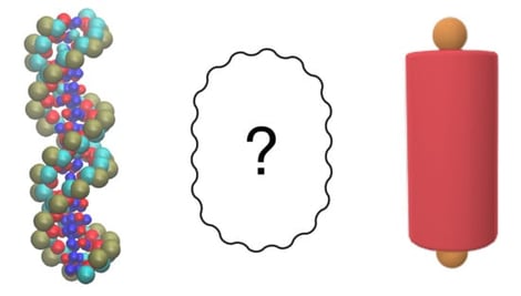
Background/Objectives: In recent years, short DNA duplexes have been studied as promising self-assembling systems and versatile building blocks for DNA-based nanotechnologies. Numerical simulations of colloidal systems incorporating such components require, as an input ingredient, reliable yet simplified force-fields capable of capturing the essential features of duplex-duplex interactions. Methods: We employed the coarse-grained SIRAH force field under an implicit solvent approximation to investigate the interactions between a pair of short, rigid double-stranded DNA (dsDNA) duplexes. We investigated the effect of duplex size by employing duplexes of 8 and 20 base pairs. Results: Using this realistic coarse-grained model, we obtained detailed insights into how the interaction force depends on the relative positions and orientations of the duplexes, as well as on salt concentration. Conclusions: Our findings provide a foundational step toward the systematic development of simplified, yet qualitatively accurate model potentials for DNA-based colloidal systems. Beyond nanotechnology, the short-range interaction features captured here are also relevant to biological contexts, including chromatin compaction, homologous recombination, and DNA repair.
2 February 2026
![Qualitative and Quantitative Alterations of mtDNA in HPV-Associated Cancers. Coding-region mutations (e.g., ND1, ND2, COI, ND5), deletions (such as the common 4.977 bp deletion), D-loop variants (C150T, T16172T, D310 repeat), and haplogroup associations contribute to HPV-driven carcinogenesis. Quantitative alterations include both increases and decreases in mtDNA copy number, with changes correlating with cancer risk, tumor stage, and progression. Mixed associations, such as the 10398 polymorphism, further highlight the prognostic complexity of mtDNA alterations in HPV-related disease [19,50,51,52,53,54,55,56,57,58,59,60,61]. The arrows indicate two major categories of mitochondrial DNA (mtDNA) alterations observed in cancer. The left arrow represents qualitative changes, including point mutations, deletions, control region variants, and haplogroup-associated differences affecting mtDNA sequence integrity. The right arrow represents quantitative alterations, reflecting changes in mtDNA copy number, including increases, decreases, or mixed associations depending on cancer type, HPV status, and disease progression. The mitochondrion is shown in brown tones, with cristae indicated by darker inner folds. Mitochondrial DNA (mtDNA) is illustrated as circular structures within the matrix. Coloured markers are used to visually distinguish qualitative and quantitative mtDNA alterations and do not represent specific molecular identities or expression levels.](https://mdpi-res.com/cdn-cgi/image/w=281,h=192/https://mdpi-res.com/dna/dna-06-00007/article_deploy/html/images/dna-06-00007-ag-550.jpg)