


Abstract
Biomagnetism occupies the interface of bioinorganic chemistry, coordination chemistry, and quantum mechanics, addressing how metal-centered electronic and spin states give rise to magnetic phenomena in living systems. This review highlights the recent developments in the field of electronic, coordinative, and spin-state parameters which control the magnetic properties in five different biological systems that are mechanistically interlinked: (i) biomineralization of nanocrystalline forms of iron oxides namely magnetite (Fe3O4), maghemite (γ-Fe2O3), and ferrihydrite core inside ferritin and magnetosomes associated proteins; (ii) coordination chemistry of paramagnetic agents used as contrast agents for magnetic resonance imaging (MRI); (iii) quantum chemistry of radical pairs responsible for avian magnetoreception through cryptochromes; (iv) processes controlling the formation of ferrihydrite core and iron storage inside ferritin proteins; and (v) role of biomagnetic field in the chemistry of free radicals and reactive oxygen species (ROS). Analysis based on the experimental results obtained by spectroscopy, magnetometry, and computation between 2020 and 2026 illustrates how changes in covalent bonding, spins of electrons, and orbital symmetries result in observable phenomena in magnetic fields that are weak (µT) and strong (T-scale). The discussion is restricted to naturally occurring and biomimetic coordination environments, deliberately excluding industrial synthetic methodologies, in order to foreground the intrinsic coordination chemistry governing these biomagnetic systems.
1. Introduction
In essence, the relationship between the magnetic field and biological substances relies heavily on the electronic structure of transition metal complexes and the quantum nature of unpaired spins. The phenomenon of magnetism, as applied to bio-inorganic chemistry, originates from the electronic structures of the molecules, which follow ligand field theory and not the physical forces [1,2,3,4,5,6]. The recent literature survey (from 2020 to 2026) reveals that biological organisms have the ability to alter the state of the spin of the d-block metals, especially those of iron and manganese [7,8,9,10,11].
Biomagnetism is based on basic concepts of transition metal chemistry where d-orbitals of transition metal complexes get split in macromolecules of proteins [12,13,14,15,16,17,18]. When a transition metal ion interacts with biological ligands like the imidazole ring of histidine, the carboxyl group of glutamate or aspartate, or porphyrin rings, the fivefold degenerate nature of d-orbitals gets lifted to different energy states. The splitting energy will dictate whether the complex exists in a high-spin state or a low-spin state (Figure 1). This will affect the overall magnetic moment of the biomolecule, denoted by μeff [19,20,21,22,23]. The formula for the spin-only value of the magnetic moment is given as , where n is the number of unpaired electrons. However, since the expression of the spin-only magnetic moment, , offers a basic estimate for the first-row transition metal species where the orbital contribution is quenched due to the biological ligand field, the full picture of biological magnetic properties can only be achieved with inclusion of orbital contribution and the spin–orbit interaction [13,14]. The real magnetic moment is affected by the spin–orbit coupling constant (λ), through the inclusion of excited states in the ground state. Also, the combination of electron spin (S) and its orbital angular momentum (L) leads to total angular momentum (J), which is the key factor controlling magnetic anisotropy. Spin–orbit interaction determines the optimal orientation of the magnetic moment within the molecular frame, a phenomenon known as zero-field splitting for mononuclear systems and magnetocrystalline anisotropy for biomineralized frameworks.
Figure 1.
Illustrations of the spin state. (a) Spatial illustration of the five d-orbitals, (b) crystal field splitting of d-orbitals in a transition metal cation when ligands interact with the cation in octahedral, tetrahedral, and square planar molecular geometries, and (c) elements reported in the previous research involving spin states, as well as d-orbital energy levels and possible spin states (low spin state, intermediate spin state, and high spin state) of Co(III) when interacting with ligands in octahedral molecular geometry as an example to illustrate the effect of d-orbital energy level splitting on the spin state of transition metal cations. Adapted with permission from Ref. [18]. Copyright 2024, RSC Publishing.
Apart from the individual characteristics of each ion, biomagnetism is often generated through the cooperative magnetic behavior of several metal ions working together. Paramagnetic transition metal ions bound with diamagnetic ligands, such as oxo or hydroxo ligands, in biological clusters generate magnetic interactions through a spin–spin superexchange mechanism [13,14]. This superexchange interaction creates long-range magnetic order throughout the biological matrix. The alignment of neighboring spins depends on the sign and strength of the exchange interaction constant in such a way that positive constants lead to parallel orientation and ferromagnetism, while negative constants lead to antiparallel orientation and antiferromagnetism and ferrimagnetism [15,16]. In the restricted biological spaces, this spin alignment phenomenon produces superparamagnetism and ferrimagnetism single-domain stability, thus connecting quantum spin phenomena in metalloproteins to macroscopic magnetic orientation in the biomineral lattice system.
According to recent structural studies, organisms change their electronic structure to create certain magnetic behavior. For instance, the transformation of a diamagnetic substance into highly paramagnetic or superparamagnetic material is one of the key processes that regulate the amount of iron and electrons in living cells [24,25,26,27,28,29,30]. These chemical nuances determine the extent of biomagnetism, which ranges from quantum-level behaviors of mononuclear metalloproteins to the magnetic orderings of biominerals in lattice systems [31,32,33,34,35,36,37,38,39,40]. In order to provide an overview of such interactions, this paper categorizes biomagnetism interactions into five continuous classes: microbial biomineralization, engineered contrast media for MRI, radical pair-based magnetoreception, iron storage in eukaryotes using ferritins, and reactive oxygen species (ROS) regulation through magnetic fields (Scheme 1).
Scheme 1.
A comprehensive overview of the chemical principles governing biomagnetism in relation to the five specified domains: biomineralization of intracellular magnetosomes, engineered MRI contrast architectures, avian magnetoreception through radical pairs, cellular iron storage within ferritin protein scaffolds, and biomagnetic field effects on reactive oxygen species (ROS).
This review promotes an integrative chemical approach by making connections between the traditional field of bioinorganic chemistry and the current ideas in quantum biology. Though recent systematic overviews have discussed in depth the microbiological and geochemical properties of biomagnetic minerals, and although most research in this field has focused mainly on applications for clinical MRI relaxivity, this review carefully separates and analyzes the underlying principles of coordination chemistry, spin state alterations, magnetic anisotropy, and exchange coupling. With this approach, this review provides an explanation of how life forms control electronic structure and spin dynamics to produce specific magnetic phenomena.
2. Biomineralization Chemistry of Intracellular Magnetosomes
One of the most complex practical examples of biomagnetism is the formation of magnetic nanoparticles with a size equal to a single domain in magnetotactic bacteria (Figure 2). Recent studies (2020–2026) have provided accurate chemical details about the biological control that occurs in these bacteria for the precipitation of magnetite (Fe3O4) or greigite (Fe3S4) minerals inside lipid vesicles called magnetosomes (Figure 3) [41,42,43,44,45]. This is an illustration of aqueous coordination chemistry occurring under strict thermodynamic and kinetic conditions. The chemical reaction starts with the process of transport of iron ions from the environment into the bacterial cell and finally into the magnetosome vesicle [46,47,48,49]. Correa et al. [50] outlined how basic science knowledge gathered from molecular biology, phylogenetics, and metagenomics of MTB might be transformed into technologically useful techniques. Moreover, Fromain et al. [51] described a process whereby bioinorganic magnetic nanoparticles were synthesized inside cells by the use of non-magnetic iron salts inside the stem cells using only the natural biological machinery of mammals. The magnetotaxis and magnetosome formation in the flagellate were examined by Leão et al. [52] through non-culture techniques. These findings convincingly demonstrate that, contrary to earlier studies on magnetotaxis in protists, the present flagellate is able to biomineralize its own magnetosomes of magnetite in an anisotropic fashion, arranged into complex structures that consist of several intracellular chains. It shows a reaction to the reversal of the magnetic field similar to that in magnetotactic bacteria. Therefore, it can be considered an early example of magnetoreception through magnetite biomineralization. The study by Ma et al. [53] constructed a magnetosome-like nanoreactor by introducing the magnetotactic bacteria (MTB) Mms6 protein into a reverse micelle system. Magnetic nanoparticles (MNPs) synthesized by thermal decomposition exhibited the same crystal morphology and magnetism (high saturation magnetization and low coercivity) as natural magnetosomes, but had a smaller particle size. The DSPE-mPEG-coated magnetosome-like MNPs exhibited good monodispersion, penetrating the lesion area of a tumor mouse model to achieve magnetic enrichment by an order of magnitude more than in the control groups, demonstrating great prospects for biomedical magnetic targeting applications. The genetics of magnetosome synthesis and biomineralization and the history of genetics of magnetotactic bacteria (MTB) and recent discoveries were reviewed by McCausland et al. [54]. Amor et al. [55] studied the environmental distribution of MTB, as well as the future applications of magnetite and greigite nanoparticles. Then, they outlined the chemistry behind the formation of iron minerals in MTB. Lastly, they provided new techniques for the detection of MTB crystals in sedimentary rocks and identified their occurrences in geological history. A model of biogenic magnetite production in eukaryotes is presented by Bellinger et al. [56], with the development of a genetic theory for geomagnetic sensing used by all forms of life on Earth. They propose a paradigm in evolutionary genetics for the production of magnetite in eukaryotes in relation to convergent studies of magnetite magnetoreception, biomineralization matrices, and eukaryogenesis.
Figure 2.
(A) Schematic representation of magnetotactic bacteria (MTB) from different phyla and classes. MTB divide into different types based on their genetics. Additional differences exist in their morphological characteristics, such as cellular shape, magnetosome chain arrangement, number of chains, number of flagella, magnetic crystal composition (magnetite or greigite), and crystal shape (Cuboctahedral, elongated-prismatic, bullet, elongated bullet). (B) Four operons from the “magnetosome island” of MSR-1. The genes marked by a single letter represent the genes and their encoded proteins that are discussed in this review. Reproduced with permission from Ref. [49], 2021, Elsevier.
Figure 3.
The proteins with experimentally identified structures have been depicted in ribbon format (MamM PDB: 3W5X; MamB PDB: 5HO5; MamO PDB: 5HM9; MamP PDB: 4JJ0). The proteins MamM and MamB help in taking up Fe2+ ions and expelling protons, thus playing a role in iron accumulation and pH maintenance inside the magnetosome lumen. Proteins such as MamH, MamZ, MamO, and MamN play a role in the transportation of ions and pH maintenance. MamE, MamT, MamP, MamX, and MamZ play a role in the homeostasis of Fe2+/Fe3+. Reproduced with permission from Ref. [49], 2021, Elsevier.
The process of magnetite (Fe3O4) and greigite (Fe3S4) crystallization using the biomineralization process leads to the unique magnetic properties that are controlled by solid-state physics and crystallography. The magnetite (Fe3O4) biomineralization process inside magnetosomes depends on the organized structure of high-spin Fe(III) (S = 5/2) and high-spin Fe(II) (S = 2) ions within an inverse spinel crystalline environment. Ferrimagnetism is seen in these inverse spinels because of the very strong antiferromagnetic superexchange interaction between the Fe(III) in the tetrahedral sites and Fe(II)/Fe(III) ions in the octahedral sites [57,58,59]. Such a coordinating environment gives rise to a non-compensated total magnetic moment. The resultant magnetic anisotropy is a macroscopic extension of the spin–orbit coupling of the constituent iron ions, which is due to the magneto-crystalline and shape anisotropy owing to the elongated chain structure, and confines the magnetic moment along the favored energy axis [44,45]. Importantly, magnetosome crystals are biologically controlled in order to keep them within certain dimensions to preserve their single-domain character [53,60,61]. Since there is a correlation between size and magnetic stability, the magnetic material cannot switch over to the multi-domain phase, where the total magnetic moment will be lower, or even to the superparamagnetic phase, where the magnetic stability may be disturbed by thermal agitation, thus allowing the bacterium to navigate using the magnetic field of Earth.
Summary: Regarding the study of magnetosomes, the formation of magnetite (Fe3O4) and greigite (Fe3S4) crystals by the regulation of biogenic processes in lipid vesicles through precise control of both thermodynamic and kinetic conditions is a known fact. Magnetotactic bacteria control the entire process of formation of highly ordered inverse spinel crystals and the achievement of a stable ferrimagnetic alignment in accordance with the Earth’s magnetic field. Even with thorough structural validation, heated debates continue over how to describe the early transient states of intracellular iron before crystallization. Moreover, determining the exact functional roles of different transmembrane transport proteins in sustaining fine-tuned metal balance and redox gradients continues to be an ongoing and unresolved research challenge.
3. Chemical Tuning of Magnetic Resonance Imaging Contrast Architectures
The magnetotactic bacteria utilize genetically determined biomineralization systems in order to precisely control the synthesis of the magnetic nanoparticles under thermodynamic and kinetic control. In modern molecular medicine, similar coordination approaches are used in the development of solution-based diagnostic probes. The change from the native intracellular arrangement of magnetic nanoparticles to artificial extracellular magnetic resonance imaging (MRI) contrast agents is a reflection of one common chemical concept—coordination of metal ions (Figure 4) [62,63,64,65,66,67,68,69,70]. In the same manner that biological systems accurately control iron transport, redox cycling, and hydration rates in order to induce the formation of minerals and avoid toxicity, artificial contrast structures require accurate chemical modifications as well. In such synthetic frameworks, it is essential to fine-tune the thermodynamic stability and kinetic stability of macrocyclic ligands in order to efficiently encapsulate strongly paramagnetic metal ions like Gd3+ and Mn2+. Thus, whether it is in the context of manipulating the geomagnetic compass by ferrihydrite cores or increasing the relaxation rate of protons for medical applications, both areas rely heavily on fine control of water exchange and electron spin relaxation under careful thermodynamic and kinetic considerations (Table 1). A single-atom gadolinium contrast agent (Gd-SA) for MRI enhancement was presented by Liu et al. [71]. The system involves individual Gd atoms surrounded by six nitrogen and two oxygen atoms, which have been incorporated into a hollow carbon nanosphere, ensuring that the Gd atoms can be utilized optimally without the risk of toxic Gd ions being released. In addition, Marasini et al. [72] considered important approaches to the introduction of small-molecule Gd3+ chelates in nanosized systems, which involved the design of liposomal-mesoporous-silica-nanoparticle (MSN)-based and polymeric-and-plasmonic nanoparticle-based GBCAs for contrast-enhanced magnetic resonance imaging (Ce-MRI). Also, McLeod et al. [73] devised a new synthetic approach to prepare gadolinium(III)-functionalized zirconium MOFs with applications in MRI contrast improvement. By employing the solvent-assisted ligand incorporation (SALI) method, the Gd(III) complex containing a terminal carboxylate moiety was incorporated into NU-1000, nanoNU-1000, and NU-901, where it interacts with the Zr6 cluster centers. Detailed characterization studies have shown that the crystalline and porous structure of the original MOF remained intact after the modification and that the coordination of the complex to the node site could be established and Gd(III) content per MOF node could be quantified. A new MRI contrast agent has been synthesized by loading Gd-DTPA on D-MON-RGD, which is reported by Chen et al. [74] and has remarkable tumor accumulation, increased relaxivity, and longer circulatory retention time. Berki et al. [75] presented an innovative synthesis process to prepare four new gadolinium (III) complexes with one or two polymerizable methacrylamide groups, able to serve as monomers and/or cross-linkers for preparing water-soluble polymeric contrast agents (CAs) through RAFT polymerization technique. The suitability of RAFT polymerization for the preparation of Gd(III) monomers and cross-linkers will result in the development of more advanced polymeric CAs, including multimodal, bio-responsive, and targeted CAs. Furthermore, Kalman et al. [76] presented a macrocyclic chelate, [Mn(PC2A-BP)], with high thermodynamic stability (log KMnL = 14.86 and pMn = 8.35), and kinetic inertness (t1/2pH=7.4 = 286.2 h) as well as remarkable relaxivity (r1p = 23.5 mM−1 s−1, 0.49 T, 37 °C) in the presence of human serum albumin, allowing a significant MRI signal intensity increase in the vasculature even at a low dose (25 μmol/kg) of the complex. The first example of an Mn(II) complex that could be activated by changes in the pH value of the environmental system was reported by Botár et al. [77]. It has been established that PC2A-EA with an ethylamine substituent is able to form a thermodynamically stable (pMn = 9.27) and kinetically inactive complex with Mn(II) in relation to trans-chelation of Cu(II) ions. As noted by Qin et al. [78], carbon-coated paramagnetic manganese (II) complexes (Mn@CCs) that encapsulated Mn2+ ions inside carbon-coated compartments were successfully synthesized with extremely high r1 relaxivity. Compared to the existing clinical contrast agent known as Magnevist, experimental results show that Mn@CCs cross the blood–brain barrier of mice without any metal deposits, concentrate only in glioma tissues, exhibit homogeneous distribution with good intracranial diffusion in an orthotopic mouse model, and demarcate tumors clearly in MRI for both ultrasmall single-nodule brain tumors and multi-nodular liver tumors. The work carried out by Chen et al. [79] clearly indicates that a new Mn(II) chelate, Mn-BnO-TyrEDTA, wherein lipophilic EDTA has been used as the ligand to regulate its biological behavior, outperforms Gd-EOB-DTPA as an MRI contrast agent for the liver.
Figure 4.
Illustration depicting behavior of protons in magnetic resonance imaging (MRI) under the influence of an external magnetic field. In cases where no external magnetic field is present, the protons have randomly oriented magnetic moments. When a static magnetic field (B0) is imposed on the protons, they will be aligned parallel or antiparallel to the field, rotating around the direction of the field lines. Adapted with permission from Ref. [66]. Copyright 2021, Springer.
The mechanism of inner sphere relaxation (Figure 5) determines the structure of T1-weighted contrast media, as described by the Solomon–Bloembergen–Morgan equations [80,81,82,83]. Relaxivity depends linearly on the number of water molecules coordinated to the metal atom (q), as well as on the square of its effective magnetic moment [84,85,86,87,88]. The electron configuration of Gadolinium is [Xe]4f7. This makes it have seven unpaired electrons (S = 7/2), which means it is highly suited for this purpose. However, free Gd3+ ions are highly toxic due to their competitive antagonism against Ca2+ ions in biological pathways; hence, they need to be complexed with polydentate organic ligands.
Figure 5.
Demonstration of the inner-sphere, second-sphere, and outer-sphere relaxation effects of MRI contrast agents with an explanation of the interaction of water molecules with the paramagnetic metal core. The relaxation effect is influenced by various aspects, including the distance of the metal protons, water exchange rates, rotational correlation time, and translational motion, which together govern how effectively MRI contrast is enhanced. Reproduced with permission from Ref. [62], 2026, American Chemical Society (ACS).
Results of coordination chemistry studies carried out in recent years (2020–2026) show a tendency toward the use of macrocyclic ligands, such as doxorubicin adducts or octadentates, such as DOTA (1,4,7,10-tetraazacyclododecane-1,4,7,10-tetraacetic acid). Ding et al. [89] concentrated on the preparation of new stable macrocyclic contrast agents (CAs). The significant enhancement of kinetic inertness of the compound was obtained by introducing a hydrophobic benzyloxy group (OBn) to the HDOTA ring (Gd-L). The presented results constitute an important progress in the field of hepatic and inflammatory MRI diagnostics. Lam et al. [90] reported the synthesis of all components of α-aryl-substituted DOTAs through a single reaction using MCRs. The derivatives obtained via this method show fast binding kinetics. In addition, we are applying the MCR technique in the construction of lanthanide luminescent probes with superior photophysical properties. In general, the application of MCRs provides us with an effective tool for creating a molecular library. Yano et al. [91] used the cyclic chained DOTA chelating agent instead of DTPA for designing the CND-based MRI contrast agents that can be used for selective lymphatic imaging. The prepared MRI contrast agents have a size range of 3–10 nm in distilled water as well as in serum, suggesting their selective uptake through the lymphatic vessels and kidney filtration. In addition, DOTA-conjugated CND-based contrast agents exhibited higher MRI visibility both in aqueous solution and in serum compared to DTPA-conjugated CND-based contrast agents, indicating that the DOTA-conjugated CND is a potential candidate for selective MR imaging of the lymphatics. This class of ligands uses the soft donor nitrogen along with the hard donor oxygen of carboxylate to fulfill the high coordination requirement of the lanthanide ion (generally 8 or 9), leaving only one coordination site vacant for a water molecule (q = 1) [92,93,94,95,96].
The residence time (τm) of the water molecule within the inner-sphere needs to be modulated chemically. If there is strong binding, then the relaxation effect does not propagate to the solvent; if there is weak binding, then there is loss of stability of the complex [97,98,99,100]. Current advances have been directed at the development of smart contrast agents, wherein an external stimulus such as a decrease in the pH value of the cancerous tissue or the variation in the redox environment results in the change in the conformation of the ligand shell in a reversible manner, leading to a change in the value of the q parameter or the electronic spin state of the metal ion.
Summary: For research related to the contrast architecture of synthetic origin, it is known that the design of efficient MRI contrast agents is achieved using paramagnetic ions of transition metals, either Gd(III) or Mn(II) ions, which have to be trapped in robust structures through the use of macrocyclic/polydentate ligands. This strict coordination guarantees not only high stability but also inertness, hence preventing the release of hazardous ions into biological systems. One of the key challenges in structure concerns the optimal residence time of water molecules inside the sphere: too much binding prevents the solvent from relaxing, while too little binding may weaken the stability of the complex. Therefore, designing an intelligent contrast agent capable of adjusting the number of hydration or electronic spins in response to physiological triggers, such as pH or redox conditions, becomes a top priority.
Table 1.
Comparative parameters of transition metal-based MRI contrast agents.
4. Spin Dynamics and Electron Transitions in Radical Pair Magnetoreception
Apart from the static properties of magnetic molecules containing transition metals, biomagnetism involves quantum processes that take place in biological macromolecules. The concept of the radical pair mechanism [101,102,103] is a quantum phenomenon in chemistry that demonstrates how weak magnetic fields (approximately 50 μT) affect chemical reactivity within living systems (Figure 6). It is a key development during the period of 2020 to 2025. The whole phenomenon is generally believed to be the chemical basis of avian magnetoreception in migratory birds, although, at the moment, it is still just a prominent theoretical model awaiting definitive in vivo physiological validation. Fay et al. [104] considered the semiclassical approximation of the dynamics of spin in radical pairs that can give an accurate account of anisotropy in the yield of the reaction product if there is no coupling between electrons and spins, which would probably not agree with the idea of a magnetic sense. They argued that radical pair avian magnetoreception can be relevant to quantum biology. Luo et al. [105] proposed a simple approach for incorporating CISS (chiral-induced spin selectivity) into the traditional models of the radical pair spin dynamics. The authors have shown that CISS can (a) enhance the angular sensitivity of the radical pairs to the weak applied magnetic field, (b) change the relationship between the magnetic field effect and reaction kinetics, and (c) break the inversion symmetry of the RPM. As Smith et al. [106] pointed out, by tuning the inter-radical spacing, it is possible to restore the sensitivity of the spin system. This phenomenon substantially enhances the geomagnetic field sensitivity of strongly coupled radical pairs via Landau–Zener–Stuckelberg–Majorana transitions from singlet to triplet states.
Figure 6.
Interconversion of the singlet and triplet states of a model radical pair. The fraction of radical pairs in the singlet state, PS(t), has been calculated as a function of time (0 ≤ t ≤ 1 µs) starting in a singlet state at t = 0. (a) In the absence of an external magnetic field. (b) In the presence of a 50 mT external magnetic field. (c) As in panel (b) but with the magnetic field rotated by 90°. This model radical pair contains two nuclear spins in one radical (with hyperfine tensors appropriate for N5 and N10 in FAD− and no nuclei in the other radical). For (b), the magnetic field is at an angle of 60° to the symmetry axis of the hyperfine interactions. Chemical reactions and spin relaxation were not included. Reproduced with permission from Ref. [104], 2020, RSC Publishing.
The molecular receptor involved in this phenomenon is known as cryptochrome, which is a flavoprotein containing a non-covalent binding of the FAD cofactor (Figure 7). Ilott et al. [107] suggested efficient numerical algorithms for simulating radical pairs for various rates of exchange and equilibrium constants. Researchers used these strategies for the Cry system, specifically examining the interconversion between [FAD●−/Trp●+C] and [FAD●−/Trp●+D], extending beyond the composite radical pair model. Furthermore, Ramsay et al. [108] introduced another theory that presents an interesting alternative to the existing static models of cryptochrome in magnetic reception and can even have applications to other biological reactions of radical pairs. This theory requires some modification in order to become realistic, especially in regard to the modeling of the protein involved and the movement of the radical toward the FAD binding site. The reaction pathway starts with the uptake of a blue photon, which initiates the rapid intramolecular electron transfer from a Trp series towards the excited FAD (Figure 7) [109,110,111,112,113,114,115,116,117,118].
Figure 7.
Structures of the flavin adenine dinucleotide (FAD) cofactor (left) and the tryptophan (Trp) residue (right). In response to blue light, these components generate a radical pair that is physically separated within the cryptochrome protein binding site. This constitutes the chemical foundation for the coherent singlet–triplet oscillations involved in avian magnetoreception. Reproduced with permission from Ref. [104], 2020, RSC Publishing.
For a strong and coherent theory of biomagnetism to be developed, it is crucial that the characteristic magnetic moments are measured and compared against the main energy scales, which are the exchange (J), dipole–dipole (D), and Zeeman interactions [104,105,119]. In collective biomagnetic systems, like magnetite (Fe3O4) in magnetosomes, the atomic magnetic moments (about 4–5 μB per iron ion) are strongly coupled by superexchange interactions. The exchange energy (J) is expected to vary between 10 and 100 meV, which is much greater than the thermal energy level for physiological temperature (kBT ≈ 26 meV at 300 K). In other words, this leads to a stabilization of the spins in the ferrimagnetic state, providing a magnetic moment of about 105 μB for a magnetosome of 50 nm size [105,115]. For the interaction with the geomagnetic field of the earth that is about 50 µT, the total Zeeman energy of the magnetic moment is significant enough to overcome thermal effects and produce the required torque for bacteria’s magnetotaxis.
Collective ferrimagnetism, which is seen in the biomineralization process of bacterial magnetosomes, arises from the anti-aligned yet unequal magnetic spins of transition metal ions present in an inverse spinel crystal lattice structure, for example, Fe3O4 (magnetite) or Fe3S4 (greigite). In such strong crystal structures, high-spin Fe3+ (S = 5/2) and Fe2+ (S = 2) ions are found in separate tetrahedral and octahedral crystallographic sites. These nearby spins are strongly coupled by the antiferromagnetic superexchange effect via oxygen or sulfur atoms [106,108]. Due to unequal numbers of total spin populations in the two sublattices, an uncompensated magnetic moment is formed in the lattice. This collective macroscopic state is additionally stabilized by anisotropy due to magneto-crystalline and shape anisotropy, resulting in a single-domain state for the particle. This means that the total Zeeman energy in this large, permanently interacting spin system easily exceeds thermal energy, thereby generating enough torque to orient the organism along Earth’s geomagnetic field lines [109,118].
In contrast to the highly coupled and energetically stable exchange regime, the mechanism of radical-pair magnetoreception at the molecular level is based on the fragile nature of the quantum dynamics of individual spins. In order for a photo-generated radical pair such as FAD•−/Trp•+ inside the cryptochrome protein complex to work as a magnetic compass, the distance between the unpaired electrons should be optimized to minimize the exchange (J) and dipolar (D) interactions to the nano-electronvolt (neV) scale [107,112,120]. The unpaired spins, if subjected to the strong superexchange interaction found in ferrimagnetic biominerals, would become locked into place, thus quenching the coherent oscillation between singlet and triplet states needed for biological sensing [114]. Thus, the radical-pair mechanism functions in a fundamentally distinct quantum regime, where the limited interactions between the pairs allow the very small Zeeman splitting (~5 × 10⁻⁹ eV) due to the magnetic fields to compete with the electron–nuclei hyperfine interactions, which, too, occur on the scale of µeV or neV [111,115,116]. This quantum mechanical effect eventually influences the yields of spin-dependent reactions and thus acts as a chemical signal, independent of the thermodynamic torque caused by the overall ferrimagnetism.
The switch from strong exchange coupling seen in the structure of biominerals to transient quantum states in biological macromolecules brings the radical-pair theory of cryptochrome into a completely different world of magnetic phenomena. It is defined by the formation of a separated pair of radicals, FAD•−/Trp•+ pair, for example [101,102], with each radical containing one electron with spin quantum number of S = 1/2. Unlike the strong superexchange seen in magnetite, the sensitivity of the radical pair to weak magnetic fields depends on maintaining certain conditions of weak exchange and electron–electron dipole interactions between the two spins [121,122]. The distance between the two spins, as well as the weak exchange interaction, keeps the coherence of the singlet state intact, thus allowing the hyperfine interaction with neighboring nuclei to induce the intersystem crossing from the singlet to the triplet state essential for subsequent biological signaling [123,124]. This intersystem crossing results from internal hyperfine coupling interaction between electron and nuclear spin of nearby nitrogen and hydrogen atoms (Figure 8).
Figure 8.
Singlet yield as a function of magnetic field direction for four models of the FAD–Trp radical pair. (a,b) Without electron coupling. (c) With electron coupling. (a,c) Seven hyperfine couplings in each radical. (b) A total of 11 hyperfine couplings in each radical. In total, 106 Monte Carlo samples of the initial spin vector orientations were used to obtain the SC results, and 1280 coherent state samples were used for the QM results in (c). Error bars for the QM results in (c) correspond to two standard errors of the mean arising from the Monte Carlo coherent spin state sampling. For further details, see the Theoretical Methods section. Reproduced with permission from Ref. [104], 2020, RSC Publishing.
The presence of the external geomagnetic field affects the quantum mechanical process. A magnetic field of strength 50 µT can affect the energy states of the triplet sub-levels due to the Zeeman effect, although this field strength is many times smaller than the thermal energy of the system (kBT) [120,125,126,127,128]. The energy modulation affects the time that the radical pair spends in either the singlet or the triplet state. The pathway of the chemical reaction that occurs in the radical pair depends on the spin state, where the singlet spin state results in charge recombination to the ground state, whereas the triplet state could begin another pathway. Hence, the magnetic field outside will affect the rate of spin-selective reactions. Even though the effect of the perturbation on the quantum level changes the yield of the reaction products, the connection to the biological pathways that follow is not straightforward; hence, the magnetic field outside affects the signaling molecules [121,129,130].
- Mathematical Formulation of radical-pair spin dynamics
The quantification of quantum mechanics responsible for the radical pair mechanism in the avian cryptochromes requires the study of the time development of transient radical pairs in terms of the density matrix formalism. In biology, the spin dynamics, spin state chemistry, and environmental decoherence are all treated within a generalized stochastic Liouville–von Neumann approach, which includes the Haberkorn reactions and Redfield relaxation theory [101,102]:
In this formulation, the symbol (t) refers to the spin density operator that includes the spin states of the electrons and nuclei of the flavintryptophan ([FAD−/Trp+]) radical pair. The coherent unitary evolution of the system is described by the spin Hamiltonian Ĥ, which accounts for the Zeeman effect of both radicals in the geomagnetic field (B0), the electron–electron exchange coupling (J), the dipolar coupling (D), and the anisotropic electron–nuclear hyperfine coupling [107,108,110,115]:
The spatial and electronic variables that make up Ĥ include the anisotropic g-tensors (gA,gB) and the hyperfine coupling tensors (AA,k,AB,l) associated with the interactions of the unpaired electron spins () and essential magnetic nuclei (), predominantly and sites in the isoalloxazine and indole rings [111,112]. Since the radical pair is formed initially in a spin-correlated state, usually by a fast photoinduced charge transfer process from a tryptophan terminal group to excited FAD (FAD*), the density operator for the initial state represents an unpolarized singlet state, , where represents the nuclear spin multiplicity.
The irreversible destruction of the radical pair is realized through spin-dependent chemical relaxation reactions that can be described through the anticommutator terms , wherein and correspond to the singlet and triplet projection operators, respectively. The first-order kinetics rate constants for singlet recombination to the diamagnetic ground state (kS) and for spin-allowed triplet decay to the different signaling or escape radical reactions (kT) determine the physical lifetime of the radical pair [111,112,113]. In magnetoreception, such recombination rates are fine-tuned to the optimal values (105 s−1 ≤ kS,T ≤ 107 s−1), balancing lifetime against rapid spin decoherence.
Thermal perturbations within the environment and the non-adiabatic motion of the protein matrix led to the irreversibility of the spin coherence, which is captured by the Redfield relaxation superoperator [115,116]. In the basis of the eigenvalues of the system Hamiltonian, the matrix elements of are expressed via the spectral density functions , where τc represents the rotational or conformational correlation time of the local macromolecular environment. If the correlation time is much shorter compared to the spin-precession period (ωτc≪1), the relaxation process follows the extreme motional narrowing limit, while being trapped in a rigid cage requires the anisotropy of the relaxation tensors in order to prevent early phase decoherence before spin-selective recombination occurs.
- 2.
- Numerical criteria for singlet–triplet coherence and directional magnetosensitivity
In order to evaluate the capability of the radical pair as a biological orientation sensor, it is important to analyze the time evolution of the density matrix concerning the steady-state reaction yields. Physiological spin dynamics, which include both asymmetric decay and environmental fluctuations using the generalized Liouville-von Neumann equation, involve strong limiting approximations to obtain an analytical expression for the singlet yield fraction, ΦS [102,104]. Specifically, the exact analytical expression holds true only for the case where the spin-selective recombination rates are equal (kS = kT = k) and environmental relaxation is negligible (). Within these clearly defined theoretical constraints, the yield is determined by integrating the singlet projector operator across the lifetime of the spin pair:
Within this restricted framework, the terms |m⟩ and |n⟩ represent the orthogonal eigenstates of the unperturbed, coherent Hermitian spin Hamiltonian, , with corresponding energy eigenvalues ℏωm and ℏωn. For systems modeled using the full stochastic Liouville–von Neumann equation, with unequal recombination rates and Redfield relaxation, the analytical trace equation is no longer mathematically sufficient. As such, the steady-state yields can only be computed via direct numerical integration of the complete density matrix. This measure describes the ratio between the closed-shell ground state formed by the radical pair and the radical pair that splits into the signaling-competent triplet state.
For the quantification of the efficiency of the biological compass in geomagnetic fields on the scale of 50 μT, the numerical estimation requires calculating the angular anisotropy, ΔΦS [105,107,122]. The angular anisotropy represents the difference in the singlet yield as the vector of the geomagnetic field B0 rotates around the molecular reference frame fixed in the cryptochrome protein, defined by the polar angle θ and azimuthal angle ϕ:
The size of ΔΦS serves as the benchmark criterion of biological viability because the more pronounced the angular anisotropy, the easier it is for the organism to determine its orientation using small inclination changes [106,122]. Furthermore, the coherence lifetime of the state is monitored through the observation of the off-diagonal terms of the density matrix in the singlet–triplet representation (, , ), ensuring that coherent mixing persists over microsecond timescales under physiological noise.
- 3.
- Methodological Frameworks for radical-pair spin simulations
Computational modeling of radical pair dynamics in cryptochromes requires dealing with high-dimensional spin systems due to the presence of many magnetic nuclei, such as nitrogen and proton nuclei in the cofactor and amino acid side chains. In turn, computer studies are based on fundamental theories, such as quantum mechanics and semiclassical theory, depending on the compromise between physics and computation (Table 2).
Table 2.
Comparative modeling approaches for radical-pair spin simulations.
Shifting from optimized computational models to biological reality, the persistence of radical pair coherence in a genuine physiological setting needs proper qualification. An important outstanding question is whether this quantum coherence can be maintained on a microsecond time scale that is functionally relevant without the rapid loss of phase coherence owing to thermal fluctuations and non-adiabatic protein dynamics in a warm, cellular environment [102,103]. Fluctuations of temperature in the environment, as well as non-adiabatic evolution processes occurring in the protein matrix, cause the irreversibility of spin coherence loss. In the density matrix theory, the decoherence due to the influence of the environment is described via the Redfield relaxation superoperator, which evaluates the longevity of coherent mixing required for spin-selective chemical reactions to take place before complete phase decoherence [115,116].
Summary: One of the most prominent theories is that avian magnetoreception is based on the radical pair theory, where blue light activates cryptochromes to form FAD–Trp radical pairs. The intersystem crossing between the singlet and triplet states of these poorly exchange-coupled pairs is particularly sensitive to weak geomagnetic field disturbances. Nevertheless, there exist significant controversies from both a theoretical and an experimental standpoint concerning the degree of resilience of quantum coherence against thermal decoherence in warm and dynamic biological settings for microsecond intervals of time. In addition, the possibility of interference by the electron–electron dipole interaction with angular magnetic sensitivity is a key area of debate at present in existing state-vector and density-matrix models.
5. Ferritin Protein Scaffolds and Cellular Spin Dynamics
Based on the mechanisms of iron precipitation through magnetosomes, there exists an entirely different approach used by eukaryotes in dealing with magnetism caused by iron [131]. The reason for this is that although magnetosomes are responsible for creating highly ordered crystals of magnetite, eukaryotes make use of ferritin to store and control the levels of iron to avoid any toxicity, which comes with unique superparamagnetic characteristics [132,133] (Figure 9). Ferritin is a hollow protein structure that is made up of 24 subunits and has a diameter of 8 nanometers, which plays a vital role in microbial biomineralization. During the years 2020–2025, the physical–chemical studies focused on the exact process that results in such a protein core containing up to 4500 atoms of iron as a mineralized superparamagnetic iron core. Specifically, Wang et al. [134] have suggested using ferritin, the protein for iron storage, and apoferritin, which is ferritin without iron, to create single-layer, double-layer, and various 3D protein lattices. It has been found that the internal voxel structure and intervoxel coding may be used to obtain ordered protein lattices, as proven by in situ X-ray scattering and cryo-electron microscopy 3D imaging. The produced protein structures remain stable and biologically active. Ricci et al. [131] explored the structure of Pyrococcus furiosus ferritin at various iron loading levels, between 50 iron ions and saturation. Using scattering methods, they discovered the core–shell structure of ferritin in solution, the iron distribution in the protein sample, and the arrangement of iron ions inside the ferritin shell. This analysis provides insight into the functioning of ferritin and possible changes in the protein cage depending on iron loading. Ahmadivand et al. [135] were the first authors to use self-assembling ferritin nanocages as an immunization strategy against a non-mammalian virus. The technique allowed for the successful production of a soluble glycoprotein of IHNV on a self-assembled ferritin nanoparticle by applying a cost-effective process (E. coli) that is biocompatible and effective for inducing antiviral activity. In this respect, Binder et al. [132] reported on the use of the ferritin cage as a drug delivery system (DDS) in the setting of T-cell agonist-induced cancer cell death. The tumor necrosis factor receptor superfamily (TNFRSF) is an attractive target in oncology.
Figure 9.
Protein shell characterization of ferritins: (A,B) SANS measurements of apo- and holo-ferritins (respectively blue and red squares, 4.6 mg/mL) with best fit reported in black solid lines. (C) SAXS measurement of apo-ferritin in solution (4 mg/mL) with best fit reported in red; (D,E) SAXS reconstruction of apo-ferritin from SAXS data using Gasbor and Denss in comparison with the pdb structure (70HF). Adapted with permission from Ref. [131], 2024, Elsevier.
Fe ions are incorporated into the ferritin protein in a soluble state (Fe2+) and interact with the ferroxidase center, which is a highly conserved catalytic domain comprising glutamate and histidine residues. This allows for the conversion of soluble Fe2+ to insoluble Fe3+ oxo-bridged dimers by the action of molecular oxygen or hydrogen peroxide [133,136,137,138,139,140,141,142,143]. These dimers act as nucleation sites for the creation of a core whose structure is similar to that of ferrihydrite, having the formula 5Fe2O3.9H2O (Figure 10). Reutovich et al. [137] studied the effect of iron loading conditions on the core structure of ferritin and discovered that natural ferritins containing an amorphous iron core formed a crystalline ferrihydrite core in vitro. Yanatori et al. [141] proposed a mechanism in which ferritin heavy chain (FTH) exhibits ferroxidase activity, which catalyzes the following reaction: 4Fe2+ + 4H+ + O2 = 4Fe3+ + 2H2O. This reaction transforms reactive Fe2+ to Fe3+, resulting in inert ferrihydrite aggregates that do not create free radicals. Longo et al. [144] performed magnetic studies on ferrihydrite, which is a naturally occurring nanophase material without any bulk counterpart. The magnetic behavior of ferrihydrite in the core of ferritin mainly results from complicated exchange effects within the protein matrix. The Fe3+ ions in the high-spin state (S = 5/2) coordinate with oxygen ions in tetrahedral and octahedral sites and form a strongly connected structure stabilized by superexchange effects via oxo-bridges. Such a structure allows for the establishment of strong antiferromagnetism in the core [57,144]. However, the limited size of ferrihydrite at the nanometer scale leads to symmetry breaking at the core surface and in its structure, which prevents spin cancelation. The non-uniform distribution of spins and the magnetic anisotropy of the nanoparticles result in a superparamagnetic state at physiological temperatures [144,145]. The magnetic property enables the protein to react to the applied alternating magnetic fields without getting permanently magnetized inside the cytoplasm of the cell. Recent spectroscopic experiments have shown that there are many physical and chemical limitations exerted by the periphery of the protein on its core structure. This affects the dynamics of spin relaxation in the iron cluster, which, in turn, influences the reaction of the protein to oscillating magnetic fields and the magnetic susceptibility of the cell cytoplasm [144,146,147,148,149].
Figure 10.
Histograms depicting the distribution of sizes of ferrihydrite mineral cores developed by (A) L-rich heteropolymers (H2/L22) and (B) H-rich heteropolymers (H21/L3), reconstituted to include about 1000 iron atoms per protein molecule. The mean core diameters of 5.876 ± 1.001 nm and 3.733 ± 0.609 nm were recorded for the L-rich and H-rich heteropolymers, respectively, indicating distinct mineralization behavior associated with subunit composition. Reproduced with permission from Ref. [144], 2023, RSC Publishing.
Summary: The cellular spin dynamics are based on the fact that the eukaryotic system neutralizes iron toxicity by forming complexes between Fe(II), which is soluble, and other elements and transforming them chemically into ferrihydrite (5Fe2O3·9H2O), which is superparamagnetic and encapsulated inside the protein shell of 24 subunits named ferritin. The exact physical and chemical conditions that are required for the nucleation process and core symmetry inside the protein shell are not completely clear yet. More specifically, understanding how symmetry breaking on a nanometer scale at the ferrihydrite surface disrupts the complete spin antiferromagnetism and how this imbalance in spins affects the cluster behavior in an oscillating magnetic field in the cytoplasm remains a significant biophysical challenge.
6. Biomagnetic Field Effects on Free Radical Chemistry
The underlying quantum spin dynamics involved in avian cryptochromes have applications other than navigation. Biomagnetism is not just applicable to specific protein receptors but also to general metabolic processes, which include the formation of ROS [150,151,152]. Radical pair reactions contribute not only to magnetoreception but also to controlling the rate of enzyme metabolic processes through radical intermediates (Table 3). The years from 2020 to 2026 saw research on the effects of static and alternating magnetic fields on these general targets within cells, establishing a direct connection between environmental magnetic exposure and oxidative stress (Figure 11). It was found by Yan et al. [153] that MF exposure can change PLT aggregation through the manipulation of intracellular calcium levels, reactive oxygen species (ROS), and many different enzymes. An additional study conducted by Gao et al. [154] has indicated an increased amount of reactive oxygen species (ROS) in microbial cells, which causes damage to lipids and polyunsaturated fatty acids. MDA is produced through the process of lipid peroxidation. MDA levels were higher in the MF-treated samples compared to the control sample, with 3.80 ± 0.70 nmol/mg CDW for the PMF group. The result was found to be 2.2 times greater than the control group (1.73 ± 0.51 nmol/mg CDW). The exposure to magnetic fields (PMF) seems to be causing a significant production of reactive oxygen species (ROS) in H. mediterranei and causing peroxidation of polyunsaturated fatty acids (PUFAs) and MDA accumulation. Despite the PMF-induced increase in superoxide dismutase (SOD) concentrations, it was unable to fully remove ROS, thus causing cell membrane destruction. This correlates with previous research on the generation of ROS due to magnetic field exposure. Time course experiments were performed by Parmagnani et al. [150] using Arabidopsis thaliana to investigate the influence of near-null mutant factor (NNMF) levels on the level of gene expression and the production of reactive oxygen species (ROS). Further, the influence of increased activity of enzymes like peroxidase (POD), superoxide dismutase (SOD), and catalase (CAT) after MF treatment on the detoxification of ROS was investigated by Mohammad et al. [151]. Pooam et al. [155] conducted a qPCR analysis of gene regulation through reactive oxygen species (ROS) through removal of cell culture from the geomagnetic field of the Earth and exposure to a low-level field condition, which results in an effect on ROS signaling similar to PEMF exposure.
Table 3.
Contrasting the Magnetosome and Radical-Pair Hypotheses.
Figure 11.
Proposed model of magnetic field effects on Arabidopsis cryptochrome. In the dark-adapted state, cryptochrome contains fully oxidized flavin (FADox). Blue light photoreduces FADox to the neutral radical state (FADH°; rate constant k1), which is the biologically active signaling form. FADH° can be further reduced to FADH− (k2) and reoxidized back to FADox through light-independent pathways (k1b and k2b). Cryptochrome activity depends on the equilibrium concentration of FADH°. Magnetic fields are proposed to alter the rate constant k2d through a radical-pair mechanism involving FADH° and a partner radical ([R1R2]). Changes in singlet–triplet interconversion affect FADox formation, thereby modifying the steady-state level of active FADH° and consequently cryptochrome signaling under continuous blue light illumination. Adapted with permission from Ref. [156]. Copyright 2020, Nature.
The superoxide radical anion (O2•−) is created through mitochondrial respiration and serves as an important factor in such phenomena. When, during biological reactions, electrons are exchanged between oxygen and other molecules, a radical pair is created in the solvent cage [157,158,159,160,161]. If there is a magnetic field outside, it affects the lifespan of the pairs because of changes in the singlet–triplet intersystem crossing rate (Table 4).
Such a prolonged lifetime allows the free radicals to escape the solvent cage and diffuse into the cellular environment, leading to an appreciable rise in the levels of steady-state ROS [162,163,164,165]. Recent measurements using electron paramagnetic resonance (EPR) spectroscopy have shown that variations in the magnetic field strength in the range of tens of mTs result in measurable changes in the rates of peroxidation of lipids.
Table 4.
Chemical Summary of Biomagnetic Phenomena.
Summary: Magnetic fields (MFs) can influence free radical chemistry through quantum spin dynamics and radical-pair mechanisms, extending beyond cryptochrome-mediated magnetoreception to broader cellular metabolic processes. Recent studies have established a potential connection between environmental magnetic field exposure and oxidative stress in biological systems. Magnetic fields can modulate platelet aggregation by influencing intracellular calcium levels, ROS production, and enzyme activities. In Halorubrum mediterranei, magnetic field exposure significantly increased ROS generation and lipid peroxidation, resulting in approximately 2.2-fold higher malondialdehyde (MDA) levels compared with the control. Although antioxidant enzymes such as superoxide dismutase (SOD) showed increased activity, their capacity was insufficient to eliminate excess ROS, leading to cellular membrane damage. Studies on Arabidopsis thaliana further demonstrated that magnetic field conditions influence ROS production, gene expression, and the activities of antioxidant enzymes, including SOD, catalase (CAT), and peroxidase (POD). Furthermore, magnetic fields may regulate cryptochrome signalling by altering singlet–triplet interconversion and the equilibrium concentration of biologically active flavin radicals. Changes in radical-pair lifetimes can facilitate radical escape from the solvent cage, increasing steady-state ROS concentrations and promoting lipid peroxidation. These findings, supported by electron paramagnetic resonance (EPR) measurements, highlight the potential role of magnetic fields in regulating oxidative stress and free radical-mediated biological processes.
7. Future Perspective
Future studies in the field of biomagnetics will continue along the lines of manipulating electronic spins and coordination spheres in biological entities. The most intriguing area of study will be that of quantum biology involving the use of the radical pair mechanism to control metabolism processes artificially. Future research could explore if the radical pairs formed within the body, such as those produced in the process of cell respiration in mitochondria, retain the level of physiological coherence necessary for modulation through weak magnetic radiofrequency fields based on current understanding of chemical kinetics and spin dynamics of cryptochromes. This strategy could provide the means to chemically regulate ROS signaling pathways directly in order to reduce oxidative stress or to selectively induce apoptosis of cancer cells without the need for traditional drug treatment.
Bio-accumulation-related problems have caused a transition from the use of gadolinium macrocycles to biomimetic manganese and iron oxide coordination systems for bio-inorganic imaging and diagnostics. The next generation of structuring will likely be achieved by developing custom-designed ferritin cages along with self-assembled peptides with the goal of improving outer sphere water coordination and electron spin relaxation. With the use of chemical conjugation of targeting peptides into these proteins, it is possible to produce intelligent contrast agents that can penetrate the blood–brain barrier and change spin states exclusively in the presence of targeted biomarkers.
The structural concepts learned from the process of biomineralization of magnetotactic bacteria are guiding towards novel strategies for nanoscale architecture design. The creation of a biomineralization environment similar to those of magnetosomes, consisting of local pH and redox gradients and rich in carboxylate proteins, will facilitate biomineralization at ambient temperature conditions. These biological-inspired models provide significant possibilities for the creation of biomaterials with the capability of responding to magnetic fields, molecules capable of being triggered, and improved targeted delivery agents which rely solely on electronic excitations and different magnetic susceptibilities.
8. Conclusions
The exploration of biomagnetic effects has highlighted the important role of magnetism in biology and revealed that these biological processes involve coordination chemistry, spin effects, and electronic transitions. From the precise inverse spinel crystal lattice of bacterial magnetosomes to the well-coordinated environments of macrocycles associated with lanthanides in MRI contrast agents, biological and biomimetic systems tune their electronic structures to achieve specific magnetic effects. Simultaneously, the established role of the radical pair mechanism in cryptochromes has provided a connection between quantum mechanics and biological signaling systems by demonstrating how singlet–triplet intersystem crossing can be influenced by weak magnetic fields. Thus, biomagnetism is not merely a secondary physical property but an essential chemical process. Understanding and controlling these processes open up opportunities in molecular imaging, targeted treatment, and quantum medicine.
Author Contributions
Conceptualization, S.G.; formal analysis, S.D.; investigation, S.G.; writing—original draft preparation, S.D. and S.G.; writing—review and editing, S.D. and S.G.; supervision, S.G.; project administration, S.G. All authors have read and agreed to the published version of the manuscript.
Funding
This research received no external funding.
Data Availability Statement
No new data were created or analyzed in this study. Data sharing is not applicable to this article.
Acknowledgment
The schematic representation presented in Scheme 1 was prepared with partial assistance from ChatGPT-6, which was utilized as a supporting tool in the generation and refinement of the graphical illustration based on relevant information compiled from the manuscript.
Conflicts of Interest
The authors declare no conflicts of interest.
References
- Ju, M.; Kim, J.; Shin, J. EPR spectroscopy: A versatile tool for exploring transition metal complexes in organometallic and bioinorganic chemistry. Bull. Korean Chem. Soc. 2024, 45, 835. [Google Scholar] [CrossRef] [Scilit]
- Maiti, B.K.; Moura, I.; Moura, J.J.G. Magnetic interactions between metal sites in complex enzymes. J. Biol. Inorg. Chem. 2025, 30, 4–5. [Google Scholar] [CrossRef] [Scilit] [PubMed]
- Policar, C. Bioinorganic chemistry: Where from and where to? J. Biol. Inorg. Chem. 2025, 30, 313–328. [Google Scholar] [CrossRef] [Scilit] [PubMed]
- Krężel, A.; Maret, W. The Bioinorganic Chemistry of Mammalian Metallothioneins. Chem. Rev. 2021, 121, 14594–14648. [Google Scholar] [CrossRef] [Scilit] [PubMed]
- Ravera, E.; Gigli, L.; Suturina, E.A.; Calderone, V.; Fragai, M.; Parigi, G.; Luchinat, C. A High-Resolution View of the Coordination Environment in a Paramagnetic Metalloprotein from its Magnetic Properties. Angew. Chem. Int. Ed. Engl. 2021, 60, 27. [Google Scholar] [CrossRef] [Scilit] [PubMed]
- Walle, A.V.D.; Tabi, J.K.; Lalatonne, Y.; Wilhelm, C. Ever-Evolving Identity of Magnetic Nanoparticles within Human Cells, the Interplay of Endosomal Confinement, Degradation, Storage, and Neo-Crystallization. Acc. Chem. Res. 2020, 53, 10. [Google Scholar] [CrossRef] [Scilit] [PubMed]
- Marques, H.M. The Bioinorganic Chemistry of the First Row d-Block Metal Ions—An Introduction. Inorganics 2025, 13, 137. [Google Scholar] [CrossRef] [Scilit]
- Byrne, J.; Amor, M. Biomagnetism: Insights into Magnetic Minerals Produced by Microorganisms. Elements 2023, 19, 4. [Google Scholar] [CrossRef] [Scilit]
- Sheikh, H.A.; Maher, B.A.; Karloukovski, V.; Lampronti, G.I.; Harrison, R.J. Biomagnetic characterization of air pollution particulates in Lahore, Pakistan. Geochem. Geophys. Geosyst. 2022, 23, e2021GC010293. [Google Scholar] [CrossRef] [Scilit]
- Costa, B.; Carvalho, J.; Gavinho, S.; Vieira, T.; Silva, J.C.; Soares, P.I.P.; Valente, M.A.; Soreto, S.; Graça, M. Preparation and Characterization of Zinc Ferrite and Gadolinium Iron Garnet Composite for Biomagnetic Applications. Materials 2024, 17, 2949. [Google Scholar] [CrossRef] [Scilit] [PubMed]
- Dai, Q.; Zhou, M.; Li, H.; Qian, X.; Yang, M.; Li, F. Biomagnetic monitoring combined with support vector machine: A new opportunity for predicting particle-bound-heavy metals. Sci. Rep. 2020, 10, 8605. [Google Scholar] [CrossRef] [Scilit] [PubMed]
- Rasaili, P.; Sharma, N.K.; Bhattarai, A. Comparison of Ferromagnetic Materials: Past Work, Recent Trends, and Applications. Condens. Matter. 2022, 7, 12. [Google Scholar] [CrossRef] [Scilit]
- Lungu, I.I.; Grumezescu, A.M.; Fleaca, C. Unexpected Ferromagnetism—A Review. Appl. Sci. 2021, 11, 6707. [Google Scholar] [CrossRef] [Scilit]
- Amrillah, T.; Hermawan, A.; Wulandari, C.P.; Muthi’Ah, A.D.; Simanjuntak, F.M. Crafting the multiferroic BiFeO3-CoFe2O4 nanocomposite for next-generation devices: A review. Mater. Manuf. Process. 2021, 36, 14. [Google Scholar] [CrossRef] [Scilit]
- Mostufa, S.; Liang, S.; Chugh, V.K.; Wang, J.-P.; Wu, K. Spintronic devices for biomedical applications. npj Spintron. 2024, 2, 26. [Google Scholar] [CrossRef] [Scilit]
- Vitayaya, O.; Nehan, P.Z.Z.; Munazat, D.R.; Manawanbc, M.T.E.; Kurniawan, B. Magnetoresistance (MR) properties of magnetic materials. RSC Adv. 2024, 14, 18617. [Google Scholar] [CrossRef] [Scilit] [PubMed]
- Rafique, M.; Shuai, Y.; Awan, S.U.; Hussain, D.; Wang, B.; Li, L.; Osama, R.A.A.; Cheng, R. Spin manipulation in 2D non-magnetic materials: Impurity-induced magnetism for spintronics. Nanoscience 2025, 11, 1–37. [Google Scholar] [CrossRef] [Scilit]
- Zhang, Y.; Wu, Q.; Seow, J.Z.Y.; Jia, Y.; Ren, X.; Xu, Z.J. Spin states of metal centers in electrocatalysis. Chem. Soc. Rev. 2024, 53, 8123. [Google Scholar] [CrossRef] [Scilit] [PubMed]
- Kumar, K.S.; Ruben, M. Sublimable Spin-Crossover Complexes: From Spin-State Switching to Molecular Devices. Angew. Chem. Int. Ed. 2021, 60, 7502–7521. [Google Scholar] [CrossRef] [Scilit] [PubMed]
- Larson, V.A.; Battistella, B.; Ray, K.; Lehnert, N.; Nam, W. Iron and manganese oxo complexes, oxo wall and beyond. Nat. Rev. Chem. 2020, 4, 8. [Google Scholar] [CrossRef] [Scilit] [PubMed]
- Mohanty, S.; Magar, A.; Singh, V.; Islam, S.S.; Guchhait, S.; Jain, A.; Yusuf, S.M.; Tsirlin, A.A.; Nath, R. Double magnetic transitions, complex field-induced phases, and large magnetocaloric effect in the frustrated garnet compound Mn3Cr2Ge3O12. Phys. Rev. B 2024, 109, 1103. [Google Scholar] [CrossRef] [Scilit]
- Ferenc, W.; Osypiuk, D.; Sarzyński, J.; Głuchowska, H. Complexes of Mn(II), Co(II), Ni(II), Cu(II) and Zn(II) with ligand formed by condensation reaction of isatin with glutamic acid. Eclét. Quím. 2020, 45, 3. [Google Scholar] [CrossRef] [Scilit]
- Dey, S.K.; Ishida, K.; Okabe, H.; Hiraishi, M.; Koda, A.; Honda, T.; Yamaura, J.; Kageyama, H.; Kadono, R. Local spin dynamics in geometrically frustrated Mo pyrochlore antiferromagnet Lu2Mo2O5-yN2. Phys. Rev. B 2023, 107, 024407. [Google Scholar] [CrossRef] [Scilit]
- Embí, A.A. Evidence of Teleported Bioelectromagnetic Energy Transfer in a Human Miniorgan Causing Delay in Crystallization. Int. J. Res. 2020, 8, 6. [Google Scholar] [CrossRef] [Scilit]
- Wang, J.; Liu, G.; Liu, Z.; Wu, Z.; Wang, Q.; Jie, S.; Zhang, S. In-situ coil constant calibration method based on electron paramagnetic resonance magnetometer response. Measurement 2025, 244, 116328. [Google Scholar] [CrossRef] [Scilit]
- Efremova, M.V.; Wiedwald, U.; Sigmund, F.; Bodea, S.-V.; Ohldag, H.; Feggeler, T.; Meckenstock, R.; Panzl, L.N.; Francke, J.; Beer, I.; et al. Genetically Controlled Iron Oxide Biomineralization in Encapsulin Nanocompartments for Magnetic Manipulation of a Mammalian Cell Line. Adv. Funct. Mater. 2025, 35, 2418013. [Google Scholar] [CrossRef] [Scilit]
- Haghighi, H.Z.; Simon, C. Magnetic field effects in biology from the perspective of the radical pair mechanism. J. R. Soc. Interface 2022, 19, 20220325. [Google Scholar] [CrossRef] [Scilit] [PubMed]
- Tao, Q.; Zhang, L.; Han, X.; Chen, H.; Ji, X.; Zhang, X. Magnetic Susceptibility Difference-Induced Nucleus Positioning in Gradient Ultrahigh Magnetic Field. Biophys. J. 2020, 118, 3. [Google Scholar] [CrossRef] [Scilit] [PubMed]
- Abdullah, N.; Ismail, Z. Finite Element Analysis of Biomagnetic Fluid Flow in a Channel with an Overlapping Stenosis. Malays. J. Fundam. Appl. Sci. 2022, 18, 618–629. [Google Scholar] [CrossRef] [Scilit]
- Jumana, S.A.; Ferdows, M.; Tzirtzilakis, E.E.; Murtaza, M.G. Flow of a biomagnetic fluid embedded by magnetic dipole over a continuously moving sheet. Z. Angew. Math. Mech. 2024, 106, e202100203. [Google Scholar] [CrossRef] [Scilit]
- Udayalaxmi, S.; Gangula, M.R.; Ravikiran, K.; Ettaiah, P. Investigation of manganese metal coordination in proteins: A comprehensive PDB analysis and quantum mechanical study. Struct. Chem. 2020, 31, 1057–1064. [Google Scholar] [CrossRef] [Scilit]
- Barbosa, E.D.; Neto, J.X.L.; Teixeira, D.G.; Bezerra, K.S.; Amaral, V.S.D.; Oliveira, J.I.N.; Lima, J.P.M.S.; Machado, L.D.; Fulco, U.L. Exploring human porphobilinogen synthase metalloprotein by quantum biochemistry and evolutionary methods. Metallomics 2021, 13, 4. [Google Scholar] [CrossRef] [Scilit] [PubMed]
- Hagen, W.R. Quantum Magnetism of the Iron Core in Ferritin Proteins—A Re-Evaluation of the Giant-Spin Model. Molecules 2024, 29, 2254. [Google Scholar] [CrossRef] [Scilit] [PubMed]
- Dehabadi, M.H.; Irani, M.; Ryde, U. Predicting Reduction Potentials of Blue Copper Proteins Using Quantum Mechanical Calculations. Inorg. Chem. 2025, 64, 3917–3929. [Google Scholar] [CrossRef] [Scilit] [PubMed]
- Gonz’alez, P.J.; Rivas, M.G.; P’erez, A.L.; Brondino, C.D. Continuous-wave electron paramagnetic resonance (CW-EPR) for studying structure-function relationships in a Cu-containing nitrite reductase and a Mo-containing aldehyde oxidoreductase. J. Magn. Reson. Open 2023, 16–17, 100117. [Google Scholar] [CrossRef] [Scilit]
- Putra, P.P.; Fitria, N.; Asnawi, A.; Djamaan, A. Metalloprotein Parameters In Molecular Dynamics Simulation for Amber, Charmm, Gromacs, And Namd: A Systematic Review. Int. J. Appl. Pharm. 2024, 16, 5. [Google Scholar] [CrossRef] [Scilit]
- Dudev, T. How Theoretical Evaluations Can Generate Guidelines for Designing/Engineering Metalloproteins with Desired Metal Affinity and Selectivity. Molecules 2023, 28, 249. [Google Scholar] [CrossRef] [Scilit] [PubMed]
- Ferraro, G.; Merlino, A. Investigation of metallodrug/protein interaction by X-ray crystallography and complementary biophysical techniques. Inorg. Chem. Front. 2025, 12, 3345. [Google Scholar] [CrossRef] [Scilit]
- Barman, A.; Mondal, S.; Sahoo, S.; De, A. Magnetization dynamics of nanoscale magnetic materials: A perspective. J. Appl. Phys. 2020, 128, 170901. [Google Scholar] [CrossRef] [Scilit]
- Rubel, M.H.K.; Hossain, M.K. Crystal Structures and Properties of Nanomagnetic Materials. In Fundamentals of Low Dimensional Magnets, 1st ed.; CRC Press: Boca Raton, FL, USA, 2022; Chapter 10. [Google Scholar]
- Wan, J.; Ji, R.; Liu, J.; Ma, K.; Pan, Y.; Lin, W. Biomineralization in magnetotactic bacteria: From diversity to molecular discovery-based applications. Cell Rep. 2024, 43, 114995. [Google Scholar] [CrossRef] [Scilit] [PubMed]
- Heintze, C.; Formanek, P.; Pohl, D.; Hauptstein, J.; Rellinghaus, B.; Kröger, N. An intimate view into the silica deposition vesicles of diatoms. BMC Mater. 2020, 2, 11. [Google Scholar] [CrossRef] [Scilit]
- Monteil, C.L.; Benzerara, K.; Menguy, N.; Bidaud, C.C.; Michot-Achdjian, E.; Bolzoni, R.; Mathon, F.P.; Coutaud, M.; Alonso, B.; Garau, C.; et al. Intracellular amorphous Ca-carbonate and magnetite biomineralization by a magnetotactic bacterium affiliated to the Alphaproteobacteria. ISME J. 2021, 15, 1. [Google Scholar] [CrossRef] [Scilit] [PubMed]
- Qin, W.; Wang, C.; Ma, Y.; Shen, M.; Li, J.; Jiao, K.; Tay, F.R.; Niu, L. Microbe-Mediated Extracellular and Intracellular Mineralization: Environmental, Industrial, and Biotechnological Applications. Adv. Mater. 2020, 32, e1907833, Correction in Adv. Mater. 2022, 34, 2109924. [Google Scholar] [CrossRef] [Scilit] [PubMed]
- Strbak, O.; Hnilicova, P.; Gombos, J.; Lokajova, A.; Kopcansky, P. Magnetotactic Bacteria: From Evolution to Biomineralization and Biomedical Applications. Minerals 2022, 12, 1403. [Google Scholar] [CrossRef] [Scilit]
- Li, Y. Redox control of magnetosome biomineralization. J. Oceanol. Limnol. 2021, 39, 2070–2081. [Google Scholar] [CrossRef] [Scilit]
- Amor, M.; Faivre, D.; Corvisier, J.; Tharaud, M.; Busigny, V.; Komeili, A.; Guyot, F. Defining Local Chemical Conditions in Magnetosomes of Magnetotactic Bacteria. J. Phys. Chem. B 2022, 126, 14. [Google Scholar] [CrossRef] [Scilit] [PubMed]
- Xu, J.; Ma, S.; Zhang, W.; Jia, L.; Zheng, H.; Bo, P.; Bai, X.; Sun, H.; Qi, L.; Zhang, T.; et al. In vitro magnetosome remineralization for silver-magnetite hybrid magnetosome biosynthesis and used for healing of the infected wound. J. Nanobiotechnol. 2022, 20, 364. [Google Scholar] [CrossRef] [Scilit] [PubMed]
- Shimon, S.B.; Stein, D.; Zarivach, R. Current view of iron biomineralization in magnetotactic bacteria. J. Struct. Biol. 2021, 5, 100052. [Google Scholar] [CrossRef] [Scilit] [PubMed]
- Correa, T.N.; Taveira, I.N.; Filho, R.P.D.S.; Abreu, F.D.A. Biomineralization of Magnetosomes: Billion-Year Evolution Shaping Modern Nanotools. In Materials at the Nanoscale; IntechOpen: London, UK, 2020. [Google Scholar]
- Fromain, A.; Walle, A.V.D.; Curé, G. Biomineralization of magnetic nanoparticles in stem cells. Nanoscale 2023, 15, 10. [Google Scholar] [CrossRef] [Scilit] [PubMed]
- Leão, P.; Le Nagard, L.; Yuan, H.; Cypriano, J.; Da Silva-Neto, I.; Bazylinski, D.A.; Acosta-Avalos, D.; de Barros, H.L.; Hitchcock, A.P.; Lins, U.; et al. Magnetosome magnetite biomineralization in a flagellated protist: Evidence for an early evolutionary origin for magnetoreception in eukaryotes? Environ. Microbiol. 2020, 22, 4. [Google Scholar] [CrossRef] [Scilit] [PubMed]
- Ma, K.; Xu, S.; Tao, T.; Qian, J.; Cui, Q.; Rehman, S.U.; Zhu, X.; Chen, R.; Zhao, H.; Wang, C.; et al. Magnetosome-inspired synthesis of soft ferrimagnetic nanoparticles for magnetic tumor targeting. Proc. Natl. Acad. Sci. USA 2022, 119, e2211228119. [Google Scholar] [CrossRef] [Scilit] [PubMed]
- McCausland, H.C.; Komeili, A. Magnetic genes: Studying the genetics of biomineralization in magnetotactic bacteria. PLoS Genet. 2020, 16, e1008499. [Google Scholar] [CrossRef] [Scilit] [PubMed]
- Amor, M.; Mathon, F.P.; Monteil, C.L.; Busigny, V.; Lefevre, C.T. Iron-biomineralizing organelle in magnetotactic bacteria: Function, synthesis and preservation in ancient rock samples. Environ. Microbiol. 2020, 22, 9. [Google Scholar] [CrossRef] [Scilit] [PubMed]
- Bellinger, M.R.; Wei, J.; Hartmann, U.; Cadiou, H.; Winklhofer, M.; Banks, M.A. Conservation of magnetite biomineralization genes in all domains of life and implications for magnetic sensing. Proc. Natl. Acad. Sci. USA 2022, 119, e2108655119. [Google Scholar] [CrossRef] [Scilit] [PubMed]
- McHenry, M.E.; Laughlin, D.E. Structure and Chemistry of Ferrites and Related Oxide Systems. In Modern Ferrites; Harris, V.G., Ed.; IEEE: New York, NY, USA, 2022; Volume 1, pp. 21–45. [Google Scholar]
- Lang, A.; Polishchuk, I.; Confalonieri, G.; Dejoie, C.; Maniv, A.; Potashnikov, D.; Caspi, E.N.; Pokroy, B. Tuning the Magnetization of Manganese (II) Carbonate by Intracrystalline Amino Acids. Adv. Mater. 2022, 34, 2201652. [Google Scholar] [CrossRef] [Scilit] [PubMed]
- Kumar, A.; Kumar, K.; Veena, P.; Singhal, N.K.; Kumar, S. Biocompatible PEGylated vanadium-doped magnetite nano-architectures: Enhanced anisotropy for magnetic fluid hyperthermia. J. Mater. Sci. 2026, 61, 27128–27144. [Google Scholar] [CrossRef] [Scilit]
- Cypriano, J.; Castro, J.; Taveira, I.; Correa, T.; Acosta-Avalos, D.; Abreu, F.; Farina, M.; Keim, C.N. Magnetosome Biomineralization by Magnetotactic Bacteria. In Mineral Formation by Microorganisms; Springer: Cham, Switzerland, 2022; Volume 6. [Google Scholar]
- Gandia, D.; Marcano, L.; Gandarias, L.; Gubieda, A.G.; García-Prieto, A.; Barquín, L.F.; Espeso, J.I.; Jefremovas, E.M.; Orue, I.; de Cerio, A.A.D.; et al. Exploring the Complex Interplay of Anisotropies in Magnetosomes of Magnetotactic Bacteria. ACS Omega 2025, 10, 16. [Google Scholar] [CrossRef] [Scilit] [PubMed]
- Chen, Y.X.; Ge, A.W.; Guo, X.; Yu, M.; Tao, J. Spin State Modulation Strategies for Transition Metal-Based MRI Contrast Agents. Chem. Biomed. Imaging. 2026, 4, 274–303. [Google Scholar] [CrossRef] [Scilit] [PubMed]
- Banerjee, A.; Blasiak, B.; Dash, A.; Tomanek, B.; Veggel, F.; Trudel, S. High-field magnetic resonance imaging: Challenges, advantages, and opportunities for novel contrast agents. Chem. Phys. Rev. 2022, 3, 11304. [Google Scholar] [CrossRef] [Scilit]
- Fu, S.; Cai, Z.; Ai, H. Stimulus-Responsive Nanoparticle Magnetic Resonance Imaging Contrast Agents: Design Considerations and Applications. Adv. Healthc. Mater. 2021, 10, e2001091. [Google Scholar] [CrossRef] [Scilit] [PubMed]
- Hu, H. Recent Advances of Bioresponsive Nano-Sized Contrast Agents for Ultra-High-Field Magnetic Resonance Imaging. Front. Chem. 2020, 8, 203. [Google Scholar] [CrossRef] [Scilit] [PubMed]
- Salehipour, M.; Rezaei, S.; Mosafer, J.; Pakdin-Parizi, Z.; Motaharian, A.; Mogharabi-Manzari, M. Recent advances in polymer-coated iron oxide nanoparticles as magnetic resonance imaging contrast agents. J. Nanopart. Res. 2021, 23, 48. [Google Scholar] [CrossRef] [Scilit]
- Ribeiro, M.; Boudoukhani, M.; Belmonte-Reche, E.; Genicio, N.; Sillankorva, S.; Gallo, J.; Rodríguez-Abreu, C.; Moulai-Mostefa, N.; Bañobre-López, M. Xanthan-Fe3O4 Nanoparticle Composite Hydrogels for Non-Invasive Magnetic Resonance Imaging and Magnetically Assisted Drug Delivery. ACS Appl. Nano Mater. 2021, 4, 8. [Google Scholar] [CrossRef] [Scilit]
- Dai, J.; Liu, Z.; Wang, L.; Huang, G.; Song, S.; Chen, C.; Wu, T.; Xu, X.; Hao, C.; Bian, Y.; et al. A Telomerase-Activated Magnetic Resonance Imaging Probe for Consecutively Monitoring Tumor Growth Kinetics and In Situ Screening Inhibitors. J. Am. Chem. Soc. 2023, 145, 2. [Google Scholar] [CrossRef] [Scilit] [PubMed]
- Reyes, D.; Sanchez, J. Performance of convolutional neural networks for the classification of brain tumors using magnetic resonance imaging. Heliyon 2024, 10, e25468. [Google Scholar] [CrossRef] [Scilit] [PubMed]
- Aderghal, K.; Afdel, K.; Pineau, J.B.; Catheline, G. Improving Alzheimer’s stage categorization with Convolutional Neural Network using transfer learning and different magnetic resonance imaging modalities. Heliyon 2020, 6, e05652. [Google Scholar] [CrossRef] [Scilit] [PubMed]
- Liu, S.; Jiang, Y.; Liu, P.; Yi, Y.; Hou, D.; Li, Y.; Liang, X.; Wang, Y.; Li, Z.; He, J.; et al. Single-Atom Gadolinium Nano-Contrast Agents with High Stability for Tumor T1 Magnetic Resonance Imaging. ACS Nano 2023, 17, 9. [Google Scholar] [CrossRef] [Scilit] [PubMed]
- Marasini, R.; Thanh Nguyen, T.D.; Aryal, S. Integration of gadolinium in nanostructure for contrast enhanced-magnetic resonance imaging. Wiley Interdiscip. Rev. Nanomed. Nanobiotechnol 2020, 12, e1580. [Google Scholar] [CrossRef] [Scilit] [PubMed]
- McLeod, S.M.; Robison, L.; Parigi, G.; Olszewski, A.; Drout, R.J.; Gong, X.; Islamoglu, T.; Luchinat, C.; Farha, O.K.; Meade, T.J. Maximizing Magnetic Resonance Contrast in Gd(III) Nanoconjugates: Investigation of Proton Relaxation in Zirconium Metal-Organic Frameworks. ACS Appl. Mater. Interfaces 2020, 12, 37. [Google Scholar] [CrossRef] [Scilit] [PubMed]
- Chen, X.; Teng, S.; Li, J.; Qiao, X.; Zhao, W.; Xue, Z.; Shi, X.; Wang, Y.; Yang, W.; Wang, T. Gadolinium (III)-Chelated Deformable Mesoporous Organosilica Nanoparticles as Magnetic Resonance Imaging Contrast Agent. Adv. Mater. 2023, 35, e2211578. [Google Scholar] [CrossRef] [Scilit] [PubMed]
- Berki, T.R.; Martinelli, J.; Tei, L.; Willcock, H.; Butler, S.J. Polymerizable Gd(III) building blocks for the synthesis of high relaxivity macromolecular MRI contrast agents. Chem. Sci. 2021, 12, 3999. [Google Scholar] [CrossRef] [Scilit] [PubMed]
- Kalman, F.K.; Nagy, V.; Váradi, B.; Garda, Z.A.N.; Molnár, E.; Trencsényi, G.; Kiss, J.; Même, S.; Meme, W.; Toth, E.; et al. Mn(II)-Based MRI Contrast Agent Candidate for Vascular Imaging. J. Med. Chem. 2020, 63, 11. [Google Scholar] [CrossRef] [Scilit] [PubMed]
- Botár, R.; Molnár, E.; Trencsényi, G.; Kiss, J.; Kálmán, F.K.; Tircsó, G. Stable and Inert Mn(II)-Based and pH-Responsive Contrast Agents. J. Am. Chem. Soc. 2020, 142, 4. [Google Scholar] [CrossRef] [Scilit] [PubMed]
- Qin, R.; Li, S.; Qiu, Y.; Feng, Y.; Liu, Y.; Ding, D.; Xu, L.; Ma, X.; Sun, W.; Chen, H. Carbonized paramagnetic complexes of Mn (II) as contrast agents for precise magnetic resonance imaging of sub-millimeter-sized orthotopic tumors. Nat. Commun. 2022, 13, 1938. [Google Scholar] [CrossRef] [Scilit] [PubMed]
- Chen, K.; Li, P.; Zhu, C.; Xia, Z.; Xia, Q.; Zhong, L.; Xiao, B.; Cheng, T.; Wu, C.; Shen, C.; et al. Mn(II) Complex of Lipophilic Group-Modified Ethylenediaminetetraacetic Acid (EDTA) as a New Hepatobiliary MRI Contrast Agent. J. Med. Chem. 2021, 64, 13. [Google Scholar] [CrossRef] [Scilit] [PubMed]
- Zhang, P.; Wang, Z.; Wang, Y.; Wang, Y.; Liu, C.; Cao, K.; Lu, Y.; Behboodpour, L.; Hou, Y.; Gao, M. An MRI contrast agent based on a zwitterionic metal-chelating polymer for hepatorenal angiography and tumor imaging. J. Mater. Chem. B 2020, 8, 6956. [Google Scholar] [CrossRef] [Scilit] [PubMed]
- Yang, L.; Wang, L.; Huang, G.; Zhang, X.; Chen, L.; Li, A.; Gao, J.; Zhou, Z.; Su, L.; Yang, H.; et al. Improving the sensitivity of T1 contrast-enhanced MRI and sensitive diagnosing tumors with ultralow doses of MnO octahedrons. Theranostics 2021, 11, 14. [Google Scholar] [CrossRef] [Scilit] [PubMed]
- Wang, R.; An, L.; He, J.; Li, M.; Jiao, J.; Yang, S. A class of water-soluble Fe(III) coordination complexes as T1-weighted MRI contrast agents. J. Mater. Chem. B 2021, 9, 7. [Google Scholar] [CrossRef] [Scilit] [PubMed]
- Nucera, A.; Carniato, F.; Baranyai, Z.; Iglesias, C.P.; Botta, M. Characterization of the Fe(III)-Tiron System in Solution through an Integrated Approach Combining NMR Relaxometric, Thermodynamic, Kinetic, and Computational Data. Inorg. Chem. 2023, 62, 4272–4283. [Google Scholar] [CrossRef] [Scilit] [PubMed]
- Devreux, M.; Henoumont, C.; Dioury, F.; Boutry, S.; Vacher, O.; Elst, L.V.; Port, M.; Muller, R.N.; Sandre, O.; Laurent, S. Mn2+ Complexes with Pyclen-Based Derivatives as Contrast Agents for Magnetic Resonance Imaging: Synthesis and Relaxometry Characterization. Inorg. Chem. 2021, 60, 6. [Google Scholar] [CrossRef] [Scilit] [PubMed]
- Jackson, C.E.; Moseley, I.P.; Martinez, R.; Sung, S.; Zadrozny, J.M. A reaction-coordinate perspective of magnetic relaxation. Chem. Soc. Rev. 2021, 50, 6684. [Google Scholar] [CrossRef] [Scilit] [PubMed]
- Li, D.; Kirberger, M.; Qiao, J.; Gui, Z.; Xue, S.; Pu, F.; Jiang, J.; Xu, Y.; Tan, S.; Salarian, M.; et al. Protein MRI Contrast Agents as an Effective Approach for Precision Molecular Imaging. Investig. Radiol. 2024, 59, 2. [Google Scholar] [CrossRef] [Scilit] [PubMed]
- Nardo, V.M.; Cassone, G.; Ponterio, R.C.; Saija, F.; Sponer, J.; Tommasini, M.; Trusso, S. Electric-Field-Induced Effects on the Dipole Moment and Vibrational Modes of the Centrosymmetric Indigo Molecule. J. Phys. Chem. A 2020, 124, 51. [Google Scholar] [CrossRef] [Scilit] [PubMed]
- Zhang, J.; Dai, L.; He, L.; Bhattarai, A.; Chan, C.-M.; Tai, W.C.-S.; Vardhanabhuti, V.; Law, G.-L. Design and synthesis of chiral DOTA-based MRI contrast agents with remarkable relaxivities. Commun. Chem. 2023, 6, 251. [Google Scholar] [CrossRef] [Scilit] [PubMed]
- Ding, Y.; Ruan, X.; Shu, K.; Xu, W.; Liu, Y.; Mo, G.; Xu, J.; Jian, Y.; Zhang, J.; Zhang, L.; et al. Rational Design of Mono-Substituted Gd-DOTA as Highly Stable and Efficient MRI Contrast Agents for Hepatobiliary and Inflammation Imaging. J. Med. Chem. 2024, 67, 17. [Google Scholar] [CrossRef] [Scilit] [PubMed]
- Lam, P.; Wu, Y.; Yao, L.; Kai, H.; Chau, H.; Zhang, Q.; Zhou, W.; Bünzli, J.G.; Wong, K. One-Step Assembly of α-Aryl-Substituted DOTAs as Superior and Universal Platforms for Multifunctional Theranostics. Angew. Chem. Int. Ed. Engl. 2025, 64, e202519204. [Google Scholar] [CrossRef] [Scilit] [PubMed]
- Yano, K.; Matsumoto, T.; Okamoto, Y.; Kurokawa, N.; Hasebe, T.; Hotta, A. Fabrication of Gd-DOTA-functionalized carboxylated nanodiamonds for selective MR imaging (MRI) of the lymphatic system. Nanotechnology 2021, 32, 23. [Google Scholar] [CrossRef] [Scilit] [PubMed]
- Gallo, E.; Rosa, E.; Diaferia, C.; Rossi, R.; Tesauro, D.; Accardo, A. Systematic overview of soft materials as a novel frontier for MRI contrast agents. RSC Adv. 2020, 10, 27064. [Google Scholar] [CrossRef] [Scilit] [PubMed]
- Kerpa, S.; Schulze, V.R.; Holzapfel, M.; Cvancar, L.; Fischer, M.; Maison, W. Decoration of 1,4,7,10-tetraazacyclododecane-1,4,7,10-tetraacetic acid (DOTA) with N-oxides increases the T1 relaxivity of Gd-complexes. ChemistryOpen 2024, 13, e202300298. [Google Scholar] [CrossRef] [Scilit] [PubMed]
- Perry, H.L.; Yoon, I.-C.; Chabloz, N.G.; Molisso, S.; Stasiuk, G.J.; Botnar, R.M.; Wilton-Ely, J.D.E.T. Metallostar Assemblies Based on Dithiocarbamates for Use as MRI Contrast Agents. Inorg. Chem. 2020, 59, 15. [Google Scholar] [CrossRef] [Scilit] [PubMed]
- Connaha, L.; Angelovski, G. Solid phase synthesis in the development of magnetic resonance imaging probes. Org. Chem. Front. 2020, 7, 4121. [Google Scholar] [CrossRef] [Scilit]
- Xu, W.; Xiao, X.; Cai, Z.; Xu, J.; Yan, W.; Yang, T.; Huang, M.; Guan, P.; Wu, X.; Jian, Y.; et al. Highly Inert Gd-DOTA-Based Contrast Agents with a Lipophilic Side Chain for Hepatobiliary and Vascular Magnetic Resonance Imaging. J. Med. Chem. 2025, 68, 24. [Google Scholar] [CrossRef] [Scilit] [PubMed]
- Oliveira, A.C.; Filipe, H.A.L.; Ramalho, J.P.P.; Salvador, A.; Geraldes, C.F.G.C.; Moreno, M.J.; Loura, L.M.S. Modeling Gd3+ Complexes for Molecular Dynamics Simulations: Toward a Rational Optimization of MRI Contrast Agents. Inorg. Chem. 2022, 61, 11837–11858. [Google Scholar] [CrossRef] [Scilit] [PubMed]
- Faridnejad, H. Physicochemical Transformation Effect on the Relaxivity Enhancement of Iron Oxide Nanoparticles in Nuclear Medicine and MRI Imaging. Biostat. Biom. Open Access J. 2023, 11, 2. [Google Scholar] [CrossRef] [Scilit]
- Singer, P.M.; Parambathu, A.V.; dos Santos, T.J.P.; Liu, Y.; Alemany, L.B.; Hirasaki, G.J.; Chapman, W.G.; Asthagiri, D. Predicting 1H NMR relaxation in Gd3+-aqua using molecular dynamics simulations. Phys. Chem. Chem. Phys. 2021, 23, 37. [Google Scholar] [CrossRef] [Scilit] [PubMed]
- Hadwani, K.; Chaturvedi, S.; Kumaran, S.S.; Kumari, S.; Singh, J.; Ghosh, M.P.; Basu, T. Breaking the Gadolinium Mold: A Metal–Organic Framework Contrast Agent for Magnetic Resonance Imaging. ACS Appl. Bio Mater. 2025, 8, 9. [Google Scholar] [CrossRef] [Scilit] [PubMed]
- Babcock, N.S.; Kattnig, D.R. Electron−Electron Dipolar Interaction Poses a Challenge to the Radical Pair Mechanism of Magnetoreception. J. Phys. Chem. Lett. 2020, 11, 2414–2421. [Google Scholar] [CrossRef] [Scilit] [PubMed]
- Luo, J. Sensitivity enhancement of radical-pair magnetoreceptors as a result of spin decoherence. J. Chem. Phys. 2024, 160, 074306. [Google Scholar] [CrossRef] [Scilit] [PubMed]
- Binhi, V. Magnetic effects in biology: Crucial role of quantum coherence in the radical pair mechanism. Phys. Rev. E 2025, 112, 014409. [Google Scholar] [CrossRef] [Scilit] [PubMed]
- Fay, T.P.; Lindoy, L.P.; Manolopoulos, D.E.; Hore, P.J. How quantum is radical pair magnetoreception? Faraday Discuss. 2020, 221, 77. [Google Scholar] [CrossRef] [Scilit] [PubMed]
- Luo, J.; Hore, P.J. Chiral-induced spin selectivity in the formation and recombination of radical pairs: Cryptochrome magnetoreception and EPR detection. New J. Phys. 2021, 23, 043032. [Google Scholar] [CrossRef] [Scilit]
- Smith, L.D.; Chowdhury, F.T.; Peasgood, I.; Dawkins, N.; Kattnig, D.R. Driven Radical Motion Enhances Cryptochrome Magnetoreception: Toward Live Quantum Sensing. J. Phys. Chem. Lett. 2022, 13, 10500–10506. [Google Scholar] [CrossRef] [Scilit] [PubMed]
- Ilott, S.I.; Pearse, C.D.; Tigg, B.; Kattnig, D.R. Electron hopping in cryptochrome: Implications for radical pair magnetoreception and the role of the fourth tryptophan. J. Chem. Phys. 2025, 163, 024110. [Google Scholar] [CrossRef] [Scilit] [PubMed]
- Ramsay, J.L.; Kattnig, D.R. Magnetoreception in cryptochrome enabled by one-dimensional radical motion. AVS Quantum Sci. 2023, 5, 022601. [Google Scholar] [CrossRef] [Scilit]
- Finkler, A.; Durga Dasari, D. Quantum Sensing and Control of Spin-State Dynamics in the Radical-Pair Mechanism. Phys. Rev. Appl. 2021, 15, 034066. [Google Scholar] [CrossRef] [Scilit]
- Yash Tiwari, Y.; Poonia, V.S. Role of chiral-induced spin selectivity in the radical pair mechanism of avian magnetoreception. Phys. Rev. E 2022, 106, 064409. [Google Scholar] [CrossRef] [Scilit] [PubMed]
- Lindoy, L.P.; Fay, T.P.; Manolopoulos, D.E. Quantum mechanical spin dynamics of a molecular magnetoreceptor. J. Chem. Phys. 2020, 152, 164107. [Google Scholar] [CrossRef] [Scilit] [PubMed]
- Luo, J.; Benjamin, P.; Gerhards, L.; Hogben, H.J.; Hore, P.J. Orientation of birds in radiofrequency fields in the absence of the Earth’s magnetic field: A possible test for the radical pair mechanism of magnetoreception. J. R. Soc. Interface 2024, 21, 20240133. [Google Scholar] [CrossRef] [Scilit] [PubMed]
- Benjamin, P.L.; Gerhards, L.; Solov’yov, I.A.; Hore, P.J. Magnetosensitivity of Model Flavin−Tryptophan Radical Pairs in a Dynamic Protein Environment. J. Phys. Chem. B 2025, 129, 5937–5947. [Google Scholar] [CrossRef] [Scilit] [PubMed]
- Kominis, I.K. Quantum relative entropy shows singlet-triplet coherence is a resource in the radical-pair mechanism of biological magnetic sensing. Phys. Rev. Res. 2020, 2, 023206. [Google Scholar] [CrossRef] [Scilit]
- Gerhards, L.; Deser, A.; Kattnig, D.R.; Matysik, J.; Solov’yov, I.A. Weak Radiofrequency Field Effects on Biological Systems Mediated through the Radical Pair Mechanism. Chem. Rev. 2025, 125, 8051–8088. [Google Scholar] [CrossRef] [Scilit] [PubMed]
- Denton, M.C.J.; Smith, L.D.; Xu, W.; Pugsley, J.; Toghill, A.; Kattnig, D.R. Magnetosensitivity of tightly bound radical pairs in cryptochrome is enabled by the quantum Zeno effect. Nat. Commun. 2024, 15, 10823. [Google Scholar] [CrossRef] [Scilit] [PubMed]
- Cai, K.; Zhang, L.; Astumian, R.D.; Stoddart, J.F. Radical-pairing-induced molecular assembly and motion. Nat. Rev. Chem. 2021, 5, 447–465, Correction in Nat. Rev. Chem. 2021, 5, 511. [Google Scholar] [CrossRef] [Scilit] [PubMed]
- Bradlaugh, A.A.; Fedele, G.; Munro, A.L.; Hansen, C.N.; Hares, J.M.; Patel, S.; Kyriacou, C.P.; Jones, A.R.; Rosato, E.; Baines, R.A. Essential elements of radical pair magnetosensitivity in Drosophila. Nature 2023, 615, 111–116, Correction in Nature 2023, 615, E27. [Google Scholar] [CrossRef] [Scilit] [PubMed]
- Keens, R.H.; Kattnig, D.R. Monte-Carlo wavefunction approach for the spin dynamics of recombining radicals. New J. Phys. 2020, 22, 083064. [Google Scholar] [CrossRef] [Scilit]
- Wong, S.Y.; Benjamin, P.; Hore, P.J. Magnetic field effects on radical pair reactions: Estimation of B1/2 for flavin-tryptophan radical pairs in cryptochromes. Phys. Chem. Chem. Phys. 2023, 25, 975. [Google Scholar] [CrossRef] [Scilit] [PubMed]
- Wong, S.Y.; Solov’yov, I.A.; Hore, P.J.; Kattnig, D.R. Nuclear polarization effects in cryptochrome-based magnetoreception. J. Chem. Phys. 2021, 154, 035102. [Google Scholar] [CrossRef] [Scilit] [PubMed]
- Bezchastnov, V.; Domratcheva, T. The arrangement of anisotropic spin couplings can optimize sensitivity of the cryptochrome radical pair to the direction of geomagnetic field. Sci. Rep. 2026, 16, 1961. [Google Scholar] [CrossRef] [Scilit] [PubMed]
- Xie, C. Searching for unity in diversity of animal magnetoreception: From biology to quantum mechanics and back. Innovation 2022, 3, 100229. [Google Scholar] [CrossRef] [Scilit] [PubMed]
- Ren, Y.; Hiscock, H.G.; Hore, P.J. Angular Precision of Radical Pair Compass Magnetoreceptors. Biophys. J. 2021, 120, 3. [Google Scholar] [CrossRef] [Scilit] [PubMed]
- Ramsay, J.; Kattnig, D.R. Radical triads, not pairs, may explain effects of hypomagnetic fields on neurogenesis. PLoS Comput. Biol. 2022, 18, e1010519. [Google Scholar] [CrossRef] [Scilit] [PubMed]
- Binhi, V.N. Magnetic Biological Effect: Quantum Constraints. Biophysics 2025, 70, 353–360. [Google Scholar] [CrossRef] [Scilit]
- Haghighi, H.Z.; Simon, C. Radical pairs can explain magnetic field and lithium effects on the circadian clock. Sci. Rep. 2022, 12, 269. [Google Scholar] [CrossRef] [Scilit] [PubMed]
- Zou, C.; Liu, Y.J.; Wang, B. The Quantum Compass Mechanism in Cryptochromes. Commun. Comput. Chem. 2025, 7, 4. [Google Scholar] [CrossRef] [Scilit]
- Babcock, N.S.; Kattnig, D.R. Radical Scavenging Could Answer the Challenge Posed by Electron−Electron Dipolar Interactions in the Cryptochrome Compass Model. JACS Au 2021, 1, 2033–2046. [Google Scholar] [CrossRef] [Scilit] [PubMed]
- Astakhova, L.A.; Rotov, A.Y.; Kavokin, K.V.; Chernetsov, N.S.; Firsov, M.L. Relationship between Avian Magnetic Compass and Photoreception: Hypotheses and Unresolved Questions. Biol. Bull. Rev. 2020, 10, 1–10. [Google Scholar] [CrossRef] [Scilit]
- Ricci, C.; Abbandonato, G.; Giannangeli, M.; Matthews, L.; Almásy, L.; Sartori, B.; Podestà, A.; Caselli, A.; Boffi, A.; Thiel, G.; et al. Ferritin at different iron loading: From biological to nanotechnological applications. Int. J. Biol. Macromol. 2024, 276, 133812. [Google Scholar] [CrossRef] [Scilit] [PubMed]
- Shatz-Binder, W.; Azumaya, C.M.; Leonard, B.; Vuong, I.; Sudhamsu, J.; Rohou, A.; Liu, P.; Sandoval, W.; Bol, K.; Izadi, S.; et al. Adapting Ferritin, a Naturally Occurring Protein Cage, to Modulate Intrinsic Agonism of OX40. Bioconjugate Chem. 2024, 35, 593–603. [Google Scholar] [CrossRef] [Scilit] [PubMed]
- Plays, M.; Müller, S.; Rodriguez, R. Chemistry and biology of ferritin. Metallomics 2021, 13, 5. [Google Scholar] [CrossRef] [Scilit] [PubMed]
- Wang, S.-T.; Minevich, B.; Liu, J.; Zhang, H.; Nykypanchuk, D.; Byrnes, J.; Liu, W.; Bershadsky, L.; Liu, Q.; Wang, T.; et al. Designed and biologically active protein lattices. Nat. Commun. 2021, 12, 3702. [Google Scholar] [CrossRef] [Scilit] [PubMed]
- Ahmadivand, S.; Krpetic, Z.; Mart’ınez, M.M.; Ordoñez, M.G.; Roher, N.; Palic’, D. Self-assembling ferritin nanoplatform for the development of infectious hematopoietic necrosis virus vaccine. Front. Immunol. 2024, 15, 1346512. [Google Scholar] [CrossRef] [Scilit] [PubMed]
- Hu, J.; Sha, X.; Li, Y.; Wu, J.; Ma, J.; Zhang, Y.; Yang, R. Multifaceted Applications of Ferritin Nanocages in Delivering Metal Ions, Bioactive Compounds, and Enzymes: A Comprehensive Review. J. Agric. Food Chem. 2023, 71, 50. [Google Scholar] [CrossRef] [Scilit] [PubMed]
- Reutovich, A.A.; Srivastava, A.K.; Smith, G.L.; Foucher, A.; Yates, D.M.; Stach, E.A.; Papaefthymiou, G.C.; Arosio, P.; Bou-Abdallah, F. Effect of Phosphate and Ferritin Subunit Composition on the Kinetics, Structure, and Reactivity of the Iron Core in Human Homo- and Heteropolymer Ferritins. Biochem. 2022, 61, 19. [Google Scholar] [CrossRef] [Scilit] [PubMed]
- Parida, A.; Behera, R.K. Iron Accumulation in Ferritin. Methods Mol. Biol. 2023, 2671, 121–134. [Google Scholar] [CrossRef] [Scilit] [PubMed]
- Masison, J.; Mendes, P. Modeling the iron storage protein ferritin reveals how residual ferrihydrite iron determines initial ferritin iron sequestration kinetics. PLoS ONE 2023, 18, e0281401. [Google Scholar] [CrossRef] [Scilit] [PubMed]
- Yanatori, I.; Kishi, F.; Toyokuni, S. New iron export pathways acting via holo-ferritin secretion. Arch. Biochem. Biophys. 2023, 746, 109737. [Google Scholar] [CrossRef] [Scilit] [PubMed]
- Yanatori, I.; Nishina, S.; Kishi, F.; Hino, K. Newly uncovered biochemical and functional aspects of ferritin. FASEB J. 2023, 37, e23095. [Google Scholar] [CrossRef] [Scilit] [PubMed]
- Arosio, P. New Advances in Iron Metabolism, Ferritin and Hepcidin Research. Int. J. Mol. Sci. 2022, 23, 14700. [Google Scholar] [CrossRef] [Scilit] [PubMed]
- Strbak, O.; Balejcikova, L.; Kmetova, M.; Gombos, J.; Trancikova, A.; Pokusa, M.; Kopcansky, P. Quantification of Iron Release from Native Ferritin and Magnetoferritin Induced by Vitamins B2 and C. Int. J. Mol. Sci. 2020, 21, 6332. [Google Scholar] [CrossRef] [Scilit] [PubMed]
- Longo, T.; Kim, S.; Srivastava, A.K.; Hurley, L.; Ji, K.; Viescas, A.J.; Flint, N.; Foucher, A.C.; Yates, D.M.; Stach, E.A.; et al. Micromagnetic and morphological characterization of heteropolymer human ferritin cores. Nanoscale Adv. 2023, 5, 208. [Google Scholar] [CrossRef] [Scilit] [PubMed]
- Dhanda, N.; Mattei, J.L.; Kharbanda, S.; Thakur, P.; Thakur, A. Introduction to ferrites: History and magnetism. In Nanoferrites for Emerging Environmental Applications, 1st ed.; Woodhead Publishing Series in Electronic and Optical Materials; Elsevier Science Ltd.: London, UK, 2025. [Google Scholar]
- Song, N.; Zhang, J.; Zhai, J.; Hong, J.; Yuan, C.; Liang, M. Ferritin: A Multifunctional Nanoplatform for Biological Detection, Imaging Diagnosis, and Drug Delivery. Acc. Chem. Res. 2021, 54, 17. [Google Scholar] [CrossRef] [Scilit] [PubMed]
- Zhu, Y.; Zhu, Y.; Cao, T. Ferritin-based nanomedicine for disease treatment. Med. Rev. 2023, 3, 1. [Google Scholar] [CrossRef] [Scilit] [PubMed]
- Yousefi, A.; Ying, C.; Parmenter, C.D.J.; Assadipapari, M.; Sanderson, G.; Zheng, Z.; Xu, L.; Zargarbashi, S.; Hickman, G.J.; Cousins, R.B.; et al. Optical Monitoring of In Situ Iron Loading into Single, Native Ferritin Proteins. Nano Lett. 2023, 23, 3251–3258. [Google Scholar] [CrossRef] [Scilit] [PubMed]
- Jiang, B.; Chen, X.; Sun, G.; Chen, X.; Yin, Y.; Jin, Y.; Mi, Q.; Ma, L.; Yang, Y.; Yan, X.; et al. A natural drug entry channel in the ferritin nanocage. Nano Today 2020, 35, 100948. [Google Scholar] [CrossRef] [Scilit]
- Parmagnani, A.S.; Mannino, G.; Maffei, M.E. Transcriptomics and Metabolomics of Reactive Oxygen Species Modulation in Near-Null Magnetic Field-Induced Arabidopsis thaliana. Biomolecules 2022, 12, 1824. [Google Scholar] [CrossRef] [Scilit] [PubMed]
- Mohammad, G. Harnessing Magnetic Fields for Sustainable Agriculture: A Study on Crop Growth, Yield, and Stress Resistance. Curr. Agric. Res. J. 2025, 12, 1077–1089. [Google Scholar] [CrossRef] [Scilit]
- Xia, X.; Pagano, A.; Macovei, A.; Padula, G.; Balestrazzi, A.; Hołubowicz, R. Magnetic field treatment on horticultural and agricultural crops: Its benefits and challenges. Folia Hort. 2024, 36, 1. [Google Scholar] [CrossRef] [Scilit]
- Yan, X.; Zhang, X.; Yue, K.; Wang, A.; Zhong, W.; Zhang, G. Characterization of platelet adhesion forces by atomic force microscope under magneto-thermal conditions. Colloids Surf. B Biointerfaces 2025, 253, 114764. [Google Scholar] [CrossRef] [Scilit] [PubMed]
- Gao, Z.-L.; Cui, Y.-W. Static Magnetic Field Increases Polyhydroxyalkanoates Biosynthesis in Haloferax mediterranei: Parameter Optimization and Mechanistic Insights from Metabolomics. Polymers 2025, 17, 1190. [Google Scholar] [CrossRef] [Scilit] [PubMed]
- Pooam, M.; Jourdan, N.; Esawi, M.E.; Sherrard, R.M.; Ahmad, M. HEK293 cell response to static magnetic fields via the radical pair mechanism may explain therapeutic effects of pulsed electromagnetic fields. PLoS ONE 2020, 15, e0243038. [Google Scholar] [CrossRef] [Scilit] [PubMed]
- Albaqami, M.; Hammad, M.; Pooam, M.; Procopio, M.; Sameti, M.; Ritz, T.; Ahmad, M.; Martino, C.F. Arabidopsis cryptochrome is responsive to Radiofrequency (RF) electromagnetic fields. Sci. Rep. 2020, 10, 11260. [Google Scholar] [CrossRef] [Scilit] [PubMed]
- Kataria, S.; Rastogi, A.; Bele, A.; Jain, M. Role of nitric oxide and reactive oxygen species in static magnetic field pre-treatment induced tolerance to ambient UV-B stress in soybean. Physiol. Mol. Biol. Plants 2020, 26, 5. [Google Scholar] [CrossRef] [Scilit] [PubMed]
- Wang, X.; Bai, R. Advances in smart delivery of magnetic field-targeted drugs in cardiovascular diseases. Drug Deliv. 2023, 30, 2256495. [Google Scholar] [CrossRef] [Scilit] [PubMed]
- Liu, X.; Yan, B.; Li, Y.; Ma, X.; Jiao, W.; Shi, K.; Zhang, T.; Chen, S.; He, Y.; Liang, X.-J.; et al. Graphene Oxide-Grafted Magnetic Nanorings Mediated Magnetothermodynamic Therapy Favoring Reactive Oxygen Species-Related Immune Response for Enhanced Antitumor Efficacy. ACS Nano 2020, 14, 2. [Google Scholar] [CrossRef] [Scilit] [PubMed]
- Jiang, Z.; Wang, H.; Zhang, Z.; Pan, J.; Yuan, H. Cartilage targeting therapy with reactive oxygen species-responsive nanocarrier for osteoarthritis. J. Nanobiotechnol. 2022, 20, 419. [Google Scholar] [CrossRef] [Scilit] [PubMed]
- Kessler, A.; Hedberg, J.; Blomberg, E.; Odnevall, I. Reactive Oxygen Species Formed by Metal and Metal Oxide Nanoparticles in Physiological Media—A Review of Reactions of Importance to Nanotoxicity and Proposal for Categorization. Nanomaterials 2022, 12, 1922. [Google Scholar] [CrossRef] [Scilit] [PubMed]
- Yu, Z.; Li, Q.; Wang, J.; Yu, Y.; Wang, Y.; Zhou, Q.; Li, P. Reactive Oxygen Species-Related Nanoparticle Toxicity in the Biomedical Field. Nanoscale Res. Lett. 2020, 15, 115. [Google Scholar] [CrossRef] [Scilit] [PubMed]
- Wang, J.; Xing, Y.; Ngatio, M.; Bies, P.; Xu, L.L.; Xing, L.; Zarea, A.; Makela, A.V.; Contag, C.H.; Li, J. Engineering Magnetotactic Bacteria as Medical Microrobots. Adv. Mater. 2025, 37, e2416966. [Google Scholar] [CrossRef] [Scilit] [PubMed]
- Zamani, W.; Rastgar, S.; Hedayati, A. Capability of TiO2 and Fe3O4 nanoparticles loaded onto Algae (Scendesmus sp.) as a novel bio-magnetic photocatalyst to degration of Red195 dye in the sonophotocatalytic treatment process under ultrasonic/UVA irradiation. Sci. Rep. 2023, 13, 18182. [Google Scholar] [CrossRef] [Scilit] [PubMed]
- Zheng, Z.; He, J.; Zhang, Z.; Kumar, A.; Khan, M.; Lung, C.W.; Lo, I.M.C. Magnetically recyclable nanophotocatalysts in photocatalysis-involving processes for organic pollutant removal from wastewater: Current status and perspectives. Environ. Sci. Nano 2024, 11, 1784. [Google Scholar] [CrossRef] [Scilit]
- Knizek, K.; Soroka, M.; Kaman, O.; Kuličková, J.; Levinský, P.; Hejtmanek, J.; Jirak, Z. Electronic and heat transport phenomena in the nanogranular thiospinel Fe3S4. Phys. Rev. B 2021, 103, 245129. [Google Scholar] [CrossRef] [Scilit]
- Pfeiffer, D.; Schüler, D. Quantifying the Benefit of a Dedicated “Magnetoskeleton” in Bacterial Magnetotaxis by Live-Cell Motility Tracking and Soft Agar Swimming Assay. Appl. Environ. Microbiol. 2020, 86, e01976-19. [Google Scholar] [CrossRef] [Scilit] [PubMed]
- Bai, F.; Chang, L.; Pei, Z.; Harrison, R.J.; Winklhofer, M. Magnetic biosignatures of magnetosomal greigite from micromagnetic calculation. Geophys. Res. Lett. 2022, 49, e2022GL098437. [Google Scholar] [CrossRef] [Scilit]
- Henoumont, C.; Devreux, M.; Laurent, S. Mn-Based MRI Contrast Agents: An Overview. Molecules 2023, 28, 7275. [Google Scholar] [CrossRef] [Scilit] [PubMed]
- Jiang, Z.; Liu, Q.; Dekkers, M.J.; Barrón, V.; Torrent, J.; Roberts, A.P. Control of Earth-like magnetic fields on the transformation of ferrihydrite to hematite and goethite. Sci. Rep. 2016, 6, 30395. [Google Scholar] [CrossRef] [Scilit] [PubMed]
- Zhang, J.; Yuan, C.; Kong, L.; Zhu, F.; Yuan, W.; Zhang, J.; Hong, J.; Deng, F.; Chen, Q.; Chen, C.; et al. H-ferritin-nanocaged gadolinium nanoparticles for ultra-sensitive MR molecular imaging. Theranostics 2024, 14, 5. [Google Scholar] [CrossRef] [Scilit] [PubMed]
- Yuan, J.-P.; Guan, Z.-J.; Lin, H.-Y.; Yan, B.; Liu, K.-K.; Zhou, H.-C.; Fang, Y. Modeling the Enzyme Specificity by Molecular Cages through Regulating Reactive Oxygen Species Evolution. Angew. Chem. Int. Ed. Engl. 2023, 62, e202303896. [Google Scholar] [CrossRef] [Scilit] [PubMed]
- Mooli, R.G.R.; Mukhi, D.; Ramakrishnan, S.K. Oxidative Stress and Redox Signaling in the Pathophysiology of Liver Diseases. Compr. Physiol. 2022, 12, 2. [Google Scholar] [CrossRef] [Scilit]
- Pooam, M.; Jourdan, N.; Aguida, B. Exposure to 1.8 GHz radiofrequency field modulates ROS in human HEK293 cells as a function of signal amplitude. Commun. Integr. Biol. 2022, 15, 1. [Google Scholar] [CrossRef] [Scilit] [PubMed]
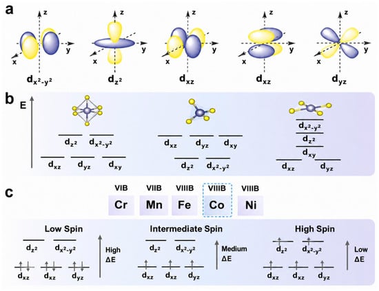
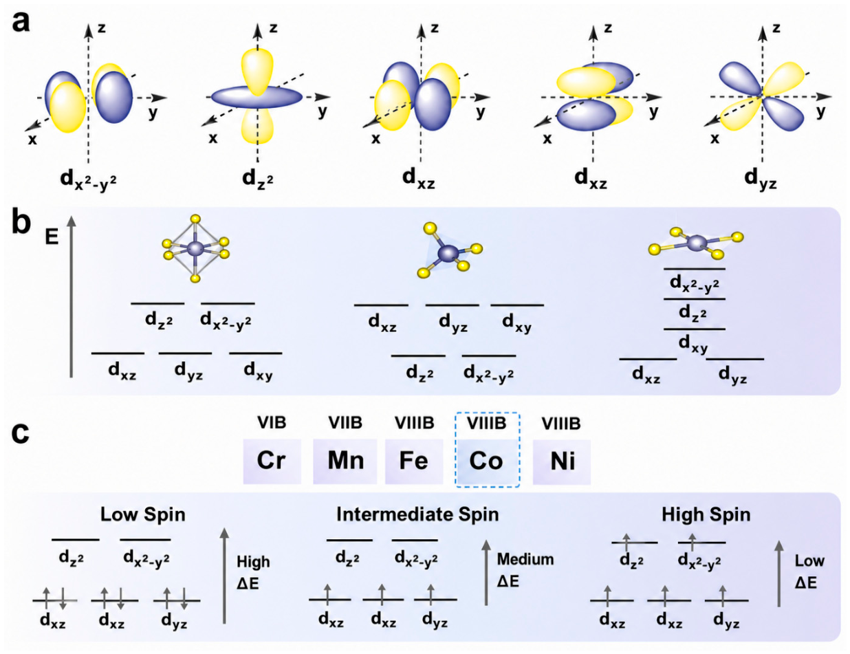
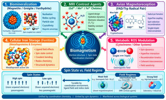
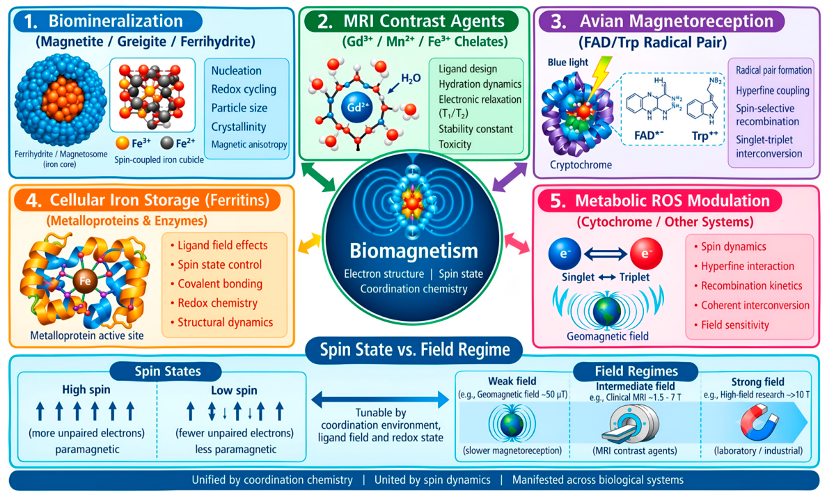
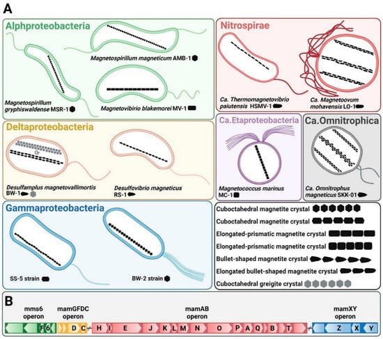
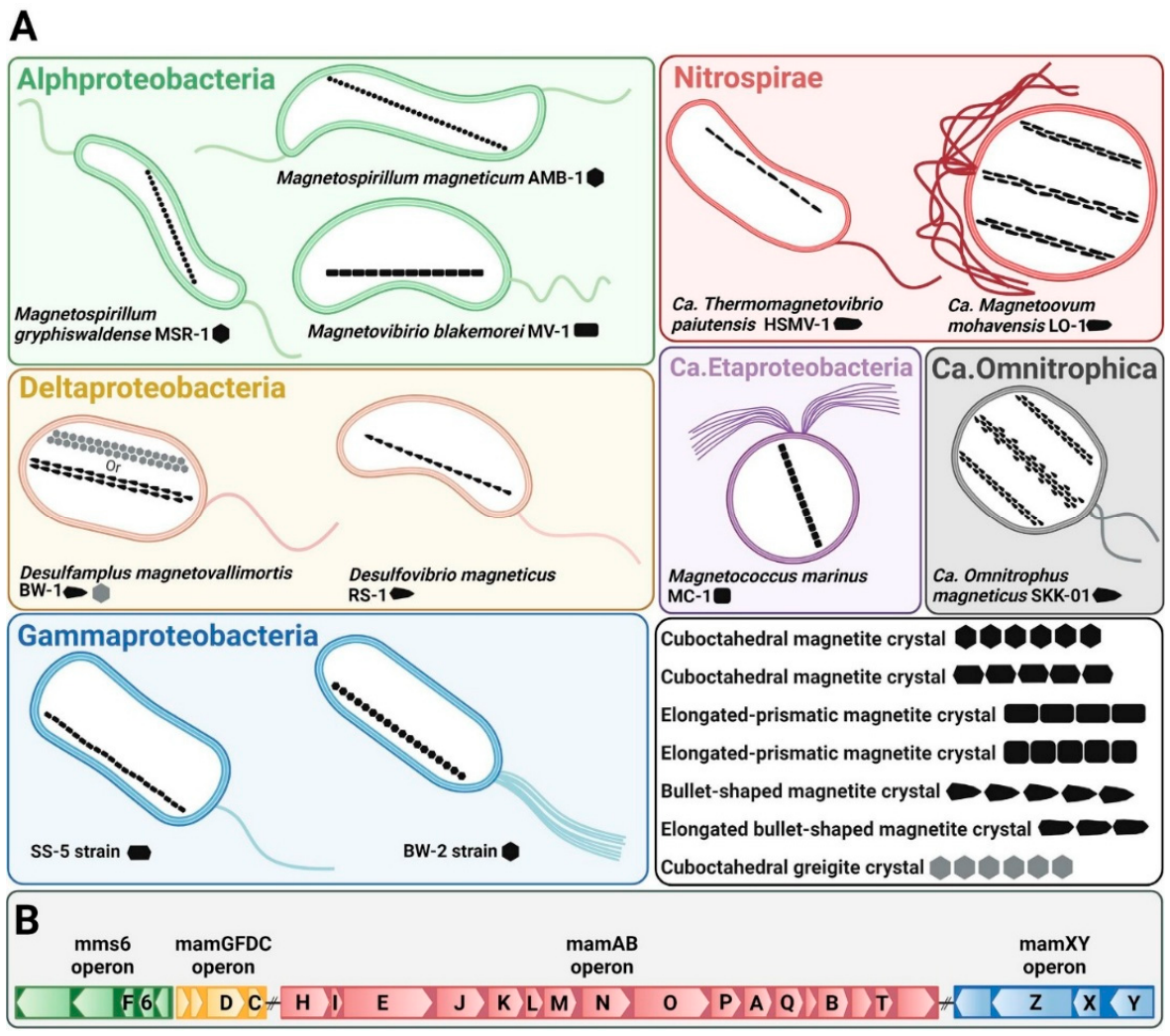
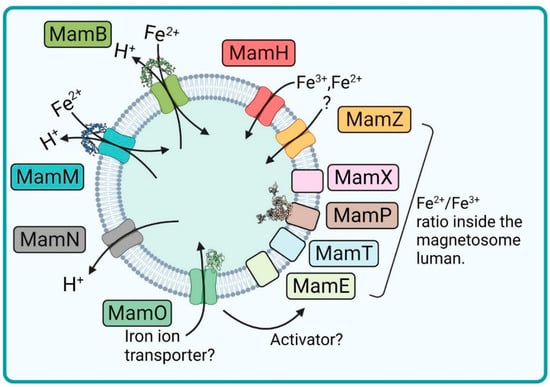
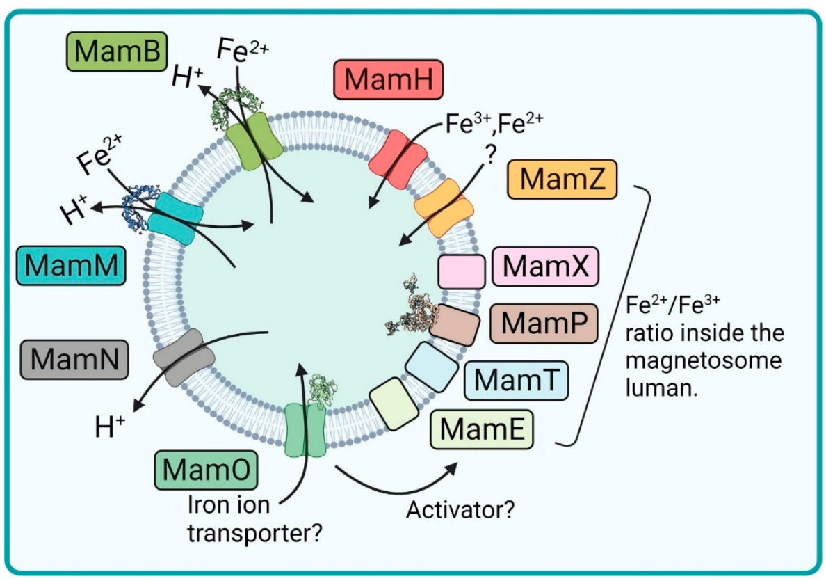
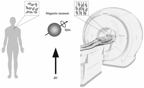
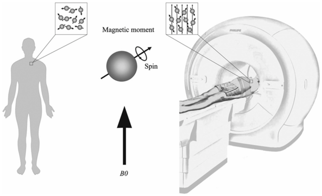
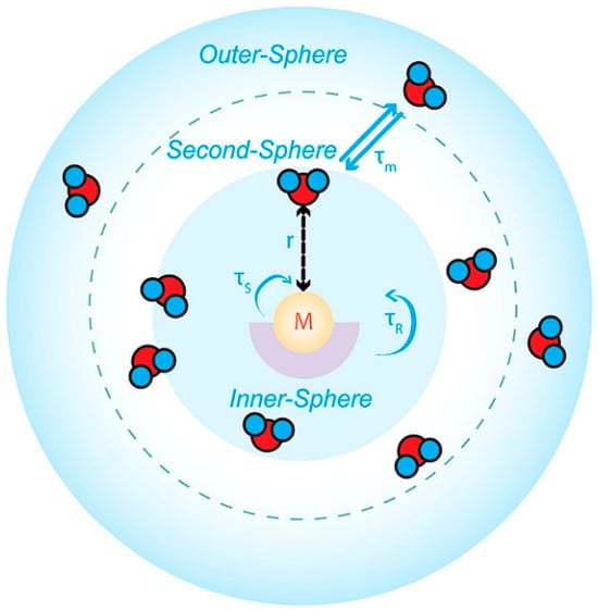
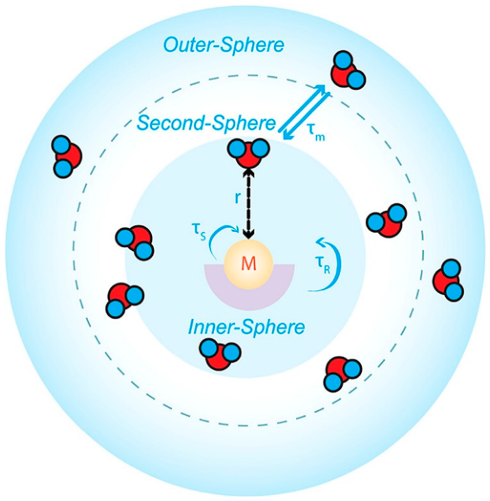
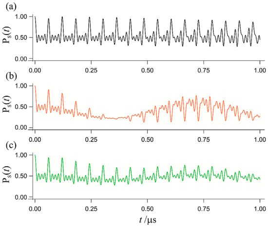
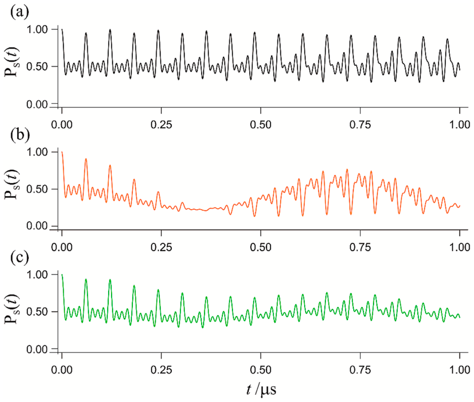
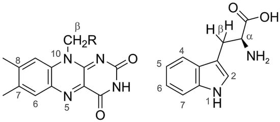
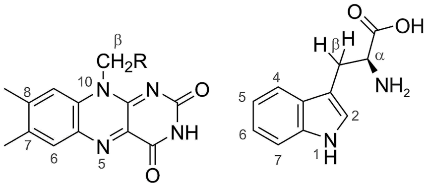
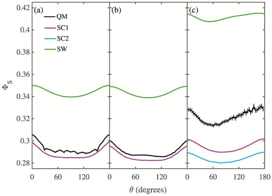
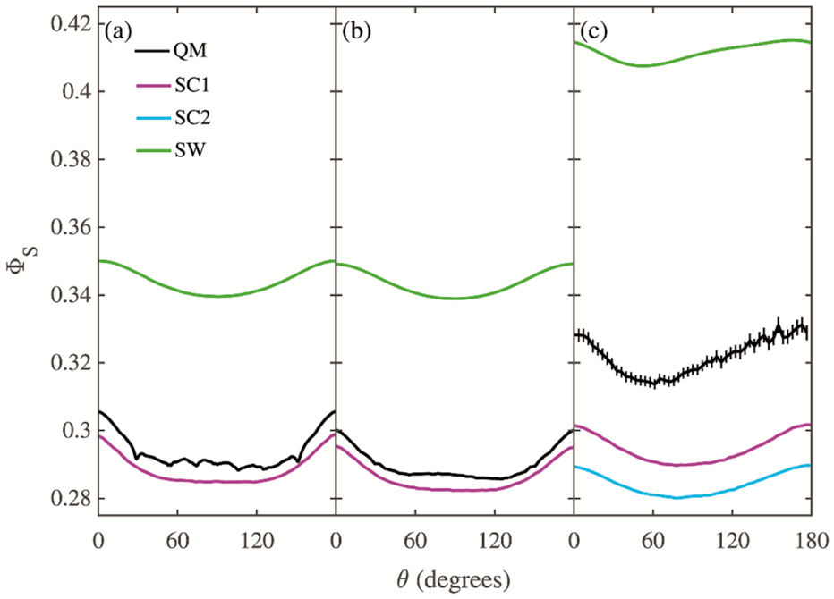
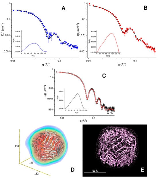
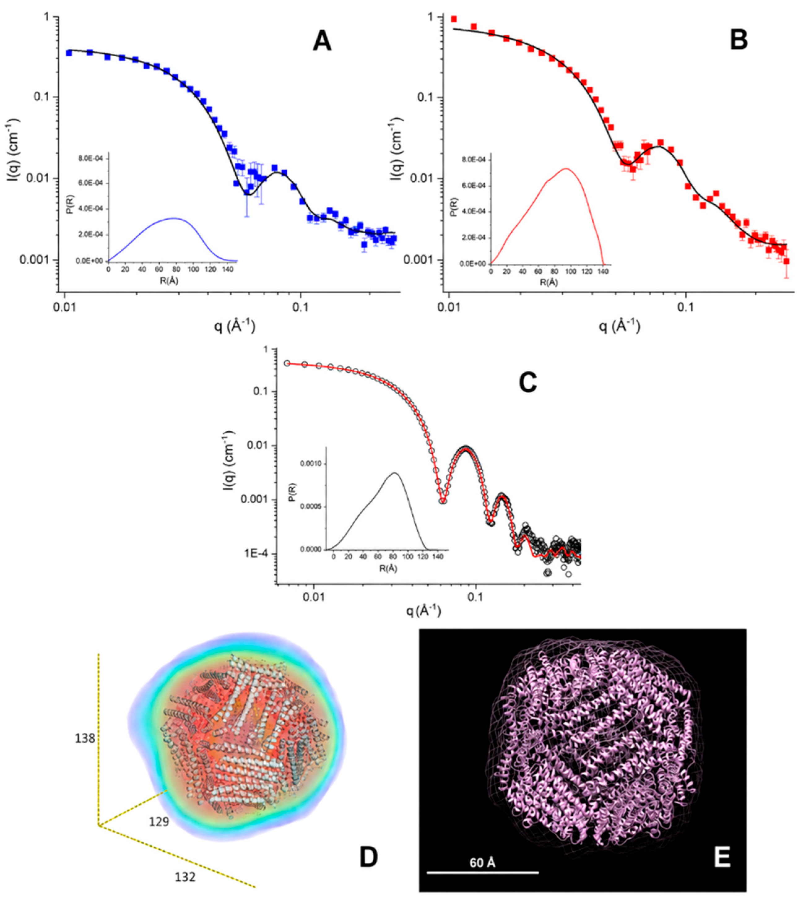
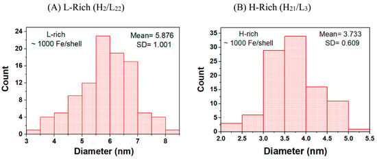
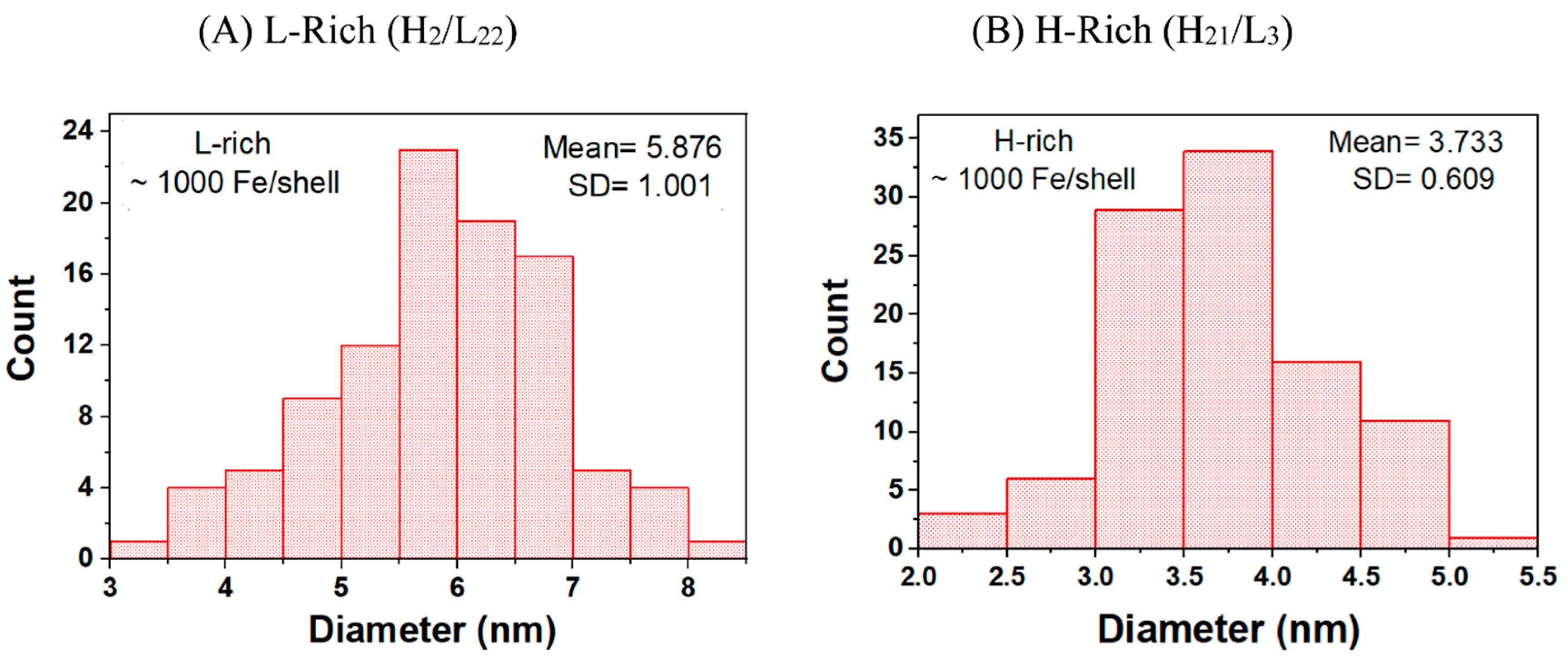
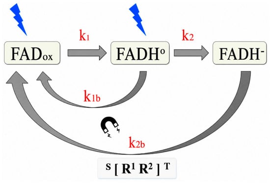
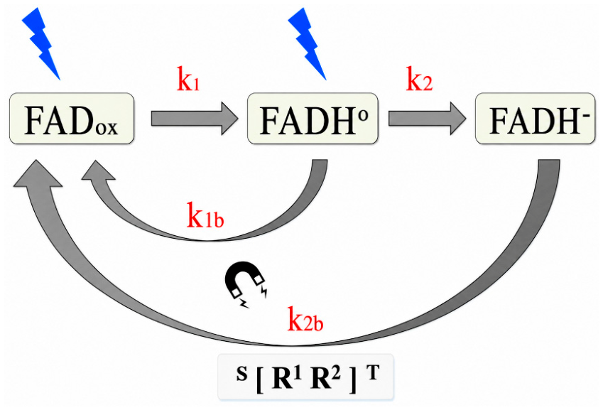

Disclaimer/Publisher’s Note: The statements, opinions and data contained in all publications are solely those of the individual author(s) and contributor(s) and not of MDPI and/or the editor(s). MDPI and/or the editor(s) disclaim responsibility for any injury to people or property resulting from any ideas, methods, instructions or products referred to in the content. |
© 2026 by the authors. Licensee MDPI, Basel, Switzerland. This article is an open access article distributed under the terms and conditions of the Creative Commons Attribution (CC BY) license.