


Abstract
Temporomandibular joint osteoarthritis (TMJ-OA) is a multifactorial degenerative disorder characterized by progressive cartilage degradation, subchondral bone remodeling, and chronic inflammation, leading to pain and functional impairment in affected individuals. Despite its clinical impact, effective disease-modifying treatments remain limited, highlighting the need for innovative therapeutic approaches for treating this condition in the future. This manuscript examines the biological rationale, clinical applications, and therapeutic potential of platelet-rich plasma (PRP) and injectable platelet-rich fibrin (i-PRF) in the management of TMJ-OA. As autologous platelet-derived biomaterials, PRP and i-PRF contain high concentrations of growth factors and bioactive molecules that can modulate inflammatory responses and support tissue repair. PRP is associated with a relatively rapid release of these mediators, whereas i-PRF forms a fibrin matrix that may enable a more sustained release profile. Current clinical evidence suggests that both therapies show potential to contribute to pain reduction and may facilitate improvements in mandibular function. However, substantial heterogeneity in preparation protocols, study designs, and outcome measures limits the comparability and generalizability of these findings to the general population. Overall, PRP and i-PRF represent promising, minimally invasive regenerative strategies for managing TMJ-OA.
1. Introduction
Temporomandibular joint osteoarthritis (TMJ-OA) is a multifactorial chronic degenerative disorder of the temporomandibular joint (TMJ) characterized by the progressive deterioration of articular cartilage, remodeling of the subchondral bone, and synovial inflammation [1,2,3,4]. Clinical diagnosis is based on patient history, physical examination, and imaging, particularly when differentiating degenerative joint disease from inflammatory or disc-related conditions [5,6,7,8]. TMJ-OA manifests as articular pain, joint sound, limited mandibular mobility, and impaired function, often accompanied by morning stiffness, and in advanced cases, occlusal changes, and facial skeletal remodeling [2,5,9]. These features can interfere with daily activities and overall well-being, highlighting the importance of early recognition and management [10,11], particularly given their association with sleep disturbances and emotional disorders that further compromise quality of life [12,13].
The pathophysiology of TMJ-OA involves the progressive degradation of the condylar fibrocartilage, specifically the breakdown of the Type I and II collagen network that provides the joint’s tensile and compressive strength. This structural failure leads to a significant loss of biomechanical stability and mechanical stiffness. The process is further driven by chondrocyte apoptosis and the release of catabolic mediators, which diminish the tissue’s natural regenerative capacity. Ultimately, as the joint’s adaptive remodeling mechanisms fail, the cartilage becomes unable to withstand normal or parafunctional forces (like bruxism), resulting in a continuous cycle of degenerative change and functional impairment [1,14,15,16,17,18,19].
Because etiological factors are multifactorial, treatment requires a multidisciplinary approach [20,21,22,23,24]. First-line therapeutic approaches are noninvasive, conservative treatments, including analgesics, anti-inflammatory drugs, occlusal splints and physiotherapy [9,22,25]. Less invasive surgical procedures encompass intraarticular injections of hyaluronic acid or corticosteroids, arthrocentesis alone and the combination of arthrocentesis and intraarticular injections [20,22,26]. While these therapies primarily provide symptomatic relief, they do not reverse tissue degeneration and are typically guided by symptom severity [5,9,20,25]. Unlike certain conventional treatments, which may exert dose-dependent chondrotoxic effects, such as corticosteroids, regenerative approaches have emerged as minimally invasive options for treating osteoarthritis (OA) [20,25,27,28]. Autologous blood-derived products can modulate inflammation, stimulate angiogenesis, and remodel the extracellular matrix [29,30,31]. Among these, platelet-rich plasma (PRP) and injectable platelet-rich fibrin (i-PRF) represent two important types of platelet concentrates, promoting tissue repair and restoring joint function. PRP is a first-generation platelet concentrate enriched with platelets and growth factors. i-PRF, a second-generation formulation, is prepared without anticoagulants and allows a sustained release of bioactive molecules, prolonging its regenerative effects. The distinct release kinetics of these two generations, the rapid ‘burst’ of PRP versus the prolonged delivery of i-PRF, form the biological basis for comparing their clinical efficacy in reducing pain, increasing maximal mouth opening, and minimizing adverse injection site responses [32,33,34,35,36].
Epidemiological studies suggest that temporomandibular disorders affect approximately 30% of the general population, with a higher prevalence among women, particularly between the second and fourth decades of life, probably reflecting the predominance of muscular and functional disorders in this age group. However, TMJ-OA specifically follows a different demographic pattern, with its incidence increasing significantly in older populations (50–60 + years) due to cumulative degenerative changes [37,38,39,40]. Hormonal influences, particularly estrogen-mediated modulation of inflammatory pathways and cartilage metabolism, have been proposed as potential contributors to sex-related disparities. In particular, estrogen-related receptors (ERRs) have been implicated in TMJ osteoarthritis, where altered signaling, such as suppression of ERRα or overexpression of ERRγ, may promote cartilage degradation and inflammatory responses, suggesting a more complex hormone-dependent regulatory network beyond classical estrogen pathways. In addition, genetic predisposition and systemic inflammatory conditions may further influence susceptibility to TMJ degenerative changes [38,41,42]. Recent genomic studies have identified specific single-nucleotide polymorphisms (SNPs), including variants in the FTO gene and intergenic regions between FILIP1 and SENP6, as potential risk factors associated with TMJ-OA, highlighting the role of genetic variability in disease onset and progression. Moreover, mechanical stress and structural abnormalities, such as disc displacement without reduction, interact with inflammatory and genetic factors, contributing to the multifactorial and often unpredictable progression of TMJ degeneration [42,43,44,45].
Advances in regenerative medicine have stimulated growing interest in orthobiologic therapies aimed at restoring joint homeostasis, rather than merely alleviating symptoms. Platelet concentrates have gained particular attention because of their autologous origin, relative ease of preparation, and capacity to deliver a concentrated mixture of growth factors and cytokines involved in tissue repair [35,46,47,48,49,50,51,52]. Within this context, a comparative evaluation of different platelet-derived formulations has become increasingly important for determining their optimal clinical application in temporomandibular joint disorders.
This narrative review aims to address which autologous formulation, PRP or i-PRF, demonstrates superior efficacy in managing TMJ-OA pain and function. By evaluating the biological impact of PRP’s rapid growth factor ‘burst’ versus i-PRF’s fibrin-mediated ‘sustained release,’ we analyze how these distinct kinetic profiles influence tissue regeneration and long-term symptom management. To support this objective, relevant studies were identified through structured searches of PubMed/MEDLINE, Scopus, and Web of Science. Priority was given to high-quality studies addressing PRP and i-PRF and their role in the treatment of TMJ-OA.
2. Pathophysiology of TMJ-OA and Rationale for Orthobiologic Therapy
TMJ-OA is characterized by complex interactions between mechanical loading, genetic predisposition, inflammatory mediators, and cellular dysregulation within the joint microenvironment [1,3,22,53,54]. Mechanical stress, microtrauma, and functional overload can disrupt the physiological homeostasis of the TMJ, initiating inflammatory responses in the synovial membrane and surrounding tissues [14,54,55,56,57]. Parafunctional habits, such as bruxism and chronic clenching, may further exacerbate mechanical overload and accelerate degenerative changes within the joint [53,54,58,59].
This inflammatory activation leads to increased production of pro-inflammatory cytokines, including interleukin (IL)-1β, IL-6, and tumor necrosis factor-α (TNF-α), which play a central role in activating signaling cascades and promoting catabolic processes within the joint [14,22,53,54,55,60]. Synovial macrophages may also contribute significantly to disease progression, as the predominance of pro-inflammatory M1 macrophages enhances cytokine release and matrix degradation, whereas M2 macrophages are generally associated with anti-inflammatory and tissue-repair responses [14,54,61]. These mediators stimulate the expression of matrix-degrading enzymes, particularly matrix metalloproteinases (MMPs), aggrecanases and a disintegrin and MMP with thrombospondin motifs (ADAMTS), which contribute to the degradation of key extracellular matrix (ECM) components, including type I and II collagen and proteoglycans in the condylar cartilage [14,53,54,55,60,62]. Concurrently, several intracellular signaling pathways, such as nuclear factor-κB (NF-κB), mitogen-activated protein kinases (MAPKs), and extracellular signal-regulated kinases (ERKs), are activated, further enhancing inflammatory responses while suppressing anabolic repair mechanisms [53,54,55].
In addition to extracellular matrix degradation, TMJ-OA is characterized by substantial disturbances in chondrocyte homeostasis. Beyond classical apoptosis, chondrocyte loss involves inflammatory-cell death pathways including pyroptosis, ferroptosis, necroptosis, and the recently identified cu-proptosis. These pathways are increasingly recognized as potential targets for PRP and i-PRF, which may mitigate such programmed cell death through the immunomodulatory effects of cytokines and the antioxidant properties of concentrated growth factors. For instance, the delivery of bioactive molecules like TGF-β and IGF-1 can help shift the joint microenvironment from a pro-inflammatory state toward one that supports chondrocyte survival and autophagy, thereby counteracting the metabolic dysregulation that triggers these diverse death cascades [54,63,64].
Compounding these processes, epigenetic alterations further downregulate protective chondrogenic programs, weaken the capacity for cellular repair, and accelerate the progression of joint deterioration [54,65,66]. Additionally, oxidative stress and hypoxic conditions within the inflamed joint microenvironment contribute to mitochondrial dysfunction, further exacerbating cellular damage [2,54,60]. Autophagy, which initially acts as a protective mechanism against cellular stress by maintaining chondrocyte homeostasis and removing damaged organelles, becomes dysregulated during disease progression, ultimately accelerating cartilage degradation [53,67]. This dual and context-dependent role, cytoprotective in early or physiological conditions but potentially cytotoxic when excessive, prolonged, or impaired, further underscores the complexity of programmed cell death in OA, particularly as it varies with disease stage, mechanical stress, and the cellular microenvironment. Collectively, these interconnected programmed cell death pathways are now recognized as central drivers of osteoarthritis pathogenesis and represent promising, yet highly nuanced, therapeutic targets [64,67,68].
Another hallmark of TMJ-OA progression is the phenotypic transition of chondrocytes to a hypertrophic state resembling endochondral ossification [53,68,69]. Hypertrophic chondrocytes alter their secretory profile and express markers such as MMPs-13, type X collagen, and alkaline phosphatase, promoting premature matrix mineralization [53,54,55,60,69]. In advanced stages, these cells may acquire osteoblast-like characteristics, directly contributing to osteophyte formation [53,54,55,60,68]. This pathological transformation is closely associated with aberrant angiogenesis, which facilitates the invasion of blood vessels into normally avascular cartilage [68,70,71,72]. The formation of these vascular networks is frequently accompanied by neurogenesis, allowing sensory nerve fibers to infiltrate damaged tissues. This process is considered a major contributor to the chronic pain and arthralgia frequently observed in TMJ-OA [54,59,68].
Emerging evidence highlights the role of the innate immune system in TMJ-OA progression. Pattern-recognition receptors, including toll-like receptors (TLRs), may be activated by damage-associated molecular patterns (DAMPs) released from degraded cartilage matrix components [73,74,75]. The pathophysiology of TMJ-OA is significantly driven by Damage-Associated Molecular Patterns (DAMPs)—intracellular molecules and matrix fragments released during tissue injury. These DAMPs act as “danger signals” that activate Toll-like receptors (TLR2/4), triggering pro-inflammatory cascades such as the NF-κB pathway. This activation leads to the production of cytokines and catabolic enzymes, which further degrade the joint matrix. Crucially, this creates a self-perpetuating feedback loop: ongoing structural damage releases more DAMPs, which in turn sustain chronic, low-grade sterile inflammation and progressive joint degeneration. Together, these mechanisms provide a direct link between mechanical tissue damage and the persistent innate immune response seen in TMJ-OA [76,77,78,79,80,81].
In addition, recent studies suggest that alterations in subchondral bone remodeling may play a critical role in initiating cartilage degeneration, rather than being solely a secondary consequence of cartilage damage [73,74,75]. Subchondral bone changes often precede cartilage degradation in TMJ-OA, suggesting a “bone-driven” pathogenesis. Abnormal mechanical loading triggers increased bone turnover and osteoclast-driven resorption, which destabilizes the mechanical support of the overlying cartilage. This disruption of the osteochondral unit enhances destructive biochemical crosstalk between bone and cartilage, while aberrant remodeling promotes angiogenesis and nerve ingrowth, further accelerating disease progression and pain [72,82,83,84,85].
Early TMJ-OA is marked by a high-turnover phase where increased bone resorption reduces bone density and enhances trabecular porosity, weakening the subchondral plate despite accelerated mineral apposition. As the disease advances, this state shifts toward suppressed resorption and excessive bone formation. This transition leads to subchondral sclerosis, characterized by increased bone volume and density, though the newly formed tissue often exhibits reduced mineralization and compromised structural quality [54,86]. As the condition advances, compensatory yet disorganized reparative mechanisms result in subchondral bone sclerosis, characterized by thickening of the bone plate, altered biomechanical properties, and progressive structural deformities [1,2,14,53,68]. These changes culminate in the crepitus and limited mandibular mobility, ultimately leading to joint dysfunction [9,10,12,54,60,68].
Conventional therapeutic strategies for TMJ-OA are primarily aimed at symptom control rather than structural regeneration of the joint. First-line management typically includes conservative approaches such as nonsteroidal anti-inflammatory drugs (NSAIDs), analgesics, occlusal splints, physiotherapy, and behavioral interventions aimed at reducing parafunctional habits and mechanical overload on the joint [9,20,24,25,87,88]. These treatments can alleviate pain, improve mandibular mobility, and reduce functional impairment, particularly in the early or mild stages of the disease. When conservative measures fail to provide sufficient relief, minimally invasive procedures may be considered, including intra-articular injections of corticosteroids or hyaluronic acid, as well as arthrocentesis to lavage inflammatory mediators from the joint space [20,26]. Although these interventions may temporarily improve clinical symptoms by reducing inflammation and improving joint lubrication, they generally do not restore damaged cartilage or halt the underlying degenerative processes [20,26,60]. Furthermore, repeated intra-articular corticosteroid injections have been associated with potential chondrotoxic effects and may accelerate cartilage degradation when used excessively, highlighting the need for therapeutic strategies capable of promoting biological repair within the joint microenvironment [20,25,28,89].
Platelet concentrates are autologous biological blood-derived products that offer a minimally invasive biological approach to modulate degenerative processes by directly delivering growth factors into the joint environment [26,90,91,92]. These bioactive mediators act synergistically to stimulate chondrocyte proliferation, angiogenesis, and ECM component synthesis, largely due to their rich content of growth factors such as VEGF, insulin-like growth factor-1 (IGF-1), PDGF, and TGF-β, which are gradually released from the fibrin matrix and provide sustained regenerative signaling [26,91,93].
Beyond their anabolic potential, these concentrates exert anti-inflammatory effects by modulating signaling pathways and reducing the release of catabolic cytokines [90,91,94]. PRP, a first-generation formulation, can reduce chondrocyte apoptosis and facilitate structural cartilage repair, which is further supported by recent findings showing that PRP counteracts inflammatory and catabolic signaling, suppresses NF-κB activation, reduces IL-1β/TNF-α-driven matrix degradation, and restores cartilage matrix homeostasis. Additionally, in osteoarthritis models, PRP has been shown to promote chondrocyte proliferation and inhibit apoptosis by downregulating Bax and caspase-3. Evidence also suggests it may enhance autophagy and mitochondrial function, indicating a potential role in reinforcing articular cartilage integrity under experimental conditions [90,95,96]. Additionally, second-generation formulations, such as injectable PRF (i-PRF), employ a naturally forming fibrin matrix that remains in a liquid, injectable state prior to coagulation, enabling uniform intra-tissue dispersion and creating an in situ three-dimensional scaffold that closely mimics the early ECM environment. With this structural advantage i-PRF can retain platelets, leukocytes, cytokines, and fibrinogen in a loosely polymerized network, resulting in a markedly prolonged and gradual release of key regenerative mediators, including PDGF, TGF-β, VEGF, IGF-1, and epidermal growth factor (EGF), over 10 to 14 days, a release profile substantially longer than the rapid burst (minutes to hours) seen with PRP. This extended kinetic pattern not only prolongs the local biological stimulus but also enhances chemotaxis, angiogenesis, and matrix remodeling, ultimately supporting a more durable and coordinated regenerative response in musculoskeletal and cartilage tissues [34,91,97,98].
Figure 1 shows the biological mechanisms associated with the rationale of the use for orthobiologic therapy.
Figure 1.
Key biological mechanisms serving as the base for orthobiologic therapy. EGF, epidermal growth factor; IL-1β, interleukin 1β; PDGF, platelet-derived growth factor; TGF-β1, transforming growth factor β1; TNF-α, tumor necrosis factor α; ↑ increase; ↓ decrease. Parts of this figure were drawn using pictures from Servier Medical Art by Servier (https://smart.servier.com, 30 March 2026) which is licensed under Attribution 4.0 International (Creative Commons CC BY 4.0).
3. Platelet-Rich Plasma in TMJ-OA
PRP is an autologous blood-derived concentrate with a platelet concentration significantly exceeding physiological baseline levels, typically ranging from two to five times the count found in the blood [46,60,92]. PRP production relies on differential centrifugation, a process that utilizes controlled centrifugal forces to separate whole blood components, such as red blood cells, leukocytes, and platelets, based on their specific gravity [90,91,92,99,100].
Standard preparation protocols generally involve a two-step centrifugation. An initial “soft spin” at a low speed separates the blood into three distinct layers: an upper plasma layer, an intermediate buffy coat rich in platelets and leukocytes, and a lower erythrocyte fraction. The plasma and buffy coat layers are separated and centrifuged again at a higher speed, typically called a “hard spin”. After this step, platelets and leukocytes precipitate, and platelet-poor plasma (PPP) remains on the top [33,100]. After removing more than half of the plasma volume, the remaining sample is gently resuspended to yield PRP, which can be gelled with calcium chloride, calcium gluconate, or thrombin [92,101].
To prevent premature coagulation and preserve platelet viability during this process, blood is collected in tubes containing anticoagulants, most commonly acid citrate dextrose (ACD) or sodium citrate (SC) [92,102,103]. The selection of an appropriate anticoagulant is a critical parameter in PRP preparation, as it directly impacts platelet recovery, membrane integrity, and growth factor release. Research has shown that ACD may yield higher levels of platelet-derived growth factor (PDGF) and VEGF, whereas SC is often associated with superior transforming growth factor-β1 (TGF-β1) concentrations [48,101]. Although ethylenediaminetetraacetic acid (EDTA) may yield higher platelet counts, it is frequently avoided because it can induce structural damage to the platelet membrane [48].
To standardize characterization, PRP is widely categorized according to the Dohan Ehrenfest Classification (2009) [104], which divides platelet concentrates into four groups based on leukocyte content and fibrin architecture: Pure Platelet-Rich Plasma (P-PRP), Leukocyte- and Platelet-Rich Plasma (L-PRP), Pure Platelet-Rich Fibrin (P-PRF), and Leukocyte- and Platelet-Rich Fibrin (L-PRF) [104,105]. Despite these frameworks, the literature highlights significant heterogeneity in preparation protocols. Currently, more than 16 commercial systems are available, exhibiting critical variations in centrifugation parameters, capture efficiency, and resulting proportions of cellular constituents [105]. This lack of standardization, compounded by donor-specific variables such as age and metabolic status, creates challenges in the clinical reproducibility of the therapeutic outcomes. Therefore, protocols should report Relative Centrifugal Force (RCF) rather than rotation per minute (RPM), and swing-out rotors are preferred to minimize platelet trauma [48,92].
The therapeutic efficacy of PRP is primarily derived from the rich composition of bioactive mediators sequestered within the alpha-granules of concentrated platelets [90,99,102]. Upon activation, initiated either exogenously by agents such as calcium chloride or thrombin or endogenously through high-affinity engagement of platelet receptors with native collagen at the injection site, platelets trigger a rapid and highly coordinated activation program characterized by α-granule and dense-granule degranulation, reported to release more than 300 soluble proteins and over 1500 bioactive mediators essential for tissue repair, immunomodulation, and angiogenesis [92,106]. This degranulation event leads to the extracellular release of a broad panel of regenerative mediators, including PDGF, TGF-β1, VEGF, IGF-1, EGF, and fibroblast growth factor (FGF) isoforms, which collectively coordinate cell proliferation, angiogenesis, ECM synthesis, and immune modulation [90,103,106,107,108,109]. Beyond these well-known factors, recent evidence has attributed significant importance to platelet-derived exosomes (extracellular vesicles) and extra platelet molecules, such as alpha-2-macroglobulin (M2), which protect cartilage from enzymatic degradation [48,110]. Additionally, mediators released from dense granules, such as serotonin (5-HT), contribute to the multifaceted analgesic effects observed in patients [48,101]. Furthermore, PRP releases stromal cell-derived factor-1 (SDF-1), which facilitates the recruitment and homing of mesenchymal stem cells to the injured TMJ site via the SDF-1/C-X-C chemokine receptor type 4 (CXCR4) axis [101,111,112].
Beyond their anabolic and reparative potential, these concentrates also exert a robust anti-inflammatory effect by suppressing the activation of the TLR4/NF-κB signaling pathway, which in turn markedly reduces the expression and release of key catabolic cytokines, including IL-1β, IL-6, and TNF-α [90,113,114]. In the OA microenvironment, PRP may interrupt NF-κB–mediated inflammatory and catabolic signaling by reducing the expression of matrix-degrading enzymes such as MMPs and aggrecanases. Evidence from experimental models suggests that PRP may facilitate macrophage polarization toward the anti-inflammatory M2 phenotype by suppressing pro-inflammatory mediators (IL-1β, IL-6, and TNF-α) and enhancing M2-associated markers including MRC1, IL-10, and TGF-β. Additionally, PRP can reprogram M1 macrophages into an M2-like state, reinforcing its immunomodulatory effects. These actions help create a pro-regenerative environment by reducing oxidative stress and mitochondrial dysfunction, while supporting mesenchymal stem cell activity, angiogenesis, and tissue remodeling, ultimately shifting OA tissues from a catabolic inflammatory state toward immune resolution and regeneration [62,92,99,115,116].
As a first-generation platelet concentrate, PRP is characterized by its liquid formulation, which facilitates intra-articular injection and allows combination with other therapeutic agents [94,101,106]. However, this fluid state also dictates its characteristic burst-type release kinetics, with platelet activation leading to rapid degranulation and a steep early discharge of bioactive molecules: approximately 70% of growth factors are released within the first 10 min and nearly 100% within 1 h [48,90,99,103]. This pattern aligns with the broader understanding that first-generation concentrates rely on anticoagulants and exogenous activators, factors known to influence both fibrin architecture and protein-release behavior [35,36,117]. While such an intense early stimulus can be advantageous in acute or inflammatory conditions, its regenerative effect is inherently short-lived, especially when compared with the more gradual and sustained release observed in second-generation preparations. L-PRF and its derivatives (including injectable PRF) have consistently been shown to provide slower, extended growth-factor delivery owing to the presence of a dense, naturally formed fibrin network and the absence of anticoagulants or chemical activators [26,35,68]. Consequently, although PRP remains useful for initiating rapid biological responses, its therapeutic window may be more transient than that of fibrin-based concentrates, which maintain bioactivity over several days and potentially offer more durable regenerative support [35,91,118].
Studies have demonstrated that platelet-derived extracellular vesicles (EVs), including exosomes and microvesicles, mediate many of the regenerative actions traditionally attributed to PRP. These nanoscale vesicles encapsulate a rich repertoire of bioactive cargos, such as microRNAs (e.g., miR-126, miR-21, and miR-223), growth factors (VEGF, PDGF-BB, basic-FGF, and TGF-β1), lipids, and cytokines, which regulate gene expression and cellular responses in recipient cells. PRP-derived EVs can enhance chondrocyte proliferation, suppress apoptosis, and activate anabolic pathways, such as Wnt/β-catenin, promoting ECM synthesis and restoring cartilage homeostasis. PRP-EVs exert potent pro-angiogenic effects by delivering VEGF, basic-FGF (b-FGF), and pro-angiogenic microRNAs, thereby activating extracellular signal-regulated kinase (ERK), phosphoinositide 3-kinase (PI3K)/protein kinase B (Akt), and yes-associated protein (YAP) signaling cascades in endothelial and stromal cells to stimulate migration, tube formation, and microvascular network assembly. Their ability to modulate inflammation, either by dampening excessive cytokine production or promoting anti-inflammatory macrophage phenotypes depending on contextual cues, further supports a microenvironment conducive to tissue repair. Moreover, because PRP-EVs protect their molecular cargo within a stable phospholipid membrane, they show superior resistance to enzymatic degradation compared with freely released PRP growth factors and may thus provide more durable biological effects in vivo. Collectively, these findings highlight PRP-derived EVs as key mediators of PRP regenerative activity and identify them as promising, more standardized, and potentially superior cell-free therapeutic candidates for cartilage repair [119,120,121,122].
4. Injectable Platelet-Rich Fibrin in TMJ-OA
i-PRF is a second-generation platelet concentrate developed to overcome some of the biological and technical limitations of conventional PRP formulations. Unlike PRP, i-PRF is produced without anticoagulants or exogenous biochemical additives, allowing the natural coagulation cascade to occur after injection. This process results in the formation of a fibrin-based three-dimensional scaffold capable of entrapping platelets, leukocytes, and circulating growth factors [32,91,123].
i-PRF is produced using low-speed centrifugation, a method specifically designed to preserve a higher concentration of viable regenerative cells (platelets, leukocytes, and circulating progenitor cells) within the liquid fibrinogen-rich plasma layer. Whole blood is collected in plastic tubes without anticoagulants and centrifuged immediately at low relative centrifugal forces, typically 40 to 60 g for 3 to 8 min depending on the protocol, which avoids premature clotting while minimizing shear-induced cellular damage [26,91,124,125]. This low-speed approach increases the recovery and uniform distribution of platelets, leukocytes, and growth factors, as demonstrated in multiple analyses of PRF matrices. Compared with high-speed centrifugation, low-speed protocols retain more bioactive components and produce a plasma fraction richer in VEGF, TGF-β1, PDGF isoforms, and other cytokines essential for angiogenesis, osteogenesis, and early immune modulation. Low-force centrifugation results in a PRF scaffold with larger fibrin pores, slower degradation, and sustained release kinetics of growth factors, thereby enhancing its regenerative capacity. After centrifugation, the upper, cell-rich liquid phase is aspirated before complete fibrin polymerization, allowing immediate injection, in which there is a gradual conversion of fibrinogen to fibrin. Preclinical evidence confirms that i-PRF prepared using low-force protocols can improve early biological responses, including enhanced vascularization, fibroblast migration, and recruitment of osteogenic cells. Overall, the low-speed i-PRF concept maximizes viable cell recovery, enhances growth factor content, improves fibrin architecture, and produces a biologically potent injectable concentrate suitable for immediate clinical use in soft and hard tissue regeneration [91,125,126,127,128,129].
Once injected into the joint space, the fibrinogen present in i-PRF undergoes gradual polymerization, forming a fibrin matrix that acts as a temporary scaffold capable of supporting cell migration and tissue regeneration. This network entraps platelets and leukocytes, allowing for a more sustained and gradual release of growth factors that PRP [32,91].
Fibrin architecture also facilitates cellular adhesion and provides a microenvironment that may promote the recruitment of mesenchymal stem cells and progenitor cells to the site of injury. i-PRF exhibits potent regenerative activity by enhancing fibroblast proliferation, cytoskeletal organization, and ECM deposition. At the cellular level, PRF and i-PRF upregulate key matrix-related proteins, heat shock protein 47 (HSP47) and lysyl oxidase) and activate Akt signaling, collectively driving osteoblast proliferation and collagen matrix production [122,123,125,130,131,132]. i-PRF also contains high concentrations of platelets, leukocytes, and progenitor cells and releases angiogenic growth factors such as PDGF, TGF-β1, VEGF, and IGF-1, in a sustained manner, supporting endothelial migration and angiogenesis, which are essential for tissue repair [26,91]. Preclinical models have further demonstrated that i-PRF accelerates early vascularization and bone remodeling when combined with graft materials, confirming its utility in bone-related applications despite limited long-term volumetric effects in some defect types. Together, these findings highlight i-PRF as a biologically active autologous matrix capable of promoting fibroblast activity, neovascularization, and ECM formation [122,123,125,130,131,132].
However, because i-PRF forms a loose three-dimensional fibrin network rather than the rapidly degrading clot characteristic of PRP, its growth factor release profile differs substantially. The fibrin mesh traps platelets, leukocytes, and their secreted mediators, allowing a gradual, sustained liberation of cytokines over several days, in contrast to the rapid, short duration burst release typical of PRP. This prolongs their biological activity and maintains a regenerative microenvironment within the tissues [32,125]. Additionally, i-PRF exerts potent immunomodulatory effects through leukocyte-derived anti-inflammatory cytokines and macrophage regulation. The sustained release of IL-10 and IL-1RA, key antagonists to the pro-inflammatory cytokines found in degenerate joints, promotes a stable anti-inflammatory environment, offering a targeted mechanism to reduce the chronic synovial inflammation and catabolic activity that characterize TMJ-OA [61,68,91,133,134].
Moreover, studies have indicated that i-PRF can promote fibroblast proliferation, cytoskeletal maturation, and alkaline phosphatase activity. These observations suggest a potential to enhance soft-tissue reparative capacity and early matrix organization, particularly in experimental models of tissue healing. PRF-derived matrices also stimulate osteoblast attachment, Akt pathway activation, and the upregulation of extracellular matrix–related proteins such as HSP47 and lysyl oxidase, collectively driving collagen synthesis and bone matrix deposition. In parallel, the sustained release of angiogenic factors supports endothelial migration and neovascularization, further enhancing regenerative properties. Together, these immunomodulatory, pro-angiogenic, and matrix-formative effects highlight the favorable regenerative potential of i-PRF for musculoskeletal and craniofacial tissues [33,91,123,134].
From a clinical perspective, the liquid consistency of i-PRF allows intra-articular administration using the same technique as PRP, after which it undergoes in situ fibrin polymerization to form a three-dimensional scaffold that entraps the cells and growth factors. This transition from a liquid to a fibrin matrix may enhance retention within the joint cavity and prolong the residence time of bioactive mediators. Optimal clinical outcomes often depend on careful patient selection; individuals in the early-to-moderate stages of TMJ-OA typically show the most favorable responses, whereas those with advanced bony ankylosis may experience more limited benefits. Standardized injection protocols generally involve delivery into the superior joint space, often following arthrocentesis to clear inflammatory debris. Early clinical evidence supports these mechanistic advantages by reporting that intra-articular i-PRF injections lead to improvements in TMJ-related symptoms, with reductions in pain and joint clicking, as well as measurable gains in mandibular mobility and overall joint function. However, routine clinical application faces challenges regarding the variability between preparation methods, as differences in centrifugation speed and tube types can significantly alter the final leukocyte and fibrin concentration. While i-PRF demonstrates sustained benefits for several months with only mild, transient adverse effects like post-injection swelling, the lack of standardized processing remains a primary limitation. Collectively, these findings suggest that the injectable nature of i-PRF, combined with its in situ fibrin scaffold formation and prolonged release of bioactive mediators, may provide a clinically meaningful advantage in modulating intra-articular inflammation and supporting functional recovery in TMJ disorders, although more robust randomized trials are still needed to fully establish its therapeutic efficacy [26,48,97,126,135,136,137].
5. PRP Versus i-PRF: Comparative Evidence
Although both PRP and i-PRF are autologous platelet concentrates designed to enhance tissue regeneration, important biological and methodological differences exist between these two formulations that may influence their therapeutic performance in TMJ-OA. Table 1 and Figure 2 present a comparative summary of the two techniques.
Table 1.
Main biological and clinical differences between PRP and i-PRF.
Figure 2.
Comparative evidence between Platelet-Rich Plasma (PRP) and Injectable Platelet-Rich Fibrin (i-PRF). Parts of this figure were drawn using pictures from Servier Medical Art by Servier (https://smart.servier.com, 30 March 2026), which is licensed under Attribution 4.0 International (Creative Commons CC BY 4.0).
One of the main distinctions between these two types of models is their preparation protocol. PRP preparation requires the use of anticoagulants and often involves a two-step centrifugation process designed to concentrate the platelets in the plasma fraction. In contrast, i-PRF is produced through low-speed centrifugation without anticoagulants, allowing natural fibrin polymerization to occur after injection [32,35,91,98,109]. These methodological differences result in distinct structural and biochemical properties that ultimately affect the growth factor release kinetics and cellular interactions.
To ensure a biologically consistent comparison between both formulations, PRP and i-PRF can be contrasted according to three major characteristics: (1) growth-factor release kinetics, with PRP exhibiting an early ‘burst’ release and i-PRF promoting a sustained release profile; (2) structural organization, where PRP lacks a stable fibrin scaffold while i-PRF forms a three-dimensional fibrin matrix that may prolong local retention and cellular migration; and (3) leukocyte incorporation, which is variable and protocol-dependent in PRP formulations but intrinsically incorporated within the fibrin network of i-PRF.
PRP is characterized by a rapid release of growth factors following platelet activation, commonly referred to as a “burst release”, in which most bioactive mediators are released within the first hours after administration [90,99,138]. This rapid biological stimulation may be advantageous in acute inflammatory settings because it quickly modulates the joint microenvironment and initiates the repair process. However, the short duration of growth factor availability may limit their regenerative effects [102,138]. Conversely, i-PRF forms a fibrin matrix that gradually releases growth factors over several days. This sustained release profile may create a more stable regenerative environment, potentially supporting prolonged cellular activity and extracellular matrix remodeling within joints [32,91]. The fibrin scaffold may also enhance cellular migration and facilitate the retention of bioactive molecules at the injection site [47,123,139].
The primary distinction of i-PRF lies in its consistent inclusion of leukocytes, which are captured within a dense fibrin scaffold through low-speed centrifugation to act as long-term “bioreactors” for joint healing. While traditional leukocyte-rich PRP products were once feared to increase irritation, research demonstrates that i-PRF actually exerts a powerful immunoregulatory effect by suppressing pro-inflammatory cytokines (such as IL-1β, IL-6, and TNF-α) and utilizing high concentrations of TGF-β to shift macrophages from an inflammatory M1 phenotype to a regenerative M2 phenotype. This unique fibrin network ensures a sustained, seven-day release of anti-inflammatory mediators like IL-10 and IL-1RA, effectively dampening synovial inflammation and curbing the catabolic processes that break down joint tissue. Ultimately, by fostering a stable microenvironment characterized by inflammation resolution rather than amplification, i-PRF provides a sophisticated biological framework specifically suited for treating the complex pathology of TMJ-OA [91,115,116,138,140,141].
Previous investigations involving intra-articular platelet-rich plasma have consistently shown that PRP provides meaningful benefits for patients with temporomandibular disorders. Across diverse patient groups, PRP has been reported to reduce pain intensity, improve maximum mouth opening, and enhance joint function, often producing better outcomes than conventional intra-articular therapies, such as hyaluronic acid or arthrocentesis alone. These improvements are observed both in early follow-up periods and several months after treatment, with many studies noting greater gains in mandibular mobility and more sustained relief from pain when PRP was used instead of standard injectables. When comparing PRP with other minimally invasive protocols, PRP, whether used alone or in combination with lavage techniques, reliably contributes to symptomatic improvement in TMJ-OA, with combined approaches sometimes producing the most pronounced pain reduction. Collectively, the available clinical evidence positions PRP as a therapeutic option capable of meaningfully improving joint biomechanics and functional outcomes in TMJ degeneration, outperforming several conventional modalities in both symptomatic relief and functional restoration [26,142,143,144,145].
Evidence regarding the use of i-PRF in TMJ-OA is limited compared to that for PRP but is steadily expanding. Early clinical investigations have indicated that intra-articular i-PRF can meaningfully reduce pain and improve mandibular mobility. From a practical standpoint, patient selection appears crucial, as the best outcomes are typically observed in patients with early-stage internal derangement or moderate degenerative changes rather than end-stage joint destruction. Studies involving single or repeated i-PRF injections have reported rapid reductions in joint clicking and pain, as well as functional improvement within weeks. Standardized injection protocols often involve 1.0 to 1.5 mL of i-PRF delivered into the superior joint space; however, the lack of a universal preparation protocol remains a significant hurdle. These findings align with experimental data suggesting that the fibrin matrix of i-PRF and the sustained release of bioactive molecules may reflect the regenerative effects of PRP [26,91,134,136,146]. However, direct head-to-head comparisons between PRP and i-PRF remain scarce, and significant variability in centrifugation parameters, leukocyte yield, and injection frequency continues to limit the routine clinical application and the formulation of evidence-based guidelines.
6. Safety Considerations
Autologous platelet concentrates are generally considered safe therapeutic options because of their biological origin and minimal risk of immunogenic reactions. Because PRP and i-PRF are derived from the patient’s own blood, the risk of disease transmission or severe immune-mediated complications is extremely low [47,48,91,92,97,147].
Reported adverse events following intra-articular injection of platelet concentrates into the TMJ are typically mild and transient. The most common complications include temporary post-injection pain, swelling, and mild inflammatory reactions at the injection site, which usually resolve spontaneously within a few days. These symptoms are generally attributed to the local inflammatory response triggered by platelet activation and the growth factor release [26,137,148,149].
Although intra-articular TMJ injections are generally considered safe, serious complications may occur, warranting careful preventive measures. Reported adverse events include localized infection, hematoma formation, transient facial nerve disturbances, sensory changes such as paresthesia or hypoesthesia, and periarticular soft-tissue reactions when injections are not performed optimally. Reviews of adverse outcomes emphasize that neurological complications, while uncommon, can arise from inadvertent needle placement or diffusion of injectates into adjacent structures, underscoring the importance of operator skill and anatomical precision in avoiding such complications. Broader safety analyses of TMJ injection procedures similarly identified postoperative pain flares, swelling, transient malocclusion, headache, and rare inflammatory responses, with most events attributed to technical variability or inadequate aseptic control rather than the biologic agent itself. For these reasons, intra-articular injections should be performed under strict sterile conditions, guided by reliable anatomical landmarks, and, when appropriate, supported by imaging techniques, namely ultrasound, to enhance accuracy and minimize procedure-related risks [137,150,151,152].
Another important safety consideration is the substantial variability in preparation protocols for platelet concentrates. Differences in centrifugation force, spin duration, platelet yield, and leukocyte inclusion can markedly alter the biological composition of the final product, leading to inconsistent therapeutic effects. Comparative analyses of platelet-rich plasma preparation techniques demonstrate wide fluctuations in platelet, leukocyte, and growth-factor concentrations across protocols, which may partly explain the variability in clinical outcomes reported in the literature. Additionally, experimental studies suggest that excessively high platelet concentrations may exert paradoxical effects, such as amplifying inflammatory signaling or even inhibiting cell proliferation, indicating the presence of a dose-dependent ceiling beyond which regenerative benefits may diminish or be reversed. These findings underscore the need for standardized preparation methods and careful protocol selection to optimize safety, minimize unwanted inflammatory responses, and enhance the reproducibility of clinical results [46,91,101,140,153].
The influence of patient-related factors on the safety and regenerative capacity of platelet concentrates should also be considered. Variations in age, systemic inflammatory burden, and metabolic status can significantly alter platelet behavior and the concentration of growth factors obtained during PRP preparation. Evidence shows that advancing age and lower baseline platelet counts reduce the final platelet yield in PRP, introducing variability, even when identical preparation protocols are used [92,102,154]. Metabolic conditions such as obesity, insulin resistance, and type 2 diabetes are associated with heightened platelet activation, increased oxidative stress, and impaired responsiveness to endogenous inhibitory mediators, all of which may modify the biological activity of PRP-derived products. Additional patient factors, including systemic inflammation, psychiatric medications, NSAIDs use, smoking, and alcohol consumption, are also known to influence platelet function and may therefore affect treatment outcomes with autologous platelet therapies [92,102,155]. Collectively, these findings underscore the need to consider individual patient characteristics when interpreting clinical responses and strive for standardized procedures that optimize the consistency and therapeutic potential of platelet concentrates.
Moreover, the technical expertise of the clinician performing the intra-articular injection plays a critical role in minimizing the procedural risks. Ultrasound-guided injection techniques have been increasingly recommended in recent years, as they allow for improved visualization of joint structures and may enhance the accuracy of orthobiologic delivery into the superior joint compartment [156,157,158,159]. Therefore, the use of image guidance may further improve the safety profile of platelet-based therapies in TMJ interventions.
Overall, current evidence suggests that both PRP and i-PRF are well-tolerated treatment modalities for TMJ disorders, with a favorable safety profile when appropriate preparation methods and injection techniques are used.
7. Limitations of Current Evidence
Despite the growing interest in orthobiologic therapies for TMJ-OA, several limitations continue to affect the interpretation and generalization of the current evidence. One of the most significant challenges is the considerable heterogeneity of the platelet concentrate preparation protocols. Variations in centrifugation speed, duration, anticoagulant use, platelet concentration, and leukocyte content can lead to substantial differences in the biological composition of the final product [105,129,140,141,160]. This lack of methodological standardization complicates comparisons between studies and limits their reproducibility across clinical settings.
Another important limitation is the relatively small number of high-quality clinical investigations specifically evaluating PRP and i-PRF for TMJ-OA. Despite the growing interest in orthobiologic therapies, most available studies have small sample sizes, short follow-up periods, or methodological constraints that limit the reliability of their findings. In addition, many trials enroll heterogeneous temporomandibular disorder populations, combining patients with disc displacement, arthralgia, myofascial pain, and degenerative joint changes rather than focusing exclusively on osteoarthritis, which makes it difficult to isolate the treatment effects in TMJ-OA. As a result, the evidence base is still evolving, and more rigorously designed studies with standardized diagnostic criteria, adequate sample sizes, and longer follow-up intervals are needed to establish the true clinical impact of PRP and i-PRF in degenerative TMJ diseases. Outcome assessment methods also varied considerably among the studies. Clinical parameters, such as pain intensity, maximum mouth opening, joint sounds, and functional scores, are measured using different scales and evaluation protocols, which further complicates the synthesis of the available evidence. Additionally, the long-term effects of orthobiologic injections remain poorly understood. Although short- and medium-term improvements in pain and function have been reported, the durability of these therapeutic effects and their potential to modify disease progression remain unclear [20,26,41,46,142,161,162,163].
Finally, direct comparative clinical studies on the use of PRP and i-PRF in the treatment of TMJ-OA are lacking. Most investigations evaluate each therapy independently, making it difficult to establish definitive conclusions regarding which platelet concentrate provides superior regenerative benefits.
8. Future Perspectives
Future research should address the current limitations, as summarized in Figure 3, by establishing standardized preparation protocols and reporting guidelines for the platelet concentrates used in TMJ therapies. The adoption of standardized classifications and reporting of parameters, such as relative centrifugal force, platelet concentration, and leukocyte content, would significantly improve study reproducibility.
Figure 3.
Future perspectives and directions of investigation in the area of platelet concentrates. Parts of this figure were drawn using pictures from Servier Medical Art by Servier (https://smart.servier.com, 30 March 2026), which is licensed under Attribution 4.0 International (Creative Commons CC BY 4.0).
Large-scale randomized controlled trials with longer follow-up periods are needed to determine the long-term efficacy and disease-modifying potential of PRP and i-PRF in TMJ-OA. Such studies should incorporate standardized clinical outcome measures, imaging-based assessments, and biomarker analyses to provide a more comprehensive evaluation of the therapeutic effects.
Emerging research is exploring the combination of platelet concentrates with other regenerative approaches, including mesenchymal stem cell therapy, EVs, and biomaterial scaffolds. These combinatorial strategies may enhance tissue regeneration by providing synergistic biological signals and structural support for cartilage repair. Notably, umbilical cord–derived mesenchymal stem cells (UC-MSCs), including those obtained from Wharton’s jelly, represent a particularly promising cell source due to their high proliferative capacity, multipotent differentiation potential into chondrogenic and osteogenic lineages, and inherent immunomodulatory properties. Preclinical evidence further demonstrates that combining UC-MSCs with biomaterial scaffolds such as PRF significantly enhances chondrogenic regeneration compared to either component alone, as reflected by increased expression of cartilage-specific markers including type II collagen, aggrecan, Sox9, and FGF18, along with improved histological outcomes. This synergistic effect is attributed to the scaffold’s ability to provide mechanical support, facilitate cell adhesion, and enable sustained delivery of bioactive factors, thereby improving UC-MSC proliferation, differentiation, and extracellular matrix deposition, while also promoting more effective tissue integration in osteochondral constructs [164,165,166,167].
Recent advances in orthobiologics indicate that combining platelet concentrates with stem-cell-based treatments or EV therapies may further enhance cartilage repair and modulate inflammatory pathways in TMJ-OA. PRP has been shown to boost mesenchymal stem-cell proliferation, migration, and paracrine activity, thereby strengthening their regenerative potential. Simultaneously, progress in bioengineering now allows the isolation of platelet-derived extracellular vesicles and exosomes, which can replicate many of PRP’s therapeutic effects while offering greater molecular stability and standardization. These acellular formulations reduce donor-dependent variability and retain the ability to modulate inflammation and support cartilage matrix preservation, with platelet-derived extracellular vesicles demonstrating strong reparative effects in preclinical OA models [47,49,93,97,116,117,120,121,168,169,170,171,172].
Additionally, the integration of orthobiologic therapies with advanced imaging and biomarker analysis may facilitate a more personalized approach to the management of TMJ-OA in the future. Molecular biomarkers reflecting cartilage degradation, inflammatory activity, and oxidative stress may help identify patients who are most likely to benefit from regenerative treatments. Such precision medicine approaches could optimize treatment timing and improve the long-term outcomes.
9. Conclusions
TMJ-OA is a complex degenerative condition characterized by progressive cartilage breakdown, chronic inflammation, and structural remodeling of the joint. Conventional therapies primarily focus on symptom control and often fail to address the underlying biological mechanisms responsible for OA tissue degeneration.
Autologous platelet concentrates have emerged as potentially promising orthobiologic therapies that may modulate inflammation and support regenerative processes in the joint environment. PRP, a first-generation platelet concentrate, provides a rapid release of growth factors that is thought to initiate tissue repair and may help attenuate inflammatory signaling in experimental models.
In contrast, i-PRF is a second-generation formulation characterized by the formation of a fibrin scaffold and a more sustained release of bioactive mediators, which has been proposed to prolong regenerative effects. Although both therapies show encouraging results in the management of TMJ disorders, direct comparative clinical evidence between PRP and i-PRF remains limited, making it premature to definitively establish the superiority of one over the other.
Current evidence suggests that platelet-based orthobiologic therapies may improve pain, mandibular mobility, and overall joint function in patients with TMJ-OA. Nevertheless, conclusions regarding their comparative efficacy should be interpreted with caution, and further well-designed head-to-head clinical trials are required to establish standardized treatment protocols and determine the long-term regenerative potential of these approaches in humans.
From a translational perspective, platelet-based orthobiologic therapies represent a promising bridge between conservative management and surgical intervention for TMJ-OA. As our understanding of platelet-derived signaling pathways continues to expand, future therapeutic strategies may involve optimized formulations tailored to specific stages of joint degeneration.
Improved standardization of preparation methods, combined with rigorous clinical evaluation, is essential for establishing evidence-based guidelines for the use of PRP and i-PRF in TMJ regenerative medicine.
Author Contributions
Conceptualization, T.M., C.S.F., and D.H.P.; investigation, T.M., B.D.C., C.S.F., and D.H.P.; imaging, B.D.C.; writing—original draft preparation, T.M., and B.D.C.; writing—review and editing, T.M., B.D.C., C.S.F., and D.H.P.; project administration, B.D.C., C.S.F., and D.H.P. All authors have read and agreed to the published version of the manuscript.
Funding
This research received no external funding.
Institutional Review Board Statement
Not applicable.
Informed Consent Statement
Not applicable.
Data Availability Statement
No new data were created or analyzed in this study. Data sharing is not applicable to this article.
Conflicts of Interest
The authors declare no relevant financial or non-financial interests to disclose.
Abbreviations
The following abbreviations are used in this manuscript:
| 5-HT | Serotonin |
| ACD | Acid Citrate Dextrose |
| ADAMTS | A Disintegrin and Metalloproteinase with Thrombospondin Motifs |
| Akt | protein kinase B |
| b-FGF | Basic Fibroblast Growth Factor |
| CXCR4 | C-X-C Chemokine Receptor Type 4 |
| DAMPs | Damage-Associated Molecular Patterns |
| ECM | Extracellular Matrix |
| EDTA | Ethylenediaminetetraacetic Acid |
| EGF | Epidermal Growth Factor |
| ERKs | Extracellular Signal-Regulated Kinases |
| EVs | Extracellular Vesicles |
| FGF | Fibroblast Growth Factor |
| HA | Hyaluronic Acid |
| HSP47 | Heat Shock Protein 47 |
| HMGB1 | High-Mobility Group Box 1 |
| IGF-1 | Insulin-Like Growth Factor-1 |
| IL | Interleukin |
| i-PRF | Injectable Platelet-Rich Fibrin |
| L-PRF | Leukocyte- and Platelet-Rich Fibrin |
| L-PRP | Leukocyte- and Platelet-Rich Plasma |
| MAPKs | Mitogen-Activated Protein Kinases |
| MMPs | Matrix Metalloproteinases |
| M2 | Alpha-2-Macroglobulin |
| MRC1 | Mannose Receptor C-Type 1 |
| NF-κB | Nuclear Factor Kappa-B |
| NSAIDs | Nonsteroidal Anti-Inflammatory Drugs |
| OA | Osteoarthritis |
| PI3K | Phosphoinositide 3-Kinase |
| P-PRF | Pure Platelet-Rich Fibrin |
| P-PRP | Pure Platelet-Rich Plasma |
| PDGF | Platelet-Derived Growth Factor |
| PPP | Platelet-Poor Plasma |
| PRF | Platelet-Rich Fibrin |
| PRP | Platelet-Rich Plasma |
| RCF | Relative Centrifugal Force |
| RPM | Rotations Per Minute |
| SC | Sodium Citrate |
| SDF-1 | Stromal Cell-Derived Factor-1 |
| TGF-β1 | Transforming Growth Factor Beta-1 |
| TLRs | Toll-Like Receptors |
| TMJ | Temporomandibular Joint |
| TMJ-OA | Temporomandibular Joint Osteoarthritis |
| TNF-α | Tumor Necrosis Factor Alpha |
| VEGF | Vascular Endothelial Growth Factor |
| YAP | Yes-Associated Protein |
References
- Cardoneanu, A.; Macovei, L.A.; Burlui, A.M.; Mihai, I.R.; Bratoiu, I.; Rezus, I.I.; Richter, P.; Tamba, B.I.; Rezus, E. Temporomandibular Joint Osteoarthritis: Pathogenic Mechanisms Involving the Cartilage and Subchondral Bone, and Potential Therapeutic Strategies for Joint Regeneration. Int. J. Mol. Sci. 2022, 24, 171. [Google Scholar] [CrossRef] [Scilit]
- Kalladka, M.; Quek, S.; Heir, G.; Eliav, E.; Mupparapu, M.; Viswanath, A. Temporomandibular joint osteoarthritis: Diagnosis and long-term conservative management: A topic review. J. Indian Prosthodont. Soc. 2014, 14, 6–15. [Google Scholar] [CrossRef] [Scilit]
- Wen, S.; Santander, J.; Barria, D.; Salazar, L.A.; Sandoval, C.; Arias, C.; Iturriaga, V. Epigenetic Biomarkers in Temporomandibular Joint Osteoarthritis: An Emerging Target in Treatment. Int. J. Mol. Sci. 2025, 26, 3668. [Google Scholar] [CrossRef] [Scilit]
- Al-Ani, Z. Temporomandibular Joint Osteoarthrosis: A Review of Clinical Aspects and Management. Prim. Dent. J. 2021, 10, 132–140. [Google Scholar] [CrossRef] [Scilit]
- Checinski, M.; Chlubek, D.; Sikora, M. Effects of Hyaluronic Acid (HA) and Platelet-Rich Plasma (PRP) on Mandibular Mobility in Temporomandibular Joint Disorders: A Controlled Clinical Trial. Biomolecules 2024, 14, 1216. [Google Scholar] [CrossRef] [Scilit]
- Nieves Del Rio, S.; Iwanaga, J.; Shane Tubbs, R.; Loukas, M. Temporomandibular joint disorder: Etiologies and treatments (part 2). Morphologie 2025, 109, 101074. [Google Scholar] [CrossRef] [Scilit] [PubMed]
- Maranini, B.; Mandrioli, S.; Ciancio, G.; Fabbian, F.; Bianchi, T.; Galiè, M.; Govoni, M. Ultrasound assessment of temporomandibular disorders: Comparative analysis between inflammatory and degenerative patterns in rheumatic and non-rheumatic patients. Clin. Rheumatol. 2026, 45, 1993–2002. [Google Scholar] [CrossRef] [Scilit] [PubMed]
- Hernández, M.; Vernal, R.; Monasterio, G.; Hernández, A.; Flores, G.; Díaz, W.; Betancur, D.; Castillo, F. Osteoarthritis of the Temporomandibular Joint: Clinical and Imagenological Diagnosis, Pathogenic Role of the Immuno-Inflammatory Response, and Immunotherapeutic Strategies Based on T Regulatory Lymphocytes. In Temporomandibular Joint Pathology—Current Approaches and Understanding; Emes, Y., Dergin, G., Aybar, B., Eds.; IntechOpen: London, UK, 2017. [Google Scholar]
- Mélou, C.; Pellen-Mussi, P.; Jeanne, S.; Novella, A.; Tricot-Doleux, S.; Chauvel-Lebret, D. Osteoarthritis of the Temporomandibular Joint: A Narrative Overview. Medicina 2023, 59, 8. [Google Scholar] [CrossRef] [Scilit]
- Saini, R.S.; Quadri, S.A.; Mosaddad, S.A.; Heboyan, A. The relationship between psychological factors and temporomandibular disorders: A systematic review and meta-analysis. Head. Face Med. 2025, 21, 46. [Google Scholar] [CrossRef] [Scilit] [PubMed]
- Bianchi, J.; de Oliveira Ruellas, A.C.; Gonçalves, J.R.; Paniagua, B.; Prieto, J.C.; Styner, M.; Li, T.; Zhu, H.; Sugai, J.; Giannobile, W.; et al. Osteoarthritis of the Temporomandibular Joint can be diagnosed earlier using biomarkers and machine learning. Sci. Rep. 2020, 10, 8012. [Google Scholar] [CrossRef] [Scilit]
- Tapia-Sierra, F.E.; Ticona-Flores, J.M.; Reichard-Monefeldt, G.; Elvira-Tapia, N.; Gallardo-Lopez, N.E.; Dieguez-Perez, M. Clinical Manifestations of Temporomandibular Disorders and Their Relationship with Sleep Disturbances and Emotional Disorders in a Spanish Pediatric Population: A Cross-Sectional Study. J. Clin. Med. 2025, 14, 3599. [Google Scholar] [CrossRef] [Scilit] [PubMed]
- Sánchez Romero, E.A.; Martínez-Pozas, O.; García-González, M.; de-Pedro, M.; González-Álvarez, M.E.; Esteban-González, P.; Cid-Verdejo, R.; Villafañe, J.H. Association between Sleep Disorders and Sleep Quality in Patients with Temporomandibular Joint Osteoarthritis: A Systematic Review. Biomedicines 2022, 10, 2143. [Google Scholar] [CrossRef] [Scilit]
- Li, B.; Guan, G.; Mei, L.; Jiao, K.; Li, H. Pathological mechanism of chondrocytes and the surrounding environment during osteoarthritis of temporomandibular joint. J. Cell. Mol. Med. 2021, 25, 4902–4911. [Google Scholar] [CrossRef] [Scilit]
- Runci Anastasi, M.; Cascone, P.; Anastasi, G.P.; Santoro, G.; Nicita, F.; Picciolo, G.; Favaloro, A.; Rizzo, G.; Cutroneo, G. Articular Disc of a Human Temporomandibular Joint: Evaluation through Light Microscopy, Immunofluorescence and Scanning Electron Microscopy. J. Funct. Morphol. Kinesiol. 2021, 6, 22. [Google Scholar] [CrossRef] [Scilit] [PubMed]
- Eder, J.; Tonar, Z.; Schmid-Schwap, M.; Bristela, M.; Skolka, A.; Traxler, H.; Piehslinger, E.; Egerbacher, M.; Trattnig, S.; Witter, K. Regional Collagen Fiber Network in the Articular Disc of the Human Temporomandibular Joint: Biochemical 3-Tesla Quantitative Magnetic Resonance Imaging Compared to Quantitative Histologic Analysis of Fiber Arrangement. J. Oral Facial Pain Headache 2018, 32, 266–267. [Google Scholar] [CrossRef] [Scilit]
- Thamm, J.R.; Jounaidi, Y.; Mueller, M.-L.; Rosen, V.; Troulis, M.J.; Guastaldi, F.P.S. Temporomandibular Joint Fibrocartilage Contains CD105 Positive Mouse Mesenchymal Stem/Progenitor Cells with Increased Chondrogenic Potential. J. Maxillofac. Oral Surg. 2023, 22, 559–570. [Google Scholar] [CrossRef] [Scilit]
- Ma, J.; Feng, Y.; Ni, X.; Yang, Q.; Lin, J. Research Progress in Tissue Engineering of Temporomandibular Joint Condylar Cartilage. Tissue Eng. Part B Rev. 2025. [Google Scholar] [CrossRef] [Scilit]
- Fazaeli, S.; Ghazanfari, S.; Everts, V.; Smit, T.H.; Koolstra, J.H. The contribution of collagen fibers to the mechanical compressive properties of the temporomandibular joint disc. Osteoarthr. Cartil. 2016, 24, 1292–1301. [Google Scholar] [CrossRef] [Scilit]
- Derwich, M.; Mitus-Kenig, M.; Pawlowska, E. Mechanisms of Action and Efficacy of Hyaluronic Acid, Corticosteroids and Platelet-Rich Plasma in the Treatment of Temporomandibular Joint Osteoarthritis-A Systematic Review. Int. J. Mol. Sci. 2021, 22, 7405. [Google Scholar] [CrossRef] [Scilit]
- Derwich, M.; Mitus-Kenig, M.; Pawlowska, E. Interdisciplinary Approach to the Temporomandibular Joint Osteoarthritis-Review of the Literature. Medicina 2020, 56, 225. [Google Scholar] [CrossRef] [Scilit] [PubMed]
- Sá, M.; Faria, C.; Pozza, D.H. Conservative versus Invasive Approaches in Temporomandibular Disc Displacement: A Systematic Review of Randomized Controlled Clinical Trials. Dent. J. 2024, 12, 244. [Google Scholar] [CrossRef] [Scilit]
- McHugh, J. New guide calls for interdisciplinary approach to TMJ arthritis symptoms. Nat. Rev. Rheumatol. 2022, 18, 612. [Google Scholar] [CrossRef] [Scilit]
- Dąbkowska, I.; Sobiech, L.; Czępińska, A.; Bęben, A.; Turżańska, K.; Gawda, P. Multimodal Approaches in the Management of Temporomandibular Disorders: A Narrative Review. J. Clin. Med. 2025, 14, 4326. [Google Scholar] [CrossRef] [Scilit]
- Alghishan, J.I.; Andor, B. Pharmacological Agents for Soft Tissue and Articular Injections: From Mechanisms to Clinical Applications. J. Clin. Med. 2026, 15, 1227. [Google Scholar] [CrossRef] [Scilit]
- Sillmann, Y.M.; Monteiro, J.; Haugstad, M.; Burris, B.; Keith, D.A.; Handa, S.; Guastaldi, F.P.S. Intra-articular injection of orthobiologics for temporomandibular joint osteoarthritis: A systematic review of randomized controlled trials. Int. J. Oral Maxillofac. Surg. 2025, 54, 624–638. [Google Scholar] [CrossRef] [Scilit]
- Torres, D.; Zaror, C.; Iturriaga, V.; Tobias, A.; Brignardello-Petersen, R. Corticosteroids for the Treatment of Internal Temporomandibular Joint Disorders: A Systematic Review and Network Meta-Analysis. J. Clin. Med. 2024, 13, 4557. [Google Scholar] [CrossRef] [Scilit]
- Pirri, C.; Sorbino, A.; Manocchio, N.; Pirri, N.; Devito, A.; Foti, C.; Migliore, A. Chondrotoxicity of Intra-Articular Injection Treatment: A Scoping Review. Int. J. Mol. Sci. 2024, 25, 7010. [Google Scholar] [CrossRef] [Scilit]
- Wessely-Szponder, J.; Zdziennicka, J.; Junkuszew, A.; Latalski, M.; Swieca, M.; Szponder, T. Prospects and Applications of Natural Blood-Derived Products in Regenerative Medicine. Int. J. Mol. Sci. 2021, 23, 472. [Google Scholar] [CrossRef] [Scilit]
- Sharifi, M.A.; Yasin, N.F.; Kamarul, T.; Sharifi, A.M. The regenerative journey: Exploring stem cell roles from injury detection to tissue repair. Stem Cell Res. Ther. 2025, 16, 541. [Google Scholar] [CrossRef] [Scilit]
- Van Bellinghen, X.; Idoux-Gillet, Y.; Pugliano, M.; Strub, M.; Bornert, F.; Clauss, F.; Schwinté, P.; Keller, L.; Benkirane-Jessel, N.; Kuchler-Bopp, S.; et al. Temporomandibular Joint Regenerative Medicine. Int. J. Mol. Sci. 2018, 19, 446. [Google Scholar] [CrossRef] [Scilit]
- Dohan, D.M.; Choukroun, J.; Diss, A.; Dohan, S.L.; Dohan, A.J.; Mouhyi, J.; Gogly, B. Platelet-rich fibrin (PRF): A second-generation platelet concentrate. Part II: Platelet-related biologic features. Oral Surg. Oral Med. Oral Pathol. Oral Radiol. Endod. 2006, 101, e45–e50. [Google Scholar] [CrossRef] [Scilit]
- Zamani, M.; Masumzadeh, M.; Mohammadi Hosn, M.; Pouladkhay, F. Blood derivatives as monotherapy and combination therapy: A promising strategy for wound healing. Regen. Ther. 2025, 30, 999–1012. [Google Scholar] [CrossRef] [Scilit]
- Bi, J.; Larjava, H.; Häkkinen, L. Beyond the clot: How the biomolecular landscape of platelet-rich fibrin directs fibroblast functions. Front. Oral Health 2025, 6, 1731949. [Google Scholar] [CrossRef] [Scilit]
- Calciolari, E.; Dourou, M.; Akcali, A.; Donos, N. Differences between first- and second-generation autologous platelet concentrates. Periodontol. 2000 2025, 97, 52–73. [Google Scholar] [CrossRef] [Scilit]
- Caruana, A.; Savina, D.; Macedo, J.P.; Soares, S.C. From Platelet-Rich Plasma to Advanced Platelet-Rich Fibrin: Biological Achievements and Clinical Advances in Modern Surgery. Eur. J. Dent. 2019, 13, 280–286. [Google Scholar] [CrossRef] [Scilit]
- Rauch, A.; Nitschke, I.; Hahnel, S.; Weber, S.; Zenthöfer, A.; Schierz, O. Prevalence of temporomandibular disorders and bruxism in seniors. J. Oral Rehabil. 2023, 50, 531–536. [Google Scholar] [CrossRef] [Scilit]
- Alqutaibi, A.Y.; Alhammadi, M.S.; Hamadallah, H.H.; Altarjami, A.A.; Malosh, O.T.; Aloufi, A.M.; Alkahtani, L.M.; Alharbi, F.S.; Halboub, E.; Almashraqi, A.A. Global prevalence of temporomandibular disorders: A systematic review and meta-analysis. J. Oral Facial Pain Headache 2025, 39, 48–65. [Google Scholar] [CrossRef] [Scilit]
- Wu, Z.; Lin, Y.; Lin, Y. Impact of age on degenerative joint disease of the temporomandibular joint: A systematic review and meta-analysis. Medicine 2025, 104, e41915. [Google Scholar] [CrossRef] [Scilit]
- Yadav, S.; Yang, Y.; Dutra, E.H.; Robinson, J.L.; Wadhwa, S. Temporomandibular Joint Disorders in Older Adults. J. Am. Geriatr. Soc. 2018, 66, 1213–1217. [Google Scholar] [CrossRef] [Scilit]
- Da-Cas, C.D.; Valesan, L.F.; Nascimento, L.P.D.; Denardin, A.C.S.; Januzzi, E.; Fernandes, G.; Stuginski-Barbosa, J.; Mendes de Souza, B.D.M. Risk factors for temporomandibular disorders: A systematic review of cohort studies. Oral Surg. Oral Med. Oral Pathol. Oral Radiol. 2024, 138, 502–515. [Google Scholar] [CrossRef] [Scilit]
- Ono, E.; Takaoka, R.; Kuyama, K.; Ono, M.; Ueda, Y.; Morika, S.; Yamamoto, R.; Yatani, H.; Ishigaki, S.; Kato, T.; et al. Association of single nucleotide polymorphisms in FILIP1-SENP6 and FTO with temporomandibular joint osteoarthritis: Clinical and in silico study. Sci. Rep. 2026. [Google Scholar] [CrossRef] [Scilit]
- Takaoka, R.; Kuyama, K.; Yatani, H.; Ishigaki, S.; Kayashima, H.; Koishi, Y.; Kato, T.; Egusa, H.; Uchiyama, Y.; Nakatani, A.; et al. Involvement of an FTO gene polymorphism in the temporomandibular joint osteoarthritis. Clin. Oral Investig. 2022, 26, 2965–2973. [Google Scholar] [CrossRef] [Scilit]
- Xiao, M.; Ni, S. Different effects of abnormal mechanical stress on temporomandibular joint cartilage, subchondral bone, and discs. Front. Physiol. 2025, 16, 1539342. [Google Scholar] [CrossRef] [Scilit]
- Neubert, J.K.; Allen, K.; Alliston, T.; Almarza, A.J.; Athanasiou, K.A.; Donertas-Ayaz, B.; de Paula, B.B.; Bavarian, R.; Bhutani, N.; Cairns, B.E.; et al. Preclinical perspectives on disorders of the temporomandibular joint: Tracing the past, navigating the present, and shaping the future. J. Pain 2025, 36, 105560. [Google Scholar] [CrossRef] [Scilit]
- Machado, E.S.; Soares, F.P.; Vianna de Abreu, E.; de Souza, T.; Meves, R.; Grohs, H.; Ambach, M.A.; Navani, A.; de Castro, R.B.; Pozza, D.H.; et al. Systematic Review of Platelet-Rich Plasma for Low Back Pain. Biomedicines 2023, 11, 2404. [Google Scholar] [CrossRef] [Scilit]
- Costa, F.R.; Pires, L.; Martins, R.A.; Santos, M.; Santos, G.S.; Lana, J.V.; Costa, B.R.; Santos, N.; de Macedo, A.P.; Kruel, A.; et al. Orthobiologics Revisited: A Concise Perspective on Regenerative Orthopedics. Curr. Issues Mol. Biol. 2025, 47, 247. [Google Scholar] [CrossRef] [Scilit]
- Costa, F.R.; Purita, J.; Martins, R.; Pires, L.; Mahmood, A.; Santos, G.S.; Kruel, A.; Protasio Netto, J.; Lana, J.F. Beyond Platelet Count: Rethinking Platelet-Rich Plasma Efficacy Through Growth Factor Biology and Functional Quality. Life 2026, 16, 188. [Google Scholar] [CrossRef] [Scilit]
- Budhiparama, N.C.; Putramega, D.; Lumban-Gaol, I. Orthobiologics in knee osteoarthritis, dream or reality? Arch. Orthop. Trauma Surg. 2024, 144, 3937–3946. [Google Scholar] [CrossRef] [Scilit]
- Quirynen, M.; Sculean, A.; Blanco, J.; Wang, H.L.; Donos, N. Introduction and overview on Autogenous Platelet Concentrates. Periodontol. 2000 2025, 97, 7–15. [Google Scholar] [CrossRef] [Scilit]
- Grzelak, A.; Hnydka, A.; Higuchi, J.; Michalak, A.; Tarczynska, M.; Gaweda, K.; Klimek, K. Recent Achievements in the Development of Biomaterials Improved with Platelet Concentrates for Soft and Hard Tissue Engineering Applications. Int. J. Mol. Sci. 2024, 25, 1525. [Google Scholar] [CrossRef] [Scilit]
- Giannotti, L.; Di Chiara Stanca, B.; Spedicato, F.; Nitti, P.; Damiano, F.; Demitri, C.; Calabriso, N.; Carluccio, M.A.; Palermo, A.; Siculella, L.; et al. Progress in Regenerative Medicine: Exploring Autologous Platelet Concentrates and Their Clinical Applications. Genes 2023, 14, 1669. [Google Scholar] [CrossRef] [Scilit] [PubMed]
- Juan, Z.; Xing-Tong, M.; Xu, Z.; Chang-Yi, L. Potential pathological and molecular mechanisms of temporomandibular joint osteoarthritis. J. Dent. Sci. 2023, 18, 959–971. [Google Scholar] [CrossRef] [Scilit]
- Tong, L.; Yu, H.; Huang, X.; Shen, J.; Xiao, G.; Chen, L.; Wang, H.; Xing, L.; Chen, D. Current understanding of osteoarthritis pathogenesis and relevant new approaches. Bone Res. 2022, 10, 60. [Google Scholar] [CrossRef] [Scilit] [PubMed]
- Tanaka, E.; Detamore, M.S.; Mercuri, L.G. Degenerative disorders of the temporomandibular joint: Etiology, diagnosis, and treatment. J. Dent. Res. 2008, 87, 296–307. [Google Scholar] [CrossRef] [Scilit]
- Liu, Y.; Jia, F.; Li, K.; Liang, C.; Lin, X.; Geng, W.; Li, Y. Critical signaling molecules in the temporomandibular joint osteoarthritis under different magnitudes of mechanical stimulation. Front. Pharmacol. 2024, 15, 1419494. [Google Scholar] [CrossRef] [Scilit]
- Shen, Y.; Sun, W.; Liu, Y.; Pan, S.; Ji, H.; Zheng, G.; Wu, J. Mechanical stress promotes synovial inflammation and osteoarthritis development via the NF-κB p52/IL-6 signalling pathway. Rheumatology 2026, 65, keaf553. [Google Scholar] [CrossRef] [Scilit]
- Zemowski, M.; Yushchenko, Y.; Wieczorek, A. The Impact of Parafunctional Habits on Temporomandibular Disorders in Medical Students. J. Clin. Med. 2025, 14, 5301. [Google Scholar] [CrossRef] [Scilit]
- Zhang, Z.; Yu, S.; Wu, S.; Jin, Z.; Fan, Z.; Huang, J. Pain-Affective Disorder Comorbidity in the Temporomandibular Disorders: From Mechanistic Insights to Clinical Management. Neurosci. Bull. 2026. [Google Scholar] [CrossRef] [Scilit]
- Wang, X.D.; Zhang, J.N.; Gan, Y.H.; Zhou, Y.H. Current understanding of pathogenesis and treatment of TMJ osteoarthritis. J. Dent. Res. 2015, 94, 666–673. [Google Scholar] [CrossRef] [Scilit]
- Kulakova, K.; Lawal, T.R.; McCarthy, E.; Floudas, A. The Contribution of Macrophage Plasticity to Inflammatory Arthritis and Their Potential as Therapeutic Targets. Cells 2024, 13, 1586. [Google Scholar] [CrossRef] [Scilit] [PubMed]
- Bartolotti, I.; Roseti, L.; Petretta, M.; Grigolo, B.; Desando, G. A Roadmap of In Vitro Models in Osteoarthritis: A Focus on Their Biological Relevance in Regenerative Medicine. J. Clin. Med. 2021, 10, 1920. [Google Scholar] [CrossRef] [Scilit] [PubMed]
- Deng, Y.; Liu, J.; Bamahel, A.S.; Zhang, P.; Yu, A.; Cui, H.; Xu, H.; Fang, Y. Chondrocyte inflammatory-immune responses in osteoarthritis: From pathogenesis to therapeutic intervention. Int. Immunopharmacol. 2026, 173, 116270. [Google Scholar] [CrossRef] [Scilit]
- Huang, J.; Wu, L.; Zhao, Y.; Zhao, H. Programmed Cell Death of Chondrocytes, Synovial Cells, Osteoclasts, and Subchondral Bone Cells in Osteoarthritis. J. Inflamm. Res. 2025, 18, 12323–12360. [Google Scholar] [CrossRef] [Scilit] [PubMed]
- Veronesi, F.; Costa, V.; Bellavia, D.; Basoli, V.; Giavaresi, G. Epigenetic Modifications of MiRNAs in Osteoarthritis: A Systematic Review on Their Methylation Levels and Effects on Chondrocytes, Extracellular Matrix and Joint Inflammation. Cells 2023, 12, 1821. [Google Scholar] [CrossRef] [Scilit]
- Núñez-Carro, C.; Blanco-Blanco, M.; Villagrán-Andrade, K.M.; Blanco, F.J.; de Andrés, M.C. Epigenetics as a Therapeutic Target in Osteoarthritis. Pharmaceuticals 2023, 16, 156. [Google Scholar] [CrossRef] [Scilit] [PubMed]
- Lee, D.Y.; Bahar, M.E.; Kim, C.W.; Seo, M.S.; Song, M.G.; Song, S.Y.; Kim, S.Y.; Kim, D.R.; Kim, D.H. Autophagy in Osteoarthritis: A Double-Edged Sword in Cartilage Aging and Mechanical Stress Response: A Systematic Review. J. Clin. Med. 2024, 13, 3005. [Google Scholar] [CrossRef] [Scilit]
- He, Z.; Hao, H.; Chen, B.; Li, X.; Mao, D.; Jin, Y.; Zhao, K.; Chen, G. Pathogenesis and therapeutic strategies of osteoarthritis: Roles of immune cells, inflammatory mediators, and pathogenic signaling pathways. Immun. Inflamm. 2026, 2, 3. [Google Scholar] [CrossRef] [Scilit]
- Chawla, S.; Mainardi, A.; Majumder, N.; Dönges, L.; Kumar, B.; Occhetta, P.; Martin, I.; Egloff, C.; Ghosh, S.; Bandyopadhyay, A.; et al. Chondrocyte Hypertrophy in Osteoarthritis: Mechanistic Studies and Models for the Identification of New Therapeutic Strategies. Cells 2022, 11, 4034. [Google Scholar] [CrossRef] [Scilit]
- Yao, Q.; Wu, X.; Tao, C.; Gong, W.; Chen, M.; Qu, M.; Zhong, Y.; He, T.; Chen, S.; Xiao, G. Osteoarthritis: Pathogenic signaling pathways and therapeutic targets. Signal Transduct. Target. Ther. 2023, 8, 56. [Google Scholar] [CrossRef] [Scilit]
- Dong, D.-L.; Jin, G.-Z. Targeting Chondrocyte Hypertrophy as Strategies for the Treatment of Osteoarthritis. Bioengineering 2025, 12, 77. [Google Scholar] [CrossRef] [Scilit]
- Wang, T.; Yao, H.; Jin, F.; Yang, Y.; Zhang, C.; Du, C.; Zhang, Z.; Zheng, L.; Chen, J.; Fang, X. Bidirectional regulation between bone and vasculature: Mechanisms of osteogenesis and angiogenesis. J. Adv. Res. 2026. [Google Scholar] [CrossRef] [Scilit]
- Goldring, M.B.; Otero, M. Inflammation in osteoarthritis. Curr. Opin. Rheumatol. 2011, 23, 471–478. [Google Scholar] [CrossRef] [Scilit]
- Loeser, R.F.; Goldring, S.R.; Scanzello, C.R.; Goldring, M.B. Osteoarthritis: A disease of the joint as an organ. Arthritis Rheum. 2012, 64, 1697–1707. [Google Scholar] [CrossRef] [Scilit] [PubMed]
- Andia, I.; Maffulli, N. Platelet-rich plasma for managing pain and inflammation in osteoarthritis. Nat. Rev. Rheumatol. 2013, 9, 721–730. [Google Scholar] [CrossRef] [Scilit] [PubMed]
- Chen, Y.H.; Wu, K.H.; Wu, H.P. Unraveling the Complexities of Toll-like Receptors: From Molecular Mechanisms to Clinical Applications. Int. J. Mol. Sci. 2024, 25, 5037. [Google Scholar] [CrossRef] [Scilit]
- Lambert, C.; Zappia, J.; Sanchez, C.; Florin, A.; Dubuc, J.E.; Henrotin, Y. The Damage-Associated Molecular Patterns (DAMPs) as Potential Targets to Treat Osteoarthritis: Perspectives From a Review of the Literature. Front. Med. 2020, 7, 607186. [Google Scholar] [CrossRef] [Scilit]
- Piccinini, A.M.; Midwood, K.S. DAMPening inflammation by modulating TLR signalling. Mediat. Inflamm. 2010, 2010, 672395. [Google Scholar] [CrossRef] [Scilit]
- Lou, L.; Qiang, X.; Zhu, C.S.; Xiong, B.; Chen, W.; Li, J.; Tracey, K.J.; Wang, H. A comparative analysis of HMGB1 and pCTS-L immunomodulatory properties in human peripheral blood mononuclear cells. Front. Immunol. 2026, 17, 1764230. [Google Scholar] [CrossRef] [Scilit]
- Lin, H.; Xiong, W.; Fu, L.; Yi, J.; Yang, J. Damage-associated molecular patterns (DAMPs) in diseases: Implications for therapy. Mol. Biomed. 2025, 6, 60. [Google Scholar] [CrossRef] [Scilit] [PubMed]
- Chen, R.; Zou, J.; Zhong, X.; Liu, J.; Kang, R.; Tang, D. DAMP signaling networks: From receptors to diverse pathophysiological functions. J. Adv. Res. 2025, 83, 183–218. [Google Scholar] [CrossRef] [Scilit]
- Zhu, X.; Chan, Y.T.; Yung, P.S.H.; Tuan, R.S.; Jiang, Y. Subchondral Bone Remodeling: A Therapeutic Target for Osteoarthritis. Front. Cell Dev. Biol. 2020, 8, 607764. [Google Scholar] [CrossRef] [Scilit] [PubMed]
- Hu, Y.; Chen, X.; Wang, S.; Jing, Y.; Su, J. Subchondral bone microenvironment in osteoarthritis and pain. Bone Res. 2021, 9, 20. [Google Scholar] [CrossRef] [Scilit] [PubMed]
- Lin, C.; Liu, L.; Zeng, C.; Cui, Z.-K.; Chen, Y.; Lai, P.; Wang, H.; Shao, Y.; Zhang, H.; Zhang, R.; et al. Activation of mTORC1 in subchondral bone preosteoblasts promotes osteoarthritis by stimulating bone sclerosis and secretion of CXCL12. Bone Res. 2019, 7, 5. [Google Scholar] [CrossRef] [Scilit]
- Zhou, X.; Cao, H.; Yuan, Y.; Wu, W. Biochemical Signals Mediate the Crosstalk between Cartilage and Bone in Osteoarthritis. Biomed. Res. Int. 2020, 2020, 5720360. [Google Scholar] [CrossRef] [Scilit]
- Dudaric, L.; Dumic-Cule, I.; Divjak, E.; Cengic, T.; Brkljacic, B.; Ivanac, G. Bone Remodeling in Osteoarthritis-Biological and Radiological Aspects. Medicina 2023, 59, 1613. [Google Scholar] [CrossRef] [Scilit]
- Gil-Martínez, A.; Paris-Alemany, A.; López-de-Uralde-Villanueva, I.; La Touche, R. Management of pain in patients with temporomandibular disorder (TMD): Challenges and solutions. J. Pain Res. 2018, 11, 571–587. [Google Scholar] [CrossRef] [Scilit] [PubMed]
- Jogna, F.; Graenicher A, A.; Rey-Millet, Q.; Groz, A.; De Grasset, J.; Stollar, F.; Coen, M.; Faivre, A. Pharmacological and non-pharmacological approaches to temporomandibular disorder chronic pain: A narrative review. Pain Manag. 2025, 15, 285–296. [Google Scholar] [CrossRef] [Scilit]
- Wernecke, C.; Braun, H.J.; Dragoo, J.L. The Effect of Intra-articular Corticosteroids on Articular Cartilage: A Systematic Review. Orthop. J. Sports Med. 2015, 3, 2325967115581163. [Google Scholar] [CrossRef] [Scilit]
- Badran, Z.; Abdallah, M.N.; Torres, J.; Tamimi, F. Platelet concentrates for bone regeneration: Current evidence and future challenges. Platelets 2018, 29, 105–112. [Google Scholar] [CrossRef] [Scilit]
- Mariani, E.; Pulsatelli, L. Platelet Concentrates in Musculoskeletal Medicine. Int. J. Mol. Sci. 2020, 21, 1328. [Google Scholar] [CrossRef] [Scilit]
- Wu, W.S.; Chen, L.R.; Chen, K.H. Platelet-Rich Plasma (PRP): Molecular Mechanisms, Actions and Clinical Applications in Human Body. Int. J. Mol. Sci. 2025, 26, 10804. [Google Scholar] [CrossRef] [Scilit]
- Li, X.-h.; Xiao, H.-x.; Wang, Z.-x.; Tang, X.-r.; Yu, X.-f.; Pan, Y.-p. Platelet Concentrates Preconditioning of Mesenchymal Stem Cells and Combined Therapies: Integrating Regenerative Strategies for Enhanced Clinical Applications. Cell Transplant. 2024, 33, 09636897241235460. [Google Scholar] [CrossRef] [Scilit] [PubMed]
- Xu, J.; Chen, X.; Zhang, H.; Zhang, X.; Liu, R.; Li, X.; Wang, J.; Li, T. Platelet-rich plasma relieves inflammation and pain by regulating M1/M2 macrophage polarization in knee osteoarthritis rats. Sci. Rep. 2025, 15, 12805. [Google Scholar] [CrossRef] [Scilit]
- Kennedy, M.I.; Whitney, K.; Evans, T.; LaPrade, R.F. Platelet-Rich Plasma and Cartilage Repair. Curr. Rev. Musculoskelet. Med. 2018, 11, 573–582. [Google Scholar] [CrossRef] [Scilit]
- Zhang, J.-Y.; Xiang, X.-N.; Yu, X.; Liu, Y.; Jiang, H.-Y.; Peng, J.-L.; He, C.-Q.; He, H.-C. Mechanisms and applications of the regenerative capacity of platelets-based therapy in knee osteoarthritis. Biomed. Pharmacother. 2024, 178, 117226. [Google Scholar] [CrossRef] [Scilit]
- Costa, F.R.; de Souza, S.A.L.; Martins, R.A.; Costa, B.R.; Pires, L.; de Macedo, A.P.; Santos, N.; Huber, S.C.; Santos, G.S.; Kruel, A.; et al. The Role of Injectable Platelet-Rich Fibrin in Orthopedics: Where Do We Stand? Curr. Issues Mol. Biol. 2025, 47, 239. [Google Scholar] [CrossRef] [Scilit] [PubMed]
- Pavlovic, V.; Ciric, M.; Jovanovic, V.; Trandafilovic, M.; Stojanovic, P. Platelet-rich fibrin: Basics of biological actions and protocol modifications. Open Med. 2021, 16, 446–454. [Google Scholar] [CrossRef] [Scilit]
- Sanchez, M.; Mercader Ruiz, J.; Marijuan Pinel, D.; Sanchez, P.; Fiz, N.; Guadilla, J.; Azofra, J.; Beitia, M.; Delgado, D. Increasing the concentration of plasma molecules improves the biological activity of platelet-rich plasma for tissue regeneration. Sci. Rep. 2025, 15, 4523. [Google Scholar] [CrossRef] [Scilit]
- Tanzadehpanah, H.; Nobari, S.; Hoseini, A.J.; Ghotbani, F.; Mehrabzadeh, M.; Jalili Shahri, J.; Alipour, A.; Sheykhhasan, M.; Manoochehri, H.; Darroudi, S.; et al. Effect of platelet-rich plasma on angiogenic and regenerative properties in patients with critical limb ischemia. Regen. Ther. 2025, 28, 517–526. [Google Scholar] [CrossRef] [Scilit]
- Rahman, M.; Nur, M.G.; Dip, T.M.; Hossain, N.B.; Padhye, R.; Houshyar, S. The Healing Potential of Platelet-Rich Plasma: Advances in Preparation Methods, Biomedical Applications, and Emerging Challenges. Adv. Ther. 2025, 8, e00350. [Google Scholar] [CrossRef] [Scilit]
- Everts, P.; Onishi, K.; Jayaram, P.; Lana, J.F.; Mautner, K. Platelet-Rich Plasma: New Performance Understandings and Therapeutic Considerations in 2020. Int. J. Mol. Sci. 2020, 21, 7794. [Google Scholar] [CrossRef] [Scilit]
- Pineda-Cortel, M.R.; Suarez, C.; Cabrera, J.-T.; Daya, M.; Bonifacio, R.B.L.; Vergara, R.C.; Dacanay, A.T.L.; Villavieja, A. Complexity of Platelet-Rich Plasma: Mechanism of Action, Growth Factor Utilization and Variation in Preparation. Plasmatology 2024, 18, 26348535241277625. [Google Scholar] [CrossRef] [Scilit]
- Dohan Ehrenfest, D.M.; Rasmusson, L.; Albrektsson, T. Classification of platelet concentrates: From pure platelet-rich plasma (P-PRP) to leucocyte- and platelet-rich fibrin (L-PRF). Trends Biotechnol. 2009, 27, 158–167. [Google Scholar] [CrossRef] [Scilit]
- Tey, R.V.; Haldankar, P.; Joshi, V.R.; Raj, R.; Maradi, R. Variability in Platelet-Rich Plasma Preparations Used in Regenerative Medicine: A Comparative Analysis. Stem Cells Int. 2022, 2022, 3852898. [Google Scholar] [CrossRef] [Scilit]
- Mormone, E.; D’Esposito, V.; De Luca, P.; Ferrara, F.E.O.; Bellotti, F.P.; Formisano, P.; Caradonna, E. Platelet-Rich Plasma from the Research to the Clinical Arena: A Journey Toward the Precision Regenerative Medicine. Int. J. Mol. Sci. 2026, 27, 1058. [Google Scholar] [CrossRef] [Scilit]
- Husak, V.; Povelychenko, O.; Maltseva, V.; Romanenko, K.; Vorontsov, P.; Pazdnikov, R. Growth factor and cell content in Platelet-Rich Plasma (PRP), Leukocyte- and Platelet-Rich Plasma (L-PRP), Platelet-Rich Fibrin (PRF) in patients with long bone defects from combat injuries. BMC Musculoskelet. Disord. 2025, 26, 1106. [Google Scholar] [CrossRef] [Scilit]
- Cecerska-Heryć, E.; Goszka, M.; Serwin, N.; Roszak, M.; Grygorcewicz, B.; Heryć, R.; Dołęgowska, B. Applications of the regenerative capacity of platelets in modern medicine. Cytokine Growth Factor Rev. 2022, 64, 84–94. [Google Scholar] [CrossRef] [Scilit]
- Pavlovic, V.; Ciric, M.; Jovanovic, V.; Stojanovic, P. Platelet Rich Plasma: A short overview of certain bioactive components. Open Med. 2016, 11, 242–247. [Google Scholar] [CrossRef] [Scilit]
- Liu, M.; Ding, J.; Peng, Y.; Fang, J.; Zhao, M.; Zhang, W.; Chen, H.; Zhang, J.; Peng, H.; Wang, Q. Platelet-rich plasma-contained drug delivery systems to treat orthopedic injuries. Int. J. Pharm. X 2025, 10, 100372. [Google Scholar] [CrossRef] [Scilit]
- Lau, T.T.; Wang, D.A. Stromal cell-derived factor-1 (SDF-1): Homing factor for engineered regenerative medicine. Expert Opin. Biol. Ther. 2011, 11, 189–197. [Google Scholar] [CrossRef] [Scilit]
- Kitaori, T.; Ito, H.; Schwarz, E.M.; Tsutsumi, R.; Yoshitomi, H.; Oishi, S.; Nakano, M.; Fujii, N.; Nagasawa, T.; Nakamura, T. Stromal cell-derived factor 1/CXCR4 signaling is critical for the recruitment of mesenchymal stem cells to the fracture site during skeletal repair in a mouse model. Arthritis Rheum. 2009, 60, 813–823. [Google Scholar] [CrossRef] [Scilit]
- Alonso-Frías, P.; Francés-Herrero, E.; Bueno-Fernandez, C.; Gómez-Álvarez, M.; Agustina-Hernández, M.; Cervelló, I.; Cozzolino, M. Beneficial Effects of Infiltration of Platelet-Rich Plasma in the Endometrium. Biology 2025, 14, 319. [Google Scholar] [CrossRef] [Scilit]
- Chen, J.; Liu, B.; He, J.; Wei, Y.; He, Y.; Zhou, H.; Zhang, Z.; Weng, Y.; Cheng, M. Multimodal mechanisms of platelet-rich plasma in bone defect repair: Angiogenesis, inflammation modulation, and metabolic regulation. Regen. Ther. 2025, 30, 920–932. [Google Scholar] [CrossRef] [Scilit]
- Uchiyama, R.; Toyoda, E.; Maehara, M.; Wasai, S.; Omura, H.; Watanabe, M.; Sato, M. Effect of Platelet-Rich Plasma on M1/M2 Macrophage Polarization. Int. J. Mol. Sci. 2021, 22, 2336. [Google Scholar] [CrossRef] [Scilit]
- Yang, S.; Li, K.; Wang, N.; Li, Z.; Zhang, Z.; Tuan, R.S.; Jiang, Y. Engineering Approaches to Modify Immunomodulatory Functions of Mesenchymal Stromal Cells (MSCs): Tissue Regeneration and Clinical Application. Adv. Sci. 2026, e22601. [Google Scholar] [CrossRef] [Scilit]
- Li, Y.; You, H.; Ou, C.; Zhu, H.; Cheng, B.; Tian, J. The evolution of three generations of platelet concentrates products: A leap from classical formulations to the era of extracellular vesicles. Front. Bioeng. Biotechnol. 2025, 13, 1628565. [Google Scholar] [CrossRef] [Scilit]
- Xie, X.; Zhang, C.; Tuan, R.S. Biology of platelet-rich plasma and its clinical application in cartilage repair. Arthritis Res. Ther. 2014, 16, 204. [Google Scholar] [CrossRef] [Scilit]
- Wu, J.; Piao, Y.; Liu, Q.; Yang, X. Platelet-rich plasma-derived extracellular vesicles: A superior alternative in regenerative medicine? Cell Prolif. 2021, 54, e13123. [Google Scholar] [CrossRef] [Scilit]
- Tao, S.C.; Guo, S.C.; Zhang, C.Q. Platelet-derived Extracellular Vesicles: An Emerging Therapeutic Approach. Int. J. Biol. Sci. 2017, 13, 828–834. [Google Scholar] [CrossRef] [Scilit]
- Antich-Rosselló, M.; Forteza-Genestra, M.A.; Monjo, M.; Ramis, J.M. Platelet-Derived Extracellular Vesicles for Regenerative Medicine. Int. J. Mol. Sci. 2021, 22, 8580. [Google Scholar] [CrossRef] [Scilit]
- Feng, L.; Xue, Y.; Kuang, J.; Tang, H.; Xu, P. Research progress of exosomes derived from platelet-rich plasma in angiogenesis of chronic diabetic wounds. J. Tissue Viability 2026, 35, 100985. [Google Scholar] [CrossRef] [Scilit] [PubMed]
- Altay, C.; Güngörmüş, M.; Bal, C.; Gürbüz, S.; Kurtis, B. Effects of injectable platelet-rich fibrin (i-PRF) application as root surface biomodification on gingival fibroblasts: An in vitro study. BMC Oral Health 2025, 25, 1391. [Google Scholar] [CrossRef] [Scilit] [PubMed]
- Farshidfar, N.; Amiri, M.A.; Jafarpour, D.; Hamedani, S.; Niknezhad, S.V.; Tayebi, L. The feasibility of injectable PRF (I-PRF) for bone tissue engineering and its application in oral and maxillofacial reconstruction: From bench to chairside. Biomater. Adv. 2022, 134, 112557. [Google Scholar] [CrossRef] [Scilit] [PubMed]
- Soares, L.F.F.; Carrera, T.M.I.; Alves, R.O.; Marcantonio, C.C.; Neves, J.D.S.; Cirelli, J.A.; Oliveira, G.; Pigossi, S.C. Platelet-Rich Fibrin Low-Speed Centrifugation Protocols and Particulate Bovine Bone on Bone Defects Repair. Braz. Dent. J. 2025, 36, e256428. [Google Scholar] [CrossRef] [Scilit]
- Jia, K.; You, J.; Zhu, Y.; Li, M.; Chen, S.; Ren, S.; Chen, S.; Zhang, J.; Wang, H.; Zhou, Y. Platelet-rich fibrin as an autologous biomaterial for bone regeneration: Mechanisms, applications, optimization. Front. Bioeng. Biotechnol. 2024, 12, 1286035. [Google Scholar] [CrossRef] [Scilit]
- Menon, S.S.; Balakrishnan, B.; Kurumathur Vasudevan, A.; Rajan Peter, M.; Suresh, R. Effect of Reduced Centrifugation Time on Leukocyte, Platelet, and Growth Factor Levels in Platelet-Rich Fibrin (PRF) Prepared Using Low-Speed Relative Centrifugal Force (RCF): An Ex Vivo Study. Cureus 2026, 18, e100846. [Google Scholar] [CrossRef] [Scilit]
- Fujioka-Kobayashi, M.; Miron, R.J.; Hernandez, M.; Kandalam, U.; Zhang, Y.; Choukroun, J. Optimized Platelet-Rich Fibrin with the Low-Speed Concept: Growth Factor Release, Biocompatibility, and Cellular Response. J. Periodontol. 2017, 88, 112–121. [Google Scholar] [CrossRef] [Scilit]
- Bunyatratchata, O.; Nimlamool, W.; Sangin, S. The Impact of Centrifugation Devices and Collection Tubes on Fibrin Characteristics and Growth Factor Release Under High- and Low-Speed Protocols. Dent. J. 2025, 13, 476. [Google Scholar] [CrossRef] [Scilit]
- Miron, R.J.; Fujioka-Kobayashi, M.; Hernandez, M.; Kandalam, U.; Zhang, Y.; Ghanaati, S.; Choukroun, J. Injectable platelet rich fibrin (i-PRF): Opportunities in regenerative dentistry? Clin. Oral Investig. 2017, 21, 2619–2627. [Google Scholar] [CrossRef] [Scilit] [PubMed]
- Narayanaswamy, R.; Patro, B.P.; Jeyaraman, N.; Gangadaran, P.; Rajendran, R.L.; Nallakumarasamy, A.; Jeyaraman, M.; Ramani, P.; Ahn, B.-C. Evolution and Clinical Advances of Platelet-Rich Fibrin in Musculoskeletal Regeneration. Bioengineering 2023, 10, 58. [Google Scholar] [CrossRef] [Scilit]
- Wu, C.L.; Lee, S.S.; Tsai, C.H.; Lu, K.H.; Zhao, J.H.; Chang, Y.C. Platelet-rich fibrin increases cell attachment, proliferation and collagen-related protein expression of human osteoblasts. Aust. Dent. J. 2012, 57, 207–212. [Google Scholar] [CrossRef] [Scilit] [PubMed]
- Wang, Z.; Mudalal, M.; Sun, Y.; Liu, Y.; Wang, J.; Wang, Y.; Sun, X.; Zhou, Y. The Effects of Leukocyte-Platelet Rich Fibrin (L-PRF) on Suppression of the Expressions of the Pro-Inflammatory Cytokines, and Proliferation of Schwann Cell, and Neurotrophic Factors. Sci. Rep. 2020, 10, 2421. [Google Scholar] [CrossRef] [Scilit]
- da Silva, M.T.; de Almeida Barros Mourão, C.F.; Mello-Machado, R.C.; Montemezzi, P.; de Lima Barbosa, R.; Sartoretto, S.C.; Leite, P.E.C.; Javid, K.; Kawase, T.; Alves, G.G.; et al. Effects of Leukocyte-Platelet-Rich Fibrin (L–PRF) on Pain, Soft Tissue Healing, Growth Factors, and Cytokines after Third Molar Extraction: A Randomized, Split-Mouth, Double-Blinded Clinical Trial. Appl. Sci. 2021, 11, 1666. [Google Scholar] [CrossRef] [Scilit]
- Manafikhi, M.; Ataya, J.; Heshmeh, O. Evaluation of the efficacy of platelet rich fibrin (I-PRF) intra-articular injections in the management of internal derangements of temporomandibular joints—A controlled preliminary prospective clinical study. BMC Musculoskelet. Disord. 2022, 23, 454. [Google Scholar] [CrossRef] [Scilit]
- Sielski, M.; Chęcińska, K.; Chęciński, M.; Sikora, M. Injectable Platelet-Rich Fibrin (I-PRF) Administered to Temporomandibular Joint Cavities: A Scoping Review. J. Clin. Med. 2023, 12, 3326. [Google Scholar] [CrossRef] [Scilit] [PubMed]
- Chęciński, M.; Chęcińska, K.; Chyży, I.; Walkowiak, K.; Turosz, N.; Kosiński, B.; Zduński, S.; Chlubek, D.; Sikora, M. Neurological Complications Following Temporomandibular Joint Injections in Patients with Temporomandibular Disorders: A Systematic Review of Reported Adverse Events. J. Clin. Med. 2025, 14, 5770. [Google Scholar] [CrossRef] [Scilit]
- Everts, P.A.; Lana, J.F.; Alexander, R.W.; Dallo, I.; Kon, E.; Ambach, M.A.; van Zundert, A.; Podesta, L. Profound Properties of Protein-Rich, Platelet-Rich Plasma Matrices as Novel, Multi-Purpose Biological Platforms in Tissue Repair, Regeneration, and Wound Healing. Int. J. Mol. Sci. 2024, 25, 7914. [Google Scholar] [CrossRef] [Scilit]
- Li, S.; Dan, X.; Chen, H.; Li, T.; Liu, B.; Ju, Y.; Li, Y.; Lei, L.; Fan, X. Developing fibrin-based biomaterials/scaffolds in tissue engineering. Bioact. Mater. 2024, 40, 597–623. [Google Scholar] [CrossRef] [Scilit] [PubMed]
- Martín-Vega, M.; Gómez-Carrión, Á.; Zaragoza-García, I.; Ortuño-Soriano, I.; Marcelino-Argemi, A.; Sanz-Wozniak, P.; Saludes-Llamas, R.; Sánchez-Gómez, R. Leukocyte-rich versus leukocyte-poor platelet-rich plasma for Osteoarthritis: A systematic review. Regen. Ther. 2026, 31, 101078. [Google Scholar] [CrossRef] [Scilit]
- Blanco, J.; García, A.; Hermida-Nogueira, L.; Castro, A.B. How to explain the beneficial effects of leukocyte- and platelet-rich fibrin. Periodontol. 2000 2025, 97, 74–94. [Google Scholar] [CrossRef] [Scilit]
- Tsai, J.H.; Tam, K.W.; Yang, J.D.; Hsu, T.H. Effectiveness of platelet-rich plasma for treating temporomandibular joint disorders: A systematic review and meta-analysis of randomized controlled trials. Pain Med. 2025, 26, 655–672. [Google Scholar] [CrossRef] [Scilit]
- Liu, S.S.; Xu, L.L.; Liu, L.K.; Lu, S.J.; Cai, B. Platelet-rich plasma therapy for temporomandibular joint osteoarthritis: A randomized controlled trial. J. Craniomaxillofac. Surg. 2023, 51, 668–674. [Google Scholar] [CrossRef] [Scilit]
- Mathpati, S.K.; Jain, G.; Mishra, V.; Singh, A.K.; Mishra, R.; Yadav, B.K. Platelet-Rich Plasma in the Management of Temporomandibular Joint Pain in Young Adults with Temporomandibular Disorder. Cureus 2024, 16, e55609. [Google Scholar] [CrossRef] [Scilit] [PubMed]
- Li, F.; Wu, C.; Sun, H.; Zhou, Q. Effect of Platelet-Rich Plasma Injections on Pain Reduction in Patients with Temporomandibular Joint Osteoarthrosis: A Meta-Analysis of Randomized Controlled Trials. J. Oral Facial Pain Headache 2020, 34, 149–156. [Google Scholar] [CrossRef] [Scilit] [PubMed]
- Kumar, N.; Francis, M.; Sindhu Vk, S.; Ramachandra, V.; Anilkumar, P.V.; Fahad Khan, M. Efficacy of Injectable Platelet-Rich Fibrin (I-PRF) in Managing Temporomandibular Joint Pain: A Prospective Clinical Study. Cureus 2024, 16, e54367. [Google Scholar] [CrossRef] [Scilit]
- Kim, S.; Yang, S.; Kim, S. Role of Platelet Derivatives and Their Therapeutic Potential in Wound Healing. Biocell 2025, 49, 2335–2364. [Google Scholar] [CrossRef] [Scilit]
- He, J.; Wang, D. Integrating platelet-rich plasma therapy into nursing practice: A review of biological mechanisms and clinical applications. Front. Bioeng. Biotechnol. 2026, 14, 1711058. [Google Scholar] [CrossRef] [Scilit]
- Oo, W.M.; Hunter, D.J. Efficacy, Safety, and Accuracy of Intra-articular Therapies for Hand Osteoarthritis: Current Evidence. Drugs Aging 2023, 40, 1–20. [Google Scholar] [CrossRef] [Scilit]
- Jogigowda, S.C.; Shastry, S.P.; Christopher, V.S.; Patil, K.; Basavarajappa, R.; Doddawad, V.G.; Deepa, B.V. Intra-articular injections in temporomandibular arthralgia: A systematic review. Oral Maxillofac. Surg. 2025, 29, 155. [Google Scholar] [CrossRef] [Scilit] [PubMed]
- Orzeszek, S.; Malysa, A.; Jenca, A., Jr.; Gebska, M.; Sluzalec-Wieckiewicz, K.; Zietek, M.; Seweryn, P. Autogenous Injections in Temporomandibular Disorders: A Systematic Review. J. Clin. Med. 2025, 14, 6640. [Google Scholar] [CrossRef] [Scilit]
- De Nordenflycht, D.; Ayala, A.; Orellana, L.; Tesch, R.S. Intra-articular injections in the TMJ inferior joint space: A scoping review. J. Oral Rehabil. 2023, 50, 1316–1329. [Google Scholar] [CrossRef] [Scilit] [PubMed]
- Pachito, D.V.; Bagattini, Â.M.; de Almeida, A.M.; Mendrone-Júnior, A.; Riera, R. Technical Procedures for Preparation and Administration of Platelet-Rich Plasma and Related Products: A Scoping Review. Front. Cell Dev. Biol. 2020, 8, 598816. [Google Scholar] [CrossRef] [Scilit]
- Moheimani, R.S.; Kajbaf, J.; Chang Chien, G.C. Patient Factors Affecting Regenerative Medicine Outcomes. In Regenerative Medicine: A Complete Guide for Musculoskeletal and Spine Disorders; Hunter, C.W., Davis, T.T., DePalma, M.J., Eds.; Springer International Publishing: Cham, Switzerland, 2023; pp. 209–213. [Google Scholar]
- Schneider, D.J. Factors Contributing to Increased Platelet Reactivity in People with Diabetes. Diabetes Care 2009, 32, 525–527. [Google Scholar] [CrossRef] [Scilit]
- Champs, B.; Corre, P.; Hamel, A.; Laffite, C.D.; Le Goff, B. US-guided temporomandibular joint injection: Validation of an in-plane longitudinal approach. J. Stomatol. Oral Maxillofac. Surg. 2019, 120, 67–70. [Google Scholar] [CrossRef] [Scilit]
- Abdel Lateef Hassan, T.; Al-Anee, A.M.; Basim Mohammed, L.; Basim Mohammed, H. The Effectiveness of Ultrasound-Guided Temporomandibular Joint Injection in Reduction of Complications Rate Associated with 50% Hypertonic Prolotherapy: Comparative Study. J. Craniofac. Surg. 2026. [Google Scholar] [CrossRef] [Scilit]
- Cha, Y.H.; O, J.; Park, J.K.; Yang, H.M.; Kim, S.H. Ultrasound-guided versus blind temporomandibular joint injections: A pilot cadaveric evaluation. Int. J. Oral Maxillofac. Surg. 2019, 48, 540–545. [Google Scholar] [CrossRef] [Scilit]
- Zhang, J.; Guo, Y.; Lu, B.; Li, G.; Li, J. Efficacy of ultrasound-guided intra-articular injection in the treatment of knee osteoarthritis in early and middle stages: A network meta-analysis. Front. Med. 2025, 12, 1700950. [Google Scholar] [CrossRef] [Scilit]
- Ferreira, F.; Faria, C.; Pozza, D.H. Autologous Platelet Concentrates in the Management of Medication-Related Osteonecrosis of the Jaw: A Systematic Review. Medicina 2025, 61, 1496. [Google Scholar] [CrossRef] [Scilit] [PubMed]
- Xu, J.; Ren, H.; Zhao, S.; Li, Q.; Li, C.; Bao, G.; Kang, H. Comparative effectiveness of hyaluronic acid, platelet-rich plasma, and platelet-rich fibrin in treating temporomandibular disorders: A systematic review and network meta-analysis. Head Face Med. 2023, 19, 39. [Google Scholar] [CrossRef] [Scilit] [PubMed]
- Bousnaki, M.; Bakopoulou, A.; Koidis, P. Platelet-rich plasma for the therapeutic management of temporomandibular joint disorders: A systematic review. Int. J. Oral Maxillofac. Surg. 2018, 47, 188–198. [Google Scholar] [CrossRef] [Scilit] [PubMed]
- Zotti, F.; Albanese, M.; Rodella, L.F.; Nocini, P.F. Platelet-Rich Plasma in Treatment of Temporomandibular Joint Dysfunctions: Narrative Review. Int. J. Mol. Sci. 2019, 20, 277. [Google Scholar] [CrossRef] [Scilit] [PubMed]
- Aratikatla, A.; Ghandour, S.; Maffulli, N.; Gupta, M.; Gupta, A. Allogenic umbilical cord tissue for temporomandibular joint injuries. Front. Pain Res. 2023, 4, 1281277. [Google Scholar] [CrossRef] [Scilit] [PubMed]
- Wang, M.; Wu, Y.; Li, G.; Lin, Q.; Zhang, W.; Liu, H.; Su, J. Articular cartilage repair biomaterials: Strategies and applications. Mater. Today Bio 2024, 24, 100948. [Google Scholar] [CrossRef] [Scilit]
- Nguyen, M.T.; Gronthos, S.; Zhao, Y.; Chandrakanthan, V.; Truong, V.K.; Vasilev, K. Overcoming challenges in cartilage regeneration: The role of chondrogenic inducers. Bioeng. Transl. Med. 2026, 11, e70079. [Google Scholar] [CrossRef] [Scilit]
- Soltanmohammadi, F.; Mahmoudi Gharehbaba, A.; Javadzadeh, Y. Synergistic strategies in tissue engineering: The role of exosomes and decellularized extracellular matrix hydrogels. Biomed. Pharmacother. 2025, 188, 118200. [Google Scholar] [CrossRef] [Scilit]
- Zhao, H.; Zhao, Z.; Li, D.; Wang, X.; Dai, D.; Fu, H. Effect study of exosomes derived from platelet-rich plasma in the treatment of knee cartilage defects in rats. J. Orthop. Surg. Res. 2023, 18, 160. [Google Scholar] [CrossRef] [Scilit]
- Liu, X.; Chen, R.; Cui, G.; Feng, R.; Liu, K. Exosomes derived from platelet-rich plasma present a novel potential in repairing knee articular cartilage defect combined with cyclic peptide-modified β-TCP scaffold. J. Orthop. Surg. Res. 2024, 19, 718. [Google Scholar] [CrossRef] [Scilit] [PubMed]
- Nie, X.; Liu, Y.; Yuan, T.; Yu, T.; Yun, Z.; Xue, W.; Yu, T.; An, J.; Dai, A.; Wu, K.; et al. Platelet-rich plasma-derived exosomes promote blood-spinal cord barrier repair and attenuate neuroinflammation after spinal cord injury. J. Nanobiotechnol. 2024, 22, 456. [Google Scholar] [CrossRef] [Scilit]
- Dai, P.; Wu, Y.; Gao, Y.; Li, M.; Zhu, M.; Xu, H.; Feng, X.; Jin, Y.; Zhang, X. Multiomics analysis of platelet-rich plasma promoting biological performance of mesenchymal stem cells. BMC Genom. 2024, 25, 564. [Google Scholar] [CrossRef] [Scilit]
- Anitua, E.; Troya, M.; Falcon-Pérez, J.M.; López-Sarrio, S.; González, E.; Alkhraisat, M.H. Advances in Platelet Rich Plasma-Derived Extracellular Vesicles for Regenerative Medicine: A Systematic-Narrative Review. Int. J. Mol. Sci. 2023, 24, 13043. [Google Scholar] [CrossRef] [Scilit]
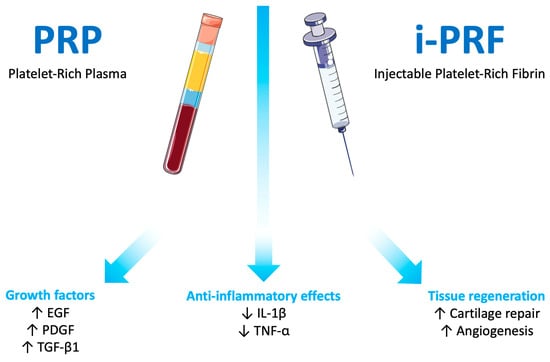
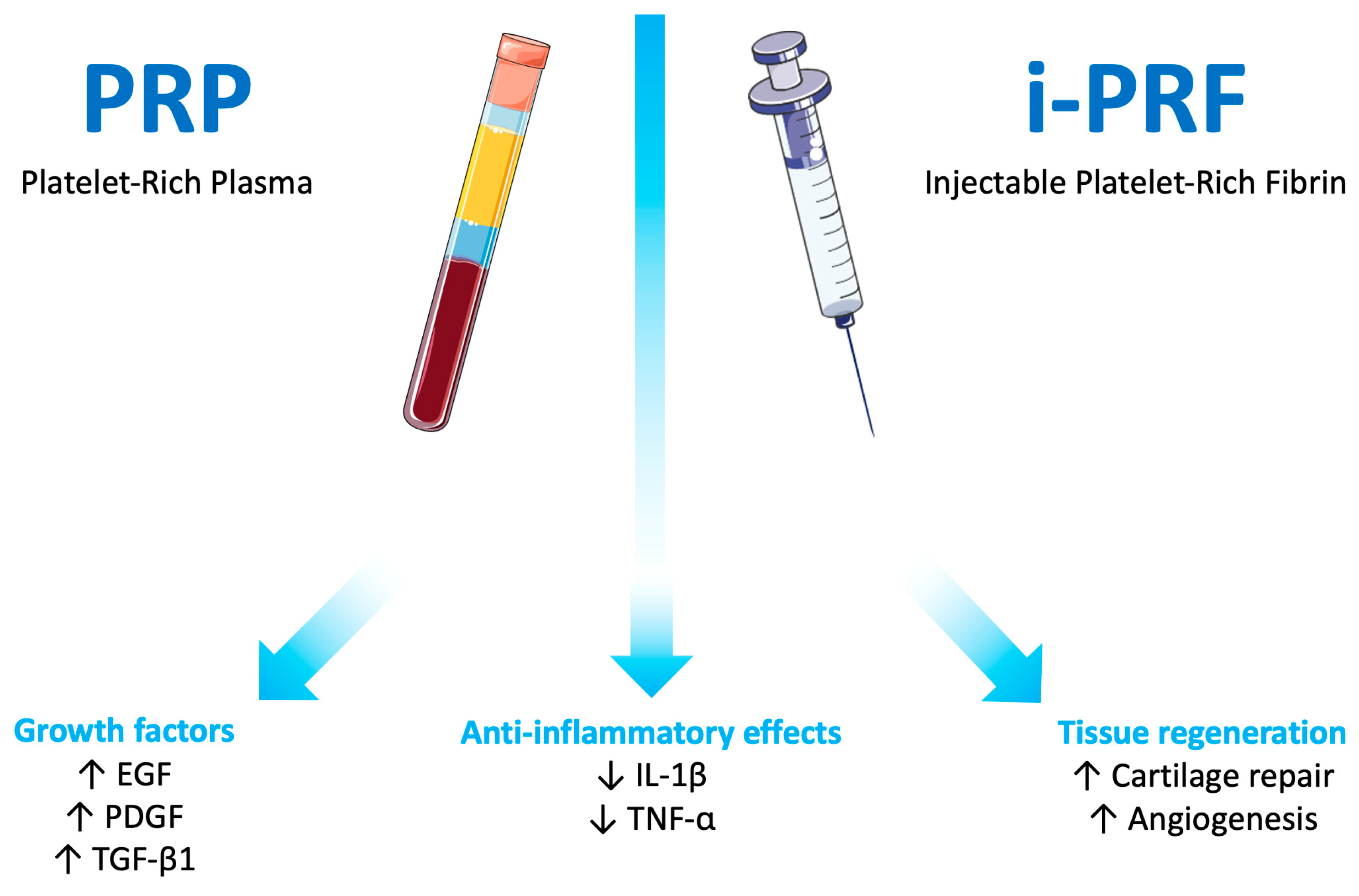
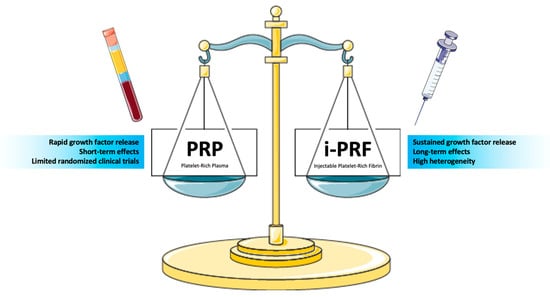
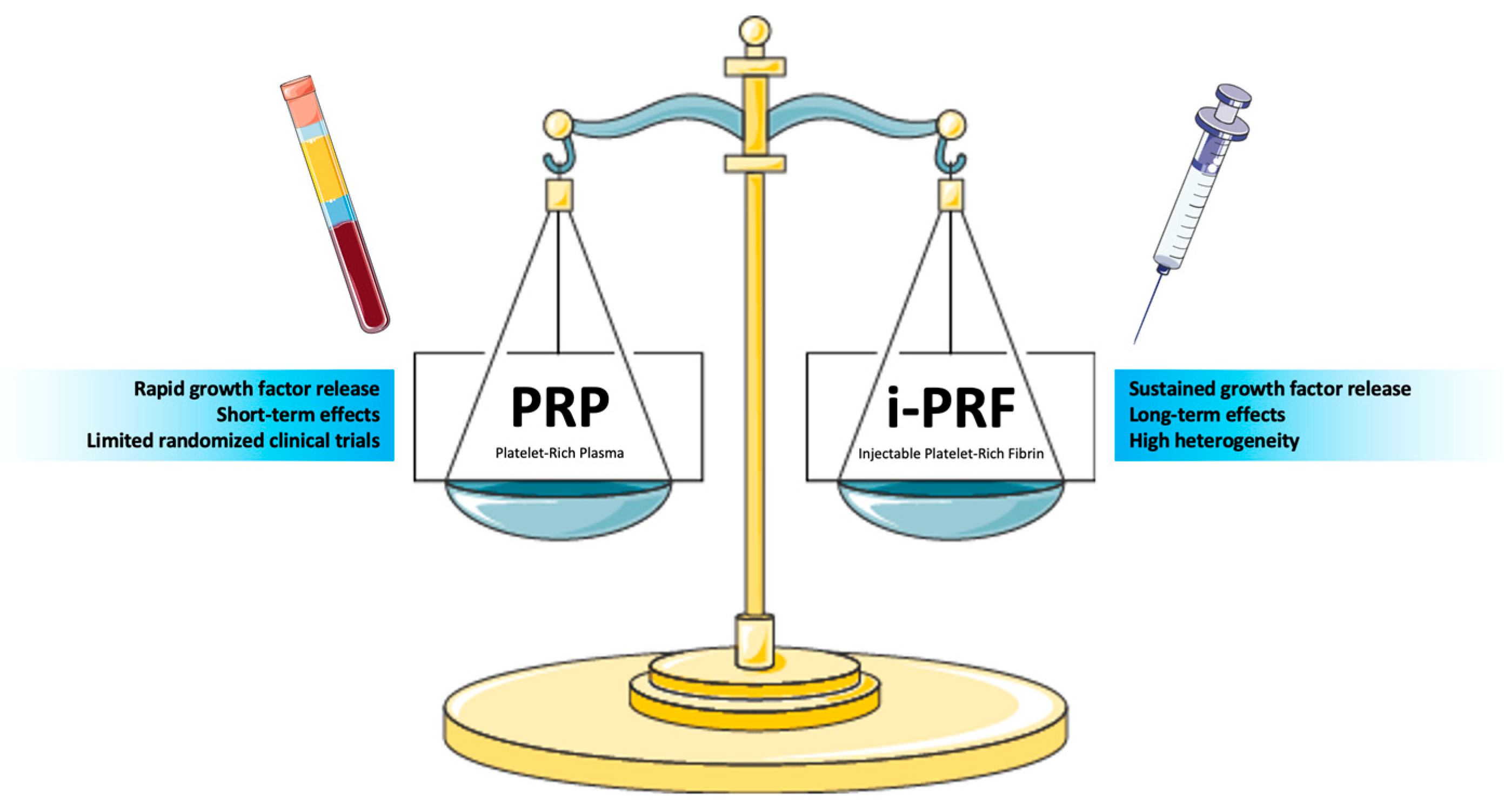
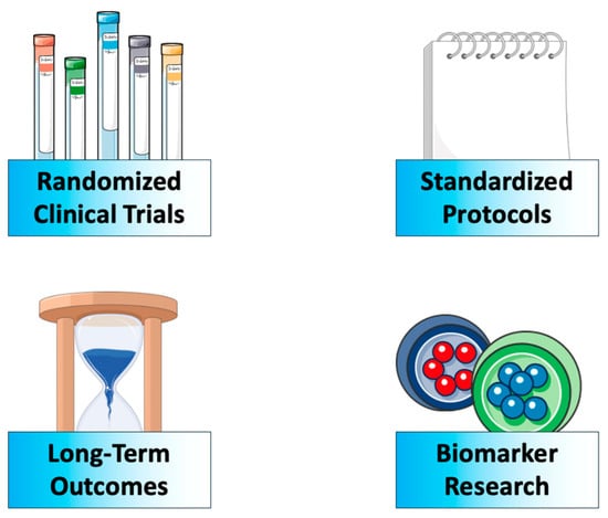
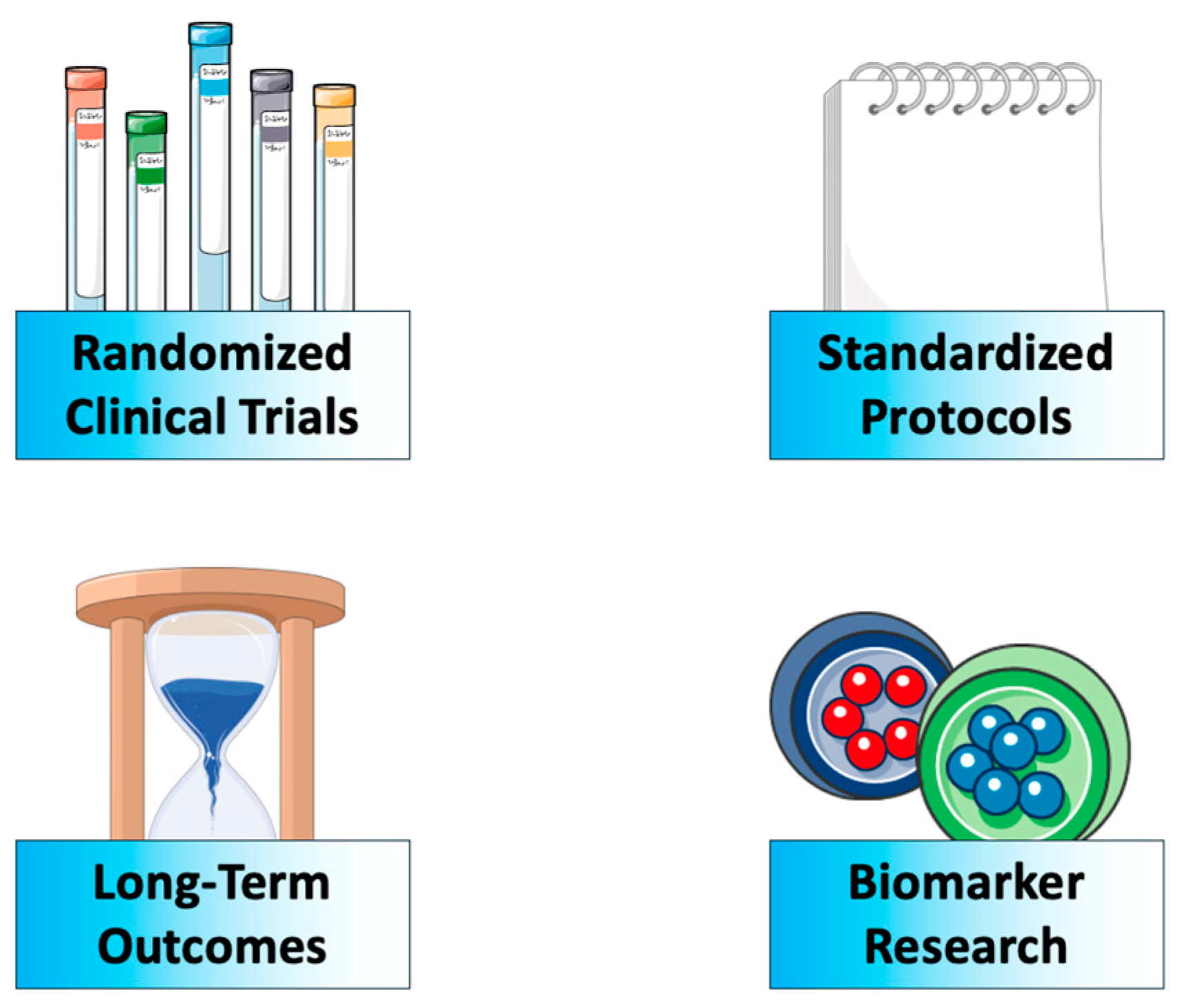

Disclaimer/Publisher’s Note: The statements, opinions and data contained in all publications are solely those of the individual author(s) and contributor(s) and not of MDPI and/or the editor(s). MDPI and/or the editor(s) disclaim responsibility for any injury to people or property resulting from any ideas, methods, instructions or products referred to in the content. |
© 2026 by the authors. Licensee MDPI, Basel, Switzerland. This article is an open access article distributed under the terms and conditions of the Creative Commons Attribution (CC BY) license.