


Abstract
High cardio-ankle vascular index (CAVI) and elevated plasma homocysteine are non-traditional cardiovascular biomarkers associated with adverse outcomes in chronic kidney disease. This study evaluated the 3-year all-cause mortality rate and the prognostic value of CAVI and plasma homocysteine in patients with end-stage kidney disease (ESKD) initiating maintenance hemodialysis (MHD). A prospective longitudinal study was conducted in 178 patients with ESKD who initiated MHD at Duc Giang General Hospital, Hanoi, Vietnam, between August 2020 and August 2021. Plasma homocysteine was measured using a chemiluminescent assay, and CAVI was assessed before hemodialysis. Patients were followed for 3 years until August 2024. During follow-up, 42 patients died, yielding an all-cause mortality rate of 23.6%. Multivariable Cox regression analysis identified low HDL-C, elevated hs-CRP, elevated plasma homocysteine, and higher CAVI as independent factors associated with all-cause mortality (all p ≤ 0.001). Receiver operating characteristic analysis showed good discriminatory ability for predicting 3-year mortality, with an area under the curve (AUC) of 0.905 (p < 0.001; cutoff, 9.45; sensitivity, 83.3%; specificity, 84.6%) for CAVI and 0.865 (p < 0.001; cutoff, 32.23 μmol/L; sensitivity, 76.2%; specificity, 84.6%) for plasma homocysteine. Kaplan–Meier analysis demonstrated significantly lower survival in patients with higher CAVI and plasma homocysteine levels (log-rank, p < 0.001). In conclusion, higher CAVI and elevated plasma homocysteine were independently associated with increased all-cause mortality and showed good discriminatory ability for risk stratification during the first 3 years of MHD.
1. Introduction
Chronic kidney disease (CKD) has become a major global public health challenge, with its prevalence steadily increasing worldwide. Current estimates indicate that approximately 13.4% of the world’s population is affected by CKD, while 5–7 million individuals have progressed to end-stage kidney disease (ESKD) requiring renal replacement therapy [1,2,3,4,5]. In addition to progressive loss of kidney function, CKD is recognized as an important cardiovascular risk factor, markedly increasing both the incidence and severity of cardiovascular complications. Coronary artery disease, heart failure, and cardiac arrhythmias occur substantially more frequently in patients with CKD than in the general population, particularly among those with stage 5 CKD (estimated glomerular filtration rate [eGFR] < 15 mL/min/1.73 m2) [6,7]. Despite advances in renal replacement therapies, including dialysis and kidney transplantation, cardiovascular disease remains a leading cause of morbidity and mortality in patients with ESKD [8,9,10].
Arterial stiffness is a characteristic vascular abnormality in CKD and is strongly associated with adverse cardiovascular outcomes. Its development is driven by multiple pathological mechanisms, including disturbances in calcium–phosphate metabolism, hypertension, oxidative stress, and chronic inflammation [11,12,13]. Together, these factors induce structural and functional changes in the arterial wall, resulting in vascular thickening, reduced elasticity, and impaired blood flow, thereby accelerating cardiovascular damage [11]. Homocysteine has been implicated in this process through its detrimental effects on the vascular endothelium, promotion of oxidative stress and inflammation, and enhancement of atherosclerotic plaque formation [14,15]. Plasma homocysteine concentrations are frequently elevated in patients with CKD, particularly those with ESKD, owing to impaired renal clearance and metabolic disturbances, and have been linked to increased arterial stiffness [14,15]. The cardio-ankle vascular index (CAVI) is a non-invasive measure of arterial stiffness that has gained widespread clinical use because it provides a reliable assessment of arterial stiffness from the aortic root to the ankle [16,17]. CAVI reflects the stiffness of the arterial system from the aortic root to the ankle. Unlike conventional pulse wave velocity (PWV) measurements, CAVI has the significant advantage of being independent of blood pressure at the time of measurement [16,17]. Murakami K et al. have confirmed that CAVI has predictive value for all-cause mortality in MHD patients [18]. Homocysteine has been recognized as a classic cardiovascular risk factor in the general population; however, the relationship between homocysteine levels and mortality in ESKD patients is complex [19].
Although several studies have demonstrated the role of elevated serum homocysteine levels and high CAVI in relation to cardiovascular events in MHD patients [19,20], there have been no studies on the combined role of both homocysteine and CAVI in predicting all-cause mortality in the early years of MHD patients. Therefore, we conducted this study with the objective of determining the proportion and value of plasma homocysteine and CAVI in predicting all-cause mortality in the first 3 years of hemodialysis in ESKD patients.
2. Materials and Methods
2.1. Sample of the Study
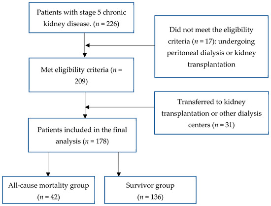
A prospective longitudinal observational study was conducted at Duc Giang General Hospital, Hanoi, Vietnam, among patients with stage 5 chronic kidney disease (CKD) who were receiving or initiating maintenance dialysis during the study recruitment period. Between August 2020 and August 2021, a total of 226 patients with stage 5 CKD were assessed for eligibility.
Patients were eligible if they were aged ≥18 years, had stage 5 CKD (estimated glomerular filtration rate [eGFR] < 15 mL/min/1.73 m2), and initiated or were receiving maintenance hemodialysis (MHD) at our center. Patients were excluded if they did not meet the predefined eligibility criteria, including those undergoing peritoneal dialysis or kidney transplantation. Seventeen patients were excluded for these reasons, leaving 209 patients who met the selection and exclusion criteria.
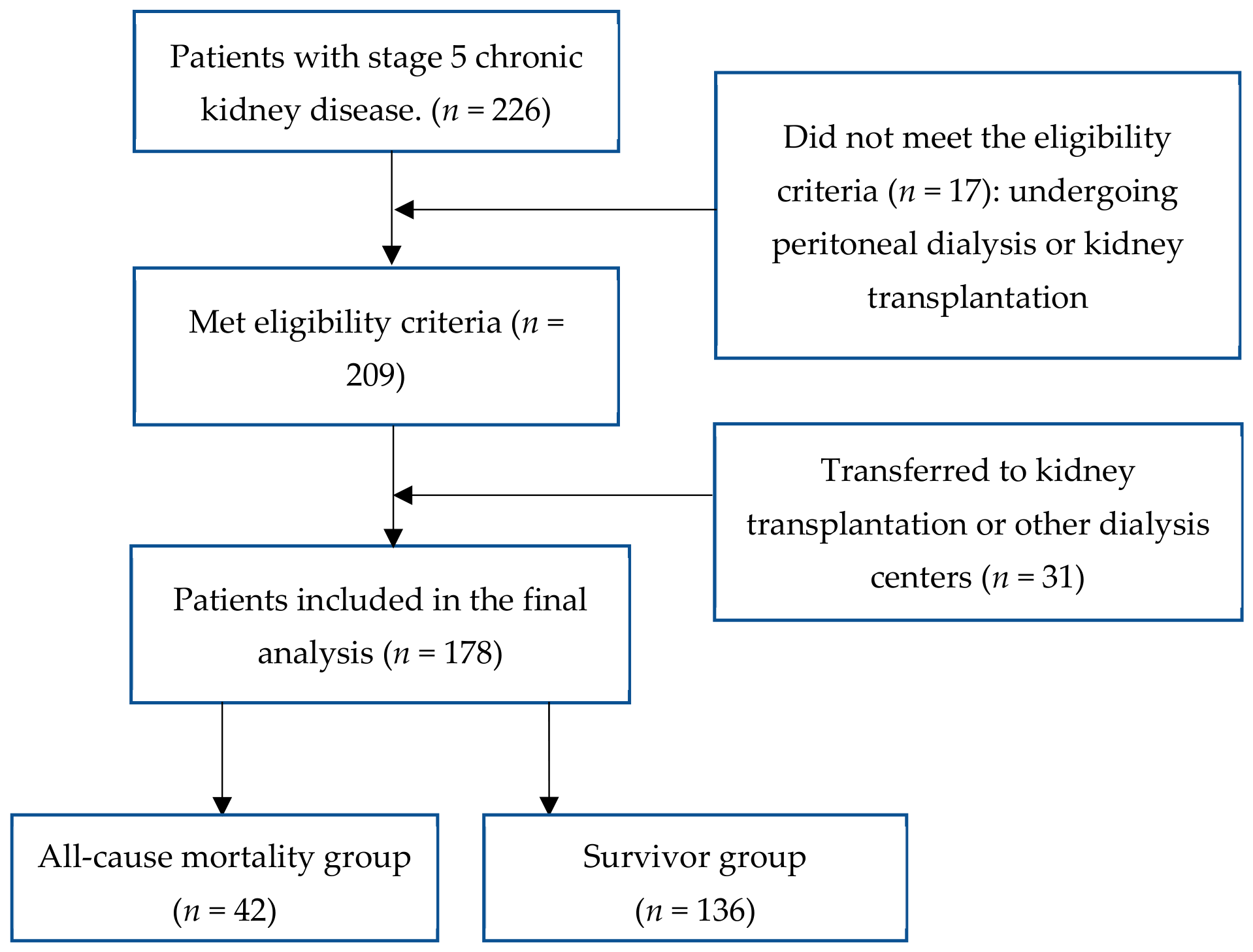
During the follow-up period, 31 patients were subsequently transferred to another dialysis center or underwent kidney transplantation and were therefore unavailable for continued follow-up at our center. These patients were excluded from the final analysis. Consequently, 178 patients were included in the study and prospectively followed for 3 years. The patient selection process is illustrated in Figure 1.
Figure 1.
Flow diagram.
2.2. Clinical and Laboratory Assessment
Demographic and clinical information, including age, sex, underlying cause of kidney failure, hypertension, diabetes mellitus, and relevant comorbidities, was collected at baseline from medical records and direct patient interviews. The underlying causes of CKD, including chronic glomerulonephritis, chronic pyelonephritis, diabetic kidney disease, hypertensive nephropathy, polycystic kidney disease, gout-related kidney disease, lupus nephritis, and other etiologies, were determined according to the clinical criteria recommended by the Kidney Disease: Improving Global Outcomes (KDIGO) 2012 Clinical Practice Guideline for the Evaluation and Management of Chronic Kidney Disease [21]. Fasting venous blood samples were obtained in the morning under standardized conditions for routine hematological and biochemical analyses, including glucose, urea, creatinine, uric acid, total protein, albumin, lipid profile (total cholesterol, triglycerides, LDL-C, and HDL-C), calcium, phosphorus, parathyroid hormone (PTH), β2-microglobulin (β2-M), high-sensitivity C-reactive protein (hs-CRP), iron, and ferritin. Renal function was estimated using the CKD-EPI equation. Serum biochemical parameters were measured using automated clinical chemistry analyzers according to the manufacturer’s instructions and the laboratory’s validated analytical procedures. Plasma homocysteine was quantified using a standardized automated chemiluminescent immunoassay.
Arterial stiffness was assessed using the cardio-ankle vascular index (CAVI), a blood-pressure-independent index of arterial stiffness derived from pulse-wave velocity. CAVI was measured using an automated vascular screening device. Before CAVI measurement, patients rested in the supine position for at least 10–15 min in a quiet room. Measurements were performed approximately 30 min before a scheduled hemodialysis session to minimize the acute hemodynamic effects of dialysis. Blood pressure cuffs were placed on both upper arms and ankles according to the manufacturer’s instructions, and electrocardiographic and phonocardiographic signals were simultaneously recorded. CAVI was automatically calculated by the device based on pulse-wave velocity and blood pressure measurements. The mean CAVI value obtained from the available limbs was used for statistical analysis.
All predefined clinical, laboratory, and CAVI measurements were available for the 178 included patients, and no missing data were identified for the variables included in the final analyses.
All patients underwent standard hemodialysis three times weekly, with each session lasting 3.5–4.5 h and a target Kt/V of at least 1.2, in accordance with the 2019 International Society of Nephrology recommendations [22]. Glycemic control and the management of anemia, hypertension, and CKD-related complications followed the KDIGO 2012 guidelines [21]. The primary outcome was all-cause mortality during the 36-month follow-up period. Based on survival status at the end of follow-up, patients were classified into the mortality group (n = 42) or the survivor group (n = 136).
2.3. Statistical Analyses
Continuous variables were expressed as mean ± standard deviation (SD) for normally distributed data or median (interquartile range [IQR]) for non-normally distributed data. Comparisons between groups were performed using Student’s t-test for normally distributed variables and the Mann–Whitney U test or Kruskal–Wallis test for non-normally distributed variables, as appropriate. Categorical variables were summarized as frequencies and percentages and compared using the Chi-square test.
Before multivariable analysis, multicollinearity among candidate variables was assessed using the variance inflation factor (VIF) and tolerance. Variables with a VIF ≥ 5 or a tolerance < 0.20 were considered to exhibit substantial multicollinearity and were excluded from further analysis. All remaining variables were entered into a multivariable Cox proportional hazards regression model, and independent prognostic factors were identified using a forward stepwise selection procedure based on the likelihood ratio test.
Receiver operating characteristic (ROC) curve analysis was performed to evaluate the discriminatory ability of CAVI and plasma homocysteine for predicting 3-year all-cause mortality. The area under the curve (AUC), optimal cutoff values, sensitivity, and specificity were calculated. Kaplan–Meier survival curves were constructed to compare cumulative survival between groups, and differences were assessed using the log-rank test. All statistical analyses were performed using SPSS version 22.0 (IBM SPSS Statistics, Chicago, IL, USA), and a two-sided p-value < 0.05 was considered statistically significant.
2.4. Ethical Considerations
The study protocol was reviewed and approved by the Ethics Committee of Duc Giang General Hospital, Hanoi, Vietnam, under Decision No. 136/QD-BVDKDG dated 22 May 2020. The study was conducted in accordance with the principles of the Declaration of Helsinki. All participants provided written informed consent before enrollment.
3. Results
3.1. Clinical and Paraclinical Characteristics of the Study Patient Group
Table 1 illustrates that age, the proportion of lipid disorders, and levels of plasma phosphorus, B2-M, PTH, hs-CRP, ferritin, homocysteine, and CAVI were significantly higher, while HDL-C levels were lower in the mortality group compared to the survivor group (p < 0.001).
Table 1.
Clinical and paraclinical comparison between mortality group and survivor group.
3.2. Predictive Value of All-Cause Mortality from Several Clinical and Paraclinical Indicators
As shown in Table 2, multivariable Cox proportional hazards regression identified four independent factors associated with all-cause mortality: HDL-C, hs-CRP, plasma homocysteine, and CAVI. After adjustment for the variables entered into the model using forward selection, lower HDL-C was independently associated with a higher risk of mortality (HR = 0.023, 95% CI: 0.004–0.144, p < 0.001). Conversely, higher hs-CRP, plasma homocysteine, and CAVI were independently associated with increased mortality risk, with HRs of 1.006 (95% CI: 1.003–1.009), 1.238 (95% CI: 1.124–1.363), and 1.389 (95% CI: 1.152–1.674), respectively.
Table 2.
Multivariable Cox regression of factors associated with mortality.
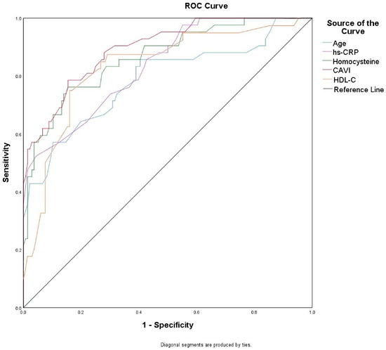
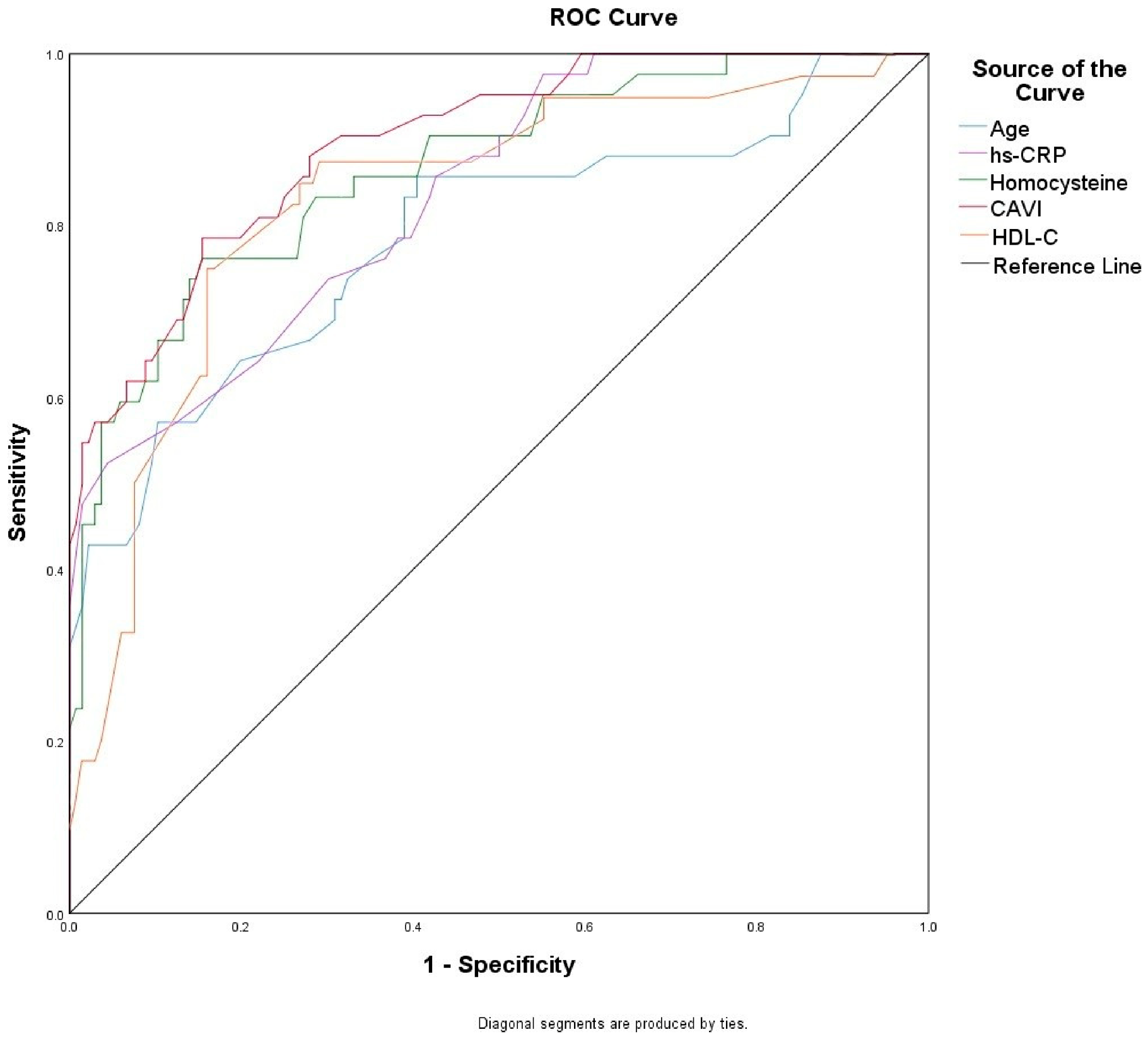
ROC curve analysis demonstrated that all five baseline variables had significant discriminatory ability for all-cause mortality (Figure 2). The AUCs were 0.786 for age, 0.804 for HDL-C, 0.835 for hs-CRP, 0.865 for plasma homocysteine, and 0.905 for CAVI. Among the evaluated variables, CAVI showed the highest discriminatory performance, followed by plasma homocysteine. At the optimal cut-off values, CAVI ≥ 9.45 yielded a sensitivity of 83.3% and specificity of 84.6%, while plasma homocysteine ≥ 32.23 µmol/L yielded a sensitivity of 76.2% and specificity of 84.6%.
Figure 2.
Receiver operating characteristic (ROC) curves predicting mortality by some factors. Age: AUC = 0.786; p < 0.001; cut-off value = 66.5 years old; sensitivity = 57.1%, specificity = 89.7%; hs-CRP: AUC = 0.835; p < 0.001; cut-off value = 3.0 mg/L; sensitivity = 52.4%, specificity = 95.6%; plasma homocysteine: AUC = 0.865; p < 0.001; cut-off value = 32.23 µmol/L; sensitivity = 76.2%, specificity = 84.6%; CAVI: AUC = 0.905; p < 0.001; cut-off value = 9.45; sensitivity = 83.3%, specificity = 84.6%; HDL-C: AUC = 0.804; p < 0.001; cut-off value = 0.875 mmol/L; sensitivity = 76.2%, specificity = 80.1%.
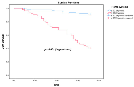
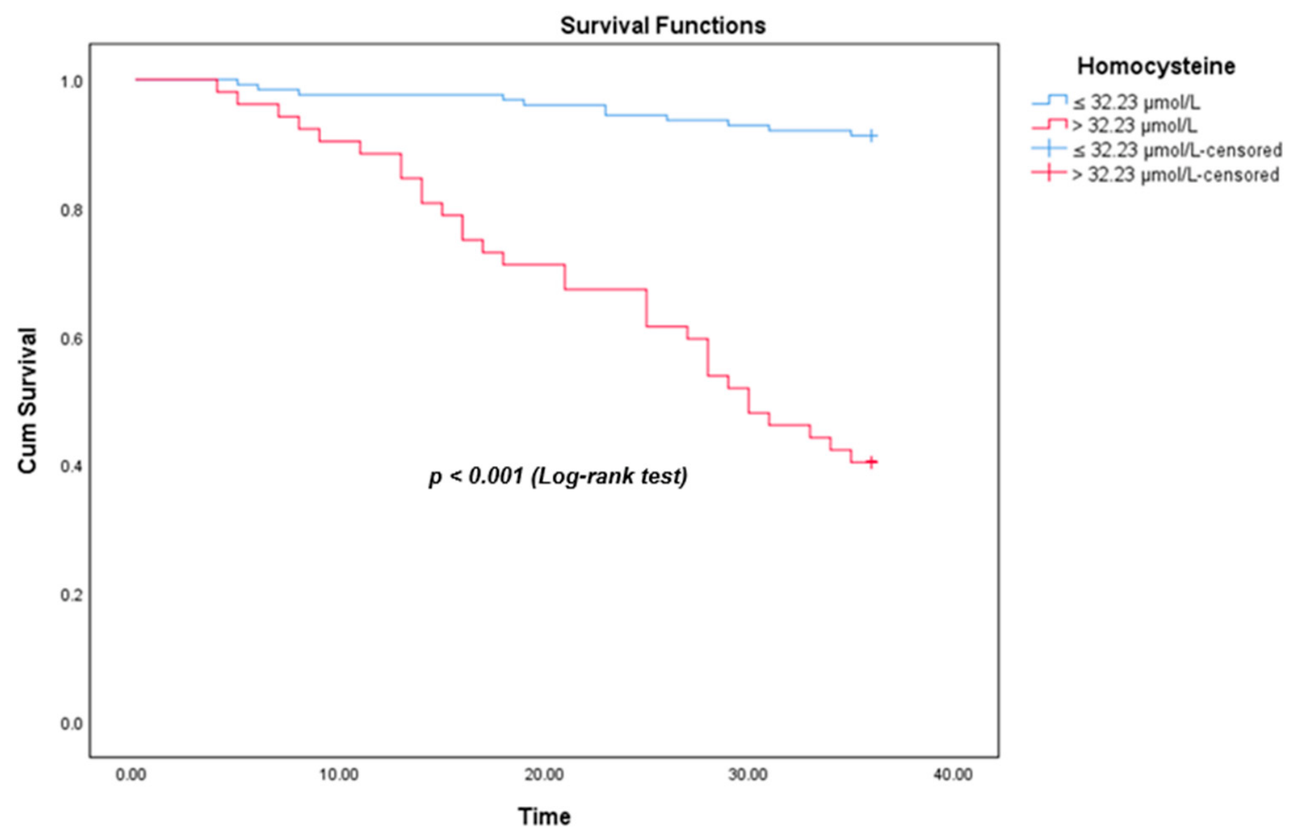
Kaplan–Meier survival analysis in Figure 3 demonstrated that patients with plasma homocysteine levels > 32.23 μmol/L had a significantly lower cumulative survival probability than those with plasma homocysteine levels ≤ 32.23 μmol/L. The difference in survival between the two groups was statistically significant (log-rank test, p < 0.001).
Figure 3.
Kaplan–Meier curves for all-cause mortality according to plasma homocysteine level.
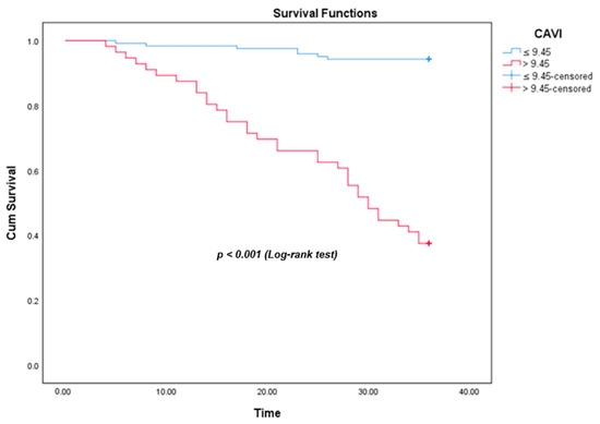
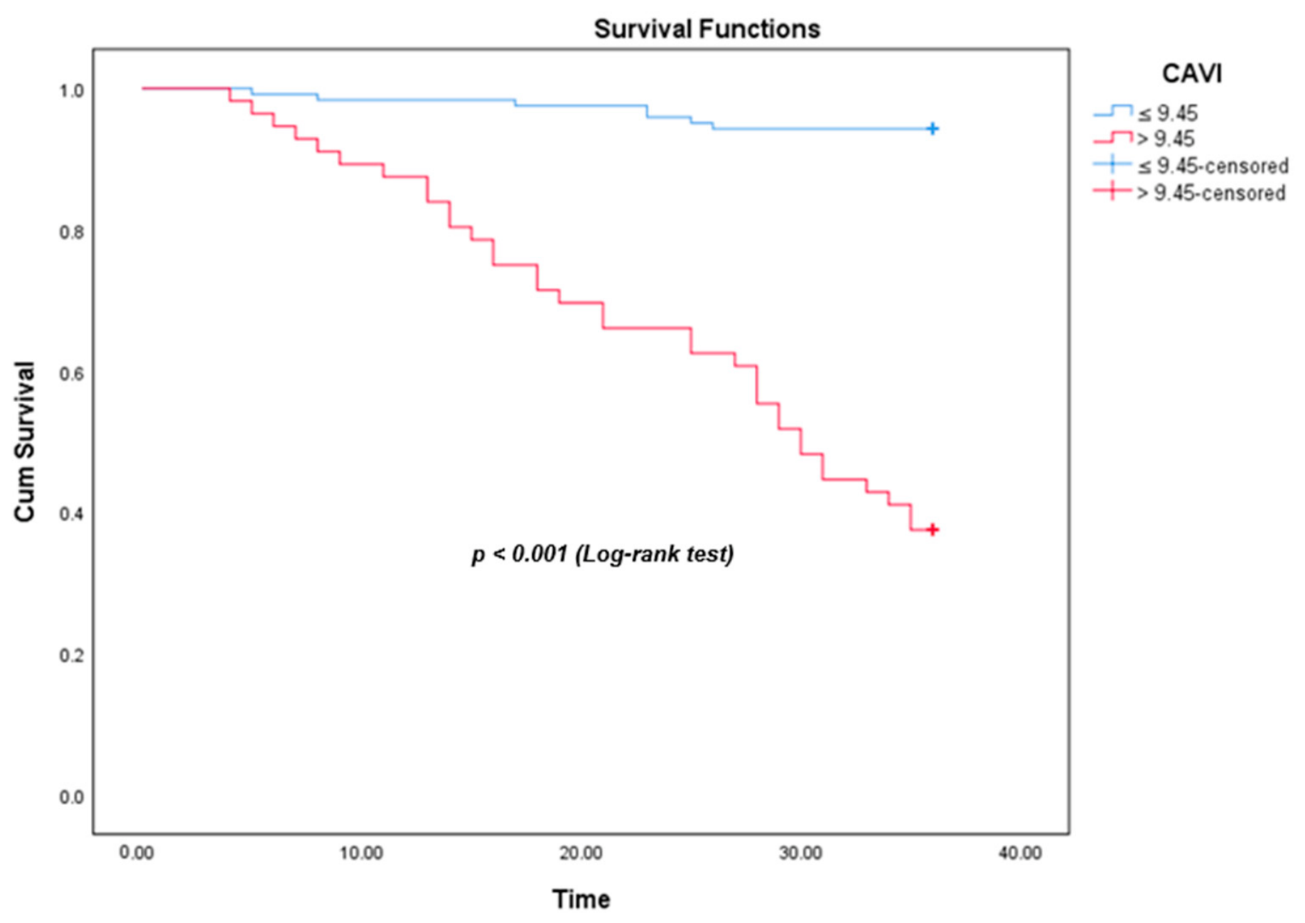
Kaplan–Meier survival analysis in Figure 4 demonstrated that patients with CAVI > 9.45 had a significantly lower cumulative survival probability than those with CAVI ≤ 9.45. The difference in survival between the two groups was statistically significant (log-rank test, p < 0.001).
Figure 4.
Kaplan–Meier curves for all-cause mortality according to CAVI.
4. Discussion
4.1. Prevalence of All-Cause Mortality in the Study
Our results show that the all-cause mortality rate during the first 3 years of hemodialysis in ESKD patients was 23.6% (Table 1). When compared with previous studies, our findings are consistent. Wang Q. et al. reported results from 863 hemodialysis patients with a follow-up period of 37 months, showing an all-cause mortality rate of 23.8%, with 14% of deaths related to cardiovascular disease [23]. However, in the study by Li Q. et al., which included 285 patients with end-stage renal disease undergoing dialysis, the overall mortality rate was only 14.7% [24]. In another study by Dimitrijevic Z. et al. involving 138 hemodialysis patients, the all-cause mortality rate over 3 years of follow-up was 23.9% (33/138 patients) [25].
Several factors were significantly associated with mortality in our study, most of which were modifiable, including dyslipidemia, elevated phosphorus, β2-microglobulin (B2-M), PTH, CRP, and homocysteine (Table 1). In patients newly initiated on hemodialysis, early mortality is associated with clinical and procedural risk factors. Cardiovascular disease and systemic inflammation are factors associated with mortality within the first few months of hemodialysis. In addition, unmodifiable factors such as advanced age and the presence of comorbidities such as heart failure and diabetes significantly increase this risk. Nutritional status, electrolyte imbalance, and hyperphosphatemia are strongly associated with low early survival rates. In particular, uncontrolled fluid overload and cardiovascular burden prior to the first hemodialysis session also increase the risk of early mortality in this patient group [26,27,28]. Wang Q et al. [23] also showed that poor nutrition, as well as its dynamic changes over time, was shown to be associated with both all-cause mortality and cardiovascular mortality in ESKD patients at the start and at months 6, 12 and 18 of hemodialysis (p < 0.001).
Cardiovascular calcification, particularly in diabetic patients, has also been confirmed to be associated with mortality in ESKD patients during the early period of hemodialysis, as reported by Li Q. et al. [24]. Metabolic syndrome has likewise been identified as a factor associated with mortality in ESKD patients during the first years of hemodialysis. Factors related to the dialysis procedure are also associated with early mortality. The use of central venous catheters instead of arteriovenous fistulas to access blood vessels significantly increases the rate of infection-related and all-cause mortality [27,28]. Thus, multiple factors contribute to mortality in hemodialysis patients, and studies identifying different risk factors are essential to support multifactorial interventions aimed at reducing mortality rates in this population.
4.2. High Cardio-Ankle Vascular Index (CAVI) and Plasma Homocysteine Concentration Related to All-Cause Mortality
Our multivariable Cox regression analysis (Table 2) demonstrated that low HDL-C, elevated hs-CRP, elevated plasma homocysteine, and higher CAVI were independently associated with an increased risk of all-cause mortality (all p ≤ 0.001). Notably, both CAVI and homocysteine were usable predictors of all-cause mortality in ESKD patients during the first 3 years of maintenance hemodialysis (Figure 2). Kaplan–Meier analysis in Figure 3 and Figure 4 further demonstrated that elevated plasma homocysteine and CAVI were associated with significantly poorer long-term survival. Patients with higher homocysteine and CAVI experienced a progressive decline in cumulative survival during follow-up, whereas survival remained relatively stable among those with lower homocysteine and CAVI (log-rank test, p < 0.001).
Elevated plasma homocysteine levels have been shown to be an independent risk factor for atherosclerosis [29]. Numerous studies have demonstrated that elevated homocysteine levels are common and are associated with mortality in CKD patients undergoing maintenance hemodialysis or in the pre-dialysis stage, both of which are associated with a high prevalence of atherosclerosis [19,29]. To explain this, we suggest that in the early years of hemodialysis, elevated plasma homocysteine levels directly cause severe endothelial dysfunction and atherosclerosis. This metabolic burden significantly increases the incidence of fatal cardiovascular and cerebrovascular events in this vulnerable patient group. Furthermore, high homocysteine levels often correlate with the malnutrition-inflammation complex, a condition that severely impairs patient survival [19,29].
A high CAVI serves as a crucial prognostic indicator of early mortality in MHD patients. This specific index reflects increased arterial stiffness, independent of blood pressure fluctuations during measurement. In the early years of hemodialysis, rapid vascular calcification and chronic fluid overload exacerbate arterial remodeling [29,30]. Murakami K. et al. demonstrated that CAVI has predictive value for all-cause mortality in a cohort of 209 maintenance hemodialysis patients followed for 6 years [31]. The CAVI measurement is crucial for the early prediction of cardiovascular events and mortality in MHD patients [20,32]. Unlike traditional PWV, CAVI provides an independent assessment of systemic arterial stiffness, separate from blood pressure. This unique characteristic is vital for hemodialysis patients who experience constant circulatory volume variability (especially fluid overload) and unstable blood pressure. Studies have shown that elevated CAVI correlates strongly with silent myocardial injury and severe left ventricular dysfunction. Therefore, high baseline CAVI strongly predicts major adverse cardiovascular events (MACE) such as heart failure and stroke. It also serves as a robust, independent predictor of all-cause mortality in this patient group. Utilizing CAVI early allows clinicians to identify high-risk patients early. This early detection optimizes dry weight management and guides aggressive cardiovascular protective therapies. Ultimately, routine CAVI screening transforms routine risk stratification into timely interventions, saving the lives of ESKD patients [18].
4.3. Limitations
Several limitations should be acknowledged. First, this was a single-center study with a relatively small sample size and a limited number of mortality events, which may have resulted in model instability and potential overfitting despite an acceptable events-per-variable ratio (EPV = 10.5). Second, the ROC-derived AUC values were estimated from the same cohort used for analysis and may therefore be subject to optimism. Thus, they should be interpreted as measures of discriminatory ability within this cohort rather than validated predictive performance. The absence of internal and external validation limits the generalizability and clinical applicability of the findings, and further validation in larger independent cohorts is warranted. Third, the association between homocysteine and mortality may be influenced by nutritional and inflammatory status. Because comprehensive nutritional assessments were not available, residual confounding and the potential effect of the “reverse epidemiology” phenomenon cannot be excluded. Finally, CAVI and homocysteine were measured at baseline only, and their changes over time were not assessed.
5. Conclusions
In ESKD patients during the first three years of maintenance hemodialysis, the all-cause mortality rate was 23.6%. A high CAVI and elevated plasma homocysteine levels were independent factors associated with all-cause mortality. The CAVI and plasma homocysteine levels can be used to predict all-cause mortality in the patients.
Author Contributions
Conceptualization: D.N.T.T., T.N.V. and T.L.V.; Methodology: D.N.T.T., T.N.V. and T.L.V.; Formal analysis and investigation: D.N.T.T., T.N.V., H.B.M., K.T.Q., H.N.T.T. and K.N.T.; Writing—original draft preparation: D.N.T.T., T.N.V. and H.B.M.; Writing—review and editing: K.T.Q., H.N.T.T. and K.N.T.; Supervision: T.L.V. All authors have read and agreed to the published version of the manuscript.
Funding
This research received no external funding.
Institutional Review Board Statement
The study was conducted in accordance with the Declaration of Helsinki and approved by the Ethics Committee of Duc Giang General Hospital, Hanoi, Vietnam (No. 136/QĐ-BVĐKĐG, 22 May 2020) for studies involving humans.
Informed Consent Statement
Informed consent was obtained from all subjects involved in the study. Written informed consent has been obtained from the patient to publish this paper.
Data Availability Statement
The data that support the findings of this study are not publicly available due to privacy reasons but are available from the corresponding author upon reasonable request.
Acknowledgments
We gratefully acknowledge the invaluable support provided by our affiliated hospital and university, which greatly contributed to the successful completion of this research.
Conflicts of Interest
The authors declare no conflicts of interest.
References
- Ammirati, A.L. Chronic Kidney Disease. Rev. Assoc. Med. Bras. 2020, 66, s03–s09. [Google Scholar] [CrossRef] [Scilit] [PubMed]
- Lv, J.C.; Zhang, L.X. Prevalence and Disease Burden of Chronic Kidney Disease. Adv. Exp. Med. Biol. 2019, 1165, 3–15. [Google Scholar] [CrossRef] [Scilit] [PubMed]
- Liyanage, T.; Toyama, T.; Hockham, C.; Ninomiya, T.; Perkovic, V.; Woodward, M.; Fukagawa, M.; Matsushita, K.; Praditpornsilpa, K.; Hooi, L.S.; et al. Prevalence of chronic kidney disease in Asia: A systematic review and analysis. BMJ Glob. Health 2022, 7, e007525. [Google Scholar] [CrossRef] [Scilit] [PubMed]
- Thurlow, J.S.; Joshi, M.; Yan, G.; Norris, K.C.; Agodoa, L.Y.; Yuan, C.M.; Nee, R. Global Epidemiology of End-Stage Kidney Disease and Disparities in Kidney Replacement Therapy. Am. J. Nephrol. 2021, 52, 98–107. [Google Scholar] [CrossRef] [Scilit] [PubMed]
- Gupta, R.; Woo, K.; Yi, J.A. Epidemiology of end-stage kidney disease. Semin Vasc. Surg. 2021, 34, 71–78. [Google Scholar] [CrossRef] [Scilit] [PubMed]
- Zoccali, C.; Mallamaci, F.; Adamczak, M.; de Oliveira, R.B.; Massy, Z.A.; Sarafidis, P.; Agarwal, R.; Mark, P.B.; Kotanko, P.; Ferro, C.J.; et al. Cardiovascular complications in chronic kidney disease: A review from the European Renal and Cardiovascular Medicine Working Group of the European Renal Association. Cardiovasc Res. 2023, 119, 2017–2032. [Google Scholar] [CrossRef] [Scilit] [PubMed]
- Schuett, K.; Marx, N.; Lehrke, M. The Cardio-Kidney Patient: Epidemiology, Clinical Characteristics and Therapy. Circ. Res. 2023, 132, 902–914. [Google Scholar] [CrossRef] [Scilit] [PubMed]
- Ayer, A.; Banerjee, U.; Mills, C.; Donovan, C.; Nelson, L.; Shah, S.J.; Dubin, R.F. Left atrial strain is associated with adverse cardiovascular events in patients with end-stage renal disease: Findings from the Cardiac, Endothelial Function and Arterial Stiffness in ESRD (CERES) study. Hemodial. Int. 2022, 26, 323–334. [Google Scholar] [CrossRef] [Scilit] [PubMed]
- Fu, E.L.; Evans, M.; Carrero, J.J.; Putter, H.; Clase, C.M.; Caskey, F.J.; Szymczak, M.; Torino, C.; Chesnaye, N.C.; Jager, K.J.; et al. Timing of dialysis initiation to reduce mortality and cardiovascular events in advanced chronic kidney disease: Nationwide cohort study. BMJ. 2021, 375, e066306. [Google Scholar] [CrossRef] [Scilit] [PubMed]
- Andersson, C.; Hansen, D.; Sørensen, S.S.; McGrath, M.; McCausland, F.R.; Torp-Pedersen, C.; Schou, M.; Køber, L.; Pfeffer, M.A. Long-term cardiovascular events, graft failure, and mortality in kidney transplant recipients. Eur. J. Intern Med. 2024, 121, 109–113. [Google Scholar] [CrossRef] [Scilit] [PubMed]
- Townsend, R.R. Arterial Stiffness in CKD: A Review. Am. J. Kidney Dis. 2019, 73, 240–247. [Google Scholar] [CrossRef] [Scilit] [PubMed]
- Vervloet, M.G. Can we reverse arterial stiffness by intervening on CKD-MBD biomarkers? Clin. Kidney J. 2023, 16, 1766–1775. [Google Scholar] [CrossRef] [Scilit] [PubMed]
- Yao, Z.; Ishigami, J.; Kim, E.; Ballew, S.H.; Sang, Y.; Tanaka, H.; Meyer, M.L.; Coresh, J.; Matsushita, K. Arterial Stiffness and Subsequent Incidence of CKD and Kidney Function Decline in a Large Longitudinal Community Cohort: The Atherosclerosis in Communities (ARIC) Study. Am. J. Kidney Dis. 2025, 86, 32–42. [Google Scholar] [CrossRef] [Scilit] [PubMed]
- Piko, N.; Bevc, S.; Naji, F.H.; Ekart, R. Association of Homocysteine with Arterial Stiffness and Kidney Injury Biomarkers in Patients with Suspected Coronary Artery Disease. J. Clin. Med. 2026, 15, 4961. [Google Scholar] [CrossRef] [Scilit] [PubMed]
- Adeoye, M.; Hamdallah, H.; Adeoye, A.M. Homocysteine levels and cardiovascular disease risk factors in chronic kidney disease (CKD), hypertensive and healthy Nigerian adults: A comparative retrospective study. BMJ Open 2025, 15, e089644. [Google Scholar] [CrossRef] [Scilit] [PubMed]
- Saiki, A.; Ohira, M.; Yamaguchi, T.; Nagayama, D.; Shimizu, N.; Shirai, K.; Tatsuno, I. New Horizons of Arterial Stiffness Developed Using Cardio-Ankle Vascular Index (CAVI). J. Atheroscler. Thromb. 2020, 27, 732–748. [Google Scholar] [CrossRef] [Scilit] [PubMed]
- Giudici, A.; Khir, A.W.; Reesink, K.D.; Delhaas, T.; Spronck, B. Five years of cardio-ankle vascular index (CAVI) and CAVI0: How close are we to a pressure-independent index of arterial stiffness? J. Hypertens. 2021, 39, 2128–2138. [Google Scholar] [CrossRef] [Scilit] [PubMed]
- Murakami, K.; Inayama, E.; Itoh, Y.; Tuchiya, S.; Iwasaki, M.; Tamura, N.; Suzuki, T.; Iwai, N.; Utino, J.; Masai, M.; et al. The Role of Cardio-Ankle Vascular Index as a Predictor of Mortality in Patients on Maintenance Hemodialysis. Vasc. Health Risk Manag. 2021, 17, 791–798. [Google Scholar] [CrossRef] [Scilit] [PubMed]
- Guéant, J.L.; Guéant-Rodriguez, R.M.; Oussalah, A.; Zuily, S.; Rosenberg, I. Hyperhomocysteinemia in Cardiovascular Diseases: Revisiting Observational Studies and Clinical Trials. Thromb. Haemost. 2023, 123, 270–282. [Google Scholar] [CrossRef] [Scilit] [PubMed]
- Tavolinejad, H.; Erten, O.; Maynard, H.; Chirinos, J.A. Prognostic Value of Cardio-Ankle Vascular Index for Cardiovascular and Kidney Outcomes: Systematic Review and Meta-Analysis. JACC Adv. 2024, 3, 101019. [Google Scholar] [CrossRef] [Scilit] [PubMed]
- KDIGO 2012 Clinical Practice Guideline for the Evaluation and Management of Chronic Kidney Disease. Kidney Int. Suppl. 2013, 3, 1–150.
- Ashby, D.; Borman, N.; Burton, J.; Corbett, R.; Davenport, A.; Farrington, K.; Flowers, K.; Fotheringham, J.; Andrea Fox, R.N.; Franklin, G.; et al. Renal Association Clinical Practice Guideline on Haemodialysis. BMC Nephrol. 2019, 20, 379. [Google Scholar] [CrossRef] [Scilit] [PubMed]
- Wang, Q.; Ye, Z.; Rao, J.; Zhao, W.; Liu, J.; Chen, Y.; Xiang, J.; Zhen, X.; Li, J.; Zhang, Y.; et al. Assessing the predictive power of nutritional indices on all-cause and cardiovascular mortality in hemodialysis patients: A longitudinal study. Ren. Fail. 2024, 46, 2431140. [Google Scholar] [CrossRef] [Scilit] [PubMed]
- Li, Q.; Li, P.; Xu, Z.; Lu, Z.; Yang, C.; Ning, J. Association of diabetes with cardiovascular calcification and all-cause mortality in end-stage renal disease in the early stages of hemodialysis: A retrospective cohort study. Cardiovasc Diabetol. 2024, 23, 259. [Google Scholar] [CrossRef] [Scilit] [PubMed]
- Dimitrijevic, Z.; Jovanovic, A.; Cvetkovic, M.; Vrecic, T.; Kostic, E.; Mitic, B. Associations of Cardiovascular and All-Cause Mortality with Metabolic Syndrome in Hemodialysis Patients: A Prospective Single-Center Study. Medicina 2019, 55, 694. [Google Scholar] [CrossRef] [Scilit] [PubMed]
- Lukowsky, L.R.; Kheifets, L.; Arah, O.A.; Nissenson, A.R.; Kalantar-Zadeh, K. Patterns and predictors of early mortality in incident hemodialysis patients: New insights. Am. J. Nephrol. 2012, 35, 548–558. [Google Scholar] [CrossRef] [Scilit] [PubMed]
- McQuillan, R.; Trpeski, L.; Fenton, S.; Lok, C.E. Modifiable risk factors for early mortality on hemodialysis. Int. J. Nephrol. 2012, 2012, 435736. [Google Scholar] [CrossRef] [Scilit] [PubMed]
- Heaf, J.; Heiro, M.; Petersons, A.; Vernere, B.; Povlsen, J.V.; Sørensen, A.B.; Clyne, N.; Bumblyte, I.; Zilinskiene, A.; Randers, E.; et al. First-year mortality in incident dialysis patients: Results of the Peridialysis study. BMC Nephrol. 2022, 23, 229. [Google Scholar] [CrossRef] [Scilit] [PubMed]
- ALSolami, A.A.; Almalki, A.A.; Alhedyan, S.Y.; Alghamdi, A.; Alzahrani, S.M.; Dause, W.R.; Hamdi, F.A.; Howladar, M.T.; Ibrahim, I.A. Plasma Homocysteine Levels and Cardiovascular Events in Patients With End-Stage Renal Disease: A Systematic Review. Cureus 2023, 15, e40357. [Google Scholar] [CrossRef] [Scilit] [PubMed]
- Namba, T.; Masaki, N.; Takase, B.; Adachi, T. Arterial Stiffness Assessed by Cardio-Ankle Vascular Index. Int. J. Mol. Sci. 2019, 20, 3664. [Google Scholar] [CrossRef] [Scilit] [PubMed]
- Matsushita, K.; Ding, N.; Kim, E.D.; Budoff, M.; Chirinos, J.A.; Fernhall, B.; Hamburg, N.M.; Kario, K.; Miyoshi, T.; Tanaka, H.; et al. Cardio-ankle vascular index and cardiovascular disease: Systematic review and meta-analysis of prospective and cross-sectional studies. J. Clin. Hypertens. 2019, 21, 16–24. [Google Scholar] [CrossRef] [Scilit] [PubMed]
- Bäck, M.; Topouchian, J.; Labat, C.; Gautier, S.; Blacher, J.; Cwynar, M.; de la Sierra, A.; Pall, D.; Duarte, K.; Fantin, F.; et al. Cardio-ankle vascular index for predicting cardiovascular morbimortality and determinants for its progression in the prospective advanced approach to arterial stiffness (TRIPLE-A-Stiffness) study. EBioMedicine 2024, 103, 105107. [Google Scholar] [CrossRef] [Scilit] [PubMed]

Disclaimer/Publisher’s Note: The statements, opinions and data contained in all publications are solely those of the individual author(s) and contributor(s) and not of MDPI and/or the editor(s). MDPI and/or the editor(s) disclaim responsibility for any injury to people or property resulting from any ideas, methods, instructions or products referred to in the content. |
© 2026 by the authors. Licensee MDPI, Basel, Switzerland. This article is an open access article distributed under the terms and conditions of the Creative Commons Attribution (CC BY) license.