


Abstract
Background: COVID-19 is primarily a respiratory disease, but increasing evidence suggests possible oral and maxillofacial complications. This study presents a case series of post-COVID maxillary osteonecrosis (PC-RONJ) cases from western Romania and explores the possible association between SARS-CoV-2 infection, its treatment, and this complication. Methods: We conducted a multicenter retrospective case series of two patients with recent PCR-confirmed SARS-CoV-2 infection who subsequently developed maxillary osteonecrosis (ONC) between 2021 and 2023. Clinical examination, CT imaging (including 3D reconstructions), and ENT assessment were used to assess the severity of the disease. All medical records were reviewed to identify comorbidities, details of COVID-19 treatment, and the appearance of maxillofacial symptoms. Results: Both patients had been hospitalized for severe COVID-19 and treated according to the national protocol with systemic corticosteroids, oxygen therapy, anticoagulation, and antivirals. CT scans revealed marked osteolytic destruction of the maxilla and maxillary sinus walls, with extension toward adjacent facial bones. Microbiological analysis revealed a complex polymicrobial profile, including Gram-positive and Gram-negative bacteria as well as opportunistic fungal species, consistent with a chronic biofilm-associated infectious process. Patients received surgical treatment, followed by local care and, in both cases, prosthetic rehabilitation with maxillary obturators, which improved speech, chewing, and oral function. Conclusions: This case series suggests a possible association between severe COVID-19, its treatment, and subsequent maxillary osteonecrosis in susceptible patients; however, the small number of cases precludes causal inference. To our knowledge, this is the first Romanian report describing such cases in patients without prior antiresorptive therapy. These findings highlight the need for careful use of systemic corticosteroids and vigilant post-recovery monitoring of maxillofacial complications. Further studies are required to clarify the underlying mechanisms and risk factors.
1. Introduction
Severe acute respiratory syndrome (SARS-CoV-2) has had a dramatic impact on national healthcare systems worldwide [1]. Although the pandemic reached its most devastating phase between 2020 and 2023, COVID-19 continues to pose ongoing clinical challenges worldwide [2,3].
COVID-19 is increasingly regarded as a systemic disease with multisystem involvement, driven by endothelial dysfunction, immune imbalance, and prothrombotic changes. Growing attention has therefore shifted toward post-acute complications including those affecting the oral and maxillofacial region [4]. The oral cavity has increasingly been recognized as a potential site of SARS-CoV-2 involvement and related pathological changes. A wide range of oral manifestations has been reported in COVID-19 patients, including non-specific ulcerations, erythema, petechiae, maculopapular eruptions and vesiculobullous lesions [5]. Osteonecrosis (ONC), also referred to as avascular necrosis (AVN), has emerged as a potential post-infectious or treatment-related complication [6]. ONC is characterized by the death of bone tissue, which results from impaired blood supply. It typically progresses from vascular compromise or thrombosis to cellular death, bone marrow necrosis, and eventual structural collapse. This condition is referred to as osteonecrosis of the jaw (ONJ) when it involves the maxillofacial region [7,8].
Management of severe COVID-19 infection often included symptomatic and supportive measures (antipyretics, analgesics, oxygen therapy), as well as specific pharmacologic agents to control the hyperinflammatory response. Treatments such as tocilizumab, a monoclonal antibody that targets the interleukin-6 receptor, and corticosteroids like dexamethasone or methylprednisolone were widely used for patients with progressive COVID-19 pneumonia. However, their immunosuppressive effects, combined with the prothrombotic and metabolic disturbances of COVID-19, may have predisposed some patients to secondary complications [9,10,11].
Post-COVID maxillofacial complications, including osteomyelitis and maxillary osteonecrosis, have been reported in multiple regions. COVID-19-associated mucormycosis and related maxillary necrosis have been particularly described in patients with diabetes mellitus and corticosteroid exposure. In this context, post-COVID-19 osteonecrosis of the jaw (PC-RONJ) and COVID-related osteonecrosis of the jaws (CRONJ) have been increasingly recognized, typically in association with systemic risk factors such as diabetes and corticosteroid therapy [12,13,14,15,16,17]. However, reports from Eastern Europe, and particularly from Romania, remain extremely scarce, and it remains unclear whether post-COVID maxillary osteonecrosis represents a distinct clinical entity or a multifactorial complication influenced by viral infection, treatment-related factors, and underlying comorbidities. To date, only one other case report from Romania has been documented. Pădurariu et al. described ONC of the jaw in a patient with prior bisphosphonate exposure who received short-course corticosteroids during COVID-19 treatment [18].
Although the acute phase of the pandemic has passed, the long-term sequelae on bone metabolism remain an active clinical challenge. In this context, the present study addresses a gap in the literature by presenting, for the first time, a series of PC-RONJ cases in Romania, with patients who were not previously treated with antiresorptive medication. The main objective of this study is to highlight the delay in diagnosis and treatment and the advanced stage of disease at presentation by describing two rare cases of maxillary bone necrosis associated with COVID-19 infection. Specifically, the present work aims to: (i) describe the clinical and radiological features of these cases; (ii) analyze the possible contribution of SARS-CoV-2 infection and its treatment, particularly systemic corticosteroids, to the development of maxillary osteonecrosis; and (iii) discuss these findings in the context of COVID-19-related mechanisms and known risk factors reported in the existing literature.
2. Materials and Methods
2.1. Study Design and Patient Selection
This study was designed as a multicenter retrospective case series. It includes two patients who were diagnosed with maxillary osteonecrosis following confirmed SARS-CoV-2 infection. Cases were identified based on clinical, radiological, microbiological, and histopathological findings, and only patients without prior exposure to antiresorptive or antiangiogenic therapy were included.
Although the number of cases is limited, they were considered clinically relevant due to their temporal association with SARS-CoV-2 infection, the absence of classical risk factors, and the severity of maxillary involvement at presentation. Furthermore, their inclusion aimed to highlight a potentially emerging pattern of post-COVID-19 maxillary osteonecrosis that remains underreported in the current literature, particularly within the Romanian population. The shared clinical and pathological features observed across cases support their relevance for hypothesis generation regarding the underlying pathogenic mechanisms.
Both patients underwent comprehensive clinical evaluation, imaging investigations, and multidisciplinary management.
2.2. Clinical and Radiological Assessment
Clinical examination included both extraoral and intraoral evaluation, with particular attention to facial asymmetry, swelling, pain, hypoesthesia in the distribution of the infraorbital nerve, mucosal changes, and bone exposure.
Radiological assessment was performed using computed tomography (CT) of the craniofacial region, consisting of axial, coronal, and sagittal sections, as well as three-dimensional (3D) reconstructions. Imaging findings were analyzed for osteolytic lesions, bone sequestra, sinus involvement, and extension to adjacent anatomical structures. Imaging findings were interpreted in correlation with clinical and intraoperative observations.
ENT evaluation, which included nasal endoscopy and anterior rhinoscopy, was performed to establish sinonasal involvement and the presence of necrotic tissue or oroantral/oronasal communications.
2.3. Therapeutic Management
The patients received standard-of-care treatment according to the national COVID-19 treatment protocol in place at the time. Systemic corticosteroid therapy consisted of dexamethasone 6 mg daily administered intravenously or orally for 7–10 days, or an equivalent dose of methylprednisolone (32–40 mg daily intravenously), depending on clinical evolution. Anticoagulant therapy included low-molecular-weight heparin (enoxaparin) administered at a prophylactic dose of 40 mg subcutaneously once daily. Antiviral therapy consisted of remdesivir, administered as a loading dose of 200 mg intravenously on day 1, followed by 100 mg intravenously once daily for 5 days. Oxygen therapy was provided according to the severity of respiratory insufficiency.
Management of maxillary osteonecrosis included surgical debridement or necrosectomy of devitalized bone, performed in collaboration with ENT and oral and maxillofacial surgery teams.
Postoperative care followed institutional standard protocols including antibiotic therapy with cefuroxime 500 mg orally twice daily for 10–14 days and antifungal therapy with voriconazole 200 mg orally twice daily for 14–21 days. Analgesic and supportive treatments were administered as clinically indicated.
2.4. Microbiological Evaluation
Microbiological investigations were performed using samples collected from the oral mucosa, maxillary sinus, and prosthetic obturator surfaces, when present. Samples were cultured for aerobic bacteria and fungi, and antimicrobial susceptibility testing was conducted using standard laboratory methods.
The analysis focused on identifying polymicrobial colonization patterns, potential cross-compartment contamination, and the persistence of microbial flora over time, with particular attention to features suggestive of biofilm-associated infection.
In addition, specific attention was given to the exclusion of COVID-19-associated mucormycosis. Fungal cultures and microbiological analyses were performed in both cases, and no evidence of mucormycosis was identified. Instead, the findings supported the presence of a chronic bacterial infection, including Actinomyces species. The diagnosis was further supported by the absence of characteristic clinical and radiological features of invasive fungal disease.
2.5. Ethical Considerations
The study was conducted in accordance with the Declaration of Helsinki and was approved by the Institutional Review Board of the Timișoara Municipal Emergency Clinical Hospital (Approval No. 94/04.10.2021, 4 October 2021). Written informed consent was obtained from both patients for participation and the publication of clinical data and images.
3. Case Presentation
3.1. Case 1
A 69-year-old female patient, known to have hypertension and grade I/II obesity, presented to the emergency department of the Arad County Emergency Clinical Hospital on 20 September 2021, two days after the onset of the disease, with cough, headache, loss of appetite, and dyspnea. She underwent RT-PCR SARS-CoV-2 testing, with a positive result on 20 September 2021. She was therefore admitted to the Infectious Diseases I department for investigation and specialist treatment. During her hospitalization, the patient received treatment with antibiotics, anticoagulants, corticosteroids, antivirals, immunomodulators, gastric protectors, hepatoprotectors, vitamins, antitussives, bronchodilators, probiotics, hydroelectrolytic rebalancing solutions, and analgesics, with favorable clinical evolution. Serial RT-PCR tests were performed, the last being negative on 10 October 2021, and the patient was discharged on 12 October 2021. Approximately one week later (around 20 October 2021), the patient reported right-sided facial swelling involving the periorbital and genian regions, associated with eyelid ptosis, mild pain, and hypoesthesia in the distribution of the right infraorbital nerve.
On 27 October 2021, the patient presented to the Neurology Department, complaining of migraine-like headache. The neurologist diagnosed pansinusitis, for which reason the patient was referred to the ENT Department.
On 15 November 2021, ENT evaluation revealed acute right sinusitis and right facial paresis.
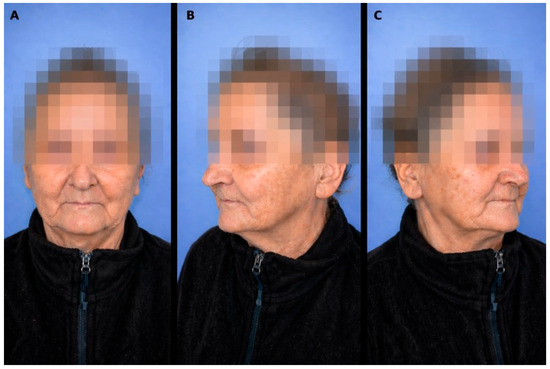
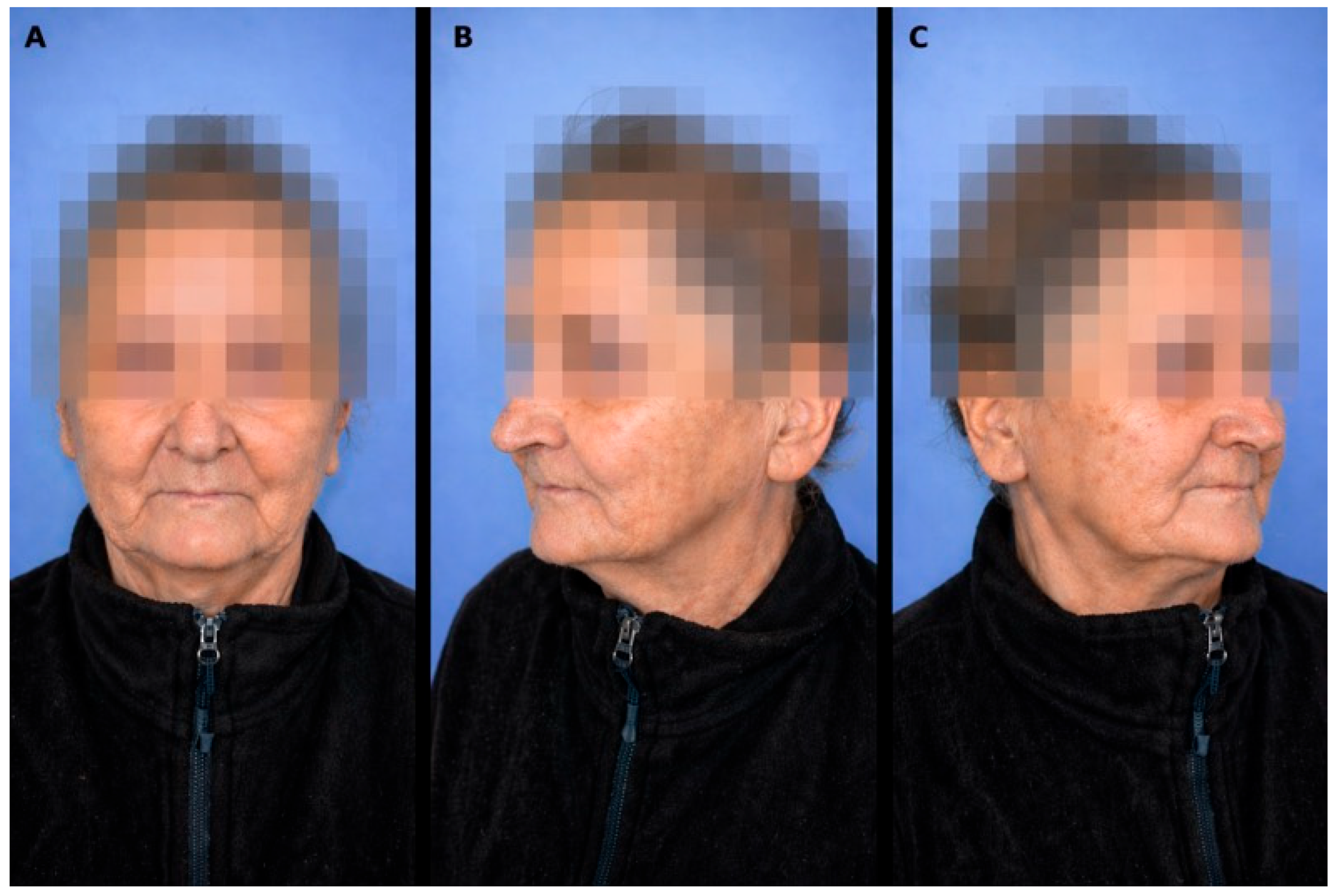
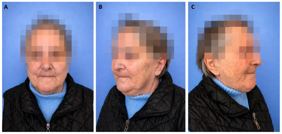
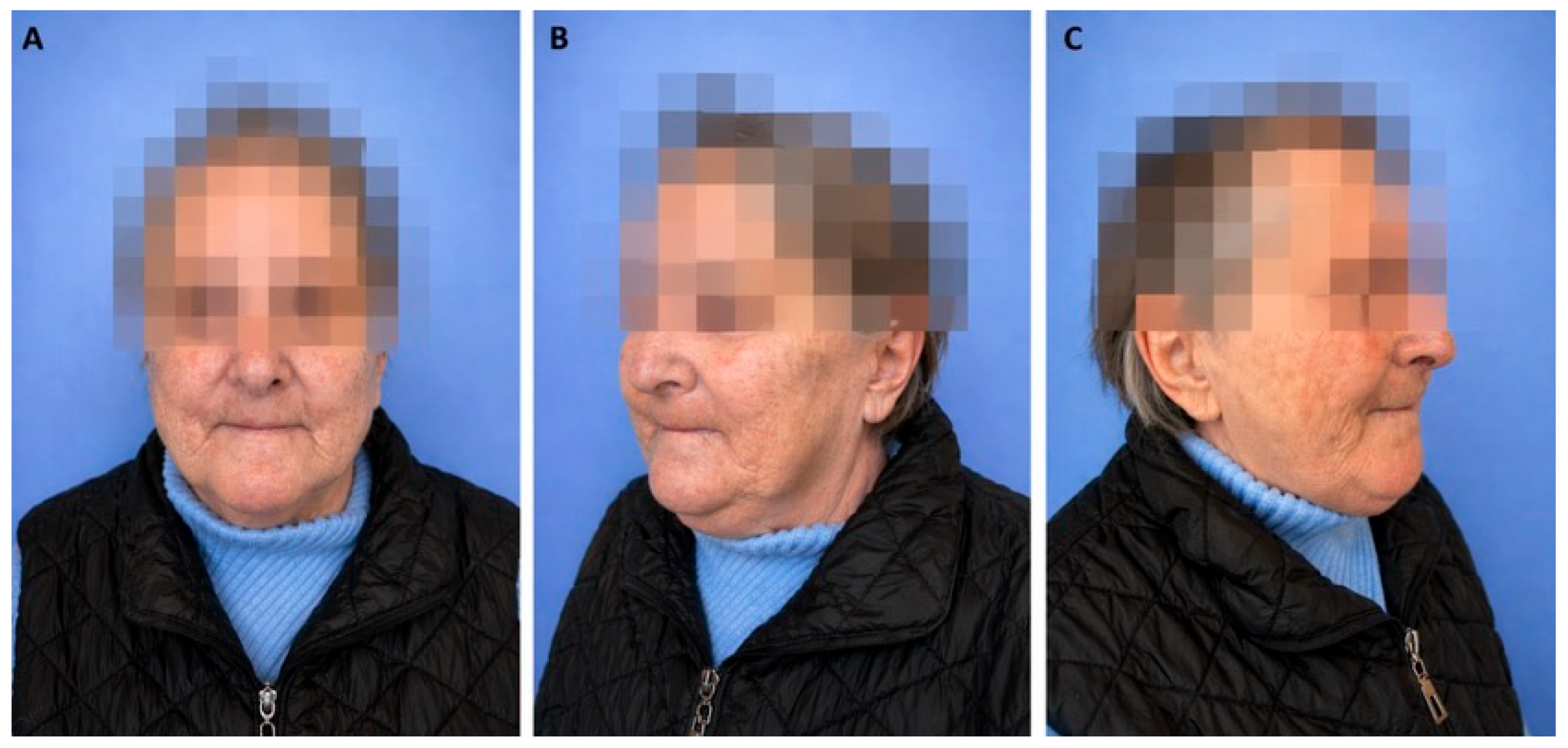
On 15 February 2022, the patient presented to the Oral and Maxillofacial Surgery Outpatient Clinic in Timișoara, reporting progressive numbness of the right maxilla and swelling of the right hard palate. She also reported that in December 2021, an area of exposed bone had appeared in the right edentulous maxillary ridge. Following dental evaluation, she was referred for specialized care. Clinical examination revealed tenderness over the right maxillary sinus and hypoesthesia in the distribution of the right infraorbital nerve (Figure 1).
Figure 1.
On extraoral examination, a tender maxillary sinus point on the right side was detected on palpation, along with hypoesthesia in the distribution territory of the right infraorbital nerve. (A) Frontal view; (B) Left oblique view; (C) Right oblique view.
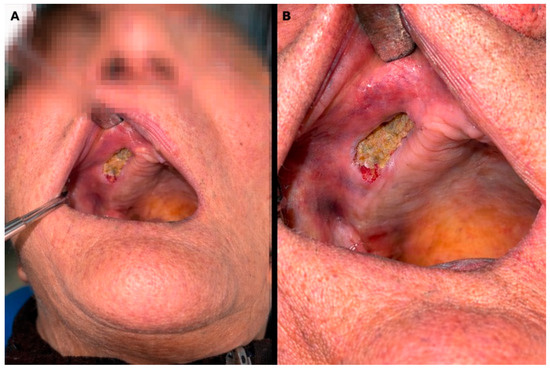
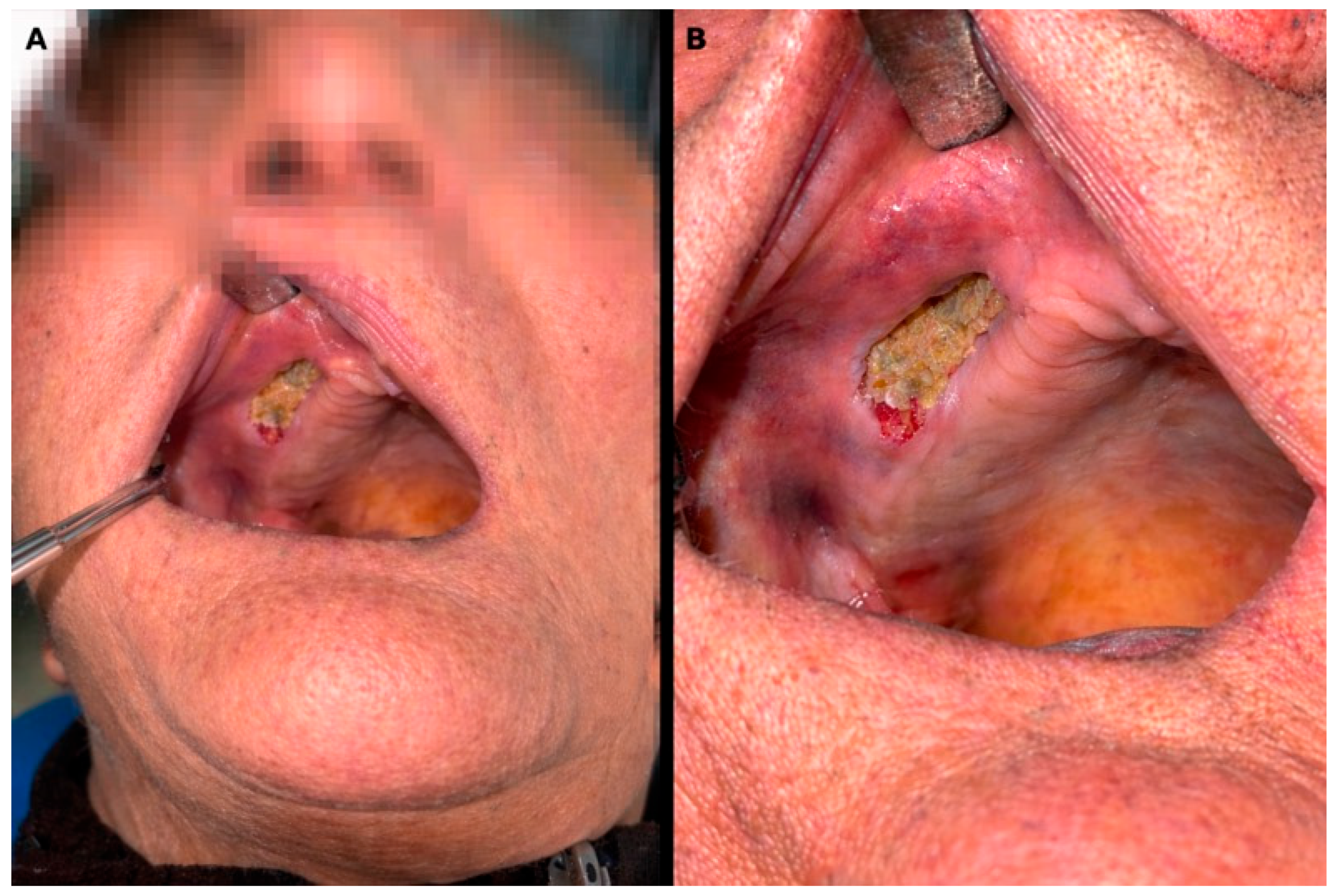
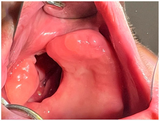
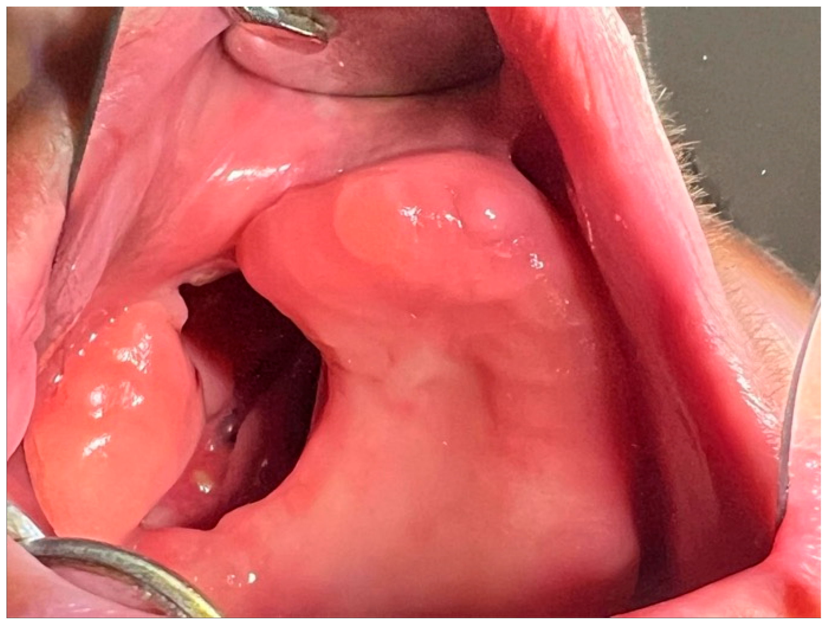
The intraoral examination revealed bone denudation at the level of the edentulous maxillary alveolar ridge, canine-molar region, approximately 3/1 cm, with a spongy bone appearance, yellowish-white in color, with edematous and erythematous perilesional mucosa, without purulent secretions (Figure 2).
Figure 2.
In the intraoral examination, a completely edentulous, non-prosthetized maxilla and mandible were observed, with bone denudation along the right edentulous maxillary alveolar ridge, in the canine–molar region, measuring approximately 3 × 1 cm. The exposed bone presented a spongy, yellowish-white necrotic bone sequestration, with edematous and erythematous perilesional mucosa, and no purulent discharge. (A) Intraoral context view; (B) Close-up view.
On 16 February 2022, ENT examination revealed a completely edentulous patient with necrosis of the right maxillary mucosa, exposing the maxillary bone, which also showed necrotic areas.
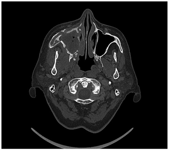
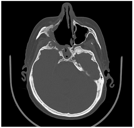
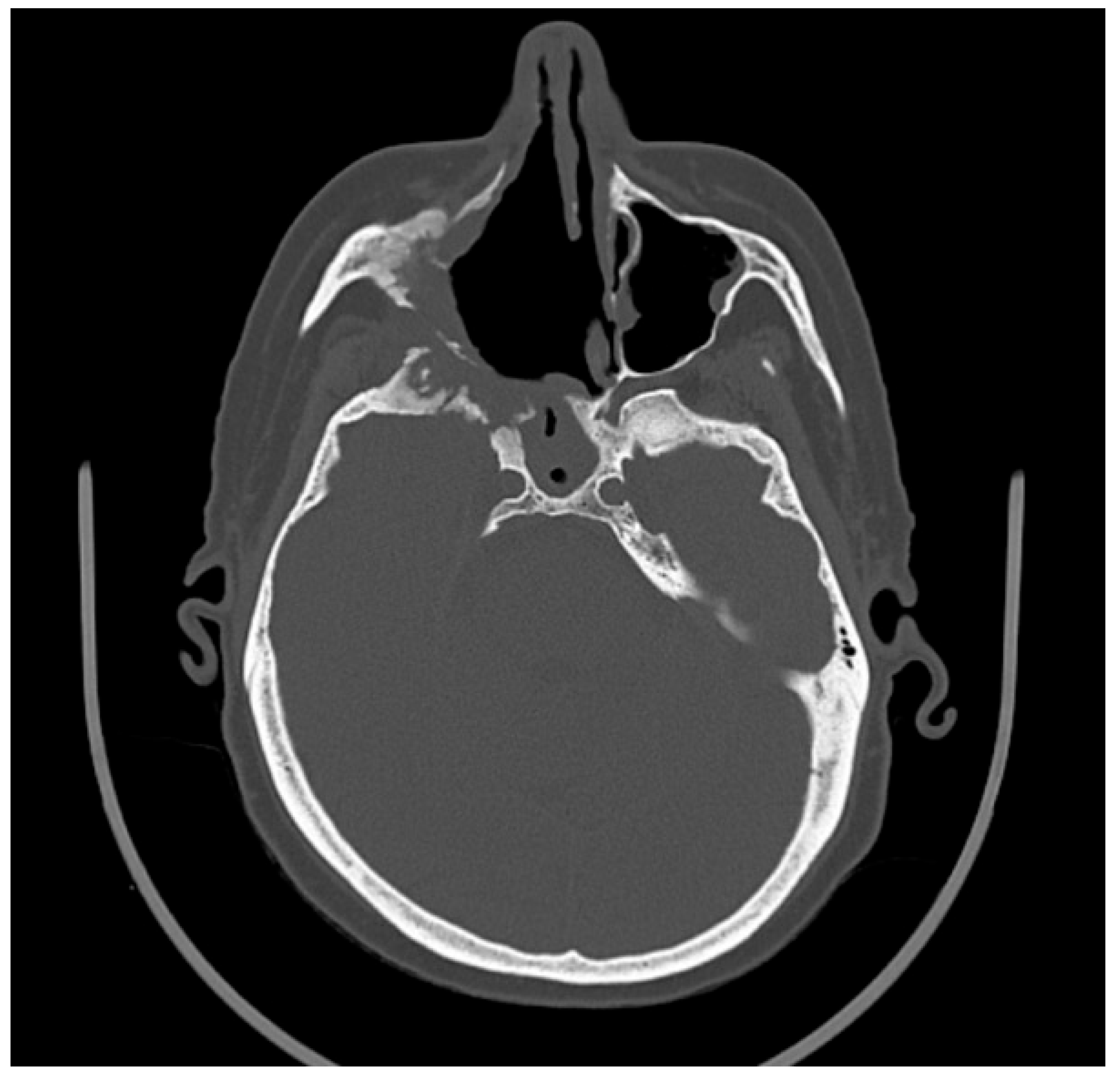
A contrast-enhanced CT scan of the skull performed the same day demonstrated extensive osteolytic bone destruction on a background of irregular osteosclerosis, predominantly involving the right maxilla and all walls of the right maxillary sinus. An extensive parasagittal bone sequestrum extending contralaterally was also identified. Additional involvement included the temporal process of the right zygomatic bone, ipsilateral pterygoid processes, lamina papyracea, and adjacent nasal and frontal bones. These findings were associated with irregular, non-homogeneous mucosal thickening of the right maxillary sinus and ethmoid cells, with minimal extension to the right frontal sinus (Figure 3).
Figure 3.
The axial CT section showed almost complete opacification of the left maxillary sinus, associated with irregular destruction of the medial and superior bone walls, with loss of cortical contour and an osteolytic appearance. The contralateral (right) maxillary sinus remained aerated, demonstrating preserved osseous architecture and providing a baseline for the localized destructive process observed on the left.
On 17 February 2022, the patient was discharged through inter-hospital transfer to the ENT-SCMUT department. The patient is currently hospitalized at the ENT-SCMUT Clinic. The patient is under medical supervision and undergoes regular check-ups. During the course of treatment, the bone sequestrum was identified and surgically removed at the maxillofacial surgery clinic in Timișoara (Figure 4).
Figure 4.
Bone defect of the right edentulous maxillary ridge, extending toward the maxillary buccal vestibule, with a wide oroantral communication. The surrounding mucosa appeared normally colored and mildly tender on palpation. The clinical appearance reflects the result after demarcation and surgical removal of the bony sequestrum.
Histopathological examination of multiple tissue fragments, including bone and maxillary sinus mucosa, using hematoxylin–eosin and periodic acid–Schiff–Alcian blue staining, revealed areas of compact bone embedded within acellular, afibrillar material, suggestive of necrotic bone tissue. PAS–Alcian blue staining highlighted the presence of bacterial colonies morphologically consistent with Actinomyces species.
The overall histopathological findings support the diagnosis of chronic inflammatory changes associated with maxillary osteonecrosis, complicated by secondary actinomycotic infection, in correlation with the clinical and radiological features.
To further evaluate the severity of the bone defects, a 3D reconstruction was obtained (Figure 5). The 3D reconstruction of the skull demonstrated extensive destruction of the right hemimaxilla, with loss of normal bone architecture, multiple areas of collapse, and marked irregularity of the maxillary contour. The right alveolar process was severely compromised, and the maxillary sinus walls appeared fragmented and discontinuous. These findings are consistent with advanced maxillary ONC/osteomyelitis, correlating with the destructive changes observed on the axial, coronal, and sagittal CT sections.
Figure 5.
The 3D reconstruction demonstrated a severe destructive process involving the right hemimaxilla, with extensive loss of maxillary osseous components, structural collapse, and marked contour irregularities. Destruction of the sinus walls, the alveolar process, and the right zygomatic region was evident, with fragmented surfaces and areas of heterogeneous osteolysis. The appearance is consistent with advanced right maxillary osteonecrosis, in concordance with the abnormalities described on the preceding CT images.
Under private health insurance, the patient received a removable full upper denture with an acrylic obturator and a removable full lower denture (Figure 6).
Figure 6.
Removable maxillary complete denture with acrylic obturator. (a) Occlusal view of the denture incorporating the obturator component used to seal the oro-antral communication and restore oral function. (b) Palatal view of the acrylic obturator, illustrating the extension and sealing surface adapted to the palatal defect.
After insertion, the patient demonstrated successful adaptation to the prosthesis, with improvements in phonation, mastication, and aesthetics (Figure 7).
Figure 7.
Removable maxillary complete denture with an acrylic obturator and a removable mandibular complete denture.
The microbiological analysis was performed at two separate intervals (April 2025 and October 2025). Samples that were collected from the oral mucosa, maxillary sinus, and prosthetic obturator surface indicated the existence of a polymicrobial flora (Table 1), while Table 2 summarizes the microbiological interpretation of these findings.
Table 1.
Microbiological findings.
In the initial set of investigations (April 2025), Staphylococcus aureus and Serratia marcescens were concomitantly isolated from all sampled sites, together with fungal colonization by Candida tropicalis.
The microbiological assessment conducted in October 2025 revealed a change in the bacterial spectrum. Klebsiella pneumoniae was present at the level of the obturator and palatal fibromucosa, alongside Staphylococcus aureus and Candida parapsilosis.
In terms of antimicrobial susceptibility, the majority of bacterial isolates exhibited sensitivity to frequently used antibiotics, such as cephalosporins, carbapenems, and fluoroquinolones. Meanwhile, the identified fungal species continued to be susceptible to azole antifungals and amphotericin B.
Table 2 presents the microbiological interpretations.
Table 2.
Microbiological interpretations.
3.2. Case 2
A 65-year-old female patient, known to have grade II arterial hypertension and type II diabetes mellitus, was admitted to Oravița City Hospital, Internal Medicine Department—COVID Unit, between 29 September and 13 October 2021, with the diagnosis of SARS-CoV-2 infection and acute respiratory failure requiring oxygen therapy.
On 20 December 2021, the patient presented to the ENT Department of the Timișoara Municipal Emergency Clinical Hospital, reporting right-sided nasal obstruction, predominantly oral breathing, headache, fatigue, right hemifacial paresthesia, and mucopurulent nasal discharge with an onset approximately 2 months prior. She was admitted to the ENT ward for diagnostic evaluation and specialized treatment (Figure 8).
Figure 8.
Extraoral clinical appearance at presentation, showing right hemifacial asymmetry, mild swelling, and signs of discomfort. The patient exhibited predominantly oral breathing and reported right hemifacial paresthesia. (A) Frontal view; (B) Right oblique view; (C) Right lateral view.
A complete ENT clinical examination, including nasal endoscopy and anterior rhinoscopy, revealed complete obstruction of the right nasal cavity by ulcerative-necrotic false membranes and sanguinolent crusts, as well as bony lysis involving the medial wall of the right maxillary sinus, the bony nasal septum, and the vomer.
A cranial computed tomography scan was performed on 21 December 2021, demonstrating a soft-tissue mass within the right frontal sinus and the right ethmoidal cells, producing osteolysis of the adjacent bony walls, with extension into the right orbit. Additional findings included involvement of the right sphenoid sinus with osteolysis of its lateral and inferior walls, as well as of the greater wing of the sphenoid (lateral orbital wall), and partial osteolysis of the right pterygoid processes and right sphenoid body. At the level of the right maxillary sinus, partial destruction of the anterior and lateral walls was noted, along with partial osteolysis of the right maxillary alveolar process. A minimal dependent fluid level was present within the right maxillary sinus. Mucosal thickening of the left sphenoid sinus and polypoid mucosal thickening of the left maxillary sinus were also observed. Additionally, a midline non-union of the bony palate was described (Figure 9).
Figure 9.
On the axial CT section, a destructive osseous process was observed involving the right hemimaxilla, characterized by irregular osteolysis, loss of continuity of the maxillary sinus walls, and complete opacification of the sinus cavity. The right zygomatic and maxillary bony structures showed deformation and fragmentation, an aspect compatible with advanced osteonecrosis extending into the sinus. The contralateral side demonstrates preserved osseous structure, underscoring the unilateral nature of the process.
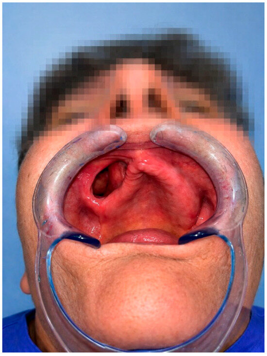
On 23 April 2022, the patient presented to the outpatient clinic of Maxillofacial Surgery at the Timișoara Municipal Emergency Clinical Hospital for further evaluation. Clinical examination revealed a right palatal vault bone defect, a wide oronasal communication, normally colored mucosa partially covered by mucopurulent deposits, and pain on palpation (Figure 10).
Figure 10.
Intraoral clinical examination revealed a bony defect in the right palatal vault, accompanied by a wide oronasal and maxillary sinus communication. The perilesional mucosa appeared normally colored but was painful on palpation.
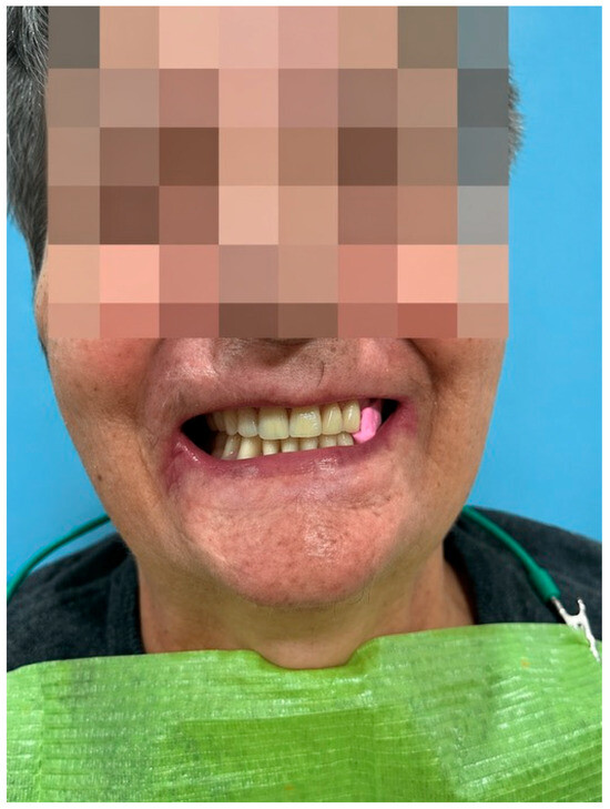
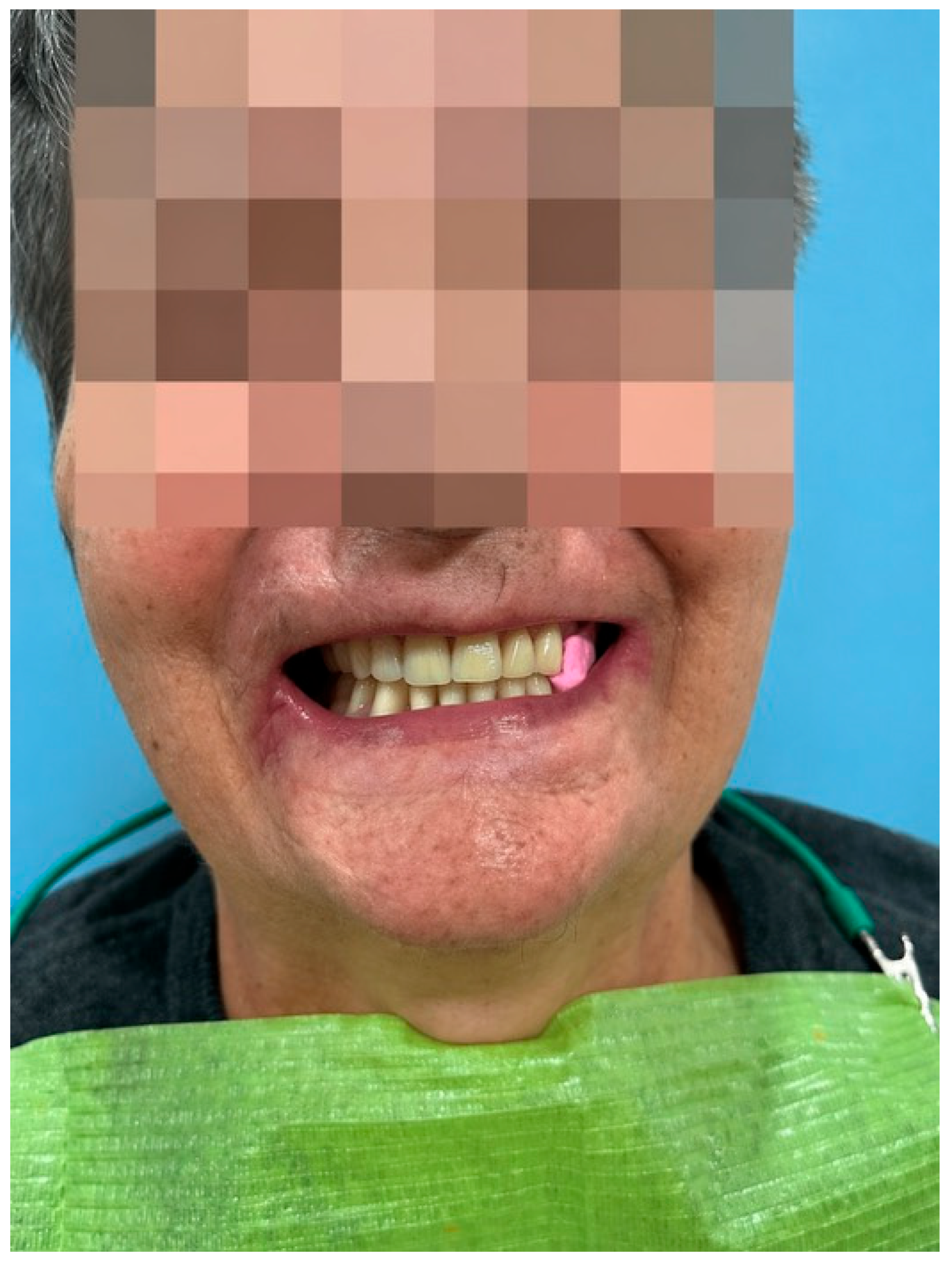
On extraoral clinical examination, the facial skin presented normal coloration, with multiple pigmented facial moles, and thinning of the vermilion border due to unrehabilitated bimaxillary edentulism. The patient reported hypoesthesia in the distribution of the right infraorbital nerve, associated with tenderness on palpation.
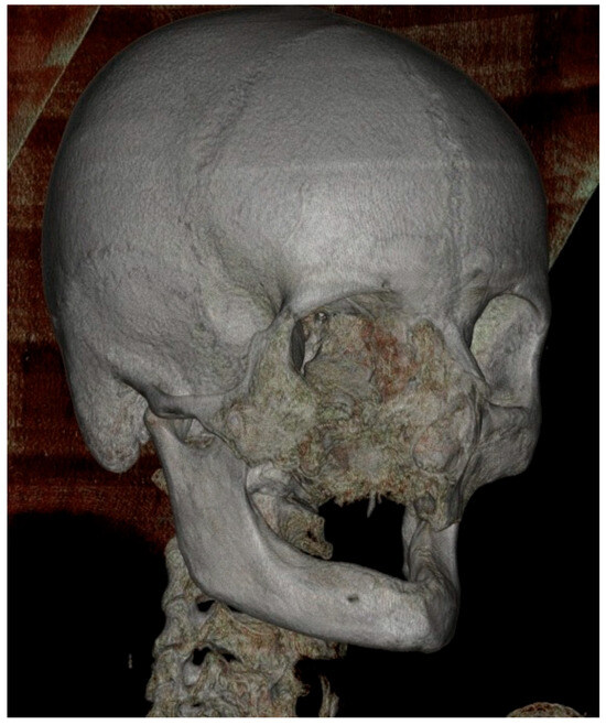
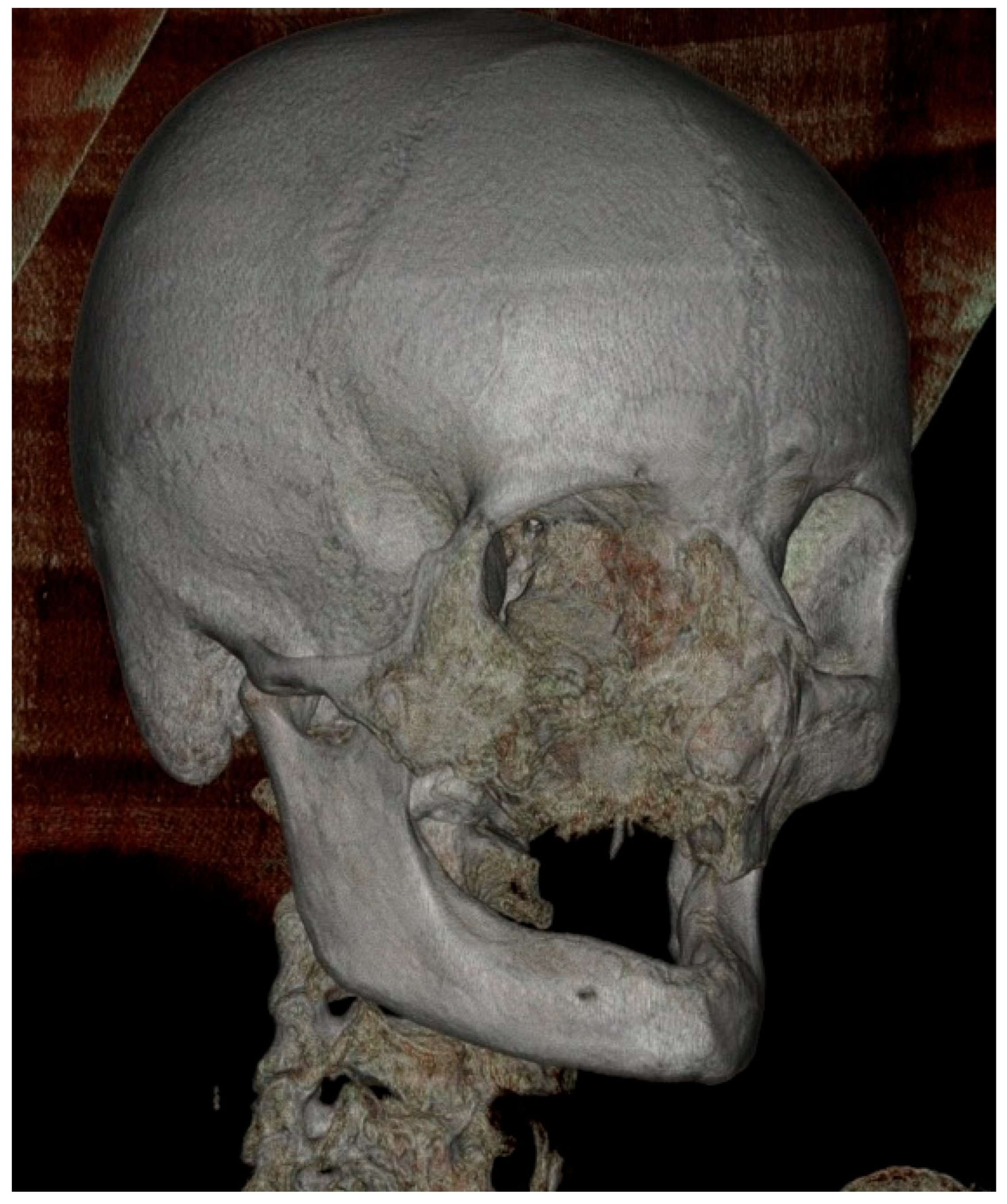
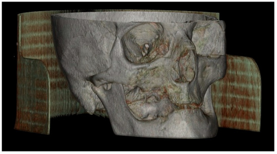
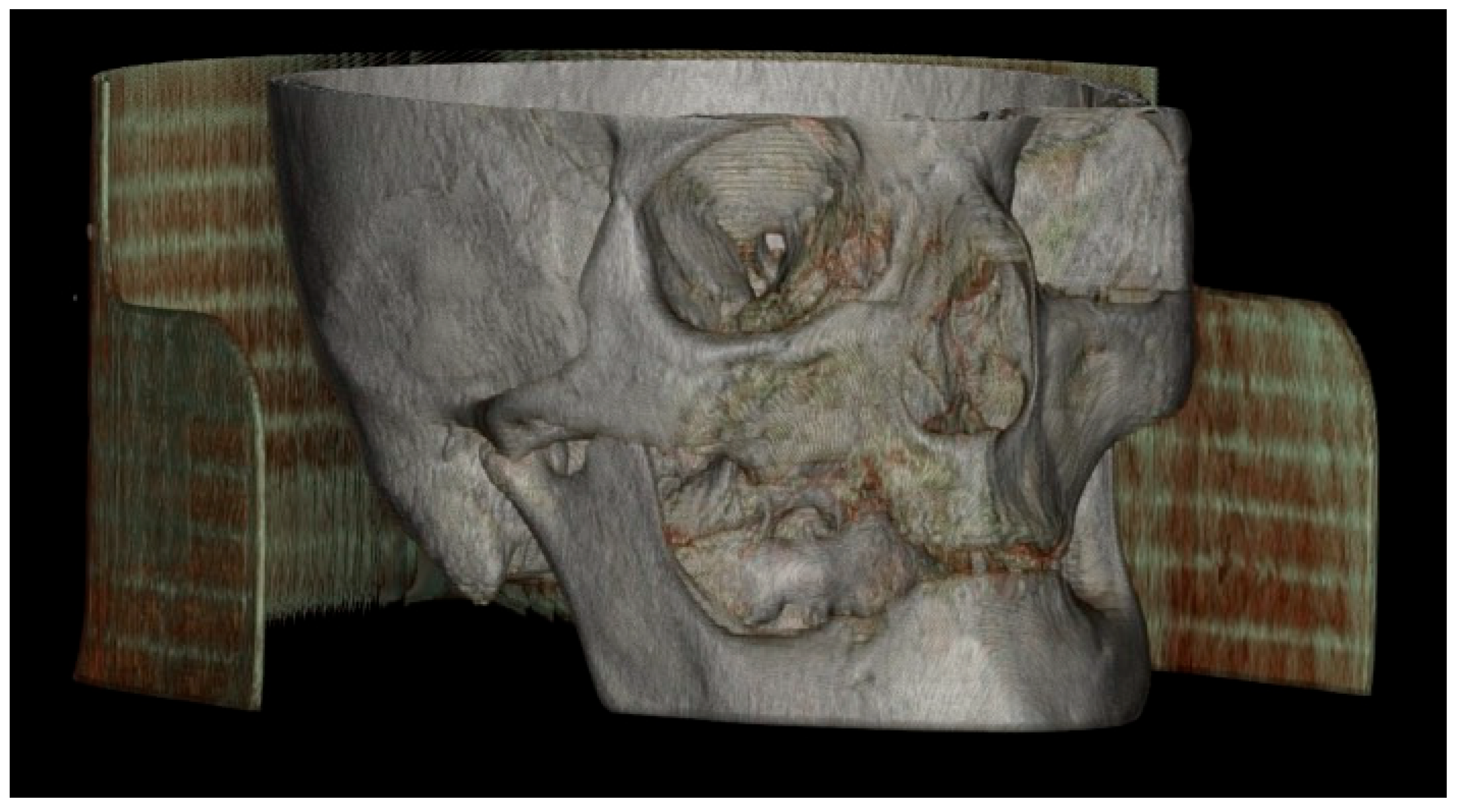
To further characterize the extent of the maxillary destruction, a 3D lateral CT reconstruction was obtained, which revealed marked collapse of the right maxillary sinus walls, fragmentation of the alveolar process, and severe alteration of the normal maxillary and zygomatic architecture (Figure 11). These findings highlight the advanced stage of ONC, with extension toward the zygomatic and suborbital regions.
Figure 11.
The lateral 3D reconstruction revealed severe osseous destruction of the right hemimaxilla, with marked collapse of the sinus walls, fragmentation of the alveolar process, and loss of the normal architecture of the maxillary and zygomatic skeleton. The affected regions appeared irregular, eroded, and displayed multiple areas of heterogeneous osteolysis, an aspect typical of advanced right maxillary osteonecrosis with extension toward the zygomatic and suborbital regions.
After appropriate preoperative preparation, under general anesthesia with orotracheal intubation and endoscopic guidance, an oronasal communication was visualized at the level of the right half of the hard palate, together with erosion of the superior wall of the right maxillary sinus extending up to the orbit, as well as erosion of the bony nasal septum and vomer, which were subsequently removed. Necrosectomy was performed within the nasal cavity. A right maxillary antrostomy, right sphenoidotomy, and right anterior nasal packing were carried out. Postoperative treatment included Zinnat (cefuroxime) 500 mg, voriconazole 200 mg, dexketoprofen, algocalmin, vitamin C, paracetamol, etamsylate, phytomenadione, adrenostazin, and controloc.
Histopathological examination of the submitted maxillary bone fragment was carried out using hematoxylin–eosin (H&E) and periodic acid–Schiff (PAS) staining. It revealed necrotic bone tissue characterized by compact osseous lamellae delimiting areas containing basophilic granular material and filamentous structures. PAS staining highlighted the presence of bacterial colonies morphologically consistent with Actinomyces israelii.
The histopathological aspect supports the diagnosis of a chronic infectious inflammatory process associated with bone necrosis. These discoveries are consistent with advanced maxillary osteonecrosis complicated by secondary actinomycotic infection, in correlation with the clinical and radiological features.
On 27 May 2022, the patient presented to the ENT Department in Cluj-Napoca, reporting phonation disorders, masticatory and feeding difficulties, and oronasal reflux. Clinical examination identified a right maxillary bony defect, as well as right oronasal and oroantral communications secondary to maxillary osteonecrosis involving the midface and skull base. On the same day, the patient was evaluated in the Maxillofacial Surgery Department in Cluj-Napoca. Examination showed a right maxillary bone defect, with right oronasal and oroantral communications secondary to maxillary osteonecrosis involving the midface and skull base, of infectious etiology, accompanied by foul-smelling nasal secretions, open rhinophonia, and objective cacosmia. The recommended treatment included empirical antifungal therapy, surgical debridement of demarcated necrotic lesions, and a removable maxillary prosthesis with an obturator following resolution of the infectious process.
On 6 June 2022, the patient was admitted to the Infectious Diseases II Ward of the “Dr. Victor Babeș” Clinical Hospital for Infectious Diseases and Pulmonology in Timișoara for specialized antibacterial and antifungal treatment. At admission, the patient presented with an altered general condition, afebrile, with loss of appetite. Local clinical examination revealed a bone defect of approximately 2 cm at the level of the hard palate, with a sinus communication and foul-smelling, purulent secretions. The right hemiface was swollen and tender on palpation, with mildly pale skin and mucosa. The patient was discharged on 4 July 2022 in improved general condition, afebrile, with restored appetite, cardio-pulmonary stability, and a persistent right palatal bony defect with congested perilesional margins but without purulent secretions.
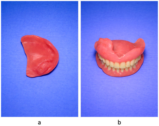
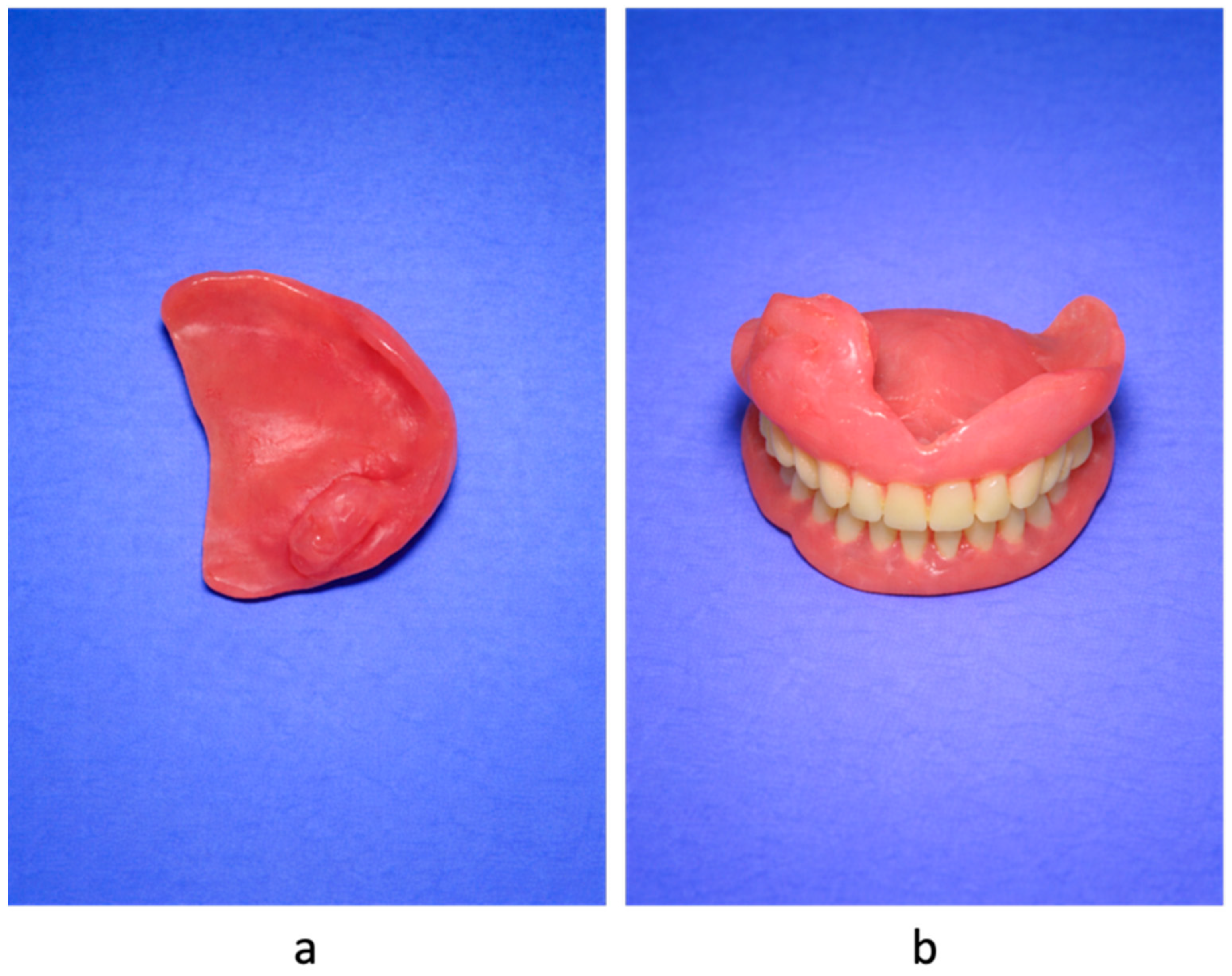
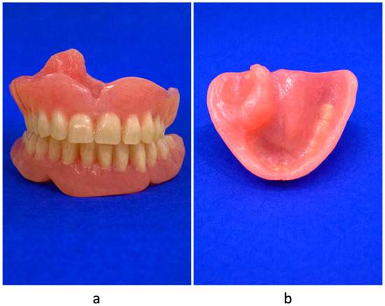
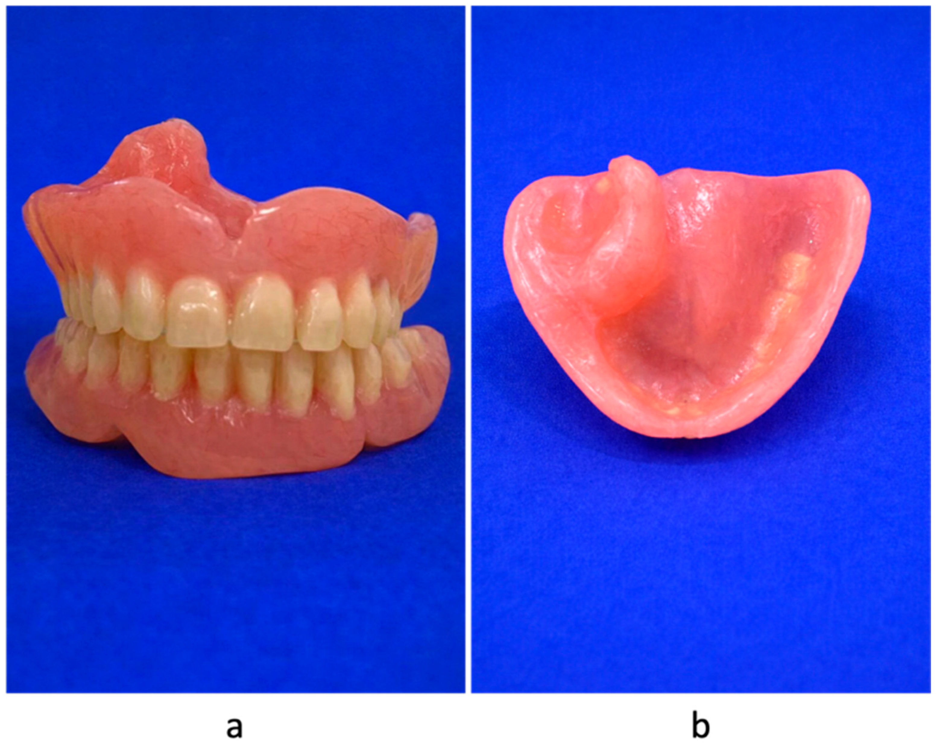
On 17 October 2022, an ENT examination was performed, revealing an oropharyngeal mucosa with normal appearance, a free and mobile soft palate, and a perforated hard palate fitted with a prosthesis, with no evidence of fungal deposits on the mucosa. In the private healthcare system, the patient received a new removable maxillary complete denture with an acrylic obturator (Figure 12).
Figure 12.
Removable maxillary complete denture with acrylic obturator. (a) Occlusal view showing the obturator extension designed to seal the oronasal communication. (b) Palatal surface of the prosthesis demonstrating the acrylic obturation area adapted to the palatal defect.
At follow-up, the patient demonstrated good adaptation to the maxillary obturator, with restoration of oral competence and improvement in speech and eating. Clinical examination confirmed stable prosthetic retention and healthy peri-prosthetic mucosa (Figure 13).
Figure 13.
Removable total maxillary prosthesis with obturator, made of acrylate.
Based on samples taken from nasal secretions, oral mucosa, maxillary sinus, prosthetic obturator, and mandibular gingival mucosa, the microbiological analysis conducted at two different time points (April 2025 and October 2025) revealed a complex microbial ecosystem with a broad distribution of microorganisms across multiple anatomical compartments. Klebsiella oxytoca and Staphylococcus aureus were found in all collected sites in the first microbiological evaluation. Concurrently, mycological studies revealed the existence of Candida species, especially Candida tropicalis.
The next microbiological analysis (October 2025) showed a significant change in the bacterial spectrum, with Citrobacter freundii appearing in the maxillary sinus, obturator, palatal fibromucosa, and mandibular gingival mucosa, although Staphylococcus aureus continued to be present.
Most bacterial isolates demonstrated sensitivity to cephalosporins and other broad-spectrum agents (Table 3), while the identified fungal species remained susceptible to azole antifungals and amphotericin B, while Table 4 summarizes the microbiological interpretation of these findings.
Table 3.
Microbiological findings.
Table 4 presents the microbiological interpretations.
Table 4.
Microbiological interpretations.
3.3. Comparative Analysis of the Two Cases
A comparative analysis highlighting the key clinical features of both cases is provided in Table 5. Both patients presented with severe COVID-19 requiring hospitalization and corticosteroid therapy, followed by the development of maxillary osteonecrosis. However, differences were observed in comorbidities, extent of bone involvement, and clinical evolution.
Table 5.
Comparative summary of the two cases.
4. Discussion
Although all patients in this series exhibited maxillary osteonecrosis following SARS-CoV-2 infection, the current findings are not sufficient to establish a conclusive causal linkage to COVID-19 alone. Both patients received systemic corticosteroid therapy during hospitalization. Classical steroid-induced osteonecrosis is associated with cumulative corticosteroid exposure and results from lipid metabolism disturbances and microvascular compromise [19]. In contrast, post-COVID osteonecrosis may also involve SARS-CoV-2-related endothelial injury and hypercoagulability, impairing bone perfusion even after short corticosteroid courses. Reports of post-COVID ONJ have also described relatively early symptom onset and frequent maxillary involvement [20,21]. Therefore, the distinction between the effects of SARS-CoV-2 infection and those of steroid therapy or underlying comorbidities cannot be fully separated in this case series. Rather, our findings support a multifactorial pathogenic model in which COVID-19-related vascular dysfunction may act synergistically with corticosteroid exposure, metabolic disease, and secondary infection to precipitate ischemic bone damage. Current evidence is limited, so it remains unclear whether post-COVID ONJ represents a distinct entity or a variant of osteonecrosis occurring in a highly pro-thrombotic and inflammatory setting [20,22,23]. In this context, the cases presented suggest a potential association between SARS-CoV-2 infection, its treatment, and the development of osteonecrosis of the jaw.
Systemic inflammation in COVID-19 may promote endothelial dysfunction, hypercoagulability, and impaired bone perfusion, thereby favoring local ischemia [19]. Al-Mahalawy et al. described a post-COVID-19-related PC-RONJ series, mostly involving the maxilla, and according to their classification, both of our cases can be considered severe forms, characterized by extensive osteolysis, sinus involvement, and oroantral/oronasal communications [20]. Grillo et al. documented two instances of COVID-19-associated CRONJ in patients undergoing treatment in Iran, underscoring the significance of acknowledging this emerging post-COVID condition [24]. Mañón et al. reported COVID-19-associated avascular necrosis of the maxilla in a patient treated in the U.S., attributing it to COVID-induced hypercoagulability with arterial thrombosis and microvascular injury, mechanisms analogous to those proposed for ONC of long bones [13]. Additional publications have shown maxillary ONC or osteomyelitis in the setting of COVID-19-related mucormycosis, frequently in diabetic patients and commonly associated with corticosteroid treatment, although cases of non-diabetic patients have also been reported [14,15,16,17]. Other possible diagnoses, such as medication-related osteonecrosis of the jaw (MRONJ), should also be considered. As defined by the AAOMS, MRONJ occurs in patients with current or prior exposure to antiresorptive or antiangiogenic agents and is characterized by exposed bone, or bone that can be probed through an intraoral or extraoral fistula, persisting for more than 8 weeks [25]. The main risk factors include bisphosphonates (mainly intravenous formulations) and denosumab, with risk increasing according to treatment potency, duration, and cumulative dose. Local factors such as tooth extraction, inflammatory oral disease, and poor oral hygiene, together with systemic factors including corticosteroid therapy, diabetes mellitus, smoking, advanced age, and neoplasia, may further contribute [26]. However, none of the patients in this study had a history of antiresorptive or antiangiogenic therapy, making a diagnosis of MRONJ unlikely. In our cases, the clinical and therapeutic context instead supports the diagnosis of post-COVID-related osteonecrosis. Beyond the jaws, AVN in the pandemic time has been increasingly described in weight-bearing joints, such as the femoral head, sometimes appearing months after recovery. Several reports describe AVN following systemic corticosteroid therapy [4], with some case series suggesting that up to 5% of patients treated with various doses of corticosteroids may develop AVN within 2 years [27]. In addition, osteonecrosis of the jaw has long been recognized in non-COVID settings as part of the broader spectrum of drug- and steroid-associated osteonecrosis, particularly in patients receiving corticosteroids together with other predisposing therapies or comorbidities. Hamadeh et al. emphasized that ONJ is multifactorial and may develop in the setting of altered bone remodeling, impaired angiogenesis, dental triggers, diabetes, smoking, and concomitant corticosteroid exposure [28]. However, other reports also documented AVN occurring in patients without prior steroid exposure, indicating that virus-induced endothelial damage, thrombosis, and immune dysregulation can directly predispose to bone infarction [28,29]. For example, Murugesan et al. described a 29-year-old man who, after conservative management of COVID-19, developed bilateral femoral head avascular necrosis without prior corticosteroid exposure or other major traditional risk factors, supporting the possibility that SARS-CoV-2-related endothelial injury and hypercoagulability may themselves contribute to bone ischemia. However, because this was a single femoral-head case report rather than a jaw-specific incidence study, reliable incidence comparisons for ONJ between steroid-treated non-COVID patients and steroid-naïve COVID patients are not yet available [30]. Thus, both steroid-dependent and steroid-independent mechanisms likely operate in COVID-19-associated ONC.
In the maxillofacial region, these mechanisms may be further aggravated by local factors such as impaired sinus drainage, mucosal damage, and secondary infection. Although corticosteroid regimens used in severe COVID-19 are usually shorter than those classically associated with glucocorticoid-induced osteonecrosis, cumulative dose and patient-related risk factors remain important considerations [23,31].
One patient in this series had diabetes mellitus, which is known to affect microcirculation, delay wound healing, and increase susceptibility to secondary infection. Superimposed infections contribute to disease progression by invading devitalized and ischemic tissue, which accelerates bone destruction [6,7,12,14,16,17,29,30,32].
From a histopathological perspective, both cases showed a similar pattern. There was necrotic bone tissue, along with associated chronic inflammatory changes and secondary microbial colonization. Chronic actinomycotic infection was identified in the devitalized bone and could have contributed to the progression of osteonecrosis. The first case presented a more extensive local involvement, reflecting a broader distribution of pathological changes. On the other hand, Case 2 revealed mainly bone-centered histopathological changes, suggesting a more localized pattern of involvement. The patients shared a common pathological mechanism of ischemic bone necrosis complicated by secondary infection. However, the extent and distribution of tissue involvement may differ, which could influence the clinical presentation and progression of the disease.
Maxillary ONC has been reported in association with multiple systemic and local risk factors. In the context of COVID-19, these factors may be amplified, contributing to increased susceptibility to bone necrosis [33,34,35,36,37], as also observed in our cases.
Our series adds several important observations. Both patients were from western Romania, representing, to our knowledge, the first Romanian case series of PC-RONJ in patients without prior antiresorptive or craniofacial radiotherapy. One patient had type 2 DM, and both were treated with systemic corticosteroids and oxygen therapy for severe COVID-19. None had a history of bisphosphonate or denosumab use or craniofacial radiotherapy, which helps distinguish these cases from classic MRONJ or osteoradionecrosis. Third, the onset of maxillary symptoms occurred typically 3–8 weeks after COVID-19 treatment, consistent with a delayed post-infectious/post-therapeutic process. Furthermore, the relatively early debut of symptoms following COVID-19 infection and the short duration of corticosteroid therapy observed in our patients are different from other cases described in the literature. A recent Romanian report by Pădurariu et al. described ONC of the jaw associated with short-course, low-dose corticosteroid therapy in a patient with prior bisphosphonate exposure during COVID-19 infection [18]. Although that case differs from our patients (who had no active antiresorptive therapy), it still reinforces the idea that corticosteroids used in COVID-19 management may contribute to the development of jaw osteonecrosis and highlights the need for increased clinical awareness. The Romanian National COVID-19 Treatment Protocol in 2021 recommended oxygen therapy, systemic corticosteroids (dexamethasone 6 mg/day for 7–10 days in hypoxic patients, or methylprednisolone in moderate-to-severe cases), anticoagulation, antiviral agents, immunomodulators, and supportive measures [38], and both patients in our series were managed under this framework. While these interventions are essential and lifesaving, they may also increase the risk of ONC in susceptible individuals, particularly in association with additional systemic and local risk factors [39].
A central message of our study is that COVID-19 should be regarded as a potential trigger factor for ONC of the jaw, acting in concert with steroids, diabetes, and local infectious or traumatic factors rather than as a passive background condition. In our patients, these interacting systemic and local factors combined to produce severe maxillary bone destruction. This pattern is consistent with the “perfect storm” hypothesis proposed in recent reviews, where multiple prothrombotic, immunosuppressive, and infectious mechanisms converge on a vulnerable anatomical site.
In both cases, there was a persistent polymicrobial infection that affected interconnected anatomical compartments, including the oral cavity, maxillary sinus, and prosthetic obturator (Table 1 and Table 2). Similar microorganisms from various locations were identified. This indicates a disruption of typical compartmentalization and the occurrence of pathological oroantral communication, promoting microbial cross-contamination and the formation of a cohesive orosinusal infectious milieu. Staphylococcus aureus was detected across multiple sampling intervals and locations, indicating a stable bacterial population, which is capable of prolonged colonization. Temporal variations in Gram-negative bacterial species were observed across both patients, showing adaptive modifications within the microbial ecosystem. In Case 1, Serratia marcescens was substituted by Klebsiella pneumoniae, and in Case 2, Klebsiella oxytoca was replaced by Citrobacter freundii. It appears that the microorganisms adapted to modified local conditions, including tissue hypoxia, diminished vascularization, compromised sinus drainage, and persistent inflammation. Even though these elements were similar, the differences were evident. Case 1 presented a more localized yet interconnected infection impacting the oral mucosa, maxillary sinus, and prosthetic obturator. In contrast, Case 2 displayed a broader distribution, which involved nasal secretions, palatal fibromucosa, and mandibular gingival tissues, indicating more extensive structural disruption. Fungal colonization in both patients reflects opportunistic adaptation to a disrupted local environment and prosthetic surfaces, contributing to the complexity of the polymicrobial infection.
The presence of a developed microbial biofilm is supported in both situations by the frequent identification of microorganisms on the surface of the prosthetic obturator. This is an important pathogenic mechanism that contributes to prolonged infection and increases resistance to antimicrobial therapy [40]. The obturator probably serves as a microbial reservoir in this situation, allowing for ongoing reinoculation of nearby tissues.
This case series has several limitations. First, only two patients from a single region in western Romania were involved. Therefore, the findings cannot be generalized to all individuals recovering from COVID-19. Second, the absence of a control group of post-COVID patients without ONC limits the ability to quantify the relative contributions of several factors (e.g., corticosteroid exposure, oxygen therapy, or glycemic control). Third, detailed information on cumulative corticosteroid doses, oxygen delivery parameters (flow, fraction of inspired oxygen, and duration), and the precise timing of each pharmacologic intervention relative to symptom onset was not consistently available in the medical records, precluding more granular analysis. Fourth, one patient had type 2 DM and concomitant invasive or opportunistic infections, making it difficult to distinguish the direct effects of SARS-CoV-2 infection from those of underlying comorbidities or secondary pathogens; these factors likely acted synergistically. Furthermore, imaging markers and laboratory parameters that could further substantiate COVID-19-related vascular involvement, such as MRI assessment of bone marrow edema, coagulation markers (e.g., D-dimer or fibrinogen), and biomarkers of endothelial dysfunction, were not systematically evaluated in these patients. The lack of this information limits the ability to objectively evaluate possible COVID-specific microvascular changes that could be causing osteonecrosis.
Fifth, patient-reported outcome measures, such as quality of life, masticatory efficiency, and speech intelligibility after prosthetic rehabilitation, were not systematically assessed.
Finally, due to the histopathological evaluation being performed only after the establishment of advanced lesions, the early microvascular and marrow changes preceding overt osteonecrosis could not be directly determined. Therefore, another limitation of this study is that histopathological evaluation was available only as written reports, and microscopic images were not accessible for inclusion. Nevertheless, the reported findings were considered reliable and showed concordance with the clinical, radiological, and microbiological data. Despite these limitations, this series provides novel data from an underrepresented region and supports the hypothesis that COVID-19 and its intensive-care treatments may trigger maxillary ONC in high-risk patients. Larger prospective, multicenter studies with appropriate control groups are needed to confirm these findings and to refine preventive and therapeutic strategies.
5. Conclusions
This case series suggests that maxillary osteonecrosis (PC-RONJ) may emerge as a debilitating complication of severe COVID-19, even in patients without prior exposure to antiresorptive therapy. A possible synergistic effect between SARS-CoV-2-induced microvascular injury, systemic glucocorticoid therapy, and metabolic comorbidities may contribute to ischemic bone collapse. However, a causal relationship cannot be definitively established based on the present data.
Early screening and a multidisciplinary approach, integrating oral and maxillofacial surgery, ENT, and microbiology, are essential to limit extensive bone loss and improve quality of life through timely prosthetic rehabilitation.
Author Contributions
Conceptualization, G.C.A., D.C. and T.R.O.; methodology, Ș.D., M.O.P. and C.I.R.; software, C.D. and M.R.; validation, G.C.A., D.C. and T.R.O.; formal analysis, A.M.K. and C.D.; investigation, G.C.A., C.M., C.I.R. and T.R.O.; resources, Ș.D., L.-N.G. and M.O.P.; data curation, A.M.K., M.R. and L.-N.G.; writing—original draft preparation, G.C.A., D.C. and T.R.O.; writing—review and editing, G.C.A., D.C., Ș.D., C.I.R. and T.R.O.; visualization, Ș.D.; supervision, G.C.A. and T.R.O.; project administration, D.C. and C.M.; funding acquisition, G.C.A. and T.R.O. All authors have read and agreed to the published version of the manuscript.
Funding
This research received no external funding.
Institutional Review Board Statement
The study was conducted in accordance with the Declaration of Helsinki and approved by the Institutional Review Board (or Ethics Committee) of Spitalul Clinic Municipal de Urgenta Timisoara (Approval No. 94/04.10.2021 rev 2025, 4 October 2021).
Informed Consent Statement
Written informed consent was obtained from the patients to publish this paper.
Data Availability Statement
The original contributions presented in this study are included in the article. Further inquiries can be directed to the corresponding author.
Acknowledgments
The authors would like to acknowledge “Victor Babeş” University of Medicine and Pharmacy, Timişoara, Romania, for its support in covering the costs of the publication of this review paper.
Conflicts of Interest
The authors declare no conflicts of interest.
References
- Ali, S.A.; Baloch, M.; Ahmed, N.; Ali, A.A.; Iqbal, A. The Outbreak of Coronavirus Disease 2019 (COVID-19)—An Emerging Global Health Threat. J. Infect. Public Health 2020, 13, 644–646. [Google Scholar] [CrossRef] [Scilit]
- Huang, G.; Guo, F.; Liu, L.; Taksa, L.; Cheng, Z.; Tani, M.; Zimmermann, K.F.; Franklin, M.; Silva, S.S.M. Changing Impact of COVID-19 on Life Expectancy 2019–2023 and Its Decomposition: Findings from 27 Countries. SSM Popul. Health 2024, 25, 101568. [Google Scholar] [CrossRef] [Scilit]
- World Health Organization (WHO). WHO Coronavirus (COVID-19) Dashboard > More Resources Dashboard. 2023. Available online: https://data.who.int/dashboards/covid19/more-resources (accessed on 3 December 2025).
- Alexandru, G.C.; Gligor, L.-N.; Chioran, D.; Roi, C.I.; Riviș, M.; Pricop, M.O.; Urîtu, A.; Pacnejer, A.-M.; Manea, H.C.; Olariu, T.R. Post-COVID-19 Jaw Osteonecrosis: A Narrative Review. Medicina 2026, 62, 641. [Google Scholar] [CrossRef] [Scilit]
- Aliberti, A.; Gasparro, R.; Mignogna, M.; Canfora, F.; Spagnuolo, G.; Sammartino, G.; Coppola, N. Unveiling the Oral Lesions, Dysgeusia and Osteonecrosis Related to COVID-19: A Comprehensive Systematic Review. J. Clin. Med. 2025, 14, 1267. [Google Scholar] [CrossRef] [Scilit]
- Baimukhamedov, C.; Botabekova, A.; Lessova, Z.; Abshenov, B.; Kurmanali, N. Osteonecrosis amid the COVID-19 Pandemic. Rheumatol. Int. 2023, 43, 1377–1378. [Google Scholar] [CrossRef] [Scilit] [PubMed]
- Matthews, A.H.; Davis, D.D.; Fish, M.J.; Stitson, D. Avascular Necrosis. In StatPearls; StatPearls Publishing: Treasure Island, FL, USA, 2023. Available online: https://www.ncbi.nlm.nih.gov/books/NBK537007/ (accessed on 3 December 2025).
- Goncharov, E.N.; Koval, O.A.; Bezuglov, E.N.; Vetoshkin, A.A.; Goncharov, N.G.; Encarnación Ramirez, M.D.J.; Montemurro, N. Conservative Treatment in Avascular Necrosis of the Femoral Head: A Systematic Review. Med. Sci. 2024, 12, 32. [Google Scholar] [CrossRef] [Scilit] [PubMed]
- Werner, M.T.; Carey, R.M.; Albergotti, W.G.; Lukens, J.N.; Brody, R.M. Impact of the COVID-19 Pandemic on the Management of Head and Neck Malignancies. Otolaryngol. Head Neck Surg. 2020, 162, 816–817. [Google Scholar] [CrossRef] [Scilit] [PubMed]
- Ralli, M.; Colizza, A.; Cambria, F.; Gilardi, A.; Meliante, P.; Cavalcanti, L.; Minni, A.; Greco, A.; de Vincentiis, M. Effects of COVID-19 Pandemic on Head and Neck Oncology Activity: The Experience of Our University Hospital. Eur. Rev. Med. Pharmacol. Sci. 2021, 25, 7268–7271. [Google Scholar] [CrossRef] [Scilit]
- Mikulska, M.; Nicolini, L.A.; Signori, A.; Di Biagio, A.; Sepulcri, C.; Russo, C.; Dettori, S.; Berruti, M.; Sormani, M.P.; Giacobbe, D.R.; et al. Tocilizumab and Steroid Treatment in Patients with COVID-19 Pneumonia. PLoS ONE 2020, 15, e0237831. [Google Scholar] [CrossRef] [Scilit]
- Boymuradov, S.A.; Rustamova, D.A.; Bobamuratova, D.T.; Kurbanov, Y.X.; Karimberdiyev, B.I.; Yusupov, S.S.; Umarov, R.Z. Aspects of Maxillar Osteomyelitis in Patients Who Had COVID-19 in Uzbekistan. Adv. Oral Maxillofac. Surg. 2021, 3, 100106. [Google Scholar] [CrossRef] [Scilit]
- Mañón, V.A.; Balandran, S.; Young, S.; Wong, M.; Melville, J.C. COVID-Associated Avascular Necrosis of the Maxilla—A Rare, New Side Effect of COVID-19. J. Oral Maxillofac. Surg. 2022, 80, 1254–1259. [Google Scholar] [CrossRef] [Scilit]
- Rao, V.U.S.; Arakeri, G.; Madikeri, G.; Shah, A.; Oeppen, R.S.; Brennan, P.A. COVID-19 Associated Mucormycosis (CAM) in India: A Formidable Challenge. Br. J. Oral Maxillofac. Surg. 2021, 59, 1095–1098. [Google Scholar] [CrossRef] [Scilit]
- Zameer, S.; Syed, R.; Afzal, A.; Amir, R.; Faridi, S.; Nayyar, A. COVID-19 Associated Mucormycosis: A Case Report in Pakistan and a Review of 144 Cases. J. Pak. Med. Assoc. 2022, 72, 2313–2316. [Google Scholar] [CrossRef] [Scilit]
- Anand, T.; Mukherjee, A.; Satija, A.; Velamuri, P.S.; Singh, K.J.; Das, M.; Josten, K.; Yadav, P.D.; Sahay, R.R.; Keche, A.Y.; et al. A Case Control Investigation of COVID-19 Associated Mucormycosis in India. BMC Infect. Dis. 2022, 22, 856. [Google Scholar] [CrossRef] [Scilit]
- Paavai, T.T.; Vasanthi, V.; Rameshkumar, A.; Rajkumar, K.; Krishnakumar Raja, V.B.; Muthusubramanian, V. Maxillary Mucormycotic Osteonecrosis as a Manifestation of Post-COVID-19 Infection in Non-Diabetic Patients: Report of Two Cases. J. Microsc. Ultrastruct. 2024, 12, 99–103. [Google Scholar] [CrossRef] [Scilit] [PubMed]
- Pădurariu, L.C.; Dincă, O.M.; Vlădan, G.C.; Niță, T.; Bucur, A. Osteonecrosis of the Jaw Associated with Short-Course and Low-Dose Corticosteroid Therapy. Rom. J. Oral Rehabil. 2023, 15, 28–33. [Google Scholar]
- Motta, F.; Timilsina, S.; Gershwin, M.E.; Selmi, C. Steroid-Induced Osteonecrosis. J. Transl. Autoimmun. 2022, 5, 100168. [Google Scholar] [CrossRef] [Scilit] [PubMed]
- Al-Mahalawy, H.; El-Mahallawy, Y.; Dessoky, N.Y.; Ibrahim, S.; Amer, H.; Ayad, H.M.; El Sherif, H.M.; Shabaan, A.A. Post-COVID-19 Related Osteonecrosis of the Jaw (PC-RONJ): An Alarming Morbidity in COVID-19 Surviving Patients. BMC Infect. Dis. 2022, 22, 544. [Google Scholar] [CrossRef] [Scilit] [PubMed]
- Khan, M.A.; Rahman, T.; Rahman, S.A.; Hashmi, G.S.; Ansari, M.K.; Danish, M.; Arman, M.; Ahmad, R.; Ahmad, W. Is Post-COVID Osteonecrosis of Jaw (PCONJ) Masquerading as Osteomyelitis? A Largest Unicentric Report of 13 Cases. Adv. Oral Maxillofac. Surg. 2023, 11, 100435. [Google Scholar] [CrossRef] [Scilit]
- Sourani, A.; Kalantari, F.; El-Rabbany, M.; Shahmoradi, M.; Haridas, A.; Vahdat, N.; Mirza, S.; Sourani, A.; Foroughi, M.; Baradaran Mahdavi, S. COVID-19 and Jaw Osteonecrosis: A Systematic Review on Clinical Presentations and Treatment Outcomes. Curr. Probl. Surg. 2025, 73, 101897. [Google Scholar] [CrossRef] [Scilit]
- Sakellariou, E.; Argyropoulou, E.; Karampinas, P.; Galanis, A.; Varsamos, I.; Giannatos, V.; Vasiliadis, E.; Kaspiris, A.; Vlamis, J.; Pneumaticos, S. A Comprehensive Review of COVID-19-Infection- and Steroid-Treatment-Associated Bone Avascular Necrosis: A Multi-Study. Anal. Diagn. 2024, 14, 247. [Google Scholar] [CrossRef] [Scilit]
- Grillo, R.; Samieirad, S.; Alizadeh, O.; Hosseini Zarch, S.H.; Khodashahi, R.; Kazemian, N.; da Graça Naclério-Homem, M. Two Cases of COVID-Related Osteonecrosis of the Jaws: A New and Worrying Entity Is Emerging. World J. Plast. Surg. 2024, 13, 96–102. [Google Scholar] [CrossRef] [Scilit]
- Thomas, J.G.; Ouanounou, A. Medication-related osteonecrosis of the jaw: A narrative review of risk factors, diagnosis, and management. Front. Oral Maxillofac. Med. 2023, 5, 31. [Google Scholar] [CrossRef] [Scilit]
- AlRowis, R.; Aldawood, A.; AlOtaibi, M.; Alnasser, E.; AlSaif, I.; Aljaber, A.; Natto, Z. Medication-Related Osteonecrosis of the Jaw (MRONJ): A Review of Pathophysiology, Risk Factors, Preventive Measures and Treatment Strategies. Saudi Dent. J. 2022, 34, 202–210. [Google Scholar] [CrossRef] [Scilit]
- Veizi, E.; Erdoğan, Y.; Sezgin, B.S.; Karaman, Y.; Kılıçarslan, K.; Fırat, A. The Painful Joint after COVID-19 Treatment: A Study on Joint Osteonecrosis Following COVID-19-Related Corticosteroid Use. Jt. Dis. Relat. Surg. 2023, 34, 75–83. [Google Scholar] [CrossRef] [Scilit] [PubMed]
- Hamadeh, I.S.; Ngwa, B.A.; Gong, Y. Drug-Induced Osteonecrosis of the Jaw. Cancer Treat. Rev. 2015, 41, 455–464. [Google Scholar] [CrossRef] [Scilit] [PubMed]
- Daltro, G.; Silva, I.C.F.; Daltro, P.B.; Botelho, V.L. SARS-CoV-2/COVID-19 and Its Implications in the Development of Osteonecrosis. J. Regen. Biol. Med. 2020, 2, 1–19. [Google Scholar]
- Murugesan, L.; Sivakumar, N.; Ramamoorthy, L.; Farooq, U. COVID-19-Associated Bilateral Avascular Necrosis of the Femoral Head in a Young Male Without Corticosteroid Exposure: A Case Report. Cureus 2024, 16, e57525. [Google Scholar] [CrossRef] [Scilit]
- Bhimraj, A.; Morgan, R.L.; Shumaker, A.H.; Lavergne, V.; Baden, L.; Cheng, V.C.-C.; Edwards, K.M.; Gandhi, R.; Gallagher, J.; Muller, W.J.; et al. Infectious Diseases Society of America Guidelines on the Treatment and Management of Patients with COVID-19. Infectious Diseases Society of America. 2021. Available online: https://www.idsociety.org/COVID19guidelines (accessed on 23 March 2026).
- Zhao, R.; Wang, H.; Wang, X.; Feng, F. Steroid Therapy and the Risk of Osteonecrosis in SARS Patients: A Dose–Response Meta-Analysis. Osteoporos. Int. 2017, 28, 1027–1034. [Google Scholar] [CrossRef] [Scilit]
- Nischal, V.; Gupta, N. Bisphosphonate-Related Jaw Osteonecrosis. In StatPearls; StatPearls: Treasure Island, FL, USA, 2026. Available online: https://www.ncbi.nlm.nih.gov/books/NBK534771/ (accessed on 3 March 2026).
- Rodriguez-Lozano, F.J.; Oñate-Sánchez, R.E. Treatment of Osteonecrosis of the Jaw Related to Bisphosphonates and Other Antiresorptive Agents. Med. Oral Patol. Oral Cir. Bucal 2016, 21, e595–e600. [Google Scholar] [CrossRef] [Scilit]
- Anastasilakis, A.D.; Pepe, J.; Napoli, N.; Palermo, A.; Magopoulos, C.; Khan, A.A.; Zillikens, M.C.; Body, J.J. Osteonecrosis of the Jaw and Antiresorptive Agents in Benign and Malignant Diseases: A Critical Review Organized by the ECTS. J. Clin. Endocrinol. Metab. 2022, 107, 1441–1460. [Google Scholar] [CrossRef] [Scilit] [PubMed]
- Lončar Brzak, B.; Horvat Aleksijević, L.; Vindiš, E.; Kordić, I.; Granić, M.; Vidović Juras, D.; Andabak Rogulj, A. Osteonecrosis of the Jaw. Dent. J. 2023, 11, 23. [Google Scholar] [CrossRef] [Scilit]
- Căpraru, I.D.; Marian, C.; Vulcănescu, D.D.; Tănăsescu, S.; Dragomir, T.L.; Marți, T.D.; Boru, C.; Avram, C.R.; Susan, M.; Vlad, C.S. Understanding the Impact of COVID-19 on Roma Vulnerable Communities in Western Romania: Insights and Predictive Factors from a Retrospective Study. Viruses 2024, 16, 435. [Google Scholar] [CrossRef] [Scilit] [PubMed]
- Ministerul Sănătăţii (MS). Protocolul de Tratament al Infecţiei cu Virusul SARS-CoV-2, din 29.12.2021. Monitorul Oficial al României, Partea I, nr. 1246. 2021. Available online: https://legislatie.just.ro/ (accessed on 23 March 2026).
- Weinstein, R.S. Glucocorticoid-Induced Osteonecrosis. Endocrine 2012, 41, 183–190. [Google Scholar] [CrossRef] [Scilit]
- Zhao, A.; Sun, J.; Liu, Y. Understanding bacterial biofilms: From definition to treatment strategies. Front. Cell. Infect. Microbiol. 2023, 13, 1137947. [Google Scholar] [CrossRef] [Scilit] [PubMed]

Disclaimer/Publisher’s Note: The statements, opinions and data contained in all publications are solely those of the individual author(s) and contributor(s) and not of MDPI and/or the editor(s). MDPI and/or the editor(s) disclaim responsibility for any injury to people or property resulting from any ideas, methods, instructions or products referred to in the content. |
© 2026 by the authors. Licensee MDPI, Basel, Switzerland. This article is an open access article distributed under the terms and conditions of the Creative Commons Attribution (CC BY) license.