


Abstract
Bacterial diseases are one of the major causative factors for fish mortality and economic losses in aquaculture sector. Most of the pathogenic bacteria exists in biofilms, which offer protection from antibiotics and other infection control measures. In the present study, the antibacterial and antibiofilm activities of green synthesized copper oxide nanoparticles were tested against selected fish pathogens. Copper oxide nanoparticles (CuO-NPs) were synthesized using brown seaweed, Sargassum vulgare, extract. The biosynthesized NPs were characterized using UV-visible spectroscopy analysis, Fourier transform infrared (FTIR) spectroscopy, scanning electron microscopy (SEM) analysis, an energy dispersive X-ray analyzer (EDX) and X-ray diffraction (XRD). The SEM examination of morphology revealed predominantly spherical particles with some degrees of aggregation in CuO-NPs. The polycrystalline nature of CuO-NPs was confirmed in the XRD analysis. Furthermore, CuO-NPs exhibited antioxidant activity against 1,1-diphenyl-2-picrylhydrazyl (DPPH). The prepared NPs also demonstrated antibacterial and biofilm formation inhibition activities against fish pathogens with a maximum biofilm inhibition of 95% against Vibrio anguillarum. Overall, the findings suggest that the S. vulgare mediated CuO-NPs have potential for further investigation in fish disease control in aquaculture due to their antibacterial and biofilm inhibition effects against fish pathogens.
1. Introduction
The aquaculture sector suffers heavy losses as a result of disease outbreaks, especially bacterial infections [1]. Biofilms, which are an aggregation of microbial communities on substrates, are considered a potential reservoir of pathogenic microorganisms that may cause infections in aquaculture [2,3]. The aquaculture sector is facing severe economic losses due to biofilm-related fish infections [4]. The microbial community in a biofilm assemblage is encased by an extracellular polymeric substances (EPSs) matrix that shields bacteria from bactericidal and bacteriostatic substances. Moreover, biofilms provide a suitable environment for pathogens by improving drug resistance to commercial antibiotics and provide protection against immune system defenses [5].
Conventional biofilm control measures in aquaculture settings include the use of chemical compounds or antibiotics. However, long-term use of antibiotics has been linked to the emergence of resistant strains and other environmental impacts [6]. Due to the growing multidrug resistance of bacterial strains and a lower efficiency of traditional biofilm control measures, alternative control measures are necessary for aquaculture health management [7]. Biofilm control using nanoparticles is one of the alternative approaches for effective fish disease management [8]. Previous studies reported the efficiency of nanoparticles against the attachment of bacteria or establishment of biofilms related to aquaculture pathogens [7,9,10,11]. Further, nanoparticles are considered as a promising alternative in managing disease outbreaks in the aquaculture industry [12,13,14] due to their ability to reduce antibiotic resistance and enhanced drug delivery. Many nanoparticles, such as Cu-NPs, Ag-NPs, chitosan Au-NPs, and others, show antibacterial activity against bacteria causing diseases in fish, such as Escherichia coli, Aeromonas spp. and Vibrio spp. [15,16,17,18]. Further, the antibiofilm properties of Cu-NPs were reported previously. Cu-NPs presented antibiofilm effect on aquaculture relevant bacteria such as Vibrio alginolyticus, Vibrio parahaemolyticus, and Aeromonas hydrophila and more than 60% biofilm reduction was reported [7]. Likewise, the fabricated CuO-NPs using leaves from the herb Ocimum americanum L. were active against aquatic pathogens such as Pseudomonas aeruginosa, Aeromonas hydrophila, and Staphylococcus aureus [9].
Among the metal nanoparticles, copper oxide nanoparticles (CuO-NPs) have high thermal and chemical stability, comparatively less toxicity, an easy synthesis route, compatibility, a nanoscale structure, improved oxygen absorption potential, a high specific surface area and low cost [19,20]. Recently, several well-defined CuO nanostructures, including nanoflowers [21], leaves [22], nanospheres [23], nanotubes [24], and nanorings [25], have been reported to improve the properties of nanoparticles and surface reactivity. CuO-NPs are synthesized via several physical and chemical methods. However, both of these methods require long-term growth and high cost, involve multistep procedures, and are hazardous to the environment. Hence, attempts have been made in recent years to develop nontoxic, clean and cost-effective methods to reduce environmental pollution [26].
Eco-friendly green methods have been established to synthesize NPs from several biological sources, including seaweeds. Seaweed extracts contain phytochemicals such as flavonoids, polyphenols, and tannins, which act as reducing, recovery, capping, and stabilizing agents [27]. Brown seaweed, including the genus Sargassum, have the ability to accumulate and transform metals, and are considered nanofactories for various types of zinc oxide nanoparticles and CuO-NPs, which possess many beneficial properties [28]. The green synthesis of CuO-NPs using brown algae usually relies on aqueous extracts, which act as reducing and capping agents, resulting in stable, biofunctionalized, and non-toxic particles [29,30,31]. Many brown algae have been successfully used in the synthesis of CuO-NPs and their use in many different biotechnology applications were documented [29,32,33]. Brown seaweed such as Macrocystis pyrifera [29], Bifurcaria bifurcate [34], Cystoseira trinodis [35], Sargassum polycystum [36], Sargassum longifolium [37], Sargassum wightii [38,39], Sargassum cervicorne [40], Padina boergesenii [41], Padina tetrastromatica [42] and Turbinaria conoides [43] have been commonly used for the synthesis of CuO-NPs. The synthesis of CuO-NPs by brown algae has many proven advantages and applications, such as antimicrobial activities [44,45,46], antioxidant activity [38,43], anticancer [44,46,47], catalytic/decolorization [43], anti-biofilm [48], and environmental bioremediation [33,40].
Previous studies have reported antibacterial and antibiofilm activities of green-synthesized CuO-NPs with varying effects depending on the algal source used for nanoparticle synthesis, bacterial strains used as targets and synthesis conditions [41,48,49,50]. Though several previous studies have reported the seaweed-mediated synthesis of metal nanoparticles, studies evaluating brown seaweed (Sargassum vulgare)-mediated CuO-NPs against fish pathogenic bacteria remain limited. Hence, in this study, the multifunctional performance, such as antioxidant, antibacterial and biofilm formation inhibition activities, of the seaweed-mediated CuO-NPs were evaluated. The objective of this study was to synthesize CuO-NPs using Sargassum vulgare extract and evaluate their antibacterial and biofilm inhibition activities against fish pathogenic bacteria that are considered relevant to aquaculture.
2. Materials and Methods
2.1. Seaweed Collection and Extract Preparation
The brown alga Sargassum vulgare was collected from the Jeddah coastal region, Saudi Arabia and brought to the laboratory. The brown seaweed was washed with filtered seawater and further washed with tap water. The sample was then dried for several days at room temperature and ground mechanically using a mixer grinder. Seaweed extraction was carried out following the methods described by Fatima et al. [51] and Jayarambabu et al. [52], with slight modifications. Briefly, seaweed was mixed with ethanol at a 1:25 ratio on a hot plate and incubated at 70 °C for 4 h under continuous stirring. After 4 h of extraction, the extract was sonicated using a sonicator (505 Sonic Dismembrator) (Fisher Scientific, Waltham, MA, USA) for one minute at 40% amplitude. The solvent extract was finally collected after centrifugation for 10 min at 4000× g at 4 °C, and the final extract was dissolved in ethanol solution for NP synthesis.
2.2. Determination of Total Polyphenol and Total Flavonoid Content
The total polyphenol content in the ethanol extract was determined by the Folin–Ciocalteu method. Folin–Ciocalteu reagent, sodium carbonate, sodium nitrate (NaNO2) gallic acid and quercetin were purchased from Sigma-Aldrich (St. Louis, MO, USA). About 0.1 mL of ethanol extract (5 mg/mL) was mixed with 1.9 mL of Folin–Ciocalteu reagent and 1 mL of sodium carbonate (7.5%, w/v), followed by incubation in the dark for 30 min. Then, the absorbance of the sample was read at 765 nm using a spectrophotometer, and ethanol was used as a negative control. Gallic acid was prepared at various concentrations (0.02 to 0.1 mg/mL), and the result was expressed as mg gallic acid equivalents/g of dry ethanol extract (mg GAE/g) [53]. The amount of total flavonoid content was estimated as described previously by Kim et al. [54], with little modification. Briefly, 0.1 mL of extract was mixed with 0.9 mL of double distilled water and 5% of 0.1 mL of NaNO2 and incubated for 5 min. Then, 0.1 mL of 10% AlCl3 was added and incubated in the dark for a further 5 min. Then, 0.5 mL of 1 M sodium hydroxide and 0.25 mL of distilled water were added. The absorbance of the sample was read at 510 nm, and quercetin (0.02 to 0.1 mg/mL) was used as the positive control.
2.3. Green Synthesis of Copper Oxide Nanoparticles (CuO-NPs)
CuO-NP preparation was performed by mixing a copper acetate solution (100 mM) and the seaweed suspension (10 mg/mL) at a 1:2 (v/v) ratio. CuO-NP synthesis was achieved via a microwave-assisted method. Briefly, the copper acetate–seaweed extract mixture was placed in a microwave oven for 40 s at 500 W, and this process was repeated 10 times. The reduction of copper acetate was achieved when the color of the mixture changed from blue to greenish blue. After that, the pellet was obtained after centrifugation in a refrigerated centrifuge (10,000× g, 10 min at 4 °C) and was dried overnight at ambient temperature. The final pellet was composed of organic matter from the seaweed phytochemicals and CuO-NPs [55].
2.4. Characterization of the CuO-NPs
The development of NPs was analyzed by a UV–Vis spectrophotometer (Eutech, Singapore, Singapore) operated at a wavelength between 200 and 700 nm (Shimadzu UV-1800 series, Kyoto, Japan). Approximately 1.5 mL of the nanoparticle suspension was placed in a quartz-cuvette, and the maximum surface plasmon resonance was detected within the optimum scan range. The structural properties of NPs were characterized by X-ray diffraction (XRD) analysis using a diffractometer with a Cu Kα radiation source (λ = 1.5406 Å), and a Ni filter was used to suppress the role of Kβ. Soller slits (5.0° angle) were applied to the diffracted and incident beams, and the results were detected via a HyPix3000 detector. Analysis was carried out with a step size of 0.05 and a range of 10 to 80° 2θ, and the analysis was performed in continuous ID mode. Scanning electron microscopy was used for morphology analysis (Thermo Fisher Scientific, Waltham, MA, USA). A clear NP suspension was made in ethanol and further deposited on a mica substrate. The optimum brightness, accelerating image, contrast and working distance of the microscope were adjusted prior to imaging [56]. The elemental analysis of the green synthesized CuO-NPs was performed using an energy dispersive X-ray analyzer (EDX) attached to SEM equipment (Thermo Fisher Scientific, Waltham, MA, USA). A Fourier transform infrared (FT-IR) spectrometer was used to analyze the biocatalytic properties of the extract responsible for reducing, stabilizing and capping the CuO-NPs with 500–4000 cm−1. The KBr pellets were prepared using CuO-NPs, and the diffuse reflectance mode was used for analysis. The dried CuO-NPs (10 mg) were compressed, and further spectra were obtained (Bruker Corporation, Billerica, MA, USA).
2.5. Antibacterial Activity of the CuO-NPs
The antibacterial properties of the NPs were evaluated as described earlier by Balasubramanian et al. [57]. The pathogenic bacteria, such as Vibrio anguillarum (ATCC19264, isolation source: fish), Pseudomonas aeruginosa (MTCC1034, isolation source: soil), Flavobacterium columnare (ATCC49512, isolation source: fish), Streptococcus iniae (ATCC29178, isolation source: freshwater dolphin), Edwardsiella tarda (ATCC15947, isolation source: feces), and Aeromonas salmonicida (ATCC14174, isolation source: fish) were used for the antibacterial assay. These bacterial strains were selected based on their potential significance as emerging microbial pathogens in aquaculture [58]. Further, all the selected bacterial strains are reported to form biofilms in aquaculture settings [3,59]. Pseudomonas aeruginosa (MTCC1034) was obtained from the Microbial Type Culture Collection and Gene Bank facility, Chandigarh, India. All the ATCC strains were originally procured from HiMedia, Mumbai, India and were subsequently provided as generous gifts from Smykon Biotech, Nagercoil, India. For the antibacterial assay, CuO-NPs were prepared at a concentration of 1 mg/mL. The result was compared to the standard antibiotic (chloramphenicol, 10 µg). In addition, 25 µL of copper acetate (100 mM) and 25 µL of seaweed extract (10 mg/mL) were also used as controls in order to determine the effect of the copper salt precursor and seaweed extract on antibacterial activity. The inhibitory zone was measured after incubation for 24 h around the sample or standard disc. The experiment was conducted in replicates (n = 3) and the mean ± SD values were considered.
2.6. Minimum Inhibitory Concentration of CuO-NPs
CuO-NPs were prepared at various concentrations, and the minimum inhibitory concentration (MIC) was established using the broth dilution method as described previously. Briefly, the bacterial cultures were cultivated on Mueller–Hinton agar medium and incubated at 37 °C for 24 h. Then, freshly grown bacteria were picked in Mueller–Hinton broth (MHB) medium and incubated for 12 to 18 h at 37 °C. Further, CuO-NPs were diluted in MHB medium and bacterial suspension was inoculated and growth inhibition was analyzed [60]. The experiment was conducted in replicates (n = 3) and the mean ± SD values were considered.
2.7. Determination of the Release of Copper Ions
The Cu release was monitored using an inductively coupled plasma mass spectrometry (ICP-MS). Briefly, 2 mg of CuO-NPs were dispersed in 20 mL of MHB medium. It was further dialyzed at 37 °C against MHB medium and the sampling was performed after 0, 6, 12, 18, and 24 h. An aliquot of the external medium was collected, and the same quantity of the newly prepared medium was supplemented with the CuO-NPs. The standard Cu solution was prepared at 10 mg/L concentration from stock solutions (Merck, Darmstadt, Germany). An external calibration method was used for the analysis. Triplicate experiments were performed to ensure precisions and repeatability of measurement.
2.8. Time-Kill Assay
A time-kill analysis was carried out to test the killing kinetics of CuO-NPs against the bacteria. Briefly, the overnight culture of bacterial strain was adjusted to 6 × 10⁵ CFU/mL concentration in Mueller–Hinton Broth medium (MHB). CuO-NPs was added at 2 × MIC for selected bacterial strains. Then, it was incubated at 37 °C. An aliquot of 10 µL sample was placed on MHA plates and incubated at 37 °C for 24 h. The developed bacterial colonies were counted using an automated colony counter after every four hours up to 24 h. The assay was conducted in replicates (n = 3) and the mean ± SD values were considered.
2.9. Antioxidant Activity
The ability of CuO-NPs to scavenge free radical property was assayed. Briefly, methanol was used for the preparation of DPPH at a 0.1 mM concentration, and 3 mL of methanolic solution was added to a test tube. Then, the CuO-NPs were prepared at various concentrations [61]. Then, it was incubated for 60 min at 28 ± 1 °C and the sample was measured at 517 nm against a reagent blank using a UV–visible spectrophotometer. Methanol was used to set zero, and DPPH with methanol was considered a blank sample. The free radical scavenging potential of CuO-NPs was analyzed at five different concentrations in replicates (n = 3). The radical scavenging powers of the NPs were analyzed, and the results are presented as inhibition (%) using the following Equation (1).
The blank sample and test sample are denoted by AB, AA, respectively. A calibration curve was plotted to determine the percentage inhibition required for 50% inhibition (IC50).
2.10. Analysis of the Biofilm Formation Inhibition Activity and Viability Assay
The biofilm formation inhibition activity of green synthesized CuO-NPs was analyzed by a microtiter plate assay. Briefly, the bacterial suspension (V. anguillarum, P. aeruginosa, F. columnare, S. iniae, E. tarda, and A. salmonicida) was prepared in Mueller–Hinton Broth (MHB) medium and incubated for 24 h at 6 × 10⁵ CFU/mL concentration. About 200 µL of bacterial culture was loaded in microtiter plate and incubated with 0.5 × MIC (sub inhibitory concentration) of green synthesized CuO-NPs at 37 °C for 24 h. The wells without CuO-NPs were considered as the control. After incubation, the culture medium was decanted and used for a bacterial viability assay. The aliquots (100 µL) of sample were diluted in PBS and 100 μL of each dilution was gently spread on the nutrient agar plates. Then, it was incubated at 24 h for 37 °C and the developed bacterial colonies were counted. A triplicate experiment was performed and the mean number of bacterial colony reductions for each sample with respect to the control samples (untreated sample) was analyzed for a viability test. Further, the microtiter plates were washed with double distilled water and air-dried for 20 min. Then, 200 µL of 1% (v/v) crystal violet was flooded and incubated for 20 min. The staining solution was removed and 200 µL of ethanol was added. The absorbance of the sample was read at 570 nm against reagent blank. The experiment was conducted in replicates (n = 3) and the mean values were considered. The biofilm inhibition potential was evaluated using the following Equation (2).
where
- A = Absorbance of the control at 570 nm
- B = Absorbance of the CuO-NPs treated samples at 570 nm
2.11. Statistical Analysis
Data are presented as mean ± SD from three independent experiments. The data were analyzed using Analysis of variance (ANOVA) followed by Tukey’s post hoc test to understand the variation between controls and treatment groups as well as between target bacteria. A p-value < 0.05 was considered statistically significant. All statistical analysis were performed using R (version.4.6.1).
3. Results and Discussion
3.1. Total Polyphenol and Total Flavonoid Content of the Seaweed Extract
The seaweed S. vulgare extract showed a total phenolic content of 202 ± 8.4 GAE/g, and the flavonoid level was measured as 89 ± 1.4 mg QE/g. The total polyphenol and flavonoid contents in macroalgae are involved in antioxidant activity and protect against lipid oxidation [62]. Hence, these two biochemical constituents were analyzed to understand the potential influence on the bioactivity of the S. vulgare synthesized CuO-NPs. Though flavonoid content recorded in this study was similar to previous reports, the observed total phenolic content was higher than the values reported for other Sargassum species [63].
3.2. UV–Visible Spectrophotometry Determination of the NPs
Seaweed extract was effective in the preparation of CuO-NPs. The development of CuO-NPs was continuously analyzed using UV–Vis spectroscopy. Figure 1 shows the UV–Vis spectra of the CuO-NPs produced using the algal extract. At 250 nm, a peak absorbance was observed, which indicated the development of CuO-NPs. The surface plasmon resonance peak observed in this study between 240 and 250 nm was characteristic of CuO-NPs. The present findings agreed with those of previous reports, which revealed that the absorbance of CuO-NPs between 250 and 274 nm [64,65]. Moreover, a UV–Vis absorbance shift has been reported in some cases due to an increase in NP size, and the intensity of the UV–Vis absorbance peak varied according to the quantity of NPs in the medium and the available phenolic compounds in the seaweed and influences the phase transition. The peak observed in this study was similar with previous reports of CuO-NP synthesis, where plant phytochemicals have green synthesis potential [66,67]. Further, Liang et al. [27] recently characterized CuO-NPs, and the calculated direct bandgap energies (ΔEg) and indirect bandgap energies were 5.26 eV and 4.80 eV, respectively, revealing the quantum confinement effect in CuO-NPs.
Figure 1.
UV–visible spectrum of the CuO-NPs synthesized using seaweed extract. CuO-NP preparation was performed by mixing copper acetate solution (100 mM) and Sargassum vulgare extract (10 mg/mL) at a 1:2 (v/v) ratio. CuO-NP synthesis was achieved via a microwave-assisted method.
3.3. Scanning Electron Microscopy Analysis of the CuO-NPs
Scanning electron microscopy analysis revealed the morphological features of the particles. Figure 2 reveals the morphology of NPs, and agglomeration was detected in the particles. The CuO-NPs observed in this study displayed some degrees of aggregation and some spherical. The degree of aggregation observed in green-synthesized NPs typically results from incomplete stabilization/capping by seaweed extract phytochemicals. The high extract concentration or high dry temperature during NP preparation may cause aggregation, or a low concentration of active phytochemicals may fail to cap the NPs, leaving a high surface energy that drives NPs to aggregate each other [68]. Moreover, monodisperse nanosphere particles have been reported previously in CuO-NPs [69,70,71]. The presence of monodisperse nanosphere particles may be due to rapid bioreduction of the copper salt to form nanoparticles and a homogeneous nucleation process [72]. The shape observed in this study was similar to that of a previous report [73].
Figure 2.
Scanning electron microscopy images of CuO-NPs synthesized using S. vulgare extract. (a) 1 µm, (b) 2 µm, (c) 300 nm and (d) 500 nm.
3.4. Energy Dispersive X-Ray Analysis (EDX)
EDX analysis of seaweed-mediated green synthesized CuO-NPs indicated the presence of copper and oxygen (Figure 3). The spectrum showed characteristics Cu peaks between 0.9 and 8 keV and oxygen peak around 0.5 keV. Previous studies also confirmed the Cu and oxygen peaks around these ranges in green synthesized CuO-NPs [74,75]. EDX analysis further revealed that copper and oxygen constituted 60.49 and 39.51 wt%, respectively. Overall, EDX analysis confirmed the presence of copper and oxygen in seaweed (S. vulgare)-mediated synthesis of CuO-NPs.
Figure 3.
Energy dispersive X-ray spectrum showing the elemental composition of synthesized CuO-NPs.
3.5. X-Ray Diffraction Spectroscopy
The XRD pattern of the seaweed-mediated CuO-NPs is plotted between 2θ, and the intensity and XRD pattern are described in Figure 4. The crystalline CuO-NPs were detected through XRD analysis. The polycrystalline nature of the seaweed-mediated CuO-NPs was similar to the results reported previously for other CuO-NPs [76]. The characteristic peaks were observed for both copper oxide and copper. Moreover, the characteristic peak of CuO may vary with respect to the reaction temperature used for green synthesis, as optimal temperature is required for the complete reduction of Cu2+ into CuO [77]. In this study, a fixed reaction in a microwave oven at 500 W for 40 s was used, which might have enabled complete bioreduction. The sharp, well-defined CuO images observed in XRD analysis were comparable with the results of previous studies by Kannan et al. [73] and Karthik et al. [78]. The XRD analysis showed the coexistence of copper oxide (CuO) and metallic copper (Cu) phases in CuO-NPs mainly arises from partial oxidation during green synthesis or physical synthesis. During the Cu2+ reducing process, biomolecules such as polyphenols, enzymes, and sugars in plant extracts act as electron donors, converting Cu2+ ions into Cu0, resulting in a multi-phase composite nanoparticle structure. The multi-phase nanostructure showed variation in NPs size, which impacts the material’s mechanical, physical, structural, and chemical performance [79].
Figure 4.
X-ray diffraction spectrum of the CuO-NPs synthesized using seaweed extract.
3.6. FT-IR Analysis
The FT-IR spectrum of seaweed-mediated CuO-NPs is depicted in Figure 5. The wave number analysis revealed a prominent peak at 3420 cm–1 in the CuO-NPs sample, which confirmed N–H groups and the wave number 2626 cm–1 indicated OH stretching vibration. The peak at wave number 1635 cm–1 indicated C=O stretching vibration and the wave number 1462.04 cm–1 indicated C–H bending vibration. The vibration at 3170 cm–1, and 2546 cm– 1 evidenced C–H stretching and C=C stretching. For the –C=O stretching of ketones, aldehydes and water, peaks were detected at 2845 cm−1 and 1215 cm−1 [80,81]. The seaweed extract used in this study was the primary source of phytochemical compounds, including proteins that bind to the surface of the prepared NPs and act as capping and stabilizing agents. These compounds reduce copper salt to NPs. Several studies have reported the role of seaweed phytochemicals with reducing power [29,37,82]. Previously, phytochemical profiling of brown algae revealed the presence of several compounds with COOH and OH groups; ethyl, OH, and methyl functional groups; –CH3, and COOH functional groups; and methyl groups and benzene rings with OH groups [49,50,83]. The peaks observed in our study were consistent with the CuO-NPs synthesized using green, brown, and red algal extract [29,37,84]. Seaweed-derived phytochemicals play a critical role as reducing and capping agents in NPs synthesis. Seaweeds are densely packed with polyphenols, sulphated polysaccharides, pigments, and proteins. The –OH and –COO− groups of polysaccharides shield the nanoparticle core, and anionic groups show negative surface charges to the NPs that cause them to repel each other. The amine and thiol groups of seaweeds may bind tightly to the surface of the NPs and form a protective layer. The carboxyl groups of brown seaweed and sulphate groups of brown/red seaweeds impart a strong net charge to the NPs surface and create a repulsive force between the NPs [37,85]. These previous studies corroborated the functional properties of seaweed extract capping NPs and were confirmed by FT-IR analysis.
Figure 5.
FT-IR spectrum of CuO-NPs synthesized using seaweed extract.
3.7. Antioxidant Activities of the CuO-NPs
The importance of CuO-NPs synthesized using S. vulgare lies in their antioxidant function, which works to remove free radicals. They are extremely important for reducing oxidative stress associated with many diseases [32,86]. Ther percentage DPPH activity was analyzed, and it showed dose-dependent variation (p < 0.05). The DPPH scavenging activities of CuO-NPs at concentrations of 0.2, 0.4, 0.6, 0.8, and 1 mg/mL were analyzed, and the IC50 values were 0.28 ± 0.02, 0.31 ± 0.02, 0.33 ± 0.02, 0.38 ± 0.02, and 0.43 ± 0.05 mg/mL, respectively. The IC50 values of CuO-NPs and ascorbic acid (standard) are presented in Table 1. In cells, the interactions of oxygen molecules with other biomolecules are involved in the production of free-radicals [87]. DPPH activity analysis is a frequently used analytic method to analyze antioxidant capacity of the molecules [88]. Compared with those in previous studies, the IC50 values and percentage inhibitory effects of the NPs used in this study indicated good antioxidant potential [89,90]. The observed DPPH radical scavenging activity in the CuO-NPs may be due to the active phytochemicals that are adsorbed or retained on the surface of NPs. In a previous study, Khairy et al. [26] prepared CuO-NPs using ethanolic extracts of Simmondsia chinensis and Azadirachta indica and the NPs exhibited antioxidant activity due to the synergistic effect of CuO core and bioactive compounds derived from Simmondsia chinensis and Azadirachta indica. In general, CuO-NPs synthesized through algal or plant extracts may generate ROS, and it is one of the antibacterial mechanisms that trigger intracellular oxidative stress and subsequent bacterial cell destruction [91]. However, the individual contributions of CuO and seaweed extract to the observed DPPH radical scavenging activity were not analyzed separately in this study. Therefore, the antioxidant activity should not be directly correlated with the antibacterial and antibiofilm activities of CuO-NPs observed in this study.
Table 1.
Percent antioxidant activity and IC50 values (mg/mL) of CuO-NPs at various concentrations (0.2 to 1 mg/mL). The results were compared with those of the control and are expressed as the means ± standard deviations (n = 3).
3.8. Antibacterial Activity
The results of antibacterial activity of CuO-NPs against fish pathogens, such as V. anguillarum, P. aeruginosa, F. columnare, S. iniae, E. tarda, and A. salmonicida, are described in Figure 6a. Figure 6b shows the zone of inhibition (mm) observed against the selected bacterial pathogens. The zone of inhibition varies from 10 ± 2 to 16 ± 2, and maximum activity was found against V. anguillarum. The antibacterial effect of the copper salt precursor varied from a 6 ± 1 to 7 ± 0 mm zone of inhibition. The seaweed extract showed a maximum of a 10 ± 1 mm zone of inhibition against V. anguillarum. The present finding revealed that CuO-NPs exhibited higher antibacterial activity than the seaweed extract control. The zone of inhibition varied significantly among the bacterial species and between the controls and CuO-NP treatment (p < 0.05). The concentration of chloramphenicol in a disc was 10 µg, and the efficacy was higher against selected bacteria than CuO-NPs because of its purity. Chloramphenicol was included in the experiment as a standard reference for the disc diffusion assay rather than as a control for direct comparison of the antibacterial performance of CuO-NPs. The comparison was made between the CuO-NPs, seaweed extract, and Cu precursor, as shown in Figure 6b. Nanoparticles are considered useful as they contribute to maintaining the health of fish and farmed organisms and change the methods and techniques of managing bacterial diseases, making the aquaculture industry more sustainable [8,13].The design of these particles can be modified to meet specific requirements, such as shape, size, and surface chemical composition. This, in turn, enhances their effectiveness against bacterial diseases in various fish species and pathogens [92]. This characteristic gives them an edge over traditional antimicrobial agents, potentially enabling the development of numerous nanostructures suited to aquaculture conditions and addressing the challenges posed by these diseases [93].
Figure 6.
Antibacterial effect of CuO-NPs on fish pathogenic bacteria: (a) inhibitory zone of CuO-NPs against fish pathogens determined by the disc diffusion method and (b) the zone of inhibition described in millimeters. Error bars represent the SD (n = 3). Interpretation of letters: a–e represents the highest to lowest zone of inhibition, where ‘a’ is most effective and ‘e’ is the least effective. Same letters indicate no significant different between bacterial strains. Different letters indicate significant difference (p < 0.05) between bacterial species.
CuO-NPs exhibited activity against all the tested fish pathogens and the results were species specific (Figure 6). The results indicated that CuO-NPs showed moderate bioactivity against S. iniae and expressed high activity against V. anguillarum, E. tarda and A. salmonicida. Among the tested organisms, the maximum zone of inhibition (16 ± 1 mm) was observed against V. anguillarum. The present findings were similar with the report of Andualem et al. [94], who found that CuO-NPs exhibited potential activity against gram-negative bacteria. Previous studies have suggested many possible mechanisms of action of CuO-NPs against microorganisms, which include Cu release, complete inactivation of metabolic processes, disruption of cellular processes and membrane disruption [95,96,97].
The MIC of CuO-NPs ranged from 12.5 ± 1.25 to 50 ± 1.1 µg/mL, and the findings are depicted in Figure 7. The MIC values of CuO-NPs observed in this study were similar with previous reports [98,99,100]. However, the observed MIC range was lower than the value (125 μg/mL) reported for a ZIF-8−based nanocomposite material that was fabricated using Ag/Cu, hyaluronic acid and oregano essential oil and tested against the foodborne pathogen Listeria monocytogenes [101]. The antibacterial activity of the CuO-NPs obtained in this study revealed that they are useful alternatives to costly antibiotics. The bacterial cell membranes are negatively charged; thus, the bacterial membrane significantly attracts CuO-NPs [102]. Owing to this interaction, the NPs significantly enter bacterial cells via adsorption and further penetrate bacteria. Qamar et al. [103] have synthesized rod-shaped CuO-NPs that exhibited bactericidal effect against the tested bacterial pathogens. The significant antimicrobial property reported by Qamar et al. [103] in CuO-NPs was mainly due to the shape of the NPs. Specifically, rod-shaped CuO-NPs had a large surface area-to-volume ratio, thereby significantly affecting bacterial growth [103]. Moreover, the CuO-NPs prepared in this study were spherical or rod shaped, had a polymorphic structure and exhibit good antimicrobial activity. This finding is similar to those of studies of polymorphic CuO-NPs with good antibacterial activity and lower MIC values [104,105]. Similarly, spherical CuO-NPs also reported to have good antimicrobial activity [60,103,106].
Figure 7.
The minimum inhibitory concentration of CuO-NPs on fish pathogenic bacteria. Error bars represent the SD (n = 3). Interpretation of letters above error bars: ‘a’ represents the highest concentration (least sensitive), ‘b’ represents second rank, ‘c’ represents third rank and ‘d’ represents the lowest concentration (most sensitive). Same letters indicate no significant difference between the bacterial strains; different letters explain a statistically significant (p < 0.05) difference between the bacteria.
Despite the potential benefits of using CuO-NPs to combat bacterial pathogens in the aquaculture industry due to the observed antibacterial activity, it may involve several challenges that require further analysis to overcome these problems [107]. When nanoparticles enter aquatic systems, many transformations may occur that affect their chemical and physical properties, even though they are designed to target pathogens of infected farmed fish, ponds, other surfaces, and aquaculture water [108,109]. All of these transformations include dissolution, sedimentation, absorption, agglomeration, and other various reactions that may negatively affect the surrounding environment [110,111]. These particles may accumulate in the environment, sediments, and living organisms, where they may cause harm to non-target organisms [112,113]. Pathogens affecting the aquaculture industry may adapt to nanoparticle-based treatments over time by acquiring various treatment resistance mechanisms or by undergoing genetic mutations [114,115]. In this study, the ecotoxicity of the CuO-NPs against fish and other aquatic organisms was not evaluated to establish their safety for aquaculture use. Therefore, further studies are required to assess the toxicity of the synthesized nanoparticles on non-target aquatic organisms. These limitations should be considered when interpreting the results in the context of aquaculture applications.
3.9. Copper Ion Release from the CuO-NPs
The Cu release from CuO-NPs was analyzed for 24 h in MHB medium to understand the behavior of nanostructures dispersed at 100 µg/mL of culture medium. The release of Cu is depicted in Figure 8. The release of 1.3 ± 0.02 µg/mL Cu was achieved after 12 h, and it exhibited maximum Cu release after 24 h (8.4 ± 0.04 µg/mL). A significant increase in Cu release was observed with an increase in duration (p < 0.05). The increased amount of Cu release in the medium indicated the maximum level of bioavailable copper species after 24 h of incubation at 37 °C. Bacterial cell surfaces absorb these ions and induce intracellular oxidation stress and disruption of membrane function. The release of Cu shows potential stronger antimicrobial effects. The release of Cu ions from CuO-NPs observed in this study was two times lower than Cu release from Cu2-xS SPs [116].
Figure 8.
The concentration of Cu released from CuO-NPs at different time intervals. The initial concentration of CuO-NPs in the medium was 100 µg/mL, and the amount of Cu release was determined by inductively coupled plasma mass spectrometry (ICP-MS). Values are mean ± SD (n = 3). Letters ‘a’ to ‘e’ represents highest to lowest Cu release. Different letters indicate a statistically significant variation (p < 0.05) in Cu release.
3.10. Time-Kill Assay
The time-dependent kill kinetics of CuO-NPs shows a gradual decrease in the viable cell count during experimental period. The number of viable cells decreased significantly compared to the control (p < 0.05) in all the tested bacteria. The time-kill assay also showed a significant reduction (p < 0.05) in viable cells with increasing incubation time. The time-dependent assay revealed that the CuO-NPs were highly effective against V. anguillarum, P. aeruginosa, and A. salmonicida (Figure 9). The CuO-NPs killed bacteria within 16 h of incubation. Numerous studies show that biosynthesized copper oxide nanoparticles (CuO-NPs) using brown algae such as S. vulgare possess high dynamic efficiency in eliminating fish pathogenic bacteria and destroying their biofilms [11,117]. It employs a concentration- and time-dependent lethal behavior using nanoparticles against pathogenic bacteria [11,118]. It has been reported that the green synthesized CuO-NPs exhibited significant antibacterial activity than chemically synthesized molecules. Because Gram-negative bacterial strains exhibited weaker cell wall than Gram-positive bacteria cells, the green synthesized NPs more rapidly damaged Gram-negative bacteria than Gram-positive bacteria. Previous studies have shown that the positive surface charge and high surface area of green-synthesized NPs can attract the negatively charged bacterial cell membrane, enhancing in cell permeability and improving NP penetration into the cell, ultimately leading to bacterial cell destruction [119,120]. The present finding was similar to previous studies on the antibacterial activity of NPs on fish pathogens [121]. In addition, the results revealed that time-kill kinetics of CuO-NPs against bacteria depend on incubation duration and concentration of the NPs, which is consistent with the literature [118]. When bacteria of the genus Vibrio (such as V. alginolyticus and V. harveyi) are exposed to a concentration equal to the minimum inhibitory threshold (MIC), a noticeable decline in the typical bacterial curve begins within the first 3 to 6 h of contact [122], In contrast, kinetic measurements show that increasing the concentration to a two- or three-fold increase leads to complete inhibition of live bacterial colonies in just 60 to 120 min [122].
Figure 9.
Time-dependent antibacterial activity of CuO-NPs against V. anguillarum (a), P. aeruginosa (b), F. columnare (c), S. iniae (d), E. tarda (e), and A. salmonicida (f). The result is expressed as log CFU/h. Error bars represent SD (n = 3).
3.11. Biofilm Formation Inhibition Activity of the CuO-NPs Against Fish Pathogens
The CuO-NPs exhibited strong biofilm formation inhibition activity against the selected bacterial strains. Figure 10 shows the biofilm formation inhibition activity of CuO-NPs against fish pathogenic bacteria used in this study. CuO-NPs exhibited very effective biofilm inhibition activity against of 95 ± 1.1% and 91.3 ± 2.2% against V. anguillarum and P. aeruginosa, respectively, and significant variation was observed among bacterial species (p < 0.05). The biofilm inhibition activity of CuO-NPs varied based on the type of bacteria used in this study because bacteria have the ability to produce biofilm, which varied among species. Among the tested bacterial strains, F. columnare was moderately resistant to the CuO-NPs, which was evidenced from the less than 50% (49.2 ± 1.4%) of biofilm inhibition. The observed higher biofilm inhibition activity of above 90% against some of the bacterial strains indicated the effectives of CuO-NPs in controlling biofilms. Generally, the compounds or materials that exhibit a biofilm inhibition of above 50% are considered as strong antibiofilm agents [123]. Further, the results showed a marked reduction of bacterial cell viability, as evidenced from CFU enumeration assay method (Table 2). This indicated the possible reduction of proliferation bacterial cells on adhered surfaces, thus confirming biofilm-inhibition activity of CuO-NPs. However, the cell viability reduction observed in the biofilm inhibition assay was lower than reductions in bacterial cell viability reported for the recently developed nanocomposite systems like a multifunctional metal-doped (Ag and Fe) zeolitic imidazolate framework-8 [124]. While this comparison is used here as a benchmark, direct quantitative comparison with previous studies is difficult due to the composition of nanoparticles, synthesis method, target bacteria and concentrations used for the tests.
Figure 10.
Biofilm inhibition activity of CuO-NPs against tested bacterial pathogens. The results are presented as mean ± SD (n = 3). Letters above error bars from ‘a’ to ‘e’ represent the highest to lowest biofilm inhibition percentage exhibited by CuO-NPs. Same letters indicate no statistically significant variation between the groups. Different letters indicate significant variation (p < 0.05) between the target bacteria.
Table 2.
Effect of CuO-NPs on the viability of bacteria in the biofilm formation inhibition assay. The bacteria were inoculated and treated with CuO-NPs and the viable cells were enumerated by plating method (Control = without CuO-NPs). Values are presented as mean ± SD (n = 3). Different letters in superscript in the column indicates significant variation in CFU reduction between bacterial strains (* indicates no significant variation in CFU reduction between these bacterial strain pairs, results are based on Tukey’s post hoc test, p < 0.05 = significant).
While the results indicate the inhibitory activity of CuO-NPs against the attachment of bacteria on hard substrate, the mechanism of biofilm inhibition needs further investigation. In addition, the effects of green synthesized CuO-NPs on established biofilms are not analyzed in this study. The potential antibacterial mechanism of synthesized CuO-NPs was also not studied through ROS, changes in membrane structures or metabolic activity assays. Though many previous studies reported the mode of action of the NPs on biofilms; they can act through several simultaneous mechanisms, such as damaging cell membranes, inducing oxidative stress, interfering with bacterial survival processes and disruption of extracellular polymeric substances [125]. This multi-targeted action is especially important in aquaculture, where pathogens face changing conditions, including environmental stress, high organic loads, and frequent opportunities for re-establishment. Generally, NPs exhibited significant antibiofilm activity and ability to disrupt biofilm structure due to their large surface area, small size, make them an alternative to traditional antibiotics for industrial and medicinal applications [7]. Therefore, green synthesized CuO-NPs could serve a promising molecule for combating biofilm-associated infections caused by fish bacteria. The significance of this study lies in its presentation of a crucial approach to fish disease control. It supports a potential antibiotic-free approach to managing fish diseases, thereby contributing to reducing disease spread in aquaculture systems. This study supports the trend toward using marine-derived nanomaterials within integrated fish health management, particularly in regions where antibiotic resistance is a major challenge.
4. Conclusions
In this study, copper oxide nanoparticles were effectively synthesized through an ecofriendly approach using brown seaweed S. vulgare extract. The present findings revealed that the seaweed extract served as an excellent reducing and capping agent. The synthesized CuO-NPs showed polycrystalline structure, nanoscale size and good stability. The seaweed S. vulgare-mediated CuO-NPs also exhibited strong antibacterial and biofilm inhibition activities against tested fish pathogens. The nanoparticles also exhibited antioxidant activity and measurable copper release under the tested conditions. Further studies on characterization and ecotoxicology assessments involving fish and other aquatic organisms are necessary to determine the suitability of S. vulgare-mediated CuO-nanoparticles for aquaculture applications.
Author Contributions
Conceptualization, S.S.; methodology, S.S., M.P.; investigation, S.S., M.P. and R.H.A.; resources, A.A.J.K.; data curation, S.S., M.P. and A.A.J.K.; writing—original draft preparation, M.P., S.S. and R.H.A. writing—review and editing, S.S., M.P., R.H.A. and A.A.J.K.; and funding acquisition, S.S. All authors have read and agreed to the published version of the manuscript.
Funding
This project was funded by the Deanship of Scientific Research (DSR) at King Abdulaziz University, Jeddah, Saudi Arabia under the grant No. (IPP: 1021-150-2025). The authors, therefore, acknowledge with thanks DSR for technical and financial support.
Institutional Review Board Statement
Not applicable.
Informed Consent Statement
Not applicable.
Data Availability Statement
The original contributions presented in this study are included in the article. Further inquiries can be directed to the corresponding author.
Conflicts of Interest
The authors declare no conflict of interest.
References
- Maldonado-Miranda, J.J.; Castillo-Pérez, L.J.; Ponce-Hernández, A.; Carranza-Álvarez, C. Summary of economic losses due to bacterial pathogens in aquaculture industry. In Bacterial Fish Diseases; Elsevier: Amsterdam, The Netherlands, 2022; pp. 399–417. [Google Scholar]
- Karunasagar, I.; Otta, S.; Karunasagar, I. Biofilm formation by Vibrio harveyi on surfaces. Aquaculture 1996, 140, 241–245. [Google Scholar] [CrossRef] [Scilit]
- Cai, W.; Arias, C.R. Biofilm formation on aquaculture substrates by selected bacterial fish pathogens. J. Aquat. Anim. Health 2017, 29, 95–104. [Google Scholar] [CrossRef] [Scilit] [PubMed]
- Arunkumar, M.; LewisOscar, F.; Thajuddin, N.; Pugazhendhi, A.; Nithya, C. In vitro and in vivo biofilm forming Vibrio spp.: A significant threat in aquaculture. Process Biochem. 2020, 94, 213–223. [Google Scholar] [CrossRef] [Scilit]
- Husain, F.M.; Hasan, I.; Qais, F.A.; Khan, R.A.; Alam, P.; Alsalme, A. Fabrication of zinc oxide-xanthan gum nanocomposite via green route: Attenuation of quorum sensing regulated virulence functions and mitigation of biofilm in gram-negative bacterial pathogens. Coatings 2020, 10, 1190. [Google Scholar] [CrossRef] [Scilit]
- Hamza, F.; Zinjarde, S. Use of marine microorganisms in designing anti-infective strategies for sustainable aquaculture production. J. Appl. Microbiol. 2023, 134, lxad128. [Google Scholar] [CrossRef] [Scilit] [PubMed]
- Chari, N.; Felix, L.; Davoodbasha, M.; Ali, A.S.; Nooruddin, T. In vitro and in vivo antibiofilm effect of copper nanoparticles against aquaculture pathogens. Biocatal. Agric. Biotechnol. 2017, 10, 336–341. [Google Scholar] [CrossRef] [Scilit]
- Abdelkarim, E.A.; Elsamahy, T.; El Bayomi, R.M.; Hussein, M.A.; Darwish, I.A.; El-tahlawy, A.S.; Alahmad, W.; Darling, R.J.; Hafez, A.E.-S.E.; Sobhi, M. Nanoparticle-driven aquaculture: Transforming disease management and boosting sustainable fish farming practices. Aquac. Int. 2025, 33, 288. [Google Scholar] [CrossRef] [Scilit]
- Manikandan, D.B.; Arumugam, M.; Abdul, U.; Sridhar, A.; Abdul Kari, Z.; Téllez-Isaías, G.; Ramasamy, T. Green synthesized AgNPs, CuONPs, and Ag-CuO NCs to effective antibiofilm activities and their potential application in aquaculture. Appl. Organomet. Chem. 2023, 37, e7293. [Google Scholar] [CrossRef] [Scilit]
- Rozi, R.; Tyasningsih, W.; Rahmahani, J.; Herupradoto, E.B.A.; Yunus, M.; Al Arif, M.A.; Kuncorojakti, S.; Sari, P.D.W.; Salleh, A.; Suwarno, S. Multiphase antibiofilm potential of shrimp-shell–derived chitosan nanoparticles against Aeromonas hydrophila isolated from tropical aquaculture environments. Vet. World 2025, 18, 3870–3887. [Google Scholar] [CrossRef] [Scilit] [PubMed]
- Ismail, E.T.; El-Son, M.A.; Ragab, W.; Ramadan, H.; El-Gohary, F.A.; Zahran, E. In vitro comparative evaluation of disinfectant-loaded nanoparticles against biofilm-forming Vibrio spp. isolated from gilthead seabream (Sparus aurata). Sci. Rep. 2026, 16, 12460. [Google Scholar] [CrossRef] [Scilit] [PubMed]
- Okeke, E.S.; Chukwudozie, K.I.; Nyaruaba, R.; Ita, R.E.; Oladipo, A.; Ejeromedoghene, O.; Atakpa, E.O.; Agu, C.V.; Okoye, C.O. Antibiotic resistance in aquaculture and aquatic organisms: A review of current nanotechnology applications for sustainable management. Environ. Sci. Pollut. Res. 2022, 29, 69241–69274. [Google Scholar] [CrossRef] [Scilit] [PubMed]
- Vibhute, P.; Jaabir, M.; Sivakamavalli, J. Applications of nanoparticles in aquaculture. In Nanotechnological Approaches to the Advancement of Innovations in Aquaculture; Springer: Berlin/Heidelberg, Germany, 2023; pp. 127–155. [Google Scholar]
- Rakhi, R.; Shahariar, M.; Alam, M. Nanoparticles (NPs) in aquaculture industry: Current status and future perspectives. Ann. Bangladesh Agric. 2022, 26, 113–125. [Google Scholar] [CrossRef] [Scilit]
- Van Khanh, N.; Van Cu, P. Antibacterial activity of silver nanoparticles against Aeromonas spp. and Vibrio spp. isolated from aquaculture water environment in Thua Thien Hue. Hue Univ. J. Sci. Agric. Rural Dev. 2019, 128, 5–16. [Google Scholar] [CrossRef] [Scilit]
- Ravikumar, S.; Gokulakrishnan, R.; Raj, J.A. Nanoparticles as a source for the treatment of fish diseases. Asian Pac. J. Trop. Dis. 2012, 2, S703–S706. [Google Scholar] [CrossRef] [Scilit]
- Baisakhi, B.; Mohanty, D.; Das, B.; Ananta, S.; Jena, K.; Pradhan, J. Synthesis of chitosan nanoparticles from Tasar silkworm, Antheraea mylitta pupae and its antioxidant efficacy and antibacterial activity against fish pathogens. J. Environ. Biol. 2023, 44, 491–497. [Google Scholar] [CrossRef] [Scilit]
- Ghetas, H.A.; Abdel-Razek, N.; Shakweer, M.S.; Abotaleb, M.M.; Paray, B.A.; Ali, S.; Eldessouki, E.A.; Dawood, M.A.; Khalil, R.H. Antimicrobial activity of chemically and biologically synthesized silver nanoparticles against some fish pathogens. Saudi J. Biol. Sci. 2022, 29, 1298–1305. [Google Scholar] [CrossRef] [Scilit] [PubMed]
- Arunkumar, B.; Jeyakumar, S.J.; Jothibas, M. A sol-gel approach to the synthesis of CuO nanoparticles using Lantana camara leaf extract and their photo catalytic activity. Optik 2019, 183, 698–705. [Google Scholar] [CrossRef] [Scilit]
- Vinothkumar, P.; Manoharan, C.; Shanmugapriya, B.; Bououdina, M. Effect of reaction time on structural, morphological, optical and photocatalytic properties of copper oxide (CuO) nanostructures. J. Mater. Sci. Mater. Electron. 2019, 30, 6249–6262. [Google Scholar] [CrossRef] [Scilit]
- Bhanjana, G.; Dilbaghi, N.; Kim, K.-H.; Kumar, S. Low temperature synthesis of copper oxide nanoflowers for lead removal using sonochemical route. J. Mol. Liq. 2017, 244, 506–511. [Google Scholar] [CrossRef] [Scilit]
- Jafari, M.; Eshghi, H. Synthesis and characterization of CuO nano-leaves prepared by the simple hot-water-vapor method, the influence of oxidation time duration for solar cell applications. Opt. Quantum Electron. 2025, 57, 242. [Google Scholar] [CrossRef] [Scilit]
- Wu, Z.; Xu, X.; Qi, Y. Synthesis of irregular Cu2O nanospheres with remarkable electrocatalytic performance for a nonenzymatic glucose sensor. ACS Appl. Electron. Mater. 2025, 7, 1975–1984. [Google Scholar] [CrossRef] [Scilit]
- Ilyas, M.T.; Fazal, A.; Rehman, Z.U.; Raza, M.A.; Almutairi, B.S.; Iqbal, M.J.; Ali, S. Substantial performance of copper sulfide nanotubes at high current densities for energy storage applications. J. Energy Storage 2024, 85, 111055. [Google Scholar] [CrossRef] [Scilit]
- Baami González, X.; Petrov, P.K.; Sutherland, D.S. Thermoplasmonic Nanorings via Sputter Deposition. Materials 2025, 18, 4371. [Google Scholar] [CrossRef] [Scilit] [PubMed]
- Khairy, T.; Amin, D.H.; Salama, H.M.; Elkholy, I.M.A.; Elnakib, M.; Gebreel, H.M.; Sayed, H.A.E. Antibacterial activity of green synthesized copper oxide nanoparticles against multidrug-resistant bacteria. Sci. Rep. 2024, 14, 25020. [Google Scholar] [CrossRef] [Scilit] [PubMed]
- Liang, Y.-P.; Chan, Y.-B.; Aminuzzaman, M.; Shahinuzzaman, M.; Djearamane, S.; Thiagarajah, K.; Leong, S.-Y.; Wong, L.-S.; Tey, L.-H. Green synthesis and characterization of copper oxide nanoparticles from durian (Durio zibethinus) husk for environmental applications. Catalysts 2025, 15, 275. [Google Scholar] [CrossRef] [Scilit]
- Mohamed, R.M.; Fawzy, E.M.; Shehab, R.A.; Ali, D.M.; Salah El Din, R.A.; Abd El Fatah, H.M. Green biosynthesis, structural characterization and anticancer activity of copper oxide nanoparticles from the brown alga Cystoseira myrica. Egypt. J. Aquat. Biol. Fish. 2021, 25, 341. [Google Scholar] [CrossRef] [Scilit]
- Araya-Castro, K.; Chao, T.-C.; Durán-Vinet, B.; Cisternas, C.; Ciudad, G.; Rubilar, O. Green synthesis of copper oxide nanoparticles using protein fractions from an aqueous extract of brown algae Macrocystis pyrifera. Processes 2020, 9, 78. [Google Scholar] [CrossRef] [Scilit]
- Kumar, V.; Singh, Y.; Kaushal, S.; Kumar, R. Bioinspired synthesis of copper oxide nanoparticles using aqueous extracts of Cladophora glomerata (L.) Kuetz and their potential biomedical applications. Bioprocess Biosyst. Eng. 2025, 48, 633–646. [Google Scholar] [CrossRef] [Scilit] [PubMed]
- Mobarak, M.B.; Sikder, M.F.; Muntaha, K.S.; Islam, S.; Rabbi, S.F.; Chowdhury, F. Plant extract-mediated green-synthesized CuO nanoparticles for environmental and microbial remediation: A review covering basic understandings to mechanistic study. Nanoscale Adv. 2025, 7, 2418–2445. [Google Scholar] [CrossRef] [Scilit] [PubMed]
- Naz, S.; Gul, A.; Zia, M.; Javed, R. Synthesis, biomedical applications, and toxicity of CuO nanoparticles. Appl. Microbiol. Biotechnol. 2023, 107, 1039–1061. [Google Scholar] [CrossRef] [Scilit] [PubMed]
- Balasubramani, S.R.; Ewe, L.S.; Senthilkumar, S.; Narayana Murthy, V.; Yew, W.K.; Baskaran, R.; Tiong, S.K. A critical review on green synthesis of copper oxide nanoparticles: Characterization and its application in wastewater treatment. Environ. Technol. Rev. 2025, 14, 797–819. [Google Scholar] [CrossRef] [Scilit]
- Abboud, Y.; Saffaj, T.; Chagraoui, A.; El Bouari, A.; Brouzi, K.; Tanane, O.; Ihssane, B. Biosynthesis, characterization and antimicrobial activity of copper oxide nanoparticles (CONPs) produced using brown alga extract (Bifurcaria bifurcata). Appl. Nanosci. 2014, 4, 571–576. [Google Scholar] [CrossRef] [Scilit]
- Gu, H.; Chen, X.; Chen, F.; Zhou, X.; Parsaee, Z. Ultrasound-assisted biosynthesis of CuO-NPs using brown alga Cystoseira trinodis: Characterization, photocatalytic AOP, DPPH scavenging and antibacterial investigations. Ultrason. Sonochem. 2018, 41, 109–119. [Google Scholar] [CrossRef] [Scilit] [PubMed]
- Bhattacharya, P.; Swarnakar, S.; Ghosh, S.; Majumdar, S.; Banerjee, S. Disinfection of drinking water via algae mediated green synthesized copper oxide nanoparticles and its toxicity evaluation. J. Environ. Chem. Eng. 2019, 7, 102867. [Google Scholar] [CrossRef] [Scilit]
- Rajeshkumar, S.; Nandhini, N.; Manjunath, K.; Sivaperumal, P.; Prasad, G.K.; Alotaibi, S.S.; Roopan, S.M. Environment friendly synthesis copper oxide nanoparticles and its antioxidant, antibacterial activities using Seaweed (Sargassum longifolium) extract. J. Mol. Struct. 2021, 1242, 130724. [Google Scholar] [CrossRef] [Scilit]
- Pei, J.; Govindaraj, H.; Sivaramakrishnan, R.; Kanniappan, G.V.; Pandurangan, V.; Jayaraman, S.; Dhayalan, V.K.; Mironescu, M.; Mironescu, I.D.; Palanisamy, C.P. Sustainable fabrication of biopolymer-based (starch/PVA) nanoscaffolds loaded with green-synthesized CuO nanoparticles from Sargassum wightii (marine macroalgae): Cytocompatibility and antioxidant activity. Biomater. Adv. 2025, 182, 214660. [Google Scholar] [CrossRef] [Scilit] [PubMed]
- Umapathy, P.; Saral Antoneyraj, M. Biosynthesis of Copper Oxide Nanoparticles Using Sargassum wightii and Their Biological Applications. ChemistrySelect 2026, 11, e04051. [Google Scholar] [CrossRef] [Scilit]
- Anwar, S.J.; Yusoff, H.M.; Bhat, I.U.H.; Ern, L.K. Remediation of dye-contaminated water using brown algae seaweed supported copper nanoparticles. Arab. J. Sci. Eng. 2024, 49, 475–496. [Google Scholar] [CrossRef] [Scilit]
- Balaji, T.; Manushankar, C.M.; Al-Ghanim, K.A.; Kamaraj, C.; Thirumurugan, D.; Thanigaivel, S.; Nicoletti, M.; Sachivkina, N.; Govindarajan, M. Padina boergesenii-mediated copper oxide nanoparticles synthesis, with their antibacterial and anticancer potential. Biomedicines 2023, 11, 2285. [Google Scholar] [CrossRef] [Scilit] [PubMed]
- Pei, J.; Ganesh, N.S.; Subramaniyam, S.; Sivaramakrishnan, R.; Kanniappan, G.V.; Jayaraman, S.; Palanisamy, C.P. Sustainable fabrication of starch/PVA/CuO electrospun nanoscaffolds from Padina tetrastromatica (Brown macroalgae) extract: Physicochemical characterization and antidiabetic assessment. Sustain. Chem. One World 2026, 9, 100195. [Google Scholar] [CrossRef] [Scilit]
- Shree, P.; Sivaramakrishnan, R.; Kanniappan, G.V.; Pandurangan, V.; Jayaraman, S.; Dhayalan, V.K.; Mironescu, M.; Mironescu, I.D.; Palanisamy, C.P. Antioxidant and biocompatible CuO-starch/PVA nanoscaffolds via ultrasonic green synthesis using Turbinaria conoides (brown marine macroalgae). Med. Drug Discov. 2025, 28, 100238. [Google Scholar] [CrossRef] [Scilit]
- Elattar, K.M.; Elmetwalli, A.; Alzahrani, O.R.; Ghoniem, A.A.; Hassan, M.G.; Abdelsayed, S.; Zaher, A.A.; Sabir, D.K.; Elsayed, A. Green-synthesized silver–copper nanocomposites from Sargassum latifolium: Antibacterial, anticancer, and in silico pharmacokinetic evaluation. Med. Oncol. 2025, 42, 339. [Google Scholar] [CrossRef] [Scilit] [PubMed]
- Wiradana, P.A.; Sunarno, S.; Widhiantara, I.G.; Lestari, C.S.W.; Nugraheny, E.; Permatasari, A.A.A.P.; Sari, N.K.Y.; Effendi, I.; Samira, J.; Devita, A. Brown macroalgae as natural marine wealth with multiple antimicrobial activities on human pathogens: A mini-review. In Proceedings of the 5th ASEAN Microbial Biotechnology Conference (AMBC 2025), Surabaya, Indonesia, 13–14 June 2025; p. 04001. [Google Scholar]
- Gebreslassie, Y.T.; Gebremeskel, F.G. Green and cost-effective biofabrication of copper oxide nanoparticles: Exploring antimicrobial and anticancer applications. Biotechnol. Rep. 2024, 41, e00828. [Google Scholar] [CrossRef] [Scilit] [PubMed]
- Kunamalla, R.; Panagal, M.; Palanisamy, C.P. Biogenic synthesis and fabrication of starch/PVA/CuO electrospun nanoscaffolds from Sargassum ilicifolium (brown marine macroalgae) extract for anticancer activity on HT-29 and MCF-7 cancer cells. Biocatal. Agric. Biotechnol. 2026, 72, 103923. [Google Scholar] [CrossRef] [Scilit]
- Marzban, A.; Mirzaei, S.Z.; Karkhane, M.; Ghotekar, S.K.; Danesh, A. Biogenesis of copper nanoparticles assisted with seaweed polysaccharide with antibacterial and antibiofilm properties against methicillin-resistant Staphylococcus aureus. J. Drug Deliv. Sci. Technol. 2022, 74, 103499. [Google Scholar] [CrossRef] [Scilit]
- Anbalagan, G.; Subramanian, B.; Suresh, V.; Sivaperumal, P. Green synthesis of copper and copper oxide nanoparticles from brown algae Turbinaria species’ aqueous extract and its antibacterial properties. Cureus 2024, 16, e57366. [Google Scholar] [CrossRef] [Scilit] [PubMed]
- Sivakumar, S.R.; Manimaran, K.; Govindasamy, M.; Alzahrani, F.M.; Alsaiari, N.S. Green synthesis and characterization of CuO nanoparticles using Halymenia dilatata extract and its evaluation of antimicrobial, anticancer activity. Biomass Convers. Biorefinery 2024, 14, 27623–27632. [Google Scholar] [CrossRef] [Scilit]
- Fatima, H.; Khan, K.; Zia, M.; Ur-Rehman, T.; Mirza, B.; Haq, I.-u. Extraction optimization of medicinally important metabolites from Datura innoxia Mill.: An in vitro biological and phytochemical investigation. BMC Complement. Altern. Med. 2015, 15, 376. [Google Scholar] [CrossRef] [Scilit] [PubMed]
- Jayarambabu, N.; Rao, K.V.; Rajendar, V. Biogenic synthesis, characterization, acute oral toxicity studies of synthesized Ag and ZnO nanoparticles using aqueous extract of Lawsonia inermis. Mater. Lett. 2018, 211, 43–47. [Google Scholar] [CrossRef] [Scilit]
- Huie, C.W. A review of modern sample-preparation techniques for the extraction and analysis of medicinal plants. Anal. Bioanal. Chem. 2002, 373, 23–30. [Google Scholar] [CrossRef] [Scilit] [PubMed]
- Kim, D.; Jeong, S.; Moon, J. Synthesis of silver nanoparticles using the polyol process and the influence of precursor injection. Nanotechnology 2006, 17, 4019–4024. [Google Scholar] [CrossRef] [Scilit] [PubMed]
- Jayarambabu, N.; Akshaykranth, A.; Rao, T.V.; Rao, K.V.; Kumar, R.R. Green synthesis of Cu nanoparticles using Curcuma longa extract and their application in antimicrobial activity. Mater. Lett. 2020, 259, 126813. [Google Scholar] [CrossRef] [Scilit]
- Fatima, R.; Priya, M.; Indurthi, L.; Radhakrishnan, V.; Sudhakaran, R. Biosynthesis of silver nanoparticles using red algae Portieria hornemannii and its antibacterial activity against fish pathogens. Microb. Pathog. 2020, 138, 103780. [Google Scholar] [CrossRef] [Scilit] [PubMed]
- Balasubramanian, B.; Soundharrajan, I.; Al-Dhabi, N.A.; Vijayaraghavan, P.; Balasubramanian, K.; Valan Arasu, M.; Choi, K.C. Probiotic characteristics of Ligilactobacillus salivarius AS22 isolated from sheep dung and its application in corn-fox tail millet silage. Appl. Sci. 2021, 11, 9447. [Google Scholar] [CrossRef] [Scilit]
- Gundi, V.A.; Bogireddy, D.; Vundru, A.K.; Arthala, P.K.; Vadela, M.B.; Karri, S.; Allam, U.S.; Gujjula, M.S.; Kodali, V.P. Microbial pathogens in aquaculture: A review of emerging threats. Acad. Biol. 2025, 3. [Google Scholar] [CrossRef] [Scilit]
- De Silva, L.; Heo, G.-J. Biofilm formation of pathogenic bacteria isolated from aquatic animals. Arch. Microbiol. 2023, 205, 36. [Google Scholar] [CrossRef] [Scilit] [PubMed]
- Wu, S.; Rajeshkumar, S.; Madasamy, M.; Mahendran, V. Green synthesis of copper nanoparticles using Cissus vitiginea and its antioxidant and antibacterial activity against urinary tract infection pathogens. Artif. Cells Nanomed. Biotechnol. 2020, 48, 1153–1158. [Google Scholar] [CrossRef] [Scilit] [PubMed]
- Jiménez-Escrig, A.; Jiménez-Jiménez, I.; Pulido, R.; Saura-Calixto, F. Antioxidant activity of fresh and processed edible seaweeds. J. Sci. Food Agric. 2001, 81, 530–534. [Google Scholar] [CrossRef] [Scilit]
- Chan, P.T.; Matanjun, P.; Yasir, S.M.; Tan, T.S. Antioxidant activities and polyphenolics of various solvent extracts of red seaweed, Gracilaria changii. J. Appl. Phycol. 2015, 27, 2377–2386. [Google Scholar] [CrossRef] [Scilit]
- Baek, S.H.; Cao, L.; Jeong, S.J.; Kim, H.-R.; Nam, T.J.; Lee, S.G. The Comparison of Total Phenolics, Total Antioxidant, and Anti-Tyrosinase Activities of Korean Sargassum Species. J. Food Qual. 2021, 2021, 6640789. [Google Scholar] [CrossRef] [Scilit]
- Khashan, K.; Jabir, M.; Abdulameer, F. Preparation and characterization of copper oxide nanoparticles decorated carbon nanoparticles using laser ablation in liquid. J. Phys. Conf. Ser. 2018, 1003, 012100. [Google Scholar] [CrossRef] [Scilit]
- Berra, D.; Laouini, S.; Benhaoua, B.; Ouahrani, M.; Berrani, D.; Rahal, A. Green synthesis of copper oxide nanoparticles by Pheonix dactylifera L leaves extract. Dig. J. Nanomater. Biostructures 2018, 13, 1231–1238. [Google Scholar]
- Saif, S.; Tahir, A.; Asim, T.; Chen, Y. Plant mediated green synthesis of CuO nanoparticles: Comparison of toxicity of engineered and plant mediated CuO nanoparticles towards Daphnia magna. Nanomaterials 2016, 6, 205. [Google Scholar] [CrossRef] [Scilit] [PubMed]
- Rathi, V.H.; Jeice, A.R. Green-fabrication of CuO nanoparticles using various plant extracts and their multifaceted applications in photocatalytic cationic dye degradation and antimicrobial activities. Biomass Convers. Biorefinery 2024, 14, 18551–18562. [Google Scholar] [CrossRef] [Scilit]
- Venkatappa, U.; Bhat, S.; Dixit, M.; Aishwarya, H.; Kumar, A.; Devi, K.D.; Esuralla, S. Few-shot learning for classification of SEM images from green-synthesized nanoparticles of Momordica cymbalaria. Sci. Rep. 2026, 16, 16185. [Google Scholar] [CrossRef] [Scilit] [PubMed]
- Potbhare, A.K.; Chaudhary, R.G.; Chouke, P.B.; Yerpude, S.; Mondal, A.; Sonkusare, V.N.; Rai, A.R.; Juneja, H.D. Phytosynthesis of nearly monodisperse CuO nanospheres using Phyllanthus reticulatus/Conyza bonariensis and its antioxidant/antibacterial assays. Mater. Sci. Eng. C 2019, 99, 783–793. [Google Scholar] [CrossRef] [Scilit] [PubMed]
- Ringu, T.; Das, A.; Ghosh, S.; Pramanik, N. Exploring the potential of copper oxide nanoparticles (CuO NPs) for sustainable environmental bioengineering applications. Nanotechnol. Environ. Eng. 2024, 9, 679–707. [Google Scholar] [CrossRef] [Scilit]
- Jabeen, S.; Siddiqui, V.U.; Bala, S.; Mishra, N.; Mishra, A.; Lawrence, R.; Bansal, P.; Khan, A.R.; Khan, T. Biogenic synthesis of copper oxide nanoparticles from aloe vera: Antibacterial activity, molecular docking, and photocatalytic dye degradation. ACS Omega 2024, 9, 30190–30204. [Google Scholar] [CrossRef] [Scilit] [PubMed]
- Chaudhary, R.G.; Sonkusare, V.N.; Bhusari, G.S.; Mondal, A.; Shaik, D.P.; Juneja, H.D. Microwave-mediated synthesis of spinel CuAl2O4 nanocomposites for enhanced electrochemical and catalytic performance. Res. Chem. Intermed. 2018, 44, 2039–2060. [Google Scholar] [CrossRef] [Scilit]
- Kannan, K.; Radhika, D.; Nesaraj, A.; Sadasivuni, K.K.; Krishna, L.S. Facile synthesis of NiO-CYSO nanocomposite for photocatalytic and antibacterial applications. Inorg. Chem. Commun. 2020, 122, 108307. [Google Scholar] [CrossRef] [Scilit]
- Meena, J.; Kumaraguru, N.; Sami Veerappa, N.; Shin, P.-k.; Tatsugi, J.; Kumar, A.S.; Santhakumar, K. Copper oxide nanoparticles fabricated by green chemistry using Tribulus terrestris seed natural extract-photocatalyst and green electrodes for energy storage device. Sci. Rep. 2023, 13, 22499. [Google Scholar] [CrossRef] [Scilit] [PubMed]
- Chattaraj, S.P.; Sri, V.S.P.S.; Mohanraju, R.; Jha, D.K.; Dharani, G.; Ramakrishnan, B. Efficacy of Phallusia arabica mediated tenorite nanoparticles in growth inhibition of clinical pathogens and A549 human lung cancer cells. Sci. Rep. 2026, 16, 20481. [Google Scholar] [CrossRef] [Scilit] [PubMed]
- Khandelwal, M.; Choudhary, S.; Kumawat, A.; Misra, K.P.; Rathore, D.S.; Khangarot, R.K. Asterarcys quadricellulare algae-mediated copper oxide nanoparticles as a robust and recyclable catalyst for the degradation of noxious dyes from wastewater. RSC Adv. 2023, 13, 28179–28196. [Google Scholar] [CrossRef] [Scilit] [PubMed]
- Bui, D.D.; Le, D.T.; Le, T.N.A.; Doan, G.N.; Tran, T.P.; Chu, K.T. Effect of Cu2+ content on the size of copper-based nanoparticles deposited on coffee husk synthesized via green chemistry and its nematicidal activity against Meloidogyne incognita on coffee plants. Mater. Res. Express 2025, 12, 035002. [Google Scholar] [CrossRef] [Scilit]
- Karthik, K.; Dhanuskodi, S.; Gobinath, C.; Prabukumar, S.; Sivaramakrishnan, S. Dielectric and antibacterial studies of microwave assisted calcium hydroxide nanoparticles. J. Mater. Sci. Mater. Electron. 2017, 28, 16509–16518. [Google Scholar] [CrossRef] [Scilit]
- Martínez-Gómez, J. Functional Composite Nanomaterials: Synthesis Strategies, Structure–Property Relationships, and Emerging Applications. Processes 2026, 14, 1428. [Google Scholar] [CrossRef] [Scilit]
- Raul, P.K.; Senapati, S.; Sahoo, A.K.; Umlong, I.M.; Devi, R.R.; Thakur, A.J.; Veer, V. CuO nanorods: A potential and efficient adsorbent in water purification. Rsc Adv. 2014, 4, 40580–40587. [Google Scholar] [CrossRef] [Scilit]
- Amin, F.; Fozia; Khattak, B.; Alotaibi, A.; Qasim, M.; Ahmad, I.; Ullah, R.; Bourhia, M.; Gul, A.; Zahoor, S. Green synthesis of copper oxide nanoparticles using Aerva javanica leaf extract and their characterization and investigation of in vitro antimicrobial potential and cytotoxic activities. Evid.-Based Complement. Altern. Med. 2021, 2021, 5589703. [Google Scholar] [CrossRef] [Scilit] [PubMed]
- Ramaswamy, S.V.P.; Narendhran, S.; Sivaraj, R. Potentiating effect of ecofriendly synthesis of copper oxide nanoparticles using brown alga: Antimicrobial and anticancer activities. Bull. Mater. Sci. 2016, 39, 361–364. [Google Scholar] [CrossRef] [Scilit]
- Phull, A.-R.; Ali, A.; Dhong, K.R.; Zia, M.; Mahajan, P.G.; Park, H.-J. Synthesis, characterization, anticancer activity assessment and apoptosis signaling of fucoidan mediated copper oxide nanoparticles. Arab. J. Chem. 2021, 14, 103250. [Google Scholar] [CrossRef] [Scilit]
- Marmiroli, M.; Villani, M.; Scarponi, P.; Carlo, S.; Pagano, L.; Sinisi, V.; Lazzarini, L.; Pavlicevic, M.; Marmiroli, N. Green synthesis of CuO nanoparticles from macroalgae Ulva lactuca and Gracilaria verrucosa. Nanomaterials 2024, 14, 1157. [Google Scholar] [CrossRef] [Scilit] [PubMed]
- Rajaboopathi, S.; Thambidurai, S. Evaluation of UPF and antibacterial activity of cotton fabric coated with colloidal seaweed extract functionalized silver nanoparticles. J. Photochem. Photobiol. B Biol. 2018, 183, 75–87. [Google Scholar] [CrossRef] [Scilit] [PubMed]
- Siddiqi, K.S.; Husen, A. Current status of plant metabolite-based fabrication of copper/copper oxide nanoparticles and their applications: A review. Biomater. Res. 2020, 24, 11. [Google Scholar] [CrossRef] [Scilit] [PubMed]
- Pambuk, C.A.; Muhammad, F. Free radicals: The types generated in biological system. MOJ Cell Sci. Rep. 2018, 5, 72–73. [Google Scholar] [CrossRef] [Scilit]
- Muthuvel, A.; Jothibas, M.; Manoharan, C. Synthesis of copper oxide nanoparticles by chemical and biogenic methods: Photocatalytic degradation and in vitro antioxidant activity. Nanotechnol. Environ. Eng. 2020, 5, 14. [Google Scholar] [CrossRef] [Scilit]
- Abhimanyu, P.; Arvind, M.; Kishor, N. Biosynthesis of CuO Nanoparticles Using Plant Extract as a Precursor: Characterization, Antibacterial, and Antioxidant Activity. Nano Biomed. Eng. 2023, 15, 369–377. [Google Scholar] [CrossRef] [Scilit]
- Jadhav, M.S.; Kulkarni, S.; Raikar, P.; Barretto, D.A.; Vootla, S.K.; Raikar, U. Green biosynthesis of CuO & Ag–CuO nanoparticles from Malus domestica leaf extract and evaluation of antibacterial, antioxidant and DNA cleavage activities. New J. Chem. 2018, 42, 204–213. [Google Scholar] [CrossRef] [Scilit]
- Kaningini, A.G.; Motlhalamme, T.; More, G.K.; Mohale, K.C.; Maaza, M. Antimicrobial, antioxidant, and cytotoxic properties of biosynthesized copper oxide nanoparticles (CuO-NPs) using Athrixia phylicoides DC. Heliyon 2023, 9, e15265. [Google Scholar] [CrossRef] [Scilit] [PubMed]
- Nasr-Eldahan, S.; Nabil-Adam, A.; Shreadah, M.A.; Maher, A.M.; El-Sayed Ali, T. A review article on nanotechnology in aquaculture sustainability as a novel tool in fish disease control. Aquac. Int. 2021, 29, 1459–1480. [Google Scholar] [CrossRef] [Scilit] [PubMed]
- Mahavir, J.; Sneh, L.; Preeti, K.; Tulika, M. Application of nanostructures in antimicrobial therapy. Int. J. Appl. Pharm. 2018, 10, 11–25. [Google Scholar] [CrossRef] [Scilit]
- Andualem, W.W.; Sabir, F.K.; Mohammed, E.T.; Belay, H.H.; Gonfa, B.A. Synthesis of copper oxide nanoparticles using plant leaf extract of Catha edulis and its antibacterial activity. J. Nanotechnol. 2020, 2020, 2932434. [Google Scholar] [CrossRef] [Scilit]
- Haq, S.; Abbasi, F.; Ben Ali, M.; Hedfi, A.; Mezni, A.; Rehman, W.; Waseem, M.; Khan, A.R.; Shaheen, H. Green synthesis of cobalt oxide nanoparticles and the effect of annealing temperature on their physiochemical and biological properties. Mater. Res. Express 2021, 8, 075009. [Google Scholar] [CrossRef] [Scilit]
- Shah, A.; Haq, S.; Rehman, W.; Waseem, M.; Shoukat, S.; Rehman, M.-u. Photocatalytic and antibacterial activities of Paeonia emodi mediated silver oxide nanoparticles. Mater. Res. Express 2019, 6, 045045. [Google Scholar] [CrossRef] [Scilit]
- Shah, A.; Tauseef, I.; Ali, M.B.; Yameen, M.A.; Mezni, A.; Hedfi, A.; Haleem, S.K.; Haq, S. In-vitro and in-vivo tolerance and therapeutic investigations of phyto-fabricated iron oxide nanoparticles against selected pathogens. Toxics 2021, 9, 105. [Google Scholar] [CrossRef] [Scilit] [PubMed]
- Hassanien, R.; Husein, D.Z.; Al-Hakkani, M.F. Biosynthesis of copper nanoparticles using aqueous Tilia extract: Antimicrobial and anticancer activities. Heliyon 2018, 4, e01077. [Google Scholar] [CrossRef] [Scilit] [PubMed]
- Arya, A.; Gupta, K.; Chundawat, T.S.; Vaya, D. Biogenic synthesis of copper and silver nanoparticles using green alga Botryococcus braunii and its antimicrobial activity. Bioinorg. Chem. Appl. 2018, 2018, 7879403. [Google Scholar] [CrossRef] [Scilit] [PubMed]
- Wu, X.; Al Farraj, D.A.; Rajaselvam, J.; Alkufeidy, R.M.; Vijayaraghavan, P.; Alkubaisi, N.A.; Agastian, P.; Alshammari, M.K. Characterization of biofilm formed by multidrug resistant Pseudomonas aeruginosa DC-17 isolated from dental caries. Saudi J. Biol. Sci. 2020, 27, 2955–2960. [Google Scholar] [CrossRef] [Scilit] [PubMed]
- Nguyen, H.L.; Moreira, R.G.; Castell-Perez, M.E. Metal-Doped ZIF-8 Nanoparticles Coated with Hyaluronic Acid and Loaded with Oregano Essential Oil for Inactivation of Listeria monocytogenes on Spinach (Spinacia oleracea). ACS Agric. Sci. Technol. 2026, 6, 1969–1986. [Google Scholar] [CrossRef] [Scilit]
- Ssekatawa, K.; Byarugaba, D.K.; Kato, C.D.; Ejobi, F.; Tweyongyere, R.; Lubwama, M.; Kirabira, J.B.; Wampande, E.M. Nanotechnological solutions for controlling transmission and emergence of antimicrobial-resistant bacteria, future prospects, and challenges: A systematic review. J. Nanopart. Res. 2020, 22, 117. [Google Scholar] [CrossRef] [Scilit]
- Qamar, H.; Rehman, S.; Chauhan, D.K.; Tiwari, A.K.; Upmanyu, V. Green synthesis, characterization and antimicrobial activity of copper oxide nanomaterial derived from Momordica charantia. Int. J. Nanomed. 2020, 15, 2541–2553. [Google Scholar] [CrossRef] [Scilit] [PubMed]
- Murthy, H.A.; Desalegn, T.; Kassa, M.; Abebe, B.; Assefa, T. Synthesis of green copper nanoparticles using medicinal plant Hagenia abyssinica (Brace) JF. Gmel. leaf extract: Antimicrobial properties. J. Nanomater. 2020, 2020, 3924081. [Google Scholar] [CrossRef] [Scilit]
- Chandraker, S.K.; Lal, M.; Ghosh, M.K.; Tiwari, V.; Ghorai, T.K.; Shukla, R. Green synthesis of copper nanoparticles using leaf extract of Ageratum houstonianum Mill. and study of their photocatalytic and antibacterial activities. Nano Express 2020, 1, 010033. [Google Scholar] [CrossRef] [Scilit]
- Siddiqi, K.S.; Rashid, M.; Rahman, A.; Tajuddin; Husen, A.; Rehman, S. Green synthesis, characterization, antibacterial and photocatalytic activity of black cupric oxide nanoparticles. Agric. Food Secur. 2020, 9, 17. [Google Scholar] [CrossRef] [Scilit]
- Dube, E. Antibacterial activity of engineered nanoparticles against fish pathogens. Aquac. Rep. 2024, 37, 102240. [Google Scholar] [CrossRef] [Scilit]
- Dube, E.; Okuthe, G.E. Engineered nanoparticles in aquatic systems: Toxicity and mechanism of toxicity in fish. Emerg. Contam. 2023, 9, 100212. [Google Scholar] [CrossRef] [Scilit]
- Schultz, A.G.; Boyle, D.; Chamot, D.; Ong, K.J.; Wilkinson, K.J.; McGeer, J.C.; Sunahara, G.; Goss, G.G. Aquatic toxicity of manufactured nanomaterials: Challenges and recommendations for future toxicity testing. Environ. Chem. 2014, 11, 207–226. [Google Scholar] [CrossRef] [Scilit]
- Kaushik, G.; Bhattacharyya, S.; Tesia, C.; Bordoloi, S. Nanotechnology in Aquaculture: A Review on Current Applications with Future Directions. In Nanobiomaterials in Animal Husbandry; Springer: Singapore, 2026; pp. 343–362. [Google Scholar] [CrossRef] [Scilit]
- Kumah, E.A.; Fopa, R.D.; Harati, S.; Boadu, P.; Zohoori, F.V.; Pak, T. Human and environmental impacts of nanoparticles: A scoping review of the current literature. BMC Public Health 2023, 23, 1059. [Google Scholar] [CrossRef] [Scilit] [PubMed]
- Exbrayat, J.-M.; Moudilou, E.N.; Lapied, E. Harmful effects of nanoparticles on animals. J. Nanotechnol. 2015, 2015, 861092. [Google Scholar] [CrossRef] [Scilit]
- Côa, F.; Bortolozzo, L.S.; Petry, R.; Da Silva, G.H.; Martins, C.H.; de Medeiros, A.M.; Sabino, C.M.; Costa, R.S.; Khan, L.U.; Delite, F.S. Environmental toxicity of nanopesticides against non-target organisms: The state of the art. In Nanopesticides: From Research and Development to Mechanisms of Action and Sustainable Use in Agriculture; Springer: Cham, Switzerland, 2020; pp. 227–279. [Google Scholar] [CrossRef] [Scilit]
- Parvin, N.; Joo, S.W.; Mandal, T.K. Nanomaterial-based strategies to combat antibiotic resistance: Mechanisms and applications. Antibiotics 2025, 14, 207. [Google Scholar] [CrossRef] [Scilit] [PubMed]
- Bagherzadeh Lakani, F. Application of nanotechnology in diagnosis, prevention, and treatment of the fish diseases. Int. J. Vet. Res. 2023, 3, 29–37. [Google Scholar] [CrossRef] [Scilit]
- Zhu, H.; Zhao, M.; Chao, Y.; Yao, J.; Yu, Q.; Sun, N. Morphology-Dependent Antibacterial Activity of Cu2-xS Nanostructures: Nanoplates Versus Superparticles. Nanomaterials 2026, 16, 636. [Google Scholar] [CrossRef] [Scilit] [PubMed]
- Nasri, N.; Mansouri-Tehrani, H.-A.; Dini, G.; Keyhanfar, M. Synthesis of alga-coated copper oxide nanoparticles with potential applications in shrimp farming. Fish Shellfish Immunol. 2024, 151, 109754. [Google Scholar] [CrossRef] [Scilit] [PubMed]
- Shehabeldine, A.M.; Amin, B.H.; Hagras, F.A.; Ramadan, A.A.; Kamel, M.R.; Ahmed, M.A.; Atia, K.H.; Salem, S.S. Potential antimicrobial and antibiofilm properties of copper oxide nanoparticles: Time-kill kinetic essay and ultrastructure of pathogenic bacterial cells. Appl. Biochem. Biotechnol. 2023, 195, 467–485. [Google Scholar] [CrossRef] [Scilit] [PubMed]
- Shehabeldine, A.; Hasanin, M. Green synthesis of hydrolyzed starch–chitosan nano-composite as drug delivery system to gram negative bacteria. Environ. Nanotechnol. Monit. Manag. 2019, 12, 100252. [Google Scholar] [CrossRef] [Scilit]
- Venault, A.; Chen, S.-J.; Lin, H.-T.; Maggay, I.; Chang, Y. Bi-continuous positively-charged PVDF membranes formed by a dual-bath procedure with bacteria killing/release ability. Chem. Eng. J. 2021, 417, 128910. [Google Scholar] [CrossRef] [Scilit]
- Chatterjee, T.; Chatterjee, B.K.; Chakrabarti, P. Modelling of growth kinetics of Vibrio cholerae in presence of gold nanoparticles: Effect of size and morphology. Sci. Rep. 2017, 7, 9671. [Google Scholar] [CrossRef] [Scilit] [PubMed]
- Techaoei, S. Time-kill kinetics and antimicrobial activities of Thai medical plant extracts against fish pathogenic bacteria. J. Adv. Pharm. Technol. Res. 2022, 13, 25–29. [Google Scholar] [CrossRef] [Scilit] [PubMed]
- Sandasi, M.; Leonard, C.; Van Vuuren, S.; Viljoen, A. Peppermint (Mentha piperita) inhibits microbial biofilms in vitro. S. Afr. J. Bot. 2011, 77, 80–85. [Google Scholar] [CrossRef] [Scilit]
- Nguyen, H.L.; Moreira, R.G.; Castell-Perez, M.E. Multifunctional OEO-ZIF-8-HA Nanoparticles for Antibacterial Control on Latex Surfaces and Baby Arugula (Eruca sativa) Leaves. J. Food Sci. 2026, 91, e70896. [Google Scholar] [CrossRef] [Scilit] [PubMed]
- Sedighi, O.; Bednarke, B.; Sherriff, H.; Doiron, A.L. Nanoparticle-based strategies for managing biofilm infections in wounds: A comprehensive review. ACS Omega 2024, 9, 27853–27871. [Google Scholar] [CrossRef] [Scilit] [PubMed]
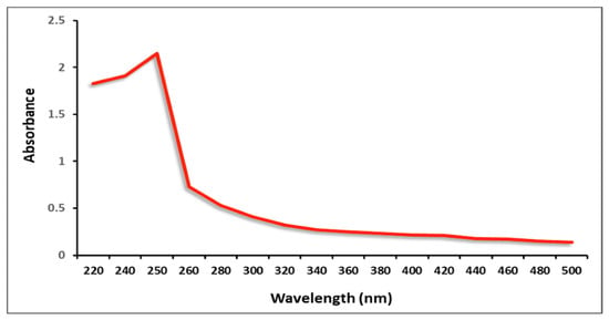
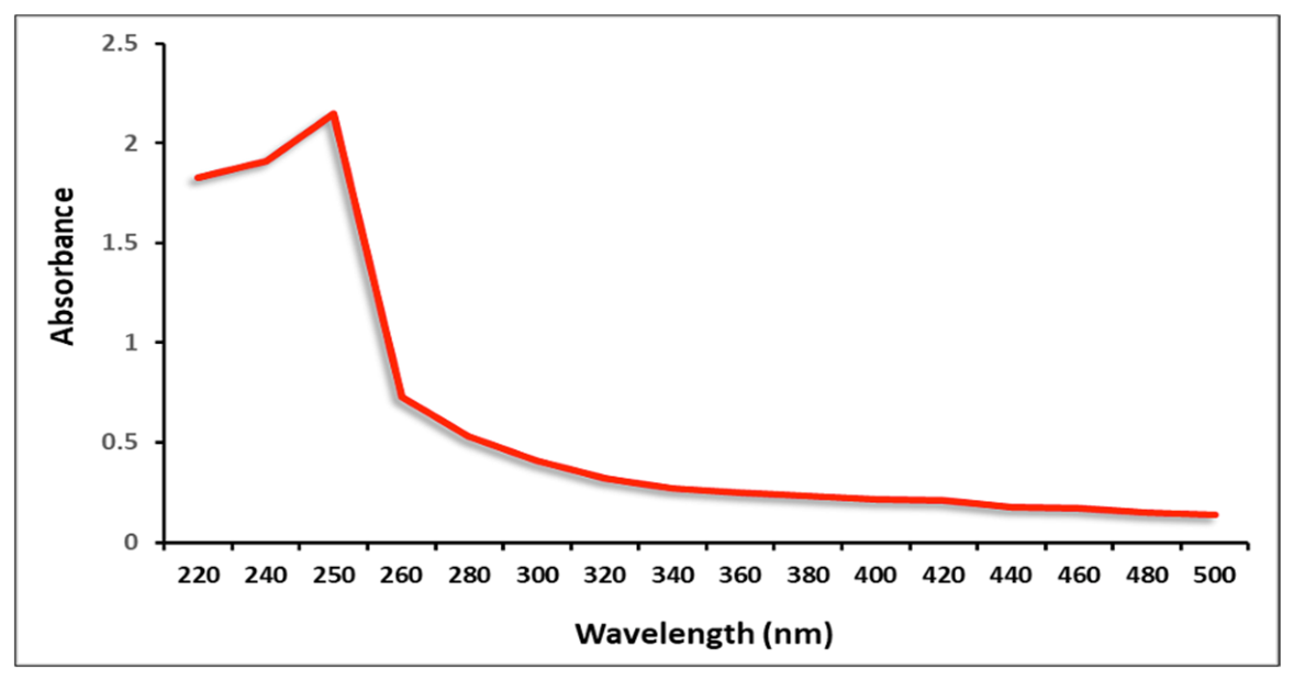
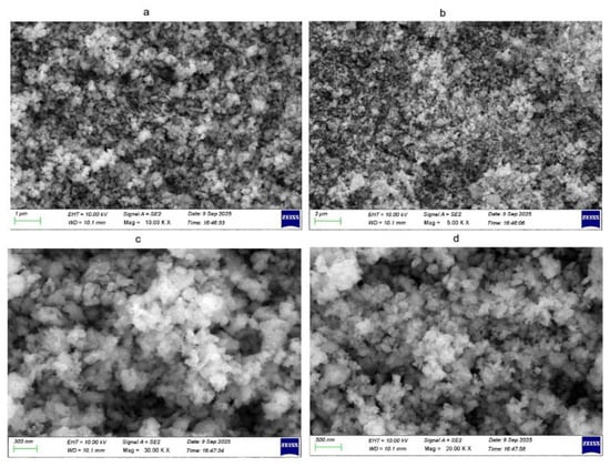
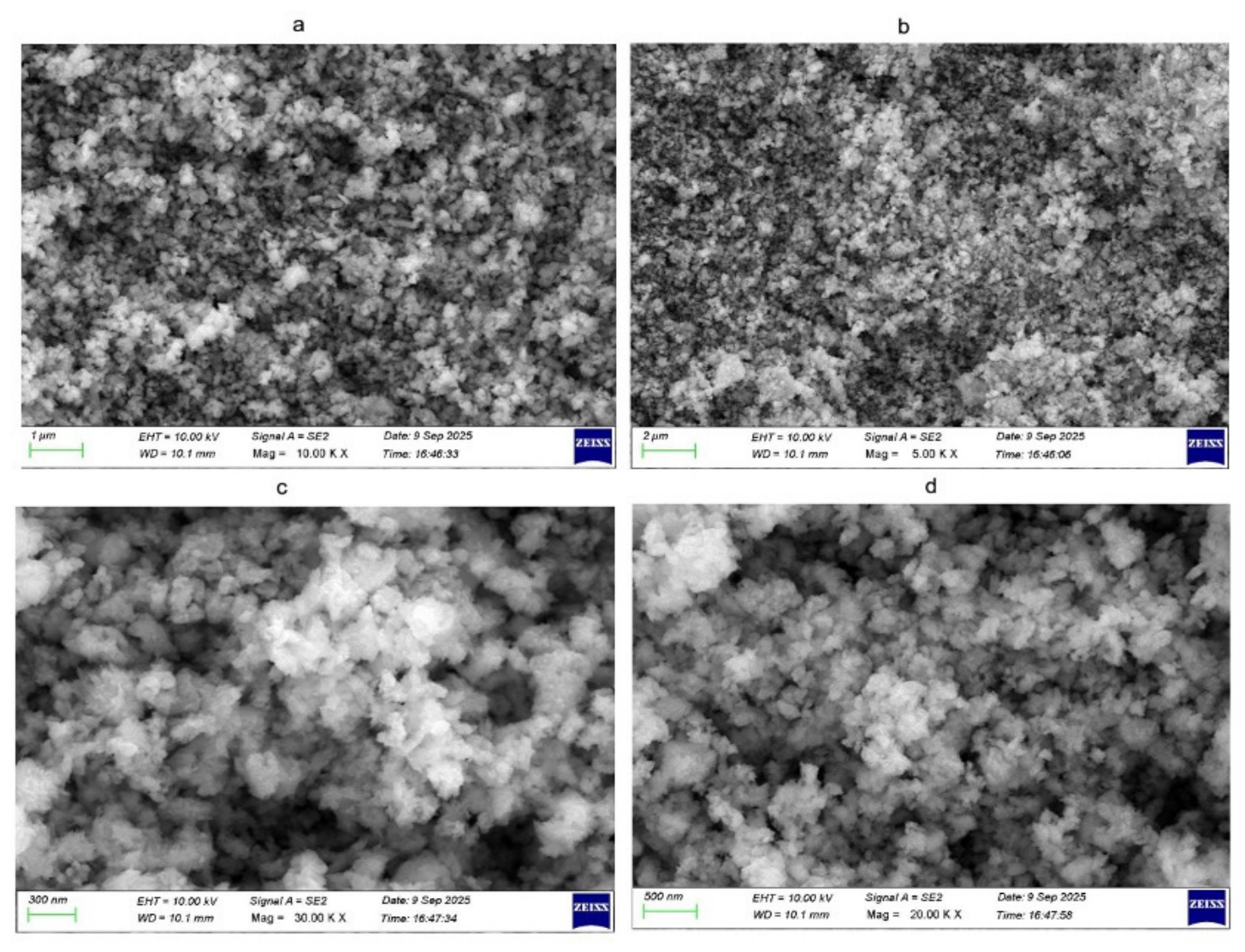
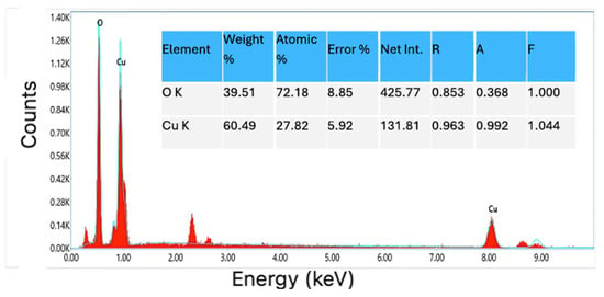
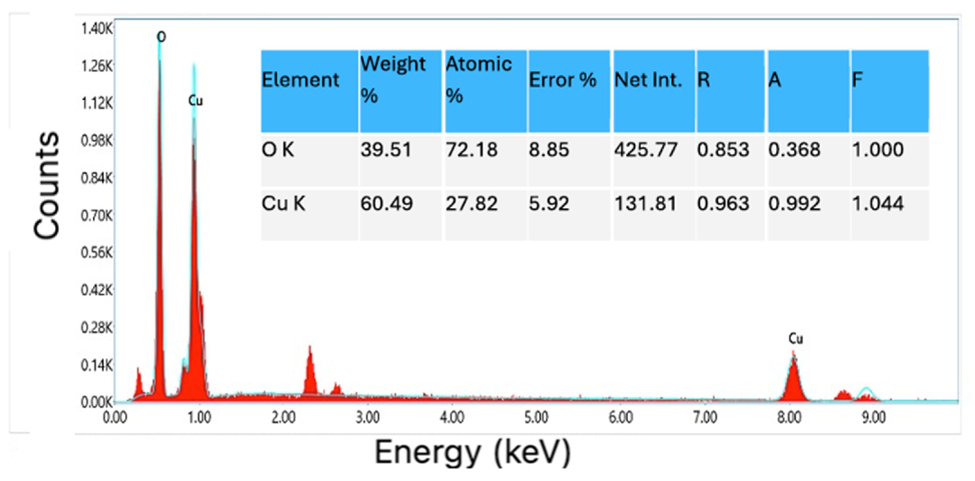
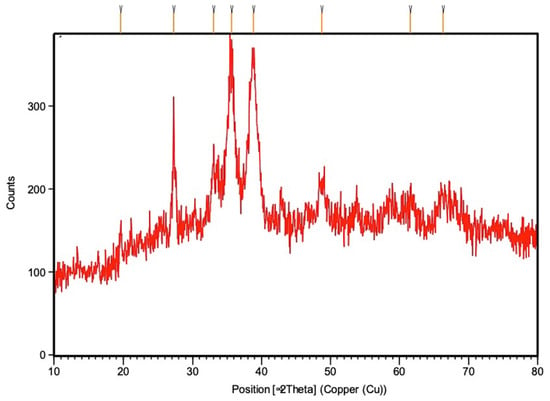
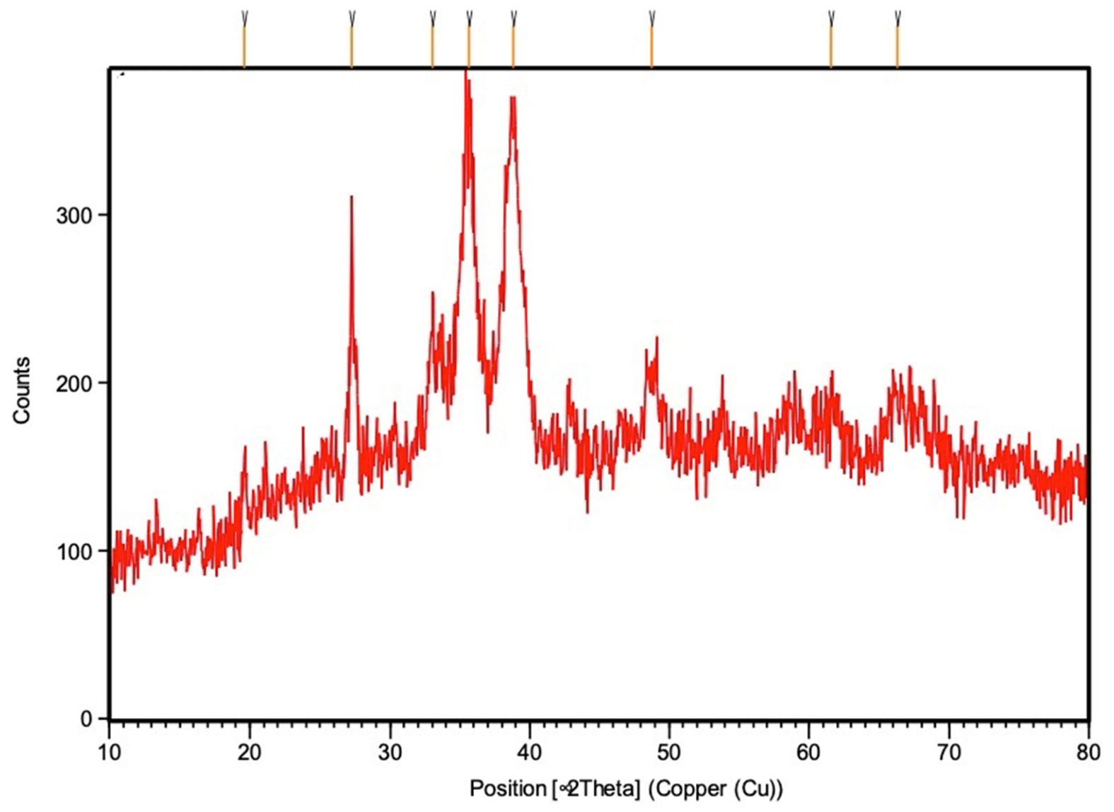
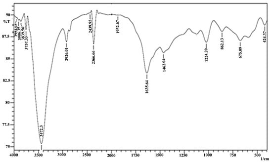
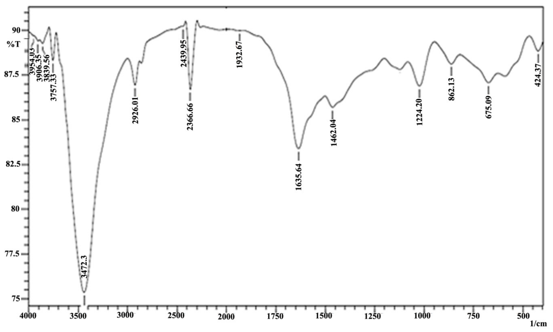
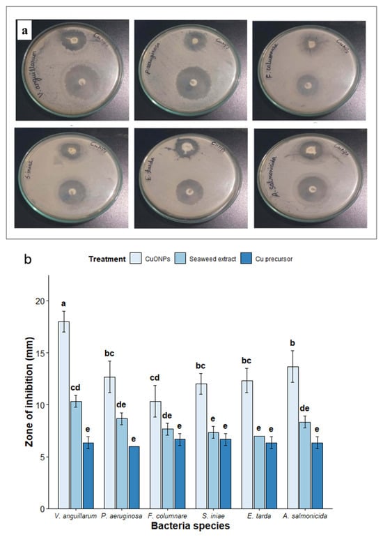
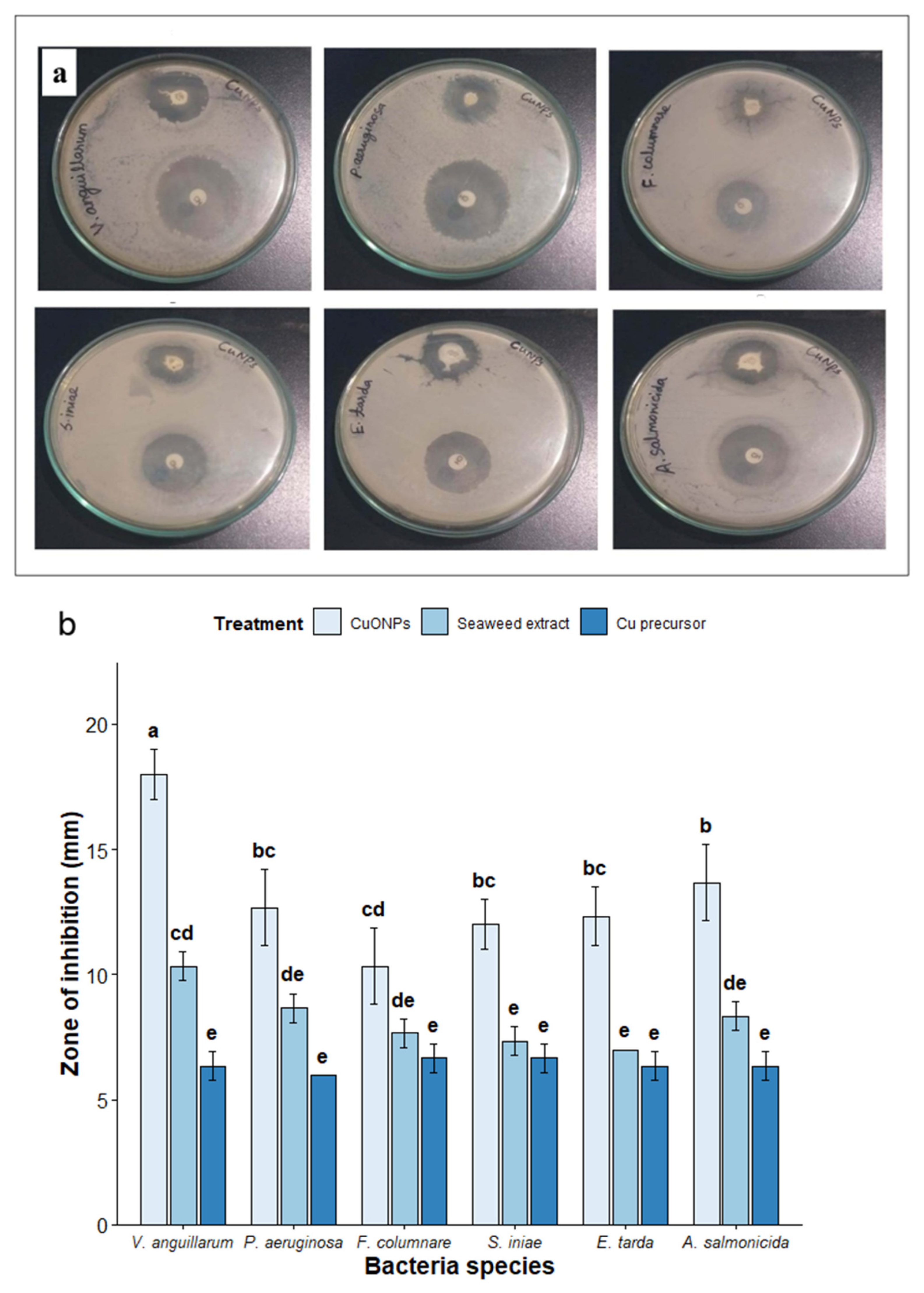
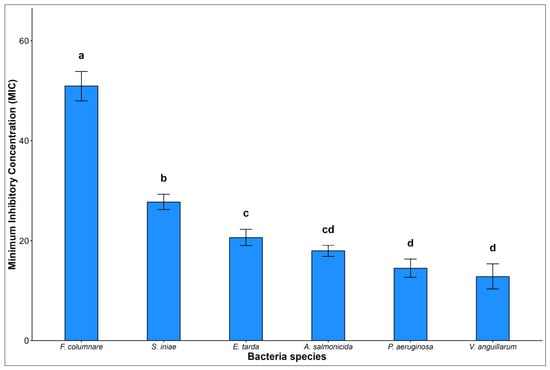
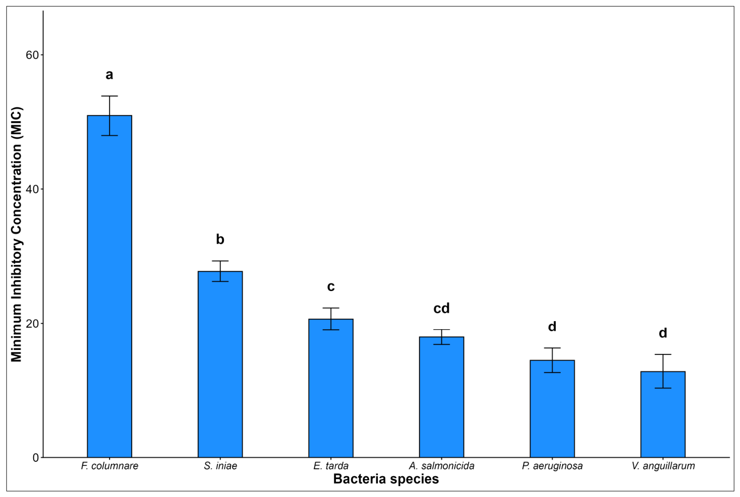
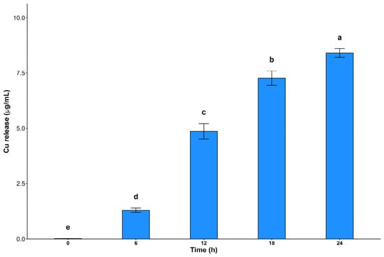
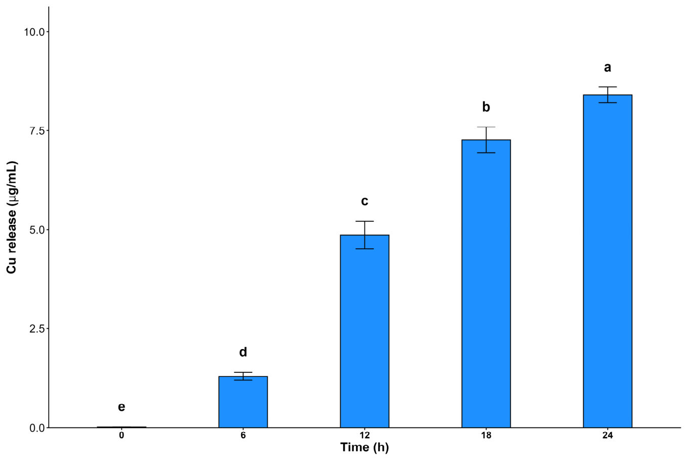
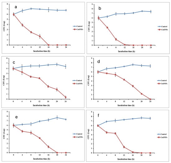
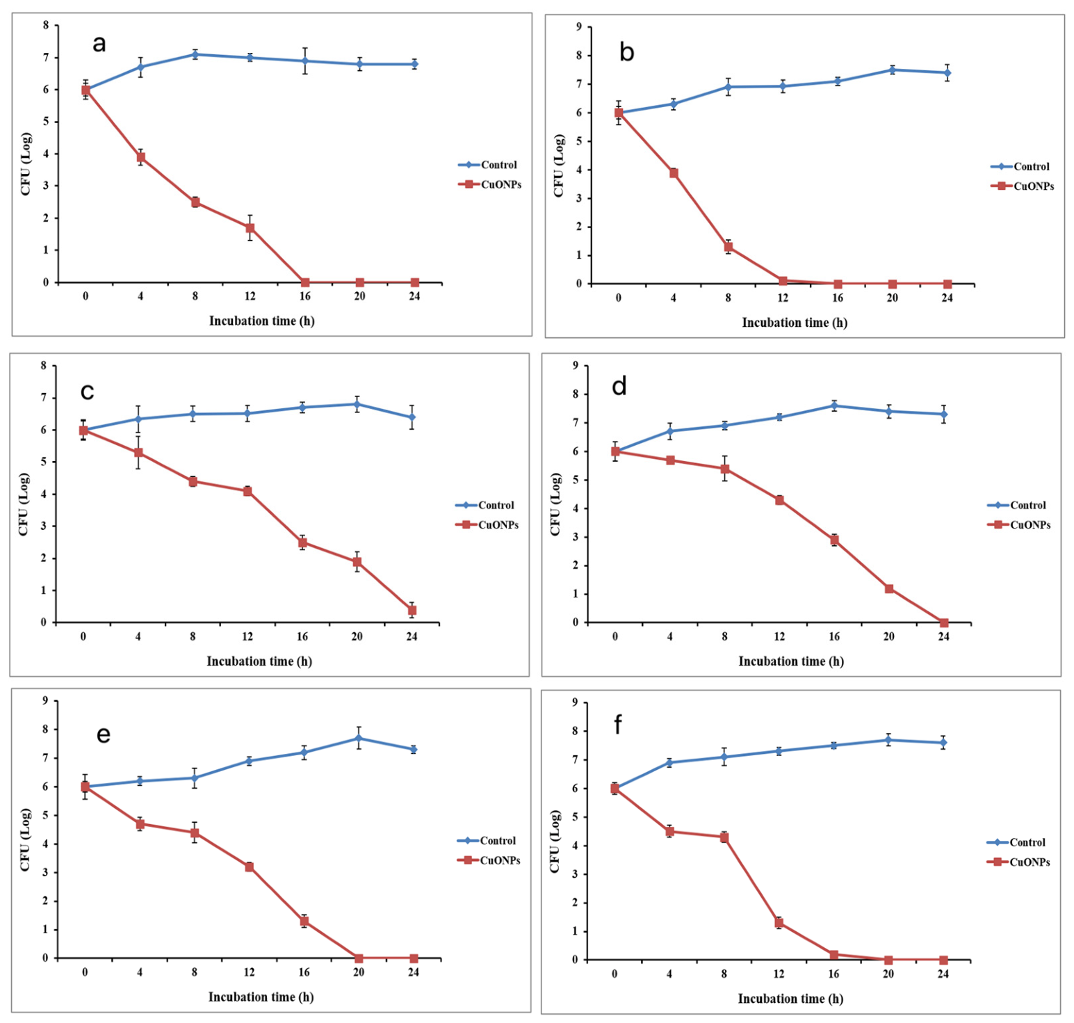
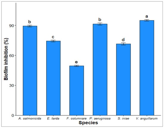
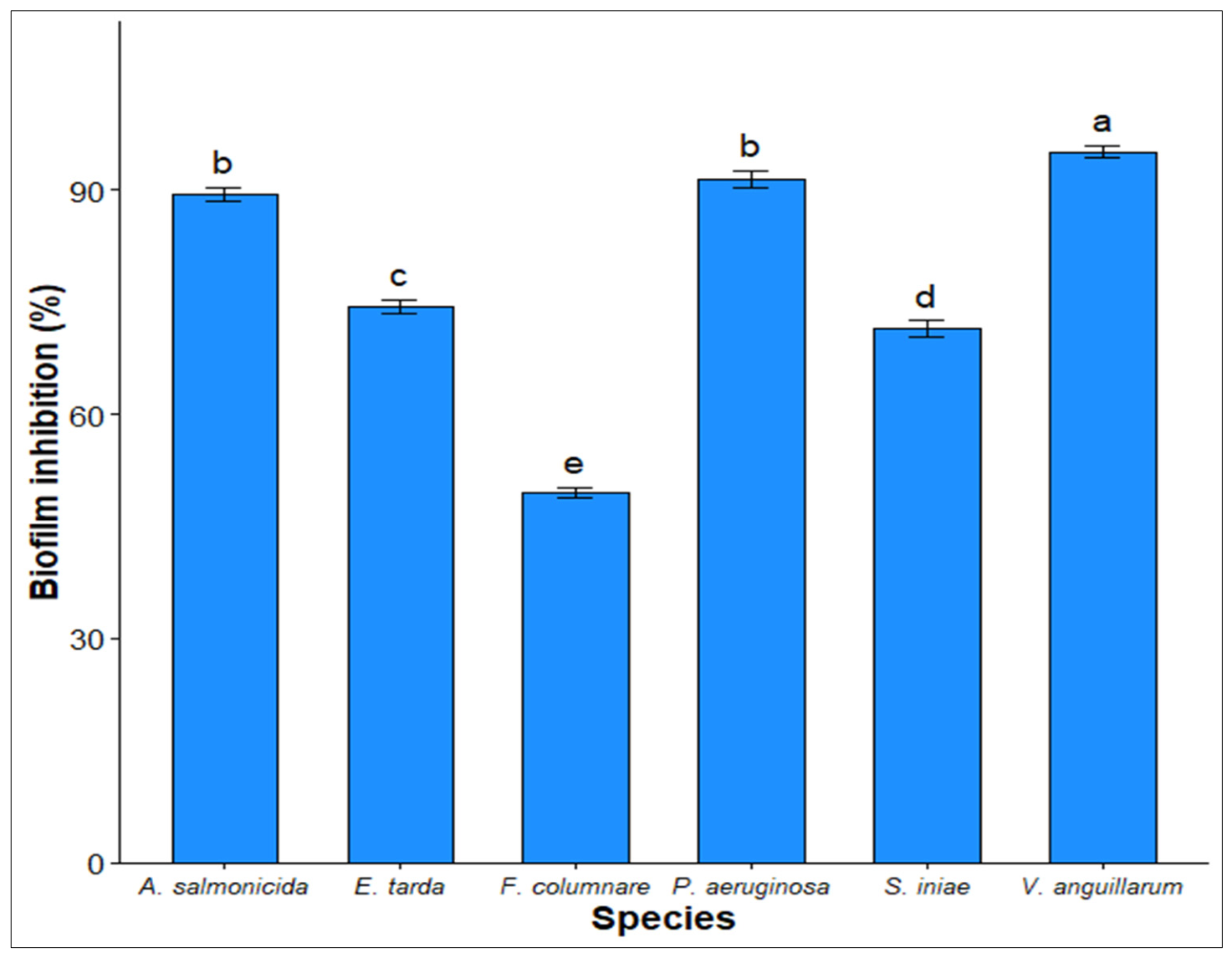

Disclaimer/Publisher’s Note: The statements, opinions and data contained in all publications are solely those of the individual author(s) and contributor(s) and not of MDPI and/or the editor(s). MDPI and/or the editor(s) disclaim responsibility for any injury to people or property resulting from any ideas, methods, instructions or products referred to in the content. |
© 2026 by the authors. Licensee MDPI, Basel, Switzerland. This article is an open access article distributed under the terms and conditions of the Creative Commons Attribution (CC BY) license.