


Abstract
The development of alternative strategies for gluten detoxification represents an important approach for the management of celiac disease and other gluten-related disorders. This study aimed to identify gluten-degrading Bacillus strains with probiotic potential by integrating enzymatic screening with comprehensive biosafety and functional characterization. A total of 45 isolates obtained from diverse ecological niches in Uzbekistan were evaluated for glutenolytic activity using an agar diffusion assay. Fifteen strains demonstrated detectable gluten hydrolysis (16–32 mm). The highest activity was observed in Bacillus cereus isolates (up to 32 mm), whereas Bacillus amyloliquefaciens and Bacillus licheniformis exhibited moderate activity (16–24 mm). However, biosafety profiling revealed pronounced hemolytic, lecithinase, and DNase activities in most B. cereus strains, along with reduced antibiotic susceptibility, which precludes their probiotic application. In contrast, B. amyloliquefaciens and B. licheniformis strains exhibited γ-hemolysis, lacked lecithinase and DNase activities, showed high susceptibility to clinically relevant antibiotics, and demonstrated satisfactory tolerance to simulated gastrointestinal conditions. None of the strains demonstrated detectable antimicrobial activity under standard in vitro conditions. Culture supernatants exhibited pronounced antioxidant activity (73.38–90.34% DPPH radical scavenging), indicating the production of extracellular bioactive metabolites. The results demonstrate functional divergence within the Bacillus genus between maximum glutenolytic capacity and probiotic safety. Species belonging to the Bacillus group, including Bacillus amyloliquefaciens, B. subtilis, and B. licheniformis, are widely recognized as safe for food and probiotic applications (GRAS and/or QPS when appropriately characterized at the strain level). Accordingly, while B. cereus strains represent promising sources of industrial gluten-degrading enzymes. B. amyloliquefaciens 6/4/2, together with selected B. subtilis and B. licheniformis strains, demonstrated the most favorable balance between glutenolytic activity and biosafety, making them the most promising candidates for further probiotic development.
1. Introduction
Wheat is one of the key food crops worldwide and the main source of plant-derived protein in the human diet. Its unique rheological and technological properties are determined by the presence of gluten—a complex protein matrix composed of gliadins and glutenins that form a viscoelastic dough structure and ensure gas retention during baking [1,2]. However, these very proteins represent the principal pathogenic factor in gluten-related disorders, primarily celiac disease.
The key biochemical feature of gluten is the exceptional resistance of its prolamin fractions to proteolytic digestion in the human gastrointestinal tract. Proline- and glutamine-rich peptides are poorly hydrolyzed by pepsin, trypsin, and chymotrypsin, leading to the accumulation of long oligopeptides with pronounced immunogenic activity [3,4]. Of particular clinical relevance is the 33-mer fragment of α-gliadin, which contains multiple overlapping T-cell epitopes efficiently recognized by HLA-DQ2/DQ8–associated T lymphocytes [5]. The initiation of the autoimmune cascade in celiac disease involves deamidation of glutamine residues by tissue transglutaminase, increased intestinal permeability, activation of pro-inflammatory cytokines, and the development of chronic inflammation of the small intestinal mucosa, resulting in villous atrophy, malabsorption syndrome, and systemic metabolic disturbances [6,7]. Thus, celiac disease should be regarded not as a localized gastroenterological disorder but as a systemic immunometabolic pathology.
At present, a strict gluten-free diet remains the only standard therapy for celiac disease. However, even trace amounts of gluten (10–50 mg/day) are sufficient to maintain inflammation and prevent complete regeneration of the intestinal mucosa [8,9]. Moreover, long-term adherence to a gluten-free diet is associated with deficiencies in dietary fiber, B vitamins, iron, magnesium, and zinc, as well as dysbiotic alterations, which significantly reduce nutritional adequacy and quality of life [10,11].
These limitations necessitate the development of alternative strategies aimed not at the complete exclusion of gluten but at its controlled biochemical detoxification. In recent years, microbial approaches to gluten degradation have garnered considerable interest, owing to the use of microorganisms that synthesize specialized proteases capable of degrading immunogenic prolamin epitopes [2,12]. Unlike exogenous enzyme preparations, probiotic microorganisms offer a fundamental advantage: the ability to perform localized and prolonged in situ biotransformation of gluten within the dynamic intestinal environment, along with systemic modulatory effects on the microbiota and immune response [13].
Traditionally, representatives of the genera Lactobacillus and Bifidobacterium have been used as probiotics; however, their limited resistance to acid and bile stress, as well as their relatively low glutenolytic activity, substantially reduce the effectiveness of this strategy [14,15,16]. In this context, bacteria of the genus Bacillus are of particular interest, as they combine spore formation, high stress tolerance, and a highly developed secretory apparatus. Bacillus species synthesize a broad spectrum of extracellular serine and metalloproteases capable of efficiently hydrolyzing complex protein substrates, including the prolamin fractions of gluten [17,18,19].
However, representatives of the genus Bacillus exhibit pronounced interstrain variability in both glutenolytic activity and biosafety profiles. In particular, Bacillus cereus strains demonstrate high enzymatic potential but simultaneously express virulence factors, including hemolysins, enterotoxins, and phospholipases, which severely limit their probiotic application [20,21]. In contrast, B. subtilis and B. licheniformis are recognized by regulatory agencies as conditionally safe microorganisms (QPS/GRAS status), yet their glutenolytic potential remains insufficiently characterized [22,23].
Therefore, this study aimed to develop a comprehensive strategy for the selection of gluten-degrading Bacillus strains based on an integrated assessment of their enzymatic activity, resistance to gastrointestinal conditions, and biosafety indicators. It was hypothesized that natural isolates could yield safe strains combining high glutenolytic activity with pronounced probiotic potential, making them promising candidates for the development of next-generation functional probiotic and synbiotic products.
2. Materials and Methods
2.1. Microorganisms and Cultivation Conditions
A total of 45 Bacillus isolates obtained from diverse ecological niches across Uzbekistan were used in this study, including oil sludge-contaminated soils and industrial wastewater, arid soils from the Kashkadarya, Samarkand, and Tashkent regions, agricultural soils fertilized with poultry manure, and fermented dairy products (kefir and sour cream).
All strains were deposited in the collection of the Enzymology Laboratory, Institute of Microbiology, Academy of Sciences of the Republic of Uzbekistan, and were stored in two forms: lyophilized cultures and frozen suspensions in 10% (v/v) glycerol at −80 °C.
Before experiments, strains were reactivated on LB agar or in tryptic soy broth (TSB) at 30–32 °C for 18–24 h.
2.2. Screening for Glutenolytic Activity
Gluten hydrolysis was assessed using an agar diffusion assay with the following medium composition (g/L): wheat gluten, 5.0; KH2PO4·7H2O, 1.0; Na2HPO4·7H2O, 1.0; NaCl, 5.0; agar, 15.0; distilled water up to 1000 mL; pH 7.0 ± 0.2.
Due to the poor solubility of gluten in water, it was first dispersed in 70% ethanol (5 g in 70 mL) under constant stirring for 15–30 min. The resulting suspension was aseptically added to the sterile medium after autoclaving (121 °C, 15 min), followed by pouring into Petri dishes.
Inoculation was performed by streaking, and plates were incubated at 30–32 °C for 5 days. The formation of clear hydrolysis zones around colonies was recorded daily. The diameters of the hydrolysis zones were measured in millimeters and used as an indicator of relative glutenolytic activity [24]. The assay was performed in triplicate, and the mean hydrolysis zone diameter was used for data analysis.
2.3. Morphological, Cultural, and Physiological–Biochemical Characterization
Strains were cultivated on LB agar and in TSB at 30–32 °C until the stationary growth phase. Cell morphology was examined by light microscopy after Gram staining. Sporulation was assessed using phase-contrast microscopy.
2.4. The Bacterial Isolates Were Initially Identified Using a MALDI-TOF MS Biotyper (Bruker Daltonics, Bremen, Germany)
According to the manufacturer’s instructions. Spectra were analyzed using the Bruker Biotyper database. Identification scores ≥ 2.0 were considered reliable for species-level assignment.
Because MALDI-TOF mass spectrometry has limited resolution for species identification of Bacillus bacteria, the obtained identifications were interpreted in conjunction with the morphological, physiological, and biochemical characteristics described in the Bergey Manual of Systematic Bacteriology. For two strains selected for subsequent biotechnological studies (Bacillus amyloliquefaciens 6/4/2 and Bacillus licheniformis 9/2), species identification was further confirmed by 16S rRNA gene sequencing, and the sequences were deposited in GenBank under accession numbers PV664479 and PQ757915, respectively. The remaining isolates were used only for primary screening and did not undergo additional phylogenetic analyses (e.g., gyrB sequencing or whole-genome sequencing).
Colony morphology (shape, size, color, transparency, and surface characteristics) was evaluated after 24–48 h of incubation on solid media.
Growth at different NaCl concentrations (4–15%) was assessed in TSB diluted 1:1 with distilled water. Temperature tolerance was studied within the range of 20–55 °C using TSB and nutrient agar (HiMedia Laboratories, Mumbai, India). Growth at different pH values (4.0–9.0) was evaluated in TSB adjusted with appropriate buffer systems.
Biochemical characterization was performed using standard microbiological methods, including starch hydrolysis, the Voges–Proskauer reaction, citrate utilization, carbohydrate fermentation, and acid production, following established protocols described in Bergey’s Manual of Systematic Bacteriology [25] and Microbiology: A Laboratory Manual by Cappuccino and Welsh [26]. The biochemical profiles obtained were compared with the diagnostic characteristics reported in Bergey’s Manual of Systematic Bacteriology to support phenotypic identification of the isolates [25].
2.5. Determination of Hemolytic Activity
Hemolytic activity was evaluated by streaking cultures onto blood agar containing 5% defibrinated sheep blood. Plates were incubated at 30–32 °C for 24–48 h. Hemolysis was classified as α-hemolysis (partial), β-hemolysis (complete), or γ-hemolysis (absence of hemolysis) based on visual inspection of zones surrounding the colonies. The assay was performed in triplicate.
2.6. Determination of Lecithinase Activity
Lecithinase activity was assessed in a liquid egg yolk–based medium. Aseptically prepared egg yolk (one egg) was added to 400 mL of sterile meat-peptone broth (MPB), thoroughly mixed, dispensed into tubes, and tested for sterility.
Each tube was inoculated with 24 h bacterial cultures and incubated at 30 ± 2 °C for 24 h. Lecithinase activity was determined by the formation of turbidity or flocculent precipitates. The assay was performed in triplicate.
2.7. Determination of DNase Activity
DNase activity was evaluated on DNase agar. Strains were inoculated by streaking and incubated at 30 ± 2 °C for 24–48 h. After incubation, the agar surface was flooded with 1 N HCl. The presence of clear zones around colonies indicated DNA hydrolysis. The assay was performed in triplicate.
2.8. Assessment of Tolerance to Simulated Gastrointestinal Conditions
Tolerance to simulated gastric conditions was evaluated using a solution containing 0.5% NaCl and 0.3% pepsin at pH 2.0. The medium was sterilized by membrane filtration (0.22 μm).
Cells grown to the logarithmic phase were harvested by centrifugation (7000 rpm, 5 min), washed twice with physiological saline, and resuspended. A 200 µL aliquot of bacterial suspension was added to 1 mL of the simulated gastric solution and incubated at 37 °C. Viability was assessed after 0, 30, 60, and 90 min.
To simulate intestinal conditions, a solution containing 0.1 g pancreatin, 0.3 g bovine bile, and 0.5% NaCl at pH 8.0 was used. Incubation was performed at 37 °C, and viability was determined after 0, 1, and 2 h.
2.9. Antibiotic Susceptibility Testing
Antibiotic susceptibility was determined by the disk diffusion method on Mueller–Hinton agar. Inoculation was performed using the lawn culture technique. Antibiotic disks of standard concentrations (HiMedia Laboratories, Mumbai, India) were placed on the agar surface, and plates were incubated at 30 ± 2 °C for 18–24 h.
Results were evaluated based on inhibition zone diameters (mm). The antibiotic susceptibility assay was performed to compare susceptibility profiles among the environmental Bacillus isolates as an additional biosafety criterion during probiotic strain selection. Susceptibility categories were assigned with reference to CLSI interpretive criteria; however, because incubation was performed at 30 ± 2 °C rather than the CLSI-recommended 35 ± 2 °C, the results should be considered comparative rather than intended for clinical antimicrobial susceptibility classification. All susceptibility tests were performed in triplicate, and inhibition zone diameters were recorded in millimeters [27,28,29].
2.10. Assessment of Antimicrobial Activity
Antimicrobial activity was evaluated using three agar diffusion methods: spot-on-lawn, agar block, and well diffusion assays. Escherichia coli, Bacillus subtilis, Pseudomonas aeruginosa, and Staphylococcus aureus were used as indicator strains.
In all assays, activity was assessed by measuring the diameter of growth inhibition zones after 24 h of incubation at 37 °C. All antimicrobial activity assays were performed in triplicate.
2.11. Assessment of Antioxidant Activity
The antioxidant activity of culture supernatants and intact cells was determined using the DPPH radical scavenging assay. Strains were cultivated in TSB at 30 ± 2 °C for 48 h, followed by centrifugation at 13,000× g for 15 min. The supernatant and cell pellet were separated. Cells were resuspended in phosphate buffer (pH 7.4).
One milliliter of DPPH solution (0.05 mM in ethanol) was mixed with 1 mL of the test sample and incubated in the dark for 60 min. After centrifugation, absorbance was measured at 517 nm. Ascorbic acid (1 mg/mL) served as a positive control.
The DPPH radical scavenging activity was calculated using the following equation:
where Asample is the absorbance of the sample containing DPPH solution, Ablank is the absorbance of the sample without DPPH, and Acontrol is the absorbance of the DPPH solution without the sample.
All measurements were performed in triplicate.
2.12. Experimental Replicates and Data Presentation
Unless otherwise stated, all qualitative screening and biosafety assays were performed in triplicate. Quantitative measurements were carried out in at least three technical replicates, and results are presented as mean ± standard deviation or mean ± standard error, where applicable.
3. Results
A total of 45 bacterial isolates obtained from oil sludge-contaminated soils, industrial wastewater, and fermented dairy products were included in the study. All isolates were initially assigned to the genus Bacillus based on MALDI-TOF mass spectrometry in combination with morphological, cultural, physiological, and biochemical characterization. The 15 isolates exhibiting glutenolytic activity (Table 1) were further assigned to the species level using the same approach.
Table 1.
Selected Bacillus strains after qualitative screening.
Molecular confirmation by 16S rRNA gene sequencing was performed only for the two strains selected for further biotechnological investigation (Bacillus amyloliquefaciens 6/4/2 and Bacillus licheniformis 9/2).
3.1. Screening of Glutenolytic Activity
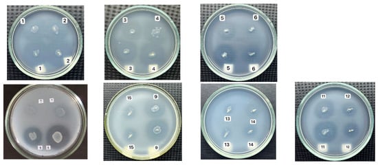
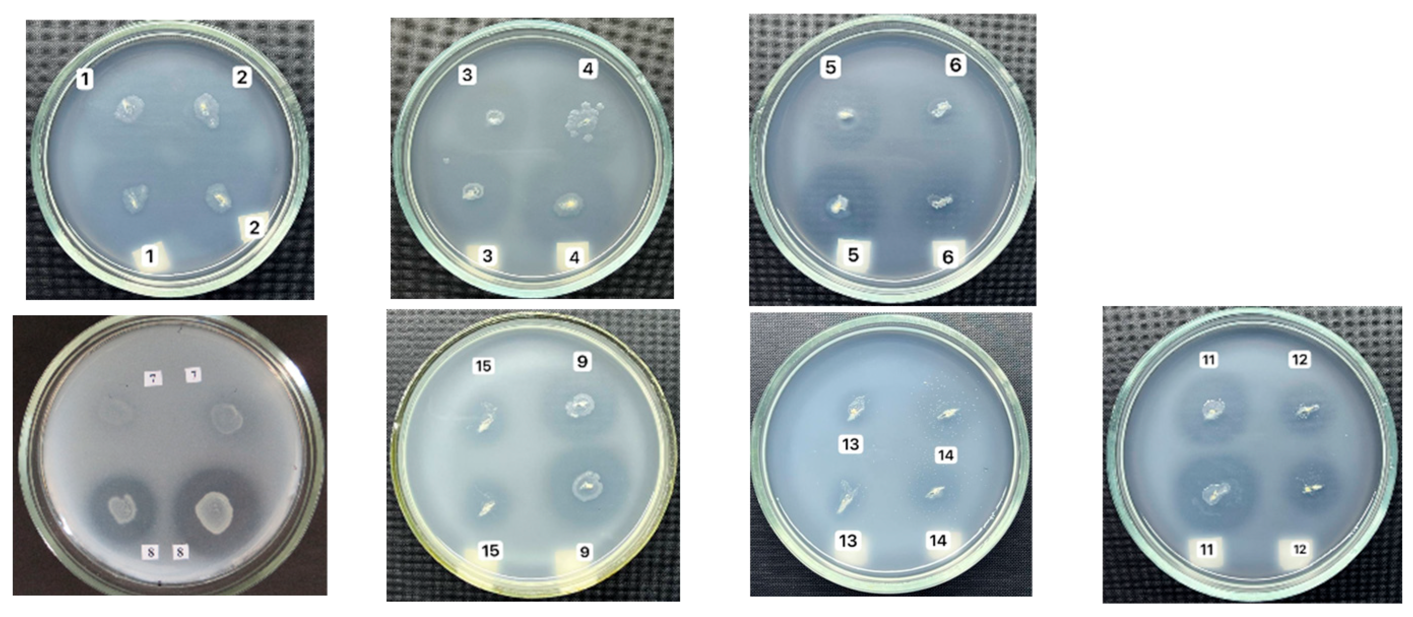
Qualitative screening revealed that 15 of the 45 screened isolates produced distinct hydrolysis zones on gluten-containing agar, indicating detectable extracellular glutenolytic activity (Figure 1, Table 1).
Figure 1.
Representative examples of glutenolytic activity exhibited by selected Bacillus isolates on gluten-containing agar. Extracellular gluten degradation is indicated by the formation of clear hydrolysis zones surrounding bacterial colonies after incubation. The hydrolysis zone diameter was used as a qualitative measure of glutenolytic activity during the primary screening of isolates. Reference strains are not shown because this figure is intended to illustrate representative examples of gluten hydrolysis among the screened environmental isolates rather than provide a comparative analysis of all tested strains and controls.
The diameter of the gluten hydrolysis zones ranged from 16 to 32 mm. Bacillus cereus isolates exhibited the highest overall glutenolytic activity (mean ± SD, 24.7 ± 5.4 mm), followed by B. amyloliquefaciens (27 mm; one isolate), B. subtilis (19.2 ± 3.0 mm), and B. licheniformis (19.5 ± 5.0 mm).
The largest hydrolysis zones were observed for B. cereus 2B (32 mm) and B. cereus 2/1/2 (30 mm), suggesting high extracellular proteolytic activity, consistent with previous reports describing the strong protease-producing capacity of Bacillus cereus strains [30]. Moderate activity (20–27 mm) was detected in B. amyloliquefaciens 6/4/2, B. subtilis 5B and 24, B. cereus 48 and Rp1-Ch, and B. licheniformis 9/2. The smallest hydrolysis zones (<19 mm) were observed in B. cereus NB and Rp1-M, and B. subtilis DK, CB1, and CB2. Although B. cereus 2B and B. cereus 2/1/2 exhibited the greatest gluten hydrolysis during the primary qualitative screening, strain selection for subsequent probiotic evaluation was based not only on glutenolytic activity but also on biosafety characteristics, including hemolytic, lecithinase, DNase, and antibiotic susceptibility profiles.
Species-level analysis showed that B. cereus exhibited the widest variability in glutenolytic activity (16–32 mm), whereas B. subtilis and B. licheniformis demonstrated narrower activity ranges (16–24 mm and 16–23 mm, respectively). The sole B. amyloliquefaciens isolate (6/4/2) exhibited high activity (27 mm), consistent with previous reports describing a highly developed secretory protease system in this species.
It should be noted that the agar diffusion assay reflects overall proteolytic activity toward gluten but does not permit direct evaluation of the degradation of immunogenic gluten epitopes responsible for the pathogenesis of celiac disease. Therefore, the results obtained should be regarded as preliminary and suitable for the primary screening of candidate glutenase-producing strains before detailed enzymatic characterization and quantitative assessment of gluten detoxification.
3.2. Evaluation of Safety Factors
Hemolysis, Lecithinase, and DNase Activity. Assessment of virulence-associated factors is a critical step in selecting probiotic candidates. Therefore, hemolytic, lecithinase, and Dnase activities were analyzed (Table 2).
Table 2.
Hemolytic, Dnase, and lecithinase activities of selected Bacillus strains.
B. subtilis (24, DK, CB1, CB2) and B. licheniformis (9/2, B1) exhibited γ-hemolysis, indicating the absence of hemolytic activity and confirming their favorable biosafety profiles. In contrast, most B. cereus isolates displayed α-hemolysis, while strains Rp1-M and Rp1-Ch exhibited pronounced β-hemolysis, reflecting cytotoxic potential.
Lecithinase activity was predominantly observed in B. cereus, consistent with phospholipase C being a major virulence factor responsible for host membrane disruption. Conversely, B. subtilis and B. licheniformis strains lacked lecithinase activity.
Dnase activity was detected predominantly among B. cereus strains, including isolates 2/1/2, 2B, 4B, Rp1-M, Rp1-Ch, and NB, consistent with the established role of extracellular nucleases in invasion and immune evasion. The absence of Dnase activity in B. subtilis, B. amyloliquefaciens, and B. licheniformis constitutes an additional biosafety criterion during probiotic strain selection.
Collectively, these findings indicate that although B. cereus strains possess high glutenolytic potential, their virulence-associated profiles preclude their probiotic use but render them suitable candidates for industrial enzyme production. Among the evaluated isolates, Bacillus amyloliquefaciens 6/4/2 represented the most favorable compromise between glutenolytic activity and biosafety, making it the principal candidate for further probiotic development. Selected B. subtilis and B. licheniformis isolates also exhibited favorable biosafety characteristics but showed lower glutenolytic activity.
The evaluation of these virulence-associated traits was performed as part of the biosafety assessment of candidate probiotic strains. Hemolytic activity, lecithinase production, and Dnase activity are widely recognized as indicators of pathogenic potential in Bacillus species. Therefore, the absence of these activities supports the biosafety profile of candidate probiotic strains, whereas their presence constitutes an important exclusion criterion for further probiotic development [31].
3.3. Tolerance to Simulated Gastrointestinal Conditions
Tolerance to simulated gastric (pH 2.0) and intestinal (pH 8.0) conditions is considered an essential characteristic of candidate probiotic strains. Cell viability was estimated spectrophotometrically (OD600) and expressed as estimated cell density (approximate CFU/mL based on OD calibration).
Exposure to simulated gastric conditions (pH 2.0) for 30 and 60 min resulted in a marked reduction in cell viability across all tested strains (Table 3), reflecting the sensitivity of vegetative Bacillus cells to acidic stress. Nevertheless, B. subtilis DK exhibited the highest tolerance after 60 min of exposure, maintaining an estimated cell density of approximately 7.2 × 10⁷ CFU/mL. This enhanced acid tolerance may be associated with strain-specific physiological characteristics, including increased sporulation capacity and more efficient acid stress response mechanisms.
Table 3.
Survival of selected Bacillus strains under simulated gastric (pH 2.0) and intestinal (pH 8.0) conditions estimated by optical density (OD600).
Under simulated intestinal conditions (pH 8.0), all tested strains maintained relatively high viability, with estimated cell densities ranging from 1.7 × 10⁸ to 2.5 × 10⁸ CFU/mL after 60 min of exposure (Table 3), indicating good adaptation to alkaline environments comparable to intestinal conditions. These findings are consistent with previous studies reporting the high tolerance of Bacillus spp. To alkaline stress and their ability to maintain metabolic activity within the pH range of 7.0–8.5.
It should be noted that the viability estimates presented in Table 3 are based on optical density measurements and should therefore be regarded as approximate. Optical density does not distinguish between vegetative cells and spores and may also be influenced by cellular aggregation and morphological changes. Nevertheless, this approach provides a reliable comparative assessment of strain survival under simulated gastrointestinal conditions.
3.4. Antibiotic Susceptibility Profiles
Antibiotic susceptibility testing revealed pronounced interspecies and strain-specific variability. B. subtilis and B. licheniformis strains exhibited comparatively larger inhibition zones for most tested antibiotics than the B. cereus isolates, indicating a more favorable antibiotic susceptibility profile as an additional biosafety characteristic. Because susceptibility testing was performed at 30 ± 2 °C, these results should be interpreted as comparative rather than as clinical susceptibility classifications.
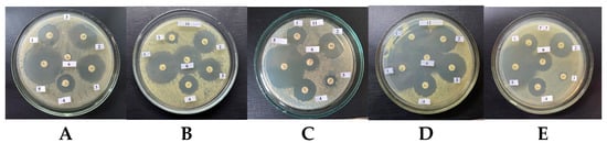
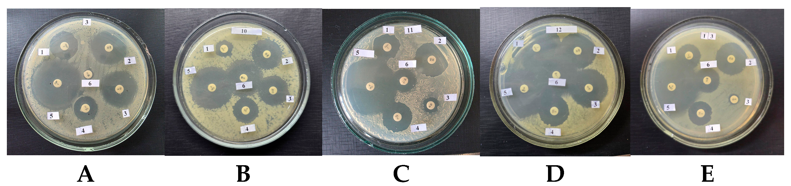
In contrast, Bacillus cereus isolates demonstrated reduced susceptibility to the tested β-lactam (ampicillin) and glycopeptide (vancomycin) antibiotics, likely owing to intrinsic β-lactamase activity and structural features of the cell wall. These characteristics constitute a major limitation for their use as probiotics (Figure 2).
Figure 2.
Representative antibiotic susceptibility profiles of selected Bacillus strains were determined using the disk diffusion method. (A) Bacillus amyloliquefaciens 6/4/2; (B) B. cereus NB; (C) B. licheniformis 9/2; (D) B. subtilis DK; (E) B. licheniformis B1. Antibiotic discs: 1, Ampicillin (AMP); 2, Amikacin (AK); 3, Erythromycin (ERY); 4, Vancomycin (VA); 5, Ciprofloxacin (CIP); 6, Chloramphenicol (CHL).
Notably, B. amyloliquefaciens 6/4/2 exhibited resistance to chloramphenicol. Although the genetic basis of this phenotype was not investigated in the present study, further molecular characterization will be required to determine whether it is associated with acquired and transferable antimicrobial resistance determinants. According to current EFSA recommendations, probiotic strains intended for commercial application should not harbor acquired transferable antimicrobial resistance genes (Table 4).
Table 4.
Antibiotic susceptibility of Bacillus strains.
Overall, based on antibiotic resistance profiles, the most promising probiotic candidates were B. subtilis strains 24, DK, CB1, CB2, and B. licheniformis 9/2.
3.5. Antimicrobial Activity
None of the tested strains exhibited antagonistic activity against E. coli, P. aeruginosa, S. aureus, or Candida albicans in any of the diffusion-based assays.
The absence of antimicrobial effects may be attributed to low expression levels of bacteriocin genes under standard cultivation conditions. It is well established that bacteriocin synthesis in Bacillus is induced by stress signals, nutrient limitation, or quorum-sensing mechanisms. Thus, negative in vitro results do not exclude the presence of latent antagonistic potential.
3.6. Antioxidant Activity
The DPPH radical scavenging assay demonstrated high antioxidant activity of culture supernatants (73.38–90.34%), comparable to that of ascorbic acid (94.31%). In contrast, intact cells exhibited markedly lower activity (10.45–32.45%), indicating that extracellular metabolites play the dominant role in antioxidant effects (Table 5).
Table 5.
DPPH radical scavenging activity of selected strains (%). (Mean ± SE, n ≥ 3).
Pronounced interstrain variability highlights metabolic diversity among Bacillus spp. and supports their potential as producers of natural antioxidants. However, considering the primary objectives of this study, antioxidant capacity should be regarded as an auxiliary functional trait rather than a decisive criterion for probiotic positioning.
4. Discussion
The present study implements an integrative approach to the selection of gluten-degrading probiotic Bacillus strains, combining primary enzymatic screening with comprehensive biosafety assessment and evaluation of physiological robustness. Unlike most previous studies focused primarily on proteolytic activity, we aimed to establish a functional biotechnological framework oriented toward the practical realization of probiotic-mediated gluten detoxification.
Our results revealed a fundamental divergence between maximal glutenolytic activity and probiotic suitability. Bacillus cereus strains exhibited the highest levels of gluten hydrolysis (up to 32 mm), reflecting their highly developed extracellular protease systems. However, the simultaneous presence of β-hemolytic activity, lecithinase, and DNase production, together with reduced susceptibility to clinically relevant antibiotics, unequivocally excludes their application as probiotic microorganisms.
Therefore, despite their high glutenolytic activity, the B. cereus isolates were excluded from further consideration as live probiotic candidates and may instead be regarded as potential producers of gluten-degrading enzymes.
Our findings agree with previous reports demonstrating that Bacillus cereus possesses highly active extracellular protease systems capable of degrading complex protein substrates. Nevertheless, the well-documented production of enterotoxins, hemolysins, phospholipases, and other virulence factors makes this species unsuitable for probiotic use despite its high enzymatic potential [32,33]. Therefore, our results support the current consensus that B. cereus should be considered primarily as a source of industrial enzymes rather than as a live probiotic microorganism.
In contrast, B. amyloliquefaciens 6/4/2 was selected for further investigation because it combined high glutenolytic activity with a favorable biosafety profile.
Candidate probiotic strains in the present study were selected not only according to their gluten-degrading activity but also on the basis of a comprehensive set of functional and biosafety criteria. Potential candidates were required to exhibit efficient gluten degradation, absence of major virulence factors, including hemolytic activity, lecithinase, and DNase production, a comparatively favorable antibiotic susceptibility profile, and resistance to simulated gastrointestinal conditions. These criteria are consistent with current recommendations for the evaluation of Bacillus probiotics, according to which biosafety assessment should precede functional characterization [34]. Although these criteria are widely accepted for the preliminary screening of candidate probiotics, confirmation of probiotic functionality requires additional strain-specific investigations, including adhesion assays, immunomodulatory studies, and in vivo validation.
Among all tested isolates, B. amyloliquefaciens 6/4/2 emerged as the principal probiotic candidate, whereas selected B. subtilis and B. licheniformis strains also demonstrated favorable characteristics and may be considered complementary candidates for further investigation. Together, these strains demonstrated the most balanced combination of glutenolytic activity, physiological robustness, and biosafety. Although their native glutenolytic activity was lower than that of B. cereus, their favorable safety profiles make them considerably more suitable for further development as probiotics.
It should also be noted that antibiotic susceptibility testing was performed at a non-standard incubation temperature (30 ± 2 °C). Therefore, the obtained results should be interpreted as a comparative biosafety assessment rather than a clinical classification of antimicrobial susceptibility.
Our findings are consistent with previous reports describing B. subtilis and B. licheniformis as promising probiotic species because of their ability to survive gastrointestinal passage, produce extracellular enzymes, and maintain favorable safety characteristics. Similarly, B. amyloliquefaciens has attracted increasing attention as a producer of industrially important proteases and as a potential probiotic owing to its high secretory capacity and low pathogenic potential [35]. The present study expands these observations by demonstrating that probiotic suitability is determined not by maximal enzymatic activity alone but by an optimal balance between functional efficacy and biosafety.
Compared with classical probiotic genera such as Lactobacillus, Bifidobacterium, and Streptococcus, members of the genus Bacillus constitute a distinct group of spore-forming probiotic candidates. Unlike lactic acid bacteria, which exert their beneficial effects primarily through metabolic interactions with the intestinal microbiota and transient colonization of the intestinal mucosa, Bacillus species produce endospores that provide exceptional resistance to food-processing conditions, long-term storage, gastric acidity, bile salts, and other environmental stresses. These characteristics make Bacillus particularly attractive as carriers of biologically active enzymes, including gluten-degrading proteases. Nevertheless, the probiotic efficacy of Bacillus strains cannot be inferred solely from these technological advantages. Properties such as epithelial adhesion, mucin binding, competitive exclusion of pathogens, and immunomodulatory activity remain strain-specific characteristics that require dedicated investigation. Because these properties were not evaluated in the present study, no conclusions can be drawn regarding the capacity of the selected isolates to adhere to the intestinal epithelium or intestinal mucus. Therefore, the selected strains should presently be regarded as promising probiotic candidates rather than fully validated probiotics [36,37].
Despite the recognized probiotic potential of several Bacillus species, their application requires careful strain-specific safety assessment. The genus comprises both well-established probiotic strains and opportunistic pathogens, and individual isolates may differ considerably in their capacity to produce toxins, harbor transferable antibiotic resistance determinants, or express other virulence-associated traits. Consequently, phenotypic biosafety evaluation should ideally be complemented by whole-genome sequencing to confirm the absence of toxin genes, mobile antimicrobial resistance elements, and other undesirable genetic determinants before commercial or clinical application. These considerations further emphasize that probiotic safety cannot be generalized at the species level but must be demonstrated individually for each candidate strain [20,34]. In this context, whole-genome sequencing of our principal candidate strain, Bacillus amyloliquefaciens 6/4/2, has recently been completed. The genomic data are beyond the scope of the present study and will be reported separately, providing additional support for the comprehensive safety evaluation of this strain.
Collectively, these observations indicate that probiotic suitability depends on the integration of functional performance with biosafety rather than on glutenolytic activity alone.
Although our study did not include in vivo validation, the selected strains fulfilled the principal in vitro criteria currently recommended for candidate Bacillus probiotics, including biosafety, gastrointestinal tolerance, and functional activity. Therefore, their probiotic potential should be regarded as preliminary but well supported by established selection criteria and consistent with previous reports describing probiotic representatives of B. subtilis, B. licheniformis, and B. amyloliquefaciens.
Despite their moderate glutenolytic activity, these strains represent an optimal compromise between functional efficacy and regulatory acceptability, rendering them promising probiotic candidates. At present, however, our data do not demonstrate that the native glutenolytic activity of these strains is sufficient for complete detoxification of dietary gluten in vivo. Therefore, optimization of enzyme production and validation under physiological conditions will be essential to determine their practical efficacy. Their clinical applicability requires further confirmation in animal models and human intervention studies.
From an applied biotechnology perspective, the obtained results support the development of a dual-component strategy for gluten detoxification, comprising: (i) the use of safe probiotic strains represented by B. amyloliquefaciens 6/4/2 together with selected B. subtilis and B. licheniformis isolates; and (ii) the application of highly active enzyme preparations derived from B. cereus as technological aids or food-processing enzymes. An alternative and particularly promising direction involve the engineering of recombinant probiotic platforms expressing gluten-degrading proteases from B. cereus in safe genetic backgrounds of B. subtilis. This strategy enables the integration of superior catalytic efficiency with an established biosafety profile, thus overcoming the intrinsic limitations of native strains.
The principal limitation of the present study is the use of qualitative methods for evaluating gluten degradation, which do not allow quantitative assessment of the destruction of immunodominant epitopes, particularly the pathogenic 33-mer α-gliadin peptide. Furthermore, partial gluten hydrolysis may generate shorter peptides that retain immunogenic epitopes. Consequently, the qualitative assays employed in this study cannot be interpreted as evidence of complete gluten detoxification or complete elimination of celiac disease-inducing peptides. These limitations emphasize the need for further investigations using high-resolution analytical techniques, including HPLC, LC-MS/MS, and immunological assays specifically targeting immunogenic gluten epitopes [38,39].
An additional limitation is that species identification of the entire collection of isolates was performed using a polyphasic approach combining MALDI-TOF MS and phenotypic characterization, whereas only the two strains selected for further biotechnological development were additionally confirmed by 16S rRNA gene sequencing. Future studies should incorporate higher-resolution phylogenetic approaches, such as gyrB gene analysis or whole-genome sequencing, particularly for members of the B. cereus and B. subtilis species complexes.
Overall, the present study demonstrates that the successful development of gluten-degrading probiotics requires simultaneous consideration of enzymatic performance, biosafety, and physiological robustness. The integrative selection strategy proposed here provides a practical framework for the development of safe probiotic strains and enzyme-based technologies for future gluten detoxification applications.
5. Conclusions
This study establishes a systematic framework for the identification and functional evaluation of Bacillus strains with potential probiotic application for gluten detoxification. The integration of glutenolytic activity screening, assessment of gastrointestinal stress tolerance, and biosafety profiling demonstrated that probiotic suitability is determined not solely by maximal proteolytic capacity but by an optimal balance between functional efficacy and biosafety.
Among the evaluated isolates, B. amyloliquefaciens 6/4/2, together with selected B. subtilis and B. licheniformis strains, exhibited the most favorable combination of glutenolytic activity and biosafety characteristics, making them the most promising candidates for further probiotic development. In contrast, although B. cereus isolates showed the highest glutenolytic activity, the presence of virulence-associated traits precluded their use as live probiotic candidates but highlights their potential as sources of industrial gluten-degrading enzymes.
These findings provide a scientific basis for the development of next-generation enzymatic probiotics and gluten-reduced functional foods. However, the proposed mechanism of action and the therapeutic potential of the selected strains remain hypothetical and require confirmation through quantitative gluten degradation assays, in vivo studies, and clinical trials before practical application.
Author Contributions
Conceptualization, A.B. (Alimova Barno), A.M. and S.M.; methodology, A.B. (Alimova Barno) and A.M.; validation, A.B. (Abdraimova Barno), M.S. and A.D.; formal analysis, A.B. (Abdraimova Barno); investigation, S.I., M.M., B.G., A.A., P.O., M.S. and A.D.; resources, A.B. (Alimova Barno); data curation, A.B. (Alimova Barno), E.N. and P.O.; writing—original draft preparation, A.B. (Alimova Barno) and B.G.; writing—review and editing, A.B. (Alimova Barno), B.G., P.O. and S.M.; visualization, A.B. (Alimova Barno) and M.M.; supervision, A.B. (Alimova Barno); project administration, A.B. (Alimova Barno) and B.G. All authors have read and agreed to the published version of the manuscript.
Funding
This work was supported by a fundamental research project of the Laboratory of Enzymology, Institute of Microbiology, Academy of Sciences of the Republic of Uzbekistan, and by the Ministry of Higher Education, Science and Innovation of the Republic of Uzbekistan under Grant No. AL-9224104369, entitled “Isolation of Proteases from Microorganisms and Development of Their Applications in Dairy and Bakery Production”.
Institutional Review Board Statement
Not applicable.
Informed Consent Statement
Not applicable.
Data Availability Statement
The data presented in this study are available from the corresponding author upon reasonable request.
Conflicts of Interest
The authors declare no conflict of interest.
References
- Khalid, A.; Hameed, A.; Tahir, M.F. Wheat quality: A review on chemical composition, nutritional attributes, grain anatomy, types, classification, and function of seed storage proteins in bread-making quality. Front. Nutr. 2023, 10, 1053196. [Google Scholar] [CrossRef] [Scilit] [PubMed]
- Bradauskiene, V.; Vaiciulyte-Funk, L.; Martinaitiene, D.; Andruskiene, J.; Verma, A.K.; Lima, J.P.M.; Serin, Y.; Catassi, C. Wheat consumption and prevalence of celiac disease: Correlation from a multilevel analysis. Crit. Rev. Food Sci. Nutr. 2023, 63, 18–32. [Google Scholar] [CrossRef] [Scilit] [PubMed]
- Kõiv, V.; Tenson, T. Gluten-degrading bacteria: Availability and applications. Appl. Microbiol. Biotechnol. 2021, 105, 3045–3059. [Google Scholar] [CrossRef] [Scilit] [PubMed]
- Wei, G.; Helmerhorst, E.J.; Darwish, G.; Blumenkranz, G.; Schuppan, D. Gluten Degrading Enzymes for Treatment of Celiac Disease. Nutrients 2020, 12, 2095. [Google Scholar] [CrossRef] [Scilit] [PubMed]
- Russell, L.A.; Alliston, P.; Armstrong, D.; Verdu, E.F.; Moayyedi, P.; Pinto-Sanchez, M.I. Micronutrient deficiencies associated with a gluten-free diet in patients with celiac disease and non-celiac gluten or wheat sensitivity: A systematic review and meta-analysis. J. Clin. Med. 2025, 14, 4848. [Google Scholar] [CrossRef] [Scilit] [PubMed]
- Sharma, N.; Bhatia, S.; Chunduri, V.; Kaur, S.; Sharma, S.; Kapoor, P.; Kumari, A.; Garg, M. Pathogenesis of Celiac Disease and Other Gluten-Related Disorders in Wheat and Strategies for Mitigating Them. Front. Nutr. 2020, 7, 6. [Google Scholar] [CrossRef] [Scilit] [PubMed]
- Parzanese, I.; Qehajaj, D.; Patrinicola, F.; Aralica, M.; Chiriva-Internati, M.; Stifter, S.; Elli, L.; Grizzi, F. Celiac disease: From pathophysiology to treatment. World J. Gastrointest. Pathophysiol. 2017, 8, 27–38. [Google Scholar] [CrossRef] [Scilit] [PubMed]
- Kounatidis, D.; Pavlou, A.; Evangelopoulos, A.; Psaroudaki, M.; Kotsi, E.; Petrakou, I.; Paraskevopoulos, P.; Stamatopoulos, V.; Mylona, E.; Vallianou, N.G. Rethinking Celiac Disease Management: Treatment Approaches Beyond the Gluten-Free Diet. Biomedicines 2026, 14, 29. [Google Scholar] [CrossRef] [Scilit] [PubMed]
- Herrera, M.G.; Dodero, V.I. Gliadin proteolytically resistant peptides: The interplay between structure and self-assembly in gluten-related disorders. Biophys. Rev. 2021, 13, 1147–1154. [Google Scholar] [CrossRef] [Scilit] [PubMed]
- Bianchi, P.I.; Aronico, N.; Santacroce, G.; Broglio, G.; Lenti, M.V.; Di Sabatino, A. Nutritional Consequences of Celiac Disease and Gluten-Free Diet. Gastroenterol. Insights 2024, 15, 878–894. [Google Scholar] [CrossRef] [Scilit]
- Saturni, L.; Ferretti, G.; Bacchetti, T. The gluten-free diet: Safety and nutritional quality. Nutrients 2010, 2, 16–34. [Google Scholar] [CrossRef] [Scilit]
- Caputo, I.; Lepretti, M.; Martucciello, S.; Esposito, C. Enzymatic strategies to detoxify gluten: Implications for celiac disease. Enzym. Res. 2010, 2010, 174354. [Google Scholar] [CrossRef] [Scilit] [PubMed]
- Cristofori, F.; Indrio, F.; Miniello, V.L.; De Angelis, M.; Francavilla, R. Probiotics in celiac disease. Nutrients 2018, 10, 1824. [Google Scholar] [CrossRef] [Scilit] [PubMed]
- Chibbar, R.; Dieleman, L.A. The Gut Microbiota in Celiac Disease and Probiotics. Nutrients 2019, 11, 2375. [Google Scholar] [CrossRef] [Scilit] [PubMed]
- Francavilla, R.; De Angelis, M.; Rizzello, C.G.; Cavallo, N.; Dal Bello, F.; Gobbetti, M. Selected Probiotic Lactobacilli Have the Capacity to Hydrolyze Gluten Peptides during Simulated Gastrointestinal Digestion. Appl. Environ. Microbiol. 2017, 83, e00376-17. [Google Scholar] [CrossRef] [Scilit] [PubMed]
- Scherer, T.C.; De Marco, I.; Ferreira, N.R.C.; Pimentel, T.C.; Magnani, M.; Hassemer, G.S.; Nascimento, A.B.D.; Verruck, S. Bifidobacteria and Celiac Disease: Mechanisms of Probiotic Action in Reducing Gluten-Induced Cytotoxicity and Inflammation. Mol. Nutr. Food Res. 2025, 69, e70222. [Google Scholar] [CrossRef] [Scilit] [PubMed]
- Zhang, J.; Zhu, B.; Li, X.; Xu, X.; Li, D.; Zeng, F.; Zhou, C.; Liu, Y.; Li, Y.; Lu, F. Multiple Modular Engineering of Bacillus amyloliquefaciens Cell Factories for Enhanced Production of Alkaline Proteases from B. clausii. Front. Bioeng. Biotechnol. 2022, 10, 866066. [Google Scholar] [CrossRef] [Scilit] [PubMed]
- Sorokulova, I. Modern status and perspectives of Bacillus bacteria as probiotics. J. Probiotics Health 2013, 1, e106. [Google Scholar] [CrossRef]
- Elshaghabee, F.M.F.; Rokana, N.; Gulhane, R.D.; Sharma, C.; Panwar, H. Bacillus as potential probiotics: Status, concerns, and future perspectives. Front. Microbiol. 2017, 8, 1490. [Google Scholar] [CrossRef] [Scilit] [PubMed]
- Lee, N.K.; Kim, W.S.; Paik, H.D. Bacillus strains as human probiotics: Characterization, safety, microbiome, and probiotic carrier. Food Sci. Biotechnol. 2019, 28, 1297–1305. [Google Scholar] [CrossRef] [Scilit] [PubMed]
- Mohammed, A.R.; El-Said, E.I.; Abd ElAal, S.F.; Kamal, R.M. Bacillus cereus Characteristics, Virulence Factors Profiles and Toxin Production. J. Adv. Vet. Res. 2023, 13, 1243–1247. Available online: https://advetresearch.com/index.php/AVR/article/view/1355 (accessed on 12 July 2026). [CrossRef] [Scilit]
- EFSA Panel on Biological Hazards (BIOHAZ); Allende, A.; Alvarez-Ordóñez, A.; Bortolaia, V.; Bover-Cid, S.; De Cesare, A.; Dohmen, W.; Guillier, L.; Jacxsens, L.; Nauta, M.; et al. Update of the list of QPS-recommended biological agents intentionally added to food or feeds as notified to EFSA. EFSA J. 2026, 24, e9823. [Google Scholar] [CrossRef] [Scilit] [PubMed]
- Mao, Y.; Lou, X.; Che, J.; Huang, X.; Chen, Y.; Lan, J.; Chen, M.; Liu, X.; Huang, Q.; Huang, X.; et al. Screening and Evaluation In Vitro of Bacillus-Based Probiotics for Feed Additives. Microorganisms 2026, 14, 834. [Google Scholar] [CrossRef] [Scilit] [PubMed]
- Rashmi, B.S.; Gayathri, D.; Vasudha, M.; Prashantkumar, C.S.; Swamy, C.T.; Sunil, K.S.; Somaraja, P.K.; Prakash, P. Gluten hydrolyzing activity of Bacillus spp. isolated from sourdough. Microb. Cell Fact. 2020, 19, 130. [Google Scholar] [CrossRef] [Scilit] [PubMed]
- Vos, P.; Garrity, G.M.; Jones, D.; Krieg, N.R.; Ludwig, W.; Rainey, F.A.; Schleifer, K.H.; Whitman, W.B. (Eds.) Bergey’s Manual of Systematic Bacteriology, 2nd ed.; Springer: Berlin/Heidelberg, Germany, 2009; Volume 3. [Google Scholar]
- Cappuccino, J.G.; Welsh, C.T. Microbiology: A Laboratory Manual, 12th ed.; Pearson: London, UK, 2020. [Google Scholar]
- CLSI M02; Performance Standards for Antimicrobial Disk Susceptibility Tests. Clinical and Laboratory Standards Institute: Berwyn, PA, USA, 2024.
- Balouiri, M.; Sadiki, M.; Ibnsouda, S.K. Methods for in vitro evaluating antimicrobial activity: A review. J. Pharm. Anal. 2016, 6, 71–79. [Google Scholar] [CrossRef] [Scilit] [PubMed]
- Jorgensen, J.H. Methods for Antimicrobial Dilution and Disk Susceptibility Testing of Infrequently Isolated or Fastidious Bacteria; Approved Guideline; Clinical and Laboratory Standards Institute (CLSI): Berwyn, PA, USA, 2010. [Google Scholar]
- Yang, X.; Wang, Z.; Zhang, C.; Wang, L.; Pang, L.; Zhang, D.; Man, C.; Jiang, Y. Assessment of the production of Bacillus cereus protease and its effect on the quality of ultra-high temperature-sterilized whole milk. J. Dairy Sci. 2021, 104, 6577–6587. [Google Scholar] [CrossRef] [Scilit] [PubMed]
- Churin, A.A.; Sokolyanskaya, L.O.; Lukina, A.P.; Karnachuk, O.V. Current Concepts in Probiotic Safety and Efficacy. Nutrients 2026, 18, 696. [Google Scholar] [CrossRef] [Scilit] [PubMed]
- Dietrich, R.; Jessberger, N.; Ehling-Schulz, M.; Märtlbauer, E.; Granum, P.E. The Food Poisoning Toxins of Bacillus cereus. Toxins 2021, 13, 98. [Google Scholar] [CrossRef] [Scilit] [PubMed]
- Jessberger, N.; Dietrich, R.; Granum, P.E.; Märtlbauer, E. The Bacillus cereus Food Infection as a Multifactorial Process. Toxins 2020, 12, 701. [Google Scholar] [CrossRef] [Scilit] [PubMed]
- Brutscher, L.M.; Borgmeier, C.; Garvey, S.M.; Spears, J.L. Preclinical Safety Assessment of Bacillus subtilis BS50 for Probiotic and Food Applications. Microorganisms 2022, 10, 1038. [Google Scholar] [CrossRef] [Scilit] [PubMed]
- Chen, Y.W.; Yu, Y.H. Differential effects of Bacillus subtilis- and Bacillus licheniformis-fermented products on growth performance, intestinal morphology, intestinal antioxidant and barrier function gene expression, cecal microbiota community, and microbial carbohydrate-active enzyme composition in broilers. Poult. Sci. 2023, 102, 102670. [Google Scholar] [CrossRef] [Scilit] [PubMed]
- Nicolas, G.M. Secondary Metabolites from Bacillus spp. probiotics as potential treatments for multidrug-resistant pathogens: A comprehensive review. Curr. Res. Microb. Sci. 2025, 8, 100392. [Google Scholar] [CrossRef] [Scilit] [PubMed]
- Sachdeva, A.; Tomar, T.; Malik, T.; Bains, A.; Karnwal, A. Exploring probiotics as a sustainable alternative to antimicrobial growth promoters. Front. Sustain. Food Syst. 2025, 8, 1523678. [Google Scholar] [CrossRef] [Scilit]
- Nguyen, H.A.; Tran, P.T.; Dam, H.T.; Nguyen, H.V.; Le, T.H.; Ho, P.H.; Lan Huong, N. Whole genome sequence analysis of Bacillus amyloliquefaciens strain S2.5 as a potential probiotic for feed supplement in livestock production. J. Genet. Eng. Biotechnol. 2024, 22, 100404. [Google Scholar] [CrossRef] [Scilit] [PubMed]
- Su, Y.; Liu, C.; Fang, H.; Zhang, D. Bacillus subtilis: A universal cell factory for industry, agriculture, biomaterials, and medicine. Microb. Cell Fact. 2020, 19, 173. [Google Scholar] [CrossRef] [Scilit] [PubMed]

Disclaimer/Publisher’s Note: The statements, opinions and data contained in all publications are solely those of the individual author(s) and contributor(s) and not of MDPI and/or the editor(s). MDPI and/or the editor(s) disclaim responsibility for any injury to people or property resulting from any ideas, methods, instructions or products referred to in the content. |
© 2026 by the authors. Licensee MDPI, Basel, Switzerland. This article is an open access article distributed under the terms and conditions of the Creative Commons Attribution (CC BY) license.