


Abstract
Water-miscible metalworking fluids are widely used in industrial processes. Despite the fact that they typically contain biocides, they are almost always colonized by microorganisms, which degrade different components of the liquid, may clog machines due to biofilm formation, and might pose a health risk to workers. In this study, samples from four metalworking machines operated with the same metalworking concentrate were analyzed with respect to microbial growth. Twenty-seven bacterial species and one fungus were identified. From these, twenty species were not observed before as colonizers of metalworking fluids. Growth of microorganisms, putative contamination sources, metabolic pathways involved in biodegradation, and resulting health risks are analyzed and discussed in this study.
Keywords:
biodeterioration; biofilm; coolant; culturomics; lubricant; microbial community; microbiome 1. Introduction
Metalworking fluids (MWFs) are widely used in industry for cooling, lubrication, removal of metal chips and forming processes. In addition, MWFs support proper transformation of workpieces and protect tools from wearing down. Water-miscible MWFs, which account for approximately 90% of MWFs, typically consist of oil, water and application-specific additives, e.g., anti-corrosives, anti-foam agents, and metal passivators [1,2]. During use, MWFs are prone to microbial colonization [3,4], which leads to severe detrimental effects. Biodeterioration of MWFs can change the stability of the water-oil emulsion, reduce viscosity, and decrease pH, rendering the coolant and lubrication fluid dysfunctional, while fungal growth and highly resistant biofilms formed by microbes may cause clogging of machine parts and biofilm-based corrosion [2,5,6,7]. In addition to these technical problems, microbes may pose a considerable health risk to workers. Skin contact with contaminated MWFs may cause dermatitis [8,9,10,11], while inhalation may lead to lung problems such as hypersensitivity pneumonitis or chronic obstructive pulmonary disease [12,13,14,15,16,17,18,19,20].
Taken together, evaluation of bacterial contamination and control is crucial to optimize the performance of MWFs and reduce health risks [21]. To monitor microbial growth, dip slides (paddles) are routinely used [2]. While this method allows a semi-quantitative analysis of growth, it can only distinguish between bacteria and fungi. For deeper insight into the microbiology of MWFs, a number of studies were carried out to identify microorganisms present in the MWF on species or genus level (for an example, see [22,23]; for a review, see [6]). While often only a few species or genera are dominating MWF samples, the general microbiome composition seems to be highly variable [24]. Main health risks observed in several studies seem to be members of the genus Mycobacterium [14,15,16,25,26,27,28].
Despite the industrial importance of MWFs and a number of studies carried out, our recent knowledge on the composition and function of microbes in MWFs is still limited in the respect that the different studies are unconnected, taxonomical analyses often stopped at genus or family level, and different types of MWFs were analyzed. To add further detailed data on MWF-colonizing microorganisms and biofilm formation, we present a cultivation-based approach here, which allows us to identify members of the microbiome of different machines operated with the same type of MWF and correlate the species-level taxonomical information obtained with available genome data.
2. Methods
2.1. Sample Collection
MWFs analyzed in this study were prepared using the mineral oil-based MWF concentrate ECO COOL GLOBAL 1000 (Fuchs GmbH, Mannheim, Germany). Liquid samples of the metalworking fluid were collected from four different machines: a band saw, a lathe, and two milling machines. The band saw was used to cut various materials such as aluminum, stainless steel, and plastic. Its irregular operation results in extended downtimes for the MWF in the tank, as continuous circulation cannot be guaranteed due to the lack of a circulation pump. MWFs of the lathe and one of the milling machines located at the Chair of Resource and Energy-Efficient Production Machines had a service life of approximately 1 to 1 1/2 years. The other milling machine was used to process gray cast iron components on an industrial scale in three shifts and five days a week at DMG Mori (Pfronten, Germany). The exact service life of the corresponding MWF emulsion was unknown, but the concentration of MWF was checked every two to three days and, if necessary, adjusted by adding water or concentrate to ensure stable conditions.
2.2. Cultivation of Samples and Isolation of Pure Cultures
For the isolation of microbes 100 µL aliquots of MWF samples were spread in triplicate on following media: Columbia Blood Agar (Oxoid, Wesel, Germany), Luria Bertani (LB) Agar, Meat Peptone Agar, Muller Hinton agar (Sigma, Steinheim, Germany), MWF agar, Sabouraud Agar (Merck, Darmstadt, Germany), and Tryptone Soy Agar (Roth, Karlsruhe, Germany) to cover different growth requirements of putative MWF-colonizing microorganisms (Table 1).
Table 1.
Cultivation media used in this study.
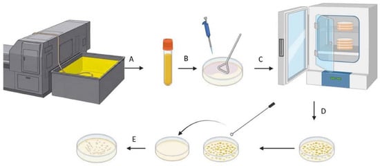
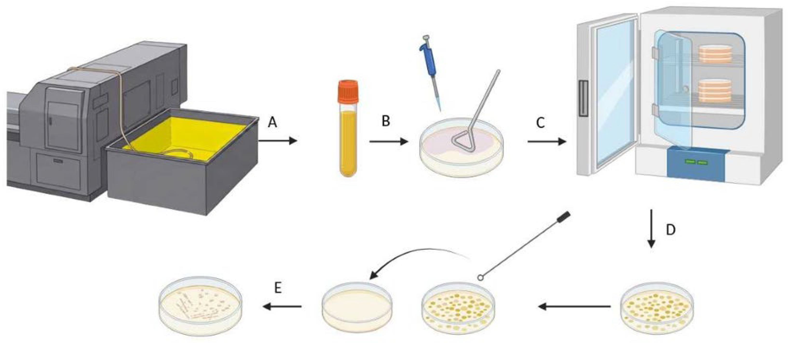
Subsequently, samples were incubated at 21, 28, and 37 °C, respectively, to mimic room temperature and elevated temperatures during the work process. Plates were incubated between three days and two weeks until considerable growth of colonies was detectable and pure cultures were isolated from these media (Figure 1).
Figure 1.
Schematic presentation of sample preparation and isolation of microorganisms. Samples taken from metalworking machines (A) were plated in serial dilutions on solid media (B) and incubated at different temperatures (C). Emerging colonies (D) were purified and isolates (E) were subjected to MALDI-ToF MS.
To determine the growth properties of members of the core microbiome, corresponding type strains were purchased from the German Collection of Microorganisms and Cell Cultures (DSMZ, Braunschweig, Germany) and cultivated in LB medium (10 g tryptone, 5 g yeast extract, 10 g NaCl) under constant shaking at 28 °C. Doubling times were determined from the exponential phase of the growth curves (see Supplementary Figure S1) using the equation
with g (one generation time) being the time (t) it takes for the bacteria to double in number (n).
g = t/n
2.3. Statistical Analysis
Growth experiments were carried out at least in triplicate as independent biological replicates, and mean values ± standard deviations were calculated using the STABW.N function of Excel.
2.4. Identification of the MWF Core Microbiome
In order to identify the microorganisms most frequently found in metalworking fluid, a literature screen was carried out in PubMed and Google Scholar using the query “metalworking fluid” and “oleovorans”. The reference sections of the resulting papers were subsequently screened for further literature.
2.5. Identification of Microorganisms
MALDI-ToF MS analysis was carried out by Labor Dr. Risch (Buchs, Switzerland) using a Bruker MALDI Biotyper (Bruker, Billerica, MA, USA) and the Bruker Daltonics Library version 2023. Fresh streak-outs of the isolates were analyzed as this technique requires fresh biological material that contains proteins that have not yet undergone significant degradation [29,30]. MALDI scores >2.0 are considered as accurate identification at the species level, values between 1.7 and 2.0 represent moderate identification confidence, and <1.7 no identification [31].
2.6. Analysis of Metabolic Pathways
To screen for metabolic pathways involved in hydrocarbon degradation, the Kyoto Encyclopedia of Genes and Genomes (KEGG, last assessed on 16 March 2026) was screened for alkane degradation and fatty acid metabolism (pathway map “map00071”; [32]).
3. Results
3.1. Growth Characteristics of the MWF Core Microbiome
Based on a literature screening carried out as a basis for our study, members of the MWF core microbiome, the microorganisms most often found, are Brevundimonas diminuta, Comamonas testosteroni, Morganella morganii, Mycobacterium immunogenum, Pseudomonas aeruginosa, Pseudomonas fluorescens, Pseudomonas putida, Pseudomonas oleovorans, Pseudomonas stutzeri, Shewanella putrefaciens, and Stenotrophomonas maltophilia [3,7,21,22,23,24,27,33,34,35,36,37,38,39,40,41,42]. We suspected that their frequent observation in MWFs might be caused by easy-to-fulfill growth conditions and fast growth under standard laboratory conditions. In fact, when cultivated under aeration in LB medium at 28 °C, these bacteria reached doubling times between 30 min and 230 min and an optical density at 600 nm (OD600) between 3.8 and 8.6 after 24 h (Table 2; Supplementary Figure S1). Growth rates in a similar range were also described in the literature for M. immunogenum [16], M. morganii [43], P. aeruginosa [44], P. putida [45], P. fluorescens [46], P. oleovorans [47], and P. stutzeri [48].
Table 2.
Growth properties of the MWF core microbiome.
3.2. Variability of Microbial Colonization of Metalworking Fluids
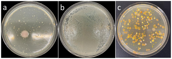
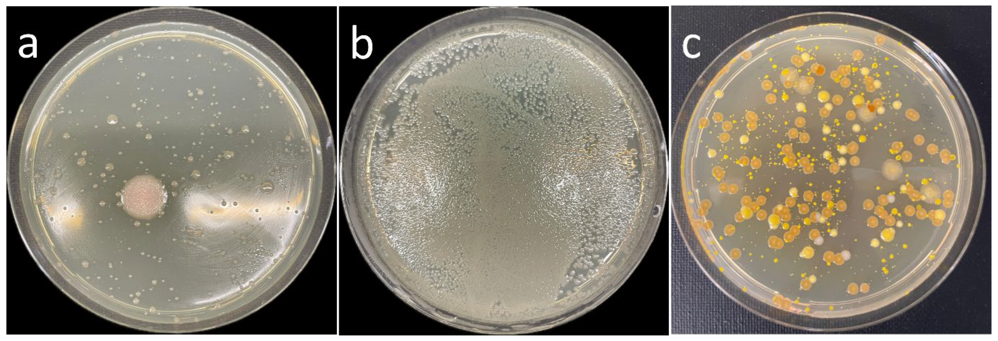
Samples were taken from four different MWFs and plated on the media described in Section 2.2. To avoid a bias towards fast-growing microorganisms and an overgrowth of slow-growing microbes, serial dilutions were carried out, and plates were incubated between three days and two weeks. Depending on the MWF and incubation conditions, different microbial populations were observed (Figure 2).
Figure 2.
Examples of microorganisms isolated in this study. Streak-outs of (a), (b) MWFs, (c) water supply system.
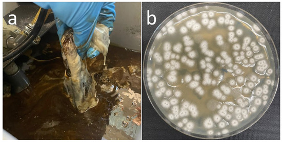
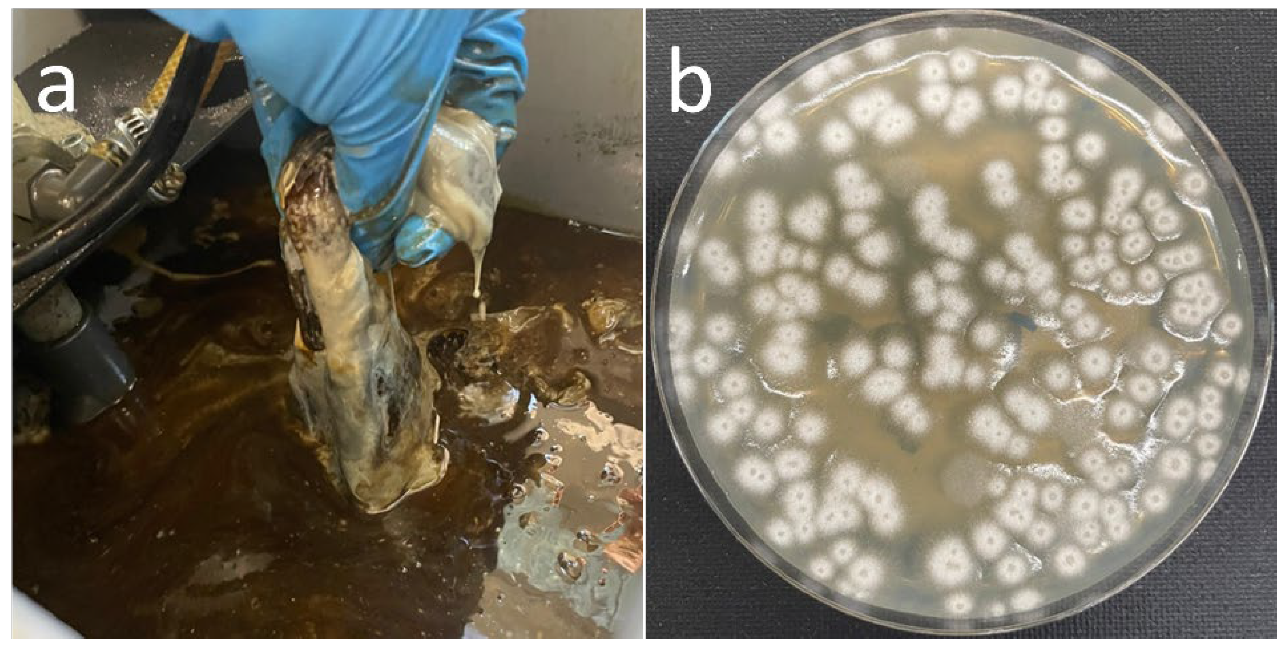
In the case of one of the machines, a band saw, which was only irregularly used, resulting in extended downtimes for the MWF in the tank, massive biofilm formation was observed (Figure 3). Cultivation of the biofilm-forming microorganism and MALDI-ToF MS-assisted identification revealed that the leather-like structured biofilm was formed by Scopulariopsis brevicaulis.
Figure 3.
Biofilm formation in MWF. (a) Biofilm sample from band saw. (b) Scopulariopsis brevicaulis isolated from biofilm sample.
3.3. Identification of Culturable Microorganisms by MALDI-ToF MS
Microbial populations can be characterized by DNA extraction, 16S rRNA gene sequencing, and subsequent taxonomic analysis. However, in the case of MWFs, DNA extraction may be disturbed by components of the coolant (e.g., oil) and by contamination from the workpieces (e.g., metal ions). Furthermore, identification by next-generation sequencing often ends at the family or genus level, and the approach cannot distinguish between living and dead microbes. Therefore, we decided to concentrate on a culture-based approach based on the idea that microbes living in MWF should be rather robust and easy to handle. In fact, we were able to isolate twenty-seven different bacteria and one fungus on the species level in this study (Table 3). To our best knowledge, twenty of these have not been found before in MWF. Twenty-two isolates could not be identified by MALDI-ToF-MS based on lacking databank entrances and were not further characterized in this study.
Table 3.
Microorganisms identified in MWFs.
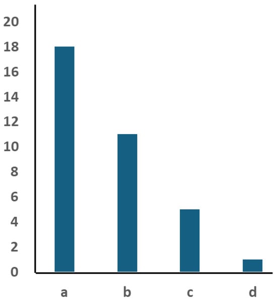
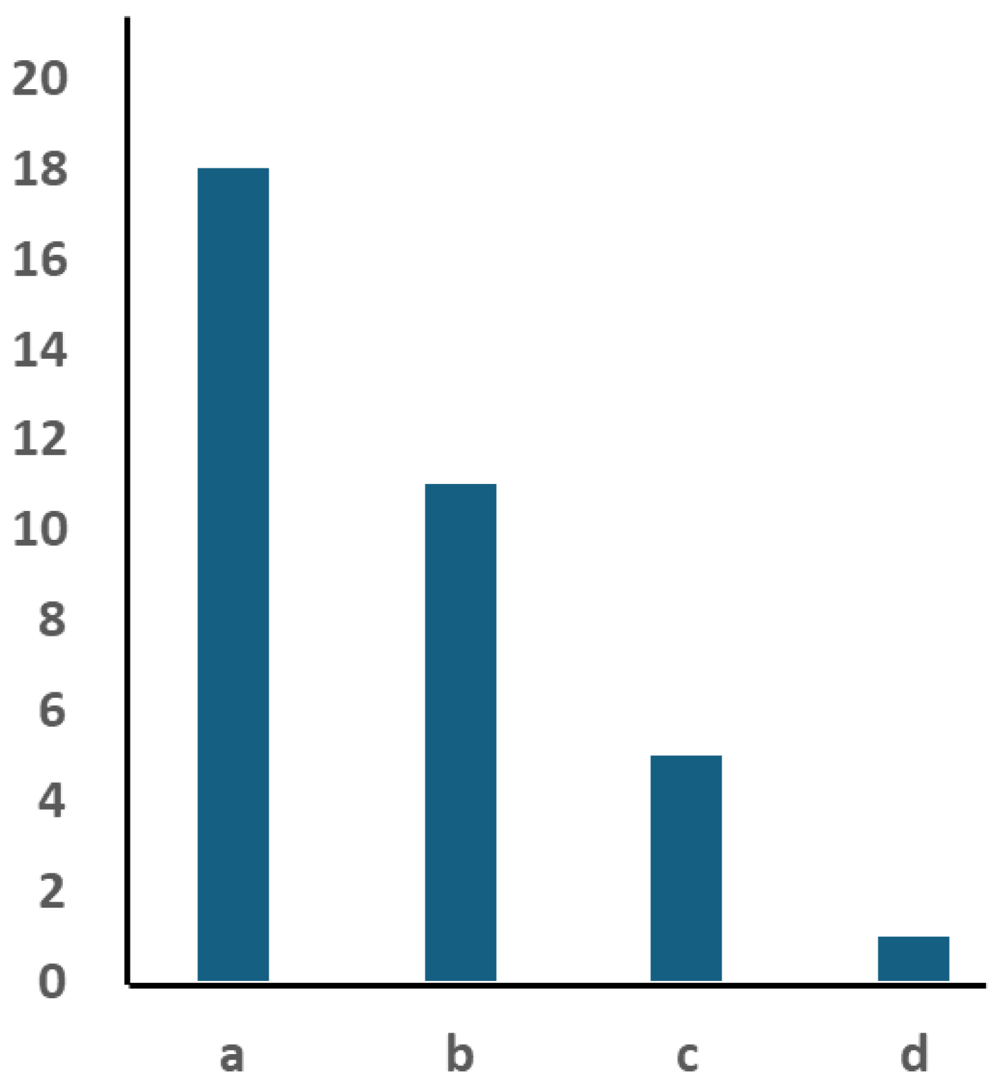
When the microbiome of the MWFs from the different machines was analyzed with respect to the number of species identified, clear differences were observed. A maximum of eighteen species was found in one of the milling machines, which had a service life of approximately 1 to 1 1/2 years, while a mono-species biofilm was found in the MWF of the band saw analyzed in this study (Figure 4). As ECO COOL GLOBAL 1000 (Fuchs GmbH, Mannheim, Germany) was used as MWF concentrate in all machines, differences based on the MWF itself can be excluded, and variations in colonization may be due to water used for MWF preparation, operation time, and materials processed.
Figure 4.
Species identified in MWFs of machines analyzed in this study. Columns indicate the number of identified species in MWF of the different machines. (a) milling machine and (b) lathe at the Chair of Resource and Energy-Efficient Production Machines, (c) milling machine at DMG Mori, (d) band saw at the Chair of Resource and Energy-Efficient Production Machines.
3.4. Putative Contamination Pathways
Microorganisms can contaminate MWFs via planktonic cells in the water to prepare them or as part of biofilm consortia in pipes and water hoses used to add water to the MWFs. In fact, when testing the water supply system, a number of bacteria and a yeast were found (Table 4), including putative primary colonizers: Acidovorax facilis and P. aeruginosa, which are species exhibiting alkane and fatty acid degradation pathways, while Pseudomonas putida is able to degrade fatty acids by β-oxidation. Other putative water-borne contaminations found in this study are C. sediminis and P. oleovorans.
Table 4.
Microorganisms identified in the water supply system.
Putative skin colonizers, especially corynebacteria and staphylococci, may be introduced in the MWF by the personnel handling machines, tools, and metal pieces; soil bacteria such as bacilli, streptomycetes, and Arthrobacter and Priestia species may be contaminating MWFs as part of dirt and dust, while M. luteus is a typical air-borne contamination.
3.5. Analysis of Hydrocarbon Degradation Pathways
MWFs contain 1 to 5% petroleum oil and less than 0.1% fatty acids, which serve as the primary carbon sources for colonizing microorganisms [6]. Alkanes are converted via oxidation into fatty acids [49,50,51], which are subsequently degraded by the β-oxidation pathway to acetyl-CoA and feed into the tricarboxylic acid cycle (TCA).
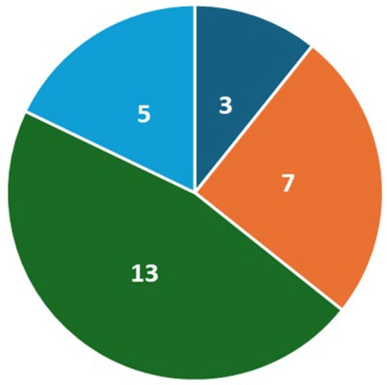
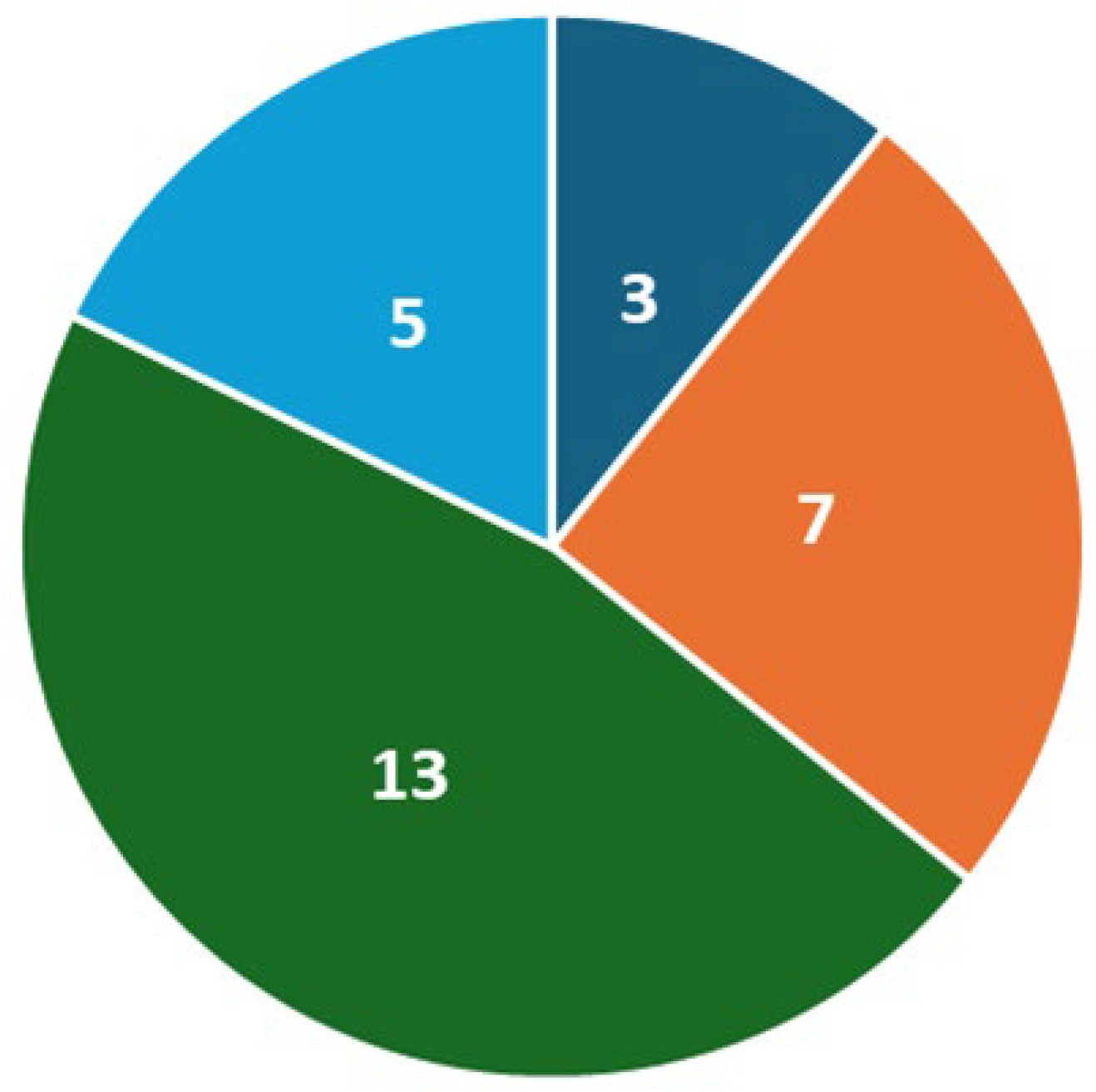
As part of this study, we screened the KEGG database for the corresponding degradation pathways. Two species identified in this study as part of the MWF microbiome were able to degrade alkanes, starting with an initial oxidation step of the carbon chain, followed by β-oxidation, P. yeei, and P. aeruginosa. Carbon utilization restricted to fatty acid consumption was found for B. pumilus, C. sediminis, C. aurimucosum, K. palustris, P. megaterium, S. decoyicus, and S. goshikiensis. Thirteen species identified here lacked any alkane and fatty acid degradation capacity, namely C. accolens, C. singulare, K. indica, K. rhizophila, M. luteus, N. dassonvillei, S. auricularis, S. capitis, S. epidermidis, S. haemolyticus, S. hominis, S. nepalensis, and S. warneri. No data were provided by KEGG for A. globiformis, C. tuberculostearicum, C. horneckiae, S. brevicaulis, and S. pragensis (Figure 5).
Figure 5.
Pie chart of metabolic pathways involved in hydrocarbon degradation. Dark blue, number of species with alkane and β-oxidation pathways; orange, number of species with β-oxidation pathways; green, number of species without alkane and fatty acid degradation capacity; light blue, no genomic data available in KEGG.
4. Discussion
To our best knowledge, almost 100 microbial species were identified in MWFs up to now [3,7,21,22,23,24,27,33,34,35,36,37,38,39,40,41,42]. In this study, twenty new species were identified by a cultivation-based approach, adding further information to our knowledge of the MWF microbiome.
Based on the literature, B. diminuta, C. testosteroni, M. morganii, M. immunogenum, P. aeruginosa, P. fluorescens, P. putida, P. oleovorans, P. stutzeri, S. putrefaciens, and S. maltophilia form the MWF core microbiome. While Pseudomonas species were frequently found in the different samples, and Brevundimonas and Comamonas species were also identified, mycobacteria were not observed in this study. A putative reason for this result might be the fact that this study was based on the analysis of one distinct MWF, i.e., ECO COOL GLOBAL 1000 (Fuchs GmbH, Mannheim, Germany), instead of different ones as in other studies. Interestingly, even the tested samples of one distinct MWF showed a rather variable and individual colonization with a limited number of species, as observed before for different MWFs [22,23,24].
The detrimental effect of microorganisms on MWFs may be caused by different metabolic properties. On the one hand, various bacteria and fungi can produce different classes of biosurfactants, surface-active agents that form micelles or vesicles around hydrocarbons to enhance uptake of hydrophobic substrates [52]. In MWFs, biosurfactants work as unwanted additional emulsifiers and support degradation of hydrocarbons. P. aeruginosa is well-known for its production of rhamnolipids [53] and also P. putida, found in a biofilm sample of a hose in this study, naturally produces rhamnolipids, although to a lesser extent [54]. In addition, glycolipid production was reported for Arthobacter strains [55,56].
On the other hand, alkanes and fatty acids of the MWFs can be degraded and used as carbon and energy sources by microorganisms. According to the KEGG database, only two species identified in this study as part of the MWF microbiome were able to completely degrade alkanes, starting with an initial oxidation step of the carbon chain followed by β-oxidation, P. yeei, and P. aeruginosa. Although not annotated in KEGG, an alkane oxidation pathway was also described before for P. oleovorans [49], a species that was also found in this study. A further seven species identified were able to degrade MWFs by the consumption of fatty acids. Growth of these microorganisms is only supported by alkanes, which are oxidized and converted to fatty acids by the primary alkane degraders. Thirteen species lacked any alkane and fatty acid utilization pathway. These bacteria will either metabolize other components of the MWFs or the metabolic products of alkane and fatty acid degraders.
In addition to detrimental effects on MWFs, machines, and metal processing, members of the MWF microbiome may pose a risk to human health. Particular members of the genus Mycobacterium were described in this respect before [14,15,16,25,26,27,28]. Interestingly, mycobacteria were not identified as MWF colonizers in this study. However, three groups of potentially harmful bacteria, which are often found in the environment, were observed here: corynebacteria, pseudomonads, and staphylococci. From the four species of the genus Corynebacterium identified, three are at least occasional pathogens. C. accolens is in rare cases associated with inflammatory diseases such as endocarditis, osteomyelitis, and pneumonia [57,58,59,60,61]. Interestingly, this species seems to have a protective effect against Staphylococcus aureus and Streptococcus pneumoniae infections [62,63]. C. aurimucosum was isolated from bone and joint infections, urinary tract infections, cases of bacteremia, and skin and soft-tissue infections, and is considered an actual opportunistic pathogen [64], while C. tuberculostearicum is a human skin colonizer that induces inflammatory signaling in skin cells, and its relevance to chronic inflammatory diseases of the skin and cutaneous oncology is discussed [65]. While P. aeruginosa is a notorious nosocomial pathogen infecting cystic fibrosis patients [66] and is also causing severe wound, ocular, and bloodstream infections [67,68,69], P. oleovorans is less in focus as a health risk. However, at least disinfectant-resistant biofilms formed by P. oleovorans were detected in endoscopes [70], and a fatal case of P. oleovorans sepsis was reported [71]. Many isolates of the genus Staphylococcus found in MWF in this study, namely S. auricularis, S. capitis, S. epidermidis, S. haemolyticus, S. hominis, and S. warneri, are coagulase-negative staphylococci, which can cause skin and soft tissue infections, particularly in older and/or immunocompromised individuals [72]. Epidemiologic and clinical studies have shown that S. capitis, S. epidermidis, S. haemolyticus, S. hominis and S. warneri may also be associated with meningitis [73]. The latter is also known for its ability to colonize artificial prosthetic materials and its tropism for musculoskeletal and cardiovascular tissues [74]. In addition, the biofilm analyzed in this study was formed by S. brevicaulis, the arsenic fungus, a difficult-to-treat human pathogen causing onychomycosis [75].
The frequent colonization of MWFs with Pseudomonas species documented in the literature and observed in this study is not only detrimental to MWF quality and function but may also pose a serious health risk to workers, which can be minimized with an optimized water supply. As described, tap water or water from wells and rivers may be used for MWF preparation [24]. Reduction of water-borne microbial contaminations from these sources may be achieved by UV treatment [76,77], nano- and ultra-filtration processes [78], or ozone and hydrogen peroxide addition [79,80]. A further, very cost-effective approach may be the implementation of Boron-doped diamond electrodes for water sanitation [81,82,83]. These treatments may help to prolong the MWF life cycle and reduce health risks for workers.
Future studies on MWFs may implement more quantitative analyses, e.g., real-time RT-PCR flanked by the determination of colony-forming units, and a time-resolved analysis of colonization, since our analyses of metabolic pathways hint at a succession of microbes, starting with primary alkane degraders. In addition, resistance to high pH and microbial acid production might be important parameters influencing the composition of the MWF microbiome and MWF biodegradation.
Supplementary Materials
The following supporting information can be downloaded at: https://www.mdpi.com/article/10.3390/applmicrobiol6050060/s1, Figure S1: Growth of MWF core microbiome species.
Author Contributions
A.H.: investigation, methodology, formal analysis, visualization. B.O.: investigation, formal analysis, visualization. J.R.: project administration. N.H.: funding acquisition. A.B.: conceptualization, resources, writing—original draft preparation, writing—review and editing. All authors have read and agreed to the published version of the manuscript.
Funding
This research was funded by the Bundesministerium für Ernährung und Landwirtschaft in the frame of the DiaCool project (grant number 2222NR081C).
Institutional Review Board Statement
Not applicable.
Informed Consent Statement
Not applicable.
Data Availability Statement
The raw data supporting the conclusions of this article will be made available by the authors on request.
Acknowledgments
The authors wish to thank Michael Kirbach (DMG Mori, Pfronten, Germany) for providing samples of used metalworking fluid.
Conflicts of Interest
The authors declare no competing interests.
Abbreviations
| KEGG | Kyoto Encyclopedia of Genes and Genomes |
| LB | Luria Broth |
| MALDI-ToF MS | matrix-assisted laser desorption/ionization time of flight mass spectrometry |
| MWF | metalworking fluid |
| OD600 | optical density at 600 nm |
| PBS | phosphate-buffered saline |
| RT PCR | reverse transcriptase polymerase chain reaction |
| TCA | tricarboxylic acid cycle |
| UV | ultraviolet |
References
- Brinksmeier, E.; Meyer, D.; Huesmann-Cordes, A.G.; Herrmann, C. Metalworking fluids—Mechanisms and Performance. CIRP Ann.–Manufact. Tech. 2015, 64, 605–628. [Google Scholar] [CrossRef] [Scilit]
- Passman, F.J.; Küenzi, P. Microbiology in Water-Miscible Metalworking Fluids. Tribol. Trans. 2020, 63, 1147–1171. [Google Scholar] [CrossRef] [Scilit]
- Mattsby-Baltzer, I.; Sandin, M.; Ahlström, B.; Allenmark, S.; Edebo, M.; Falsen, E.; Pedersen, K.; Rodin, N.; Thompson, R.A.; Edebo, L. Microbial growth and accumulation in industrial metal-working fluids. Appl. Environ. Microbiol. 1989, 55, 2681–2689. [Google Scholar] [CrossRef] [Scilit] [PubMed]
- Sloyer, J.L.; Novitsky, T.J.; Nugent, S. Rapid bacterial counts in metal working fluids. J. Ind. Microbiol. Biotechnol. 2002, 29, 323–324. [Google Scholar] [CrossRef]
- Theaker, D.; Thompson, I. The Industrial Consequences of Microbial Deterioration of Metal-Working Fluid. In Handbook of Hydrocarbon and Lipid Microbiology; Timmis, K.N., Ed.; Springer: Berlin/Heidelberg, Germany, 2010; pp. 2641–2650. [Google Scholar] [CrossRef] [Scilit]
- Saha, R.; Donofrio, R.S. The microbiology of metalworking fluids. Appl. Microbiol. Biotechnol. 2012, 94, 1119–1130. [Google Scholar] [CrossRef] [Scilit]
- Trafny, E.A.; Lewandowski, R.; Kozłowska, K.; Zawistowska-Marciniak, I.; Stępińska, M. Microbial contamination and biofilms on machines of metal industry using metalworking fluids with or without biocides. Int. Biodeterior. Biodegrad. 2015, 99, 31–38. [Google Scholar] [CrossRef] [Scilit]
- Grattan, C.E.; English, J.S.; Foulds, I.S.; Rycroft, R.J. Cutting fluid dermatitis. Contact Dermat. 1989, 20, 372–376. [Google Scholar] [CrossRef] [Scilit]
- Foulds, I.S.; Koh, D. Dermatitis from metalworking fluids. Clin. Exp. Dermatol. 1990, 15, 157–162. [Google Scholar] [CrossRef] [Scilit] [PubMed]
- Alomar, A. Occupational skin disease from cutting fluids. Dermatol. Clin. 1994, 12, 537–546. [Google Scholar] [CrossRef] [Scilit] [PubMed]
- Goh, C.L.; Gan, S.L. The incidence of cutting fluid dermatitis among metalworkers in a metal fabrication factory: A prospective study. Contact Dermat. 1994, 31, 111–115. [Google Scholar] [CrossRef] [Scilit]
- Bernstein, D.I.; Lummus, Z.L.; Santilli, G.; Siskosky, J.; Bernstein, I.L. Machine operator’s lung. A hypersensitivity pneumonitis disorder associated with exposure to metalworking fluid aerosols. Chest 1995, 108, 636–641. [Google Scholar] [CrossRef] [Scilit] [PubMed]
- Kreiss, K.; Cox-Ganser, J. Metalworking fluid-associated hypersensitivity pneumonitis: A workshop summary. Am. J. Ind. Med. 1997, 32, 423–432. [Google Scholar] [CrossRef]
- Shelton, B.G.; Flanders, W.D.; Morris, G.K. Mycobacterium sp. as a possible cause of hypersensitivity pneumonitis in machine workers. Emerg. Infect. Dis. 1999, 5, 270–273. [Google Scholar] [CrossRef] [Scilit]
- Hodgson, M.J.; Bracker, A.; Yang, C.; Storey, E.; Jarvis, B.J.; Milton, D.; Lummus, Z.; Bernstein, D.; Cole, S. Hypersensitivity pneumonitis in a metal-working environment. Am. J. Ind. Med. 2001, 39, 616–628. [Google Scholar] [CrossRef] [Scilit]
- Wilson, R.W.; Steingrube, V.A.; Böttger, E.C.; Springer, B.; Brown-Elliott, B.A.; Vincent, V.; Jost, K.C.; Zhang, Y.; Garcia, M.J.; Chiu, S.H.; et al. Mycobacterium immunogenum sp. nov., a novel species related to Mycobacterium abscessus and associated with clinical disease, pseudo-outbreaks and contaminated metalworking fluids: An international cooperative study on mycobacterial taxonomy. Int. J. Syst. Evol. Microbiol. 2001, 51, 1751–1764. [Google Scholar] [CrossRef] [Scilit]
- Fishwick, D.; Tate, P.; Elms, J.; Robinson, E.; Crook, B.; Gallagher, F.; Lennox, R.; Curran, A. Respiratory symptoms, immunology and organism identification in contaminated metalworking fluid workers. What you see is not what you get. Occup. Med. 2025, 55, 238–241. [Google Scholar] [CrossRef] [Scilit]
- Rosenman, K.D. Asthma, hypersensitivity pneumonitis and other respiratory diseases caused by metalworking fluids. Curr. Opin. Allergy Clin. Immunol. 2009, 9, 97–102. [Google Scholar] [CrossRef] [Scilit]
- Tillie-Leblond, I.; Grenouillet, F.; Reboux, G.; Roussel, S.; Chouraki, B.; Lorthois, C.; Dalphin, J.C.; Wallaert, B.; Millon, L. Hypersensitivity pneumonitis and metalworking fluids contaminated by mycobacteria. Eur. Respir. J. 2011, 37, 640–647. [Google Scholar] [CrossRef] [Scilit]
- Burge, P.S. Hypersensitivity Pneumonitis Due to Metalworking Fluid Aerosols. Curr. Allergy Asthma Rep. 2016, 16, 59. [Google Scholar] [CrossRef] [Scilit] [PubMed]
- Marchand, G.; Lavoie, J.; Racine, L.; Lacombe, N.; Cloutier, Y.; Bélanger, E.; Lemelin, C.; Desroches, J. Evaluation of bacterial contamination and control methods in soluble metalworking fluids. J. Occup. Environ. Hyg. 2010, 7, 358–366. [Google Scholar] [CrossRef] [Scilit]
- van der Gast, C.J.; Whiteley, A.S.; Lilley, A.K.; Knowles, C.J.; Thompson, I.P. Bacterial community structure and function in a metal-working fluid. Environ. Microbiol. 2003, 5, 453–461. [Google Scholar] [CrossRef] [Scilit]
- Gilbert, Y.; Veillette, M.; Duchaine, C. Metalworking fluids biodiversity characterization. J. Appl. Microbiol. 2010, 108, 437–449. [Google Scholar] [CrossRef] [Scilit] [PubMed]
- Lodders, N.; Kämpfer, P. A combined cultivation and cultivation-independent approach shows high bacterial diversity in water-miscible metalworking fluids. Syst. Appl. Microbiol. 2012, 35, 246–252. [Google Scholar] [CrossRef] [Scilit]
- Moore, J.S.; Christensen, M.; Wilson, R.W.; Wallace, R.J., Jr.; Zhang, Y.; Nash, D.R.; Shelton, B. Mycobacterial contamination of metalworking fluids: Involvement of a possible new taxon of rapidly growing mycobacteria. AIHAJ 2000, 61, 205–213. [Google Scholar] [CrossRef] [Scilit]
- Wallace, R.J., Jr.; Zhang, Y.; Wilson, R.W.; Mann, L.; Rossmoore, H. Presence of a single genotype of the newly described species Mycobacterium immunogenum in industrial metalworking fluids associated with hypersensitivity pneumonitis. Appl. Environ. Microbiol. 2002, 68, 5580–5584. [Google Scholar] [CrossRef] [Scilit]
- Khan, I.U.; Selvaraju, S.B.; Yadav, J.S. Occurrence and characterization of multiple novel genotypes of Mycobacterium immunogenum and Mycobacterium chelonae in metalworking fluids. FEMS Microbiol. Ecol. 2005, 54, 329–338. [Google Scholar] [CrossRef] [Scilit]
- Thorne, P.S.; Adamcakova-Dodd, A.; Kelly, K.M.; O’neill, M.E.; Duchaine, C. Metalworking fluid with mycobacteria and endotoxin induces hypersensitivity pneumonitis in mice. Am. J. Respir. Crit. Care Med. 2006, 173, 759–768. [Google Scholar] [CrossRef] [Scilit]
- Reeve, M.A.; Buddie, A.G. A simple and inexpensive method for practical storage of field-sample proteins for subsequent MALDI-TOF MS analysis. Plant Methods 2018, 14, 90. [Google Scholar] [CrossRef] [Scilit] [PubMed]
- Topić Popović, N.; Kazazić, S.P.; Bojanić, K.; Strunjak-Perović, I.; Čož-Rakovac, R. Sample preparation and culture condition effects on MALDI-TOF MS identification of bacteria: A review. Mass Spectrom. Rev. 2023, 42, 1589–1603. [Google Scholar] [CrossRef] [Scilit] [PubMed]
- Croxatto, A.; Prod’hom, G.; Greub, G. Applications of MALDI-TOF mass spectrometry in clinical diagnostic microbiology. FEMS Microbiol. Rev. 2012, 36, 380–407. [Google Scholar] [CrossRef] [Scilit]
- Parekh, V.R.; Traxler, R.W.; Sobek, J.M. N-Alkane oxidation enzymes of a pseudomonad. Appl. Environ. Microbiol. 1977, 33, 881–884. [Google Scholar] [CrossRef] [Scilit]
- Cook, P.E.; Gaylarde, C.C. Biofilm formation in aqueous metal working fluids. Int. Biodeterior. 1988, 24, 265–270. [Google Scholar] [CrossRef] [Scilit]
- Zacharisen, M.C.; Kadambi, A.R.; Schlueter, D.P.; Kurup, V.P.; Shack, J.B.; Fox, J.L.; Anderson, H.A.; Fink, J.N. The Spectrum of Respiratory Disease Associated with Exposure to Metal Working Fluids. J. Occup. Environ. Med. 1998, 40, 640. [Google Scholar] [CrossRef] [Scilit]
- Veillette, M.; Thorne, P.; Gordon, T.; Duchaine, C. Six Month Tracking of Microbial Growth in a Metalworking Fluid After System Cleaning and Recharging. Ann. Occup. Hyg. 2004, 48, 541–546. [Google Scholar] [CrossRef] [Scilit]
- Dilger, S.; Fluri, A.; Sonntag, H.-G. Bacterial contamination of preserved and non-preserved metal working fluids. Int. J. Hyg. Environ. Health 2005, 208, 467–476. [Google Scholar] [CrossRef] [Scilit] [PubMed]
- Cyprowski, M.; Piotrowska, M.; Zakowska, Z.; Szadkowska-Stańczyk, I. Microbial and endotoxin contamination of water-soluble metalworking fluids. Int. J. Occup. Med. Environ. Health 2007, 20, 365–371. [Google Scholar] [CrossRef] [Scilit]
- Rabenstein, A.; Koch, T.; Remesch, M.; Brinksmeier, E.; Kuever, J. Microbial degradation of water miscible metal working fluids. Int. Biodeterior. Biodegrad. 2009, 63, 1023–1029. [Google Scholar] [CrossRef] [Scilit]
- Perkins, S.D.; Angenent, L.T. Potential pathogenic bacteria in metalworking fluids and aerosols from a machining facility: Potential pathogens in metalworking fluids and aerosols. FEMS Microbiol. Ecol. 2010, 74, 643–654. [Google Scholar] [CrossRef] [Scilit] [PubMed]
- Murat, J.-B.; Grenouillet, F.; Reboux, G.; Penven, E.; Batchili, A.; Dalphin, J.-C.; Thaon, I.; Millon, L. Factors Influencing the Microbial Composition of Metalworking Fluids and Potential Implications for Machine Operator’s Lung. Appl. Environ. Microbiol. 2012, 78, 34–41. [Google Scholar] [CrossRef] [Scilit]
- Koch, T.; Passman, F.; Rabenstein, A. Comparative study of microbiological monitoring of water-miscible metalworking fluids. Int. Biodeterior. Biodegrad. 2015, 98, 19–25. [Google Scholar] [CrossRef] [Scilit]
- Kapoor, R.; Selvaraju, S.B.; Subramanian, V.; Yadav, J.S. Microbial Community Establishment, Succession, and Temporal Dynamics in an Industrial Semi-Synthetic Metalworking Fluid Operation: A 50-Week Real-Time Tracking. Microorganisms 2024, 12, 267. [Google Scholar] [CrossRef] [Scilit]
- Sun, Z.; Zhang, Y.; Lin, X.; Zhang, S.; Chen, Y.; Ji, C. Inhibition Mechanism of Lactiplantibacillus plantarum on the Growth and Biogenic Amine Production in Morganella morganii. Foods 2023, 12, 3625. [Google Scholar] [CrossRef] [Scilit]
- Mahmoud, M.; Richter, P.; Lebert, M.; Burkovski, A. Photodynamic Activity of Chlorophyllin and Polyethylenimine on Pseudomonas aeruginosa Planktonic, Biofilm and Persister Cells. Int. J. Mol. Sci. 2023, 24, 12098. [Google Scholar] [CrossRef] [Scilit] [PubMed]
- Jensen, J.W.; Nikel, P.I.; Woodley, J.M.; Junicke, H. Genome-Reduced Pseudomonas putida Outcompetes the Wild-Type Strain Upon Oxygen Depletion. Eng. Life Sci. 2025, 25, e70046. [Google Scholar] [CrossRef] [Scilit] [PubMed]
- Gill, C.O.; Tan, K.H. Effect of carbon dioxide on growth of Pseudomonas fluorescens. Appl. Environ. Microbiol. 1979, 38, 237–240. [Google Scholar] [CrossRef] [Scilit]
- Wang, H.; Sun, C.; Chen, X.; Yan, K.; He, H. Isolation of Pseudomonas oleovorans Carrying Multidrug Resistance Proteins MdtA and MdtB from Wastewater. Molecules 2023, 28, 5403. [Google Scholar] [CrossRef] [Scilit]
- Sommer, M.; Xie, H.; Michel, H. Pseudomonas stutzeri as an alternative host for membrane proteins. Microb. Cell Fact. 2017, 16, 157. [Google Scholar] [CrossRef] [Scilit] [PubMed]
- van Beilen, J.B.; Wubbolts, M.G.; Witholt, B. Genetics of alkane oxidation by Pseudomonas oleovorans. Biodegradation 1994, 5, 161–174. [Google Scholar] [CrossRef] [Scilit]
- van Beilen, J.B.; Funhoff, E.G. Alkane hydroxylases involved in microbial alkane degradation. Appl. Microbiol. Biotechnol. 2007, 74, 13–21. [Google Scholar] [CrossRef] [Scilit]
- Guo, X.; Zhu, S.; Zhu, N.; Zhang, S.; Yang, S.; Luo, G.; Li, H.; Wang, Y.; Sun, J.; Ma, B. Pan-Genomic and Phenotypic Characterisation of Petroleum Hydrocarbon Degradation by Pseudomonas Species. Environ. Microbiol. Rep. 2026, 18, e70300. [Google Scholar] [CrossRef] [Scilit]
- Dini, S.; Bekhit, A.E.A.; Roohinejad, S.; Vale, J.M.; Agyei, D. The Physicochemical and Functional Properties of Biosurfactants: A Review. Molecules 2024, 29, 2544. [Google Scholar] [CrossRef] [Scilit]
- Das, P.V.; Valentine, M.E.; Long, T.E.; Yu, H.D. Attenuated Strains of Pseudomonas aeruginosa: A Promising Cell Factory for Rhamnolipid Production. Microb. Biotechnol. 2025, 18, e70239. [Google Scholar] [CrossRef] [Scilit]
- Toribio, J.; Escalante, A.E.; Soberón-Chávez, G. Production of rhamnolipids in bacteria other than Pseudomonas aeruginosa. Eur. J. Lipid Sci. Technol. 2010, 112, 1082–1087. [Google Scholar] [CrossRef] [Scilit]
- Campos, J.M.; Stamford, T.L.; Sarubbo, L.A.; de Luna, J.M.; Rufino, R.D.; Banat, I.M. Microbial biosurfactants as additives for food industries. Biotechnol. Prog. 2013, 29, 1097–1108. [Google Scholar] [CrossRef] [Scilit]
- Santos, D.K.; Rufino, R.D.; Luna, J.M.; Santos, V.A.; Sarubbo, L.A. Biosurfactants: Multifunctional Biomolecules of the 21st Century. Int. J. Mol. Sci. 2016, 17, 401. [Google Scholar] [CrossRef] [Scilit]
- Claeys, G.; Vanhouteghem, H.; Riegel, P.; Wauters, G.; Hamerlynck, R.; Dierick, J.; de Witte, J.; Verschraegen, G.; Vaneechoutte, M. Endocarditis of native aortic and mitral valves due to Corynebacterium accolens: Report of a case and application of phenotypic and genotypic techniques for identification. J. Clin. Microbiol. 1996, 34, 1290–1292. [Google Scholar] [CrossRef] [Scilit]
- Wong, J.S.; Seaward, L.M.; Ho, C.P.; Anderson, T.P.; Lau, E.O.; Amodeo, M.R.; Metcalf, S.C.; Pithie, A.D.; Murdoch, D.R. Corynebacterium accolens-associated pelvic osteomyelitis. J. Clin. Microbiol. 2010, 48, 654–655. [Google Scholar] [CrossRef] [Scilit]
- Liu, B.M.; Beck, E.M.; Fisher, M.A. The Brief Case: Ventilator-Associated Corynebacterium accolens Pneumonia in a Patient with Respiratory Failure Due to COVID-19. J. Clin. Microbiol. 2021, 59, e0013721. [Google Scholar] [CrossRef] [Scilit] [PubMed]
- Liu, B.M.; Beck, E.M.; Fisher, M.A. Closing the Brief Case: Ventilator-Associated Corynebacterium accolens Pneumonia in a Patient with Respiratory Failure Due to COVID-19. J. Clin. Microbiol. 2021, 59, e0013821. [Google Scholar] [CrossRef] [Scilit] [PubMed]
- Poucke, L.V.; Hinton, J.B.; Heck, H.C.; Heck, B.E.; Heck, B.E. Systemic Sclerosis Presenting as Osteomyelitis of the Finger: Physicians Must Maintain a High Index of Suspicion for Systemic Sclerosis when Evaluating Patients with Fingertip Ulceration or Infection. J. Orthop. Case Rep. 2025, 15, 52–55. [Google Scholar] [CrossRef] [Scilit] [PubMed]
- Bomar, L.; Brugger, S.D.; Yost, B.H.; Davies, S.S.; Lemon, K.P. Corynebacterium accolens Releases Antipneumococcal Free Fatty Acids from Human Nostril and Skin Surface Triacylglycerols. mBio 2016, 7, e01725-15. [Google Scholar] [CrossRef] [Scilit] [PubMed]
- Menberu, M.A.; Liu, S.; Cooksley, C.; Hayes, A.J.; Psaltis, A.J.; Wormald, P.J.; Vreugde, S. Corynebacterium accolens Has Antimicrobial Activity against Staphylococcus aureus and Methicillin-Resistant S. aureus Pathogens Isolated from the Sinonasal Niche of Chronic Rhinosinusitis Patients. Pathogens 2021, 10, 207. [Google Scholar] [CrossRef] [Scilit]
- Lefèvre, C.R.; Pelletier, R.; Le Monnier, A.; Corvec, S.; Bille, E.; Potron, A.; Fihman, V.; Farfour, E.; Amara, M.; Degand, N.; et al. Clinical relevance and antimicrobial susceptibility profile of the unknown human pathogen Corynebacterium aurimucosum. J. Med. Microbiol. 2021, 70, 001334. [Google Scholar] [CrossRef] [Scilit] [PubMed]
- Altonsy, M.O.; Kurwa, H.A.; Lauzon, G.J.; Amrein, M.; Gerber, A.N.; Almishri, W.; Mydlarski, P.R. Corynebacterium tuberculostearicum, a human skin colonizer, induces the canonical nuclear factor-κB inflammatory signaling pathway in human skin cells. Immun. Inflamm. Dis. 2020, 8, 62–79. [Google Scholar] [CrossRef] [Scilit]
- Núñez-García, L.Á.; Córdova-Fletes, C.; Barboza-Cerda, M.C.; Garza-González, E. Pseudomonas aeruginosa Biofilms in Cystic Fibrosis: Interactions, Methods, and Therapeutic Strategies. Biomed. Res. Int. 2026, 2026, 5328382. [Google Scholar] [CrossRef] [Scilit]
- Alharbi, M.S.; Moursi, S.A.; Alshammari, A.; Aboras, R.; Rakha, E.; Hossain, A.; Alshubrumi, S.; Alnazha, K.; Khaja, A.S.S.; Saleem, M. Multidrug-resistant Pseudomonas aeruginosa: Pathogenesis, resistance mechanisms, and novel therapeutic strategies. Virulence 2025, 16, 2580160. [Google Scholar] [CrossRef] [Scilit] [PubMed]
- Ather, M.; Conrady, C.D. Emerging Ocular Pathogen Resistance and Clinically Used Solutions: A Problem That Is More than Meets the Eye. Pharmaceuticals 2025, 19, 31. [Google Scholar] [CrossRef] [Scilit]
- Jabar, A.K.; Romli, N.I.A.; Vellasamy, K.M.; Pallath, V.; Al-Maleki, A.R. Predictors of Mortality in Pseudomonas aeruginosa Bloodstream Infections: A Scoping Review. Pathogens 2026, 15, 61. [Google Scholar] [CrossRef] [Scilit]
- Leeb-Zatorska, B.; Van den Nest, M.; Ebner, J.; Moser, D.; Spettel, K.; Bovier-Azula, L.; Diab-El Schahawi, M.; Presterl, E. Tolerance of Pseudomonas oleovorans biofilms to disinfectants commonly used in endoscope reprocessing? Biofilm 2024, 8, 100221. [Google Scholar] [CrossRef] [Scilit]
- Gautam, L.; Kaur, R.; Kumar, S.; Bansal, A.; Gautam, V.; Singh, M.; Ray, P. Pseudomonas oleovorans Sepsis in a Child: The First Reported Case in India. Jpn. J. Infect. Dis. 2015, 68, 254–255. [Google Scholar] [CrossRef] [Scilit] [PubMed]
- Natsis, N.E.; Cohen, P.R. Coagulase-Negative Staphylococcus Skin and Soft Tissue Infections. Am. J. Clin. Dermatol. 2018, 19, 671–677. [Google Scholar] [CrossRef] [Scilit]
- Azimi, T.; Mirzadeh, M.; Sabour, S.; Nasser, A.; Fallah, F.; Pourmand, M.R. Coagulase-negative staphylococci (CoNS) meningitis: A narrative review of the literature from 2000 to 2020. New Microbes New Infect. 2020, 37, 100755. [Google Scholar] [CrossRef] [Scilit]
- Ravaioli, S.; De Donno, A.; Bottau, G.; Campoccia, D.; Maso, A.; Dolzani, P.; Balaji, P.; Pegreffi, F.; Daglia, M.; Arciola, C.R. The Opportunistic Pathogen Staphylococcus warneri: Virulence and Antibiotic Resistance, Clinical Features, Association with Orthopedic Implants and Other Medical Devices, and a Glance at Industrial Applications. Antibiotics 2024, 13, 972. [Google Scholar] [CrossRef] [Scilit]
- Mayser, P. Scopulariopsis brevicaulis: Der “Arsenpilz”—Ein schwierig zu behandelnder Erreger [Scopulariopsis brevicaulis: The “arsenic fungus”—A difficult-to-treat pathogen]. Dermatologie 2025, 76, 533–543. [Google Scholar] [CrossRef] [Scilit]
- Barkhudarov, E.M.; Christofi, N.; Kossyi, I.A.; Misakyan, M.A.; Sharp, J.; Taktakishvili, I.M. Killing bacteria present on surfaces in films or droplets using microwave UV lamps. World J. Microbiol. Biotechnol. 2008, 24, 761–769. [Google Scholar] [CrossRef] [Scilit]
- O’Flaherty, E.; Membré, J.-M.; Cummins, E. Meta-analysis of the reduction of sensitive and antibiotic-resistant Escherichia coli as a result of low and medium pressure UV lamps. Water Sci. Technol. 2018, 2017, 612–620. [Google Scholar] [CrossRef] [Scilit]
- Schwermer, C.U.; Krzeminski, P.; Wennberg, A.I.; Vogelsang, C.; Uhl, W. Removal of antibiotic-resistant E. coli in two Norwegian wastewater treatment plants and by nano- and ultrafiltration processes. Water Sci. Technol. 2018, 77, 1115–1126. [Google Scholar] [CrossRef] [Scilit] [PubMed]
- Cho, M.; Kim, J.; Kim, Y.J.; Yoon, J.; Kim, J.-H. Mechanisms of Escherichia coli inactivation by several disinfectants. Water Res. 2010, 44, 3410–3434. [Google Scholar] [CrossRef] [Scilit]
- Martirosyan, V.; Moosavi, E.; Ayrapetyan, S. The study of the effects of carbon dioxide-induced elevation of hydrogen peroxide toxicity on microbes as a novel tool for water purification. World J. Microbiol. Biotechnol. 2011, 27, 1091–1098. [Google Scholar] [CrossRef] [Scilit]
- Schorr, B.; Ghanem, H.; Rosiwal, S.; Geißdörfer, W.; Burkovski, A. Elimination of bacterial contaminations by treatment of water with diamond electrodes. World J. Microbiol. Biotechnol. 2019, 35, 48. [Google Scholar] [CrossRef] [Scilit] [PubMed]
- Koch, M.; Rosiwal, S.; Burkovski, A. Environmentally sustainable elimination of microbes using boron-doped diamond electrodes: From water treatment to medical applications. In Microbial Biotechnology: Role in Ecological Sustainability and Research; Chowdhary, P., Mani, S., Chaturvedi, P., Eds.; John Wiley & Sons, Inc.: Hoboken, NJ, USA, 2022; pp. 355–364. [Google Scholar]
- Kraft, A. Doped diamond: A compact review on a new, versatile electrode material. Int. J. Electrochem. Sci. 2007, 2, 355–385. [Google Scholar] [CrossRef] [Scilit]

Disclaimer/Publisher’s Note: The statements, opinions and data contained in all publications are solely those of the individual author(s) and contributor(s) and not of MDPI and/or the editor(s). MDPI and/or the editor(s) disclaim responsibility for any injury to people or property resulting from any ideas, methods, instructions or products referred to in the content. |
© 2026 by the authors. Licensee MDPI, Basel, Switzerland. This article is an open access article distributed under the terms and conditions of the Creative Commons Attribution (CC BY) license.