


Abstract
Probiotic bacteria are increasingly being explored as engineering platforms for local production of bioactive proteins, but the outcome may depend on both the recombinant protein and the bacterial strain. Azurin-producing derivatives of Escherichia coli Nissle 1917 (EcN) and E. coli M17 were generated using the same lacUV5-pelB-azu cassette and evaluated in HCT116 colorectal carcinoma cells. Western blotting confirmed azurin production in both strains, predominantly in cell-associated fractions and at lower levels in concentrated culture supernatants. A separately produced recombinant azurin preparation obtained from E. coli NiCo21(DE3) reduced HCT116 cell biomass after 48 h, with an estimated IC50 of 320 µg/mL. In bacterial co-culture, the azurin-producing derivatives showed lower mean SRB-assessed HCT116 biomass than their corresponding wild-type strains, but these differences did not reach statistical significance. In contrast, YO-PRO-1/PI analysis at a bacteria-to-cell ratio of 500 identified a significant overall increase in the total apoptosis-associated fraction in the azurin-producing groups, whereas the necrosis-associated fraction remained low. The overall response was strain-dependent: EcN showed strong baseline inhibitory activity, whereas the lower baseline effect of M17 allowed the azurin-associated contribution to be distinguished more clearly. Thus, both strains supported recombinant azurin production, but the host–cell response was strongly influenced by the bacterial background. These findings highlight strain selection as an important factor in the development and evaluation of engineered probiotic systems for bioactive protein production.
1. Introduction
Research on probiotic bacteria has expanded beyond their inherent physiological roles to include their use as genetically engineered systems for the local production and delivery of bioactive molecules [1,2]. These engineered strains can be used to produce proteins in situ where they can directly interact with host cells, which may help overcome some of the limitations associated with repeated administration of purified proteins [3,4]. However, bacterial strain selection is no trivial matter since the chassis is not a biologically neutral carrier [5]. The bacterial background can influence growth, cellular physiology, genetic-circuit performance, and recombinant protein production [6,7,8], while strain-specific interactions with host cells may independently affect the resulting biological response [9,10]. Consequently, the observed activity of an engineered probiotic reflects not only the recombinant protein but also the intrinsic properties of the bacterial strain in which it is produced.
Probiotic Escherichia coli strains are attractive for early-stage development and engineering because they have a history of gastrointestinal application, are compatible with standard molecular biology methods, and grow under routine laboratory conditions [11,12,13]. Among them, E. coli Nissle 1917 (EcN) is one of the best studied probiotic E. coli backgrounds and used in several engineered live bacterial systems [12,14]. Its extensive characterization positions EcN as an established reference strain for the development of recombinant probiotic platforms. At the same time, EcN has its own biological activity, including immunomodulatory effects and interaction with epithelial cells, which may influence host–cell responses independently of an introduced recombinant protein [9,15].
E. coli M17 is a less extensively characterized probiotic E. coli strain with a history of use in live bacterial preparation for gastrointestinal disorders [16]. Fitzpatrick and colleagues examined E. coli M17 in vitro and in vivo and reported effects on NF-κB signaling, cytokine secretion, and dextran sulfate sodium-induced murine colitis, suggesting that its activity is partly related to modulation of immune responses [17]. Despite this background, M17 has received far less attention than EcN as an engineered platform. Its inclusion in the present study therefore provides a complementary bacterial background for testing the same expression system. A direct comparison of EcN and M17 makes it possible to determine whether the differences in protein production, localization, and host–cell interaction are influenced by the bacterial strain rather than by the recombinant construct alone.
Azurin is a copper-containing protein from Pseudomonas aeruginosa that has shown antiproliferative and pro-apoptotic activity in several cancer-cell models [18,19]. Previous studies have associated azurin activity with entry into cancer cells, interaction with and stabilization of p53, modulation of cell-cycle regulators, and activation of mitochondrial apoptosis-associated pathways [18,19,20,21]. Nevertheless, direct administration of purified azurin would depend on achieving sufficient and sustained protein exposure at the target side. Engineered bacteria may provide an alternative approach by acting as a localized factory producing azurin in proximity to host cells. Azurin-expressing EcN has previously been shown to accumulate in tumors and release azurin in vivo [22]. However, when live bacteria are used as the production platform, the biological activity of the bacterial chassis itself may complicate attribution of the host–cell response specifically to the recombinant payload.
Despite increasing interest in engineered probiotic bacteria, direct comparisons of distinct probiotic E. coli backgrounds carrying the same therapeutic expression cassette remain limited. Consequently, the extent to which an observed host–cell response reflects the recombinant payload rather than the intrinsic activity of the bacterial chassis remains insufficiently determined.
The present study therefore compared EcN and M17 as probiotic E. coli backgrounds for recombinant azurin production using the same lacUV5-pelB-azu cassette. We first examined whether both strains could produce detectable recombinant azurin and how the protein was distributed between cell-associated and culture-supernatant fractions. Their effects on HCT116 colorectal carcinoma cells were then evaluated using the SRB assay and YO-PRO-1/PI flow cytometry. By keeping the expression cassette constant while changing the bacterial background, the study was designed to distinguish the contribution associated with recombinant azurin from the intrinsic biological effects of the chassis itself.
2. Materials and Methods
2.1. Bacterial Strains and Culture Conditions
The bacterial strains used in this study were wild-type E. coli Nissle 1917 ((Mutaflor®, Ardeypharm GmbH, Herdecke, Germany)), wild-type E. coli M17 kindly provided by the Pasteur’s Research Institute of Epidemiology and Microbiology (St. Petersburg, Russia), E. coli DH5α (New England Biolabs, Ipswich, MA, USA), and E. coli NiCo21 (DE3). E. coli DH5α was used for plasmid construction and isolation, whereas E. coli NiCo21 (DE3) was used for protein production. EcN and M17 were used as probiotic E. coli backgrounds for recombinant azurin production. Standard techniques were used for E. coli cultivation, transformation, and plasmid DNA extraction [23].
For induction of recombinant protein production: bacteria were grown for 16 h, then diluted 100-fold into fresh LB medium and grown until the optical density reached 0.4–0.6, followed by adding 1 mM of IPTG to induce expression and shaking for 3 h unless specified otherwise.
2.2. Plasmid Design
The in silico experimental design was performed using the Benchling web platform. PelB (iGEM parts; id: BBa_J32015) was fused to the N-terminal of mature azurin (NCBI data base, accession number: M30389) after removing the native signal peptide (first 20 amino acids). LacUV5 promoter with lac operator (TTTACACTTTATGCTTCCGGCTCGTATAATGggaattgtgagcggataacaattcc) sequence followed by RBS was placed upstream from pelB-azu, the resulting promoter-signal peptide-mature protein sequence was flanked by XbaI (Sibenzyme, Novosibirsk, Russia) and Sfr274 (Sibenzyme, Novosibirsk, Russia) restriction sites. Afterwards, DNAWorks v3.2.4 software was used to fragment the sequence into overlapping oligonucleotides. The 18 resulting oligonucleotides were assembled in two-step polymerase chain assembly and subsequently cloned into pET303-CT/His (Thermofisher, Waltham, MA, USA), directly upstream from His tag provided by the plasmid (Figure S1). The constructed plasmid with the correct sequences was transformed into EcN and M17.
Chemically competent EcN and M17 cells were transformed with 500 ng of purified plasmid by heat shock at 42 °C for 40 s, returned to ice for 5 min, recovered in 600 µL SOC medium for 1 h at 37 °C, and plated on LB agar containing 100 µg/mL ampicillin.
2.3. Western Blot
Recombinant azurin production was evaluated by Western blotting after 16 h of induction with 1 mM IPTG. For each strain, 1 mL of bacterial culture was collected and centrifuged to separate the cellular and culture-supernatant fractions. The bacterial pellet was resuspended and lysed in 200 µL TES lysis buffer. A 20 µL aliquot of the resulting lysate was mixed with 20 µL of 4× Laemmli sample buffer, and 20 µL of this mixture was loaded onto the gel. Thus, each pellet lane contained 10 µL of the original lysate, corresponding to 5% of the cellular fraction obtained from 1 mL of culture.
The corresponding culture supernatant was clarified by centrifugation at 5000× g for 15 min and filtered through a 0.22 µm membrane. Supernatant proteins were precipitated with trichloroacetic acid (Diaem, Moscow, Russia) at a final concentration of 10% (w/v). The resulting protein precipitate was resuspended directly in 20 µL of 2× Laemmli sample buffer, and the entire 20 µL sample was loaded onto the gel. Therefore, each supernatant lane represented proteins recovered from the complete supernatant fraction of 1 mL of bacterial culture.
Proteins were separated by 15% SDS-PAGE and transferred onto PVDF membranes. His-tagged recombinant azurin was detected using an HRP-conjugated mouse monoclonal antibody against the 6×His tag (Arigo Biolaboratories, Zhubei City, Taiwan), followed by chemiluminescent visualization. Wild-type EcN and M17 and the corresponding empty-vector strains were included as negative controls. Because the pellet and supernatant fractions underwent different sample-preparation procedures and represented different starting-culture equivalents, the Western blot was used for qualitative assessment of azurin production and extracellular detection rather than quantitative comparison of protein abundance between fractions.
Cell-associated azurin was additionally estimated by semi-quantitative Western blot densitometry. Bacterial fractions corresponding to bacteria-to-cell ratios of 125, 250, 500, and 1000 were analyzed together with known amounts of purified recombinant azurin (0.21, 0.42, 0.84, and 1.68 µg/lane), which were used to generate a calibration curve. Band intensities were quantified using Image Lab software version 6.0.1 (Bio-Rad Laboratories, Hercules, CA, USA). Azurin amounts were interpolated only for samples whose band intensities fell within the calibration range; signals below the lowest standard were considered below the quantifiable range.
2.4. Cell Line
The human colorectal carcinoma cell line HCT116 was provided by the Blokhin National Medical Research Center of Oncology (Moskow, Russia). Cells were cultured in DMEM (Biolot, St. Petersburg, Russia) supplemented with 5–10% (v/v) FBS (Biolot, St. Petersburg, Russia) and 50 μg/mL gentamicin (Biolot, Russia) at 37 °C in a humidified atmosphere containing 5% CO2. In all experiments, cells in the logarithmic growth phase and passage numbers not exceeding 15 were used. Cells were passaged using trypsin-EDTA followed by incubation at 37 °C for 5 min. Once the cells began to detach, trypsin-EDTA was removed, and the appropriate medium was added.
2.5. Recombinant Azurin Treatment
To obtain recombinant purified azurin, an additional plasmid was constructed. pET303[T7-azu] contained the full-length azu gene cloned under the control of the T7 promoter. The azu sequence was amplified from P. aeruginosa genomic DNA (Figure S2) (kindly provided by the Pasteur’s Research Institute of Epidemiology and Microbiology) and cloned into pET303-CT/His using restriction-ligation cloning.
Recombinant azurin for the purified-protein experiments was produced using E. coli NiCo21(DE3) carrying pET303[T7-azu]. An overnight culture was grown in 25 mL LB medium supplemented with 100 µg/mL ampicillin. Cells were collected by centrifugation and used to inoculate 250 mL of modified autoinduction medium based on the formulation described by Studier. Cultures were grown at 37 °C to an OD600 of approximately 0.5 and subsequently incubated for 20 h at 220 rpm. Cells were harvested by centrifugation, resuspended in Tris-HCl buffer, pH 8.0, containing 0.3% Tween-20, and disrupted by sonication. Following centrifugation at 18,000× g, the insoluble fraction containing recombinant azurin inclusion bodies was collected. The pellet was washed twice with the initial lysis buffer and once with the same buffer supplemented with 1 M NaCl. Inclusion bodies were solubilized in 30 mM Tris-HCl, pH 8.0, containing 8 M urea, 0.5 M NaCl, and 10 mM imidazole. Following sonication, centrifugation at 18,000× g, and filtration through a 0.45 µm PVDF membrane, the clarified preparation was subjected to immobilized metal-affinity chromatography using a 1 mL HisTrap HP column on a Bio-Rad NGC Discover 10 chromatography system. The column was equilibrated and washed with the solubilization buffer, and bound recombinant protein was eluted using increasing concentrations of imidazole up to 1 M while maintaining 8 M urea. Azurin-containing fractions were subsequently dialyzed against PBS to remove urea and imidazole. No centrifugal concentrator was used before SDS-PAGE analysis. Protein purity was assessed by densitometric analysis (GelAnalyzer 19.1) of a Coomassie-stained SDS-PAGE gel and was estimated to be approximately 88% for the preparation used in the cell-based experiments.
The purified-protein construct differed from the probiotic expression cassette. EcN(azu) and M17(azu) expressed PelB fused to the mature azurin sequence followed by a C-terminal His tag, whereas the NiCo21(DE3) construct contained the full-length azu sequence, including its native signal peptide, followed by the same C-terminal His tag. The calculated molecular masses were approximately 17 kDa for both precursor constructs and approximately 15 kDa for mature azurin–His after removal of the signal peptide.
To estimate recombinant azurin activity, HCT116 cells were seeded in 96-well plates at 5000 cells per well in 100 µL of DMEM, 5% FBS and incubated for 48 h. Protein was diluted in a growth medium and added in concentrations from 7.8 to 1000 μg/mL. The plates were incubated for 48 h and the total effect was estimated by SRB assay as described below. The 48 h treatment duration was selected to match the endpoint of the bacterial co-culture experiments. A matched buffer control was included under the same conditions, and the responses were normalized accordingly (Relative HCT116 biomass (%) = (A570, treated/A570, matched buffer control) × 100). The dose–response curve was fitted using a four-parameter logistic model to estimate the IC50.
Etoposide was included as a positive cytotoxicity control at 20 µM and was applied under the same assay conditions.
2.6. Bacterial Co-Culture and SRB Assay
HCT116 cells were seeded in 96-well plates at 5000 cells per well in 100 µL of DMEM, 5% FBS and incubated for 48 h. Bacterial cultures grown for 16–18 h, reseeded in 100-fold in fresh LB media. Bacteria grown until 0.6 OD600, then added 1 mM IPTG (Sibenzyme, Novosibirsk, Russia) and continued growing for 3 h. Bacteria were washed three times in PBS (Gibco, Thermo Fisher Scientific, Waltham, MA, USA) and resuspended in DMEM. For each strain, the relationship between OD600 and viable bacterial concentration (CFU/mL) was established experimentally using serial 10-fold dilutions and colony counting. After washing and resuspension in DMEM, strain-specific OD600-to-CFU values were used to calculate the bacterial volumes required for the indicated bacteria-to-cell ratios. Bacterial suspensions were added to HCT116 cells at bacteria-per-cell ratios of 125, 250, 500, and 1000. Cells were incubated with bacteria for 2 h, after which gentamicin was added to remove bacteria at a final concentration of 0.1 mg/ml. Cells were incubated under standard culture conditions for 48 h. Untreated cells served as negative control.
For SRB staining, cells were fixed with cold 10% (w/v) trichloroacetic acid for 10 min at 4 °C, washed by deionized H2O, stained with 0.4% sulforhodamine B (Sigma Aldrich, St. Louis, MO, USA), washed 3 times by 1% acetic acid (EKOS-1, Moscow, Russia), and air-dried. Protein-bound dye was solubilized in 20 mM Tris-OH (Merck, Darmstadt, Germany), and absorbance was measured at 570 nm. Results were expressed relative to untreated control cells.
2.7. Cell-Death Profiling by YO-PRO-1/PI Flow Cytometry
Cells and bacteria were seeded as described previously, cells were exposed to wild-type M17, M17(azu), wild-type EcN, or EcN(azu) at a bacteria-to-cell ratio of 500. Following 2 h of bacterial exposure, gentamicin was added as described for the SRB experiments, and cells were incubated until the 48 h endpoint. Recombinant azurin was included as a reference treatment. To characterize the cell-death profile, cells were stained with 1 μM YO-PRO-1 (Sigma-Aldrich, St. Louis, MO, USA) and 1.5 μM propidium iodide (PI) (Sigma-Aldrich, St. Louis, MO, USA) for 5 min at room temperature. YO-PRO-1 was used to detect apoptosis-associated changes in membrane permeability, whereas PI was used to detect loss of plasma membrane integrity. Samples were analyzed by flow cytometry using CytoFlex S (Beckman Coulter Life Sciences, Indianapolis, IN, USA) with FITC and TRITC detection channels. At least 10,000 events were acquired per sample. Events were first gated on the cell population to exclude debris (Figure S7A) and subsequently gated on singlets (Figure S7B) before YO-PRO-1/PI analysis (Figure S7C–E). Population boundaries were defined using untreated and single-stained controls and were applied consistently during analysis. Data were processed using CytExpert software version 2.5 (Beckman Coulter Life Sciences, Indianapolis, IN, USA).
YO-PRO-1−/PI− cells were classified as viable, YO-PRO-1+/PI− cells as early apoptosis-associated, YO-PRO-1+/PI+ cells as late apoptosis-associated, and YO-PRO-1−/PI+ cells as a necrosis-associated membrane-compromised population. Representative gating plots are provided in Figure S8. The total apoptosis-associated fraction was calculated as the sum of the early and late apoptosis-associated populations.
2.8. Statistical Analysis
Statistical analysis was performed using GraphPad Prism version 8.4.3. Data are presented as mean ± SD unless otherwise stated. For YoPRO/PI analysis of bacterial treatments, two-way ANOVA was used with bacterial strain and azurin as factors, followed by Sidak’s multiple-comparison test for multiple comparisons. ANOVA assumptions were assessed using residual diagnostics in GraphPad Prism. The percentage of total variation explained by each factor was additionally reported as a descriptive measure of the relative contribution of azurin expression, bacterial strain, and their interaction. A p-value < 0.05 was considered statistically significant. Each biological replicate was measured in three technical replicates for the SRB assay and two technical replicates for YO-PRO-1/PI cell-death profiling. Technical replicates were averaged within each biological replicate before statistical analysis and were not treated as independent observations.
3. Results
3.1. Generation of Azurin-Producing EcN and M17 Strains
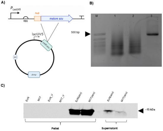
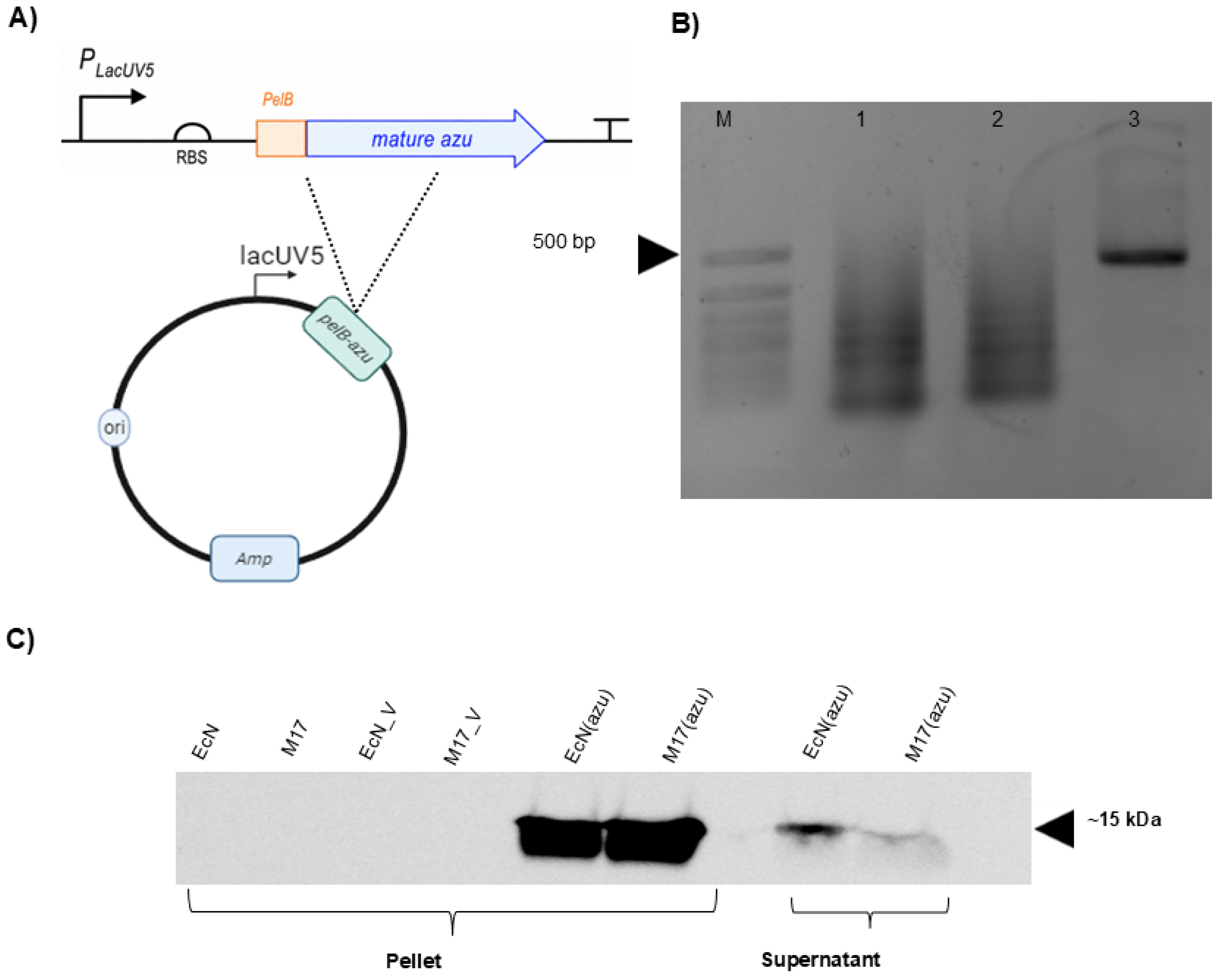
The lacUV5-pelB-azu expression cassette (Figure 1A) was generated for recombinant azurin production in EcN and M17. The cassette contained the mature azurin sequence fused to the pelB signal peptide and placed downstream of the lacUV5 promoter and ribosome-binding site (RBS). Agarose gel electrophoresis confirmed assembly of the expected cassette (Figure 1B), and the final plasmid sequence was verified by Sanger sequencing. The resulting construct was introduced into EcN and M17 to generate EcN(azu) and M17(azu), respectively.
Figure 1.
Generation and verification of azurin producing probiotic E. coli strains. (A) Schematic representation of pET303[lacUV5-pelB-azu] expression plasmid used for azurin production in E. coli Nissle 1917 (EcN) and E. coli M17. The mature azu coding sequence was placed downstream of the lacUV5 promoter, RBS, and pelB signal peptide to direct recombinant azurin towards the periplasmic compartment. (B) Agarose gel electrophoresis confirming assembly of the azurin expression cassette, where M, 50 bp DNA ladder (Evrogen, Moscow, Russia); lanes 1–2, PCR products of the first round of oligonucleotides assembly; lane 3, PCR product of the second round of oligonucleotides assembly. The expected product corresponding to the lacUV5-pelB-azu cassette is visible at approximately 587 bp. (C) Western blot detection of His-tagged recombinant azurin in engineered EcN and M17 strains. Pellet fractions and concentrated culture supernatants were analyzed after 16 h of 1 mM IPTG induction. No azurin signal was detected in wild-type EcN, wild-type M17, or empty-vector controls (EcN_V, M17_V.). Recombinant azurin was detected in EcN(azu) and M17(azu), predominantly in the pellet fraction and at lower intensity in concentrated supernatants. Approximate molecular weight markers are shown on the right in kDa.
3.2. EcN(azu) and M17(azu) Produced Recombinant Azurin Predominantly in the Cell-Associated Fraction
Western blotting was performed to evaluate azurin production in EcN(Azu) and M17(Azu). Clear immunoreactive bands corresponding to recombinant azurin were detected in the pellet fractions of both engineered strains, whereas no corresponding signal was observed in wild-type or empty-vector control samples (Figure 1C). These data confirmed that the lacUV5-pelB-azu cassette supported recombinant azurin production in both probiotic E. coli backgrounds.
Azurin was detected more strongly in the bacterial pellet fraction than in the supernatant fraction. This indicates that most detectable recombinant azurin remained cell-associated, most likely in the periplasmic compartment. Lower intensity azurin signals were also observed in concentrated culture supernatant fractions.
Semi-quantitative densitometric analysis was subsequently used to estimate cell-associated azurin at different bacterial inputs (Figure S3, Table S2). At bacteria-to-cell ratios of 500 and 1000, the estimated azurin amounts were 0.35 and 0.68 µg/lane for EcN(azu), respectively, and 0.28 and 0.35 µg/lane for M17(azu). At ratios of 100 and 250, the azurin signal was below the lower limit of the calibration range (0.21 µg/lane) and was therefore not quantified. These measurements indicate increased detectable cell-associated azurin at the higher bacterial inputs, while the analysis was not designed to determine extracellular azurin availability during mammalian-cell co-culture.
3.3. Purified Recombinant Azurin Reduced SRB-Assessed HCT116 Cell Biomass/Viability
Before evaluating the engineered bacterial strains, a purified recombinant azurin preparation was tested as a functional reference independent of the EcN and M17 backgrounds. SDS-PAGE analysis showed a predominant band corresponding to recombinant azurin, and the purity of the preparation used for the cell-based experiments was estimated at approximately 88% via densitometric analysis (Figure S4).
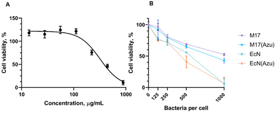
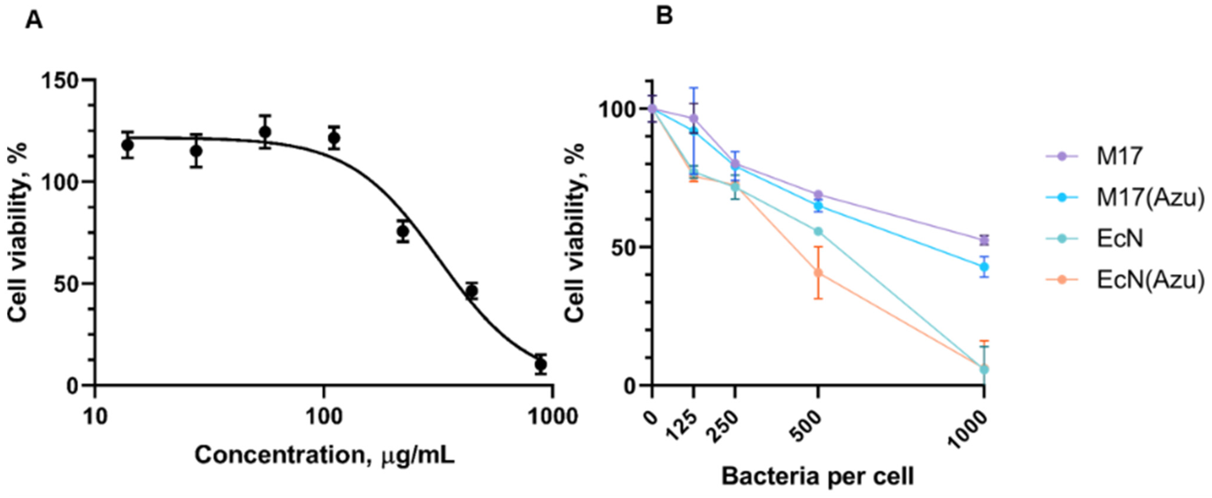
Following purification and dialysis into PBS, HCT116 cells were treated with increasing concentrations of the recombinant azurin preparation for 48 h. Recombinant azurin produced a concentration-dependent reduction in SRB signal (Figure 2A). Fitting the dose–response data with a four-parameter logistic model gave an estimated IC50 of 320 µg/mL.
Figure 2.
Effects of purified recombinant azurin and azurin-producing probiotic E. coli strains on HCT116 cell biomass/viability. (A) HCT116 cells were treated with increasing concentrations of purified recombinant azurin from 7.8 to 1000 µg/mL for 48 h, and cell biomass/viability was assessed using the SRB assay. Purified-protein values were normalized to the matched buffer control. The dose–response curve was fitted using a four-parameter logistic model, giving an estimated IC50 of approximately 320 µg/mL. (B) HCT116 cells were exposed to wild-type M17, M17(azu), wild-type EcN, and EcN(azu) at bacteria-to-cell ratios of 125, 250, 500, and 1000. After 2 h of bacterial exposure, gentamicin was added, and cells were incubated for 48 h, after SRB assay was performed. Results are expressed relative to untreated control cells. All data are presented as mean ± SD, N = 3.
These results indicate that the recombinant azurin preparation retained measurable biological activity following purification and dialysis. The relatively high concentration required to produce a pronounced SRB response also indicates that substantial extracellular exposure was required under the conditions used here.
3.4. Effects of Wild-Type and Azurin-Producing EcN and M17 on SRB-Assessed HCT116 Cell Biomass
The effects of wild-type and azurin-producing EcN and M17 on HCT116 cells were evaluated after 48 h using the SRB assay. HCT116 cells were exposed to bacteria for 2 h at bacteria-per-cell ratios of 125, 250, 500, and 1000, followed by gentamicin treatment and incubation to the 48 h endpoint.
All bacterial treatments reduced HCT116 cell biomass/viability in a bacteria-per-cell-dependent manner, but the magnitude of the response differed between the two strain backgrounds (Figure 2B). Wild-type M17 caused a gradual reduction in SRB signal, with approximately half of the cell biomass remaining at the highest bacterial dose. M17(Azu) showed lower mean SRB values than wild-type M17; however, the difference did not reach statistical significance.
Wild-type EcN showed a stronger inhibitory effect than wild-type M17. At the highest bacterial dose wild-type EcN reduced HCT116 biomass/viability to very low levels. The numerical difference between EcN and EcN(azu) was most apparent at intermediate bacteria-to-cell ratios, although this difference also did not reach statistical significance. At a bacteria-to-cell ratio of 1000, both EcN and EcN(azu) produced similarly strong reductions, limiting the ability to distinguish an additional azurin-associated effect under this condition.
Across the SRB assay, the azurin-producing derivatives showed lower mean HCT116 biomass values than their corresponding wild-type strains; however, these differences did not reach statistical significance under the tested conditions. The strain-dependent pattern remained apparent descriptively, with EcN producing a stronger baseline reduction than M17 and with EcN and EcN(azu) converging at the highest bacterial dose. The SRB results were therefore interpreted as evidence of differences in the overall HCT116 biomass response rather than as definitive evidence of azurin-induced cell death. This observation motivated subsequent analysis of the cell-death profile by YO-PRO-1/PI flow cytometry.
3.5. Azurin-Producing E. coli Increased the Apoptosis-Associated Fraction of HCT116 Cells
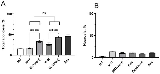
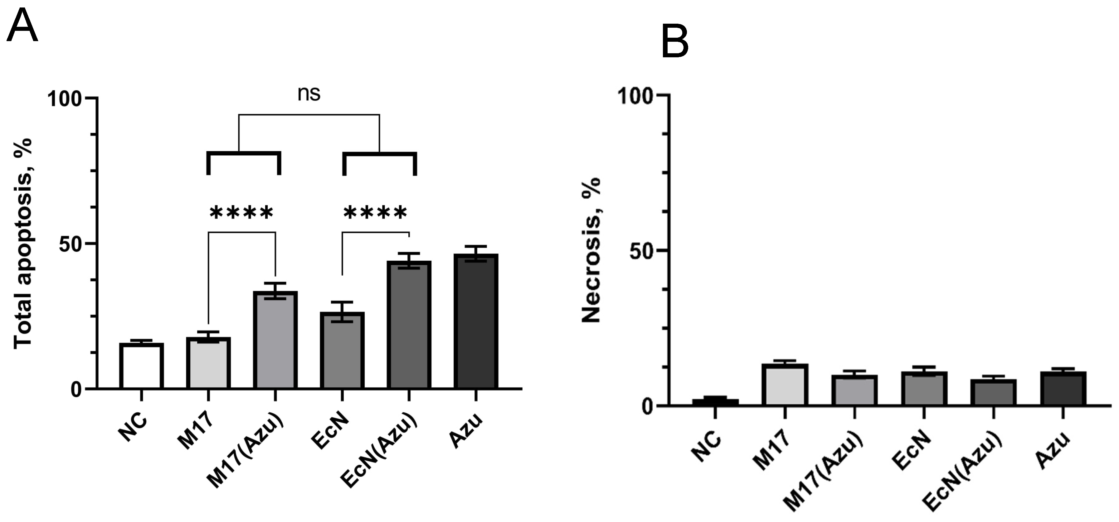
To further characterize whether the changes observed in the SRB assay were accompanied by cell-death-associated membrane changes, HCT116 cells were analyzed after 48 h by YO-PRO-1/PI flow cytometry at a bacteria-to-cell ratio of 500 (Figure 3 and Figure S7). This condition was selected for comparative cell-death profiling and was not intended to establish a dose–response relationship. Untreated cells, wild-type M17 and EcN, the corresponding azurin-producing strains, and the recombinant azurin preparation were included in the analysis.
Figure 3.
Cell-death profile of HCT116 cells following treatment with wild-type and azurin-producing E. coli. HCT116 cells were treated with wild-type M17, M17(azu), wild-type EcN, EcN(azu), or recombinant azurin and analyzed after 48 h. Bacterial treatments were performed at a bacteria-to-cell ratio of 500. Cell-death-associated membrane permeability was assessed by YO-PRO-1/PI staining followed by flow cytometry. (A) Total apoptosis-associated fraction, calculated as the sum of YO-PRO-1+/PI− early apoptosis-associated cells and YO-PRO-1+/PI+ late apoptosis-associated cells. (B) YO-PRO-1−/PI+ necrosis-associated population. Data are presented as mean ± SD from three biological replicates. Statistical analysis of the bacterial treatment groups was performed using two-way ANOVA with bacterial background and azurin production as factors, followed by Sidak’s multiple-comparison test. Recombinant azurin was included as a reference treatment and was not included in the two-way ANOVA model. ns, not significant; ****—p-value < 0.0001.
Wild-type M17 produced only a limited increase in the total apoptosis-associated population compared with the untreated control, whereas M17(azu) showed a substantially higher fraction. Wild-type EcN exhibited a higher baseline apoptosis-associated fraction than wild-type M17, consistent with the stronger intrinsic effect of EcN observed in the SRB experiments. EcN(azu) further increased this population relative to wild-type EcN. The recombinant azurin preparation produced the highest apoptosis-associated fraction among the tested conditions, consistent with its independently observed activity in the SRB assay.
Two-way ANOVA identified a significant main effect of azurin expression on the total apoptosis-associated fraction (p < 0.0001), accounting for 68.99% of the total variation. Bacterial strain accounted for 29.22% of the variation, whereas the strain × azurin interaction accounted for only 0.48% and was not significant (p = 0.4349), indicating that the magnitude of the azurin-associated increase did not differ significantly between the M17 and EcN backgrounds. Relative to their corresponding wild-type strains, azurin production increased the apoptosis-associated population approximately 1.77-fold in M17 and 1.56-fold in EcN.
In contrast to the apoptosis-associated response, the necrosis-associated population remained low across the bacterial treatment groups, and no statistically significant differences were detected (Figure 3B). Thus, under the tested conditions, the principal change detected by YO-PRO-1/PI staining was an increase in the apoptosis-associated population rather than a pronounced increase in acute membrane-compromised cells.
4. Discussion
EcN and M17 were successfully modified using the same lacUV5-pelB-azu expression cassette, and recombinant azurin was detected in both strain backgrounds. Both azurin-producing derivatives showed lower mean SRB-assessed HCT116 cell biomass than their corresponding wild-type strains, although these differences did not reach statistical significance in the SRB assay. Complementary YO-PRO-1/PI analysis demonstrated that the azurin-producing derivatives were associated with a significant overall increase in the total apoptosis-associated population, providing additional evidence that the recombinant payload contributed to the HCT116 response.
The design of the expression cassette was guided by the natural localization of azurin. Native P. aeruginosa azurin is a periplasmic copper protein synthesized with an N-terminal signal peptide [24,25]. Therefore, the mature azurin sequence was fused to the PelB signal peptide to promote its transport towards the periplasm in EcN and M17 and thereby increase its potential availability beyond the bacterial cytoplasm [22,26]. A similar strategy was used by Zhang et al., who expressed mature azurin with a PelB leader sequence in EcN and detected the protein in both bacterial pellets and culture supernatants [22]. In the same study, full-length azurin carrying its native sequence was not detectably secreted by EcN, showing that the native localization signal from P. aeruginosa did not function equivalently in this heterologous bacterial background.
In the present work, azurin was detected in both pellet and culture-supernatant fractions, although the signal was predominantly cell-associated. This general distribution is consistent with previous findings, but it also indicates that PelB-mediated targeting did not result in efficient release of most of the recombinant protein into the culture medium. The mechanism responsible for extracellular azurin detection was not investigated in the present study.
The apparent difference between the EcN(azu) and M17(azu) supernatant signals is potentially relevant because the same construct may behave differently in distinct strain backgrounds. Differences in envelope physiology, stress response, proteolysis or membrane permeability could affect the amount of recombinant protein detected outside the cell [6,8,27]. However, the qualitive pellet/supernatant Western blot shown in Figure 1C was not designed for quantitative comparison between strains.
The recombinant azurin preparation produced a concentration-dependent reduction in SRB-assessed HCT116 biomass, with an estimated IC50 of approximately 320 µg/mL.
This value is comparable to that reported by Ma et al. for purified recombinant azurin in colorectal cancer cell models, where IC50 values of approximately 405.5 µg/mL and 415.8 µg/mL were obtained for human HT29 and murine CT26 cells, respectively [28]. However, direct quantitative comparison should be made cautiously because Ma et al. used a CCK-8 assay after 24 h of treatment, whereas the present study used an SRB endpoint after 48 h.
Because responses were normalized to a matched buffer control, the observed reduction cannot be attributed to the dialysis vehicle itself. The experiment provides an independent functional reference for the bacterial co-culture experiments and was used qualitatively to establish that independently produced recombinant azurin was biologically active in HCT116 cells; however, because the preparation was approximately 88% pure, a contribution from residual co-purified proteins cannot be fully excluded.
The purified-protein and bacterial treatment formats should nevertheless not be directly equated. The recombinant preparation was applied as a defined bulk concentration, whereas bacterial treatment involved ongoing protein production, predominantly cell-associated azurin, and direct interaction between bacteria and mammalian cells.
The substantial concentration of purified azurin required to produce a marked SRB response suggests protein availability may be an important consideration for the current bacterial design. Semi-quantitative Western blot analysis provided an estimate of cell-associated azurin at the tested bacterial inputs, with detectable amounts within the calibration range at bacteria-to-cell ratios of 500 and 1000. However, these measurements do not determine the extracellular concentration or local azurin exposure experienced by HCT116 cells during co-culture. Local exposure near bacterial cells may differ substantially from the average concentration in the medium. It therefore cannot be concluded that the tested bacterial doses failed to reach a biologically relevant local concentration.
The absence of statistically significant differences between wild-type and azurin-producing bacteria in the SRB assay should also be considered in relation to what this assay measures. SRB staining provides an endpoint estimate of adherent cellular protein and therefore reflects the combined effects of cell number, proliferation and cell loss rather than distinguishing between cytostatic and cytotoxic responses. The SRB experiment therefore provided the overall dose–response context for subsequent cell-death profiling.
Empty-vector controls were also evaluated to determine whether plasmid carriage contributed detectably to the HCT116 response. Across the tested bacteria-to-cell ratios, the empty-vector derivatives closely followed the SRB response profiles of their corresponding wild-type strains (Figure S5). No significant main effect of vector carriage or vector-by-dose interaction was detected. This observation was consistent with the supplementary bacterial growth data (Figure S6), in which plasmid carriage and the induction conditions did not produce a detectable growth disadvantage. Together, these controls support the use of the wild-type strains as the principal chassis comparators.
The M17 background produced the clearest separation between wild-type and azurin-producing bacteria. Wild-type M17 caused a moderate, dose-dependent reduction in HCT116 biomass, while M17(azu) showed numerically lower mean biomass values than the wild-type strain. M17 has received considerably less attention than EcN as an engineered protein-delivery strain, and the current results do not establish that it is therapeutically superior. They do, however, suggest that its lower baseline effect may be useful when the experimental objective is to distinguish the contribution of a recombinant payload from that of the bacterial strain.
EcN showed a different activity profile. Wild-type EcN strongly reduced HCT116 cell biomass, with an almost complete loss of SRB signal at the highest bacterial dose. This shows that EcN itself was not biologically neutral in the present co-culture system. Alizadeh et al. similarly reported inhibitory effects of EcN-related treatments on HT-29 colorectal cancer cells. In their study, conditioned medium generated during EcN–HT-29 co-culture and heat-inactivated EcN produced apoptosis-associated changes involving PTEN, AKT1, Bax and Bcl-xL [15]. This baseline effect can also arise from direct bacteria-cell interaction or released bacterial metabolites [29,30]. The strong response at a bacteria-to-cell ratio of 1000 also created a floor effect, as little residual cell biomass remained against which an additional contribution from azurin could be measured. The convergence of EcN and EcN(azu) at this dose indicates that the assay had limited capacity to resolve an additional effect once the chassis alone had produced near-maximal inhibition. This distinction is important when comparing bacterial platforms as the strain that produces the greatest total reduction in cancer-cell biomass is not necessarily the strain that provides the clearest measure of recombinant payload activity.
A bacteria-to-cell ratio of 500 was selected for the YO-PRO-1/PI experiment because it represented an intermediate condition at which the SRB data showed separation between wild-type and azurin-producing bacteria while avoiding the near-complete loss of HCT116 biomass observed with EcN at a ratio of 1000. In the SRB assay, lower bacterial doses produced smaller overall effects, whereas the highest dose reduced the dynamic range available for distinguishing chassis- and payload-associated responses. The intermediate ratio therefore provided a more informative condition for evaluating whether the biomass phenotype was accompanied by changes in the cell-death profile.
The YO-PRO-1/PI analysis provides an additional perspective on this strain-dependent response. At a bacteria-to-cell ratio of 500, the azurin-producing derivatives showed a significant overall increase in the total apoptosis-associated fraction across the two bacterial backgrounds. When normalized to the corresponding wild-type strains, this increase was approximately 1.77-fold for M17(azu) and 1.56-fold for EcN(azu). However, the absolute increases were similar, approximately 14.6 and 16.0 percentage points, respectively, and no significant interaction between bacterial background and azurin production was detected. Thus, the apparently larger relative effect in M17 primarily reflects its lower baseline activity rather than a substantially greater responsiveness to azurin. In EcN, a similar azurin-associated increment occurred on top of a stronger chassis-derived response.
The response to the recombinant azurin preparation was also consistent across the two assay formats. The preparation reduced SRB-assessed HCT116 biomass in a concentration-dependent manner and produced a pronounced apoptosis-associated population in the YO-PRO-1/PI analysis.
In contrast, the necrosis-associated population remained comparatively low and showed no significant differences between bacterial treatment groups. This suggests that the dominant membrane-permeability change detected under these conditions was associated with an apoptosis-associated rather than an acute membrane-destructive phenotype. Nevertheless, YO-PRO-1/PI staining does not establish the upstream molecular mechanism of apoptosis and, therefore, it is only considered as evidence of an apoptosis-associated cell-death profile rather than definitive demonstration of a specific apoptotic pathway.
The present findings are consistent with previous work using alternative bacterial platforms for azurin delivery. Ma et al. generated non-replicating EcN-derived minicells containing recombinant azurin and reported increased apoptosis in both CT26 and HT29 colorectal cancer cells compared with control minicells. In their study, early apoptosis increased approximately 2.3-fold in CT26 cells and 1.3-fold in HT29 cells following treatment with azurin-containing minicells [28]. The two experimental systems are nevertheless distinct. Minicells provide a non-replicating delivery platform, whereas the present study used live E. coli in direct co-culture with HCT116 cells. Consequently, the response measured here reflects both recombinant azurin production and the intrinsic biological activity of the bacterial chassis. This distinction is particularly apparent for EcN, which produced a substantial baseline response even in the absence of the recombinant payload.
5. Conclusions
Several limitations remain. Although cell-associated azurin was estimated by semi-quantitative Western blotting, its extracellular concentration and local availability to HCT116 cells were not determined. YO-PRO-1/PI analysis was performed at a single selected bacteria-to-cell ratio and does not establish the underlying apoptotic pathway. In addition, the study used a single colorectal carcinoma cell line without a non-malignant intestinal epithelial control, and the in vivo safety of the engineered strains was not evaluated.
This work demonstrates that the effect of an engineered probiotic strain cannot be attributed to the recombinant payload alone, as the biological properties of the bacterial chassis can substantially influence the final host–cell response. In the present system, M17 provided a lower-background platform in which the additional response associated with azurin production was more readily distinguished from the intrinsic response of the wild-type chassis, whereas the stronger intrinsic activity of EcN complicated their separation. These findings support the importance of considering chassis-dependent activity when evaluating engineered bacteria for recombinant protein delivery.
Supplementary Materials
The following supporting information can be downloaded at https://www.mdpi.com/article/10.3390/applmicrobiol6100126/s1. Figure S1. Annotated nucleotide sequence of the lacUV5-pelB-azu-His expression cassette used in EcN(azu) and M17(azu); Figure S2. Electrophoresis of Azurin obtained from Pseudomonas aeruginosa DNA; Figure S3. Semi-quantitative Western blot analysis of cell-associated recombinant azurin; Figure S4. SDS-PAGE analysis of independently purified recombinant azurin preparations; Figure S5. Effect of empty-vector carriage on HCT116 cell biomass during co-culture with M17 and EcN; Figure S6. Growth analysis of EcN(azu), and M17(azu); Figure S7. Representative YO-PRO-1/PI flow-cytometry gating strategy of HCT116 cells following bacterial and recombinant azurin treatment; Figure S8. Representative YO-PRO-1/PI flow-cytometry gating of HCT116 cells following bacterial and recombinant azurin treatment; Table S1. Oligonucleotides used to assemble the lacUV5_PelB_azu cassette; Table S2. Semi-quantitative estimates of cell-associated recombinant azurin in EcN(azu) and M17(azu).
Author Contributions
Conceptualization, N.D. and G.O.; methodology, N.D., A.S. and G.O.; validation, A.T. and E.M.; formal analysis, N.D., A.S. and G.O.; investigation, N.D. and G.O.; data curation, G.O.; writing—original draft preparation, N.D.; writing—review and editing, G.O. and E.K.; supervision, E.K. All authors have read and agreed to the published version of the manuscript.
Funding
This research received no external funding.
Institutional Review Board Statement
Not applicable.
Informed Consent Statement
Not applicable.
Data Availability Statement
The original contributions presented in this study are included in the article/Supplementary Materials. Further inquiries can be directed to the corresponding author.
Acknowledgments
We would like to thank the laboratory of Amyloid Biology St. Petersburg State University for the extraction and purification of azurin.
Conflicts of Interest
The authors declare no conflicts of interest.
Abbreviations
The following abbreviations are used in this manuscript:
| DMEM | Dulbecco’s Modified Eagle Medium |
| EcN | Escherichia coli Nissle 1917 |
| FBS | Fetal bovine serum |
| HRP | Horseradish peroxidase |
| IC50 | Half-maximal inhibitory concentration |
| IPTG | Isopropyl β-D-1-thiogalactopyranoside |
| PCR | Polymerase chain reaction |
| RBS | Ribosome-binding site |
| SDS-PAGE | Sodium dodecyl sulfate-polyacrylamide gel electrophoresis |
| SRB | Sulforhodamine B |
References
- Muhammad, S.; Li, M.; Jia, Q.; Ijaz, M.; Liang, S.; Zeng, W.; Chen, D.; Zhang, Y.; Du, X.; Song, W.; et al. Advances in the engineering of living probiotics for cancer immunotherapy. Theranostics 2026, 16, 1164–1226. [Google Scholar] [CrossRef] [Scilit] [PubMed]
- Pan, W.; Liu, H.; Wu, D. Advances in engineered bacteria for cancer therapy. Precis. Med. Eng. 2025, 2, 100017. [Google Scholar] [CrossRef] [Scilit]
- Kumar, V.; Barwal, A.; Sharma, N.; Mir, D.S.; Kumar, P.; Kumar, V. Therapeutic proteins: Developments, progress, challenges, and future perspectives. 3 Biotech 2024, 14, 112. [Google Scholar] [CrossRef] [Scilit] [PubMed]
- Gurbatri, C.R.; Lia, I.; Vincent, R.; Coker, C.; Castro, S.; Treuting, P.M.; Hinchliffe, T.E.; Arpaia, N.; Danino, T. Engineered probiotics for local tumor delivery of checkpoint blockade nanobodies. Sci. Transl. Med. 2020, 12, eaax0876. [Google Scholar] [CrossRef] [Scilit] [PubMed]
- Charbonneau, M.R.; Isabella, V.M.; Li, N.; Kurtz, C.B. Developing a new class of engineered live bacterial therapeutics to treat human diseases. Nat. Commun. 2020, 11, 1738. [Google Scholar] [CrossRef] [Scilit] [PubMed]
- Marisch, K.; Bayer, K.; Cserjan-Puschmann, M.; Luchner, M.; Striedner, G. Evaluation of three industrial Escherichia coli strains in fed-batch cultivations during high-level SOD protein production. Microb. Cell Factories 2013, 12, 58. [Google Scholar] [CrossRef] [Scilit] [PubMed]
- Chan, D.T.C.; Baldwin, G.S.; Bernstein, H.C. Revealing the host-dependent nature of an engineered genetic inverter in concordance with physiology. BioDesign Res. 2023, 5, 0016. [Google Scholar] [CrossRef] [Scilit] [PubMed]
- Cardinale, S.; Joachimiak, M.P.; Arkin, A.P. Effects of genetic variation on the E. coli host-circuit interface. Cell Rep. 2013, 4, 231–237. [Google Scholar] [CrossRef] [Scilit] [PubMed]
- Ukena, S.N.; Westendorf, A.M.; Hansen, W.; Rohde, M.; Geffers, R.; Coldewey, S.; Suerbaum, S.; Buer, J.; Gunzer, F. The host response to the probiotic Escherichia coli strain Nissle 1917: Specific up-regulation of the proinflammatory chemokine MCP-1. BMC Med. Genet. 2005, 6, 43. [Google Scholar] [CrossRef] [Scilit] [PubMed]
- Kittana, H.; Gomes-Neto, J.C.; Heck, K.; Geis, A.L.; Muñoz, R.R.S.; Cody, L.A.; Schmaltz, R.J.; Bindels, L.B.; Sinha, R.; Hostetter, J.M.; et al. Commensal Escherichia coli strains can promote intestinal inflammation via differential interleukin-6 production. Front. Immunol. 2018, 9, 2318. [Google Scholar] [CrossRef] [Scilit] [PubMed]
- Dangi, S.; Liew, D.S.; Subramaniyan, V.; Narayanan, K. Engineered Escherichia coli as a microbial cell factory for intracellular protein delivery: Strains, vectors, mechanisms, and therapeutic applications. Microb. Cell Factories 2026, 25, 94. [Google Scholar] [CrossRef] [Scilit] [PubMed]
- Ba, F.; Zhang, Y.; Ji, X.; Liu, W.; Ling, S.; Li, J. Expanding the toolbox of probiotic Escherichia coli Nissle 1917 for synthetic biology. Biotechnol. J. 2024, 19, e2300327. [Google Scholar] [CrossRef] [Scilit] [PubMed]
- Behrouzi, A.; Mazaheri, H.; Falsafi, S.; Tavassol, Z.H.; Moshiri, A.; Siadat, S.D. Intestinal effect of the probiotic Escherichia coli strain Nissle 1917 and its OMV. J. Diabetes Metab. Disord. 2020, 19, 597–604. [Google Scholar] [CrossRef] [Scilit] [PubMed]
- Yu, X.; Lin, C.; Yu, J.; Qi, Q.; Wang, Q. Bioengineered Escherichia coli Nissle 1917 for tumour-targeting therapy. Microb. Biotechnol. 2019, 13, 629. [Google Scholar] [CrossRef] [Scilit] [PubMed]
- Alizadeh, S.; Esmaeili, A.; Omidi, Y. Anti-cancer properties of Escherichia coli Nissle 1917 against HT-29 colon cancer cells through regulation of Bax/Bcl-xL and AKT/PTEN signaling pathways. Iran. J. Basic Med. Sci. 2020, 23, 886. [Google Scholar] [CrossRef] [Scilit] [PubMed]
- Bondarenko, V.M.; Fialkina, S.V. Presence of genes of genotoxin associated with pks pathogenicity island in Escherichia coli M-17 probiotic strain. J. Microbiol. Epidemiol. Immunobiol. 2012, 89, 25–27. [Google Scholar]
- Fitzpatrick, L.R.; Small, J.; Hoerr, R.A.; Bostwick, E.F.; Maines, L.; Koltun, W.A. In vitro and in vivo effects of the probiotic Escherichia coli strain M-17: Immunomodulation and attenuation of murine colitis. Br. J. Nutr. 2008, 100, 530–541. [Google Scholar] [CrossRef] [Scilit] [PubMed]
- Yamada, T.; Goto, M.; Punj, V.; Zaborina, O.; Chen, M.L.; Kimbara, K.; Majumdar, D.; Cunningham, E.; Das Gupta, T.K.; Chakrabarty, A.M. Bacterial redox protein azurin, tumor suppressor protein p53, and regression of cancer. Proc. Natl. Acad. Sci. USA 2002, 99, 14098–14103. [Google Scholar] [CrossRef] [Scilit] [PubMed]
- Punj, V.; Bhattacharyya, S.; Saint-Dic, D.; Vasu, C.; Cunningham, E.A.; Graves, J.; Yamada, T.; Constantinou, A.I.; Christov, K.; White, B.; et al. Bacterial cupredoxin azurin as an inducer of apoptosis and regression in human breast cancer. Oncogene 2004, 23, 2367–2378. [Google Scholar] [CrossRef] [Scilit] [PubMed]
- Taylor, B.N.; Mehta, R.R.; Yamada, T.; Lekmine, F.; Christov, K.; Chakrabarty, A.M.; Green, A.; Bratescu, L.; Shilkaitis, A.; Beattie, C.W.; et al. Noncationic peptides obtained from azurin preferentially enter cancer cells. Cancer Res. 2009, 69, 537–546. [Google Scholar] [CrossRef] [Scilit] [PubMed]
- Yamada, T.; Hiraoka, Y.; Ikehata, M.; Kimbara, K.; Avner, B.S.; Das Gupta, T.K.; Chakrabarty, A.M. Apoptosis or growth arrest: Modulation of tumor suppressor p53′s specificity by bacterial redox protein azurin. Proc. Natl. Acad. Sci. USA 2004, 101, 4770–4775. [Google Scholar] [CrossRef] [Scilit] [PubMed]
- Zhang, Y.; Zhang, Y.; Xia, L.; Zhang, X.; Ding, X.; Yan, F.; Wu, F. Escherichia coli Nissle 1917 targets and restrains mouse B16 melanoma and 4T1 breast tumors through expression of azurin protein. Appl. Environ. Microbiol. 2012, 78, 7603–7610. [Google Scholar] [CrossRef] [Scilit] [PubMed]
- Sambrook, J.; Fritsch, E.R. Molecular Cloning: A Laboratory Manual, 2nd ed.; Cold Spring Harbor Laboratory Press: Cold Spring Harbor, NY, USA, 1989. [Google Scholar]
- Raimunda, D.; Padilla-Benavides, T.; Vogt, S.; Boutigny, S.; Tomkinson, K.N.; Finney, L.A.; Argüello, J.M. Periplasmic response upon disruption of transmembrane Cu transport in Pseudomonas aeruginosa. Metallomics 2013, 5, 144–151. [Google Scholar] [CrossRef] [Scilit] [PubMed]
- Canters, G.W. The azurin gene from Pseudomonas aeruginosa codes for a pre-protein with a signal peptide. Cloning and sequencing of the azurin gene. FEBS Lett. 1987, 212, 168–172. [Google Scholar] [CrossRef] [Scilit] [PubMed]
- Zhang, W.; Lu, J.; Zhang, S.; Liu, L.; Pang, X.; Lv, J. Development an effective system to expression recombinant protein in E. coli via comparison and optimization of signal peptides: Expression of Pseudomonas fluorescens BJ-10 thermostable lipase as case study. Microb. Cell Factories 2018, 17, 50. [Google Scholar] [CrossRef] [Scilit] [PubMed]
- Xia, X.X.; Han, M.; Lee, S.Y.; Yoo, J. Comparison of the extracellular proteomes of Escherichia coli B and K-12 strains during high cell density cultivation. Proteomics 2008, 8, 2089–2103. [Google Scholar] [CrossRef] [Scilit] [PubMed]
- Ma, Y.; Zhu, G.; Feng, L.; Jiang, S.; Xiang, Q.; Wang, J. Efficient Cytotoxicity of Recombinant Azurin in Escherichia coli Nissle 1917-Derived Minicells against Colon Cancer Cells. Bioengineering 2023, 10, 1188. [Google Scholar] [CrossRef] [Scilit] [PubMed]
- Damoogh, S.; Vosough, M.; Hadifar, S.; Rasoli, M.; Gorjipour, A.; Falsafi, S.; Behrouzi, A. Evaluation of E. coli Nissle 1917 derived metabolites in modulating key mediator genes of the TLR signaling pathway. BMC Res. Notes 2021, 14, 156. [Google Scholar] [CrossRef] [Scilit] [PubMed]
- Cañas, M.A.; Giménez, R.; Fábrega, M.-J.; Toloza, L.; Baldomà, L.; Badia, J. Outer membrane vesicles from the probiotic Escherichia coli Nissle 1917 and the commensal ECOR12 enter intestinal epithelial cells via clathrin-dependent endocytosis and elicit differential effects on DNA damage. PLoS ONE 2016, 11, e0160374. [Google Scholar] [CrossRef] [Scilit] [PubMed]
Disclaimer/Publisher’s Note: The statements, opinions and data contained in all publications are solely those of the individual author(s) and contributor(s) and not of MDPI and/or the editor(s). MDPI and/or the editor(s) disclaim responsibility for any injury to people or property resulting from any ideas, methods, instructions or products referred to in the content. |
© 2026 by the authors. Licensee MDPI, Basel, Switzerland. This article is an open access article distributed under the terms and conditions of the Creative Commons Attribution (CC BY) license.