


Simple Summary
The spread of cancer to distant organs, known as metastasis, is the primary cause of cancer-related deaths. Historically, researchers focused on permanent genetic mutations as the main driver of this process. However, it is now clear that cancer cells also heavily rely on “epigenetics,” reversible chemical tags added to DNA and proteins that act like switches to turn specific genes on or off. By hijacking these switches, cancer cells gain the ability to detach from the original tumor, hide from the immune system, and colonize new organs. This review breaks down exactly how these epigenetic mechanisms orchestrate the spread of cancer and how tumors manipulate their surrounding environment to fuel this process. Crucially, because epigenetic changes are reversible, they present excellent targets for new medical treatments. We highlight how emerging “epigenetic drugs” can reset these cellular switches and how combining them with therapies that boost the immune system could stop cancer metastasis, offering a promising roadmap for more effective and precise cancer treatments.
Abstract
Cancer metastasis is the leading cause of cancer-related mortality, accounting for more than 90% of cancer deaths worldwide. However, the epigenetic mechanisms governing the metastatic cascade remain incompletely understood. Epigenetic reprogramming, including reversible changes in histone modifications, DNA methylation, chromatin remodeling, and non-coding RNA (ncRNA)-mediated regulation, enables tumor cells to acquire invasive, migratory, stem-like, and immune-evasive characteristics. During epithelial-to-mesenchymal transition (EMT), key epigenetic regulators such as histone deacetylases (HDACs), the Polycomb repressive complex 2 (PRC2) subunit EZH2, lysine-specific demethylase 1 (LSD1/KDM1A), and bromodomain and extraterminal (BET) proteins repress epithelial gene expression while activating mesenchymal transcriptional programs, promoting invasion and dissemination. At distant sites, epigenetic plasticity facilitates metastatic colonization through mesenchymal-to-epithelial transition (MET) and adaptive chromatin remodeling. Because these changes are reversible, they represent attractive therapeutic targets. HDAC, EZH2, LSD1/KDM1A, BET, and DNA methyltransferase (DNMT) inhibitors have shown promise in preclinical models of metastasis, with several advancing through clinical trials. Long non-coding RNAs, particularly HOTAIR, function as epigenetic scaffolds that reinforce metastatic programs, while reciprocal interactions between tumor cells and the tumor microenvironment (TME) drive epigenetic adaptations that promote immune evasion and metastatic progression. In addition, circulating tumor DNA (ctDNA) methylation signatures are emerging as minimally invasive biomarkers for assessing metastatic risk and monitoring treatment. This review summarizes current insights into the epigenetic regulation of cancer metastasis, evaluates emerging epigenetic therapies, and highlights translational opportunities to advance precision anti-metastatic strategies and improve patient outcomes.
1. Introduction
Cancer remains one of the leading causes of mortality worldwide, with an estimated 20.0 million new cancer cases and 9.7 million cancer-related deaths reported globally in 2022 [1]. Metastasis continues to be the primary cause of cancer mortality, accounting for approximately 90% of cancer-related deaths, yet the epigenetic mechanisms orchestrating each stage of the metastatic cascade remain incompletely characterized [2,3,4]. Despite decades of research, the molecular basis of metastatic competence remains only partially understood, partly because the dominant conceptual framework has historically privileged genetic drivers. Emerging evidence, however, positions epigenetic reprogramming as an equally and in some contexts more fundamental determinant of metastatic potential.
Epigenetic regulation refers to heritable, reversible modifications that alter gene expression without changing the underlying DNA sequence. The principal epigenetic layers include DNA methylation at CpG dinucleotides, post-translational modifications of histone tails, ATP-dependent chromatin remodeling, and regulation by non-coding RNAs. These mechanisms operate in concert to define transcriptional programs that govern cell identity, differentiation, and plasticity. In cancer, the epigenome undergoes widespread, non-random reprogramming that enables tumor cells to acquire capabilities for invasiveness, resistance to anoikis, immune evasion, and stem-like self-renewal, which are prerequisites for successful metastasis [5,6].
A defining feature of epigenetic changes, and a key reason for their therapeutic appeal, is their dynamic and pharmacologically reversible nature. Unlike genetic mutations, aberrant epigenetic states can, in principle, be reset by inhibitors targeting the enzymatic writers, erasers, and readers of histone marks or DNA methylation. This property has catalyzed intense drug development efforts, yielding several clinically approved agents and a robust pipeline of investigational compounds. Yet translating mechanistic insights into effective anti-metastatic therapies requires a comprehensive understanding of how epigenetic alterations are wired into the metastatic cascade [7].
Several excellent reviews have addressed individual epigenetic mechanisms or specific types of modifications in the context of cancer progression [5,8,9]. While the recent literature, including the 2026 review by Sun et al. [7], provides a comprehensive general overview of epigenetic regulation in cancer, the present review establishes a distinct, stage-gated translational framework. We systematically examine the epigenetic landscape across all major regulatory layers: histone modifications, DNA methylation, non-coding RNAs, and critically, ATP-dependent chromatin remodeling and 3D genome architecture. Furthermore, we explicitly address the epigenetic regulation of metastatic dormancy and organ-specific colonization, which represent critical frontiers in anti-metastatic therapy. By mapping these specific chromatin state transitions across the metastatic cascade to clinical trial readiness, we aim to provide a highly reproducible, mechanism-to-clinic roadmap that advances precision anti-metastatic strategies.
Literature Search Strategy and Selection Criteria
To ensure a reproducible and comprehensive assessment of the literature, a systematic search strategy was employed. Electronic databases, including PubMed/MEDLINE, Web of Science, Embase, and ClinicalTrials.gov, were searched for articles published up to May 2026. The search utilized combinations of the following MeSH terms and keywords: (“epigenetics” OR “DNA methylation” OR “histone modification” OR “chromatin remodeling” OR “lncRNA”) AND (“cancer metastasis” OR “epithelial-mesenchymal transition” OR “metastatic dormancy” OR “tumor microenvironment”) AND (“epigenetic therapy” OR “HDAC inhibitors” OR “EZH2 inhibitors” OR “clinical trials”). Language limits were restricted to English peer-reviewed publications. Inclusion criteria prioritized original research articles, high-impact clinical trial reports, and comprehensive meta-analyses providing mechanistic or clinical evidence of epigenetic regulation in metastatic progression. Exclusion criteria included non-peer-reviewed preprints, abstracts without full texts, and studies that focused solely on primary tumorigenesis without assessing metastatic phenotypes. Clinical trials were selected based on their focus on solid tumor metastasis and on the use of epigenetic-modifying agents, either as monotherapies or in combination with immune checkpoint blockade, prioritizing trials with reported Phase I–III outcomes.
2. Epigenetic Landscape of the Metastatic Cascade
Metastasis is not a singular event but an ordered, multistep biological process comprising local invasion, intravasation into the vasculature or lymphatics, survival in circulation, extravasation, and colonization of distant organs. At each stage, tumor cells must overcome formidable physical, immunological, and metabolic barriers while acquiring and shedding distinct phenotypic states. Epigenetic reprogramming is now understood to serve as the molecular infrastructure that endows tumor cells with the transcriptional plasticity required to traverse these steps [8,10].
A central conceptual advance is the recognition that epigenetic and genetic drivers of metastasis are neither redundant nor mutually exclusive. Whereas genetic mutations are largely irreversible, epigenetic changes are inherently dynamic; the same cell can shift between epithelial and mesenchymal states, between proliferative and quiescent programs, or between drug-sensitive and drug-resistant phenotypes without accumulating new somatic mutations. This epigenetic plasticity is both the vulnerability that enables metastatic dissemination and the biological rationale for pharmacological intervention [6].
Crosstalk between epigenetic layers adds complexity. DNA methylation at promoter CpG islands is mechanistically coupled to the histone modification landscape: hypermethylated promoters characteristically carry repressive H3K27me3 or H3K9me3 marks, whereas actively transcribed genes display H3K4me3 and H3K27 acetylation. These associations reflect direct molecular interactions, for example, the PRC2–DNMT axis, in which the PRC2 complex both deposits H3K27me3 and recruits DNMTs to enforce and stabilize transcriptional silencing. Non-coding RNAs, particularly lncRNAs, add another regulatory dimension by serving as molecular scaffolds that recruit chromatin-modifying complexes to specific genomic loci. Understanding these layers individually and their mutual interactions is essential for identifying the most effective intervention points.
To contextualize these complex regulatory networks for translational application, it is critical to map when and where these epigenetic enzymes act during cancer progression. Rather than functioning in isolation, specific epigenetic writers, erasers, and readers dominate distinct phases of metastasis. Table 1 provides a stage-by-mechanism matrix that synthesizes these regulators across the metastatic cascade, from local invasion and circulation to dormancy, colonization, and therapy resistance, and links them to specific cancer types and their current evidence levels in the drug development pipeline.
Table 1.
Epigenetic Regulators Across the Metastatic Cascade: A Stage-by-Mechanism Matrix.
3. Histone Modifications in Metastatic Reprogramming
3.1. Histone Acetylation and HDACs
Histone acetylation catalyzed by histone acetyltransferases (HATs) and reversed by histone deacetylases (HDACs) regulates chromatin accessibility and transcriptional output. Acetylation of histone H3 at lysine 9 (H3K9ac) and lysine 27 (H3K27ac) marks active enhancers and promoters, while HDAC-mediated deacetylation promotes chromatin compaction and transcriptional repression. In cancer, class I HDACs (HDAC1, 2, 3, 8) are frequently overexpressed and contribute to the silencing of tumor suppressor genes, anti-invasive programs, and immune recognition machinery [9]. Dynamic changes in chromatin structure heavily dictate the epigenetic landscape of metastasis. A summary of key histone modifications, their respective enzymatic writers and erasers, and their functional roles across various cancer types is provided in Table 2.
Table 2.
Key Histone Modifications in Cancer Metastasis.
HDAC-mediated reprogramming is particularly critical during EMT. HDAC1 and HDAC2 associate with the SNAIL and ZEB transcription factors to deacetylate and silence the CDH1 promoter (which encodes E-cadherin), a pivotal event in the acquisition of an invasive phenotype. Class II HDACs, notably HDAC6, regulate the acetylation of non-histone substrates, including cortactin and alpha-tubulin, thereby modulating the cytoskeletal dynamics required for cell migration. HDAC inhibitors (HDACi) have demonstrated the capacity to reverse EMT-associated gene expression programs, restore E-cadherin expression, and suppress cancer cell migration and invasion in multiple preclinical models [9,13].
The functional consequences of HDAC activity extend beyond direct transcriptional silencing. HDAC-mediated deacetylation modulates the activity of key metastatic signaling nodes, including the TGF-β, Wnt/β-catenin, and NF-κB pathways, either by modifying histone marks at their target gene promoters or by directly deacetylating pathway components. This dual role as chromatin regulator and signal modifier underscores the broad influence of HDAC activity on metastatic programs and provides multiple mechanistic rationales for therapeutic targeting.
3.2. Histone Methylation: EZH2 and the PRC2 Axis
Histone methylation is distinguished from acetylation by its context dependence: mono-, di-, and tri-methylation of the same lysine residue can confer distinct, even opposing, functional consequences. Trimethylation of histone H3 at lysine 27 (H3K27me3), catalyzed by EZH2, the enzymatic subunit of PRC2, is one of the best-characterized repressive marks in cancer biology. EZH2 is overexpressed or mutated in a broad spectrum of malignancies, including breast, prostate, bladder, ovarian, and non-Hodgkin lymphoma, and its overexpression strongly correlates with advanced-stage disease and metastatic dissemination [8,11].
Mechanistically, EZH2-mediated H3K27me3 deposits at the promoters of metastasis suppressor genes, including CDH1, RASSF1A, DAB2IP, and members of the SLIT-ROBO pathway, enforcing their transcriptional silencing and facilitating the invasive transition [11,14]. PRC2 does not act in isolation; it functions in concert with DNA methylation machinery, and EZH2-deposited H3K27me3 at CpG-rich promoters can serve as a docking platform for DNMT recruitment, converting transient chromatin silencing into stable, heritable methylation. Beyond its role in primary tumor progression, EZH2 has been shown to regulate the pre-metastatic niche by reprogramming immune cell function and activating the stroma at secondary sites [15].
In contrast to H3K27me3, trimethylation of H3K4 (H3K4me3) is associated with active transcription. The MLL/KMT2 family of methyltransferases deposits H3K4me3 at promoters of actively expressed genes, including metastasis suppressors. KMT2C loss has been shown to promote metastasis through DNMT3A-mediated epigenetic reprogramming in small-cell lung cancer, illustrating how the loss of an activating mark can collaborate with DNA methylation to silence anti-metastatic programs [5].
3.3. LSD1/KDM1A and Histone Demethylation
LSD1, encoded by KDM1A, was the first histone demethylase to be identified. It catalyzes the removal of mono- and di-methyl groups from H3K4 (suppressing active marks) and H3K9 (removing repressive marks in certain contexts), using flavin adenine dinucleotide (FAD) as a cofactor. LSD1 is aberrantly overexpressed in diverse cancers, including breast, prostate, gastric, hepatocellular, and esophageal carcinomas, where it promotes tumor cell invasion and migration and maintains cancer stem cell (CSC) identity [16,17].
In the context of metastasis, LSD1’s most consequential function is the silencing of epithelial differentiation programs by demethylating H3K4me1/2 at enhancers of epithelial genes. LSD1 forms a functional, direct biochemical complex with SNAIL and HDAC1/2 at the CDH1 promoter, cooperating to deposit a fully repressive chromatin state [18]. Beyond EMT, LSD1 is a critical enforcer of CSC gene programs: it maintains hypomethylation of H3K9 at stemness gene loci, including SOX2, NANOG, and OCT4, enabling self-renewal and tumor-initiating capacity that underlie metastatic colonization. Pharmacological inhibition of LSD1 reduces CSC properties, impairs invasion, and re-sensitizes tumor cells to differentiation-inducing stimuli [13].
A particularly intriguing dimension of LSD1 biology is its interaction with lncRNAs. LSD1 is one of the two chromatin-modifying complexes recruited by the lncRNA HOTAIR (together with PRC2), mediating H3K4 demethylation to reinforce gene silencing at HOTAIR target loci. LSD1 also regulates the sorting of metastasis-relevant miRNAs into exosomes through its interaction with hnRNPA2B1, thereby shaping epigenetic communication between primary tumor cells and the pre-metastatic niche [12].
3.4. BET Bromodomain Proteins: Reading the Acetylation Code
Bromodomain and extra-terminal (BET) domain proteins BRD2, BRD3, BRD4, and BRDT function as epigenetic readers that recognize acetylated lysine residues on histone tails, primarily at active enhancers and super-enhancers. Recruiting transcription elongation factors, including P-TEFb and BET proteins, particularly BRD4, drives the expression of oncogenes and pro-invasive gene programs. In cancer cells, BRD4 accumulates at super-enhancers, driving the transcription of key metastatic regulators including MYC, SNAIL, and matrix metalloproteinases [19].
Pharmacological BET inhibitors, most notably the prototype compound JQ1 and clinically evaluated agents such as OTX015/MK-8628, competitively displace BET proteins from acetylated chromatin, leading to transcriptional downregulation of BRD4-dependent gene programs. In gastric cancer, JQ1 downregulates chromatin accessibility at RUNX2-binding motifs, suppresses NID1 expression, and inhibits cancer cell invasion and migration [20]. In triple-negative breast cancer, BET inhibition impairs hypoxia-induced transcriptional responses, suppressing CA9 and VEGF-A and thereby attenuating angiogenesis and metastatic adaptation. Notably, BRD4-mediated transcription of CD274 (encoding PD-L1) has been identified, suggesting that BET inhibitors may simultaneously suppress metastatic gene programs and restore anti-tumor immunity [21].
Despite a compelling preclinical rationale, BET inhibitors have faced clinical challenges, including dose-limiting toxicities and the emergence of resistance via compensatory activation of oncogenic pathways (PI3K/AKT/mTOR, Wnt/β-catenin). Next-generation strategies, including BRD4-selective degraders (PROTACs) and dual BET/kinase inhibitors, are under investigation to improve the therapeutic index and overcome resistance mechanisms [19].
3.5. ATP-Dependent Chromatin Remodeling and 3D Genome Architecture
In addition to covalent histone modifications, ATP-dependent chromatin-remodeling complexes such as the SWI/SNF (BAF), ISWI, CHD, and INO80 subfamilies play a pivotal role in governing the chromatin accessibility required for metastatic dissemination [22]. These complexes utilize ATP hydrolysis to restructure chromatin, repositioning nucleosomes to dictate transcription factors’ binding at metastasis-associated loci. Beyond localized nucleosome sliding, the structural reorganization of the 3D genome architecture, including alterations in topologically associating domains (TADs), is an emerging hallmark of cancer progression [23]. Altered chromatin topology facilitates aberrant enhancer–promoter interactions, frequently resulting in “enhancer hijacking,” where oncogenes or pro-metastatic drivers exploit active enhancers due to massive structural genomic rearrangements [24]. This 3D epigenomic rewiring underscores a critical, higher-order regulatory layer that coordinates the extensive transcriptional reprogramming required for cancer cells to breach the primary tumor microenvironment and successfully metastasize.
3.6. Summary of Causal Regulatory Axes in Histone Modification
While large-scale epigenomic profiling often reveals widespread correlative changes in chromatin states during cancer progression, establishing definitive causal regulatory pathways requires linking a specific epigenetic enzyme and its catalytic modification to a direct downstream target gene. The mechanistic examples detailed throughout this section, such as EZH2 silencing CDH1 or LSD1 maintaining SOX2 expression, demonstrate that histone-modifying enzymes do not merely accompany metastatic progression; they actively drive it by acting as highly specific transcriptional switches at critical genomic loci. To synthesize these mechanistic links, Table 3 highlights several clearly defined causal axes, mapping specific histone writers and erasers to their direct target genes and the resulting metastatic phenotypes.
Table 3.
Causal Regulatory Axes of Histone-Modifying Enzymes in Metastasis.
3.7. Lineage-Specific Epigenetic Reprogramming
While core epigenetic mechanisms, such as the coordinated recruitment of LSD1 and PRC2 to the CDH1 promoter, are frequently observed across epithelial-to-mesenchymal transitions, the biological significance of specific histone modifications is largely dictated by cellular lineage and the unique tumor microenvironmental context. Epigenetic vulnerabilities are rarely uniform; a mutation or altered expression of a chromatin modifier that drives metastasis in one cancer type may be entirely passenger or even contextually tumor-suppressive in another. To help distinguish universal epigenetic principles from lineage-dependent mechanisms, Table 4 summarizes representative, context-specific epigenetic drivers across major solid and hematological malignancies. This synthesis illustrates how distinct cellular origins leverage different chromatin alterations and downstream target genes to achieve metastatic competence.
Table 4.
Lineage-Specific Epigenetic Dependencies in Cancer Metastasis.
4. DNA Methylation Reprogramming and Metastatic Gene Regulation
Aberrant DNA methylation is among the most consistent epigenetic alterations in human cancer, encompassing two seemingly paradoxical patterns: global genomic hypomethylation and focal promoter hypermethylation at CpG islands. Global hypomethylation, affecting repetitive elements and gene bodies, promotes chromosomal instability and reactivates silenced oncogenes and transposable elements. Focal promoter hypermethylation, catalyzed by DNMT3A and DNMT3B (de novo methyltransferases) with maintenance by DNMT1, silences tumor suppressor genes whose products function as metastasis brakes [6,7].
Key metastasis suppressors subject to promoter hypermethylation include CDH1 (E-cadherin), RASSF1A, TIMP3 (tissue inhibitor of metalloproteinase 3), SLIT2, and PTEN. The silencing of CDH1 through cooperative DNMT activity and PRC2-mediated H3K27me3 deposition is a critical early event in EMT across multiple cancer types. RASSF1A silencing, frequently observed in lung, breast, and colorectal cancers, de-represses RAS-driven proliferative and migratory signaling. TIMP3 silencing removes a brake on matrix metalloproteinase activity, thereby enhancing proteolytic remodeling of the extracellular matrix and enabling invasion [7].
Active DNA demethylation through the TET family of dioxygenases (TET1, TET2, TET3) adds dynamic bidirectionality to the methylation landscape. TET enzymes convert 5-methylcytosine (5mC) to 5-hydroxymethylcytosine (5hmC), which can undergo further oxidation and base excision repair to regenerate unmodified cytosine. Loss of TET enzyme function through mutation, promoter hypermethylation, or metabolic perturbation (e.g., accumulation of the oncometabolite 2-hydroxyglutarate in IDH-mutant cancers) leads to hypermethylation at loci that require active demethylation for anti-metastatic gene expression. Conversely, aberrant TET activation can promote hypomethylation at pro-invasive gene loci. The precise consequences of TET dysregulation are thus context-dependent and cancer-type-specific [6].
5. Epigenetic Orchestration of EMT and Metastatic Phenotype
Epithelial-to-mesenchymal transition is the master phenotypic switch that endows epithelial cancer cells with the migratory and invasive properties required for metastatic dissemination. EMT is not a binary state but a continuum of partial, hybrid phenotypes in which cells co-express epithelial and mesenchymal markers and exhibit intermediate migratory and invasive capacities. This phenotypic heterogeneity is fundamentally epigenetic; it reflects incomplete or context-dependent reprogramming of chromatin states at EMT-regulatory gene loci.
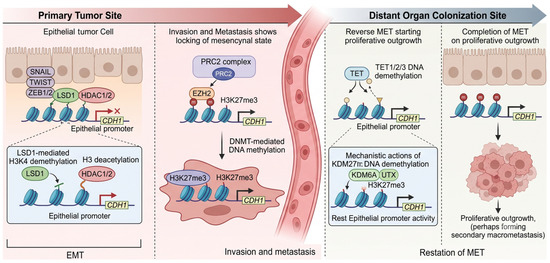
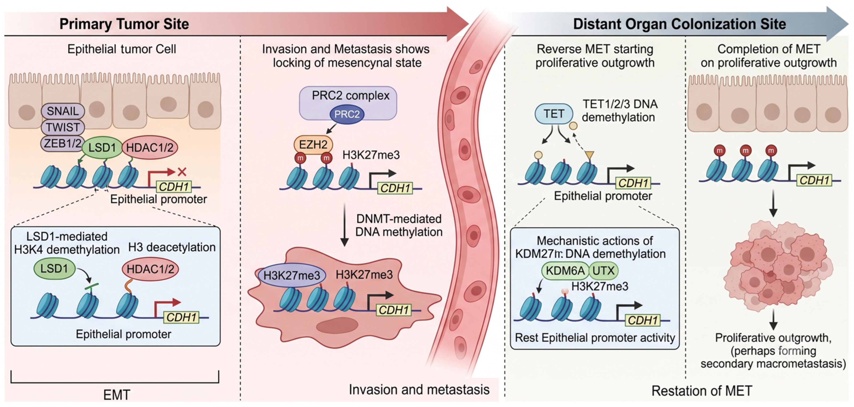
At the molecular level, EMT is governed by the transcription factors SNAIL, TWIST, ZEB1, and ZEB2, which repress epithelial gene programs (notably CDH1) and activate mesenchymal gene programs (including CDH2, VIM, and fibronectin). These transcription factors recruit chromatin-modifying complexes to enforce stable transcriptional reprogramming, though the universality of these mechanisms varies. For instance, direct biochemical evidence from specific in vitro models demonstrates that SNAIL recruits the LSD1-HDAC1/2 complex and the PRC2 complex to the CDH1 promoter in a coordinated fashion: LSD1 demethylates H3K4me2 (removing the active mark), while EZH2 deposits H3K27me3 and HDAC1/2 deacetylate H3K27, cumulatively establishing a densely repressive chromatin state. DNMTs are subsequently recruited, converting this reversible repression into stable, heritable methylation (Figure 1) [7]. However, it is crucial to emphasize that while these direct mechanistic links are well established in highly controlled experimental models, in bulk clinical tumor profiling, these associations are often correlational and exhibit significant tumor-type specificity. The strict, ordered sequential recruitment of PRC2 and DNMTs may not be obligate across all carcinoma subtypes, reflecting a highly context-dependent epigenetic landscape.
Figure 1.
Dynamic epigenetic reprogramming during the Epithelial-to-Mesenchymal Transition (EMT) and Mesenchymal-to-Epithelial Transition (MET). The metastatic cascade relies on reversible phenotypic plasticity as tumor cells disseminate from the primary site and colonize distant organs. At the primary site, EMT is driven by transcription factors (e.g., SNAIL) that recruit chromatin-modifying complexes (LSD1, PRC2/EZH2, and DNMTs) to establish a repressive chromatin state at epithelial gene promoters, such as CDH1. Conversely, successful colonization at secondary sites requires MET, facilitated by TET-mediated DNA demethylation and the removal of repressive histone marks (e.g., by KDM6A/UTX), thereby restoring epithelial gene expression.
The NuRD (nucleosome remodeling and deacetylase) complex also participates in EMT-associated chromatin remodeling. NuRD integrates ATP-dependent nucleosome repositioning (via CHD3/4) with histone deacetylation (via HDAC1/2) to establish and maintain the repressive chromatin architecture at epithelial gene loci. Concomitantly, the SWI/SNF chromatin remodeling complex mediates nucleosome remodeling at mesenchymal gene promoters, creating an open, accessible chromatin state permissive for mesenchymal gene transcription.
The classical paradigm dictates that at secondary colonization sites, a reverse transition mesenchymal-to-epithelial transition (MET) is required for proliferative outgrowth. When it occurs, MET involves the epigenetic resetting of the mesenchymal chromatin state: demethylation of the CDH1 promoter, removal of H3K27me3 by KDM6A/UTX or KDM6B, and restoration of H3K4me3 at epithelial promoters [30,31]. However, emerging lineage-tracing models and single-cell transcriptomics have challenged the dogma that a complete EMT is required for dissemination or that a full MET is obligatory for colonization. Epigenetic reprogramming frequently stalls at intermediate stages, giving rise to partial EMT (pEMT) or hybrid states. Cells in a pEMT state retain critical epithelial features such as localized E-cadherin-mediated cell–cell adhesion, while simultaneously acquiring mesenchymal invasive traits. This epigenetic duality facilitates collective cell migration, a mode of dissemination increasingly recognized as a dominant metastatic driver in breast carcinomas and HNSCC. Importantly, metastasis can occur largely independently of a full EMT; highly cohesive epithelial clusters can intravasate, survive in circulation, and seed distant organs without ever adopting a fully mesenchymal chromatin state [6,32].
Similarly, an obligatory MET is not universally required for colonization. Depending on the biochemical and physical permissiveness of the distant organ microenvironment, hybrid or predominantly mesenchymal tumor cells can establish secondary macrometastases through alternative, site-specific epigenetic adaptations. Understanding these intermediate and context-dependent chromatin states is critical, as therapies designed exclusively to reverse a full mesenchymal phenotype may inadvertently stabilize highly aggressive, collectively migrating pEMT clusters.
Epigenetic Regulation of Metastatic Dormancy and Organotropism
Following dissemination, cancer cells must survive in foreign microenvironments, frequently entering a prolonged state of cellular quiescence known as metastatic dormancy. This dormant state is not passive; it is heavily regulated by epigenetic plasticity in response to extrinsic niche cues [33]. Transcriptional repressors and orphan nuclear receptors, such as NR2F1 and DEC2, orchestrate intrinsic dormancy by enforcing repressive histone methylation at promoters of proliferative genes while preserving a stem-like epigenetic state [34]. The subsequent reactivation, or “awakening,” of these dormant disseminated cancer cells (DCCs) is highly dependent on dynamic epigenetic reprogramming driven by the local tissue stroma [35,36]. Furthermore, the establishment of metastasis is frequently site-specific, a phenomenon termed organotropism. The epigenetic adaptation of cancer cells to distinct microenvironments, such as the bone, liver, brain, or lung, requires site-specific epigenomic rewiring, enabling tumor cells to co-opt resident stromal signals, evade localized immune surveillance, and establish secondary macrometastases.
6. Non-Coding RNAs as Epigenetic Orchestrators of Metastasis
The pervasive transcription of non-protein-coding regions of the human genome produces a diverse repertoire of regulatory RNAs whose roles in cancer epigenetics have been rapidly elucidated. Among these, lncRNAs (>200 nucleotides) and miRNAs (~22 nucleotides) have emerged as central coordinators of epigenetic reprogramming in metastasis, operating through distinct but interconnected mechanisms.
6.1. HOTAIR: A Paradigmatic Epigenetic Scaffold
HOTAIR (HOX antisense intergenic RNA), transcribed from the HOXC locus on chromosome 12, is among the most extensively characterized oncogenic lncRNAs. Gupta et al. demonstrated that HOTAIR expression is substantially elevated in primary breast tumors and metastases and that its expression level in primary tumors is a powerful predictor of subsequent metastasis and survival [16]. Mechanistically, HOTAIR functions as a bimodular scaffold: its 5′ domain binds PRC2, while its 3′ domain recruits the LSD1/CoREST complex. By coordinating PRC2-mediated H3K27 trimethylation and LSD1-mediated H3K4 demethylation at target gene promoters, HOTAIR enforces stable transcriptional silencing of metastasis-suppressor genes, including RASSF1A, PCDH10, and members of the HOXD cluster [37,38].
HOTAIR additionally functions as a competing endogenous RNA (ceRNA), sponging tumor-suppressive miRNAs, including miR-145, to relieve their repression of EMT drivers and pro-invasive signaling molecules, such as NID1 and MMP-9. In hepatocellular carcinoma, HOTAIR recruits PRC2 to the miR-145-5p promoter, silencing this tumor-suppressive miRNA through H3K27me3 deposition and thereby de-repressing its target NUAK1, a kinase that promotes invasion and metastasis [38].
The therapeutic implications of HOTAIR have been explored through several approaches. Small molecule disruptors of the HOTAIR–EZH2 interface (AC1Q3QWB/AQB and ADQ) have shown preclinical efficacy in suppressing tumor metastasis, and antisense oligonucleotides targeting HOTAIR are under investigation. HOTAIR overexpression can be detected in plasma as a liquid biopsy biomarker, suggesting its potential for monitoring metastatic disease progression [39].
6.2. MALAT1, H19, and Other Oncogenic lncRNAs
MALAT1 (metastasis-associated lung adenocarcinoma transcript 1) is a highly abundant nuclear lncRNA whose overexpression correlates with metastatic propensity across multiple cancer types. MALAT1 promotes EMT and metastasis partly by modulating the alternative splicing of EMT-related transcripts and partly by recruiting chromatin-modifying complexes, including PRC2, to anti-metastatic gene loci. The imprinted lncRNA H19, expressed primarily from the maternal allele, promotes invasion and metastasis through multiple mechanisms, including suppression of let-7 miRNAs, which normally restrain the expression of EMT-promoting transcription factors, and through epigenetic regulation of the IGF2/H19 imprinting locus.
6.3. miRNAs Targeting Epigenetic Enzymes
A reciprocal regulatory circuit links miRNAs and epigenetic enzymes: epigenetic mechanisms regulate miRNA expression (through promoter methylation and histone modifications), while miRNAs, in turn, target epigenetic enzymes. The miR-29 family (miR-29a/b/c) targets DNMT3A, DNMT3B, and DNMT1, and its downregulation in multiple cancers contributes to global DNA hypermethylation and silencing of metastasis suppressor genes. miR-101 directly targets EZH2; its loss in multiple cancers leads to EZH2 overexpression and consequent epigenetic silencing of anti-metastatic programs. Conversely, members of the miR-200 family target ZEB1/ZEB2, key EMT transcription factors, and their downregulation via promoter hypermethylation is a critical epigenetic event in EMT activation [6,40].
7. Epigenetic Reprogramming in the Tumor Microenvironment
The tumor microenvironment (TME), comprising cancer-associated fibroblasts (CAFs), tumor-associated macrophages (TAMs), endothelial cells, immune effectors, and the extracellular matrix, exerts bidirectional epigenetic influences on both tumor cells and stromal components. This crosstalk is increasingly recognized as a major determinant of metastatic success, immune evasion, and therapeutic resistance [40,41].
7.1. Epigenetic Polarization of Tumor-Associated Macrophages
Historically, tumor-associated macrophages (TAMs) have been broadly classified using a binary M1 (pro-inflammatory) versus M2 (pro-tumoral) paradigm. However, recent advances in single-cell RNA sequencing (scRNA-seq) and spatial transcriptomics have fundamentally challenged this oversimplified framing. It is now evident that TAMs span a highly diverse, continuous transcriptional spectrum, frequently co-expressing classical M1 and M2 markers within the same cellular niche [42,43]. Rather than adopting a strict M2 polarization, TAMs differentiate into distinct spatial and functional subpopulations. For instance, single-cell analyses have identified specific subsets, such as SPP1+/TREM2+ macrophages and lipid-associated macrophages (LAMs), which are significantly enriched in metastatic niches and correlate directly with poor progression-free survival [44].
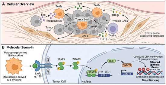
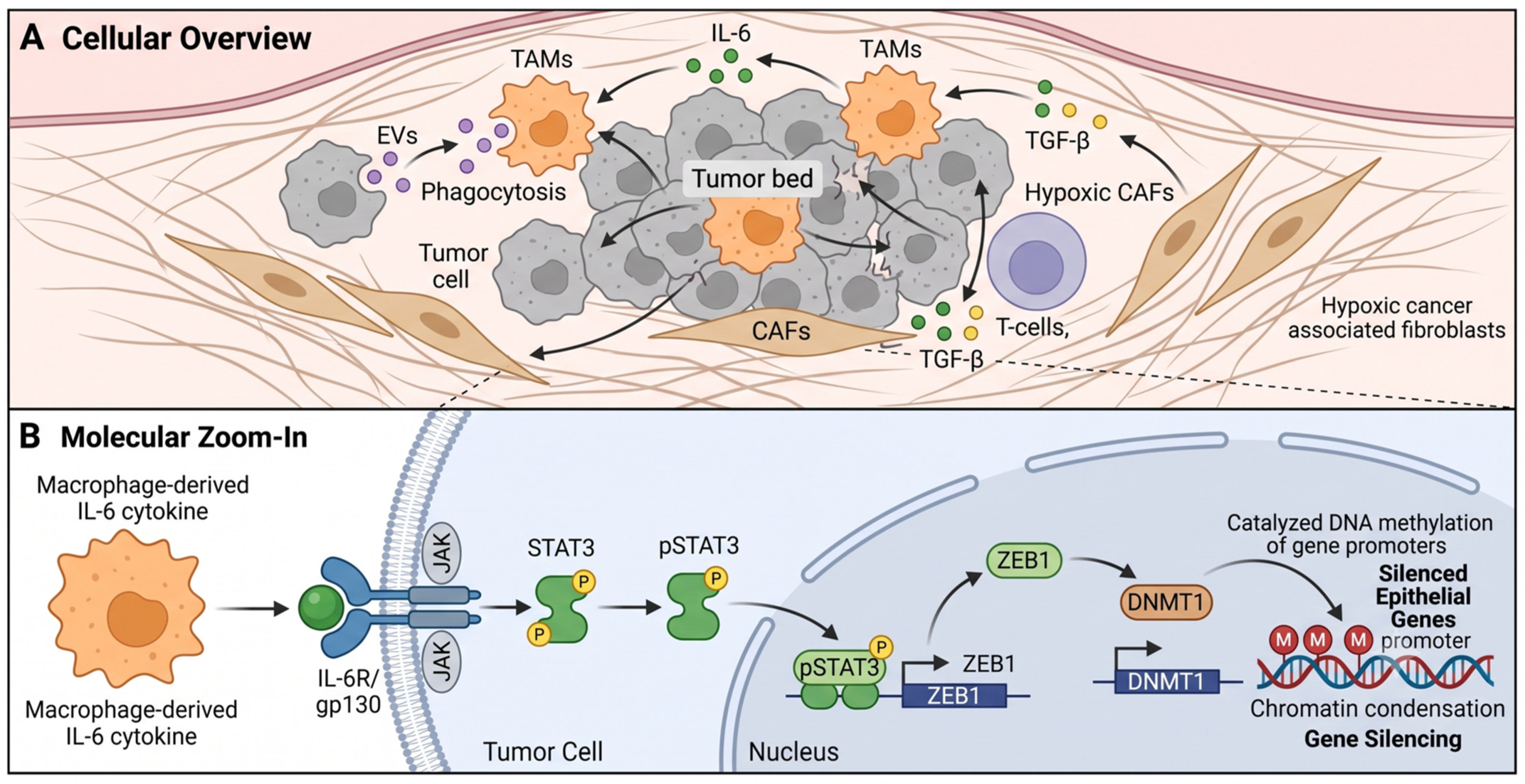
The acquisition of these diverse, pro-metastatic states is enforced and stabilized by localized epigenetic reprogramming within the TME. Environmental cues drive coordinated changes in histone acetylation and methylation at cytokine gene loci; for example, repressive H3K27me3 marks accumulate at inflammatory gene promoters (e.g., IL-12, TNF-α), while active H3K4me3 and H3K27ac marks define immunosuppressive and tissue-remodeling loci (e.g., IL-10, TGF-β). Once polarized into these highly secretory states, TAMs exert profound epigenetic influence over adjacent tumor cells. Experimental evidence demonstrates that this stromal-to-tumor crosstalk operates through direct, unbroken mechanistic cascades. Notably, TAM-derived IL-6 has been shown to directly bind tumor cell receptors, activating the intracellular IL-6–pSTAT3–ZEB1–DNMT1 axis. As validated in breast cancer models, STAT3 phosphorylation induces ZEB1, which in turn acts as a direct transcriptional activator of DNMT1, driving widespread DNA methylation and the epigenetic silencing of epithelial genes (Figure 2) [45,46,47]. This specific cytokine-to-chromatin step mechanistically couples TME signaling directly to the epigenetic EMT apparatus of the invading tumor cell.
Figure 2.
Bidirectional epigenetic crosstalk between malignant cells and the tumor microenvironment (TME). (A) Tumor cells secrete extracellular vesicles (EVs) containing non-coding RNAs and epigenetic enzymes that polarize tumor-associated macrophages (TAMs) toward an immunosuppressive M2-like state. Concurrently, hypoxia stabilizes HIF-1α in cancer-associated fibroblasts (CAFs), driving DNMT3A-dependent pro-metastatic activation. (B) In reciprocal signaling, TAM-derived cytokines, including IL-6 and TGF-β, activate the IL-6–pSTAT3–ZEB1–DNMT1 intracellular axis within tumor cells, enforcing DNA methylation-mediated silencing of epithelial genes and promoting invasion.
Reciprocally, tumor cells shape the epigenetic state of macrophages through extracellular vesicle (EV)-mediated delivery of oncogenic signals. Tumor-derived EVs carry non-coding RNAs, metabolites, and epigenetic enzymes that are taken up by macrophages and reprogram their transcriptional state toward pro-tumoral polarization. The UHRF1-mediated epigenetic pathway in hepatocellular carcinoma exemplifies this: TAM-derived prostaglandin E2 (PGE2) upregulates tumor cell UHRF1, which in turn methylates H3K9 to suppress KLF6 expression, promoting cancer growth and creating an oncogenic feedback loop that sustains further TAM accumulation [27,48].
7.2. CAF Epigenetic Activation and the Pre-Metastatic Niche
Cancer-associated fibroblasts acquire a protumoral, myofibroblastic phenotype through epigenetic reprogramming, including DNMT-mediated silencing of tumor-suppressive genes (including RASAL1 and GAS1) and HDAC-dependent deacetylation of cytoskeletal regulatory genes. Once activated, CAFs remodel the extracellular matrix by secreting MMPs and collagens, creating a physical scaffold and a biochemical milieu conducive to invasion and colonization. Hypoxia further exacerbates CAF activation: HIF-1α accumulation under low-oxygen conditions drives DNMT3A-dependent methylation-mediated silencing of antioxidant and tumor-suppressive gene programs in CAFs, thereby reinforcing the pro-metastatic stroma.
At distant organ sites, the pre-metastatic niche, a receptive microenvironment established by primary tumor-derived factors prior to the arrival of metastatic cells, is increasingly understood to involve epigenetic reprogramming of stromal and immune resident cells. Bone marrow-derived myeloid cells recruited to pre-metastatic niches undergo epigenetic polarization that suppresses anti-tumor immune programs and creates a permissive soil for metastatic seed establishment.
8. Therapeutic Vulnerabilities: Targeting the Epigenetic Machinery
When evaluating the therapeutic efficacy of epigenetic agents, it is critical to distinguish between general tumor response and specific anti-metastatic activity. In clinical settings, the general tumor response is typically measured by overall response rate (ORR) or progression-free survival (PFS) according to RECIST criteria, which assess the shrinkage of established lesions. However, true anti-metastatic efficacy, the prevention of local invasion, intravasation, or the establishment of new metastatic colonies, is more accurately captured by endpoints such as metastasis-free survival (MFS), time to new metastases, or strictly controlled preclinical in vivo assays tracking circulating tumor cells and extravasation. Therefore, the clinical and preclinical evidence presented below is evaluated using a standardized assessment to determine whether an agent primarily induces cytotoxicity in established tumors or actively halts the metastatic cascade.
8.1. HDAC Inhibitors
Historically, the FDA granted accelerated approvals for several HDAC inhibitors in hematological malignancies; however, the regulatory landscape has undergone significant recent corrections. While vorinostat and romidepsin remain approved for cutaneous T-cell lymphoma (CTCL), and belinostat retains its accelerated approval for peripheral T-cell lymphoma (PTCL), other major indications have been formally withdrawn [49,50]. Notably, the accelerated approval for panobinostat in multiple myeloma was withdrawn by the FDA after post-marketing requirements were not met, and the PTCL indication for romidepsin was voluntarily withdrawn following the failure of a confirmatory phase 3 trial to demonstrate sufficient clinical benefit over standard-of-care chemotherapy [51]. These withdrawals underscore the stringent requirements for demonstrating durable clinical efficacy and highlight the challenges of translating epigenetic therapies from early-phase signals to definitive clinical practice. Nevertheless, their application in solid tumor metastasis is under active clinical investigation, with a mechanistic rationale supported by preclinical evidence of EMT reversal, invasion suppression, and immune sensitization [13].
Entinostat, a class I-selective HDAC inhibitor, has shown clinical activity in combination strategies. A phase 3 trial of entinostat plus exemestane in Chinese patients with hormone receptor-positive advanced breast cancer (N = 354) demonstrated a statistically significant improvement in progression-free survival (median 6.32 months vs. 3.72 months; HR 0.76, CI 0.58–0.98, p = 0.046) and a meaningful overall survival benefit [52]. A phase 2 study (NCT03250273) of entinostat combined with the PD-1 inhibitor nivolumab in metastatic pancreatic ductal adenocarcinoma (N = 30) utilized ORR as a primary endpoint, demonstrating an ORR of 11% (95% CI: 2.4–29.2%) and a median response duration of 10.2 months, highlighting general tumor control rather than isolated anti-metastatic prevention [53]. In metastatic uveal melanoma, a disease notorious for its lack of response to immunotherapy, the PEMDAC phase 2 trial (NCT02697630), combining entinostat with pembrolizumab (N = 29), demonstrated a 14% ORR (95% CI: 4–32%), with Grade ≥ 3 adverse events observed in 48% of patients [54].
8.2. EZH2 Inhibitors
Tazemetostat (EPZ-6438), a selective inhibitor of EZH2 methyltransferase activity, received FDA accelerated approval in 2020 for metastatic or locally advanced epithelioid sarcoma with SMARCB1/INI1 loss, and subsequently for relapsed/refractory follicular lymphoma. In a phase 2 trial for follicular lymphoma (N = 99), tazemetostat achieved an ORR of 69% (95% CI: 53–82%) in EZH2-mutant cohorts and 34% (95% CI: 22–48%) in EZH2 wild-type disease [11]. It is important to note that, under accelerated approval, its continued regulatory status for follicular lymphoma remains contingent upon verification of clinical benefit in ongoing confirmatory phase 3 trials. For example, the SYMPHONY-1 trial (NCT04224493) is currently investigating tazemetostat in combination with lenalidomide and rituximab to confirm long-term efficacy, reflecting the FDA’s increasingly rigorous oversight of epigenetic agents [55].
Beyond its approved indications, tazemetostat is being evaluated in combination with standard therapies in solid tumor metastasis. A phase 1 trial of tazemetostat with pembrolizumab in anti-PD-1-resistant head and neck squamous cell carcinoma (N = 18) demonstrated the safety and tolerability of the combination, with 800 mg tazemetostat identified as the recommended dose [15]. The investigational dual EZH2/EZH1 inhibitor tulmimetostat (CPI-0209) has earned FDA Fast Track designation for ARID1A-mutant endometrial cancer and is under phase 1b/2 investigation in combination with pembrolizumab for advanced non-small cell lung cancer (NCT05467748) [25]. Despite these advances, clinical limitations, including acquired resistance and EZH2’s non-catalytic scaffolding functions in certain malignancies, underscore the need for next-generation strategies, such as targeted EZH2 protein degraders and combination regimens [11].
8.3. LSD1/KDM1A Inhibitors
Nine LSD1 inhibitors have entered clinical trials for hematological and solid cancers, reflecting the broad translational interest in this target [10]. Iadademstat (ORY-1001) has demonstrated the capacity to target SOX2-driven cancer stem cells in breast cancer by blocking LSD1-dependent SOX2 super-enhancer activity, thereby reducing mammosphere formation and CSC self-renewal [56]. Preclinical work has established that LSD1 inhibition reduces breast cancer cell bone metastasis, in part by disrupting hnRNPA2B1-mediated exosomal miRNA sorting, thereby modulating communication between tumor cells and the pre-metastatic niche [12]. In ovarian cancer, pharmacological LSD1 inhibition combined with estrogen receptor beta agonism synergistically inhibited tumor growth and invasion across orthotopic, syngeneic, and patient-derived xenograft models [28].
8.4. BET Bromodomain Inhibitors
More than 30 BET inhibitors have entered clinical trials. While no BET inhibitor has yet to achieve regulatory approval due to dose-limiting toxicities and early emergence of resistance, early-phase signals of efficacy have been observed. BRD4 inhibition suppresses PD-L1 expression on tumor cells and tumor-associated dendritic cells and macrophages, an effect mediated by direct BRD4 occupancy at the CD274 promoter, providing a mechanistic rationale for combining BET inhibitors with anti-PD-1/PD-L1 checkpoint blockade [21]. Resistance mechanisms, including compensatory activation of PI3K/AKT/mTOR and Wnt/β-catenin pathways and upregulation of BRD4 itself, are under investigation, and BRD4-directed PROTACs are emerging as an alternative approach to bypass bromodomain-independent oncogenic functions [19].
8.5. DNMT Inhibitors
The cytosine analogues azacitidine (5-azacytidine) and decitabine (5-aza-2’-deoxycytidine) inhibit DNMTs through covalent trapping after incorporation into DNA, leading to passive demethylation of hypermethylated gene loci and re-expression of silenced tumor suppressor genes. Their application in solid tumor metastasis is under investigation, with preclinical evidence supporting restoration of E-cadherin expression, suppression of invasion, and re-sensitization to immune checkpoint inhibitors through upregulation of endogenous retroviral element expression and innate immune sensing.
8.6. Combination Strategies: Epigenetic Priming and Immunotherapy
A compelling translational opportunity lies in combining epigenetic drugs with immune checkpoint inhibitors. HDACi and DNMTi modulate the tumor immunopeptidome, upregulate MHC class I expression, and dampen immunosuppressive mechanisms, creating a more immunologically hot tumor microenvironment. EZH2 inhibition de-represses the tumor-intrinsic antigen-presentation machinery and relieves epigenetic suppression of inflammatory cytokine genes in TAMs, thereby synergizing with anti-PD-1 blockade. BET inhibition reduces PD-L1 expression while EZH2 inhibition may restore responsiveness to checkpoint blockade in otherwise resistant tumors. These mechanistic synergies are being actively evaluated in several ongoing trials (Table 5).
Table 5.
Selected Clinical Trials of Epigenetic Agents in Metastatic Cancers.
9. Epigenetic Biomarkers for Metastatic Risk Stratification and Therapy Monitoring
The dynamic and tissue-specific nature of the cancer epigenome presents both a challenge and an opportunity for biomarker development. Unlike genetic mutations, epigenetic marks reflect a tumor’s current transcriptional state and can change rapidly in response to therapy. To successfully translate these findings into clinical practice, it is essential to clearly delineate their intended uses across the patient journey: early diagnosis, metastatic risk stratification, and longitudinal treatment monitoring.
9.1. Early Detection and Diagnostic Biomarkers
Circulating tumor DNA (ctDNA) methylation profiling has emerged as a robust approach for early cancer detection, metastatic monitoring, and assessment of therapy response. Aberrant DNA methylation patterns in ctDNA, including promoter hypermethylation of cancer-specific genes, mirror those in tumor tissue and can be detected in plasma with high sensitivity and specificity [59,60]. For instance, multi-modal epigenetic sequencing analysis (MESA), which integrates methylation profiles with nucleosome positioning data, has achieved high performance in colorectal cancer detection. In hepatocellular carcinoma (HCC), combined serum biomarker panels (incorporating the lncRNAs HOTAIR, ICR, and BRM, along with alpha-fetoprotein [AFP]) have reported diagnostic accuracy AUC values approaching 0.998 [61]. However, this exceptional diagnostic accuracy must be interpreted with extreme caution. The primary study establishing this panel was limited to a small discovery cohort (61 HCC patients, 60 liver cirrhosis patients, and healthy controls) and notably lacked predefined biomarker cutoffs, internal cross-validation, and independent external cohort validation [61]. Without rigorous, prospective validation in large independent cohorts, such extraordinarily high AUC values risk reflecting statistical model overfitting rather than definitive clinical readiness.
9.2. Metastatic Risk Stratification and Prognosis
Beyond early detection, epigenetic biomarkers are critical for predicting a patient’s baseline propensity for metastasis. High baseline serum levels of HOTAIR and other oncogenic lncRNAs strongly correlate with advanced tumor stage, vascular invasion, and subsequent metastatic burden, identifying high-risk patients who may require aggressive adjuvant interventions [61]. In lung cancer, methylation profiling of ctDNA has demonstrated the capacity to identify highly aggressive small-cell lung cancer subtypes prone to early dissemination [59]. Furthermore, tissue-based epigenetic markers, including EZH2 expression scores, HDAC expression profiles, and tumor DNA methylation signatures, are being actively evaluated as predictors of metastatic risk. The challenge of spatial epigenomic heterogeneity is increasingly being addressed by single-cell and spatial technologies that map the epigenetic architecture of primary lesions, thereby identifying highly invasive subclonal populations before widespread dissemination.
9.3. Treatment Monitoring and Minimal Residual Disease (MRD)
The reversible nature of epigenetic modifications makes them exceptionally suited for real-time therapy monitoring. Following curative-intent therapy, ctDNA methylation signatures are utilized to detect minimal residual disease (MRD), predicting metastatic relapse months before standard radiographic progression becomes visible in metastatic breast and prostate cancers [59]. Beyond DNA methylation, nucleosome footprinting in cell-free DNA provides real-time information on chromatin accessibility and transcription factor occupancy. Circulating nucleosomes carrying tumor-specific histone modifications, such as H3K27me3 and H3K4me3, are detectable in plasma and may offer a direct, non-invasive readout of the tumor’s real-time pharmacodynamic response to targeted epigenetic therapies (e.g., EZH2 or LSD1 inhibitors), facilitating rapid therapeutic adjustments.
10. Challenges, Open Questions, and Future Directions
Despite the mechanistic richness and therapeutic promise of epigenetic oncology, several fundamental challenges must be overcome to realize its potential in the anti-metastatic setting. First, the pervasive intratumoral heterogeneity of the cancer epigenome, now empirically demonstrated by single-cell ATAC-seq and single-cell bisulfite sequencing, means that bulk tissue epigenomic analyses capture population averages that may obscure therapeutically relevant minority cell states. Spatial epigenomic technologies, which preserve tissue context while capturing epigenomic data, are beginning to address this limitation by mapping the distribution of epigenetic states throughout the TME.
Second, the question of epigenetic memory and therapeutic durability is critically important. Pharmacological epigenetic reprogramming can achieve transient re-expression of silenced genes, but the stability of this reprogramming after drug withdrawal is uncertain. Residual histone-modifying complexes can rapidly restore histone modification states if the underlying transcriptional activators or repressors are not themselves durably altered. Combination strategies pairing epigenetic drugs with transcription factor-targeted agents may yield more durable reprogramming.
Third, the selectivity challenge is particularly acute not only for chromatin-modifying enzymes but broadly across all classes of epigenetic modulators, including DNA methyltransferases, epigenetic readers (such as BET proteins), and non-coding RNAs. Because these epigenetic regulators are essential for normal physiological processes, development, and cellular homeostasis, on-target toxicity in non-malignant tissues remains a recurring obstacle to the clinical development of epigenetic drugs. Tissue-targeted drug delivery approaches, including nanoparticle formulations, antibody-drug conjugates incorporating epigenetic payloads, and locally administered agents, may improve the therapeutic index. Epigenetic editing tools based on CRISPR-dCas9 fused to histone-modifying enzymes offer the prospect of locus-specific epigenetic reprogramming without the global transcriptional disruption associated with small-molecule inhibitors.
Fourth, integrating artificial intelligence with multi-omics approaches offers transformative potential for decoding the metastatic epigenome. Machine learning models trained on integrated chromatin accessibility, histone modification, DNA methylation, and transcriptomic data are beginning to identify epigenomic features predictive of metastatic behavior and therapeutic response that are not apparent from any single data type. These tools may also accelerate the identification of novel drug targets within the epigenetic machinery and the design of patient stratification strategies for clinical trials.
Finally, a relatively unexplored frontier concerns organ-specific epigenetic programs at metastatic sites. The epigenetic adaptation of cancer cells to the unique microenvironments of the lung, liver, bone, or brain, which differ substantially in their stromal composition, oxygen tension, and metabolic milieu, likely involves site-specific epigenomic reprogramming that governs metastatic colonization efficiency and drug response in a location-dependent manner. Comparative epigenomics of matched primary and metastatic specimens from different organ sites may reveal organ-specific vulnerabilities amenable to targeted epigenetic therapy.
11. Conclusions
Epigenetic reprogramming is not an epiphenomenon of cancer metastasis but a mechanistically central and causally implicated process that drives every step of the metastatic cascade from local invasion and EMT to immune evasion, metastatic colonization, and therapeutic resistance. The convergent contributions of histone modifications (acetylation, methylation, and their enzymatic regulators), DNA methylation, chromatin remodeling complexes, and non-coding RNAs create a richly interconnected regulatory network that defines the metastatic epigenome. The tumor microenvironment participates in this network through bidirectional epigenetic crosstalk, amplifying pro-metastatic programs while suppressing anti-tumor immunity.
Crucially, the pharmacological tractability of epigenetic mechanisms, demonstrated by the clinical approval of HDAC inhibitors and tazemetostat, and the robust clinical pipeline of LSD1, BET, and next-generation EZH2 inhibitors, establishes epigenetic mechanisms as a validated therapeutic target class in oncology. Emerging evidence for synergy between epigenetic drugs and immune checkpoint blockade holds particular promise for the anti-metastatic setting. Simultaneously, circulating epigenetic biomarkers, such as ctDNA methylation signatures, lncRNA levels, and nucleosome footprints, are advancing toward clinical utility as real-time monitors of metastatic disease and therapeutic response.
The translational realization of this promise will require addressing the fundamental challenges of epigenetic heterogeneity, therapeutic durability, and selectivity through innovative drug delivery, combination strategies, and precision patient selection. Advances in single-cell and spatial epigenomics, AI-driven multi-omics integration, and CRISPR-based epigenetic editing provide the technological foundation for this next generation of anti-metastatic epigenetic therapy. Collectively, the evidence reviewed here makes a compelling case that the epigenetic layer of metastatic biology represents a frontier of therapeutic opportunity warranting concerted translational investment.
Author Contributions
Conceptualization, P.P.; methodology, P.P. and K.M.; software, P.P. and K.M.; validation, D.T. and N.R.; formal analysis, D.T. and N.R.; data curation, P.P. and N.R.; writing—original draft preparation, P.P.; writing—review and editing, D.T., K.M. and N.R.; project administration, P.P. All authors have read and agreed to the published version of the manuscript.
Funding
This research received no external funding.
Institutional Review Board Statement
Not applicable.
Informed Consent Statement
Not applicable.
Data Availability Statement
No new data were created or analyzed in this study. Data sharing is not applicable to this article.
Acknowledgments
No AI tool was used to generate any content during the drafting of the original manuscript. However, the premium version of Grammarly Version 6.8.263 was used solely to edit and refine the language of the manuscript. The authors have reviewed and edited the output and take full responsibility for the content of this publication.
Conflicts of Interest
The authors declare no conflicts of interest.
References
- Bray, F.; Laversanne, M.; Sung, H.; Ferlay, J.; Siegel, R.L.; Soerjomataram, I.; Jemal, A. Global cancer statistics 2022: GLOBOCAN estimates of incidence and mortality worldwide for 36 cancers in 185 countries. CA A Cancer J. Clin. 2024, 74, 229–263. [Google Scholar] [CrossRef] [Scilit] [PubMed]
- Pandey, P.; Arya, D.K.; Deepak, P.; Ali, D.; Alarifi, S.; Srivastava, S.; Lavasanifar, A.; Rajinikanth, P.S. αvβ3 Integrin and Folate-Targeted pH-Sensitive Liposomes with Dual Ligand Modification for Metastatic Breast Cancer Treatment. Bioengineering 2024, 11, 800. [Google Scholar] [CrossRef] [Scilit] [PubMed]
- Pandey, P.; Arya, D.K.; Kumar, A.; Kaushik, A.; Mishra, Y.K.; Rajinikanth, P.S. Dual ligand functionalized pH-sensitive liposomes for metastatic breast cancer treatment: In vitro and in vivo assessment. J. Mater. Chem. B 2025, 13, 2682–2694. [Google Scholar] [CrossRef] [Scilit] [PubMed]
- Pandey, P.; Chaudhary, R.; Tripathi, D.; Lavudi, K.; Dua, K.; Weinfeld, M.; Lavasanifar, A.; Rajinikanth, P. Personalized treatment approach for HER2-positive metastatic breast cancer. Med. Oncol. 2024, 41, 252. [Google Scholar] [CrossRef] [Scilit] [PubMed]
- Na, F.; Pan, X.; Chen, J.; Chen, X.; Wang, M.; Chi, P.; You, L.; Zhang, L.; Zhong, A.; Zhao, L. KMT2C deficiency promotes small cell lung cancer metastasis through DNMT3A-mediated epigenetic reprogramming. Nat. Cancer 2022, 3, 753–767. [Google Scholar] [CrossRef] [Scilit] [PubMed]
- Gu, M.; Ren, B.; Fang, Y.; Ren, J.; Liu, X.; Wang, X.; Zhou, F.; Xiao, R.; Luo, X.; You, L.; et al. Epigenetic regulation in cancer. MedComm 2024, 5, e495. [Google Scholar] [CrossRef] [Scilit] [PubMed]
- Sun, Z.; Wu, Y.; Zong, X.; Liu, H.; Sun, X.; Wang, S.; Liu, J. Epigenetic regulation in cancer metastasis. Discov. Oncol. 2026, 17, 675. [Google Scholar] [CrossRef] [Scilit] [PubMed]
- Yang, J.; Ren, B.; Ren, J.; Yang, G.; Fang, Y.; Wang, X.; Zhou, F.; You, L.; Zhao, Y. Epigenetic reprogramming-induced guanidinoacetic acid synthesis promotes pancreatic cancer metastasis and transcription-activating histone modifications. J. Exp. Clin. Cancer Res. 2023, 42, 155. [Google Scholar] [CrossRef] [Scilit] [PubMed]
- Parveen, R.; Harihar, D.; Chatterji, B.P. Recent histone deacetylase inhibitors in cancer therapy. Cancer 2023, 129, 3372–3380. [Google Scholar] [CrossRef] [Scilit] [PubMed]
- Pandey, P.; Kumar Arya, D.; Kumar Ramar, M.; Chidambaram, K.; Rajinikanth, P.S. Engineered nanomaterials as an effective tool for HER2+ breast cancer therapy. Drug Discov. Today 2022, 22, 2526–2540. [Google Scholar] [CrossRef] [Scilit] [PubMed]
- Kim, K.H.; Roberts, C.W. Targeting EZH2 in cancer. Nat. Med. 2016, 22, 128–134. [Google Scholar] [CrossRef] [Scilit] [PubMed]
- Liu, Z.; Xin, B.; Zhang, N.; An, P.; Shi, Y.; Yang, J.; Wan, Y.; He, Y.; Hu, X. LSD1 modulates the bone metastasis of breast cancer cells through hnRNPA2B1-mediated sorting of exosomal miRNAs. Cell Death Discov. 2024, 10, 115. [Google Scholar] [CrossRef] [Scilit] [PubMed]
- Luo, J.; Gao, S.P. Histone modifications in cancer. Front. Pharmacol. 2025, 16, 1727417. [Google Scholar] [CrossRef] [Scilit] [PubMed]
- Au, S.L.; Wong, C.C.; Lee, J.M.; Wong, C.M.; Ng, I.O. EZH2-Mediated H3K27me3 Is Involved in Epigenetic Repression of Deleted in Liver Cancer 1 in Human Cancers. PLoS ONE 2013, 8, e68226. [Google Scholar] [CrossRef] [Scilit] [PubMed]
- Oppelt, P.J.; Puram, S.V.; Liu, J.; Ley, J.C.; Adkins, D. Tazemetostat, a selective EZH2 inhibitor, in combination with pembrolizumab for recurrent or metastatic head and neck squamous cell carcinoma: A phase 1 trial. Cancers 2025, 17, 437. [Google Scholar] [CrossRef] [Scilit] [PubMed]
- Johnson, J.D.; Alejo, S.; Jayamohan, S.; Sareddy, G.R. Lysine-specific demethylase 1 as a therapeutic cancer target: Observations from preclinical study. Expert Opin. Ther. Targets 2023, 27, 1177–1188. [Google Scholar] [CrossRef] [Scilit] [PubMed]
- Fang, Y.; Liao, G.; Yu, B. LSD1/KDM1A inhibitors in clinical trials: Advances and prospects. J. Hematol. Oncol. 2019, 12, 129. [Google Scholar] [CrossRef] [Scilit] [PubMed]
- Lin, Y.; Wu, Y.; Li, J.; Dong, C.; Ye, X.; Chi, Y.I.; Evers, B.M.; Zhou, B.P. The SNAG domain of Snail1 functions as a molecular hook for recruiting lysine-specific demethylase 1. EMBO J. 2010, 29, 1803–1816. [Google Scholar] [CrossRef] [Scilit] [PubMed]
- Guo, J.; Zheng, Q.; Peng, Y. BET proteins: Biological functions and therapeutic interventions. Pharmacol. Ther. 2023, 243, 108354. [Google Scholar] [CrossRef] [Scilit] [PubMed]
- Zhou, S.; Zhang, S.; Wang, L.; Huang, S.; Yuan, Y.; Yang, J.; Wang, H.; Li, X.; Wang, P.; Zhou, L.; et al. BET protein inhibitor JQ1 downregulates chromatin accessibility and suppresses metastasis of gastric cancer via inactivating RUNX2/NID1 signaling. Oncogenesis 2020, 9, 33. [Google Scholar] [CrossRef] [Scilit] [PubMed]
- Zhu, H.; Bengsch, F.; Svoronos, N.; Rutkowski, M.R.; Bitler, B.G.; Allegrezza, M.J.; Yokoyama, Y.; Kossenkov, A.V.; Bradner, J.E.; Conejo-Garcia, J.R. BET bromodomain inhibition promotes anti-tumor immunity by suppressing PD-L1 expression. Cell Rep. 2016, 16, 2829–2837. [Google Scholar] [CrossRef] [Scilit] [PubMed]
- Wang, H.; Anwaier, G.; Bai, S.; Liao, L.; Wang, Y.; Li, S. How ATP-Dependent Chromatin Remodeling Complexes Regulate Vertebrate Embryonic Development. Int. J. Mol. Sci. 2026, 27, 835. [Google Scholar] [CrossRef] [Scilit] [PubMed]
- Feng, Y.; Pauklin, S. Revisiting 3D chromatin architecture in cancer development and progression. Nucleic Acids Res. 2020, 48, 10632–10647. [Google Scholar] [CrossRef] [Scilit] [PubMed]
- Yang, J.; Zhou, F.; Luo, X.; Fang, Y.; Wang, X.; Liu, X.; Xiao, R.; Jiang, D.; Tang, Y.; Yang, G.; et al. Enhancer reprogramming: Critical roles in cancer and promising therapeutic strategies. Cell Death Discov. 2025, 11, 84. [Google Scholar] [CrossRef] [Scilit] [PubMed]
- Wang, J.; Xiang, L.; Zhang, H.; Zhang, X. SETD2 and EZH2: Two epigenetic drivers of prostate cancer. J. Cancer 2025, 16, 3673–3683. [Google Scholar] [CrossRef] [Scilit] [PubMed]
- Peinado, H.; Ballestar, E.; Esteller, M.; Cano, A. Snail mediates E-cadherin repression by the recruitment of the Sin3A/histone deacetylase 1 (HDAC1)/HDAC2 complex. Mol. Cell. Biol. 2004, 24, 306–319. [Google Scholar] [CrossRef] [Scilit] [PubMed]
- Zhang, J.; Zhang, H.; Ding, X.; Hu, J.; Li, Y.; Zhang, J.; Wang, H.; Qi, S.; Xie, A.; Shi, J. Crosstalk between macrophage-derived PGE2 and tumor UHRF1 drives hepatocellular carcinoma progression. Theranostics 2022, 12, 3776. [Google Scholar] [CrossRef] [Scilit] [PubMed]
- Venkata, P.P.; Jayamohan, S.; He, Y.; Alejo, S.; Johnson, J.D.; Palacios, B.E.; Pratap, U.P.; Chen, Y.; Liu, Z.; Zou, Y. Pharmacological inhibition of KDM1A/LSD1 enhances estrogen receptor beta-mediated tumor suppression in ovarian cancer. Cancer Lett. 2023, 575, 216383. [Google Scholar] [CrossRef] [Scilit] [PubMed]
- Béguelin, W.; Popovic, R.; Teater, M.; Jiang, Y.; Bunting, K.L.; Rosen, M.; Shen, H.; Yang, S.N.; Wang, L.; Ezponda, T. EZH2 is required for germinal center formation and somatic EZH2 mutations promote lymphoid transformation. Cancer Cell 2013, 23, 677–692. [Google Scholar] [CrossRef] [Scilit] [PubMed]
- Zhang, C.X.; Huang, R.Y.-J.; Sheng, G.; Thiery, J.P. Epithelial-mesenchymal transition. Cell 2025, 188, 5436–5486. [Google Scholar] [CrossRef] [Scilit] [PubMed]
- Hara, T.; Subramanian, M. Rewriting the Fate of Cancer: Epigenetic and Epitranscriptomic Regulators in the Metastatic Cascade. Biomolecules 2025, 15, 1573. [Google Scholar] [PubMed]
- Bastos, V.A.F.; Souza, A.G.; Guedes, V.C.S.; Cunha, T.M. Shared and Context-Specific Mechanisms of EMT and Cellular Plasticity in Cancer and Fibrotic Diseases. Int. J. Mol. Sci. 2025, 26, 9476. [Google Scholar] [CrossRef] [Scilit] [PubMed]
- Yang, S.; Seo, J.; Choi, J.; Kim, S.-H.; Kuk, Y.; Park, K.C.; Kang, M.; Byun, S.; Joo, J.-Y. Towards understanding cancer dormancy over strategic hitching up mechanisms to technologies. Mol. Cancer 2025, 24, 47. [Google Scholar] [CrossRef] [Scilit] [PubMed]
- Sun, D.; Singh, D.K.; Carcamo, S.; Filipescu, D.; Khalil, B.; Huang, X.; Miles, B.A.; Westra, W.; Sproll, K.C.; Hasson, D.; et al. MacroH2A impedes metastatic growth by enforcing a discrete dormancy program in disseminated cancer cells. Sci. Adv. 2022, 8, eabo0876. [Google Scholar] [CrossRef] [Scilit] [PubMed]
- Barnieh, F.M.; Morton, J.; Olanrewaju, O.; El-Khamisy, S.F. Decoding the adaptive survival mechanisms of breast cancer dormancy. Oncogene 2025, 44, 3759–3773. [Google Scholar] [CrossRef] [Scilit] [PubMed]
- Mackie, G.M.; Turrell, F.K. Ageing influences tumour–niche interactions: Implications for metastatic dormancy, reawakening and recurrence. Curr. Opin. Cell Biol. 2026, 100, 102630. [Google Scholar] [CrossRef] [Scilit] [PubMed]
- Gupta, R.A.; Shah, N.; Wang, K.C.; Kim, J.; Horlings, H.M.; Wong, D.J.; Tsai, M.-C.; Hung, T.; Argani, P.; Rinn, J.L. Long non-coding RNA HOTAIR reprograms chromatin state to promote cancer metastasis. Nature 2010, 464, 1071–1076. [Google Scholar] [CrossRef] [Scilit] [PubMed]
- Chu, D.-x.; Jin, Y.; Wang, B.-r.; Jiao, Y.; Zhang, C.-k.; Guo, Z.-h.; Hu, S.-z.; Li, N. LncRNA HOTAIR enhances epithelial-to-mesenchymal transition to promote the migration and invasion of liver cancer by regulating NUAK1 via epigenetic inhibition miR-145-5p expression. J. Cancer 2023, 14, 2329. [Google Scholar] [CrossRef] [Scilit] [PubMed]
- Nazari, M.; Babakhanzadeh, E.; Mollazadeh, A.; Ahmadzade, M.; Mohammadi Soleimani, E.; Hajimaqsoudi, E. HOTAIR in cancer: Diagnostic, prognostic, and therapeutic perspectives. Cancer Cell Int. 2024, 24, 415. [Google Scholar] [CrossRef] [Scilit] [PubMed]
- Sami, A.; Raza, A. Reprogramming the tumor microenvironment–macrophages emerge as key players in breast cancer immunotherapy. Front. Immunol. 2024, 15, 1457491. [Google Scholar] [CrossRef] [Scilit] [PubMed]
- Mittal, K.; Rathi, N.; Tripathi, D.; Rajinikanth, P.S.; Pandey, P. Targeting the Tumour Microenvironment in Pancreatic Cancer: From Stromal Reprogramming to Emerging Therapeutics. Future Pharmacol. 2026, 6, 12. [Google Scholar] [CrossRef] [Scilit]
- Li, J.; Yang, D.; Wang, X.; Ju, R.; Chen, S.; Zhao, J.; Xu, J.; Chen, J.; Ye, J.; Xu, B.; et al. Single-Cell Transcriptomics Reveals Immune Modulation by Telmisartan in Colorectal Cancer. Cells 2026, 15, 729. [Google Scholar] [CrossRef] [Scilit] [PubMed]
- Foca, E.; Garstea, I.; Carpenco, E.; David, V.; Saptefrati, L.; Fulga, V. Molecular profile of macrophages in prostatic carcinoma. Arta Medica 2025, 97, 55–62. [Google Scholar]
- Mei, S.; Zhang, H.; Hirz, T.; Jeffries, N.E.; Xu, Y.; Baryawno, N.; Wu, S.; Wu, C.-L.; Patnaik, A.; Saylor, P.J. Single-cell and spatial transcriptomics reveal a tumor-associated macrophage subpopulation that mediates prostate cancer progression and metastasis. Mol. Cancer Res. 2025, 23, 653–665. [Google Scholar] [CrossRef] [Scilit] [PubMed]
- Ponneganti, S.; Lavudi, K.; Thalla, M.; Narkhede, G.; Dwivedi, R.; Kokkanti, R.; Pandey, P. Metabolomic Insights into Head and Neck Cancer: Recent Advances and Future Directions. Curr. Oncol. 2026, 33, 201. [Google Scholar] [CrossRef] [Scilit] [PubMed]
- Li, Z.; Wang, P.; Cui, W.; Yong, H.; Wang, D.; Zhao, T.; Wang, W.; Shi, M.; Zheng, J.; Bai, J. Tumour-associated macrophages enhance breast cancer malignancy via inducing ZEB1-mediated DNMT1 transcriptional activation. Cell Biosci. 2022, 12, 176. [Google Scholar] [CrossRef] [Scilit] [PubMed]
- To, S.Q.; Dmello, R.S.; Richards, A.K.; Ernst, M.; Chand, A.L. STAT3 Signaling in Breast Cancer: Multicellular Actions and Therapeutic Potential. Cancers 2022, 14, 429. [Google Scholar] [CrossRef] [Scilit] [PubMed]
- Kim, S.W.; Kim, C.W.; Moon, Y.-A.; Kim, H.S. Reprogramming of tumor-associated macrophages by metabolites generated from tumor microenvironment. Anim. Cells Syst. 2024, 28, 123–136. [Google Scholar] [CrossRef] [Scilit] [PubMed]
- Mao, X.; Wang, Z.; Kong, S.; Yang, Y.; Dreyer, N.A.; Ba, Y.; Lotze, M.T.; Zhan, Q.; Li, G. The evolving global landscape of first-in-class oncology drug innovation. Signal Transduct. Target. Ther. 2026, 11, 174. [Google Scholar] [CrossRef] [Scilit] [PubMed]
- Beaver, J.A.; Pazdur, R. “Dangling” accelerated approvals in oncology. N. Engl. J. Med. 2021, 384, e68. [Google Scholar] [CrossRef] [Scilit] [PubMed]
- Cliff, E.R.S.; Russler-Germain, D.A.; Daval, C.J.R.; Kesselheim, A.S. US Food and Drug Administration’s Directive to Deal With Delayed Confirmatory Trials: Lessons From Pralatrexate and Belinostat for T-Cell Lymphoma. J. Clin. Oncol. 2024, 42, 3901–3904. [Google Scholar] [CrossRef] [Scilit] [PubMed]
- Xu, B.; Zhang, Q.; Hu, X.; Li, Q.; Sun, T.; Li, W.; Ouyang, Q.; Wang, J.; Tong, Z.; Yan, M.; et al. Entinostat, a class I selective histone deacetylase inhibitor, plus exemestane for Chinese patients with hormone receptor-positive advanced breast cancer: A multicenter, randomized, double-blind, placebo-controlled, phase 3 trial. Acta Pharm. Sin. B 2023, 13, 2250–2258. [Google Scholar] [CrossRef] [Scilit] [PubMed]
- Baretti, M.; Danilova, L.; Durham, J.N.; Betts, C.B.; Cope, L.; Sidiropoulos, D.N.; Tandurella, J.A.; Charmsaz, S.; Gross, N.; Hernandez, A.; et al. Entinostat in combination with nivolumab in metastatic pancreatic ductal adenocarcinoma: A phase 2 clinical trial. Nat. Commun. 2024, 15, 9801. [Google Scholar] [CrossRef] [Scilit] [PubMed]
- Ny, L.; Jespersen, H.; Karlsson, J.; Alsén, S.; Filges, S.; All-Eriksson, C.; Andersson, B.; Carneiro, A.; Helgadottir, H.; Levin, M.; et al. The PEMDAC phase 2 study of pembrolizumab and entinostat in patients with metastatic uveal melanoma. Nat. Commun. 2021, 12, 5155. [Google Scholar] [CrossRef] [Scilit] [PubMed]
- Salles, G.; Park, S.I.; Phillips, T.J.; Amengual, J.E.; Andorsky, D.J.; Campbell, P.; McKay, P.; Leonard, J.P.; Chen, Y.; Chen, J. Tazemetostat in combination with lenalidomide and rituximab in patients with relapsed/refractory follicular lymphoma: Updated phase 1b results of symphony-1 with 22.5 months follow-up. Blood 2023, 142, 3035. [Google Scholar] [CrossRef] [Scilit]
- Cuyàs, E.; Gumuzio, J.; Verdura, S.; Brunet, J.; Bosch-Barrera, J.; Martin-Castillo, B.; Alarcón, T.; Encinar, J.A.; Martin, Á.G.; Menendez, J.A. The LSD1 inhibitor iadademstat (ORY-1001) targets SOX2-driven breast cancer stem cells: A potential epigenetic therapy in luminal-B and HER2-positive breast cancer subtypes. Aging 2020, 12, 4794. [Google Scholar] [CrossRef] [Scilit] [PubMed]
- Wang, J.; Zhang, Q.; Hu, X.; Li, Q.; Sun, T.; Li, W.; Ouyang, Q.; Wang, J.; Tong, Z.; Yan, M. Entinostat, a class I selective histone deacetylase inhibitor, plus exemestane for Chinese patients with hormone receptor-positive advanced breast cancer: An overall survival update and long-term safety from the randomised, double-blind, placebo-controlled, phase 3 trial. J. Clin. Oncol. 2024, 42, 1050. [Google Scholar] [CrossRef] [Scilit]
- Drescher, C.; Walter, H.S.; Gastinne, T.; Lakhani, N.J.; Ribrag, V.; Rasco, D.W.; Gutierrez, M.; Sullivan, R.J.; Harvey, R.D.; Banda, K. EZH2/EZH1 inhibitor tulmimetostat (CPI-0209) in patients with advanced solid tumors or hematologic malignancies: Preliminary phase II results. J. Clin. Oncol. 2023, 41, 3094. [Google Scholar] [CrossRef] [Scilit]
- Li, L.; Sun, Y. Circulating tumor DNA methylation detection as biomarker and its application in tumor liquid biopsy: Advances and challenges. MedComm 2024, 5, e766. [Google Scholar] [CrossRef] [Scilit] [PubMed]
- Noë, M.; Mathios, D.; Annapragada, A.V.; Koul, S.; Foda, Z.H.; Medina, J.E.; Cristiano, S.; Cherry, C.; Bruhm, D.C.; Niknafs, N. DNA methylation and gene expression as determinants of genome-wide cell-free DNA fragmentation. Nat. Commun. 2024, 15, 6690. [Google Scholar] [CrossRef] [Scilit] [PubMed]
- Lou, Z.-H.; Xu, K.-Y.; Qiao, L.; Su, X.-Q.; Ou-Yang, Y.; Miao, L.-B.; Liu, F.; Wang, Y.; Fu, A.; Ren, X.-H. Diagnostic potential of the serum lncRNAs HOTAIR, BRM and ICR for hepatocellular carcinoma. Front. Biosci.-Landmark 2022, 27, 264. [Google Scholar] [CrossRef] [Scilit] [PubMed]

Disclaimer/Publisher’s Note: The statements, opinions and data contained in all publications are solely those of the individual author(s) and contributor(s) and not of MDPI and/or the editor(s). MDPI and/or the editor(s) disclaim responsibility for any injury to people or property resulting from any ideas, methods, instructions or products referred to in the content. |
© 2026 by the authors. Licensee MDPI, Basel, Switzerland. This article is an open access article distributed under the terms and conditions of the Creative Commons Attribution (CC BY) license.