


Abstract
Background/Objectives: Rabies remains a fatal viral disease, and current conventional vaccine strategies face significant challenges regarding structural stability and large-scale manufacturing. This study aimed to design a novel messenger ribonucleic acid (mRNA) vaccine candidate targeting the rabies virus glycoprotein G (RABV-G) using an integrated immunoinformatic and structural modeling framework. Methods: The RABV-G sequence was systematically evaluated for evolutionary conservation and safety, followed by 3D structural modeling. Highly antigenic B-cell and T-cell epitopes were identified based on human leukocyte antigen (HLA) binding, toxicity, and glycosylation shielding analyses. Functional interactions were assessed via molecular docking with the Toll-like Receptor 4/Myeloid Differentiation factor 2 (TLR4-MD2) complex. Finally, a full-length mRNA construct was engineered and computationally evaluated for translational efficiency, structural stability, and immune simulation. Results: The RABV-G target exhibited high evolutionary conservation, with 53.82% of positions fully identical and an additional 43.70% highly conserved (70–99%) across 87 analyzed sequences. Structural validation confirmed a high-quality model, enabling the extraction of accessible, non-glycosylated epitopes. Molecular docking simulations revealed a highly favorable predicted structural interaction with the TLR4-MD2 receptor, suggesting a potential structural capacity to engage innate immune pathways, pending experimental validation. The codon-optimized mRNA construct exhibited a predicted thermodynamically stable secondary structure (MFE = −429.50 kcal/mol) with an accessible translation initiation site, pending experimental validation. Furthermore, in silico immune simulations predicted a robust and sustained activation of both humoral and cellular immunity, notably including memory B-cell expansion. Conclusions: The computationally designed mRNA vaccine candidate demonstrates highly favorable structural characteristics and immunogenic potential. While these in silico findings present a promising foundational framework for rabies prevention, subsequent experimental laboratory validation is strictly required to confirm its functional efficacy and translation.
1. Introduction
Rabies remains a major neglected tropical disease and an important global public health concern, responsible for an estimated 59,000 human deaths annually in more than 150 countries [1,2]. Despite being entirely preventable, rabies exacts its heaviest toll on low- and middle-income countries, with the vast majority of cases concentrated in Africa and Asia [1,2]. The disease is mainly transmitted by dogs, accounting for approximately 99% of human infections. Within these regions, rural communities bear the greatest burden, and children under 15 years of age account for nearly half of all cases [1,2]. Due to substantial underreporting and limitations in surveillance systems, the true burden of rabies is likely much greater than current estimates suggest. In Africa alone, rabies contributes to more than 21,000 deaths annually [1,3]. The virus is neurotropic, entering the nervous system primarily via interaction between its glycoprotein G (RABV-G) and neuronal receptors such as nicotinic acetylcholine receptors (nAChRs) [4].
Despite the availability of effective rabies vaccines, several limitations continue to hinder their widespread impact, particularly in endemic regions. Current strategies rely on post-exposure prophylaxis (PEP), which must be administered promptly—a major challenge where healthcare access is limited [1,5]. Multiple-dose regimens, sometimes combined with rabies immunoglobulin, further complicate adherence and strain healthcare systems [5]. Production also depends on virus culture, limiting scalability and slowing adaptation to emerging needs [6]. These limitations motivate the search for more flexible, rapidly deployable platforms such as mRNA-based vaccines.
mRNA vaccines encoding full-length RABV-G have reached early clinical evaluation, most notably CV7201 in a phase 1 trial [7], alongside preclinical work on self-amplifying RNA platforms [8]. In parallel, computational immunoinformatics has been applied to rabies vaccine design, as in a recent multi-epitope RABV-G construct combining structural modeling, immune simulation, and codon optimization [9]. However, the same depth of characterization has not been applied to a full-length construct, leaving full-length and multi-epitope strategies without a direct, method-matched comparison. Given their distinct trade-offs, such a comparison is needed to guide the more effective design path forward. By retaining the complete native sequence, a full-length design is more likely to preserve conformation-dependent epitopes than multi-epitope constructs, which excise sequences from their structural context. Building on this gap, the present study computationally designs and characterizes a full-length RABV-G mRNA vaccine candidate through an integrated in silico pipeline, encompassing sequence conservation and antigenicity screening, epitope mapping and population coverage analysis, structural modeling and molecular docking, and codon and secondary-structure optimization of the mRNA construct. The novelty therefore lies in this integrated and development-oriented design and characterization framework, applied here to a full-length antigen to provide a basis for future comparison with existing multi-epitope and clinical-stage rabies mRNA vaccine candidates.
2. Materials and Methods
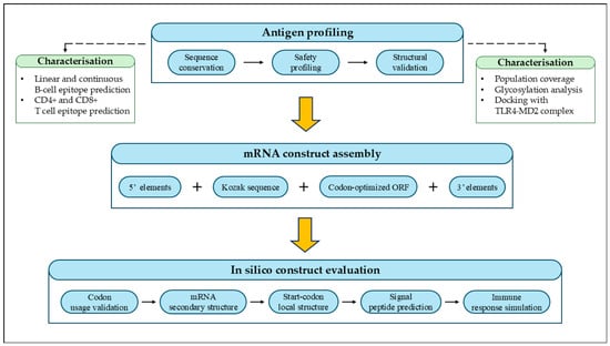
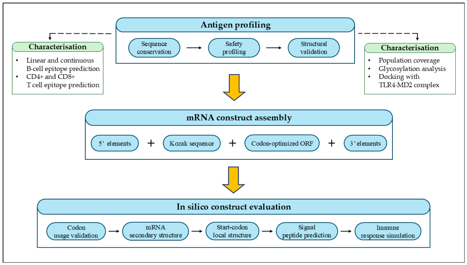
A comprehensive overview of the in silico workflow employed for the design and evaluation of the rabies virus glycoprotein G mRNA vaccine is illustrated in Figure 1.
Figure 1.
Overview of the in silico workflow for rabies virus glycoprotein G mRNA vaccine design. The workflow comprises three sequential stages that directly informed the final construct—antigen profiling, mRNA construct assembly, and in silico construct evaluation—connected by solid arrows. Immunoinformatic and structural characterization analyses performed on the selected antigen (dashed boxes, left and right) provided supporting antigenicity and safety data but did not influence the sequence or composition of the final mRNA construct.
2.1. Antigen Selection and Analysis
2.1.1. Sequence Conservation and Phylogenetic Analysis
The reference amino acid sequence of RABV-G (RefSeq accession number: NP_56796) was retrieved from the National Center for Biotechnology Information (NCBI) database [10], based on the genomic template (GCF 000859625). To account for global genetic diversity and to encompass various circulation strains, the reference sequence was supplemented with 86 additional RABV-G sequences collected from diverse geographical regions, spanning multiple countries across different continents (as detailed in Supplementary Table S1), resulting in a comprehensive set of 87 sequences. Multiple sequence alignment (MSA) was performed using the Clustal Omega web server (https://www.ebi.ac.uk/jdispatcher/msa/clustalo) (accessed on 5 March 2025) [11] to compare sequence similarity and identify conserved regions among the selected strains. Following the alignment, the conservation percentage for each of the 524 amino acid positions was calculated and categorized into three groups: fully identical (100%), highly conserved (70–99%), and variable (<70%). To visualize evolutionary relationships among the aligned sequences and assess phylogenetic conservation patterns, a phylogenetic tree was constructed using the NGPhylogeny.fr server (https://prabi-cloud192.univ-lyon1.fr/) (accessed on 5 March 2025) [12].
2.1.2. Protein Safety Prediction and Physicochemical Analysis
To evaluate the safety and physicochemical characteristics of the selected antigen, several computational analyses were performed. The allergenicity of RABV-G was predicted using the AllergenFP v1.1 server (https://ddg-pharmfac.net/AllergenFP/) (accessed on 7 March 2025) [13], while protein toxicity was evaluated using the CSM-Toxin platform (https://biosig.lab.uq.edu.au/csm_toxin/) (accessed on 7 March 2025) [14]. The antigenicity of the protein was assessed using the VaxiJen v2.1 server (https://www.ddg-pharmfac.net/vaxijen/) (accessed on 7 March 2025) [15] with a threshold value of 0.4 to assess its antigenic potential. In addition, the physicochemical properties of the protein were analyzed using the ProtParam tool (https://web.expasy.org/protparam/) (accessed on 7 March 2025) available on the ExPASy server (https://www.expasy.org/) (accessed on 7 March 2025) [16]. This tool was used to calculate several parameters, including molecular weight, theoretical isoelectric point, instability index, aliphatic index, grand average hydropathicity, the predicted in vitro half-life of the protein, and total number of atoms.
2.1.3. Structural Modeling and Validation
The three-dimensional (3D) structure of RABV-G was generated using the SWISS-MODEL serve (https://swissmodel.expasy.org/) (accessed on 9 March 2025) [17] based on template structure 7U9G chain A, which showed 100% sequence identity with the target protein, indicating an acceptable to good level of structural quality. SWISS-MODEL was selected over AI-based prediction tools such as AlphaFold and Rosetta Fold because the availability of a high-identity experimentally resolved template makes template-based homology modeling the most appropriate and reliable approach in this context, as template-free methods are primarily advantageous in the absence of suitable structural templates. The predicted structural model was downloaded for further analysis. Model validation was then performed to assess the stereochemical quality and structural reliability of the predicted protein structure. Stereochemical evaluation was carried out using the SAVES v6.0 server (https://saves.mbi.ucla.edu/) (accessed on 9 March 2025), which includes PROCHECK Ramachandran plot analysis [18]. In addition, the ERRAT algorithm [19] was used to assess the overall quality of the protein structure. Further structural validation was performed using MolProbity (https://molprobity.biochem.duke.edu/) (accessed on 9 March 2025) [20] to evaluate geometry parameters such as Ramachandran favored regions, outliers, rotamer quality, and possible bond geometry issues.
2.1.4. B-Cell Epitope Prediction
Linear B-Cell Epitope Prediction
Linear B-cell epitope prediction was performed using the Immune Epitope Database (IEDB) Analysis Resource B-cell Epitope Prediction tool (https://tools.iedb.org/bcell/) (accessed on 11 March 2025) based on the BepiPred method [21]. The full-length amino acid sequence of RABV-G was submitted to identify potential antibody-recognition regions. Regions with prediction scores above the default threshold of 0.5 were considered potential linear B-cell epitopes. From the predicted regions, candidate peptide segments of sufficient length (≥14 amino acids) were selected to ensure structural relevance as antigenic targets, prioritizing peptides distributed across distinct regions of the protein sequence to maximize antigenic coverage. Selected candidates were subsequently evaluated for antigenicity using VaxiJen v2.1 (https://www.ddg-pharmfac.net/vaxijen/) (accessed on 11 March 2025) [15] with a threshold of 0.4, toxicity using ToxinPred3.0 (https://webs.iiitd.edu.in/raghava/toxinpred3/) (accessed on 11 March 2025) [22], and allergenicity using AllerCatPro 2.0 (https://allercatpro.bii.a-star.edu.sg/) (accessed on 11 March 2025) [23]. Only peptides satisfying all three criteria were retained for downstream analysis.
Conformational B-Cell Epitope Prediction
Conformational B-cell epitope prediction was performed using the ElliPro tool (https://tools.iedb.org/ellipro/) (accessed on 13 March 2025) available in the IEDB [24]. The previously generated structural model of RABV-G was used as input. A minimum ElliPro score of 0.5 and a maximum distance of 6 Å were applied as prediction parameters. The predicted epitopes were ranked according to their ElliPro scores, and the three highest-scoring regions were selected for further analysis. To evaluate the surface accessibility of the predicted epitopes, the solvent-accessible surface area (SASA) was calculated using UCSF ChimeraX (version 1.11) [25], and epitope locations on the protein surface were visualized accordingly.
2.1.5. T-Cell Epitope Identification and Immuno-Informatics Analysis
CD4+ T-Cell Epitope Prediction and Selection (MHC Class II)
T-cell epitopes restricted to major histocompatibility complex (MHC) class II molecules were predicted using the IEDB MHC II binding prediction tool (https://tools.iedb.org/mhcii/) (accessed on 3 March 2025) [26]. The full-length protein sequence was submitted, and predictions were performed using the NetMHCIIpan method with a reference set of 7 human leukocyte antigen (HLA) alleles and a peptide length of 15-mer; all remaining parameters were kept at their default settings. The resulting dataset was filtered based on percentile rank, retaining only peptides with a rank value below 2%, yielding 19 candidate peptides. Peptide cores appearing across more than one allele context were prioritized to maximize HLA allele coverage as a prerequisite for broader population applicability, reducing the candidate list to 8 unique core sequences. Cores represented by a single allele were excluded, yielding 6 candidate epitopes. Each epitope was subsequently evaluated for antigenicity using VaxiJen v2.1 (https://www.ddg-pharmfac.net/vaxijen/) (accessed on 15 March 2025) [15], allergenicity using AllerCatPro 2.0 (https://allercatpro.bii.a-star.edu.sg/) (accessed on 15 March 2025) [23], and toxicity using ToxinPred (https://neurosnap.ai/service/ToxinPred%20Peptide%20Toxicity%20Prediction) accessed on 15 March 2025 [22] via the NeuroSnap server (https://neurosnap.ai/) (accessed on 15 March 2025). Final selection was restricted to epitopes predicted to be antigenic, non-allergenic, and non-toxic, yielding 3 final MHC-II epitopes for inclusion in downstream analyses.
CD8+ T-Cell Epitope Prediction and Selection (MHC Class I)
CD8+ T-cell epitopes were predicted using the IEDB MHC I (https://tools.iedb.org/mhci/) (accessed on 17 March 2025) binding prediction tool [27] with the NetMHCpan method and a reference set of HLA alleles. The full-length protein sequence was submitted, and the resulting dataset was filtered based on percentile rank, retaining only peptides with a rank value below or equal to 0.5%, yielding the candidate peptide dataset. To identify recurrent predicted epitopes, peptide frequency was assessed using a pivot analysis, and peptides appearing 5 or more times were prioritized. Duplicate sequences were subsequently removed to prevent redundancy. The shortlisted epitopes were then evaluated for immunogenicity using the IEDB Class I Immunogenicity tool (https://tools.iedb.org/immunogenicity/) (accessed on 17 March 2025) [28], and only epitopes with a positive immunogenicity score were considered. Antigenicity was assessed using VaxiJen v2.1 (https://www.ddg-pharmfac.net/vaxijen/) (accessed on 17 March 2025) [15], and allergenicity was evaluated using AllerCatPro 2.0 (https://allercatpro.bii.a-star.edu.sg/) (accessed on 17 March 2025) [23]. Toxicity prediction was not performed due to the short peptide length falling outside the valid input range of available prediction tools. Based on these criteria, a final set of high-confidence CD8+ T-cell epitopes were selected for further analysis.
2.1.6. Population Coverage Analysis
Population coverage analysis was performed to evaluate the potential applicability of the selected T-cell epitopes across different populations. The analysis was conducted using the IEDB Population Coverage tool (https://tools.iedb.org/population/) (accessed on 17 March 2025) [29], based on the distribution of HLA alleles associated with the selected CD4+ and CD8+ T-cell epitopes. The combined set of MHC class I and class II alleles were used as input. Population coverage was calculated for the global population and multiple regions selected to represent both high rabies-endemic areas and geographically diverse populations, including North Africa, South Asia, Southwest Asia, East Africa, West Africa, Europe, North America, and the West Indies. The analysis estimates the fraction of individuals predicted to respond to at least one epitope, in addition to the average number of epitope hits per individual and the population coverage at 90% (PC90), which represents the minimum number of epitope hits/HLA combinations recognized by 90% of the population.
2.1.7. Glycosylation Site Prediction
Prediction of potential N-linked glycosylation sites within the RABV-G protein was performed using the NetNGlyc 1.0 server (https://services.healthtech.dtu.dk/services/NetNGlyc-1.0/) (accessed on 19 March 2025) [30]. The full-length amino acid sequence of the glycoprotein was submitted to identify glycosylation motifs following the consensus sequence N-X-S/T, where X represents any amino acid except proline. The predicted glycosylation sites and their corresponding scores were recorded for further analysis. In addition, the predicted glycosylation positions were compared with the previously identified linear and conformational B-cell epitopes to evaluate the possible presence of glycan shielding that could affect epitope accessibility.
2.1.8. Molecular Docking with TLR4-MD2 Complex
Protein–protein docking was performed to investigate the potential structural interaction between RABV-G and the human TLR4-MD2 complex. Docking was performed using the experimentally resolved trimeric form of RABV-G (Protein Data Bank (PDB): 7U9G [31]), representing the prefusion trimeric state of the protein. The RABV-G trimer was isolated from this structure by removing the bound RVA122 Fab antibody chains, retaining the three glycoprotein protomers (chains A–C), and both this trimer and the TLR4-MD2 receptor structure were prepared using UCSF ChimeraX (version 1.11) [25]—including removal of solvent molecules and non-protein components, hydrogen addition, and basic energy minimization. Docking simulations were performed using two independent servers, HDOCK (http://hdock.phys.hust.edu.cn/) (accessed on 21 April 2025) [32] and ClusPro (https://cluspro.org/login.php) (accessed on 21 April 2025) [33], which employ distinct docking algorithms and scoring functions.
2.2. mRNA Vaccine Design
2.2.1. mRNA Construct Design Strategy
The mRNA vaccine construct was designed based on established mRNA vaccine design principles [34]. mRNA-based vaccines function by delivering a synthetic transcript encoding the target antigen directly into host cells, where it is translated into protein and subsequently recognized by the immune system. This approach eliminates the need for pathogen culture and enables rapid, scalable production. Each structural element of the construct was selected to fulfill a specific functional role aimed at maximizing antigen expression, transcript stability, and reduced innate immune recognition. The structural elements were arranged sequentially from the 5′ end to the 3′ end to ensure efficient translation and improved stability of the mRNA transcript.
5′ Cap Structure
The 5′ cap structure is an essential element of functional mRNA, as it protects the transcript from 5′-to-3′ exonucleolytic degradation and serves as a recognition signal for the eukaryotic translation initiation machinery, thereby ensuring both transcript stability and efficient protein synthesis. Accordingly, a 5′ cap structure was incorporated into the designed construct to protect the transcript from degradation and to promote translation initiation. In this construct, a Cap-1 structure was selected because the presence of 2′-O methylation reduces recognition of the mRNA by innate immune receptors such as retinoic acid-inducible gene I and melanoma differentiation-associated protein 5, thereby partially reducing innate immune sensing, noting that complete abrogation would require additional nucleoside modifications such as N1-methylpseudouridine (m1Ψ) in future experimental work [35].
5′ and 3′ Untranslated Regions (UTRs)
The untranslated regions located at both the 5′ and 3′ ends of the mRNA play important roles in regulating translation efficiency and transcript stability. The selection of human hemoglobin subunit alpha 1 (version 1.11)-derived UTRs was motivated by their reported ability to confer favorable transcript longevity, which may contribute to adequate antigen expression duration following vaccine administration. Accordingly, in this construct, both UTRs were derived from the HBA1 gene, which has been reported to enhance mRNA stability and translation efficiency [34]. The corresponding UTR sequences were retrieved using the UCSC Genome (https://genome.ucsc.edu/index.html) (accessed on 23 April 2025) [36] based on the GRCh38/hg38 genome assembly corresponding to the (RefSeq accession number NM_000558.5).
Kozak Sequence
The Kozak sequence is a conserved nucleotide motif surrounding the AUG start codon that plays a critical role in facilitating ribosome recognition and translation initiation in eukaryotic cells. Accordingly, a Kozak sequence was incorporated immediately upstream of the start codon AUG to support translation initiation efficiency and assist ribosomes in recognizing the correct start site [37]. The specific Kozak sequence used in this construct was retrieved from a previously published study [38].
Codon Optimization of the ORF
The open reading frame (ORF) encoding full-length RABV-G was retrieved from NCBI [10] (RefSeq: NP_056796) and codon-optimized for human expression, as detailed in Section 2.3.1.
Poly(A) Tail Design
The poly(A) tail is a key structural element of eukaryotic mRNA that contributes to transcript protection from 3′ exonucleolytic degradation and supports efficient translation by facilitating ribosome recycling. Accordingly, a poly(A) tail consisting of 130 nucleotides was incorporated into the mRNA construct to support transcript stability and translation efficiency, consistent with previously reported optimal poly(A) tail lengths ranging between 120 and 150 nucleotides (Supplementary File S1 [34].
2.3. In Silico Evaluation of the Designed Vaccine
2.3.1. Codon Optimization Using JCat
To improve the translational efficiency of the designed vaccine antigen, codon optimization was performed using the JCat server (https://www.jcat.de/) (accessed on 1 April 2025) [39]. The amino acid sequence of RABV-G was submitted to the server, and Homo sapiens was selected as the target host to ensure maximum translational compatibility with the human-based immune simulations and docking analyses performed in this study. Furthermore, since both humans and canines share a ‘general mammalian pattern’ of genome composition, they exhibit significant similarities in Guanine and Cytosine (GC) content distribution at the third codon position and codon usage preferences [40]. This suggests that the optimized sequence may potentially maintain adequate expression efficiency in both species. Additional parameters were enabled to minimize potential sequence-related issues, including avoiding Rho-independent transcription terminators, preventing prokaryotic ribosome binding sites, and excluding restriction enzyme cleavage sites. These adjustments were applied to improve the stability of the mRNA sequence and enhance its compatibility with the host cellular translation machinery.
2.3.2. Independent Codon Usage Validation Using GenRCA
To further evaluate the compatibility of the optimized coding sequence with the human expression system, codon usage analysis was performed using the GenScript Rare Codon Analysis tool (GenRCA) (https://www.genscript.com/tools/rare-codon-analysis) (accessed on 3 April 2025) [41]. The optimized gene sequence was submitted to the server to assess codon usage bias and translational efficiency. The analysis generated several parameters describing codon usage characteristics, including the Codon Adaptation Index (CAI), the Effective Number of Codons, and the GC content distribution across the sequence. These metrics were used to evaluate the suitability of the optimized sequence for expression in human cells.
2.3.3. mRNA Secondary Structure Prediction
To evaluate the structural stability of the optimized mRNA sequence, secondary structure prediction was performed using the RNAfold server (version 2.6.3) (http://rna.tbi.univie.ac.at/cgi-bin/RNAWebSuite/RNAfold.cgi) (accessed on 4 April 2025) [42]. The optimized mRNA sequence was submitted to the server to predict the most thermodynamically stable secondary structure and to calculate the Minimum Free Energy (MFE). This analysis helps identify potential internal base-pairing interactions that may influence the stability of the transcript and its accessibility during translation inside host cells. In addition, local folding analysis around the translation initiation region (start codon) was considered to evaluate the accessibility of the ribosome binding region.
2.3.4. Local Secondary Structure Analysis Around the Start Codon
To further evaluate the accessibility of the translation initiation region, local secondary structure analysis was performed for the mRNA segment surrounding the start codon using the RNAfold server (version 2.6.3) (http://rna.tbi.univie.ac.at/cgi-bin/RNAWebSuite/RNAfold.cgi) (accessed on 6 April 2025) [42]. A sequence fragment containing approximately 30 nucleotides upstream and 30 nucleotides downstream of the start codon AUG was extracted from the optimized mRNA sequence and submitted to the RNAfold server. This analysis was conducted to assess whether local folding structures could potentially interfere with ribosome binding and translation initiation.
2.3.5. Signal Peptide Prediction
To evaluate whether the encoded RABV-G could be directed to the host secretory pathway, signal peptide prediction was performed using the SignalP 6.0 server (https://services.healthtech.dtu.dk/services/SignalP-6.0/) (accessed on 6 April 2025) [43]. The amino acid sequence of RABV-G was submitted to the server with Eukarya selected as the target organism. SignalP 6.0 predicts the presence of N-terminal signal peptides, their type, and the most probable signal peptide cleavage site responsible for directing proteins toward the secretory pathway.
2.3.6. In Silico Immune Response Simulation
To evaluate the potential immunogenicity of the designed mRNA vaccine construct, an immune response simulation was performed using the C-IMMSIM server (https://kraken.iac.rm.cnr.it/C-IMMSIM/index.php) (accessed on 7 April 2025) [44]. The amino acid sequence of RABV-G was submitted as the vaccine antigen. The simulation was conducted for 1050 time steps, with three virtual injections administered at four-week intervals (time steps 1, 84, and 168). The adjuvant parameter was set to 100 according to the standard C-ImmSim parameter set, while the “vaccine (no LPS)” option was selected. The antigen concentration was set to 1000 per dose, and the random seed was set to 12345 to ensure reproducibility. This computational framework was used to predict the humoral and cellular immune responses over an extended simulation period.
3. Results
3.1. RABV-G Conservation, Safety, and Structural Validation
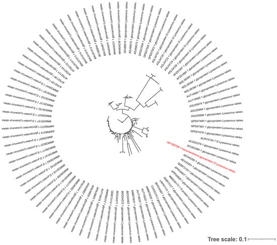
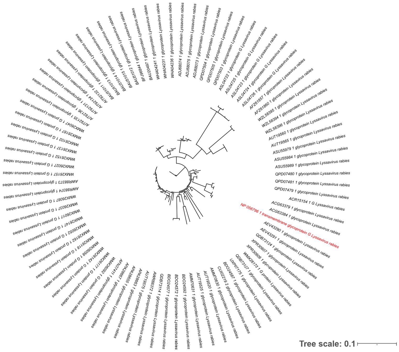
Comprehensive computational analyses—including conservation, physicochemical, and structural assessments—indicate the suitability of RABV-G as a conserved antigenic target for vaccine design. Initially, the MSA of 87 RABV-G sequences revealed substantial conservation across the protein. Analysis of the 524 amino acid positions identified 282 positions with complete sequence identity (53.82%), 229 positions with high conservation levels ranging from 70 to 99% (43.70%), and 13 variable positions below 70% (2.48%) as detailed in Supplementary Table S2. Phylogenetic analysis using NGPhylogeny.fr demonstrated that sequences from different geographical regions clustered with strong evolutionary support, indicating robust conservation of critical functional domains across diverse rabies virus strains (Figure 2).
Figure 2.
Phylogenetic analysis of rabies virus glycoprotein G (RABV-G) sequences. The tree was constructed using the NGPhylogeny.fr server based on the alignment of 87 sequences from diverse geographical regions. The reference strain used for the mRNA vaccine construct design (NP _056796) is highlighted in red. The scale bar represents an evolutionary distance of 0.1.
Furthermore, computational analyses predicted that RABV-G is non-toxic, non-allergenic, and possesses favorable antigenicity, suggesting its potential to safely stimulate an immune response. Additionally, physicochemical profiling supported its structural stability, hydrophilic nature, and optimal expression characteristics, with all detailed calculated parameters comprehensively summarized in Supplementary Table S3A.
Finally, the predicted 3D model was subjected to multiple validation analyses to assess its structural quality. Independent validation tools, including PROCHECK, ERRAT, and MolProbity, consistently indicated acceptable stereochemical quality and reliability of the modeled protein structure (Supplementary Figure S1A,B). The slight differences between PROCHECK and MolProbity Ramachandran statistics are expected due to differences in evaluation algorithms. All detailed validation scores are summarized in Supplementary Table S3B. Taken together, the high evolutionary conservation, favorable safety and antigenicity profiles, and the reliability of the predicted 3D model collectively support the viability of RABV-G as a promising vaccine target. These robust characteristics establish a solid foundation for subsequent in silico analyses, particularly for the comprehensive identification of candidate epitopes and the evaluation of their global population coverage.
3.2. Epitope Identification and Population Coverage Analysis
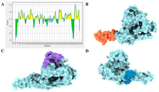
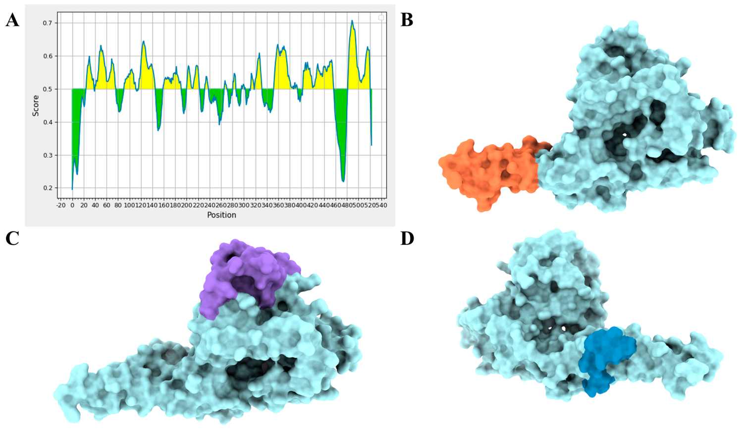
Having established a reliable RABV-G antigen model, comprehensive epitope mapping was performed to identify highly antigenic regions capable of eliciting both humoral and cellular immune responses, followed by an assessment of their global population-level applicability. For the humoral response, BepiPred analysis identified 18 regions across the RABV-G sequence with prediction scores above the threshold of 0.5, indicating potential linear B-cell epitopes (Figure 3A). Following length-based filtering (≥14 amino acids) and the selection of non-overlapping peptides distributed across distinct regions, three candidate peptides were shortlisted. Subsequent evaluation confirmed that all three peptides satisfied the selection criteria, demonstrating strong antigenic potential while being predicted as non-allergenic and non-toxic. The final selected linear B-cell epitopes are summarized in Table 1.
Figure 3.
Predicted linear and conformational B-cell epitopes of RABV-G. (A) Linear B-cell epitope propensity plot generated via the BepiPred algorithm; yellow regions represent residues with scores exceeding the 0.5 threshold indicating potential linear epitopes, whereas green regions indicate scores below the threshold. (B–D) 3D surface models of RABV-G showing the spatial localization of the top three predicted conformational B-cell epitopes. The specific epitopes are color-coded as follows: (B) CE-1 (orange), (C) CE-2 (purple), and (D) CE-3 (blue).
Table 1.
Selected linear B-cell epitopes identified from RABV-G after antigenicity, toxicity, and allergenicity filtering.
Complementing the linear epitope mapping, conformational B-cell analysis using ElliPro (minimum score 0.5, maximum distance 6 Å) identified 9 discontinuous epitopes on the antigen structure. The three top-scoring epitopes were selected for further validation. SASA calculations using ChimeraX [25] confirmed that these selected regions are highly exposed on the protein surface, ensuring optimal accessibility for antibody recognition. Specifically, the top predicted conformational epitopes—designated as CE-1 (41 residues, ElliPro score: 0.868, SASA: 3791.8 Å2), CE-2 (54 residues, ElliPro score: 0.738, SASA: 4356.5 Å2), and CE-3 (15 residues, ElliPro score: 0.680, SASA: 1773.8 Å2)—exhibited favorable surface exposure, with their spatial distribution visualized in Figure 3B–D. Having secured promising targets for humoral immunity, subsequent analyses focused on identifying robust T-cell epitopes to drive a comprehensive cellular immune response. Accordingly, both CD4+ and CD8+ T-cell epitopes were predicted using the IEDB platform and subjected to rigorous, multi-step filtering. For CD4+ T-cells, MHC class II prediction (percentile rank < 2%) initially identified 19 peptides, which were subsequently grouped by core sequences binding multiple HLA alleles to yield 6 candidates. Concurrently, MHC class I prediction for CD8+ T-cells (percentile rank ≤ 0.5%) generated the candidate peptide dataset, which were refined by prioritizing recurrent sequences appearing five or more times. Each candidate set then underwent stringent downstream evaluations: CD4+ candidates were assessed for antigenicity, allergenicity, and toxicity, while CD8+ candidates were evaluated for immunogenicity, antigenicity, and allergenicity. This filtering ultimately yielded 3 CD4+ and 5 CD8+ final epitopes (detailed in Table 2A,B, respectively) demonstrating robust antigenic (CD4+) and immunogenic (CD8+) potential, where all final CD4+ epitopes were verified as non-allergenic and non-toxic, and all final CD8+ epitopes were confirmed as non-allergenic. With these promising T-cell epitopes successfully identified, the next critical step was to evaluate their collective broad applicability across diverse human genetic backgrounds through a global population coverage analysis.
Table 2.
(A) Selected CD4+ T-cell epitopes with their corresponding predicted HLA alleles, percentile rank, and antigenicity scores. (B) Selected CD8+ T-cell epitopes with their corresponding predicted HLA alleles, immunogenicity, and antigenicity scores.
Given these favorable epitope selections, the subsequent population coverage analysis demonstrated that the selected T-cell epitopes provide broad global and regional applicability. For the global population, the combined MHC class I and II coverage reached 86.52%, with an average epitope hit of 1.58 and a PC90 value of 0.74. Coverage estimates across all analyzed regions are summarized in Table 3. Coverage exceeded 72% across all regions analyzed, with the highest values observed in Europe (93.95%) and North America (88.52%), and the lowest in East Africa (72.65%). The distribution of epitope recognition across individuals for each population is shown in Supplementary Figure S2. These results suggest that the selected epitopes may elicit immune responses in a substantial proportion of individuals across diverse HLA profiles globally. To ensure that these promising epitopes remain accessible and are not masked by post-translational modifications, the analysis next proceeded to the prediction of N-linked glycosylation sites.
Table 3.
Global and regional population coverage analysis of the selected T-cell epitopes, demonstrating combined MHC class I and II binding potential across diverse geographic regions.
Accordingly, the prediction of potential N-linked glycosylation sites in RABV-G was performed using the NetNGlyc 1.0 server. The analysis identified five potential glycosylation motifs following the consensus sequence N-X-S/T at amino acid positions 56, 177, 266, 338, and 484 (Supplementary Table S4). The corresponding prediction profile showing glycosylation potential across the protein sequence is provided in Supplementary Figure S3. These predicted glycosylation sites were further analyzed in relation to the previously identified linear and conformational B-cell epitopes. Importantly, no overlap was observed between the glycosylation sites and the selected epitopes, indicating the absence of potential glycan shielding near antigenic regions, suggesting that the identified epitopes remain structurally accessible and may be readily recognized by the immune system.
Taken together, the selected B-cell and T-cell epitopes demonstrated robust antigenic (B-cell and CD4+) and immunogenic (CD8+) profiles, favorable safety profiles, and optimal surface accessibility. The confirmed absence of glycan shielding ensures that these regions remain structurally available for immune recognition, while the extensive population coverage guarantees their broad global applicability. Consequently, these comprehensively characterized epitopes establish a reliable foundation for the next phase of the study, advancing to molecular docking analyses to evaluate their physical binding interactions with specific immune cell receptors.
3.3. Receptor Docking Analysis
Building upon the successful identification of safe, accessible, antigenic and immunogenic epitopes, the study next investigated whether the RABV-G antigen could physically interact with the human TLR4-MD2 receptor complex, a key initiator of innate immunity.
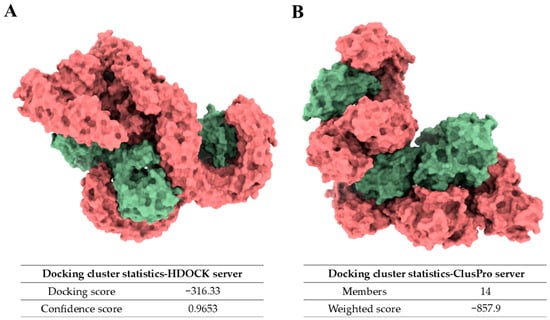
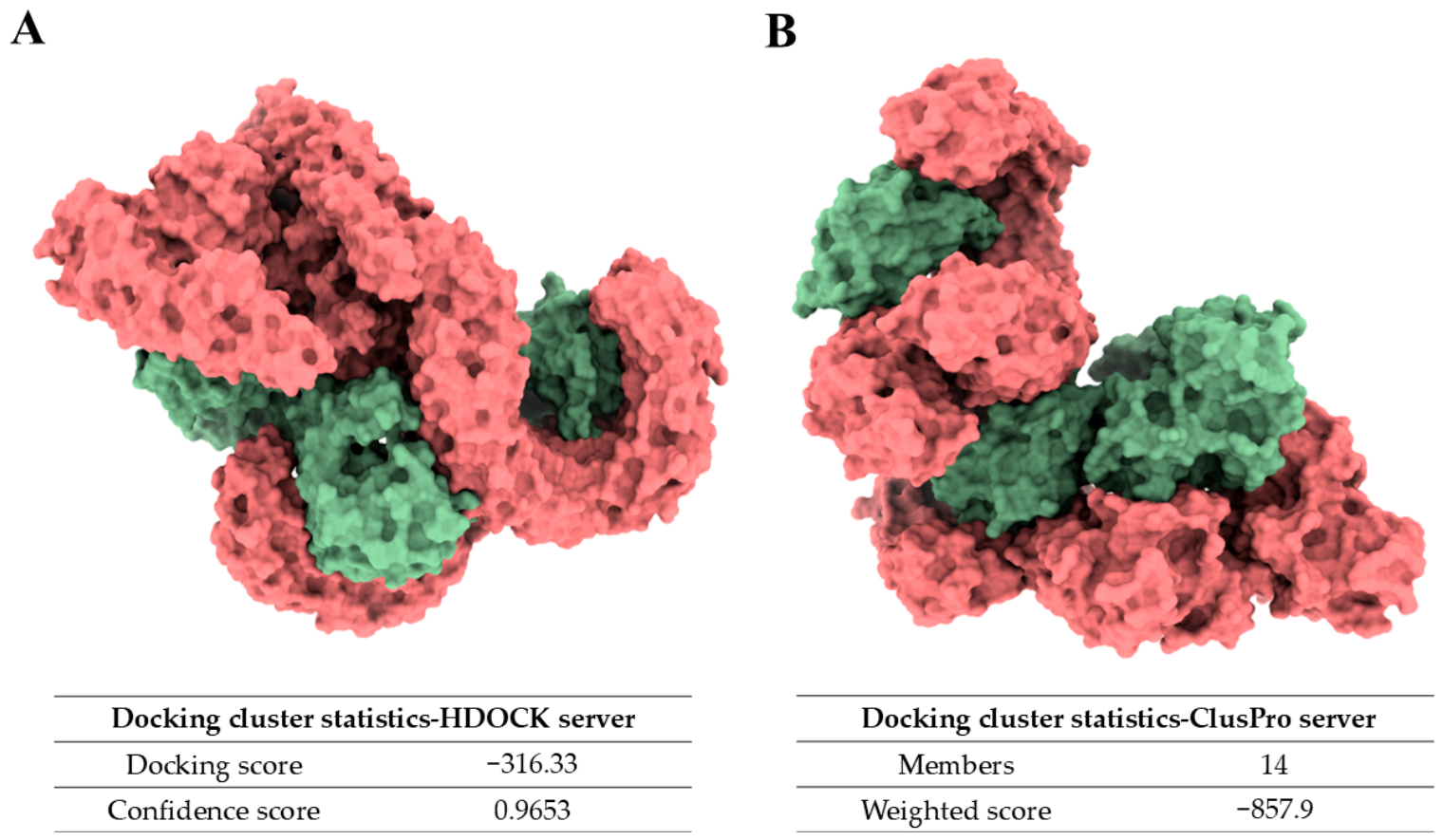
Docking analysis predicted a potential structural binding configuration between RABV-G and the human TLR4-MD2 receptor. To better represent the native oligomeric state of RABV-G, the docking analysis was performed using the experimentally resolved prefusion trimeric structure (PDB: 7U9G) [31], which was also used as the structural template for homology modeling in this study. The top-ranked docking model obtained from the HDOCK server showed a docking score of −316.33 with a confidence score of 0.9653, supporting a favorable predicted binding configuration between the two proteins (Figure 4A). In parallel, docking performed using the ClusPro server identified Cluster 0 as the most populated cluster, containing 14 members with a weighted score of −857.9 (Figure 4B).
Figure 4.
Predicted molecular docking models of RABV-G complexed with the human Toll-Like Receptor 4/Myeloid Differentiation factor 2 complex (TLR4–MD2) receptor. The 3D structural models illustrate the predicted binding interfaces, with RABV-G depicted in red and the human TLR4–MD2 complex shown in green. (A) The top-ranked docking pose generated via the HDOCK server (docking score: −316.33; confidence score: 0.9653). (B) The representative model from the most populated cluster (Cluster 0, comprising 14 members) generated via the ClusPro server, exhibiting a weighted score of −857.9.
These findings suggest a predicted structural binding pose between RABV-G and the TLR4-MD2 complex, consistent with the broader observation that viral glycoproteins can engage TLR4 [45]. However, docking simulations do not assess receptor activation or downstream signaling, and experimental validation would be required to confirm any functional interaction. Further structural flexibility and theoretical dynamic behavior of the docked complex were evaluated using Normal Mode Analysis (NMA). Detailed methodology and results are provided in Supplementary File S2, and the corresponding results are illustrated in Supplementary Figure S4.
Building on these favorable antigen and structural properties, the study next focused on the design and evaluation of the mRNA vaccine construct itself.
3.4. mRNA Construct Design and Evaluation
Building upon the favorable receptor binding interactions predicted by molecular docking, the final phase of the study focused on designing and computationally evaluating the complete mRNA vaccine construct. This in silico evaluation sequentially addressed the structural assembly of the construct, the optimization of codon usage for human expression, the validation of secondary structure stability, and the computational simulation of the vaccine-induced immunity.
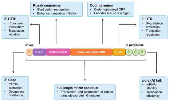
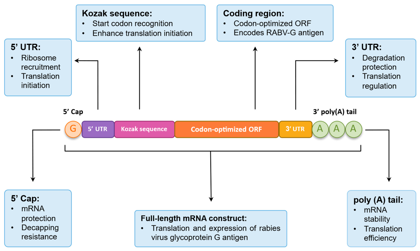
Based on the established design strategy, the final mRNA vaccine construct was successfully assembled with structural elements arranged from the 5′ end to the 3′ end as follows: a 5′ Cap (Cap-1 structure), a 5′ UTR derived from α-globin, a Kozak sequence, a codon-optimized ORF encoding the full-length RABV-G, a 3′ UTR derived from α-globin, and a poly(A) tail consisting of 130 nucleotides. The overall architecture of the designed mRNA construct is illustrated in Figure 5. With the physical elements of the construct fully assembled, the nucleotide sequence of the antigen was subsequently evaluated and optimized to ensure maximum translational efficiency within human host cells.
Figure 5.
Schematic representation of the designed mRNA vaccine construct encoding the RABV-G. The diagram illustrates the linear sequence of the structural elements arranged from the 5′ to the 3′ end. The integrated components comprise a 5′ Cap (Cap-1), a 5′ untranslated region (UTR) derived from human α-globin, a Kozak consensus sequence, the codon-optimized ORF encoding the full-length viral antigen, a 3′ UTR (α-globin), and a 130-nucleotide poly(A) tail.
To maximize translational efficiency in human host cells, the viral ORF was subjected to codon optimization. Initial optimization using JCat demonstrated strong adaptation to human codon preferences and enhanced GC content, suggesting improved transcript stability (Supplementary Figure S5). This was independently cross-validated using the GenRCA tool, which confirmed a strong bias toward preferred human codons and verified the absence of negative repeat elements or unwanted termination signals (Supplementary Figure S6). While minor variations in specific optimization metrics were observed between the two platforms due to differences in their underlying algorithms and reference datasets, both analyses consistently yielded CAI values well above the accepted threshold of 0.8. A detailed comparison of the calculated parameters from both servers is summarized in Supplementary Table S5. Collectively, these findings indicate that the optimized coding sequence possesses favorable characteristics for stable expression and efficient protein production.
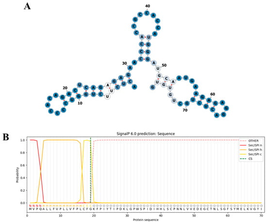
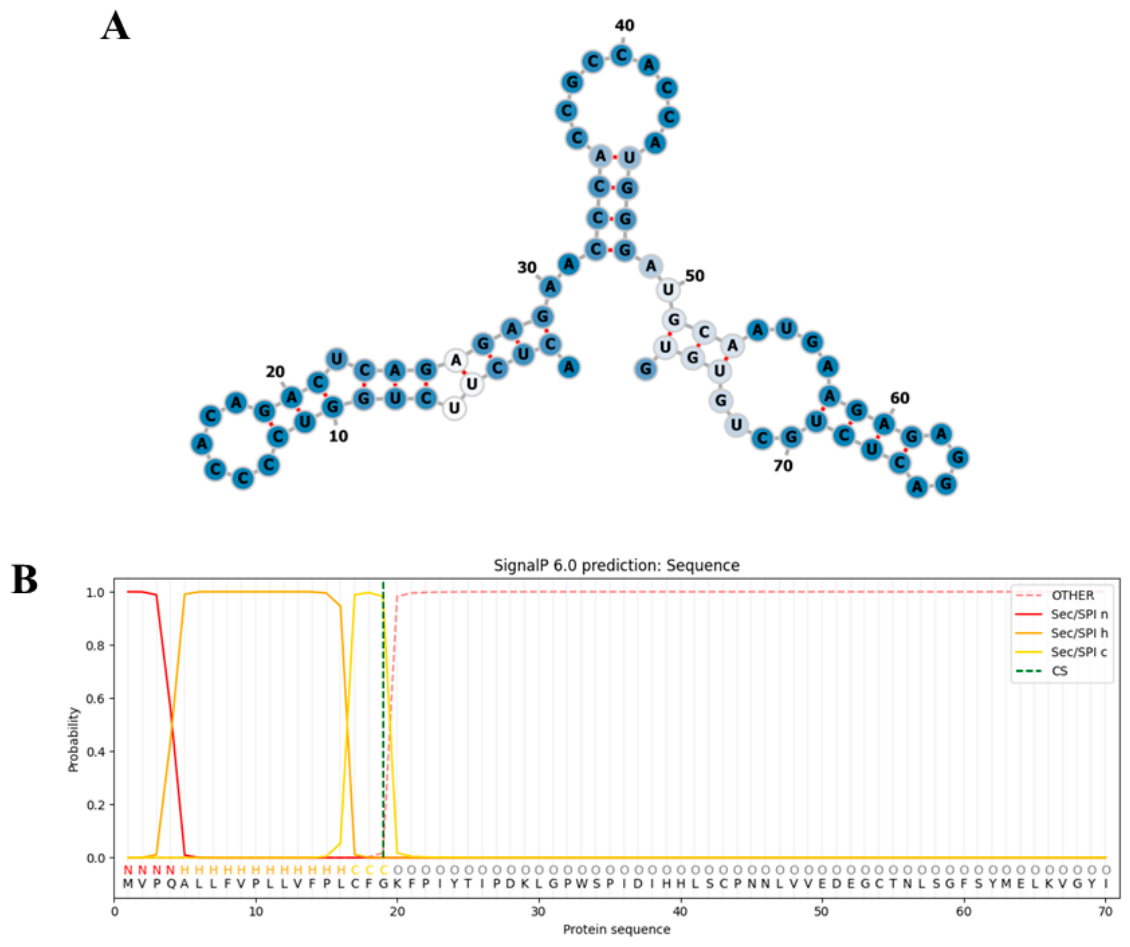
Following sequence optimization, the thermodynamic stability and translational readiness of the mRNA construct were evaluated using RNAfold. Global sequence analysis predicted a highly stable transcript, indicated by a MFE of −429.50 kcal/mol and a balanced distribution of paired and unpaired nucleotides (Supplementary Figure S7A,B). While absolute MFE values for long transcripts are inherently negative, local structural analysis confirmed that the translation initiation region remains highly accessible (local MFE = −18.80 kcal/mol). Importantly, no stable secondary structures were found overlapping the start codon AUG, reducing the likelihood of steric hindrance and supporting efficient ribosomal assembly (Figure 6A). Complementing this translational accessibility, SignalP 6.0 confidently identified a functional Sec/SPI-type signal peptide at the N-terminus (probability = 0.999), featuring a well-defined cleavage site between residues 19 and 20 (Figure 6B). Together, these findings indicate that the designed mRNA is structurally stable, sterically accessible for translation initiation, and properly equipped to direct the synthesized antigen to the host secretory pathway for optimal folding and subsequent presentation.
Figure 6.
Local mRNA secondary structure and signal peptide prediction for the optimized RABV-G construct. (A) Predicted local secondary structure of the mRNA transcript region flanking the start codon, generated via the RNAfold server. Nucleotide positions are numbered sequentially, with base-pairing interactions indicated by connecting lines. (B) Signal peptide and cleavage site prediction generated using SignalP 6.0. The plot displays the predicted probabilities for distinct regions, color-coded as follows: the N-terminal n-region (Sec/SPI n, red), the hydrophobic h-region (Sec/SPI h, orange), the c-region preceding the cleavage site (Sec/SPI c, yellow), and the non-signal-peptide sequence (OTHER, dashed pink). The predicted cleavage site is marked by a dashed vertical line between amino acid positions 19 and 20.
Finally, the immunogenic potential of the complete mRNA construct was evaluated through in silico immune simulation using the C-ImmSim server. The simulation predicted sustained activation of both humoral and cellular immune responses following a three-dose vaccination protocol. Notably, the simulated response was characterized by rapid antigen clearance and antibody isotype switching, with a progressive increase in IgG1 and IgG2 subclasses. Concurrently, memory B and T-helper cell populations expanded and remained elevated throughout the 350-day simulation period, supporting the predicted development of long-term immune memory. The simulation also revealed a cellular immune response featuring the expansion of active cytotoxic T lymphocytes and a Th1-biased profile, alongside enhanced innate immune activity and elevated IFN-γ levels. The full suite of simulation outputs, including detailed cellular subset dynamics and cytokine profiles, is presented in Supplementary Figure S8. Overall, these findings represent model-dependent in silico predictions and require experimental validation to determine their biological relevance.
4. Discussion
The RABV-G plays a critical role in the infection process, serving as the primary surface protein that binds to host cell receptors and facilitates viral entry into the nervous system [4]. Given this indispensable function, it is widely regarded as the main protective antigen [46]. Historically, rabies prevention has relied on conventional whole-virus vaccines produced using nerve tissue, embryonated eggs, and cell culture systems [47]. These platforms are detailed in Table 4 and broadly divided into two generations: traditional whole-virus vaccines (nerve tissue, avian embryo, and cell culture) and next-generation nucleic acid vaccines (full-length and multi-epitope mRNA). In contrast to traditional methods, mRNA vaccination introduces a flexible strategy by delivering genetic instructions for intracellular antigen production, offering rapid design adaptability and robust immune stimulation [48]. Within the rabies landscape, this platform allows for the exploration of both full-length RABV-G constructs and alternative targeted designs, laying the foundation for comparing structural and immunological efficacy between full-length and multi-epitope mRNA configurations. A quantitative comparison with a recent in silico multi-epitope RABV mRNA design [9] showed distinct vaccine architectures. The published construct targeted four RABV proteins (G, N, M, and P) and incorporated 21 epitopes (13 linear B-cell, 4 HTL, and 4 CTL), with a predicted population coverage of 96.01%. In contrast, our construct targets only the full-length RABV-G antigen and includes 14 prioritized epitopes (3 linear B-cell, 3 conformational B-cell, 3 CD4, and 5 CD8), achieving a predicted population coverage of 86.52%. These differences reflect the distinct strategies of epitope concatenation versus preservation of the complete RABV-G antigenic and structural context. Compared to multi-epitope mRNA designs, our full-length RABV-G construct offers a more comprehensive antigenic representation while preserving the native structural context of the antigen. While concatenated multi-epitope peptides may not fully reproduce native conformations [49], full-length expression preserves the 3D architecture and conformational epitopes of RABV-G [50]. Specifically for the virus, RABV-G serves as the primary surface antigen and the major target of VNAs [45]. This suggests that full-length targeting may provide a more focused immune response than multi-epitope approaches [51]. Such designs typically also incorporate internal proteins such as N, P, and M [52], whose primary role are associated with viral replication rather than antibody neutralization.
Table 4.
Comparative overview of the proposed full-length RABV-G mRNA vaccine candidate against existing and alternative rabies vaccine platforms, including multi-epitope protein and mRNA designs, and conventional nerve tissue, avian embryo, and cell culture-derived vaccines.
To support this selection, the protein was further evaluated for conservation, safety, and antigenicity. Analysis of 87 RABV-G sequences from diverse geographical regions revealed substantial conservation—282 positions (53.82%) showed complete sequence identity, and 229 positions (43.70%) were highly conserved at 70–99%—a pattern further supported by tight phylogenetic clustering (NGPhylogeny.fr), consistent with RABV-G’s potential for cross-protective vaccine design [53]. Safety assessments via AllergenFP and CSM-Toxin predicted the protein to be non-allergenic and non-toxic. An antigenicity score of 0.5029 (VaxiJen) exceeded the 0.4 threshold, indicating potential for immune recognition [54]. Physicochemical analysis was largely favorable: the aliphatic index (84.01) and negative GRAVY value (−0.167) are sequence-based indicators generally associated with thermostability and hydrophilicity. The instability index (40.51), slightly above the classical threshold of 40, should not be taken as a firm result: it is based only on sequence patterns and does not reflect the actual folding environment inside a cell. Experimental testing would be needed to confirm the protein’s real stability. Collectively, these findings suggest RABV-G as a conserved, low-risk, and antigenically promising candidate for mRNA vaccine development. Building on these foundational characteristics, the vaccine’s antigenic potential was further evaluated through epitope selection and structural analysis. This analysis therefore provides insight into the antigen’s predicted antigenic and immunogenic profile rather than serving as a basis for construct design. A signal peptide was identified at the N-terminus (probability = 0.999), consistent with proper trafficking of the antigen through the secretory pathway for surface presentation [43]. Structural modeling was performed on the mature, signal-peptide-cleaved sequence, consistent with the co-translational removal of the signal peptide prior to folding of the native ectodomain. Glycosylation analysis further showed that the five predicted N-glycosylation sites do not overlap with the selected B-cell epitopes, supporting their potential accessibility to antibody recognition [55]. Population coverage analysis across populations representing major rabies-endemic geographically diverse regions showed broad combined MHC class I/II coverage overall (Table 3).
Coverage was comparatively the lowest in East Africa, highlighting a gap worth addressing in future work. Immune simulation using C-ImmSim predicted a sustained immune response characterized by increases in IgM and IgG antibodies and the formation of memory B cells [44], while expansion of both CD4+ and CD8+ T cells, together with increased IFN-γ and IL-2 levels, indicated activation of cellular immunity. However, these findings are inherently model-dependent and should be interpreted as computational predictions rather than evidence of actual vaccine efficacy or parameter-independent robustness in biological systems.
Molecular docking with the TLR4-MD2 complex was performed—selected not as a documented entry receptor for RABV-G, but to investigate a potential innate immune recognition pathway distinct from viral entry. As TLR4 recognizes ligands together with MD2, the complete TLR4-MD2 complex was used to better reflect the physiological receptor configuration [56,57]. Docking was carried out using two independent servers, HDOCK and ClusPro, each employing distinct algorithms and scoring functions. Docking used the trimeric form of RABV-G, representing its biologically active conformation on the viral envelope; the predicted interaction with TLR4-MD2 may therefore be more physiologically relevant than one derived from a monomeric or computationally assembled structure [58,59]. This interaction is consistent with TLR4′s established role as a pattern recognition receptor [60] and prior evidence linking TLR4 signaling to rabies-induced humoral immunity [57], though it reflects structural complementarity only and would require experimental validation to confirm functional activation. In addition to the immunological and structural analyses, the mRNA sequence was optimized for human expression using JCat. To validate this optimization independently, codon usage was re-assessed using a second platform, GenRCA. JCat produced a CAI of 0.953 (GC 66.28%), versus 0.81 (GC 56%) for GenRCA—a difference driven by JCat’s use of the single most frequent codon per amino acid, which raises GC content, while GenRCA follows a different codon-usage model.
Despite this platform-dependent variation, both tools reported CAI values above the 0.8 threshold considered adequate for expression in human cells. Both also showed higher GC content than the unoptimized sequence (40.89%), consistent with improved transcript stability.
Balancing transcript stability against translational accessibility, RNAfold analysis of the mRNA secondary structure gave an MFE of −429.50 kcal/mol. This highly negative value suggests a stable secondary structure that may help protect the transcript from degradation [42]. However, MED values require some caution, as longer transcripts inherently yield more negative values regardless of their actual folding stability. At the same time, a highly stable structure might interfere with ribosome binding. To check this, the region around the start codon was examined separately and found to remain accessible, supporting efficient translation initiation. Taken together, these findings suggest that the mRNA design may support both transcript stability and protein expression. Comparative genomic analyses indicate that humans and canines share notable similarities in GC content and codon usage patterns [61]. suggesting the optimized sequence may also support adequate expression efficiency in canine cells. Given that dogs are the primary reservoir of human rabies transmission, this cross-species compatibility could be a valuable additional benefit of the vaccine design. Chemical modification of the mRNA is one key consideration: unmodified mRNA can trigger unwanted innate immune activation, reducing protein expression and causing inflammation [62]. Incorporating modified nucleosides such as m1Ψ substantially reduces this recognition while improving translational efficiency and transcript stability [63]. The current design already includes a Cap-1 structure to partly address this; incorporating m1Ψ in future work would likely enhance this effect further [61]. Delivery is another critical factor: naked mRNA is unstable in biological fluids and cannot efficiently enter cells on its own. Lipid nanoparticles (LNPs) remain the most clinically validated delivery system, protecting mRNA from degradation and enabling cellular uptake, as demonstrated in COVID-19 vaccines [64]. Beyond these formulation considerations, practical barriers to translation remain: LNP-based mRNA vaccines currently require cold-chain storage and specialized manufacturing capacity, both of which pose significant cost and logistical challenges in the low-resource, rabies-endemic regions where this vaccine would be most needed. For the vaccine designed in this study, the incorporation of both nucleoside modifications and an optimized LNP formulation would be important considerations in any future experimental work. Future versions of this vaccine could also add extra immune-boosting elements alongside RABV-G, use self-amplifying mRNA to improve expression, or extend the conservation and design analysis beyond RABV to cover other lyssaviruses such as LBV, MOKV, and EBLVs, broadening cross-protection. Overall, the designed mRNA vaccine targeting RABV-G shows strong computational potential and merits further experimental investigation. All results reported here are computational predictions; wet lab validation—in vitro expression, in vivo immunogenicity testing, and safety assessment—remains essential to confirm their biological relevance.
5. Limitations
Despite these promising results, several limitations should be acknowledged. This study is entirely computational: all antigenicity, structural, and immune-response predictions—including the C-ImmSim simulation—are model-based and have not been experimentally validated; in vitro expression, in vivo immunogenicity testing, and eventual clinical evaluation remain necessary before any conclusions can be drawn about real-world safety and efficacy. Each analytical stage also relied on a single tool rather than a consensus approach: B-cell epitopes from BepiPred/ElliPro, MHC binding from NetMHCpan/NetMHCIIpan, and immune simulation from C-ImmSim, none cross-validated against alternative methods. Innate immune modeling covered only TLR4-MD2; TLR7/8 and RIG-I/MDA5, potentially more relevant to mRNA sensing, were not examined. The construct also targets a single antigen from a single virus, and conservation analysis was limited to RABV strains; extension to other lyssaviruses (e.g., LBV, MOKV, EBLVs) or multi-antigen designs was not attempted. Structural analysis combined a monomeric model for epitope mapping with a trimeric model for docking, which may not fully reflect RABV-G’s native conformation; only N-linked glycosylation was modeled, leaving other modifications such as O-glycosylation unexamined.
6. Conclusions
This study presents the computational design of a full-length RABV-G mRNA vaccine candidate. Sequence analysis showed high conservation across 87 RABV-G strains, favorable antigenicity (VaxiJen score 0.5029), and a structurally reliable homology model (>90% residues in favored Ramachandran regions). Epitope mapping identified conserved, surface-accessible B- and T-cell epitopes with broad population coverage (72.65–93.95% across nine regions), and immune simulation predicted the activation of both humoral and cellular responses. The optimized mRNA construct showed favorable codon adaptation (CAI > 0.8) and a stable predicted secondary structure. Collectively, these computational findings support the feasibility of the proposed design, though they remain model-based; in vitro expression, in vivo immunogenicity testing, and safety assessment are required to confirm biological relevance.
Supplementary Materials
The following supporting information can be downloaded at: https://www.mdpi.com/article/10.3390/biomedinformatics6050085/s1. Supplementary Table S1, Dataset containing the GenBank accession numbers, geographical origins, and NCBI; Supplementary Table S2, Conservation analysis results; Supplementary Table S3, Predicted physicochemical properties and Structural validation statistics; Supplementary Table S4, NetNGlyc 1.0 prediction of N-glycosylation sites in Rabies G protein; Supplementary Table S5, Comparison of codon optimization parameters between JCat and GenRCA servers; Supplementary Figure S1, Structural validation of rabies virus glycoprotein G model; Supplementary Figure S2, Global and regional population coverage of predicted T-cell epitopes; Supplementary Figure S3, Graphical potential plot for NetNGlyc predictions in Rabies G; Supplementary Figure S4, Normal mode analysis figures; Supplementary Figure S5, Codon optimization using jcat server; Supplementary Figure S6, Codon optimization using GenRCA tool; Supplementary Figure S7, RNAfold secondary structure and mountain plot for optimized mRNA; and Supplementary Figure S8, immune simulation results; Supplementary File S1, nucleotide sequence of the designed mRNA vaccine construct; and Supplementary File S2, Normal mode analysis Methodology and Results [65].
Author Contributions
Conceptualization, I.B.A.; methodology, I.B.A.; software, I.B.A.; validation, A.R.E.; formal analysis, I.B.A.; data curation, I.B.A.; writing—original draft preparation, I.B.A.; writing—review and editing, A.R.E. and M.M.L.; supervision, A.R.E.; project administration, A.R.E. All authors have read and agreed to the published version of the manuscript.
Funding
This research received no external funding.
Institutional Review Board Statement
Not applicable.
Informed Consent Statement
Not applicable.
Data Availability Statement
Data is contained within the article and Supplementary Material.
Conflicts of Interest
The authors declare no conflicts of interest.
Abbreviations
The following abbreviations are used in this manuscript:
| COVID-19 | Coronavirus Disease 2019 |
| CAI | Codon Adaptation Index |
| GC | Guanine and Cytosine |
| GenRCA | GenScript Rare Codon Analysis tool |
| HBA1 | Human Hemoglobin Subunit Alpha 1 |
| HLA | Human Leukocyte Antigen |
| IEDB | Immune Epitope Database |
| IFN-γ | Interferon Gamma |
| IgG | Immunoglobulin G |
| IgM | Immunoglobulin M |
| IL-2 | Interleukin-2 |
| LNP | Lipid Nanoparticle |
| MFE | Minimum Free Energy |
| MHC | Major Histocompatibility Complex |
| mRNA | Messenger Ribonucleic Acid |
| MSA | Multiple Sequence Alignment |
| m1Ψ | N1-methylpseudouridine |
| nAChRs | Nicotinic Acetylcholine Receptors |
| NCBI | National Center for Biotechnology Information |
| ORF | Open Reading Frame |
| PC90 | Population Coverage at 90% |
| PDB | Protein Data Bank |
| PEP | Post-Exposure Prophylaxis |
| RABV-G | Rabies Virus Glycoprotein G |
| SASA | Solvent-Accessible Surface Area |
| TLR4-MD2 | Toll-Like Receptor 4/Myeloid Differentiation factor 2 complex |
| UTR | Untranslated Region |
| VNAs | Virus-Neutralizing Antibodies |
| 3D | Three-Dimensional |
References
- World Health Organization Rabies. Available online: https://www.who.int/southeastasia/health-topics/rabies (accessed on 15 March 2026).
- Hampson, K.; Coudeville, L.; Lembo, T.; Sambo, M.; Kieffer, A.; Attlan, M.; Barrat, J.; Blanton, J.D.; Briggs, D.J.; Cleaveland, S.; et al. Estimating the Global Burden of Endemic Canine Rabies. PLoS Negl. Trop. Dis. 2015, 9, e0003709. [Google Scholar] [CrossRef] [Scilit] [PubMed]
- Sambo, M.; Lembo, T.; Cleaveland, S.; Ferguson, H.M.; Sikana, L.; Simon, C.; Urassa, H.; Hampson, K. Knowledge, Attitudes and Practices (KAP) about Rabies Prevention and Control: A Community Survey in Tanzania. PLoS Negl. Trop. Dis. 2014, 8, e3310. [Google Scholar] [CrossRef] [Scilit] [PubMed]
- Fooks, A.R.; Cliquet, F.; Finke, S.; Freuling, C.; Hemachudha, T.; Mani, R.S.; Müller, T.; Nadin-Davis, S.; Picard-Meyer, E.; Wilde, H.; et al. Rabies. Nat. Rev. Dis. Primers 2017, 3, 17091. [Google Scholar] [CrossRef] [Scilit] [PubMed]
- Chen, Q.; Cai, L.; Lv, X.; Liu, S.; Liu, C.; Liu, J.; Liu, X.; Yin, W.; Wang, C.; Zhu, Z. Circulating Antibody’s Role During Post-Exposure Prophylaxis, and Beyond for Rabies: A Review. Vaccines 2025, 13, 775. [Google Scholar] [CrossRef] [Scilit] [PubMed]
- Fooks, A.R.; Banyard, A.C.; Horton, D.L.; Johnson, N.; McElhinney, L.M.; Jackson, A.C. Current Status of Rabies and Prospects for Elimination. Lancet 2014, 384, 1389–1399. [Google Scholar] [CrossRef] [Scilit] [PubMed]
- Alberer, M.; Gnad-Vogt, U.; Hong, H.S.; Mehr, K.T.; Backert, L.; Finak, G.; Gottardo, R.; Bica, M.A.; Garofano, A.; Koch, S.D.; et al. Safety and Immunogenicity of a mRNA Rabies Vaccine in Healthy Adults: An Open-Label, Non-Randomised, Prospective, First-in-Human Phase 1 Clinical Trial. Lancet 2017, 390, 1511–1520. [Google Scholar] [CrossRef] [Scilit] [PubMed]
- Zhao, D.; Chen, G.; Wang, S.; Suo, Y.; Ye, Q.; Xing, R.; Yang, Y.; Li, Z.; Wang, X.; Zhou, R.; et al. A Single Ultra-Low Dose Self-Amplifying RNA Rabies Vaccine Confers Rapid and Complete Protection in Mice. Vaccine 2026, 73, 128150. [Google Scholar] [CrossRef] [Scilit] [PubMed]
- Tombari, W.; Khamessi, O.; Othman, H.; Kallala, O.; Mahjoub, R.; Ghedira, K.; Trabelsi, A. A Novel mRNA-Based Multi-Epitope Vaccine for Rabies Virus Computationally Designed via Reverse Vaccinology and Immunoinformatics. Sci. Rep. 2025, 15, 30355. [Google Scholar] [CrossRef] [Scilit] [PubMed]
- O’Leary, N.A.; Cox, E.; Holmes, J.B.; Anderson, W.R.; Falk, R.; Hem, V.; Tsuchiya, M.T.N.; Schuler, G.D.; Zhang, X.; Torcivia, J.; et al. Exploring and Retrieving Sequence and Metadata for Species across the Tree of Life with NCBI Datasets. Sci. Data 2024, 11, 732. [Google Scholar] [CrossRef] [Scilit] [PubMed]
- Madeira, F.; Madhusoodanan, N.; Lee, J.; Eusebi, A.; Niewielska, A.; Tivey, A.R.N.; Lopez, R.; Butcher, S. The EMBL-EBI Job Dispatcher Sequence Analysis Tools Framework in 2024. Nucleic Acids Res. 2024, 52, W521–W525. [Google Scholar] [CrossRef] [Scilit] [PubMed]
- Lemoine, F.; Correia, D.; Lefort, V.; Doppelt-Azeroual, O.; Mareuil, F.; Cohen-Boulakia, S.; Gascuel, O. NGPhylogeny. Fr: New Generation Phylogenetic Services for Non-Specialists. Nucleic Acids Res. 2019, 47, W260–W265. [Google Scholar] [CrossRef] [Scilit] [PubMed]
- Dimitrov, I.; Naneva, L.; Doytchinova, I.; Bangov, I. AllergenFP: Allergenicity Prediction by Descriptor Fingerprints. Bioinformatics 2014, 30, 846–851. [Google Scholar] [CrossRef] [Scilit] [PubMed]
- Morozov, V.; Rodrigues, C.H.M.; Ascher, D.B. CSM-Toxin: A Web-Server for Predicting Protein Toxicity. Pharmaceutics 2023, 15, 431. [Google Scholar] [CrossRef] [Scilit] [PubMed]
- Doytchinova, I.A.; Flower, D.R. VaxiJen: A Server for Prediction of Protective Antigens, Tumour Antigens and Subunit Vaccines. BMC Bioinform. 2007, 8, 4. [Google Scholar] [CrossRef] [Scilit] [PubMed]
- Gasteiger, E.; Hoogland, C.; Gattiker, A.; Duvaud, S.; Wilkins, M.R.; Appel, R.D.; Bairoch, A. Protein Identification and Analysis Tools on the ExPASy Server. In The Proteomics Protocols Handbook; Walker, J.M., Ed.; Humana Press: Totowa, NJ, USA, 2005; pp. 571–607. ISBN 978-1-58829-343-5. [Google Scholar] [CrossRef] [Scilit]
- Waterhouse, A.; Bertoni, M.; Bienert, S.; Studer, G.; Tauriello, G.; Gumienny, R.; Heer, F.T.; de Beer, T.A.P.; Rempfer, C.; Bordoli, L.; et al. SWISS-MODEL: Homology Modelling of Protein Structures and Complexes. Nucleic Acids Res. 2018, 46, W296–W303. [Google Scholar] [CrossRef] [Scilit] [PubMed]
- Laskowski, R.A.; MacArthur, M.W.; Moss, D.S.; Thornton, J.M. PROCHECK: A Program to Check the Stereochemical Quality of Protein Structures. J. Appl. Crystallogr. 1993, 26, 283–291. [Google Scholar] [CrossRef] [Scilit]
- Colovos, C.; Yeates, T.O. Verification of Protein Structures: Patterns of Nonbonded Atomic Interactions. Protein Sci. 1993, 2, 1511–1519. [Google Scholar] [CrossRef] [Scilit] [PubMed]
- Chen, V.B.; Arendall, W.B.; Headd, J.J.; Keedy, D.A.; Immormino, R.M.; Kapral, G.J.; Murray, L.W.; Richardson, J.S.; Richardson, D.C. MolProbity: All-Atom Structure Validation for Macromolecular Crystallography. Acta Crystallogr. D Biol. Crystallogr. 2010, 66, 12–21. [Google Scholar] [CrossRef] [Scilit] [PubMed]
- Larsen, J.; Lund, O.; Nielsen, M. Improved Method for Predicting Linear B-Cell Epitopes. Immunome Res. 2006, 2, 2. [Google Scholar] [CrossRef] [Scilit] [PubMed][Green Version]
- Rathore, A.S.; Choudhury, S.; Arora, A.; Tijare, P.; Raghava, G.P.S. ToxinPred 3.0: An Improved Method for Predicting the Toxicity of Peptides. Comput. Biol. Med. 2024, 179, 108926. [Google Scholar] [CrossRef] [Scilit] [PubMed]
- Nguyen, M.N.; Krutz, N.L.; Limviphuvadh, V.; Lopata, A.L.; Gerberick, G.F.; Maurer-Stroh, S. AllerCatPro 2.0: A Web Server for Predicting Protein Allergenicity Potential. Nucleic Acids Res. 2022, 50, W36–W43. [Google Scholar] [CrossRef] [Scilit] [PubMed]
- Ponomarenko, J.; Bui, H.-H.; Li, W.; Fusseder, N.; Bourne, P.E.; Sette, A.; Peters, B. ElliPro: A New Structure-Based Tool for the Prediction of Antibody Epitopes. BMC Bioinform. 2008, 9, 514. [Google Scholar] [CrossRef] [Scilit]
- Meng, E.C.; Goddard, T.D.; Pettersen, E.F.; Couch, G.S.; Pearson, Z.J.; Morris, J.H.; Ferrin, T.E. UCSF ChimeraX: Tools for Structure Building and Analysis. Protein Sci. 2023, 32, e4792. [Google Scholar] [CrossRef] [Scilit] [PubMed]
- Nilsson, J.B.; Kaabinejadian, S.; Yari, H.; Kester, M.G.D.; Van Balen, P.; Hildebrand, W.H.; Nielsen, M. Accurate Prediction of HLA Class II Antigen Presentation across All Loci Using Tailored Data Acquisition and Refined Machine Learning. Sci. Adv. 2023, 9, eadj6367. [Google Scholar] [CrossRef] [Scilit] [PubMed]
- Reynisson, B.; Alvarez, B.; Paul, S.; Peters, B.; Nielsen, M. NetMHCpan-4.1 and NetMHCIIpan-4.0: Improved Predictions of MHC Antigen Presentation by Concurrent Motif Deconvolution and Integration of MS MHC Eluted Ligand Data. Nucleic Acids Res. 2020, 48, W449–W454. [Google Scholar] [CrossRef] [Scilit] [PubMed]
- Calis, J.J.A.; Maybeno, M.; Greenbaum, J.A.; Weiskopf, D.; De Silva, A.D.; Sette, A.; Keşmir, C.; Peters, B. Properties of MHC Class I Presented Peptides That Enhance Immunogenicity. PLoS Comput. Biol. 2013, 9, e1003266. [Google Scholar] [CrossRef] [Scilit] [PubMed]
- Bui, H.-H.; Sidney, J.; Dinh, K.; Southwood, S.; Newman, M.J.; Sette, A. Predicting Population Coverage of T-Cell Epitope-Based Diagnostics and Vaccines. BMC Bioinform. 2006, 7, 153. [Google Scholar] [CrossRef] [Scilit] [PubMed]
- Gupta, R.; Brunak, S. Prediction of Glycosylation across the Human Proteome and the Correlation to Protein Function. Pac. Symp. Biocomput. 2002, 310–322. [Google Scholar] [CrossRef] [Scilit]
- Callaway, H.M.; Zyla, D.; Larrous, F.; De Melo, G.D.; Hastie, K.M.; Avalos, R.D.; Agarwal, A.; Corti, D.; Bourhy, H.; Saphire, E.O. Structure of the Rabies Virus Glycoprotein Trimer Bound to a Prefusion-Specific Neutralizing Antibody. Sci. Adv. 2022, 8, eabp9151. [Google Scholar] [CrossRef] [Scilit] [PubMed]
- Yan, Y.; Tao, H.; He, J.; Huang, S.-Y. The HDOCK Server for Integrated Protein–Protein Docking. Nat. Protoc. 2020, 15, 1829–1852. [Google Scholar] [CrossRef] [Scilit] [PubMed]
- Kozakov, D.; Hall, D.R.; Xia, B.; Porter, K.A.; Padhorny, D.; Yueh, C.; Beglov, D.; Vajda, S. The ClusPro Web Server for Protein–Protein Docking. Nat. Protoc. 2017, 12, 255–278. [Google Scholar] [CrossRef] [Scilit] [PubMed]
- Sahin, U.; Karikó, K.; Türeci, Ö. mRNA-Based Therapeutics—Developing a New Class of Drugs. Nat. Rev. Drug Discov. 2014, 13, 759–780. [Google Scholar] [CrossRef] [Scilit] [PubMed]
- Ramanathan, A.; Robb, G.B.; Chan, S.-H. mRNA Capping: Biological Functions and Applications. Nucleic Acids Res. 2016, 44, 7511–7526. [Google Scholar] [CrossRef] [Scilit] [PubMed]
- Casper, J.; Speir, M.L.; Raney, B.J.; Perez, G.; Nassar, L.R.; Lee, C.M.; Hinrichs, A.S.; Gonzalez, J.N.; Fischer, C.; Diekhans, M.; et al. The UCSC Genome Browser Database: 2026 Update. Nucleic Acids Res. 2026, 54, D1331–D1335. [Google Scholar] [CrossRef] [Scilit] [PubMed]
- Tusup, M.; French, L.E.; De Matos, M.; Gatfield, D.; Kundig, T.; Pascolo, S. Design of in Vitro Transcribed mRNA Vectors for Research and Therapy. Chimia 2019, 73, 391. [Google Scholar] [CrossRef] [Scilit] [PubMed]
- Kozak, M. At Least Six Nucleotides Preceding the AUG Initiator Codon Enhance Translation in Mammalian Cells. J. Mol. Biol. 1987, 196, 947–950. [Google Scholar] [CrossRef] [Scilit] [PubMed]
- Grote, A.; Hiller, K.; Scheer, M.; Munch, R.; Nortemann, B.; Hempel, D.C.; Jahn, D. JCat: A Novel Tool to Adapt Codon Usage of a Target Gene to Its Potential Expression Host. Nucleic Acids Res. 2005, 33, W526–W531. [Google Scholar] [CrossRef] [Scilit] [PubMed]
- Joy, F.; Basak, S.; Gupta, S.K.; Das, P.J.; Ghosh, S.K.; Ghosh, T.C. Compositional Correlations in Canine Genome Reflects Similarity with Human Genes. BMB Rep. 2006, 39, 240–246. [Google Scholar] [CrossRef] [Scilit] [PubMed]
- Fan, K.; Li, Y.; Chen, Z.; Fan, L. GenRCA: A User-Friendly Rare Codon Analysis Tool for Comprehensive Evaluation of Codon Usage Preferences Based on Coding Sequences in Genomes. BMC Bioinform. 2024, 25, 309. [Google Scholar] [CrossRef] [Scilit] [PubMed]
- Gruber, A.R.; Lorenz, R.; Bernhart, S.H.; Neubock, R.; Hofacker, I.L. The Vienna RNA Websuite. Nucleic Acids Res. 2008, 36, W70–W74. [Google Scholar] [CrossRef] [Scilit] [PubMed]
- Teufel, F.; Almagro Armenteros, J.J.; Johansen, A.R.; Gíslason, M.H.; Pihl, S.I.; Tsirigos, K.D.; Winther, O.; Brunak, S.; Von Heijne, G.; Nielsen, H. SignalP 6.0 Predicts All Five Types of Signal Peptides Using Protein Language Models. Nat. Biotechnol. 2022, 40, 1023–1025. [Google Scholar] [CrossRef] [Scilit] [PubMed]
- Stolfi, P.; Castiglione, F.; Mastrostefano, E.; Di Biase, I.; Di Biase, S.; Palmieri, G.; Prisco, A. In-Silico Evaluation of Adenoviral COVID-19 Vaccination Protocols: Assessment of Immunological Memory up to 6 Months after the Third Dose. Front. Immunol. 2022, 13, 998262. [Google Scholar] [CrossRef] [Scilit] [PubMed]
- Müller, T. Rabies (Infection with Rabies Virus and Other Lyssaviruses). In Manual of Diagnostic Tests and Vaccines for Terrestrial Animals; WOAH: Paris, France, 2023; Chapter 3.1.19; pp. 1–38. [Google Scholar]
- Faber, M.; Pulmanausahakul, R.; Hodawadekar, S.S.; Spitsin, S.; McGettigan, J.P.; Schnell, M.J.; Dietzschold, B. Overexpression of the Rabies Virus Glycoprotein Results in Enhancement of Apoptosis and Antiviral Immune Response. J. Virol. 2002, 76, 3374–3381. [Google Scholar] [CrossRef] [Scilit] [PubMed]
- Fang, Z.; Yu, P.; Zhu, W. Development of mRNA Rabies Vaccines. Hum. Vaccines Immunother. 2024, 20, 2382499. [Google Scholar] [CrossRef] [Scilit] [PubMed]
- Rezk, N.; McClean, S. Harnessing the Potential of mRNA Vaccines Against Infectious Diseases. Microb. Biotechnol. 2025, 18, e70212. [Google Scholar] [CrossRef] [Scilit] [PubMed]
- Khalid, K.; Lim, H.X.; Hwang, J.S.; Poh, C.L. The Development of Epitope-Based Recombinant Protein Vaccines against SARS-CoV-2. AAPS J. 2024, 26, 93. [Google Scholar] [CrossRef] [Scilit] [PubMed]
- Farsiu, N.; Khodadadpour Mahani, F.; Arefinia, N.; Charostad, J.; Pardeshenas, M.; Mirzaei, H.; Nakhaie, M.; Hassandarvish, P.; AbuBakar, S. From Legacy to Innovation: A Comprehensive Review of Vaccine Platforms against Viral Infections. Virus Res. 2026, 368, 199730. [Google Scholar] [CrossRef] [Scilit] [PubMed]
- Li, Y.; Farhan, M.H.R.; Yang, X.; Guo, Y.; Sui, Y.; Chu, J.; Huang, L.; Cheng, G. A Review on the Development of Bacterial Multi-Epitope Recombinant Protein Vaccines via Reverse Vaccinology. Int. J. Biol. Macromol. 2024, 282, 136827. [Google Scholar] [CrossRef] [Scilit] [PubMed]
- Wang, P.-H.; Xing, L. The Roles of Rabies Virus Structural Proteins in Immune Evasion and Implications for Vaccine Development. Can. J. Microbiol. 2024, 70, 461–469. [Google Scholar] [CrossRef] [Scilit] [PubMed]
- Horton, D.L.; McElhinney, L.M.; Marston, D.A.; Wood, J.L.N.; Russell, C.A.; Lewis, N.; Kuzmin, I.V.; Fouchier, R.A.M.; Osterhaus, A.D.M.E.; Fooks, A.R.; et al. Quantifying Antigenic Relationships among the Lyssaviruses. J. Virol. 2010, 84, 11841–11848. [Google Scholar] [CrossRef] [Scilit] [PubMed]
- Doneva, N.; Dimitrov, I. Viral Immunogenicity Prediction by Machine Learning Methods. Int. J. Mol. Sci. 2024, 25, 2949. [Google Scholar] [CrossRef] [Scilit] [PubMed]
- Watanabe, Y.; Allen, J.D.; Wrapp, D.; McLellan, J.S.; Crispin, M. Site-Specific Glycan Analysis of the SARS-CoV-2 Spike. Science 2020, 369, 330–333. [Google Scholar] [CrossRef] [Scilit] [PubMed]
- Park, B.S.; Song, D.H.; Kim, H.M.; Choi, B.-S.; Lee, H.; Lee, J.-O. The Structural Basis of Lipopolysaccharide Recognition by the TLR4–MD-2 Complex. Nature 2009, 458, 1191–1195. [Google Scholar] [CrossRef] [Scilit] [PubMed]
- Chen, C.; Zhang, C.; Li, H.; Wang, Z.; Yuan, Y.; Zhou, M.; Fu, Z.F.; Zhao, L. Toll-Like Receptor 4 Regulates Rabies Virus-Induced Humoral Immunity through Recruitment of Conventional Type 2 Dendritic Cells to Lymph Organs. J. Virol. 2021, 95, e00829-21. [Google Scholar] [CrossRef] [Scilit] [PubMed]
- Gaudin, Y.; Ruigrok, R.W.; Knossow, M.; Flamand, A. Low-pH Conformational Changes of Rabies Virus Glycoprotein and Their Role in Membrane Fusion. J. Virol. 1993, 67, 1365–1372. [Google Scholar] [CrossRef] [Scilit] [PubMed]
- Yang, F.; Lin, S.; Ye, F.; Yang, J.; Qi, J.; Chen, Z.; Lin, X.; Wang, J.; Yue, D.; Cheng, Y.; et al. Structural Analysis of Rabies Virus Glycoprotein Reveals pH-Dependent Conformational Changes and Interactions with a Neutralizing Antibody. Cell Host Microbe 2020, 27, 441–453.e7. [Google Scholar] [CrossRef] [Scilit] [PubMed]
- Kawai, T.; Akira, S. The Role of Pattern-Recognition Receptors in Innate Immunity: Update on Toll-like Receptors. Nat. Immunol. 2010, 11, 373–384. [Google Scholar] [CrossRef] [Scilit] [PubMed]
- Plotkin, J.B.; Kudla, G. Synonymous but Not the Same: The Causes and Consequences of Codon Bias. Nat. Rev. Genet. 2011, 12, 32–42. [Google Scholar] [CrossRef] [Scilit] [PubMed]
- Karikó, K.; Buckstein, M.; Ni, H.; Weissman, D. Suppression of RNA Recognition by Toll-like Receptors: The Impact of Nucleoside Modification and the Evolutionary Origin of RNA. Immunity 2005, 23, 165–175. [Google Scholar] [CrossRef] [Scilit] [PubMed]
- Chandra, S.; Wilson, J.C.; Good, D.; Wei, M.Q. mRNA Vaccines: A New Era in Vaccine Development. Oncol. Res. 2024, 32, 1543–1564. [Google Scholar] [CrossRef] [Scilit] [PubMed]
- Polack, F.P.; Thomas, S.J.; Kitchin, N.; Absalon, J.; Gurtman, A.; Lockhart, S.; Perez, J.L.; Pérez Marc, G.; Moreira, E.D.; Zerbini, C.; et al. Safety and Efficacy of the BNT162b2 mRNA COVID-19 Vaccine. N. Engl. J. Med. 2020, 383, 2603–2615. [Google Scholar] [CrossRef] [Scilit] [PubMed]
- López-Blanco, J.R.; Aliaga, J.I.; Quintana-Ortí, E.S.; Chacón, P. iMODS: Internal Coordinates Normal Mode Analysis Server. Nucleic Acids Res. 2014, 42, W271–W276. [Google Scholar] [CrossRef] [Scilit] [PubMed]

Disclaimer/Publisher’s Note: The statements, opinions and data contained in all publications are solely those of the individual author(s) and contributor(s) and not of MDPI and/or the editor(s). MDPI and/or the editor(s) disclaim responsibility for any injury to people or property resulting from any ideas, methods, instructions or products referred to in the content. |
© 2026 by the authors. Licensee MDPI, Basel, Switzerland. This article is an open access article distributed under the terms and conditions of the Creative Commons Attribution (CC BY) license.