


Abstract
Caenorhabditis elegans has served as a model for the unfolded protein response of the endoplasmic reticulum (UPR-ER) and, separately, the oxidative stress response coordinated by SKN-1/Nrf. The oxidative protein folding in the ER generates reactive oxygen species, which is one reason why these systems are frequently studied together. Additionally, numerous stressors activate both pathways simultaneously. Although these molecular chaperones are critical to both processes, current research rarely considers them as an integrative concept, instead categorizing the interaction strictly within different pathway mechanisms. This review focuses on the chaperone systems themselves, including the HSP-4 and protein disulfide isomerases that are located in the ER, the HSP-70 family that resides in the cytoplasm and is controlled by HSF-1, and HSP-6 and HSP-60 that function in the mitochondria and are regulated by ATFS-1. Oxidative stress interacts with each compartment in a manner that is mechanistically distinct. This corresponds to direct redox chemistry in the ER, transcriptional coupling by SKN-1, and compartment-specific ROS-sensing evidence in the mitochondria and cytoplasm. Oxidative stress and chaperone induction converge through multiple independent pathways rather than a single sequential pathway, and this convergence deteriorates with age in a pathway- and tissue-specific manner, as shown by studies focusing on translational chaperone loss, particulate matter exposure, and proteasome-mediated degradation. Finally, we address some outstanding issues, such as the translational relevance to human diseases associated with ER stress and the functioning of redox-regulated chaperone mechanisms in C. elegans.
1. Introduction
Caenorhabditis elegans is a highly effective model organism for elucidating the molecular mechanisms underlying cellular stress responses. Its transparent structure, completely sequenced genome, and concise genetics make it a fundamental model for investigating the unfolded protein response of the endoplasmic reticulum (UPR-ER), a signaling pathway centered on three conserved stress sensors, IRE-1, PEK-1, and ATF-6, which identify the accumulation of misfolded proteins and promote molecular chaperones, reduce translation, and enhance the folding function [1]. Concurrently, C. elegans has been extensively used to study oxidative stress, defined as the imbalance between reactive oxygen species (ROS) production and antioxidant defense, which is countered largely through the SKN-1/Nrf transcriptional process [2,3].
There is a compelling reason why these two stress systems are extensively studied and compared. Oxidative protein folding within the endoplasmic reticulum is a source of reactive oxygen species, produced as a direct consequence of disulfide bond formation. Environmental and metabolic stressors typically activate both pathways together, as illustrated clearly by studies showing that a single stressor such as paraquat can simultaneously stimulate ER (hsp-4), cytosolic (hsp-16.2), and mitochondrial (hsp-6, hsp-60) chaperone genes together [4]. Both pathways converge on a shared functional output: the molecular chaperone network. However, chaperone machinery is rarely used as an organizing framework to evaluate this convergence, as existing reviews largely approach the topic from one of two distinct perspectives. Numerous studies on proteostasis have highlighted SKN-1/Nrf as a central figure influencing oxidative stress, detoxification, and proteostasis, with UPR-ER emerging as one of several outcomes [2,5]. Disease- and aging-focused reviews concentrate on the mitochondrial UPR and its correlates to oxidative phosphorylation, largely bypassing the ER chaperone system [6,7,8]. A recent synthesis of proteostasis and innate immunity treats chaperones, the heat shock response, and the UPR as an integrated network, but organizes that integration around immune signaling rather than oxidative stress [9].
Comprehensive reviews that consolidate organelle-specific chaperone systems across the ER (HSP-4, PDIs), cytosol (HSF-1, HSP-70 family), and mitochondria (ATFS-1, HSP-6, HSP-60) into a foundational framework to characterize differential oxidative stress interactions remain limited. The association in the ER is basic biochemistry: as disulfide-forming enzymes function, they generate ROS, and their redox equilibrium controls their activity. In the mitochondrial compartment, recent work has shown that ROS activate the UPRmt through a route mechanistically distinct from the canonical peptide-export signal used by other mitochondrial perturbations, demonstrating that oxidative stress does not activate the UPR through one uniform mechanism but several, depending on which chaperone system is engaged [10]. In the cytosol, HSF-1 protects against oxidative (peroxide) stress through both chaperone-dependent and chaperone-independent mechanisms, a distinction which has real implications for how tightly chaperone induction and oxidative defense are actually coupled [11].
This gap is not simply a matter of organization. Research on these mechanisms, including the age-dependent decline in IRE-1 endoribonuclease activity, the dual protective role of HSF-1 against peroxide-induced stress, and the role of ATFS-1 as a stress-integration center, has accumulated substantially [12,13].
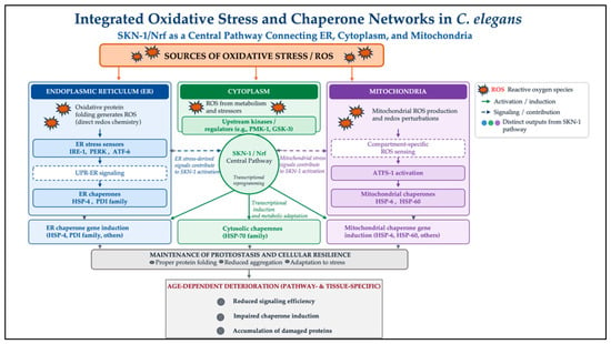
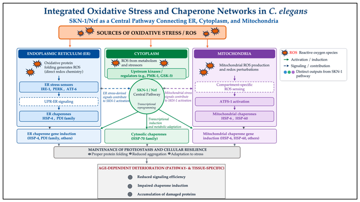
This review addresses this gap by first examining C. elegans chaperone systems according to their cellular compartments, with ER and mitochondrial chaperones summarized in two reference tables (Table 1 and Table 2). We then discuss the direct biochemical mechanisms linking oxidative stress with chaperone function, followed by the transcriptional pathways, particularly UPR-ER and SKN-1, that connect these responses, and the cross-talk between different chaperone systems (Figure 1). Next, we highlight studies showing how oxidative stress and UPR pathways converge through chaperone activation or failure, and how this interaction changes with age. Finally, we summarize the main methodological approaches used to study these mechanisms and discuss the remaining knowledge gaps and their potential translational relevance before concluding the review.
Figure 1.
Integrated oxidative stress and chaperone networks in C. elegans. Oxidative stress and reactive oxygen species (ROS) arising from protein oxidation, cellular metabolism, and mitochondrial redox perturbations are sensed through compartment-specific mechanisms in the endoplasmic reticulum (ER), cytoplasm, and mitochondria. ER stress activates the UPR-ER through IRE-1, PERK, and ATF-6, whereas mitochondrial stress engages compartment-specific ROS sensing and ATFS-1; cytosolic stress signals and upstream regulators contribute to SKN-1/Nrf activation. SKN-1/Nrf serves as a central pathway integrating stress signals across these compartments and promoting transcriptional reprogramming and induction of compartment-specific chaperones, including HSP-4 and PDI family proteins in the ER, Hsp70-family proteins in the cytoplasm, and HSP-6 and HSP-60 in the mitochondria. These coordinated responses support protein folding, limit protein aggregation, and promote cellular resilience. With aging, however, the efficiency of these pathways declines in a pathway- and tissue-specific manner, resulting in reduced stress signaling, impaired chaperone induction, and accumulation of damaged proteins.
2. Mapping the Chaperone Network in C. elegans
Protein folding in C. elegans is managed by three largely distinct chaperone systems, each housed in a different cellular compartment and regulated by its specific transcriptional regulator. Although these systems interact, a theme developed later in the discussion of cross-talk between chaperone systems, each one has evolved a compartment-specific solution to the same underlying problem: preventing misfolded proteins from accumulating and becoming toxic. The chaperone background, organized by compartment, is depicted below.
2.1. Chaperones of the Endoplasmic Reticulum
The endoplasmic reticulum controls approximately one-third of the cells’ overall protein production, and its chaperone system corresponds to that task. At the core is HSP-4, the C. elegans ortholog of BiP/GRP78, an Hsp70 family chaperone that binds unfolded substrates in the ER lumen and, under resting conditions, represses the three UPR-ER stress sensors (IRE-1, PEK-1, and ATF-6) through direct physical association [14]. When misfolded proteins accumulate, HSP-4 is sequestered away to bind the substrate, releasing the sensors to activate their downstream pathways. Induction of hsp-4, commonly monitored using hsp-4p::GFP reporters, is the standard indicator used across the field for UPR-ER activation and has been applied across contexts ranging from tunicamycin and DTT challenge to environmental exposures such as PM2.5 [15].
A family of protein disulfide isomerases (PDIs) operates alongside HSP-4, catalyzing the formation and rearrangement of disulfide bonds, a process of oxidative folding that stabilizes many secreted and membrane proteins. This is where the ER chaperone system’s relationship to oxidative stress becomes mechanistically direct rather than merely correlational, a point which we have expanded upon further in the discussion of direct biochemical mechanisms below, and the genes involved in this compartment are shown in Table 1. Direct evidence for this compartment’s biological necessity comes from the disruption of ER protein disulfide isomerase activity, which is essential for viability and extracellular matrix assembly, underscoring that ER chaperone function is not merely stress-responsive but is required for normal development [16]. More recent work has extended this connection directly into the review’s UPR framework: PDI-6 was shown to regulate the Wnt ligand secretion required for inter-tissue mitochondrial UPR activation, with PDI-6 expression declining with age and its loss shortening the lifespan [17].
Table 1.
Chaperones and folding enzymes of the endoplasmic reticulum in C. elegans.
2.2. Cytosolic Chaperones
In a separate compartment from the ER, cytosolic protein quality control is coordinated by the heat shock response (HSR), governed by the single C. elegans ortholog of the HSF family, HSF-1. Under stress, HSF-1 undergoes post-translational modification, oligomerizes, and translocates to the nucleus, where it binds heat shock elements to induce a broad set of HSP-70 family chaperones and small heat shock proteins such as HSP-16.2. In addition to its canonical role in acute proteotoxic stress, HSF-1 is also a major determinant of C. elegans lifespan, and is required for the longevity extension associated with reduced insulin/IGF-1 signaling, TOR inhibition, and dietary restriction [26]. This function is itself gated by upstream regulators: the homeodomain-interacting protein kinase HPK-1 antagonizes an inhibitory sumoylation modification of HSF-1, is required for HSF-1-dependent chaperone induction after heat shock, and independently extends longevity by driving autophagy under TORC1 inhibition [27].
Significantly, the induction of chaperones alone fails to comprehensively elucidate the relationship between HSF-1 and oxidative stress. Studies have deconstructed the transactivation domain of HSF-1 and discovered that it offers protection against peroxide stress through two different pathways. A particular approach relies on the transactivation domain (heat shock-induced protection), whereas the subsequent method does not (providing constitutive protection through targets such as asp-4/cathepsin and dapk-1) [11]. This differentiation is significant for the extent to which chaperone induction and oxidative stress protection are considered to correlate, and it will be explored later in a review of interactions between chaperone systems. Notably, most of this compartment-specific evidence is inferred from mammalian and yeast systems; whether oxidative stress differentially engages the ER, cytosolic, and mitochondrial chaperone machineries within a single tissue in these organisms needs to be investigated further, and it has yet to be determined whether these compartments respond independently or through shared redox-sensitive intermediates.
2.3. Mitochondrial Chaperones
The mitochondrial matrix has its own dedicated chaperone system, centered on HSP-6, a mitochondrial Hsp70, and HSP-60, a mitochondrial chaperonin, both nuclear-encoded and imported into the organelle. Their expression is coordinated by the mitochondrial unfolded protein response (UPRmt), whose master regulator is ATFS-1. It carries both a mitochondrial targeting sequence and a nuclear localization signal; under normal conditions it is imported into mitochondria and degraded, but under mitochondrial stress, import is impaired and ATFS-1 instead accumulates in the nucleus, where it drives transcription of hsp-6, hsp-60, and a wider pathway of detoxification and metabolic genes [28]. A complementary, translational layer of control acts upstream of this import-based switch: a v-ATPase/TORC1 signaling axis promotes ATFS-1 translation upon mitochondrial stress, independently of GCN-2/PEK-1 signaling, and is required for both UPRmt induction and mitochondrial-stress-induced longevity [29].
The transcriptional reach of ATFS-1 extends well beyond the mitochondrial chaperones themselves. Its activation is sufficient to upregulate genes across the SKN-1-mediated oxidative stress response, the DAF-16 stress response, the UPR-ER, and the HSR simultaneously, establishing ATFS-1 as plausibly the single most consequential integration point between mitochondrial chaperone biology and the rest of the stress-response network discussed in this review [12]. Whether ATFS-1 functions as a true integration hub or merely one of several parallel, non-hierarchical mitochondrial stress sensors remains subject to debate, and resolving this distinction will require systematic epistasis studies rather than single-pathway loss-of-function data.
Table 2.
Mitochondrial chaperones and the UPRmt in C. elegans.
2.4. On Non-Canonical Chaperone Activity
Not all translational chaperones fit within these three canonical systems. One non-canonical example is the nascent polypeptide-associated complex (NAC; encoded by icd-1 and icd-2), whose depletion triggers UPR-ER activation and hsp-4 upregulation, with ire-1 and hsf-1 mutants showing heightened sensitivity to its loss [33]. This example is revisited later among the studies on chaperone failure, as an illustration of how chaperone dysfunction, even outside the canonical UPR-ER machinery, can drive convergence between proteostasis and stress-response activation.
3. Oxidative Stress and Chaperone Function: Direct Mechanisms
The relationship between oxidative stress and chaperone biology goes beyond stressors happening to trigger both responses simultaneously together. In at least one compartment, the ER, the connection is built directly into the core chemistry of protein folding.
3.1. Oxidative Protein Folding as a Source of ROS
Disulfide bond formation, the folding step catalyzed by protein disulfide isomerases (PDIs), is inherently an oxidation reaction. In the canonical ERO1-PDI pathway, PDI accepts a disulfide bond from the client protein and is reoxidized by the ER oxidase ERO1, with hydrogen peroxide (H2O2) generated as a direct byproduct [18]. An additional pathway coupling PDI to peroxiredoxin 4 (PRDX4) also contributes to oxidative folding while helping to buffer the H2O2 produced [19,20,21]. The catalytic step itself is not purely inferred from other systems: C. elegans PDI-1, PDI-2, and PDI-3 have been purified and directly shown to catalyze thiol–disulfide exchange, with distinct substrate preferences suggesting the three enzymes serve different folding pathways in vivo [34]. However, the ERO1-coupled H2O2-generating step and the PDI foldase/holdase redox switch remain untested in the worm; this is addressed further in Section 3.3. Therefore, protein quality control in the ER is structurally a source of oxidative stress that other parts of the cell must contend with. This is because folding a secreted or membrane protein properly generates a substantial amount of reactive oxygen species. This has an emerging implication for the interpretation of how oxidative stress triggers the UPR-ER: a part of the oxidative stress that the UPR-ER regulates may originate internally, resulting from its own chaperone and folding functions rather than only from external stresses, such as xenobiotics, metabolic stressors, or mitochondrial ROS releases. In the toxicological and xenobiotic research covered in the particular studies listed below, oxidative stress is viewed as specifically being upstream of UPR-ER activation. This reconsiders the typically assumed perspective of direction.
3.2. Redox as a Regulator, Not a Stressor
The role of PDI goes beyond serving as a passive site of ROS production; its own redox state functions as a regulatory switch. Under ER stress, phosphorylation-associated conformational changes in PDI, well characterized for PDIA1 in mammalian systems, shift its function from a foldase to a holdase, a mode in which it limits the output of IRE1 signaling to keep the response adaptive rather than terminal [22]. A related PDI family member, PDIA6, attenuates both IRE1 and PERK signaling directly, in the latter case through disulfide interactions with the sensor itself [23]. Peroxiredoxin 4 shows an analogous logic: its own oxidation state determines whether it participates productively in oxidative folding or instead engages an off-pathway reaction, a switch that the reductase activity of PDI can suppress [19]. A parallel, organismal-level example of this redox selectivity exists directly in C. elegans: overexpression of the PDI family member PDI-6 increases resistance to oxidative, ER, and mitochondrial stress while simultaneously increasing sensitivity to reductive stress, indicating that its protective function is tuned to a specific redox direction rather than acting as a generic stress buffer [17].
Collectively, these mechanisms indicate that the redox does not just activate the UPR-ER externally. It is systematically integrated into the real-time modulation of the UPR-ER sensors, regulating the intensity and duration of the transmitted signal.
3.3. A Limitation: This Biochemical Mechanism Has Yet to Be Characterized in C. elegans
To accurately frame the current evidence base, it is necessary to delineate what has been experimentally demonstrated in C. elegans versus what is inferred from other models. Genetic studies confirm that C. elegans PDI family members are enzymatically active and vital for organismal survival [16,34], and PDI-6 exhibits a redox-selective phenotype consistent with a regulatory role [17]. The detailed molecular dynamics remain largely defined in mammalian and yeast systems. Specifically, the complete the ERO1-PDI-H2O2 cycle, the phosphorylation-dependent foldase-to-holdase conformational switch, and the direct physical attenuation of IRE-1 and PEK-1 sensors by PDIA6 have not yet been directly demonstrated in the nematode. Although the phenotypic selectivity of PDI-6 aligns with such a regulatory mechanism, it does not provide direct biochemical proof of conformational switching or sensor gating. Determining whether C. elegans PDI orthologs engage in these specific post-translational and physical interactions remains an important target for future investigation [35].
3.4. Compartment-Specific Redox Regulation Beyond the ER
The ER is not the only compartment where oxidative stress engages chaperone systems through a distinguishable mechanism. In the mitochondrial matrix, ROS generated by paraquat exposure activate the UPRmt (via hsp-6 induction) through a route that is independent of HAF-1, the peptide exporter thought to relay the canonical mitochondrial-perturbation signal to ATFS-1. This indicates that ROS-triggered UPRmt activation and ETC-perturbation-triggered UPRmt activation are mechanistically distinct pathways that converge on the same transcriptional output [10]. Direct genetic disruption of mitochondrial redox homeostasis reinforces this point: C. elegans lacking both mitochondrial glutathione reductase (gsr-1a) and thioredoxin reductase (trxr-2) shows constitutive ATFS-1 nuclear localization and UPRmt reporter activation, and this response is essential for viability under mitochondrial redox stress, even though canonical mitochondrial function, including respiration, ATP, and ROS output, remains normal [36]. In the cytosol, HSF-1 protection against peroxide stress separates into a transactivation-domain-dependent mechanism requiring canonical chaperone induction, and a transactivation-domain-independent pathway acting through targets such as asp-4/cathepsin and dapk-1, unrelated to chaperone upregulation [11]. Taken together with the ER mechanisms above, this suggests that oxidative stress activates chaperone systems through at least three distinct regulatory strategies, not one mechanism repeated across compartments, all converging on protein quality control. We discuss this further below.
4. UPR-ER as the Transcriptional Bridge Between Oxidative Stress and Chaperone Induction
The biochemical interaction between oxidative stress and chaperone function that was previously established, at least within the ER, is basically the initial level of this connection. We continue with the second level: the transcriptional mechanism that alters stress signals, whether oxidative or not, into modifications in the expression of chaperone genes.
4.1. IRE-1/XBP-1: The Primary Chaperone-Inducing Pathway
IRE-1 is the most conserved of the three UPR-ER sensors, and the most directly tied to chaperone output. Upon activation, its endoribonuclease domain splices xbp-1 mRNA, producing the active transcription factor XBP-1s, which drives the expression of ER chaperones and ERAD components [37]. This pathway is not a passive route. Studies using a gap-junction misfolding model (UNC-9 overexpression in neurons) have shown that IRE-1 activity itself is regulated upstream by the p38 MAP kinase PMK-3, which phosphorylates IRE-1 to control UPR-ER output, with hsp-4/BiP induction and autophagy induction responding to genetically separable inputs downstream of this pathway [38]. This serves as a useful depiction of IRE-1 activation being a controlled condition rather than simply an on/off mechanism activated primarily by misfolded proteins.
The IRE-1/XBP-1 pathway is also the mechanism most clearly implicated in age-related decline: IRE-1’s endoribonuclease activity, both xbp-1 splicing and its separate RIDD (regulated Ire1-dependent decay) activity, fails early in adulthood at the onset of the reproductive period, and this failure is not rescued by interventions such as reduced germline signaling that restore other age-related stress response declines [12]. This age-dependent mechanism is developed further in the discussion of aging below.
4.2. PEK-1 and ATF-6, Translational Attenuation and a Limited Transcriptional Role
PEK-1, the C. elegans PERK ortholog, attenuates global translation under ER stress by phosphorylating eIF2a, reducing the accumulating protein-folding demand while the chaperone system catches up, a mechanism distinct from direct chaperone gene induction. ATF-6, by contrast, is processed in the Golgi and drives a comparatively modest transcriptional process in C. elegans relative to its mammalian counterpart, and its specific contribution to chaperone gene regulation remains less well defined [39]. Functional data support this asymmetry. In a paraquat/tunicamycin electrotaxis assay, ire-1, pek-1, and xbp-1 were each required for normal stress-response behavior, while atf-6 was dispensable, consistent with ATF-6 playing a smaller, more specialized role in the C. elegans UPR-ER than IRE-1 or PEK-1 [4]. This role appears context-dependent; however, atf-6 is required, rather than being dispensable, for hsp-4 induction under replication-fork stress [40]. Dietary conditions add yet another variable to how this pathway is used. A high-glucose diet extends lifespan when administered post-reproductively through atf-6- and pek-1-dependent mechanisms, but shortens lifespan when administered during the reproductive period in an ire-1-independent manner. This highlights the fact that the UPR-ER system in which one is participating is not established, but rather is influenced by both age and diet together [41].
4.3. SKN-1/Nrf: The Node That Couples Oxidative Stress Genes with UPR-ER Output
The underlying regulator of the oxidative stress response and the C. elegans ortholog of Nrf2, SKN-1, is the transcriptional bridge between oxidative stress and UPR-ER that is most obvious. Genome-wide profiling has shown that SKN-1 regulates a substantial set of genes with UPR-ER- or ER-associated functions, including atf-5 (a UPR-ER transcription factor), choline kinase, prolyl carboxypeptidase, and numerous smooth ER-localized xenobiotic metabolism enzymes, and that transgenically expressed SKN-1 binds directly at the regulatory regions of ire-1, xbp-1, pek-1, and atf-6 themselves, as well as genes involved in calcium signaling and protein folding and degradation [42].
Importantly, this relationship is not just SKN-1 acting as a generic amplifier of both stress pathways. ER stress (induced by tunicamycin) activates SKN-1-dependent expression of glutathione metabolism genes (gcs-1, gst-4), canonical oxidative stress-response targets, but does not activate other SKN-1 targets that are normally induced under direct oxidative stress. This differential finding indicates that SKN-1 mediates a genuine, partially distinct response to ER stress, rather than solely re-triggering its full oxidative stress transcriptional cascade whenever the UPR-ER is engaged [42]. Consistent with this, ER-associated degradation and the UPR-ER can direct SKN-1 activation during infection, with the two mechanisms acting in a mutually inhibitory rather than purely cooperative manner [43]. In other words, SKN-1 does not treat ER stress and oxidative stress as interchangeable inputs. It interprets them as related but distinguishable signals and produces a correspondingly overlapping but non-identical transcriptional profile.
4.4. Implications for the Chaperone-Based Organizing Framework
Taken together, oxidative stress reaches chaperone gene expression through two distinguishable routes, direct redox chemistry acting on folding enzymes and transcriptional coupling through the partial overlap of SKN-1 with the UPR-ER system. Neither route alone explains the tight association between oxidative stress and chaperone induction seen in the case studies below, which is why this review frames chaperone biology, rather than either pathway alone, as its organizing structure.
5. Cross-Talk Between Chaperone Systems (HSR, UPR-ER, UPRmt)
Thus far, the ER, cytosolic, and mitochondrial chaperone systems have been presented as largely parallel tracks, each with a distinct sensor, regulator, and relationship to oxidative stress. In reality, they are not insulated from each other. We focus on the points where they communicate directly, transcriptionally, and across tissues, arguing that this cross-talk is where the relationship between oxidative stress and chaperone induction becomes most integrated.
5.1. HSF-1 and the UPR-ER: Bidirectional Cross-Talk Under Heat Stress
Although the HSR and UPR-ER are traditionally treated as separate systems responding to separate insults (proteotoxic heat stress and ER-specific misfolding, respectively), recent work has identified a non-canonical route by which heat stress activates the UPR-ER directly through coordinated action of XBP-1 and HSF-1, rather than XBP-1 acting solely downstream of IRE-1 splicing in the conventional pathway [34]. The organization of studies demonstrated that UPR-ER activation, when induced in particular tissues (neurons or glia), can heterotypically increase the HSR in peripheral tissues, consequently raising organismal thermotolerance [44]. This is a properly bidirectional mechanism: HSF-1 affects the UPR-ER outcome, and UPR-ER activation is reflected back to regulate the HSR.
5.2. Inter-Tissue Signaling: The UPR-ER as a Nonautonomous System
The previously discussed bidirectionality correlates with an expanded concept: UPR-ER activity in C. elegans is not cell-autonomous. Overexpression of constitutively active XBP-1s specifically in neurons is sufficient to activate the UPR-ER in distal intestinal cells and to extend organismal lifespan. This transcellular signal has been traced to neuropeptide signaling from a small set of glial cells, packaged into dense-core vesicles and released independently of the originating neurons [24]. This means that a chaperone-inducing signal generated in one compartment (the nervous system) can reprogram chaperone capacity in a mechanistically distant compartment (the intestine), without oxidative or ER stress ever having been directly detected in the effected tissue.
Notably, this same neuronal XBP-1s paradigm reveals that UPR-ER benefits are not exclusively mediated by chaperone induction. Activation promotes ER restructuring and lipid depletion through lipophagy, a pathway that is mechanistically distinct from chaperone induction and is independently required for the associated lifespan extension [25]. A comparable pattern operates for the mitochondrial network. Intestinal UPRmt is non-autonomously tuned by neuronal acetylcholine and GABA signaling, and this neuro-intestinal circuit is required for organismal survival of oxidative stress, indicating that cell-nonautonomous control of chaperone systems is not unique to the ER/XBP-1s axis [45]. This is an important complication for a chaperone-centered review to acknowledge directly. UPR-ER activation and more chaperones are not equivalent, even though chaperone induction is the default indicator used across the field for UPR-ER activity.
5.3. The Integrative Role of ATFS-1 in Stress Signaling
Of all the transcriptional regulators discussed in this review, ATFS-1 exhibits the most extensive documented reach across systems. Its activation is sufficient to upregulate genes spanning the SKN-1-mediated oxidative stress response, the DAF-16 stress response, the UPR-ER, the HSR, the HIF-1-mediated hypoxia response, and the p38/PMK-1 innate immune pathway, essentially simultaneously [12]. Functionally, this integration holds significance for oxidative stress resistance. In long-lived mitochondrial mutants (clk-1, isp-1, nuo-6), ATFS-1-dependent UPRmt activation is required for their enhanced resistance to paraquat, and loss of atfs-1 abolishes the coordinated upregulation of oxidative and other stress pathways that these mutants otherwise show [32]. This is consistent with ATFS-1 acting as an obligate redox-stress sensor, not just as a downstream effector of protein misfolding or ETC dysfunction [36].
5.4. A Single Stressor, Three Compartments: Paraquat/Electrotaxis
The obvious theoretical evidence that these systems act collectively, rather than simply possessing individual responses to oxidative stress, is derived from a study employing paraquat as a singular, specified oxidative stressor and electrotactic locomotor behavior as a common functional indicator. Paraquat exposure simultaneously induced hsp-4 (ER), hsp-6 and hsp-60 (mitochondrial), and hsp-16.2 (cytosolic), with ire-1, pek-1, and xbp-1 each functionally required for the resulting behavioral phenotype, while atf-6 was not [4]. This is the main single-paper evidence that a single expression of oxidative stress coordinates the ER, cytosolic, and mitochondrial chaperone systems in a functionally consequential manner rather than as three independent, redundant responses to the same source.
5.5. What Cross-Talk Implies for the Rest of This Review
The cross-talk described here has two consequences for understanding the findings and aging-related discussion that follow. First, because these systems influence each other and are co-regulated by networks like ATFS-1 identifying an oxidative stress phenotype to any chaperone system individually, which risks understating the mechanism. Second, because UPR-ER activation can produce chaperone-independent effects and HSF-1 can protect against oxidative stress through chaperone-independent targets, chaperone induction should be managed as one result for these systems rather than a valid indicator for full function.
6. Studies of Chaperone Failure Driving Oxidative Stress/UPR-ER Convergence
Of the cited research, the paraquat/electrotaxis study provides the most obvious depiction of coordinated cross-compartment chaperone engagement under oxidative stress. Three more examples show how chaperone dysfunction and oxidative stress become connected: loss of a non-canonical translational chaperone, physical environmental exposure, and an increase in the degradation mechanism that supports the chaperone system when folding is damaged.
6.1. Translational Chaperone Loss: Nascent Polypeptide-Associated Complex Depletion
The nascent polypeptide-associated complex (NAC), encoded in C. elegans by icd-1 and icd-2, is not part of the canonical UPR-ER machinery, but functions as a translational chaperone, escorting nascent polypeptides as they emerge from the ribosome. RNAi depletion of NAC subunits over time triggers UPR-ER activation and upregulates the ER chaperone hsp-4/BiP, accompanied by increased apoptosis and gut cell lipofuscin accumulation [33]. Notably, worms carrying a partially defective ire-1 allele were significantly more sensitive to NAC depletion, and hsf-1 mutants showed heightened sensitivity as well, despite HSF-1 being a cytosolic rather than ER-specific regulator. This study is particularly instructive because the initial defect is not oxidative or ER-specific; it is a failure in general translational quality control. However, it converges on the same downstream chaperone-induction pathway (UPR-ER, HSF-1) that oxidative stressors use to function.
6.2. Environmental Exposure and Chaperone Induction, PM2.5
Fine particulate matter (PM2.5) exposure offers the most complete demonstration of the full mechanistic sequence linking an environmental oxidative stressor to chaperone induction and an organismal outcome. In C. elegans, PM2.5 exposure induces oxidative stress, activates the UPR-ER, and shortens the lifespan [15]. This sequence of events was tested pharmacologically. The antioxidant N-acetylcysteine suppressed UPR activation by reducing oxidative stress upstream, while the UPR inhibitor 4-PBA acted further downstream, and both independently rescued the PM2.5-induced lifespan reduction. This remains the strongest evidence in C. elegans that oxidative stress functions upstream of UPR-ER activation, rather than the two being independent, co-occurring events.
6.3. Reinforcing the Chaperone System from the Degradation Side: Proteasome Hyperactivation
The studies above concern chaperone induction—that is, greater folding capacity in response to stress. A complementary route is strengthening the degradation machinery that clears proteins the chaperone system cannot rescue. A C. elegans model engineered for hyperactive 20S proteasome activity (via a truncated alpha3 subunit, alpha3-delta-N) shows enhanced degradation of intrinsically disordered proteins and misfolded substrates, markedly reduced oxidative damage, and improved ER-associated degradation (ERAD), all achieved independently of the superoxide dismutase (SOD) antioxidant system [46]. This reveals that oxidative stress resistance and ER proteostasis can both be improved by intervening downstream of chaperone function entirely, at the level of degradation capacity, without involving the chaperones or the canonical antioxidant machinery.
6.4. Collective Implications of These Studies
These three studies collectively show the various ways that oxidative stress and chaperone mechanisms communicate. First, the same UPR-ER and HSF-1 pathways that are triggered by oxidative agents can also be triggered by non-oxidative stress, such as the loss of translational chaperones. Second, there are pharmacologically established associations between environmental exposures such as PM2.5 and typical oxidative stress. Third, rather than inducing chaperones, proteasome hyperactivation improves oxidative resistance and ER proteostasis by targeted protein destruction. Ultimately, these examples illustrate that the chaperone network is not a conventional, direct axis that is solely driven by oxidative stress, but rather a shared intersection for various types of upstream signals.
7. Aging and Convergent Decline in Chaperone and Oxidative Stress Systems
Chaperone pathways and oxidative stress are currently thought to be widely vulnerable and frequently consolidating. The condition of this illustration decreases with age. Aging serves as the critical intersection at which oxidative stress and chaperone failure may most significantly converge, as the reduction in capacity of one system eliminates a buffer that the other system relies on.
7.1. The Decline Is Early, Pathway-Specific, and Not Explained by a Single Mechanism
UPR-ER responsiveness declines with age in C. elegans. This has been traced to a specific failure: the endoribonuclease activity of IRE-1, both canonical xbp-1 splicing and its separate RIDD function, deteriorates at the onset of the reproductive period, well before organismal aging becomes broadly apparent. This decline is not rescued by the interventions, such as reduced germline signaling, that restore other age-related stress-response declines, indicating that IRE-1 failure is mechanistically distinct from reproduction-linked decline in other longevity pathways [12].
Chaperone capacity shows a comparable but tissue-specific pattern. Young muscle is largely heat-stress-resistant via HSF-1-mediated induction but refolds poorly during recovery, while young neurons are more heat-susceptible but refold well [47]. With age, this pattern shifts rather than uniformly collapsing: aged neurons gain some stability during heat stress itself, aged muscle loses heat shock responsiveness, and both tissues lose refolding capacity during recovery [47]. Age-dependent shifts in HSF-1 regulation contribute further: young and aged animals rely on distinct, partly compensatory HSF-1/UPR-ER/SKN-1 circuits [48], and reduced proteasomal turnover of HSF-1 with age reprioritizes protein-stress management toward chronic aggregate defense at the expense of acute heat shock responsiveness [49].
7.2. Where Oxidative Damage and Chaperone Decline May Directly Intersect
In C. elegans, ER chaperones and folding enzymes are mechanistically associated with redox reactions in a manner that has not yet been directly tested, as previously established. Aging is where this gap is most consequential to explore. In mouse liver, the ER chaperones BiP and PDI have been shown to accumulate direct oxidative damage with age, contributing to the age-associated rise in the misfolded ER protein burden [50,51]. This finding offers a plausible mechanism by which oxidative stress could directly degrade chaperone function over time, rather than only triggering chaperone induction acutely. Whether the analogous damage occurs to HSP-4 or worm PDI orthologs with age has not, to our knowledge, been directly tested, and represents one of the more experimentally tractable open questions for the field, discussed further below.
7.3. Reversibility: Neuronal UPR-ER Activation as a Partial Rescue
The age-related decline in UPR-ER responsiveness is not fully fixed or irreversible at the organismal level. Constitutive expression of active XBP-1s specifically in neurons is sufficient to restore ER stress resistance and extend lifespan, acting non-cell-autonomously to activate the UPR-ER in distal intestinal tissue via glial neuropeptide signaling, as discussed above [24,52]. As also noted there, this recovery operates partly through chaperone induction and partly through a chaperone-independent lipophagy system, meaning that the rescue of age-related UPR-ER decline is not simply a restoration of youthful chaperone levels, but the activation of a partially distinct protective response overlaid on top of an otherwise still-deteriorating system.
7.4. What Aging Reveals About the Central Argument of This Review
Aging sharpens rather than resolves the questions raised throughout this review. It shows that oxidative stress and chaperone decline can be linked through direct molecular damage, established in mammals but untested in worms; that the decline itself is pathway- and tissue-specific rather than occurring evenly across the animal; and that interventions restoring UPR-ER function do not necessarily restore chaperone induction as previously assumed, but can act through parallel, chaperone-independent mechanisms.
8. Methodological Framework for C. elegans Stress-Response Analysis
This review synthesizes studies that rely on a shared, relatively brief list of genetic and reporter tools, which are summarized here. Reporter strains for individual chaperone genes are also listed alongside their respective genes in Table 1 and Table 2.
8.1. Reporter Strains
Beyond the compartment-specific chaperone reporters already listed in Table 1 and Table 2 (hsp-4p::GFP for UPR-ER, hsp-6p::GFP and hsp-60p::GFP for UPRmt), two further reporters are used throughout this review: hsp-16.2p::GFP as the standard cytosolic HSR reporter, and gst-4p::GFP as the standard SKN-1-dependent oxidative stress reporter. Studies aiming to demonstrate cross-compartment convergence, such as the paraquat/electrotaxis study discussed earlier, typically combine several of these reporters in parallel [4,53,54].
8.2. Genetic Tools
Loss-of-function mutants and RNAi knockdown are available for essentially every gene discussed in this review: the UPR-ER sensors (ire-1, pek-1, atf-6) and their downstream effector (xbp-1); the cytosolic regulator hsf-1 and its negative regulator hsb-1, whose inhibition phenocopies HSF-1 overexpression through substantially overlapping transcriptional changes and comparably extends the lifespan [55]; the mitochondrial UPR regulator atfs-1 and the peptide exporter haf-1; and the oxidative stress master regulator skn-1. Constitutively active alleles are also used in several of the studies discussed here, notably xbp-1s (spliced, active XBP-1) and activated ATFS-1 that hinder mitochondrial import [24,56,57].
8.3. Common Experimental Stressors
Chemical and physical stressors used across this literature include tunicamycin and dithiothreitol (DTT) for direct ER stress, heat shock or mild temperature increase for HSR activation, paraquat for mitochondrial and cytosolic oxidative stress, hydrogen peroxide for direct peroxide stress, and environmental exposures such as fine particulate matter (PM2.5) for oxidative and xenobiotic stress [15,58,59].
9. Remaining Questions and Future Considerations
The unresolved inquiries in this domain focus on cause-and-effect relationships, mechanisms, tissue specificity, and translation. The mechanism of oxidative damage in relation to UPR-ER activation is still unclear. The PM2.5 study offers the most specific pharmacological test of this interaction, but this has not been repeated with other stressors or tested for direct chaperone modification; assays measuring oxidative modification of HSP-4 or worm PDI orthologs, independent of transcriptional readouts, would clarify whether this is a general trigger or whether it is PM2.5-specific [15]. A second gap is worm-specific PDI redox biochemistry: the mammalian PDI foldase/holdase switch and its regulation of IRE1/PERK have never been tested in C. elegans, despite tractable genetic and reporter tools already being available [22,23,35]. Tissue specificity remains underexplored beyond the neuron/muscle comparison and glial-to-intestine signaling. The intestine and germline are largely unrevealed as independent convergence parts, which is notable given the known role of the intestine elsewhere in this review as a stress-response and ROS-generating tissue, though emerging neuro-intestinal signaling work is starting to address this gap [45,60,61]. Finally, translational relevance extends to mammalian ER stress-linked disease, including diabetes, neurodegeneration, and proteinopathies involving intrinsically disordered proteins, where the C. elegans-specific regulatory distinctions described earlier could help separate conserved disease mechanisms from those unique to C. elegans [60,62,63]. This link is illustrated concretely, rather than only asserted, through three named, established transgenic C. elegans disease models. Muscle-expressed human amyloid-beta (Aβ42) strains recapitulate proteotoxic paralysis linked to chaperone capacity [64]; more recent work in this model shows that enhancing mitochondrial proteostasis and mitophagy, an arm of the UPRmt response, directly reduces amyloid-β proteotoxicity [65]. Pan-neuronal alpha-synuclein strains model chaperone-dependent aggregate formation relevant to Parkinsonian pathology [66]. Wang et al. used this model to trace neuronal alpha-synuclein aggregation to the induction of the intestinal mitochondrial unfolded protein response via gut neuron signaling [67]. Polyglutamine (polyQ)-expansion strains show how chaperone-dependent aggregation thresholds shift with age [68]; a related polyQ/ataxin-3 model shows that pharmacological modulation of the ER unfolded protein response rescues polyQ-driven neurodegeneration [69]. Together, these three model systems anchor the chaperone networks discussed here to citable, disease-relevant phenotypes rather than mammalian extrapolation alone.
10. Conclusions
This review points out that chaperone biological activity is a highly useful perspective for analyzing the mechanism through which C. elegans manages proteotoxic and oxidative damage in a sequential manner, rather than the UPR-ER or oxidative stress response independently. Through various mechanisms, including direct redox chemistry, SKN-1-mediated transcription, and compartment-specific ROS sensing, oxidative stress engages chaperones in the ER, cytosol, and mitochondria. These mechanisms are convergent and rely on shared machinery. Research findings demonstrated that this convergence occurs through multiple pathways rather than a single pathway. Additionally, aging reveals that this convergence is the most probable site for failures in a pathway- and tissue-specific manner. Investigating whether mammalian mechanisms, such as oxidative chaperone damage and PDI redox switching, operate in C. elegans and expanding tissue-specific analysis to the intestine and germline remain critical for further investigation.
Author Contributions
Conceptualization, J.T. (Jerald Tan), J.T. (Jirapan Thongsroy) and S.C.; writing—original draft preparation, J.T. (Jerald Tan), J.T. (Jirapan Thongsroy) and S.C.; writing—review and editing, J.T. (Jerald Tan), J.T. (Jirapan Thongsroy) and S.C.; visualization, J.T. (Jerald Tan), J.T. (Jirapan Thongsroy) and S.C.; supervision, J.T. (Jerald Tan), J.T. (Jirapan Thongsroy) and S.C. All authors have read and agreed to the published version of the manuscript.
Funding
This research received no external funding.
Institutional Review Board Statement
Not applicable. This review did not involve new experiments with humans or animals.
Informed Consent Statement
Not applicable.
Data Availability Statement
No new data were created or analyzed in this study. Data sharing is not applicable to this article; all data discussed are available in the cited references.
Acknowledgments
The Center for Library Resources and Educational Media, Walailak University.
Conflicts of Interest
The authors declare no conflicts of interest.
Abbreviations
The following abbreviations are used in this manuscript:
| UPR-ER | Unfolded protein response of the endoplasmic reticulum |
| UPRmt | Mitochondrial unfolded protein response |
| HSR | Heat shock response |
| ROS | Reactive oxygen species |
| ER | Endoplasmic reticulum |
| PDI | Protein disulfide isomerase |
| ERAD | ER-associated degradation |
| RIDD | Regulated Ire1-dependent decay |
| NAC | Nascent polypeptide-associated complex |
| SOD | Superoxide dismutase |
| PM2.5 | Fine particulate matter (aerodynamic diameter <2.5 micrometers) |
| DTT | Dithiothreitol |
References
- Shen, X.; Ellis, R.E.; Lee, K.; Liu, C.Y.; Yang, K.; Solomon, A.; Yoshida, H.; Morimoto, R.; Kurnit, D.M.; Mori, K.; et al. Complementary signaling pathways regulate the unfolded protein response and are required for C. elegans development. Cell 2001, 107, 893–903. [Google Scholar] [CrossRef] [Scilit] [PubMed]
- Blackwell, T.K.; Steinbaugh, M.J.; Hourihan, J.M.; Ewald, C.Y.; Isik, M. SKN-1/Nrf, stress responses, and aging in Caenorhabditis elegans. Free Radic. Biol. Med. 2015, 88, 290–301. [Google Scholar] [CrossRef] [Scilit] [PubMed]
- Shi, L.; Yu, X.T.; Li, H.; Wu, G.S.; Luo, H.R. D-chiro-inositol increases antioxidant capacity and longevity of Caenorhabditis elegans via activating Nrf-2/SKN-1 and FOXO/DAF-16. Exp. Gerontol. 2023, 175, 112145. [Google Scholar] [CrossRef] [Scilit] [PubMed]
- Taylor, S.K.B.; Minhas, M.H.; Tong, J.; Selvaganapathy, P.R.; Mishra, R.K.; Gupta, B.P. C. elegans electrotaxis behavior is modulated by heat shock response and unfolded protein response signaling pathways. Sci. Rep. 2021, 11, 3115. [Google Scholar] [CrossRef] [Scilit] [PubMed]
- Lee, S.-K. Endoplasmic Reticulum Homeostasis and Stress Responses in Caenorhabditis elegans. In Cellular Biology of the Endoplasmic Reticulum; Agellon, L.B., Michalak, M., Eds.; Springer International Publishing: Cham, Switzerland, 2021; pp. 279–303. [Google Scholar]
- Haynes, C.M.; Hekimi, S. Mitochondrial dysfunction, aging, and the mitochondrial unfolded protein response in Caenorhabditis elegans. Genetics 2022, 222, iyac160. [Google Scholar] [CrossRef] [Scilit] [PubMed]
- Torres, A.K.; Fleischhart, V.; Inestrosa, N.C. Mitochondrial unfolded protein response (UPR(mt)): What we know thus far. Front. Cell Dev. Biol. 2024, 12, 1405393. [Google Scholar] [CrossRef] [Scilit] [PubMed]
- Tan, J.; Chiamkunakorn, C.; Boonchuay, K.; Shi, Y.; Braeckman, B.P.; Suthammarak, W. Metformin modulates the unfolded protein responses, altering lifespan and health-promoting effects in UPR-activated worms. PLoS ONE 2025, 20, e0326100. [Google Scholar] [CrossRef] [Scilit] [PubMed]
- Ghosh, A.; Singh, J. Interplay between proteostasis pathways and innate immune responses in Caenorhabditis elegans. Infect. Immun. 2026, 94, e0003726. [Google Scholar] [CrossRef] [Scilit] [PubMed]
- Runkel, E.D.; Liu, S.; Baumeister, R.; Schulze, E. Surveillance-activated defenses block the ROS-induced mitochondrial unfolded protein response. PLoS Genet. 2013, 9, e1003346. [Google Scholar] [CrossRef] [Scilit] [PubMed]
- Servello, F.A.; Apfeld, J. The heat shock transcription factor HSF-1 protects Caenorhabditis elegans from peroxide stress. Transl. Med. Aging 2020, 4, 88–92. [Google Scholar] [CrossRef] [Scilit]
- Soo, S.K.; Traa, A.; Rudich, P.D.; Mistry, M.; Van Raamsdonk, J.M. Activation of mitochondrial unfolded protein response protects against multiple exogenous stressors. Life Sci. Alliance 2021, 4, e202101182. [Google Scholar] [CrossRef] [Scilit] [PubMed]
- De-Souza, E.A.; Cummins, N.; Taylor, R.C. IRE-1 endoribonuclease activity declines early in C. elegans adulthood and is not rescued by reduced reproduction. Front. Aging 2022, 3, 1044556. [Google Scholar] [CrossRef] [Scilit] [PubMed]
- Richardson, C.E.; Kinkel, S.; Kim, D.H. Physiological IRE-1-XBP-1 and PEK-1 signaling in Caenorhabditis elegans larval development and immunity. PLoS Genet. 2011, 7, e1002391. [Google Scholar] [CrossRef] [Scilit] [PubMed]
- Zhao, Y.; Jin, L.; Chi, Y.; Yang, J.; Zhen, Q.; Wu, H. Fine Particulate Matter Leads to Unfolded Protein Response and Shortened Lifespan by Inducing Oxidative Stress in C. elegans. Oxid. Med. Cell. Longev. 2019, 2019, 2492368. [Google Scholar] [CrossRef] [Scilit] [PubMed]
- Winter, A.D.; McCormack, G.; Page, A.P. Protein disulfide isomerase activity is essential for viability and extracellular matrix formation in the nematode Caenorhabditis elegans. Dev. Biol. 2007, 308, 449–461. [Google Scholar] [CrossRef] [Scilit] [PubMed]
- Li, X.; Li, J.; Zhu, D.; Zhang, N.; Hao, X.; Zhang, W.; Zhang, Q.; Liu, Y.; Wu, X.; Tian, Y. Protein disulfide isomerase PDI-6 regulates Wnt secretion to coordinate inter-tissue UPR(mt) activation and lifespan extension in C. elegans. Cell Rep. 2022, 39, 110931. [Google Scholar] [CrossRef] [Scilit] [PubMed]
- Zito, E. ERO1: A protein disulfide oxidase and H2O2 producer. Free Radic. Biol. Med. 2015, 83, 299–304. [Google Scholar] [CrossRef] [Scilit] [PubMed]
- Zito, E.; Melo, E.P.; Yang, Y.; Wahlander, A.; Neubert, T.A.; Ron, D. Oxidative protein folding by an endoplasmic reticulum-localized peroxiredoxin. Mol. Cell 2010, 40, 787–797. [Google Scholar] [CrossRef] [Scilit] [PubMed]
- Nguyen, V.D.; Saaranen, M.J.; Karala, A.R.; Lappi, A.K.; Wang, L.; Raykhel, I.B.; Alanen, H.I.; Salo, K.E.; Wang, C.C.; Ruddock, L.W. Two endoplasmic reticulum PDI peroxidases increase the efficiency of the use of peroxide during disulfide bond formation. J. Mol. Biol. 2011, 406, 503–515. [Google Scholar] [CrossRef] [Scilit] [PubMed]
- Yang, Y.; Peng, H.; Meng, D.; Fa, Z.; Yao, C.; Lin, X.; Schick, J.; Jin, X. Stress Management: How the Endoplasmic Reticulum Mitigates Protein Misfolding and Oxidative Stress by the Dual Role of Glutathione Peroxidase 8. Biomolecules 2025, 15, 847. [Google Scholar] [CrossRef] [Scilit] [PubMed]
- Yu, J.; Li, T.; Liu, Y.; Wang, X.; Zhang, J.; Wang, X.; Shi, G.; Lou, J.; Wang, L.; Wang, C.C.; et al. Phosphorylation switches protein disulfide isomerase activity to maintain proteostasis and attenuate ER stress. EMBO J. 2020, 39, e103841. [Google Scholar] [CrossRef] [Scilit] [PubMed]
- Eletto, D.; Eletto, D.; Dersh, D.; Gidalevitz, T.; Argon, Y. Protein disulfide isomerase A6 controls the decay of IRE1alpha signaling via disulfide-dependent association. Mol. Cell 2014, 53, 562–576. [Google Scholar] [CrossRef] [Scilit] [PubMed]
- Frakes, A.E.; Metcalf, M.G.; Tronnes, S.U.; Bar-Ziv, R.; Durieux, J.; Gildea, H.K.; Kandahari, N.; Monshietehadi, S.; Dillin, A. Four glial cells regulate ER stress resistance and longevity via neuropeptide signaling in C. elegans. Science 2020, 367, 436–440. [Google Scholar] [CrossRef] [Scilit] [PubMed]
- Daniele, J.R.; Higuchi-Sanabria, R.; Durieux, J.; Monshietehadi, S.; Ramachandran, V.; Tronnes, S.U.; Kelet, N.; Sanchez, M.; Metcalf, M.G.; Garcia, G.; et al. UPR(ER) promotes lipophagy independent of chaperones to extend life span. Sci. Adv. 2020, 6, eaaz1441. [Google Scholar] [CrossRef] [Scilit] [PubMed]
- Morley, J.F.; Morimoto, R.I. Regulation of longevity in Caenorhabditis elegans by heat shock factor and molecular chaperones. Mol. Biol. Cell 2004, 15, 657–664. [Google Scholar] [CrossRef] [Scilit] [PubMed]
- Das, R.; Melo, J.A.; Thondamal, M.; Morton, E.A.; Cornwell, A.B.; Crick, B.; Kim, J.H.; Swartz, E.W.; Lamitina, T.; Douglas, P.M.; et al. The homeodomain-interacting protein kinase HPK-1 preserves protein homeostasis and longevity through master regulatory control of the HSF-1 chaperone network and TORC1-restricted autophagy in Caenorhabditis elegans. PLoS Genet. 2017, 13, e1007038. [Google Scholar] [CrossRef] [Scilit] [PubMed]
- Melber, A.; Haynes, C.M. UPR(mt) regulation and output: A stress response mediated by mitochondrial-nuclear communication. Cell Res. 2018, 28, 281–295. [Google Scholar] [CrossRef] [Scilit] [PubMed]
- Li, T.Y.; Gao, A.W.; Li, X.; Li, H.; Liu, Y.J.; Lalou, A.; Neelagandan, N.; Naef, F.; Schoonjans, K.; Auwerx, J. V-ATPase/TORC1-mediated ATFS-1 translation directs mitochondrial UPR activation in C. elegans. J. Cell Biol. 2023, 222, e202205045. [Google Scholar] [CrossRef] [Scilit] [PubMed]
- Liu, P.; Li, D.; Li, W.; Wang, D. Mitochondrial Unfolded Protein Response to Microgravity Stress in Nematode Caenorhabditis elegans. Sci. Rep. 2019, 9, 16474. [Google Scholar] [CrossRef] [Scilit] [PubMed]
- Jeong, D.E.; Lee, D.; Hwang, S.Y.; Lee, Y.; Lee, J.E.; Seo, M.; Hwang, W.; Seo, K.; Hwang, A.B.; Artan, M.; et al. Mitochondrial chaperone HSP-60 regulates anti-bacterial immunity via p38 MAP kinase signaling. EMBO J. 2017, 36, 1046–1065. [Google Scholar] [CrossRef] [Scilit] [PubMed]
- Wu, Z.; Senchuk, M.M.; Dues, D.J.; Johnson, B.K.; Cooper, J.F.; Lew, L.; Machiela, E.; Schaar, C.E.; DeJonge, H.; Blackwell, T.K.; et al. Mitochondrial unfolded protein response transcription factor ATFS-1 promotes longevity in a long-lived mitochondrial mutant through activation of stress response pathways. BMC Biol. 2018, 16, 147. [Google Scholar] [CrossRef] [Scilit] [PubMed]
- Arsenovic, P.T.; Maldonado, A.T.; Colleluori, V.D.; Bloss, T.A. Depletion of the C. elegans NAC engages the unfolded protein response, resulting in increased chaperone expression and apoptosis. PLoS ONE 2012, 7, e44038. [Google Scholar] [CrossRef] [Scilit] [PubMed]
- Karala, A.R.; Psarrakos, P.; Ruddock, L.W.; Klappa, P. Protein disulfide isomerases from C. elegans are equally efficient at thiol-disulfide exchange in simple peptide-based systems but show differences in reactivity towards protein substrates. Antioxid. Redox Signal. 2007, 9, 1815–1823. [Google Scholar] [CrossRef] [Scilit] [PubMed]
- Guo, K.; Li, X.; Liang, Q.; Chen, C.; Lin, J.; Zhan, T.; Sun, C.; Kang, N.; Qiu, F.; Zhang, Q. Redox-Encoded Proteostasis: Heat Shock Proteins as Integrators of Stress Adaptation and Therapeutic Targets. Antioxid. Redox Signal. 2026, 45, 78–112. [Google Scholar] [CrossRef] [Scilit] [PubMed]
- Valenzuela-Villatoro, M.; de la Cruz-Ruiz, P.; Guerrero-Gomez, D.; Gomez-Orte, E.; Schiavi, A.; Maglioni, S.; Montero, M.; Fonteriz, R.I.; Casas-Martinez, J.C.; Briand, N.E.; et al. Mitochondrial redox homeostasis links organellar stress surveillance to germline and somatic integrity in Caenorhabditis elegans. Redox Biol. 2026, 93, 104115. [Google Scholar] [CrossRef] [Scilit] [PubMed]
- Calfon, M.; Zeng, H.; Urano, F.; Till, J.H.; Hubbard, S.R.; Harding, H.P.; Clark, S.G.; Ron, D. IRE1 couples endoplasmic reticulum load to secretory capacity by processing the XBP-1 mRNA. Nature 2002, 415, 92–96, Correction in Nature 2002, 420, 202. [Google Scholar] [CrossRef] [Scilit] [PubMed]
- Guan, L.; Zhan, Z.; Yang, Y.; Miao, Y.; Huang, X.; Ding, M. Alleviating chronic ER stress by p38-Ire1-Xbp1 pathway and insulin-associated autophagy in C. elegans neurons. PLoS Genet. 2020, 16, e1008704. [Google Scholar] [CrossRef] [Scilit] [PubMed]
- Shen, X.; Ellis, R.E.; Sakaki, K.; Kaufman, R.J. Genetic interactions due to constitutive and inducible gene regulation mediated by the unfolded protein response in C. elegans. PLoS Genet. 2005, 1, e37. [Google Scholar] [CrossRef] [Scilit] [PubMed]
- Xu, J.; Sabatino, B.; Yan, J.; Ermakova, G.; Doering, K.R.S.; Taubert, S. The unfolded protein response of the endoplasmic reticulum protects Caenorhabditis elegans against DNA damage caused by stalled replication forks. G3 Genes Genomes Genet. 2024, 14, jkae017. [Google Scholar] [CrossRef] [Scilit] [PubMed]
- Beaudoin-Chabot, C.; Wang, L.; Celik, C.; Abdul Khalid, A.T.; Thalappilly, S.; Xu, S.; Koh, J.H.; Lim, V.W.X.; Low, A.D.; Thibault, G. The unfolded protein response reverses the effects of glucose on lifespan in chemically-sterilized C. elegans. Nat. Commun. 2022, 13, 5889. [Google Scholar] [CrossRef] [Scilit] [PubMed]
- Glover-Cutter, K.M.; Lin, S.; Blackwell, T.K. Integration of the unfolded protein and oxidative stress responses through SKN-1/Nrf. PLoS Genet. 2013, 9, e1003701. [Google Scholar] [CrossRef] [Scilit] [PubMed]
- Gabaldon, C.; Karakuzu, O.; Garsin, D.A. SKN-1 activation during infection of Caenorhabditis elegans requires CDC-48 and endoplasmic reticulum proteostasis. Genetics 2024, 228, iyae131. [Google Scholar] [CrossRef] [Scilit] [PubMed]
- Ahmed, S.; Kovács, D.; Kovács, M.; Kosztelnik, M.; Hotzi, B.; Sigmond, T.; Saskői, É.; Vincze, V.V.; Erdélyi, V.; Deák, V.; et al. Heat shock factor-1 alleviates ER-stress in Caenorhabditis elegans. Sci. Rep. 2026, 16, 9928, Correction in Sci. Rep. 2026, 16, 17863. https://doi.org/10.1038/s41598-026-50440-2. [Google Scholar] [CrossRef] [Scilit] [PubMed]
- Cornell, R.; Cao, W.; Harradine, B.; Godini, R.; Handley, A.; Pocock, R. Neuro-intestinal acetylcholine signalling regulates the mitochondrial stress response in Caenorhabditis elegans. Nat. Commun. 2024, 15, 6594. [Google Scholar] [CrossRef] [Scilit] [PubMed]
- Salcedo-Tacuma, D.; Asad, N.; Islam, M.Q.; Anderson, R.; Smith, D.M. Hyperactive 20S proteasome enhances proteostasis and ERAD in C. elegans via degradation of intrinsically disordered proteins. Sci. Adv. 2025, 11, eadx3014. [Google Scholar] [CrossRef] [Scilit] [PubMed]
- Kern, A.; Ackermann, B.; Clement, A.M.; Duerk, H.; Behl, C. HSF1-controlled and age-associated chaperone capacity in neurons and muscle cells of C. elegans. PLoS ONE 2010, 5, e8568. [Google Scholar] [CrossRef] [Scilit] [PubMed]
- Kovacs, D.; Biro, J.B.; Ahmed, S.; Kovacs, M.; Sigmond, T.; Hotzi, B.; Varga, M.; Vincze, V.V.; Mohammad, U.; Vellai, T.; et al. Age-dependent heat shock hormesis to HSF-1 deficiency suggests a compensatory mechanism mediated by the unfolded protein response and innate immunity in young Caenorhabditis elegans. Aging Cell 2024, 23, e14246. [Google Scholar] [CrossRef] [Scilit] [PubMed]
- Wang, H.; Sun, F.; He, Z.; Wang, X.; Liu, H.; Song, M.; Chen, Q.; Li, Z.; Wu, L.; Yan, X.; et al. Reduced Proteasome Degradation of HSF-1 Shifts Protein Stress Management With Age in Caenorhabditis elegans. Aging Cell 2026, 25, e70399. [Google Scholar] [CrossRef] [Scilit] [PubMed]
- Rabek, J.P.; Boylston, W.H., 3rd; Papaconstantinou, J. Carbonylation of ER chaperone proteins in aged mouse liver. Biochem. Biophys. Res. Commun. 2003, 305, 566–572. [Google Scholar] [CrossRef] [Scilit] [PubMed]
- Nuss, J.E.; Choksi, K.B.; DeFord, J.H.; Papaconstantinou, J. Decreased enzyme activities of chaperones PDI and BiP in aged mouse livers. Biochem. Biophys. Res. Commun. 2008, 365, 355–361. [Google Scholar] [CrossRef] [Scilit] [PubMed]
- Taylor, R.C.; Dillin, A. XBP-1 is a cell-nonautonomous regulator of stress resistance and longevity. Cell 2013, 153, 1435–1447. [Google Scholar] [CrossRef] [Scilit] [PubMed]
- Manjarrez, J.R.; Mailler, R. Stress and timing associated with Caenorhabditis elegans immobilization methods. Heliyon 2020, 6, e04263. [Google Scholar] [CrossRef] [Scilit] [PubMed]
- Vertti-Quintero, N.; Berger, S.; Casadevall, I.S.X.; Statzer, C.; Annis, J.; Ruppen, P.; Stavrakis, S.; Ewald, C.Y.; Gunawan, R.; deMello, A.J. Stochastic and Age-Dependent Proteostasis Decline Underlies Heterogeneity in Heat-Shock Response Dynamics. Small 2021, 17, e2102145. [Google Scholar] [CrossRef] [Scilit] [PubMed]
- Sural, S.; Lu, T.C.; Jung, S.A.; Hsu, A.L. HSB-1 Inhibition and HSF-1 Overexpression Trigger Overlapping Transcriptional Changes To Promote Longevity in Caenorhabditis elegans. G3 Genes Genomes Genet. 2019, 9, 1679–1692. [Google Scholar] [CrossRef] [Scilit] [PubMed]
- Shpilka, T.; Du, Y.; Yang, Q.; Melber, A.; Uma Naresh, N.; Lavelle, J.; Kim, S.; Liu, P.; Weidberg, H.; Li, R.; et al. UPR(mt) scales mitochondrial network expansion with protein synthesis via mitochondrial import in Caenorhabditis elegans. Nat. Commun. 2021, 12, 479. [Google Scholar] [CrossRef] [Scilit] [PubMed]
- Haeussler, S.; Conradt, B. Methods to Study the Mitochondrial Unfolded Protein Response (UPR(mt)) in Caenorhabditis elegans. Methods Mol. Biol. 2022, 2378, 249–259. [Google Scholar] [CrossRef] [Scilit] [PubMed]
- Casas-Martinez, J.C.; Xia, Q.; Li, P.; Borja-Gonzalez, M.; Miranda-Vizuete, A.; McDermott, E.; Dockery, P.; Quinlan, L.R.; Goljanek-Whysall, K.; Samali, A.; et al. Adaptive ER stress promotes mitochondrial remodelling and longevity through PERK-dependent MERCS assembly. Cell Death Differ. 2026, 33, 732–747. [Google Scholar] [CrossRef] [Scilit] [PubMed]
- Yu, J.; Gao, X.; Zhang, L.; Shi, H.; Yan, Y.; Han, Y.; Wu, C.; Liu, Y.; Fang, M.; Huang, C.; et al. Magnolol extends lifespan and improves age-related neurodegeneration in Caenorhabditis elegans via increase of stress resistance. Sci. Rep. 2024, 14, 3158. [Google Scholar] [CrossRef] [Scilit] [PubMed]
- Wodrich, A.P.K.; Scott, A.W.; Shukla, A.K.; Harris, B.T.; Giniger, E. The Unfolded Protein Responses in Health, Aging, and Neurodegeneration: Recent Advances and Future Considerations. Front. Mol. Neurosci. 2022, 15, 831116. [Google Scholar] [CrossRef] [Scilit] [PubMed]
- Kirmes, I.; Hung, G.C.C.; Hahn, A.; Dai, C.Y.; Campbell, D.; Ahier, A.; Lee, R.S.Y.; Palmer, A.; Zuryn, S. The microRNA miR-71 suppresses maladaptive UPR(mt) signaling through both cell-autonomous and cell-non-autonomous mechanisms. Nat. Commun. 2025, 17, 510. [Google Scholar] [CrossRef] [Scilit] [PubMed]
- Yuan, S.; She, D.; Jiang, S.; Deng, N.; Peng, J.; Ma, L. Endoplasmic reticulum stress and therapeutic strategies in metabolic, neurodegenerative diseases and cancer. Mol. Med. 2024, 30, 40. [Google Scholar] [CrossRef] [Scilit] [PubMed]
- Hetz, C.; Saxena, S. ER stress and the unfolded protein response in neurodegeneration. Nat. Rev. Neurol. 2017, 13, 477–491. [Google Scholar] [CrossRef] [Scilit] [PubMed]
- Link, C.D. Expression of human beta-amyloid peptide in transgenic Caenorhabditis elegans. Proc. Natl. Acad. Sci. USA 1995, 92, 9368–9372. [Google Scholar] [CrossRef] [Scilit] [PubMed]
- Sorrentino, V.; Romani, M.; Mouchiroud, L.; Beck, J.S.; Zhang, H.; D’Amico, D.; Moullan, N.; Potenza, F.; Schmid, A.W.; Rietsch, S.; et al. Enhancing mitochondrial proteostasis reduces amyloid-beta proteotoxicity. Nature 2017, 552, 187–193. [Google Scholar] [CrossRef] [Scilit] [PubMed]
- van Ham, T.J.; Thijssen, K.L.; Breitling, R.; Hofstra, R.M.; Plasterk, R.H.; Nollen, E.A. C. elegans model identifies genetic modifiers of alpha-synuclein inclusion formation during aging. PLoS Genet. 2008, 4, e1000027. [Google Scholar] [CrossRef] [Scilit] [PubMed]
- Wang, C.; Yang, M.; Liu, D.; Zheng, C. Metabolic rescue of alpha-synuclein-induced neurodegeneration through propionate supplementation and intestine-neuron signaling in C. elegans. Cell Rep. 2024, 43, 113865. [Google Scholar] [CrossRef] [Scilit] [PubMed]
- Morley, J.F.; Brignull, H.R.; Weyers, J.J.; Morimoto, R.I. The threshold for polyglutamine-expansion protein aggregation and cellular toxicity is dynamic and influenced by aging in Caenorhabditis elegans. Proc. Natl. Acad. Sci. USA 2002, 99, 10417–10422. [Google Scholar] [CrossRef] [Scilit] [PubMed]
- Fardghassemi, Y.; Tauffenberger, A.; Gosselin, S.; Parker, J.A. Rescue of ATXN3 neuronal toxicity in Caenorhabditis elegans by chemical modification of endoplasmic reticulum stress. Dis. Models Mech. 2017, 10, 1465–1480. [Google Scholar] [PubMed]
Disclaimer/Publisher’s Note: The statements, opinions and data contained in all publications are solely those of the individual author(s) and contributor(s) and not of MDPI and/or the editor(s). MDPI and/or the editor(s) disclaim responsibility for any injury to people or property resulting from any ideas, methods, instructions or products referred to in the content. |
© 2026 by the authors. Licensee MDPI, Basel, Switzerland. This article is an open access article distributed under the terms and conditions of the Creative Commons Attribution (CC BY) license.